

Submitted:
17 August 2026
Posted:
19 August 2026
You are already at the latest version
Abstract
Mosaic RASopathies are rare conditions caused by postzygotic mutations in RAS/MAPK signaling pathway genes arising during embryonic development. They are mainly characterized by a combination of epidermal nevi with extra-cutaneous manifestations. Choosing between different entities can be challenging because of their substantial clinical and genetic overlap. Our aim was to demonstrate the importance of genetic background in accurate diagnosis and further prognostic assessment of disease progression by analyzing genotype–phenotype correlations within this clinical group. The study enrolled 18 patients (10 males, 8 females) aged from 1 month to 21 years with congenital nevi of craniofacial localization and variable extracutaneous abnormalities. DNA isolated from peripheral blood lymphocytes and affected tissues (nevus, tumor) was analyzed using a targeted next-generation sequencing (NGS) panel covering RAS/MAPK pathway genes, followed by Sanger sequencing. Eight patients were diagnosed with Schimmelpenning–Feuerstein–Mims syndrome caused by postzygotic mutations in the HRAS (n=4) or KRAS (n=4) genes. Seven patients were clinically diagnosed with phacomatosis pigmentokeratotica, and mosaic mutations were identified in the HRAS (n=4), KRAS (n=2), or BRAF (n=1) genes. Two patients with similar phenotypes carried somatic mutations in the PIK3CA and FGFR2 genes, respectively, and in one case no mutation was detected in the affected skin. We compared the spectrum of extracutaneous anomalies depending on the mutation in a specific gene by combining our data with data from other clinical cases of mosaic RASopathies described in the literature. In summary, 53 cases with HRAS postzygotic mutations and 41 cases with KRAS mutations were involved in the analysis of genotype–phenotype correlations. Hypophosphatemic rickets and malignant tumors were associated with mutations in the HRAS gene, while ophthalmological, neurological, cardiovascular, and renal disorders were more common in KRAS-mutant patients. Thus, genetic testing of affected tissue is essential for accurate diagnosis of these mosaic disorders. Taking into account clinical manifestations and the identified genotype–phenotype correlations may further assist in prognosis and patient management.
Keywords:
mosaic RASopathy
; epidermal nevus syndromes
; genotype‐phenotype correlation
; children
; rhabdomyosarcoma
; Schimmelpenning‐Feuerstein‐Mims syndrome
; phacomatosis pigmentokeratotica
1. Introduction
Molecular genetic studies of epidermal nevus syndromes (ENS) clearly defined them as mosaic disorders caused by postzygotic mutation in various genes, primarily those involved in RAS/MAPK signaling pathway [1,2,3]. Epidermal nevi (EN) are cutaneous hamartomas, which include keratinocytic or organoid nevi that may involve melanocytes or other skin appendages (epidermis, hair follicles, sweat and sebaceous glands). In addition to skin lesions presenting as various types of nevi, these syndromes are characterized by a wide range of extracutaneous abnormalities affecting virtually all organ systems. Currently, however, the nevus subtype (sebaceous, spilus, melanocytic, etc.) remains the main diagnostic feature used to classify the disease [4].
At the same time, there is substantial genetic and clinical overlap within this group of mosaic disorders. Congenital craniofacial linear nevus sebaceous (LNS) is a hallmark of Schimmelpenning-Feuerstein-Mims syndrome (SFMS), but also, a necessary component of phakomatosis pigmentokeratotica (PPK), when is occurs in association with a nevus spilus (speckled lentiginous nevus) [5,6]. The combination of EN and hypophosphatemic rickets (HR) is classified as a cutaneous skeletal hypophosphatemia syndrome (CSHS) [7]. Genetic overlap between these clinical subtypes has been identified, and the same mosaic mutations in RAS/MAPK pathway genes, primarily HRAS and KRAS, have been found in SFMS, PPK, CSHS, keratinocytic epidermal nevus (KEN), congenital melanocytic nevus, and others [2,5,8,9,10,11,12]. The same KRAS variants are present in oculoectodermal syndrome (OES) and encephalocraniocutaneous lipomatosis (ECCL); the latter is also characterized by nevus sebaceous, typically in obligatory combination with nevus psiloliparus, while congenital aplasia cutis, alopecia, and skin pigmentation are also common features. Mutations in other genes, such as BRAF, PIK3CA, FGFR1-3, PTEN, may also be detected in nevi and can be accompanied by syndromic manifestations [3].
Last studies proved that involved genes play a key role in the character of extracutaneous disorders, prognosis of the disease and possible treatment options [13,14]. There was strong evidence for the significance of genetic classification, encompassing ENS under the general term “mosaic RASopathies” [8,15,16,17,18]. To date, the fundamental mechanisms underlying the development of ENS, as well as the reasons for the variability of clinical manifestations, remain largely unknown. We previously conducted a systematic review of genetically confirmed clinical cases of SFMS and identified correlations between the presence of KRAS or HRAS mutations and specific phenotypic manifestations [13].
In this study, we present a series of 18 clinical cases with cutaneous manifestations and overlapping phenotypes characteristic of mosaic RASopathies to demonstrate the importance of genetic testing for accurate diagnosis and for predicting the extracutaneous phenotypic manifestations most typically associated with pathogenic variants in various genes of the RAS/MAPK signaling pathway. We also analyzed an expanded sample of patients diagnosed with SFMS and PPK, using data from previously described clinical cases with a genetically confirmed diagnosis, in order to more clearly delineate the genotype–phenotype relationship. These data provide a better understanding of the molecular basis of mosaic diseases and may also have implications for patient management.
2. Materials and Methods
The study included 18 patients (10 males, 8 females) aged from 1 month to 21 years with clinical features of ENS. Next-generation sequencing (NGS) was performed on DNA isolated from blood leukocytes, nevus tissue, normal skin, and tumor using the QIAmp DNA Mini Kit (Qiagen, Hilden, Germany). Libraries were prepared with the KAPA HyperPrep Kit (Roche, Basel, Switzerland) according to the manufacturer's protocol, hybridized to the coding regions of 198 genes (Data S1), then pooled and sequenced on a NextSeq 2000 (Illumina, San Diego, CA, USA) (paired-end sequencing, 300 cycles, 250–300× coverage depth). Exome sequencing of blood samples was performed for a number of patients. The identified pathogenic variants were verified by Sanger sequencing.
Sequencing data were processed using an automated algorithm that included alignment of reads to the reference human genome sequence (hg38), post-processing of alignment, variant detection, and quality filtering. The annotation and interpretation of detected variants were performed as described previously [19]. Statistical analysis was performed using GraphPad Instat software (GraphPad Software, La Jolla, CA, USA) (Fisher's exact test). Differences were considered significant at p < 0.05. The false discovery rate (FDR) was used to adjust for multiple comparisons.
3. Results
This section may be divided by subheadings. It should provide a concise and precise description of the experimental results, their interpretation, and the experimental conclusions that can be drawn. In total, 18 patients were included in the study. Mutations in the genes KRAS (n=6), HRAS (n=8), BRAF (n=1), PIK3CA (n=1), and FGFR2 (n=1) were present only in affected tissues (nevus, tumor) and were absent from the peripheral blood DNA and skin without lesions. The diagnosis was assigned taking into account nevus type, extracutaneous abnormalities, and genetic data. The clinical and genetic characteristics of the patients are presented in Table S1 (Supplementary). A more detailed description of these clinical cases is provided below.
3.1. Schimmelpenning-Feuerstein-Mims Syndrome
Patient ID1, a 9-year-old boy, had congenital craniofacial LNS and EN on the trunk (Figure1 A-D). Subsequently, numerous Spitz nevi on the face developed, some of them were surgically excised (Figure 1, E-G). The patient has a delay in physical development, and from 12 months onwards, skeletal deformities began (Figure 1, C,D), and radiographs showed dysplastic foci of mixed lytic and sclerotic bone, as well as classic signs of rickets. Treatment of HR (phosphate and alfacalcidol) was ineffective, so at 4 years, pathogenetic therapy with burosumab (monoclonal antibody against fibroblast growth factor 23) was started [20]. After 15 months, the pain was relieved, independent walking was restored, and biochemical values (alkaline phosphatase (ALP), serum phosphorus and calcium) were normalized. Exome sequencing of blood DNA did not reveal any mutations. Genetic testing of the nevus identified a pathogenic variant HRAS c.182A>G (p.Gln61Arg) with a variant allele frequency (VAF) of 62%.
Patient ID2, a 8-month-old girl had craniofacial LNS (Figure 1, H-K), a pigmented nevus in the right axillary region (Figure 1J), right palpebral ptosis and a patent arterial duct since birth. MRI showed moderate expansion of the subarachnoid spaces. At 3 months, paroxysmal atrial tachycardia with atrial flutter and aplasia cutis on the vertex were diagnosed. Genetic testing of the LNS revealed a mutation HRAS c.182A>G (p.Gln61Arg) (VAF 28-36%). The patient is under follow-up due to the possible risk of developing disorders in the future.
Patient ID3, a 4-year-old boy, had an LNS on the head, face, trunk, and extremities (Figure 2 A-D). For the first 3 months, he was in intensive care unit due to CNS depression, pseudobulbar syndrome, and aspiration pneumonia. MRI revealed encephalopathy, and at 4 months, the seizures were recorded. At 6 months, he underwent a left orchiopexy – a soft tissue tumor of paratesticular region was found and subsequently removed. The tumor was identified as alveolar rhabdomyosarcoma (RMS) T1N1M1, IRS – IV, without FOXO1 gene rearrangements (the sign of favorable prognosis) and with metastases to the soft tissues of the left femur and right hand (Figure 2, E-G). After 9 courses of adjuvant polychemotherapy (PCT), the patient has been in remission for over two years. At 20 months, central precocious puberty (CPP) was diagnosed, and triptorelin acetate was prescribed. Also, he had a fracture of the left femur, and high level of ALP, hypophosphatemia and phosphaturia were defined, indicating HR. Phosphate and alfacalcidol were prescribed, followed by burosumab starting at 3 years and 9 months. After 6 months of the treatment, the laboratory values became normal, but without clinical improvement. A pathogenic variant HRAS c.37G>C (p.Gly13Arg) was revealed with a VAF from 17% to 22% in the nevus and 84% in the tumor.
Patient ID4, a 1-year-old boy was born with LNS on the scalp, face, and trunk (Figure2, H-N). Other findings were patent foramen ovale, cardiomegaly, right cryptorchism and an enlarged left testis. At 1 month, a tumor in the scrotum with intensive vascularization (spindle cell RMS, T1N0M0, IRS – IA) was revealed and left orchiectomy was performed followed by PCT. At 4.5 months, epilepsy was diagnosed, and anticonvulsive treatment was started. A mutation HRAS c.181C>A (p.Gln61Lys) was identified with a VAF from 18% to 37% in the nevus, and 76% in the tumor.
Patient ID5, a 14-years-old girl, at birth had LNS on the scalp and face, ptosis, deformation of the right palpebral region (Figure3, A-D) and congenital heart defect. At 1 month, clipping of the ductus arteriosus was performed and an orbital lipoma was removed, while during the next 2 years, multiple papillomatous lesions of the skin and palate were excised. Due to suspected Wilms' tumor at 2 years, a right nephrectomy was performed, but histology confirmed nephroblastomatosis. At 2.5 years, gliosis in the right occipital-parietal region and arachnoid cyst were detected on MRI, and focal epilepsy was diagnosed and unsuccessfully treated. At 3 years, the patient had a stroke with right-sided hemiparesis and another cerebrovascular accident after half a year. Hyperhomocysteinemia (homocysteine 12 mkmol/l) was defined and treated by dipyridamole. At the same age, CPP (thelarche, adrenarche) was observed, several benign tumors appeared (extramedullary neoplasm of the 2–3 cervical vertebrae, syringocystadenoma papilliferum in LNS), and plexiform neurofibroma which was operated on the age of 7 years (Figure 3, E-G). Now, the sexual development has normalized, but the patient has severe neurological and intellectual impairments. Exome sequencing of blood DNA did not reveal any changes. A pathogenic variant KRAS c.35G>A (p.Gly12Asp) was identified in the nevus (VAF 35%).
Patient ID6, a 5-year-old girl, was born at 36 weeks of pregnancy with low weight (1880g) and body length (42cm). At birth, LNS was presented on the scalp, face and neck (Figure 3, H-Q). At 4 months, she had seizures, and MRI revealed bilateral lipomatous lesions in the trigeminal nerves, ventriculomegaly, dysplasia of the right temporal-parietal-occipital region, white matter atrophy and hydrocephaly. Finally, focal epilepsy was diagnosed, and the patient took antiepileptic therapy without noticeable effect. At 6 months, echocardiography revealed rhabdomyoma in the right ventricle; then a central giant cell granuloma in the upper and lower jaws on the right was diagnosed, but the operation was postponed until somatic status improves. The patient has severe cognitive impairments. A mutation KRAS c.35G>A (p.Gly12Asp) was revealed in the nevus (VAF 23–28%).
Patient ID7, a 6-month-old boy had just congenital LNS on the face (Figure 3, R,S). Genetic examination of the nevus revealed a mutation KRAS c.35G>A (p.Gly12Asp), that was not observed in other tissues. Now, he is under dynamic monitoring due to suspected SFMS.
Patient ID8, a 12-year-old boy had congenital LNS on the right-sided face (Figure 3, T). In local hospital an attempt was made to partially remove the nevus surgically. Since birth he had epibulbar dermoids in the right eye which were removed at 5 months. He also had mixed non-permanent monocular non-accommodative strabismus and partial vascularized corneal opacity in the right eye (Figure 3, U). At 1.5 months he was diagnosed with epilepsy, and at 4 years, a resection of the temporal lobe was performed followed by histological examination that revealed gangliocytoma (WHO grade 1). Since then, he has not suffered from seizures; however, significant developmental delay was observed (he started walking at 1 year and 7 months, and speaking at 5 years). Genetic testing of nevus tissue revealed a mutation KRAS c.35G>A (p.Gly12Asp), that was not observed in blood DNA or skin without lesions.
3.2. Phacomatosis Pigmentokeratotica
Patient ID9, a 9-year-old girl was born with LNS involving the scalp and face and a nevus spilus on the trunk and limbs (Figure 4, A-C). At 4 months, she had seizures and was prescribed tetracosactide with positive effect. At 8 months, a hypervascular tumor (presumably rhabdomyosarcoma after biopsy) with invasion of the bladder and vagina was identified. She received 9 courses of neoadjuvant PCT, and then cystectomy, hysterectomy and vaginal resection were performed, and the tumor was defined as pelvic ectomesenchymoma, T2bN1M0, IRS-IIIA (Figure 4, D-K). A pathogenic variant HRAS c.37G>C (p.Gly13Arg) was detected (VAF 25–36% in the nevus and 41% in the tumor).
Patient ID10, a 19-year-old boy, had extensive congenital LNS, while later, a wide-spreaded nevus spilus and café-au-lait macules appeared on the body (Figure 5, A-F). At 8 years, he underwent surgery using a transpedicular system due to severe scoliosis [21]. Also, he has corneal opacity in the left eye, delayed speech development, and intellectual disability. A mutation HRAS c.37G>C (p.Gly13Arg) was revealed in the nevus (VAF 37%).
Patient ID11, a 14-year-old girl, had LNS since birth, and later, nevus spilus developed in the lumbar region (Figure 5, G, H). At 2 years, the first fracture occurred, and HR was diagnosed. The patient received phosphate and alfacalcidol for 8 years, but had multiple fractures and shortening of the left leg by 8 cm. From 11 years, she became limited in mobility, and currently receives burosumab. A pathogenic variant HRAS c.182A>G (p.Gln61Arg) was identified in the nevus tissue (VAF 33%). Now she is under follow-up without other abnormalities.
Patient ID12, a 17-year-old boy has had LNS on the scalp and nevus spilus on the right side of the face, as well as on the midline of neck, back, and chest since birth (Figure 5, I-N). Ophthalmological examination diagnosed a hemangioma of the right eye. Brain MRI revealed a dystopia of the cerebellum into the foramen magnum and vascular anomalies (hypoplasia of the proximal part of the left anterior cerebral artery, anterior trifurcation of the right internal carotid artery). Also, he was diagnosed with a bone cyst of the upper part of the right humerus at the age of 14 years. Pathology in other systems of organs was not seen. Genetic examination of both nevi types revealed a mutation HRAS c.182A>G (p.Gln61Arg), that was not observed in other tissues or blood.
Patient ID13, a 21-year-old girl, was born with LNS on the scalp, ear and neck (Figure 5, O-Q). Later, facial angiofibromas appeared, and nevus spilus was detected on the face and trunk. From 2 months of age, she began to suffer from pharmacoresistant epilepsy. MRI revealed focal cortical dysplasia in the right frontal-temporal lobe, and at 5 years, a lobectomy was done. At 14 years, she underwent embolization of an arteriovenous malformation at the level of the 4-8 thoracic vertebrae, and after 3 years, angiomyolipomas and a cyst of the kidney were identified. Also, retinal hamartomas and mild pulmonary valve stenosis were found. The patient is poorly socialized and receives antipsychotic therapy (risperidone) due to self-aggression. Exome sequencing of blood DNA did not reveal any changes, but target sequencing of the nevus identified a mutation KRAS c.35G>A (p.Gly12Asp) with a VAF of 32%.
Patient ID14, an 18-year-old boy, at birth had LNS on the face and back, and a nevus spilus on the chest (Figure 5, R, S). At the age of 1 year, his left low extremity became swollen and at 4 years, a venous malformation was detected by MR-angiography and surgically corrected. Brain MRI revealed hypoplasia in the left anterior cerebral artery and aplasia of the left posterior connective artery. Other abnormalities were not identified. A mutation KRAS c.34G>A (p.Gly12Ser) was identified in the nevus (VAF 30%). Because clinical features of both Parks-Weber syndrome (venous malformation of the leg and hypoplasia of the cerebral arteries) and PPK (congenital LNS and nevus spilus) were present simultaneously, he was diagnosed with a combination of the two syndromes.
Patient ID15, a 1-year-old boy, was born with congenital LNS on the scalp and neck (Figure 5, T). At the age of 1 month, a nevus spilus appeared in the lumbar region. Further, the patient was diagnosed with focal epilepsy, hypoplasia of the corpus callosum, a retrocerebellar cyst, nystagmus, strobism, and sensorineural hearing loss. Also, he suffered from severe recurrent infections and died from pneumonia at the age of 2 years. Primary immunodeficiency or other hereditary diseases were excluded after exome sequencing of blood DNA. A likely pathogenic variant BRAF c.1406G>C (p.Gly469Ala, rs121913355) was found only in the nevus (VAF 26%). The differential diagnosis for this patient between mosaic form of cardio-facial-cutaneous (CFC) syndrome and PPK was discussed [22].
3.3. Other Clinical Cases
Patient ID16, a 10-year-old girl (Figure 6, A, B), since birth had an EN on the scalp, face and neck [23]. At the age of 2 months, focal epilepsy was diagnosed, and MRI revealed hemimegalencephaly. At 10 years of age, she was found to have left-sided hemiparesis, delayed psychomotor development, strabismus of the left eye and nystagmus. A mutation PIK3CA c.1633G>A (p.Glu545Lys) was identified in the nevus (VAF 29%), indicating diagnosis of PIK3CA-Related Overgrowth Spectrum (PROS).
Patient ID17, a 2-year-old boy (Figure 6, C, D), was born with a hairless, well-defined linear plaque on the scalp, but without other abnormalities. Surgical excision of the skin lesion with subsequent plastic surgery of the right auricle was performed, and histology verified cerebriform nevus. Genetic testing of the nevus revealed a mutation FGFR2 c.1144T>C (p.Cys382Arg) with a VAF of 37%. He was diagnosed with rare subtype of cutis verticis gyrate and upon follow-up, there were no other pathologies.
Patient ID18, a 14-year-old girl (Figure 6, E) was born with an EN in the right scalp. No other organ involvement was detected during follow-up. At 14 years of age, surgical excision of the nevus was performed, and genetic testing did not reveal any mutations in the studied genes.
4. Discussion
The differential diagnosis of mosaic RASopathies remains a significant challenge for clinicians due to the rarity of these conditions and the similarity of their phenotypes. Typically, the first sign is characteristic skin lesions in the form of extensive epidermal nevi, which appear primarily in the craniofacial region. It is crucial for both parents and clinicians to understand the spectrum of possible clinical manifestations during the child's development and to discuss the individual patient’s prognosis. The primary factor believed to influence on disease severity is the timing of the postzygotic mutation during embryogenesis. It mainly defines the extent of tissue and organ involvement—whether ectodermal, mesodermal, or endodermal, or a combination—and, consequently, the spectrum and expressiveness of syndromic clinical manifestations [2,3,24].
We also examined the variability in clinical manifestations and disease course in relation to the specific gene mutated, drawing on both our own data and the published literature. In our study, eight patients were diagnosed with SFMS and seven with PPK. In both groups, mutations in the HRAS or KRAS genes were predominant. We combined these two clinical groups and compared our results with those from previously published studies described SFMS and PPK cases [7,9,11,14,15,17,18,25,26,27,28,29,30,31,32,33,34,35,36,37,38,39,40,41,42,43,44,45,46,47,48,49,50,51,52,53,54,55,56,57,58,59,60,61,62,63,64,65,66,67,68,69,70,71,72,73,74,75,76,77,78,79,80,81,82,83,84,85,86,87,88,89,90,91,92,93,94] to analyze the spectrum of gene variants, the variability of cutaneous and extracutaneous anomalies, and genotype-phenotype correlations (Table S1).
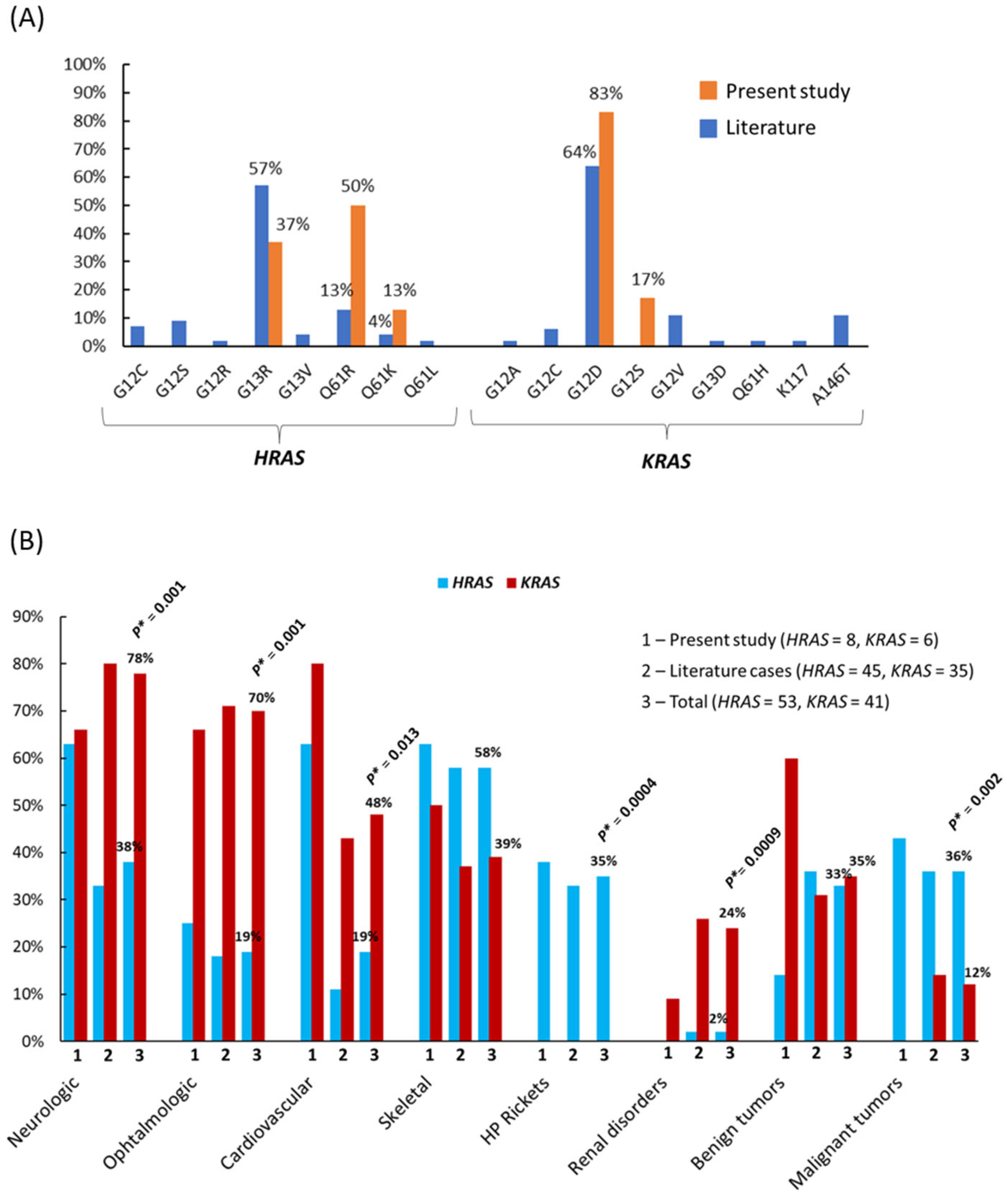
In our study, the most common substitution in HRAS was Q61R (50%), followed by G13R (38%), whereas in the literature, the most frequent was HRAS G13R (57%). The G12D substitution predominated among all KRAS mutations, both in our study (83%) and in cases from the literature (64%) (Figure 7, A). All our patients with HRAS mutations had craniofacial LNS, and four of them (50%) also had nevus spilus. Other cutaneous anomalies included EN, melanocytic nevi, and café-au-lait spots. According to the literature, LNS was present in 67% of 45 HRAS-mutated cases, often in combination with nevus spilus (40%), EN, and KEN [13,15,22]. All six of our patients with KRAS mutations also had congenital craniofacial LNS, and two of them had nevus spilus. Other skin lesions included café-au-lait spots and angiofibromas. In 35 KRAS-mutated cases from the literature, LNS was noted in 68%, and only one patient also had nevus spilus, a finding indicative of PPK.
Despite the apparent overlap of HRAS- and KRAS-mutated phenotypes, the spectrum of extracutaneous anomalies differed significantly between the two groups (Table S2, Figure 7, B). The most notable difference was the association of KRAS mutations with ophthalmic disorders (70% in KRAS-mutated vs. 19% in HRAS-mutated, OR = 10.4, p* = 0.001); coloboma, choristoma, and epibulbar/bulbar dermoid were detected only in patients with KRAS mutations, both in our study and in the literature (Table S2). Neurological disorders (including epilepsy and various brain malformations) were also more common in KRAS-mutated than in HRAS-mutated patients (78% vs. 38%, OR = 5.9, p = 0.001), as were cardiovascular disorders and lymphatic malformations (48% vs. 19%, OR = 3.7, p* = 0.013; and 22% vs. 4%, OR = 7.2, p* = 0.014, respectively). In our cases and in the literature, there is a strict association between HRAS mutations and HR, which was diagnosed exclusively in HRAS-mutated patients, occurring in 30–40% of cases (p* = 0.0004) [7,14,26,27,48,56,67,75]. Other skeletal abnormalities (fibrous dysplasia, scoliosis, limb asymmetry, cystic bone lesions, etc.) were found in HRAS- and KRAS-mutated patients, and the difference was not significant.
In total sample, benign neoplasms occurred with equal frequency in both groups (33% in HRAS-mutated patients and 35% in KRAS-mutated patients), whereas malignancies were more frequent in the HRAS-mutated group (36% vs. 12%, OR = 4.0, p* = 0.002). Among the malignancies, embryonal/alveolar RMS was more common in childhood [18,26,44,56,65,68], while skin cancer [53,56,57,63,64,93], urothelial carcinoma [59,90], and astrocytoma [32] have been described in adults. In our study, two HRAS-mutated patients (ID3 and ID4) developed embryonal RMS, and one patient (ID9) developed ectomesenchymoma; all tumors occurred in the groin area before the age of 3 years, and the pathogenic mutation had the highest VAF in the tumor tissue (84%, 76%, and 41%, respectively). KRAS mutations have previously been described in Wilms' tumor [45], embryonal RMS [17,71,91], and yolk sac tumor [62], all occurring under the age of 1 year. Besides nephroblastoma, renal anomalies (cysts, hamartomas, angiolipomas, etc.) were a very characteristic feature of KRAS-associated mosaicism (ID5) [17,28,30,37,91], as was CPP (ID3, ID4, and ID5) [46,69,77]. Hearing loss (ID13) [17,34,37,57,60,61,78,92] has been reported with equal frequency in both groups (Table S2). We have previously suggested that the expression of genetic information and the formation of specific phenotypic patterns in mosaic syndromes may depend not only on the timing of the mutation, but also on the expression levels of different RAS family genes in various organs and tissues during embryonic development [13]. In this study, we observed very similar genotype–phenotype correlations when using a significantly larger sample. Further research in this area may help elucidate this question.
Differential diagnosis of ENS associated with postzygotic mutations in RAS/MAPK pathway is complicated, and DNA testing plays a key role in establishing the diagnosis. Germline BRAF mutations can lead to CFC [95], while postzygotic mutations have been described in PPK [12,76,78]. Patient ID15 had severe psychomotor delay, minor cardiac abnormalities, immunodeficiency, and facial dysmorphism, which are characteristic of CFC. On the other hand, a diagnosis of PPK could also be considered based on the mosaic BRAF variant, congenital LNS, and nevus spilus. Patients with PROS may also have EN, besides asymmetric tissue growth [96]. In patient ID16 with EN, lower limb asymmetry, and hemimegaloencephaly, SFMS was suspected, but the presence of a mosaic PIK3CA variant allowed the correct diagnosis of PROS to be made. Germline variants in the FGFR2 gene may be associated with severe skeletal disorders [97], while mosaic mutations can lead to phacomatosis resembling SFMS [98]. The somatic mutation FGFR2 c.1144T>C (VAF 23–40%) was found in 5/8 patients with cerebriform nevus of the sebaceous glands [99], and also in patient ID17 in our study. It is also important to exclude germline mutations in patients with mosaic RASopathies. Previously, we described a patient with a congenital facial epidermal nevus (Figure 6, F) who was found to have a pathogenic PTEN variant first in the nevus, and this was subsequently confirmed by blood DNA analysis [100]. Thus, the final diagnosis was type 2 segmental Cowden disease, which is associated with linear Cowden nevus, and a lifelong surveillance was prescribed due to the high cancer risk.
In the nevus tissue of patient ID18, genetic analysis did not reveal any mutations in any of the genes associated with ENS. Furthermore, the patient had only skin manifestations, which were successfully corrected by surgery, and during further follow-up (more than 5 years) no symptoms characteristic of ENS were noted. Therefore, the case was considered as isolated skin malformation.
Based on this study, we propose the following algorithm for accurately determining somatic mosaicism in ENS. Given the varying VAF in affected tissues, biopsies should be taken from at least four different areas of the nevus (and different types of nevi, if feasible), and genetic testing of peripheral blood should be performed to exclude germline mutations. This approach can help differentiate mosaic disorders from hereditary syndromes with incomplete phenotypic manifestations. A mutation in a specific gene can certainly influence the range of clinical manifestations; however, it is important to recognize that these correlations are probabilistic in nature. Therefore, all patients with characteristic skin lesions (epidermal nevus) and suspected mosaic forms of the disease should be referred for assessment by a multidisciplinary team of specialists [15].
This study has some limitations. Due to the rarity of these mosaic diseases, we had a limited number of cases for the statistical analysis. The comparison of small patient groups led to wide confidence intervals, indicating some inconsistency in the results. Another aspect is that extracutaneous anomalies can develop over a long period, while some of patients from our study and literature cases were only 0–3 years old. Therefore, certain syndromic manifestations may not yet have become fully apparent. It is also worth noting that in the cases reported in the literature, not all patients underwent a comprehensive examination of all organ systems, which could also have influenced the final statistical calculations. Further investigations involving more genetically confirmed cases will make it possible to identify clearer correlations and the molecular basis underlying the diverse phenotypic patterns.
5. Conclusions
Genetic testing plays a crucial role in accurately diagnosing mosaic forms of the disease. The spectrum and severity of clinical manifestations of mosaic RASopathies may depend not only on the timing of the postzygotic mutation, but also on the specific gene involved. For example, it can be assumed that patients with KRAS-mediated RASopathy will have an increased risk of ophthalmological disorders (especially coloboma or choristoma), as well as neurological, renal, and vascular abnormalities, while HRAS-mediated mosaic disorder is likely to be associated with hypophosphatemic rickets and malignancies in both children and adults. The specific mosaic genotype may be taken into consideration for prognosis, patient management, and the development of targeted therapies.
Supplementary Materials
The following supporting information can be downloaded at the website of this paper posted on Preprints.org, Table S1. Clinical and genetic characteristics of patients (present study and literature cases). Table S2. Extracutaneous abnormalities in HRAS-mutant and KRAS-mutant patients.
Author Contributions
T.B.: Writing- Original draft preparation, Conceptualization, Supervision, Funding acquisition; T.N.: Writing- Original draft preparation, Supervision, Resources; E.Z.: Writing- Original draft preparation, Data curation, Investigation; V.S.: Writing - review and editing, Methodology, Investigation, N.S.: Writing - review and editing, Investigation, G.S.: Writing - review and editing, Data curation, I.K.: Writing - review and editing, Visualization; J.V.: Writing - review and editing, Visualization; E. Sh.: Writing - review and editing, Validation; K.K.: Writing - review and editing, Data curation; P.V.: Writing - review and editing, Data curation; A.B.: Writing - review and editing, Data curation; M.B.: Writing - review and editing, Data curation; M.D.: Writing - review and editing, Methodology, Data curation; T.V.: Writing - review and editing, Methodology, Conceptualization; S.V.: Writing - review and editing, Methodology, Supervision.
Funding
This research was funded by the Program of Fundamental Research in the Russian Federation for the 2021–2030 period (project No. 124032100002-1) and by the Ministry of Health of the Russian Federation (the project “Personalized approaches to the treatment of malignant neoplasms in children with genodermatoses”, code NUYO-2023-0007).
Institutional Review Board Statement
The study was reviewed and approved by the Local Ethics Committee of N.N. Blokhin National Medical Research Center of Oncology; approval #2 from 26 February 2026.
Informed Consent Statement
Written informed consent for participation in this study and for the publication of any potentially identifiable images or data included in this article was obtained from the participant or legal representative/closest relative of the participants.
Data Availability Statement
The original contributions presented in this study are included in this article/supplementary material, further inquiries can be directed to the corresponding author.
Acknowledgments
The authors would like to express their gratitude to Mr. Sergey Kaplunov and Mr. Roman Garbuzov, as well as Drs. Ella Kumirova, Anatoly Kazantsev, Elena Mikhailova and Diana Dmitrenko for their valuable contribution to the collection and description of clinical material.
Conflicts of Interest
The authors declare that the research was conducted in the absence of any commercial or financial relationships that could be construed as a potential conflict of interest.
References
- Waldman, A.R.; Garzon, M.C.; Morel, K.D. Epidermal Nevi: What Is New. Dermatol. Clin. 2022, 40(1), 61–71. [Google Scholar] [CrossRef]
- Groesser, L.; Herschberger, E.; Ruetten, A.; Ruivenkamp, C.; Lopriore, E.; Zutt, M.; Langmann, T.; Singer, S.; Klingseisen, L.; Schneider-Brachert, W.; Toll, A.; Real, F.X.; Landthaler, M.; Hafner, C. Postzygotic HRAS and KRAS mutations cause nevus sebaceous and Schimmelpenning syndrome. Nat. Genet 2012, 44(7), 783–7. [Google Scholar] [CrossRef] [PubMed]
- Jaeger, Z.J.; Maverakis; Ramirez, N.K.A.; Osborne, A.D.; Staser, K.W.; King, K.A.; Bayliss, S.J.; Mann, C. RASopathies. Part II: Cutaneous and extracutaneous manifestations. J. Am. Acad. Dermatol. 2025, S0190-9622(25), 02340–0. [Google Scholar] [CrossRef] [PubMed]
- Nicholson, C.L.; Daveluy, S. Epidermal Nevus Syndromes. 2023. In StatPearls [Internet]; StatPearls Publishing: Treasure Island (FL), 2025; Available online: https://www.ncbi.nlm.nih.gov/books/NBK559003/.
- Groesser, L.; Herschberger, E.; Sagrera, A.; Shwayder, T.; Flux, K.; Ehmann, L.; Wollenberg, A.; Torrelo, A.; Bagazgoitia, L.; Diaz-Ley, B.; Tinschert, S.; Oschlies, I.; Singer, S.; Mickler, M.; Toll, A.; Landthaler, M.; Real, F.X.; Hafner, C. Phacomatosis pigmentokeratotica is caused by a postzygotic HRAS mutation in a multipotent progenitor cell. J. Invest Dermatol. 2013, 133(8), 1998–2003. [Google Scholar] [CrossRef] [PubMed]
- Torchia, D.; Happle, R. Papular nevus spilus syndrome: old and new aspects of a mosaic RASopathy. Eur. J. Dermatol. 2019, 29(1), 2–5. [Google Scholar] [CrossRef] [PubMed]
- Lim, Y.H.; Ovejero, D.; Sugarman, J.S.; Deklotz, C.M.; Maruri, A.; Eichenfield, L.F.; Kelley, P.K.; Juppner, H.; Gottschalk, M.; Tifft, C.J.; Gafni, R.I.; Boyce, A.M.; Cowen, E.W.; Bhattacharyya, N.; Guthrie, L.C.; Gahl, W.A.; Golas, G.; Loring, E.C.; Overton, J.D.; Mane, S.M.; Lifton, R.P.; Levy, M.L.; Collins, M.T.; Choate, K.A. Multilineage somatic activating mutations in HRAS and NRAS cause mosaic cutaneous and skeletal lesions, elevated FGF23 and hypophosphatemia. Hum. Mol. Genet 2014, 23(2), 397–407. [Google Scholar] [CrossRef] [PubMed]
- Hafner, C.; Groesser, L. Mosaic RASopathies. Cell Cycle 2013, 12(1), 43–50. [Google Scholar] [CrossRef] [PubMed]
- Igawa, S.; Honma, M.; Minami-Hori, M.; Tsuchida, E.; Iizuka, H.; Ishida-Yamamoto, A. Novel postzygotic KRAS mutation in a Japanese case of epidermal nevus syndrome presenting with two distinct clinical features, keratinocytic epidermal nevi and sebaceous nevi. J. Dermatol. 2016, 43(1), 103–4. [Google Scholar] [CrossRef] [PubMed]
- Boppudi, S.; Bögershausen, N.; Hove, H.B.; Percin, E.F.; Aslan, D.; Dvorsky, R.; Kayhan, G.; Li, Y.; Cursiefen, C.; Tantcheva-Poor, I.; Toft, P.B.; Bartsch, O.; Lissewski, C.; Wieland, I.; Jakubiczka, S.; Wollnik, B.; Ahmadian, M.R.; Heindl, L.M.; Zenker, M. Specific mosaic KRAS mutations affecting codon 146 cause oculoectodermal syndrome and encephalocraniocutaneous lipomatosis. Clin. Genet 2016, 90(4), 334–42. [Google Scholar] [CrossRef] [PubMed]
- Kuroda, Y.; Ohashi, I.; Enomoto, Y.; Naruto, T.; Baba, N.; Tanaka, Y.; Aida, N.; Okamoto, N.; Niihori, T.; Aoki, Y.; Kurosawa, K. A postzygotic NRAS mutation in a patient with Schimmelpenning syndrome. Am. J. Med. Genet A 2015, 167A(9), 2223–5. [Google Scholar] [CrossRef] [PubMed]
- Sorlin, A.; Carmignac, V.; Amiel, J.; Boccara, O.; Fraitag, S.; Maruani, A.; Theiler, M.; Weibel, L.; Duffourd, Y.; Philippe, C.; Thauvin-Robinet, C.; Faivre, L.; Riviere, J.-B.; Vabres, P.; Kuentz, P. Expanding the clinical spectrum of mosaic BRAF skin phenotypes. J. Eur. Acad. Dermatol. Venereol. 2021, 35(10), e690–e693. [Google Scholar] [CrossRef] [PubMed]
- Zelenova, E.; Belysheva, T.; Sharapova, E.; Semenova, V.; Valiev, T.; Nasedkina, T. Schimmelpenning-Feuerstein-Mims syndrome: a systematic review of clinical cases to identify genotype-phenotype associations. Front Med. 2025, 12, 1681584. [Google Scholar] [CrossRef] [PubMed]
- Wu, J.; Cao, Q.; Lu, W.; Sun, C.; Li, Q.; Ye, R.; Cheng, R.; Luo, F.; Li, M. A Child with Cutaneous-Skeletal Hypophosphatemia Syndrome Caused by a Mosaic HRAS Mutation: Outcome of Treatment with Anti-FGF23 Antibody. Calcif. Tissue Int. 2025, 116(1), 65. [Google Scholar] [CrossRef] [PubMed]
- Morren, M.A.; Fodstad, H.; Brems, H.; Bedoni, N.; Guenova, E.; Jacot-Guillarmod, M.; Busiah, K.; Giuliano, F.; Gilliet, M.; Atallah, I. Mosaic RASopathies concept: different skin lesions, same systemic manifestations? J. Med. Genet 2024, 61(5), 411–419. [Google Scholar] [CrossRef] [PubMed]
- Maridet, C.; Morice-Picard, F.; Gros, A.; Crivelli, L.; de la Fouchardière, A.; Vergier, B.; Taïeb, A. Mosaic NRASopathy n a child with giant melanocytic congenital naevus, epidermal hamartoma and bilateral nephroblastomatosis: clinical implication for follow-up. J. Eur. Acad. Dermatol. Venereol. 2018, 32(7), e258–e260. [Google Scholar] [CrossRef] [PubMed]
- Chang, C.A.; Perrier, R.; Kurek, K.C.; Estrada-Veras, J.; Lehman, A.; Yip, S.; Hendson, G.; Diamond, C.; Pinchot, J.W.; Tran, J.M.; Arkin, L.M.; Drolet, B.A.; Napier, M.P.; O'Neill, S.A.; Balci, T.B.; Keppler-Noreuil, K.M. Novel findings and expansion of phenotype in a mosaic RASopathy caused by somatic KRAS variants. Am. J. Med. Genet A 2021, 185(9), 2829–2845. [Google Scholar] [CrossRef] [PubMed]
- Davies, O.M.T.; Bruckner, A.L.; McCalmont, T.; Mascarenhas, L.; Oza, V.; Williams, M.L.; Wine-Lee, L.; Shern, J.F.; Siegel, D.H. Cutaneous mosaic RASopathies associated with rhabdomyosarcoma. Pediatr. Blood Cancer 2022, 69(5), e29639. [Google Scholar] [CrossRef] [PubMed]
- Zelenova, E.; Belysheva, T.; Sofronov, D.; Semenova, V.; Radjabova, G.; Vishnevskaya, Y.; Kletskaya, I.; Sharapova, E.; Karasev, I.; Romanov, D.; Denieva, M.; Petrochenko, N.; Valiev, T.; Nasedkina, T. Cutaneous Metastasis of Rectal Cancer as a Diagnostic Challenge: A Clinical Case and Literature Review. Diagnostics 2024, 14(21), 2420. [Google Scholar] [CrossRef] [PubMed]
- Kulikova, K.S.; Raikina, E.N.; Tolmacheva, E.R.; Pomerantseva, E.A. Cutaneous skeletal hypophosphatemia syndrome: the first clinical description of two cases with genetic verification in Russia. Med. Genet. (In Russ.). 2023, 22(6), 51–61. [Google Scholar] [CrossRef]
- Gamayunov, B.N.; Korotkiy, N.G.; Baranova, E.E. Phacomatosis pigmentokeratotica or the Schimmelpenning-Feuerstein-Mims syndrome? Clin. Case Rep. 2016, 4(6), 564–7. [Google Scholar] [CrossRef] [PubMed]
- Zelenova, E.E.; Belysheva, T.S.; Vasiliev, P.A.; Sharapova, E.V.; Semenova, V.V.; Mikhaylova, E.V.; Dorofeeva, M,Yu.; Valiev, T.T.; Nasedkina, T.V. Phacomatosis Pigmentokeratotica: Case Study. Curr. Pediatr. (In Russ.). 2025, 24(4), 305–313. [Google Scholar] [CrossRef]
- Ol’shanskaya, A.S.; Dyuzhakova, A.V.; Shnayder, N.A.; Dmitrenko, D.V.; Guzey, T.N. Schimmelpenning–Feuerstein–Mims syndrome (a case report). Russ. J. Child Neurol. 2017, 12(4), 50–55. [Google Scholar] [CrossRef]
- Kinsler, V.A.; Boccara, O.; Fraitag, S.; Torrelo, A.; Vabres, P.; Diociaiuti, A. Mosaic abnormalities of the akin: review and guidelines from the European reference network for rare skin diseases. Br. J. Dermatol. 2020, 182, 552–63. [Google Scholar] [CrossRef] [PubMed]
- Vera-Duarte, G.R.; Eskenazi-Betech, R.; De la Fuente-Batta, I.; Carreño-Bolaños, D.; Chacón-Camacho, O.F.; Zenteno, J.C.; Graue-Hernandez, E.O. Unveiling the complexity of Schimmelpenning-Feuerstein-Mims syndrome: A comprehensive case study. Am. J. Ophthalmol. Case Rep. 2025, 38, 102321. [Google Scholar] [CrossRef] [PubMed]
- Mirza, A.; Rossi, C.; Kulozik, A.; Banan, R.; Sahm, F.; Harrabi, S.; Syrbe, S.; Choukair, D. Targeted treatment of hypophosphatemia with trametinib in HRAS-related mosaic RASopathy. Orphanet J. Rare Dis. 2025, 20(1), 339. [Google Scholar] [CrossRef] [PubMed]
- Del Pino, M.; Viterbo, G.; Valentini, M.A.; Salvatierra, R.; Belleri, F.; La Forgia, P.; Abbate, S.; Chinton, J.; Lazzati, J.M.; Maderna, O.; Cervini, A.B.; Fano, V. Burosumab Treatment in a Girl With Cutaneous Skeletal Hypophosphatemia Syndrome: 2-Year Follow-Up. Am. J. Med. Genet A 2025, 197(6), e64020. [Google Scholar] [CrossRef] [PubMed]
- Ohishi, A.; Enomoto, Y.; Iwafuchi, H.; Meguro, S.; Kosugi, I.; Baba, S.; Iwashita, T.; Segawa, Y.; Ueno, D.; Iijima, S. Autopsy case of linear nevus sebaceous syndrome with KRAS (G12D) mutation. Pathol. Int. 2024, 74(9), 538–545. [Google Scholar] [CrossRef] [PubMed]
- Hanna, H.; Kaisa, K.; Kristiina, A.; Sirpa, K.; Leila, J.; Sinikka, S.; Päivi, S.; Katariina, H.-J. Mosaic KRAS Mutation in Schimmelpenning-Feuerstein-Mims Syndrome With Overlapping Oculoectodermal Syndrome and Encephalocraniocutaneous Lipomatosis Features. Pediatr. Dermatol. 2025, 42(3), 591–595. [Google Scholar] [CrossRef] [PubMed]
- Dionysiou, M.; Makri, S.C.; Ahlawat, S.; Guryildirim, M.; Barañano, K.W.; Groves, M.L.; Argani, P.; Pratilas, C.A. Case report: MEK inhibitor as treatment for multi-lineage mosaic KRAS G12D-associated epidermal nevus syndrome in a pediatric patient. Front Neurol. 2024, 15, 1466946. [Google Scholar] [CrossRef] [PubMed]
- Querzoli, G.; Badiali, G.; Vitali, F.; Tarsitano, A.; De Biase, D.; Gabrielli, L.; Foschini, M.P. Oral sialadenoma papilliferum with kras mutation in a patient with linear nevus sebaceous syndrome. Pathologica 2024, 116(4), 254–257. [Google Scholar] [CrossRef] [PubMed]
- Tumova, A.; Auslands, K.; Millers, A.; Priede, Z.; Buks, M.; Ozola, A.; Ozoliņa, E.; Bicāns, K.; Ulmanis, R. Association of Schimmelpenning Syndrome with Astrocytoma (WHO Grade 3): Case Report. Medicina 2024, 60(10), 1688. [Google Scholar] [CrossRef] [PubMed]
- Haller, C.N.; Leszczynska, M.A.; Brichta, L.; Maier, E.; Riddington, I.M.; Choate, K.A.; Levy, M.L. Topical trametinib for epidermal and sebaceous nevi in a child with Schimmelpenning-Feuerstein-Mims syndrome. Pediatr. Dermatol. 2024, 41(3), 523–525. [Google Scholar] [CrossRef] [PubMed]
- Beyens, A.; Lietaer, C.; Claes, K.; De Baere, E.; Goeteyn, M.; Lerut, B.; Syryn, H.; Vanakker, O.M.; Meulen, J.V.; Vanwalleghem, L.; Callewaert, B. HRAS-related epidermal nevus syndromes: Expansion of the spectrum with first branchial arch defects. Clin. Genet 2023, 103(6), 709–713. [Google Scholar] [CrossRef] [PubMed]
- Miao, C.; He, R.; Yang, S.; Zhang, B. Schimmelpenning-Feuerstein-Mims syndrome: A case series and brief literature review of genetically ascertained cases. J. Eur. Acad. Dermatol. Venereol. 2023, 37(3), e438–e440. [Google Scholar] [CrossRef] [PubMed]
- Yeager, L.B.; Casper, D.S.; Del Portillo, A.; Marr, B.P. Schimmelpenning-Feuerstein-Mims syndrome with orbital choristoma and KRAS mutation: a current review and novel case report. Ophthalmic Genet 2024, 45(3), 252–257. [Google Scholar] [CrossRef] [PubMed]
- D'Onofrio, G.; Delrue, M.A.; Lortie, A.; Marquis, C.; Striano, P.; Jaworski, M.; Andelfinger, G.; Perreault, S. Treatment of Refractory Epilepsy With MEK Inhibitor in Patients With RASopathy. Pediatr. Neurol. 2023, 148, 148–151. [Google Scholar] [CrossRef] [PubMed]
- Ono, H.; Yamaguchi, R.; Arai, M.; Togi, S.; Ura, H.; Niida, Y.; Shimizu, A. Schimmelpenning-Feuerstein-Mims syndrome induced by HRAS Gly12Ser somatic mosaic mutation: Case report and literature review. J. Dermatol. 2023, 50(9), 1213–1215. [Google Scholar] [CrossRef] [PubMed]
- Carli, D.; Cardaropoli, S.; Tessaris, D.; Coppo, P.; La Selva, R.; Cesario, C.; Lepri, F.R.; Pullano, V.; Palumbo, M.; Ramenghi, U.; Brusco, A.; Medico, E.; DeSanctis, L.; Ferrero, G.B.; Mussa, A. Successful treatment with MEK-inhibitor in a patient with NRAS-related cutaneous skeletal hypophosphatemia syndrome. Genes Chromosom. Cancer 2022, 61(12), 740–746. [Google Scholar] [CrossRef] [PubMed]
- Friedrich, R.E.; Gosau, M.; Luebke, A.M.; Hagel, C.; Kohlrusch, F.K.; Hahn, M.; Kroge, S.; Hahn, J.; Wieland, I.; Zenker, M. Oral HRAS Mutation in Orofacial Nevus Sebaceous Syndrome (Schimmelpenning-Feuerstein-Mims-Syndrome): A Case Report With a Literature Survey. Vivo 2022, 36(1), 274–293. [Google Scholar] [CrossRef] [PubMed]
- Blanco Portals, C.; Gómez Tellado, M.; Del Pozo Losada, J.; Rodríguez Ruiz, M. Inguinal lymph nodes agenesia in a patient with Schimmelpenning-Feuerstein-Mims syndrome with proven somatic KRAS mutation. Clin. Exp. Dermatol. 2022, 47(1), 235–239. [Google Scholar] [CrossRef] [PubMed]
- Huang, L.; Zhang, J.; Chen, F.; Zhang, X.; Cao, Y.; Cheng, R.; Deng, D. The first case of Chinese phacomatosis pigmentokeratotica diagnosed by a missense HRAS mosaicism. J. Dermatol. 2022, 49(9), 921–924. [Google Scholar] [CrossRef] [PubMed]
- Green, T.E.; MacGregor, D.; Carden, S.M.; Harris, R.V.; Hewitt, C.A.; Berkovic, S.F.; Penington, A.J.; Scheffer, I.E.; Hildebrand, M. Identification of a recurrent mosaic KRAS variant in brain tissue from an individual with nevus sebaceous syndrome. Cold Spring Harb. Mol. Case Stud. 2021, 7(6), a006133. [Google Scholar] [CrossRef] [PubMed]
- Luo, Q.; Zhang, Q.; Shen, J.; Guan, W.; Li, M.; Zhang, J.; Tan, Z. Expanding mutational spectrum of HRAS by a patient with Schimmelpenning-Feuerstein-Mims syndrome. J. Dermatol. 2021, 48(8), 1273–1276. [Google Scholar] [CrossRef] [PubMed]
- Slack, J.C.; Bründler, M.A.; Chang, C.A.; Perrier, R.; Lafay-Cousin, L.; Kurek, K.C. Bilateral Nephroblastic Tumors and a Complex Renal Vascular Anomaly in a Patient With a Mosaic RASopathy: Novel Histopathologic Features and Molecular Insights. Pediatr. Dev. Pathol. 2021, 24(3), 235–240. [Google Scholar] [CrossRef] [PubMed]
- De Los Santos-La Torre, M.A.; Del Águila-Villar, C.M.; Lu-de Lama, L.R.; Nuñez-Almache, O.; Chávez-Tejada, E.M.; Espinoza-Robles, O.A.; Pinto-Ibarcena, P.M.; Calagua-Quispe, M.R. Association of Central Precocious Puberty with a Rare Presentation of Schimmelpenning-Feuerstein-Mims Syndrome in a Peruvian Girl. Case Rep. Endocrinol. 2020, 2020, 1928121. [Google Scholar] [CrossRef] [PubMed]
- Chang, C.A.; Perrier, R.; Kurek, K.C.; Estrada-Veras, J.; Lehman, A.; Yip, S.; Hendson, G.; Diamond, C.; Pinchot, J.W.; Tran, J.M.; Arkin, L.M.; Drolet, B.A.; Napier, M.P.; O'Neill, S.A.; Balci, T.B.; Keppler-Noreuil, K.M. Novel findings and expansion of phenotype in a mosaic RASopathy caused by somatic KRAS variants. Am. J. Med. Genet A 2021, 185(9), 2829–2845. [Google Scholar] [CrossRef] [PubMed]
- Khadora, M.; Mughal, M.Z. Burosumab treatment in a child with cutaneous skeletal hypophosphatemia syndrome: A case report. Bone Rep. 2021, 15, 101138. [Google Scholar] [CrossRef] [PubMed]
- Kapoor, S.; Scanga, H.L.; Reyes-Múgica, M.; Nischal, K.K. Somatic KRAS mutation affecting codon 146 in linear sebaceous nevus syndrome. Am. J. Med. Genet A 2021, 185(12), 3825–3830. [Google Scholar] [CrossRef] [PubMed]
- Pepi, C.; de Palma, L.; Trivisano, M.; Pietrafusa, N.; Lepri, F.R.; Diociaiuti, A.; Camassei, F.D.; Carfi-Pavia, G.; De Benedictis, A.; Rossi-Espagnet, C.; Vigevano, F.; Marras, C.E.; Novelli, A.; Bluemcke, I.; Specchio, N. The Role of KRAS Mutations in Cortical Malformation and Epilepsy Surgery: A Novel Report of Nevus Sebaceous Syndrome and Review of the Literature. Brain Sci. 2021, 11(6,), 793. [Google Scholar] [CrossRef] [PubMed]
- Sideris, E.; Tng, E.T.V.; Chee, P. Lymphatic malformation responsive to sirolimus in Keratinocytic Epidermal Nevus syndrome with KRAS Mutation: A case and brief literature discussion. Case Rep. Dermatol. 2021, 13, 195–201. [Google Scholar] [CrossRef] [PubMed]
- Yan, Y.; Zhang, S.; Zhou, H.; Zhou, Y.; Fu, Y. Ophthalmic Manifestation and Pathological Features in a Cohort of Patients With Linear Nevus Sebaceous Syndrome and Encephalocraniocutaneous Lipomatosis. Front Pediatr. 2021, 9, 678296. [Google Scholar] [CrossRef] [PubMed]
- Hayani, K.; Giehl, K.; Kumbrink, J.; Fischer, J.; Schlaak, M.; French, L.E.; Wollenberg, A. Gene Mutation Mapping in a Fatal Case of Phacomatosis Pigmentokeratotica Happle. Acta Derm. Venereol. 2020, 100(15), adv00241. [Google Scholar] [CrossRef] [PubMed]
- Pan, C.; Zhou, X.; Hong, A.; Fang, F.; Wang, Y. Identification, of KRAS mutation in a patient with linear nevus sebaceous syndrome: a case report. BMC Med. Genom. 2020, 13(1), 188. [Google Scholar] [CrossRef] [PubMed]
- Chaves, R.R.M.; Júnior, A.A.C.P.; Gomes, C.C.; de Castro, W.H.; Gomez, R.S. Multiple adenomatoid odontogenic tumors in a patient with Schimmelpenning syndrome. Oral Surg. Oral Med. Oral Pathol. Oral Radiol. 2020, 129(1), e12–e17. [Google Scholar] [CrossRef] [PubMed]
- Maia, T.F.; Freire, B.L.; Rodrigues, A.D.S.; Jorgetti, V.; Rocha, A.C.; Moises, C.S.; Souza, B.F.; Rodrigues, M.B.; Lima Jorge, A.A.; Martin, R.M.; Cabral, H.R. MON-333 Cutaneous Skeletal Hypophosphatemic Syndrome (Cshs) Caused by Somatic HRAS p.G13R Mutation: Long Follow-Up of Two Brazilian Women. J. Endocr. Soc. 2020, 4 (Suppl 1), MON–333. [Google Scholar] [CrossRef]
- Hoxha, E.; Linse, K.P.; Toberer, F. Das Spektrum benigner und maligner Neoplasien beim Schimmelpenning-Feuerstein-Mims-Syndrom. J. Dtsch. Dermatol. Ges. 2020, 18(12), 1493–1494. [Google Scholar] [CrossRef] [PubMed]
- Mestach, L.; Polubothu, S.; Calder, A.; Denayer, E.; Gholam, K.; Legius, E.; Levtchenko, E.; Laethem, A.V.; Brems, H.; Kinsler, V.A.; Morren, M.-A. Keratinocytic epidermal nevi associated with localized fibro-osseous lesions without hypophosphatemia. Pediatr. Dermatol. 2020, 37(5), 890–895. [Google Scholar] [CrossRef] [PubMed]
- Gadea, A.; Hernández-Muñoz, I.; Vicente, A.; Andrades, E.; García-Calvente, M.; Camacho, L.; Fernandez-Rodríguez, C.; Bellosillo, B.; Pujol, R.; Toll, A. Molecular characterisation of oncogenic urothelial mosaic mutations in patients with extensive keratinocytic epidermal naevi. J. Med. Genet 2020, 57(9), 601–604. [Google Scholar] [CrossRef] [PubMed]
- Chacon-Camacho, O.F.; Lopez-Moreno, D.; Morales-Sanchez, M.A.; Hofmann, E.; Pacheco-Quito, M.; Wieland, I.; Cortes-Gonzalez, V.; Villanueva-Mendoza, C.; Zenker, M.; Zenteno, J.C. Expansion of the phenotypic spectrum and description of molecular findings in a cohort of patients with oculocutaneous mosaic RASopathies. Mol. Genet Genom. Med. 2019, 7(5), e625. [Google Scholar] [CrossRef] [PubMed]
- Nagatsuma, M.; Takasawa, K.; Yamauchi, T.; Nakagawa, R.; Mizuno, T.; Tanaka, E.; Yamamoto, K.; Uemura, N.; Kashimada, K.; Morio, T. A postzygotic KRAS mutation in a patient with Schimmelpenning syndrome presenting with lipomatosis, renovascular hypertension, and diabetes mellitus. J. Hum. Genet 2019, 64(2), 177–181. [Google Scholar] [CrossRef] [PubMed]
- Mitchel, B.J.; Rogers, G.F.; Wood, B.C. A patient with Schimmelpenning syndrome and mosaic KRAS mutation. J. Craniofac Surg. 2019, 30, 184–5. [Google Scholar] [CrossRef] [PubMed]
- Kubo, A.; Yamada, D. Phakomatosis Pigmentokeratotica. N Engl. J. Med. 2019, 381(15), 1458. [Google Scholar] [CrossRef] [PubMed]
- Cranwell, W.C.; Walsh, M.; Winship, I. Phacomatosis pigmentokeratotica: Postzygotic HRAS mutation with malignant degeneration of the sebaceous naevus. Australas. J. Dermatol. 2019, 60(3), e245–e246. [Google Scholar] [CrossRef] [PubMed]
- Nemeth, K.; Szabo, S.; Cottrell, C.E.; McNulty, S.M.; Segura, A.; Sokumbi, O.; Browning, M.; Siegel, D.H. Mosaic pathogenic HRAS variant in a patient with nevus spilus with agminated Spitz nevi and parametrial-uterine rhabdomyosarcoma. Br. J. Dermatol. 2018, 178(3), 804–806. [Google Scholar] [CrossRef] [PubMed]
- Bessis, D.; Sandrine, M.; Vabres, P.; Quere, I. Lymphangiome vulvaire au cours du syndrome de Schimmelpenning–Feuerstein–Mims traité par l’association sirolimus-sunitinib. Ann. De Dermatol. Et. De Vénéréologie 2018, 145(12), S335–S336. [Google Scholar] [CrossRef]
- Park, P.G.; Park, E.; Hyun, H.S.; Kang, H.G.; Ha, I.S.; Cho, T.J.; Ko, J.M.; Cheong, H.I. Cutaneous Skeletal Hypophosphatemia Syndrome in Association with a Mosaic HRAS Mutation. Ann. Clin. Lab Sci. 2018, 48(5), 665–669. [Google Scholar] [PubMed]
- Prieto-Barrios, M.; Llamas-Martin, R.; Velasco-Tamariz, V.; Calleja-Algarra, A.; Ruano, Y.; Ortiz-Romero, P.L.; Rodriguez-Peralto, J.L. Phacomatosis pigmentokeratotica: a case of HRAS mosaicism causing rhabdomyosarcoma. Br. J. Dermatol. 2018, 179(5), 1163–1167. [Google Scholar] [CrossRef] [PubMed]
- Martin, R.J.; Arefi, M.; Splitt, M.; Redford, L.; Moss, C.; Rajan, N. Phacomatosis pigmentokeratotica and precocious puberty associated with HRAS mutation. Br. J. Dermatol. 2018, 178(1), 289–291. [Google Scholar] [CrossRef] [PubMed]
- Lihua, J.; Feng, G.; Shanshan, M.; Jialu, X.; Kewen, J. Somatic KRAS mutation in an infant with linear nevus sebaceous syndrome associated with lymphatic malformations: A case report and literature review. Medicine 2017, 96(47), e8016. [Google Scholar] [CrossRef] [PubMed]
- Om, A.; Cathey, S.S.; Gathings, R.M.; Hudspeth, M.; Lee, J.A.; Marzolf, S.; Lee, L.W. Phacomatosis Pigmentokeratotica: A Mosaic RASopathy with Malignant Potential. Pediatr. Dermatol. 2017, 34(3), 352–355. [Google Scholar] [CrossRef] [PubMed]
- Jennings, L.; Cummins, R.; Murphy, G.M.; Gulmann, C.; O'Kane, M. HRAS mutation in phacomatosis pigmentokeratotica without extracutaneous disease. Clin. Exp. Dermatol. 2017, 42(7), 791–792. [Google Scholar] [CrossRef] [PubMed]
- Whyte, M.P.; Griffith, M.; Trani, L.; Mumm, S.; Gottesman, G.S.; McAlister, W.H.; Krysiak, K.; Lesurf, R.; Skidmore, Z.L.; Campbell, K.M.; Rosman, I.S.; Bayliss, S.; Bijanki, V.N.; Nenninger, A.; Van Tine, B.A.; Griffith, O.L.; Mardis, E.R. Melorheostosis: Exome sequencing of an associated dermatosis implicates postzygotic mosaicism of mutated KRAS. Bone 2017, 101, 145–155. [Google Scholar] [CrossRef] [PubMed]
- Igawa, S.; Honma, M.; Minami-Hori, M.; Tsuchida, E.; Iizuka, H.; Ishida-Yamamoto, A. Novel postzygotic KRAS mutation in a Japanese case of epidermal nevus syndrome presenting with two distinct clinical features, keratinocytic epidermal nevi and sebaceous nevi. J. Dermatol. 2016, 43(1), 103–4. [Google Scholar] [CrossRef] [PubMed]
- Hucthagowder, V.; Cottrell, C.E.; Schaffer, A.; Arbelaez, A.M.; Bayliss, S.J.; Shinawi, M. Targeted Next-Generation Sequencing Identifies HRAS mutation in Schimmelpenning-Feuerstein-Mims syndrome. In American College of Medical Genetics; 2016. [Google Scholar]
- Ayala, D.; Ramón, M.D.; Martín, J.M.; Jordá, E. Atypical Phacomatosis Pigmentokeratotica as the Expression of a Mosaic RASopathy With the BRAF Glu586Lys Mutation. Actas Dermosifiliogr. 2016, 107(4), 344–6. [Google Scholar] [CrossRef] [PubMed]
- Kuentz, P.; Mignot, C.; St-Onge, J.; Duffourd, Y.; Bonniaud, B.; Faivre, L.; Riviere, J.-B.; Vabres, P. Postzygotic BRAF p.Lys601Asn Mutation in Phacomatosis Pigmentokeratotica with Woolly Hair Nevus and Focal Cortical Dysplasia. J. Invest Dermatol. 2016, 136(5), 1060–1062. [Google Scholar] [CrossRef] [PubMed]
- Watanabe, Y.; Shido, K.; Niihori, T.; Niizuma, H.; Katata, Y.; Iizuka, C.; Oba, D.; Moriya, K.; Saito-Nanjo, Y.; Onuma, M.; Rikiishi, T.; Sasahara, Y.; Watanabe, M.; Aiba, S.; Saito, R.; Sonoda, Y.; Tominaga, T.; Aoki, Y.; Kure, S. Somatic BRAF c.1799T>A p.V600E Mosaicism syndrome characterized by a linear syringocystadenoma papilliferum, anaplastic astrocytoma, and ocular abnormalities. Am. J. Med. Genet A 2016, 170A(1), 189–94. [Google Scholar] [CrossRef] [PubMed]
- Wang, H.; Qian, Y.; Wu, B.; Zhang, P.; Zhou, W. KRAS G12D mosaic mutation in a Chinese linear nevus sebaceous syndrome infant. BMC Med. Genet 2015, 16, 101. [Google Scholar] [CrossRef] [PubMed]
- Kim, S.W.; Song, J.S.; Kang, M.S.; Sin, J.B.; Ki, C.S.; Jeon, G.W. Identification of somatic KRAS mutation in a Korean baby with nevus sebaceus syndrome. Ann. Lab Med. 2015, 35(1), 178–180. [Google Scholar] [CrossRef] [PubMed]
- Kuroda, Y.; Ohashi, I.; Enomoto, Y.; Naruto, T.; Baba, N.; Tanaka, Y.; Aida, N.; Okamoto, N.; Niihori, T.; Aoki, Y.; Kurosawa, K. A postzygotic NRAS mutation in a patient with Schimmelpenning syndrome. Am. J. Med. Genet A 2015, 167A(9), 2223–5. [Google Scholar] [CrossRef] [PubMed]
- Farschtschi, S.; Mautner, V.F.; Hollants, S.; Hagel, C.; Spaepen, M.; Schulte, C.; Legius, E.; Brems, H. Keratinocytic epidermal nevus syndrome with Schwann cell proliferation, lipomatous tumour and mosaic KRAS mutation. BMC Med. Genet 2015, 16, 6. [Google Scholar] [CrossRef] [PubMed]
- Lim, Y.H.; Ovejero, D.; Sugarman, J.S.; Deklotz, C.M.; Maruri, A.; Eichenfield, L.F.; Kelley, P.K.; Juppner, H.; Gottschalk, M.; Tifft, C.J.; Gafni, R.I.; Boyce, A.M.; Cowen, E.W.; Bhattacharyya, N.; Guthrie, L.C.; Gahl, W.A.; Golas, G.; Loring, E.C.; Overton, J.D.; Mane, S.M.; Lifton, R.P.; Levy, M.L.; Collins, M.T.; Choate, K.A. Multilineage somatic activating mutations in HRAS and NRAS cause mosaic cutaneous and skeletal lesions, elevated FGF23 and hypophosphatemia. Hum. Mol. Genet 2014, 23(2), 397–407. [Google Scholar] [CrossRef] [PubMed]
- Li, J.Y.; Berger, M.F.; Marghoob, A.; Bhanot, U.K.; Toyohara, J.P.; Pulitzer, M.P. Combined melanocytic and sweat gland neoplasm: cell subsets harbor an identical HRAS mutation in phacomatosis pigmentokeratotica. J. Cutan. Pathol. 2014, 41(8), 663–71. [Google Scholar] [CrossRef] [PubMed]
- Avitan-Hersh, E.; Tatur, S.; Indelman, M.; Gepstein, V.; Shreter, R.; Hershkovitz, D.; Brick, R.; Bergman, R.; Tiosano, D. Postzygotic HRAS mutation causing both keratinocytic epidermal nevus and thymoma and associated with bone dysplasia and hypophosphatemia due to elevated FGF23. J. Clin. Endocrinol. Metab. 2014, 99(1), E132-6. [Google Scholar] [CrossRef] [PubMed]
- Sun, B.K.; Saggini, A.; Sarin, K.Y.; Kim, J.; Benjamin, L.; LeBoit, P.E.; Khavari, P.A. Mosaic activating RAS mutations in nevus sebaceus and nevus sebaceus syndrome. J. Invest Dermatol. 2013, 133(3), 824–827. [Google Scholar] [CrossRef] [PubMed]
- Groesser, L.; Herschberger, E.; Sagrera, A.; Shwayder, T.; Flux, K.; Ehmann, L.; Wollenberg, A.; Torrelo, A.; Bagazgoitia, L.; Diaz-Ley, B.; Tinschert, S.; Oschlies, I.; Singer, S.; Mickler, M.; Toll, A.; Landthaler, M.; Real, F.X.; Hafner, C. Phacomatosis pigmentokeratotica is caused by a postzygotic HRAS mutation in a multipotent progenitor cell. J. Invest Dermatol. 2013, 133(8), 1998–2003. [Google Scholar] [CrossRef] [PubMed]
- Rijntjes-Jacobs, E.,G.; Lopriore, E.; Steggerda, S.J.; Kant, S.G.; Walther, F.J. Discordance for Schimmelpenning-Feuerstein-Mims syndrome in monochorionic twins supports the concept of a postzygotic mutation. Am. J. Med. Genet A 2010, 152A(11), 2816–9. [Google Scholar] [CrossRef] [PubMed]
- Chantorn, R.; Shwayder, T. Phacomatosis pigmentokeratotica: a further case without extracutaneous anomalies and review of the condition. Pediatr. Dermatol. 2011, 28(6), 715–9. [Google Scholar] [CrossRef] [PubMed]
- Hafner, C.; Toll, A.; Real, F.X. HRAS mutation mosaicism causing urothelial cancer and epidermal nevus. N Engl. J. Med. 2011, 365(20), 1940–2. [Google Scholar] [CrossRef] [PubMed]
- Bourdeaut, F.; Hérault, A.; Gentien, D.; Pierron, G.; Ballet, S.; Reynaud, S.; Paris, R.; Schleiermacher, G.; Baumann, C.; Philippe-Chomette, P.; Gauthier-Villars, M.; Peuchmaur, M.; Radvanyi, F.; Delattre, O. Mosaicism for oncogenic G12D KRAS mutation associated with epidermal nevus, polycystic kidneys and rhabdomyosarcoma. J. Med. Genet 2010, 47(12), 859–62. [Google Scholar] [CrossRef] [PubMed]
- Wiedemeyer, K.; Hartschuh, W. Trichoblastomas with Merkel cell proliferation in nevi sebacei in Schimmelpenning-Feuerstein-Mims syndrome—histological differentiation between trichoblastomas and basal cell carcinomas. J. Dtsch. Dermatol. Ges. 2009, 7(7), 612–5. [Google Scholar] [CrossRef] [PubMed]
- Zutt, M.; Strutz, F.; Happle, R.; Habenicht, E.M.; Emmert, S.; Haenssle, H.A.; Kretschmera, L.; Neumanna, Ch. Schimmelpenning-Feuerstein-Mims syndrome with hypophosphatemic rickets. Dermatology 2003, 207(1), 72–6. [Google Scholar] [CrossRef] [PubMed]
- Torrelo, A.; Zambrano, A. What syndrome is this. Phakomatosis pigmentokeratotica (Happle). Pediatr. Dermatol. 1998, 15, 321–3. [Google Scholar] [CrossRef] [PubMed]
- Di Majo, B.E.; Leoni, C.; Cartisano, E.; Fossati, C.; Viscogliosi, G.; Trevisan, V.; Bruno, L.P.; Conti, F.; Moratti, M.; Monaco, E.; Rigante, D.; Rivalta, B.; Cancrini, C.; Szczawińska-Popłonyk, A.; Jamsheer, A.; Obara-Moszyńska, M.; Zakharova, V.; Shcherbina, A.; Rodina, J.; Tüysüz, B.; Jamuar, S.S.; Lim, J.Y.; Goh, J.; Cereda, A.; Agovino, T.; Contaldo, I.; Gambardella, M.L.; Balduzzi, A.C.; Cherubino, A.; Marrocco, G.A.; Bellesi, S.; Carusi, V.; Rumi, G.; Biondi, A.; Zampino, G.; Saettini, F. Cardiofaciocutaneous syndrome and immunodeficiency: data from an international multicenter cohort. Front Immunol. 2025, 16, 1598896. [Google Scholar] [CrossRef] [PubMed]
- Roth, G.M.; Ferguson, N.; Wanat, K.A. Segmental epidermal nevus and mucosal neuromas associated with PIK3CA-related overgrowth spectrum disorder. JAAD Case Rep. 2018, 4(10), 1080–1082. [Google Scholar] [CrossRef] [PubMed]
- Zhuang, X.R.; Zhao, H.S. A case of Pfeiffer syndrome caused by FGFR2 gene variation. Zhonghua Yan Ke Za Zhi 2022, 58(6), 453–456. [Google Scholar] [CrossRef] [PubMed]
- Wang, Y.; Zhao, L.; Chen, Y.; Pan, L.; Huang, N.; Zhou, R.; Li, L. FGFR2 variants in Schimmelpenning-Feuerstein-Mims syndrome. Br. J. Dermatol. 2025, 193(1), 167–169. [Google Scholar] [CrossRef] [PubMed]
- Theiler, M.; Weibel, L.; Christen-Zaech, S.; Carmignac, V.; Sorlin, A.; Neuhaus, K.; Chevarin, M.; Thauvin-Robinet, C.; Philippe, C.; Faivre, L.; Vabres, P.; Kuentz, P. Cerebriform sebaceous nevus: a subtype of organoid nevus due to specific postzygotic FGFR2 mutations. J. Eur. Acad. Dermatol. Venereol. 2021, 35(10), 2085–2090. [Google Scholar] [CrossRef] [PubMed]
- Zelenova, E.; Belysheva, T.; Sharapova, E.; Barinova, I.; Fedorova, A.; Semenova, V.; Vishnevskaya, Y.; Kletskaya, I.; Mitrofanova, A.; Sofronov, D.; Karasev, I.; Romanov, D.; Valiev, T.; Nasedkina, T. Atypical Manifestations of Cowden Syndrome in Pediatric Patients. Diagnostics 2025, 15(12), 1456. [Google Scholar] [CrossRef] [PubMed]
Figure 1.
Phenotype ID1 (A-E): (A, B) – LNS on the head, trunk and limbs predominantly right-sided following Blashko’s lines at 2 months; (C, D) – multiple deformities of long bones and the chest at 1 year; (E) – Spitz nevi on the face, small congenital melanocytic nevi on the forehead at 1 year; histology ID1 (F,G): (F) – Spitz nevi: symmetrical and well demarcated wedge shaped lesion with marked acanthosis (H&E, x150); (G) – the lesion is composed of epithelioid cells containing abundant cytoplasm and monomorphic nuclei, stroma is slightly fibrotic in reticular dermis (H&E, x350). Phenotype ID2 (H-J): (H, I) – LNS on the head and face predominantly right-sided following Blashko’s lines and right ptosis, (J) – linear pigmented nevus in the axillary region; histology ID2: (K) – nevus sebaceous of Jadassohn with papillomatosis and hyperkeratosis, hamartomatous sebaceous glands located higher than normal onto the epidermal surface (H&E, x250).
Figure 1.
Phenotype ID1 (A-E): (A, B) – LNS on the head, trunk and limbs predominantly right-sided following Blashko’s lines at 2 months; (C, D) – multiple deformities of long bones and the chest at 1 year; (E) – Spitz nevi on the face, small congenital melanocytic nevi on the forehead at 1 year; histology ID1 (F,G): (F) – Spitz nevi: symmetrical and well demarcated wedge shaped lesion with marked acanthosis (H&E, x150); (G) – the lesion is composed of epithelioid cells containing abundant cytoplasm and monomorphic nuclei, stroma is slightly fibrotic in reticular dermis (H&E, x350). Phenotype ID2 (H-J): (H, I) – LNS on the head and face predominantly right-sided following Blashko’s lines and right ptosis, (J) – linear pigmented nevus in the axillary region; histology ID2: (K) – nevus sebaceous of Jadassohn with papillomatosis and hyperkeratosis, hamartomatous sebaceous glands located higher than normal onto the epidermal surface (H&E, x250).
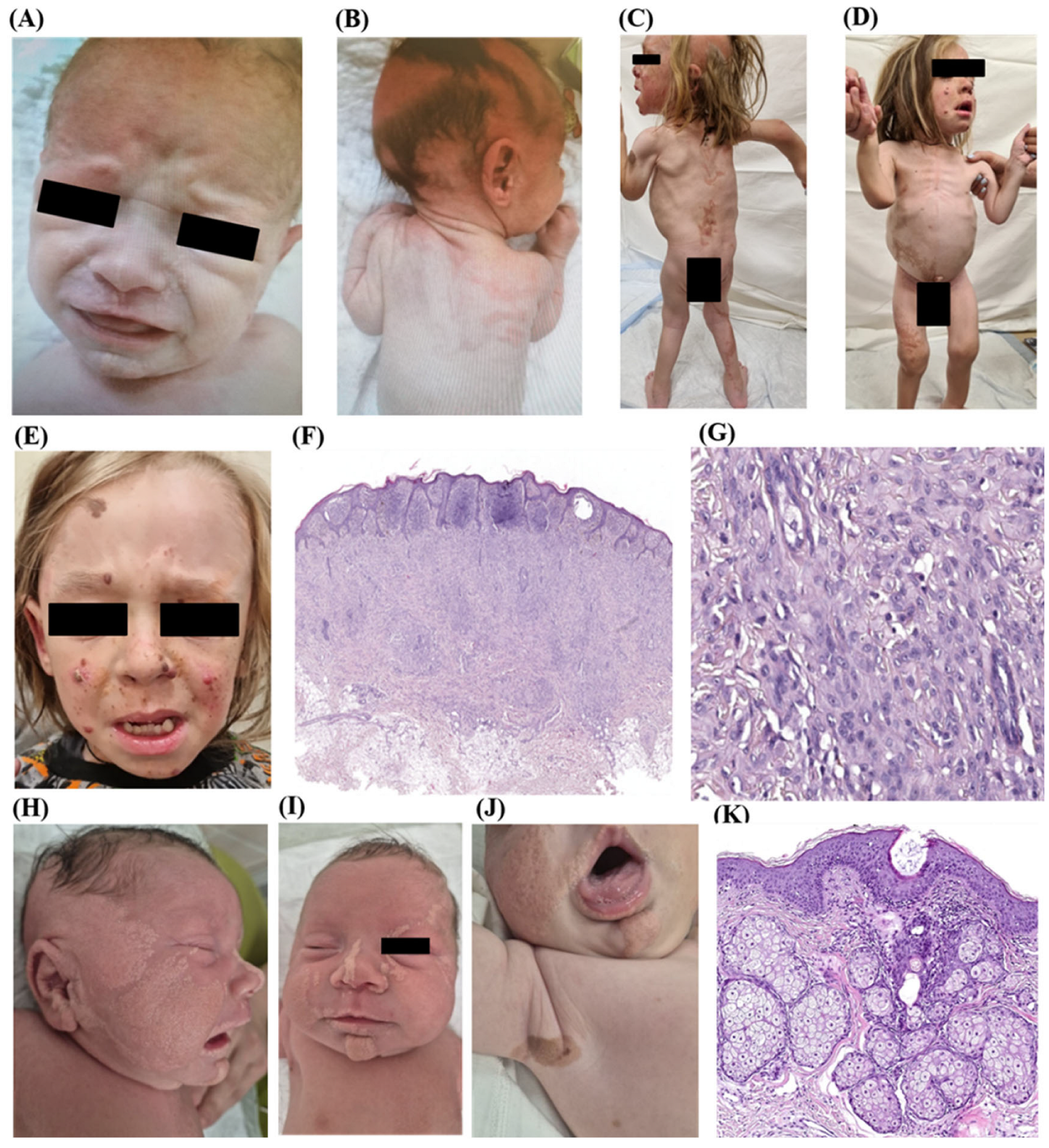
Figure 2.
Phenotype ID3: (A-D) – LNS on the scalp, face, neck, and trunk, predominantly left-sided following Blashko’s lines; a mosaic variant in the HRAS gene with VAF 22% was detected in the nevus tissue, but not in the biopsy of healthy skin. Histology ID3: (E) – trabecular-solid round-cell tumor (H&E, x100). Immunohistochemistry ID3 (F-G): (F) – diffuse expression of MyoD1 (x100) in the tumor; (G) – diffuse expression of Desmin (x200) in the tumor. Phenotype ID4: (H-J) – LNS on the face, scalp, and trunk predominantly right-sided and in the middle line following Blashko’s lines; Sanger sequencing results indicating VAF in the loci studied. Histology ID4 (K-M): (K) – hyperplastic sebaceous glands without excretory ducts and with underdeveloped hair follicles are observed in the skin (H&E, x100); (L) – moderate degree of testicular hypoplasia, seminiferous tubules unevenly spaced due to stromal sclerosis, disruption of layer stratification in the germinal epithelium, cells with light cytoplasm and cells with dystrophic changes; (M) – testicular rhabdomyosarcoma, spindle cell variant (H&E, x200). Immunohistochemistry ID4: (N) – focal expression of MyoD1 (x200) in tumor cells.
Figure 2.
Phenotype ID3: (A-D) – LNS on the scalp, face, neck, and trunk, predominantly left-sided following Blashko’s lines; a mosaic variant in the HRAS gene with VAF 22% was detected in the nevus tissue, but not in the biopsy of healthy skin. Histology ID3: (E) – trabecular-solid round-cell tumor (H&E, x100). Immunohistochemistry ID3 (F-G): (F) – diffuse expression of MyoD1 (x100) in the tumor; (G) – diffuse expression of Desmin (x200) in the tumor. Phenotype ID4: (H-J) – LNS on the face, scalp, and trunk predominantly right-sided and in the middle line following Blashko’s lines; Sanger sequencing results indicating VAF in the loci studied. Histology ID4 (K-M): (K) – hyperplastic sebaceous glands without excretory ducts and with underdeveloped hair follicles are observed in the skin (H&E, x100); (L) – moderate degree of testicular hypoplasia, seminiferous tubules unevenly spaced due to stromal sclerosis, disruption of layer stratification in the germinal epithelium, cells with light cytoplasm and cells with dystrophic changes; (M) – testicular rhabdomyosarcoma, spindle cell variant (H&E, x200). Immunohistochemistry ID4: (N) – focal expression of MyoD1 (x200) in tumor cells.
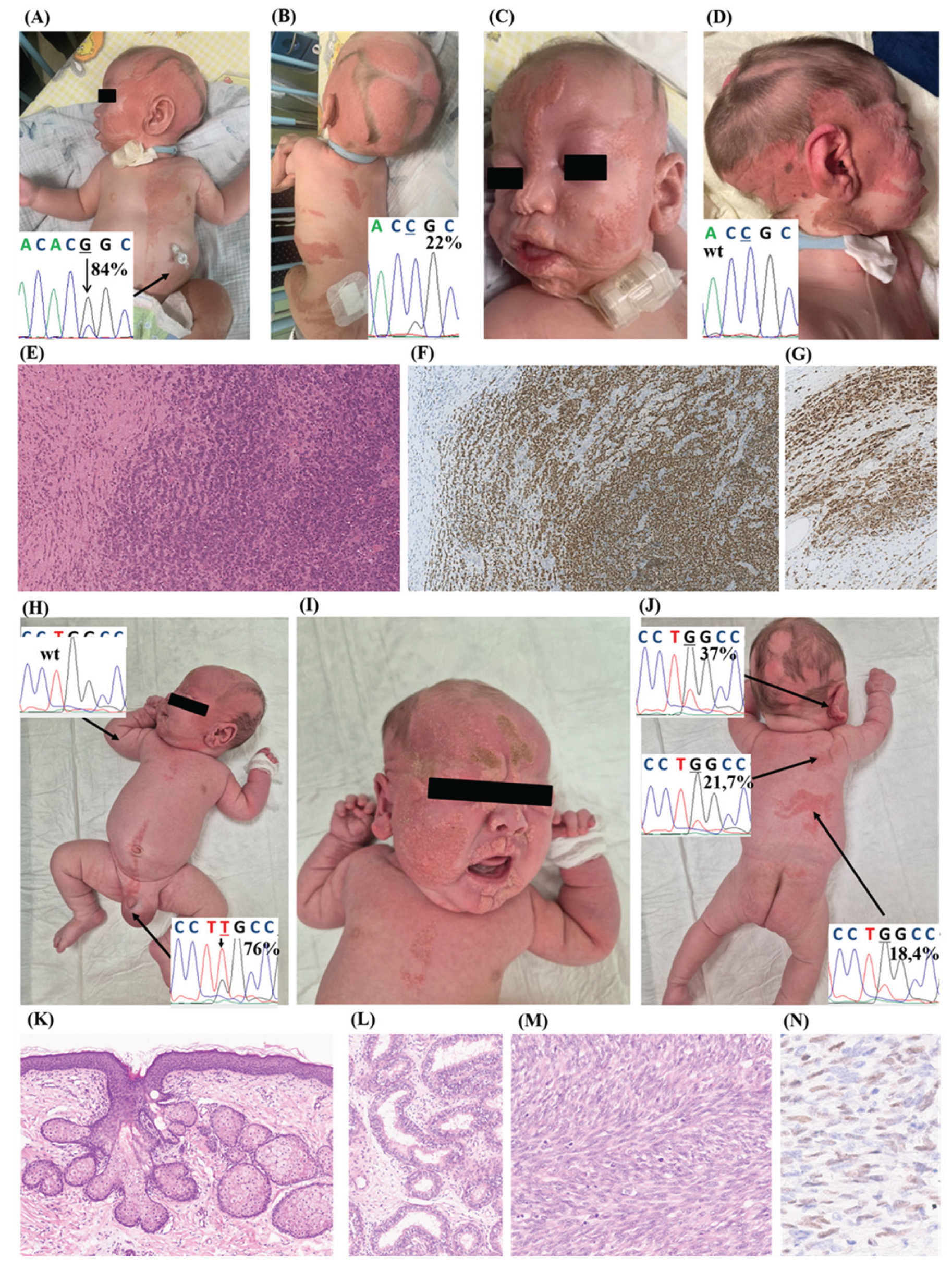
Figure 3.
SFMS patients with KRAS-mutation. Phenotype ID5 at age 2 (A, B) and 4 years (C, D): (A-C) – LNS on the scalp, face and neck predominantly right-sided following Blashko’s lines; (D) – single café-au-lait macule in the right shoulder and scapula. Histology (E-G): (E) – nevus sebaceous of Jadassohn with underlying plexiform neurofibroma (H&E, x150), (F) – marked papillomatosis and hyperkeratosis, hamartomatous sebaceous glands located higher than normal in the dermis without connection to a hair follicle and with direct opening onto the epidermal surface (H&E, x250); (G) – syringocystadenoma papilliferum presenting as focal cystic invaginations of the infundibular epithelium projecting into the dermis, covered by a double columnar and cuboidal cell layer (H&E, x350). Phenotype ID6 (H-L): (H, I) – café-au-lait macules on the trunk skin; (J-L) – LNS on the scalp, face, neck and trunk predominantly right-sided following Blashko’s lines. Brain MRI ID6 (M-Q): (M-O) – granulosa cell tumor (indicated by arrows); (P) – white matter atrophy (indicated by arrows); (Q) – retinal calcifications OU (oculus uterque) (indicated by arrows). Phenotype ID7: (R) – LNS on the face following Blashko’s lines. Histology ID7: (S) – hamartomatous sebaceous glands onto the epidermal surface (H&E, x250). Phenotype ID8 (T, U): (T) – LNS on the right-sided face after attempt to surgically excised the nevus in local hospital; (U) – partial vascularized corneal opacity in the right eye after removing the epibulbar dermoid.
Figure 3.
SFMS patients with KRAS-mutation. Phenotype ID5 at age 2 (A, B) and 4 years (C, D): (A-C) – LNS on the scalp, face and neck predominantly right-sided following Blashko’s lines; (D) – single café-au-lait macule in the right shoulder and scapula. Histology (E-G): (E) – nevus sebaceous of Jadassohn with underlying plexiform neurofibroma (H&E, x150), (F) – marked papillomatosis and hyperkeratosis, hamartomatous sebaceous glands located higher than normal in the dermis without connection to a hair follicle and with direct opening onto the epidermal surface (H&E, x250); (G) – syringocystadenoma papilliferum presenting as focal cystic invaginations of the infundibular epithelium projecting into the dermis, covered by a double columnar and cuboidal cell layer (H&E, x350). Phenotype ID6 (H-L): (H, I) – café-au-lait macules on the trunk skin; (J-L) – LNS on the scalp, face, neck and trunk predominantly right-sided following Blashko’s lines. Brain MRI ID6 (M-Q): (M-O) – granulosa cell tumor (indicated by arrows); (P) – white matter atrophy (indicated by arrows); (Q) – retinal calcifications OU (oculus uterque) (indicated by arrows). Phenotype ID7: (R) – LNS on the face following Blashko’s lines. Histology ID7: (S) – hamartomatous sebaceous glands onto the epidermal surface (H&E, x250). Phenotype ID8 (T, U): (T) – LNS on the right-sided face after attempt to surgically excised the nevus in local hospital; (U) – partial vascularized corneal opacity in the right eye after removing the epibulbar dermoid.
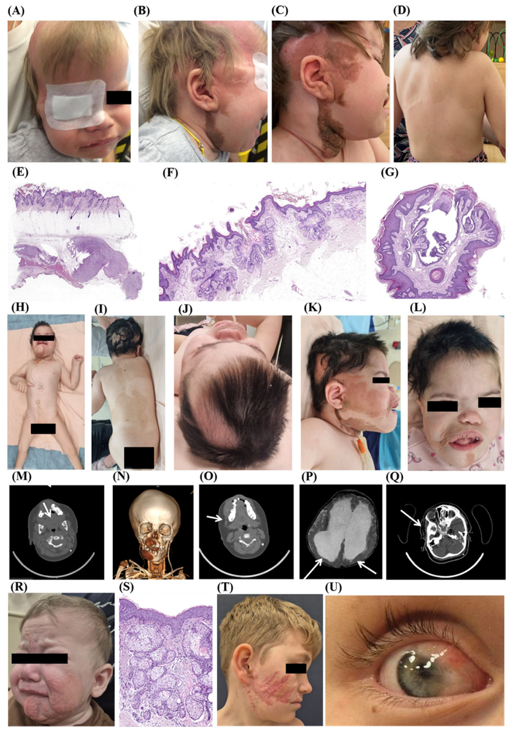
Figure 4.
Phenotype ID 9: (A-C) – LNS on the scalp, face and neck, as well as on the chest and abdomen, predominantly on the left side and in the left axillary region following Blashko’s lines, nevus spilus on the neck left-sided. Histology ID9 (D-G): (D) – ectomesenchymoma in the urinary bladder wall (H&E, x80); (E) – neoplastic tissue is composed of moderate to low cellularity and biphasic structure (H&E, x250); (F) – rhabdomyoblastic component is composed of round to oval cells with abundant eosinophylic cytoplasm and cross striations (arrowhead) (H&E, x350); (G) – second component composed of spindle cells with wavy nuclei and admixed ganglion cells (arrowhead) (H&E, x350). Immunohistochemistry ID9 (H-K): (H) – expression of Desmin (x350); (I) – expression of Myogenin in rhabdomyoblastic component (x350); (J) – expression of S100 in ganglioneuroma-like component (x350); (K) – expression of Synaptophysin in ganglion cells in ganglioneuroma-like component (arrowhead) (x350).
Figure 4.
Phenotype ID 9: (A-C) – LNS on the scalp, face and neck, as well as on the chest and abdomen, predominantly on the left side and in the left axillary region following Blashko’s lines, nevus spilus on the neck left-sided. Histology ID9 (D-G): (D) – ectomesenchymoma in the urinary bladder wall (H&E, x80); (E) – neoplastic tissue is composed of moderate to low cellularity and biphasic structure (H&E, x250); (F) – rhabdomyoblastic component is composed of round to oval cells with abundant eosinophylic cytoplasm and cross striations (arrowhead) (H&E, x350); (G) – second component composed of spindle cells with wavy nuclei and admixed ganglion cells (arrowhead) (H&E, x350). Immunohistochemistry ID9 (H-K): (H) – expression of Desmin (x350); (I) – expression of Myogenin in rhabdomyoblastic component (x350); (J) – expression of S100 in ganglioneuroma-like component (x350); (K) – expression of Synaptophysin in ganglion cells in ganglioneuroma-like component (arrowhead) (x350).
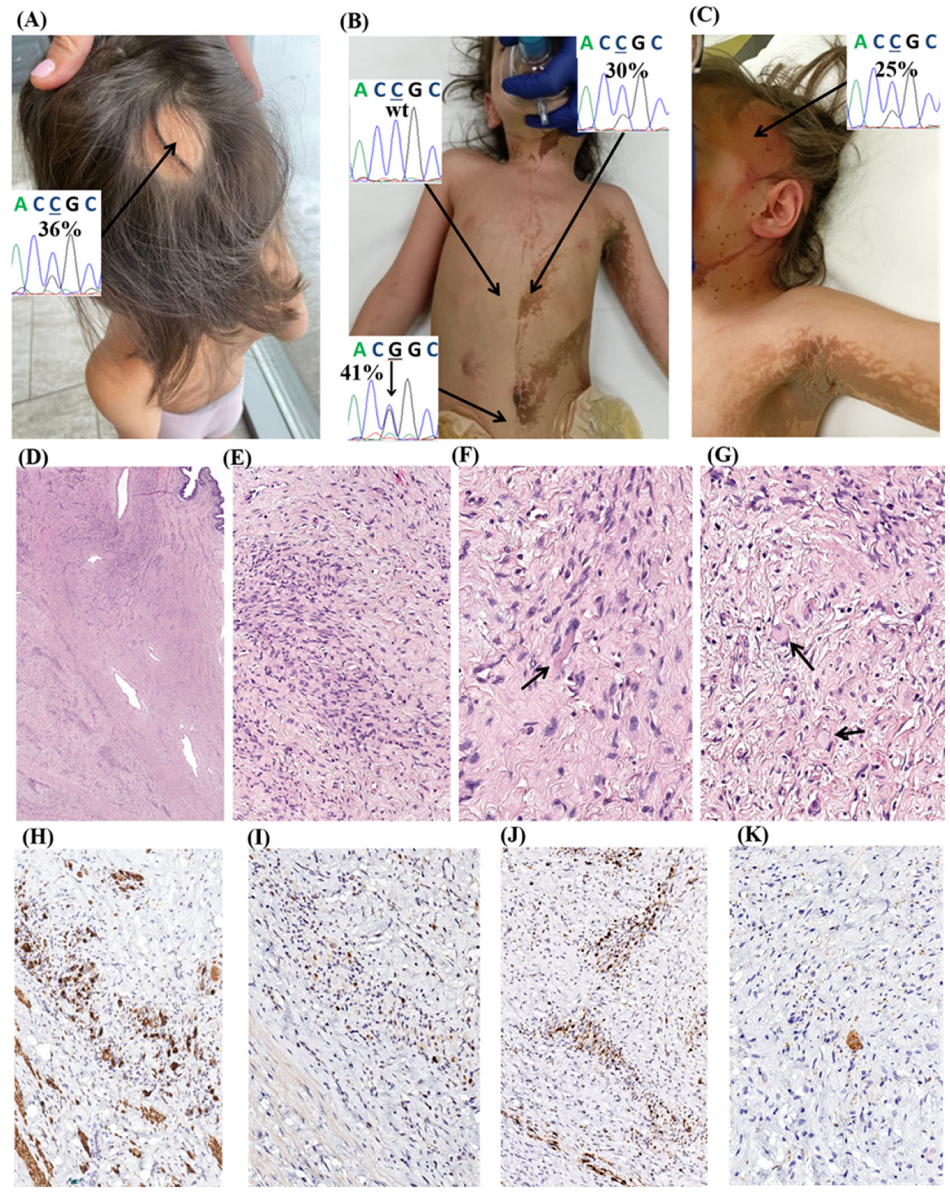
Figure 5.
Phenotype ID10 at age 3 (A, B) and 18 (C-F): LNS in the scalp, face, neck and trunk predominantly left-sided following Blashko’s lines and nevus spilus in the right-sided trunk and left arm, pronounced scoliotic deformities. Phenotype ID11 (G, H): (G) – LNS on the scalp, neck, auricle and trunk predominantly left-sided following Blashko’s lines; (H) – pronounced scoliosis of the spine and nevus spilus in lumbar region. Phenotype ID12 (I-N): LNS on the scalp and nevus spilus on the face, neck, back and chest (I-K), hemangioma of right eye (L). Nevus spilus, histology (M, N) - on the background of slight epidermal melanocytic hyperplasia, there are multiple compound melanocytic nevi (M, H&E, x40), composed of junctional and dermal nests of small monomorphic nevus cells with periadnexal distribution (N. H&E, x100). Phenotype ID13 (O-Q): LNS observed on the scalp, neck and back areas, nevus spilus on the face, left arm and in the back area. Phenotype ID14 (R, S): LNS on the scalp, face and neck predominantly left-sided following Blashko’s lines, nevus spilus on the chest left-sided. Phenotype ID15 (T): facial dysmorphism (high forehead and hypoplasia of the brow ridges, depressed bridge of the nose), nevus spilus on the trunk and LNS on the scalp and trunk in the middle line.
Figure 5.
Phenotype ID10 at age 3 (A, B) and 18 (C-F): LNS in the scalp, face, neck and trunk predominantly left-sided following Blashko’s lines and nevus spilus in the right-sided trunk and left arm, pronounced scoliotic deformities. Phenotype ID11 (G, H): (G) – LNS on the scalp, neck, auricle and trunk predominantly left-sided following Blashko’s lines; (H) – pronounced scoliosis of the spine and nevus spilus in lumbar region. Phenotype ID12 (I-N): LNS on the scalp and nevus spilus on the face, neck, back and chest (I-K), hemangioma of right eye (L). Nevus spilus, histology (M, N) - on the background of slight epidermal melanocytic hyperplasia, there are multiple compound melanocytic nevi (M, H&E, x40), composed of junctional and dermal nests of small monomorphic nevus cells with periadnexal distribution (N. H&E, x100). Phenotype ID13 (O-Q): LNS observed on the scalp, neck and back areas, nevus spilus on the face, left arm and in the back area. Phenotype ID14 (R, S): LNS on the scalp, face and neck predominantly left-sided following Blashko’s lines, nevus spilus on the chest left-sided. Phenotype ID15 (T): facial dysmorphism (high forehead and hypoplasia of the brow ridges, depressed bridge of the nose), nevus spilus on the trunk and LNS on the scalp and trunk in the middle line.
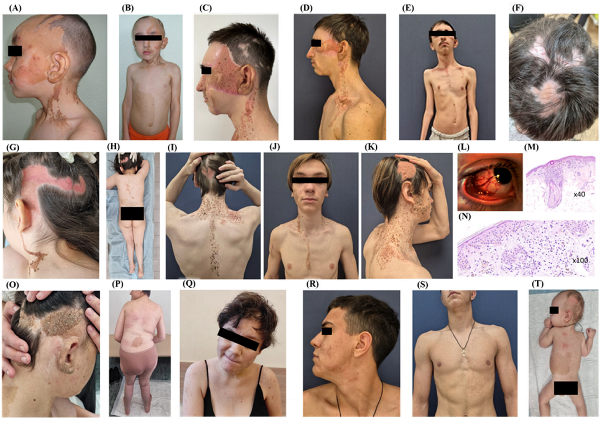
Figure 6.
Differential diagnosis. Phenotype ID16 (A): epidermal nevus in the face and neck area, more pronounced on the left. Brain MRI ID16 (B): left-sided hemimegalencephaly (enlarged hemisphere indicated by arrow). Phenotype ID17: linear cerebriform nevus before (C) and after (D) surgical excision. Phenotype ID18: linear epidermal nevus on the scalp and in the right temporal region (E). For comparison, a congenital linear epidermal nevus in the right temporal region in a patient with Cowden syndrome is presented (F).
Figure 6.
Differential diagnosis. Phenotype ID16 (A): epidermal nevus in the face and neck area, more pronounced on the left. Brain MRI ID16 (B): left-sided hemimegalencephaly (enlarged hemisphere indicated by arrow). Phenotype ID17: linear cerebriform nevus before (C) and after (D) surgical excision. Phenotype ID18: linear epidermal nevus on the scalp and in the right temporal region (E). For comparison, a congenital linear epidermal nevus in the right temporal region in a patient with Cowden syndrome is presented (F).
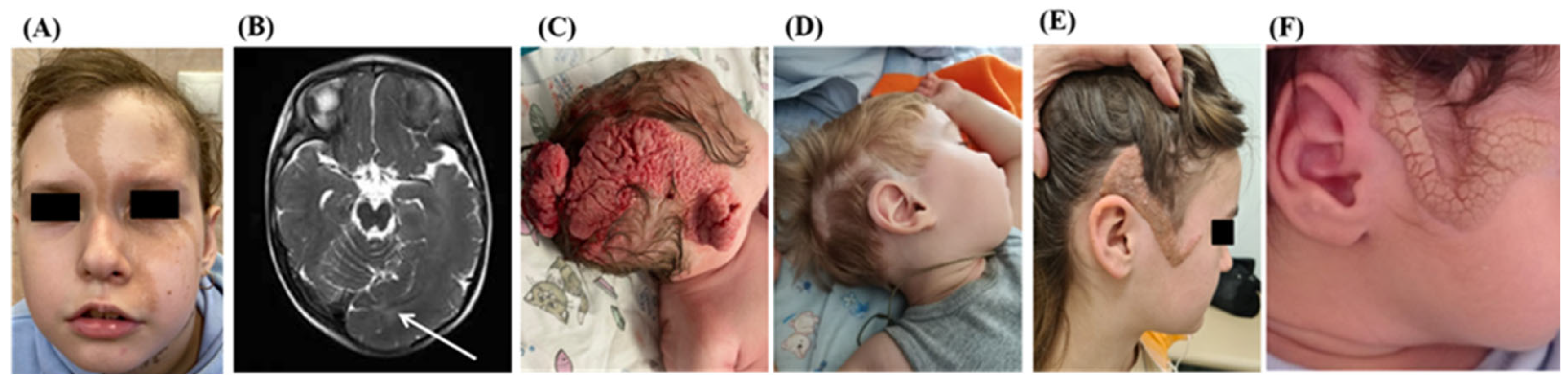
Figure 7.
(A) - Frequency of HRAS and KRAS genetic variants in the present study compared to the published literature data. (B) - Frequency of different extracutaneous abnormalities in HRAS-mutant and KRAS-mutant patients with mosaic RASopathies (p* - FDR-adjusted p-value, p* < 0.05 is considered significant).
Figure 7.
(A) - Frequency of HRAS and KRAS genetic variants in the present study compared to the published literature data. (B) - Frequency of different extracutaneous abnormalities in HRAS-mutant and KRAS-mutant patients with mosaic RASopathies (p* - FDR-adjusted p-value, p* < 0.05 is considered significant).
Disclaimer/Publisher’s Note: The statements, opinions and data contained in all publications are solely those of the individual author(s) and contributor(s) and not of MDPI and/or the editor(s). MDPI and/or the editor(s) disclaim responsibility for any injury to people or property resulting from any ideas, methods, instructions or products referred to in the content. |
© 2026 by the authors. Licensee MDPI, Basel, Switzerland. This article is an open access article distributed under the terms and conditions of the Creative Commons Attribution (CC BY) license (http://creativecommons.org/licenses/by/4.0/).
Copyright: This open access article is published under a Creative Commons CC BY 4.0 license, which permit the free download, distribution, and reuse, provided that the author and preprint are cited in any reuse.