

Submitted:
17 August 2026
Posted:
18 August 2026
You are already at the latest version
Abstract
Virus infections unfold through spatially organized events whose timing and progression vary among individual cells. Imaging complements population-based approaches by preserving spatial context and, in live experiments, temporal progression at single-cell and tissue scales. This review examines how imaging has advanced our understanding of virus infection, using influenza A virus (IAV) as the principal example. The review is organized around three complementary perspectives: visualization of viral components throughout infection, analysis of virus-induced host-cell and tissue remodeling, and investigation of heterogeneous antiviral responses. Together, these perspectives show how imaging preserves the spatial, temporal, and cell-to-cell context needed to relate viral processes to host-cell responses across scales. Because imaging approaches differ in what they directly measure, this review considers their complementary strengths and limitations and distinguishes descriptive observations from mechanistic conclusions.
Keywords:
fluorescence imaging
; influenza A virus
; live-cell imaging
; viral life cycle
; virus-host interaction
; antiviral responses
; host cell remodeling
; single-cell imaging
; infection heterogeneity
; infection dynamics
1. Introduction
Virus infection is a spatially and temporally organized process in which viral life-cycle progression, host-cell remodeling, and antiviral responses continuously interact to determine infection outcome [1,2,3]. Influenza A virus (IAV) provides a clear example of this spatial and temporal organization: its life cycle requires coordinated use of distinct cellular compartments, such as the endosomal compartment, the nucleus, intracellular trafficking networks, and the plasma membrane [4,5]. Importantly, infection progression and outcome are not uniform across infected cells. Even within genetically identical cell populations, individual cells can follow distinct trajectories due to differences in incoming virus particles, viral gene expression dynamics, host-cell state, and antiviral response activation [2,6,7,8]. As a result, measurements that average across populations can obscure cell-to-cell heterogeneity, including divergent infection trajectories and rare infection states. Recent advances in single-particle tracking, live-cell microscopy, multiplexed imaging of viral RNAs, super-resolution microscopy, and tissue-scale imaging have begun to fill important gaps between population-averaged descriptions of infection and the spatially and temporally organized events occurring in individual cells [9,10,11]. These imaging modalities reveal when and where viral and host processes occur, resolve heterogeneous infection trajectories, and allow multiple features of infection to be related within the same cell or tissue. However, insights obtained through imaging remain distributed across studies focused on different viruses, life-cycle stages, host processes, imaging modalities, and spatial scales. Here, we integrate diverse imaging studies to assess how imaging has collectively expanded our understanding of virus infection.
With a primary focus on IAV, this review draws on selected examples from other viruses that offer relevant methodological or biological insights and examines infection from three complementary perspectives: viral components and infection progression, virus-induced remodeling of host-cell organization, and antiviral responses. Together, these perspectives show how viral life-cycle progression, host-cell remodeling, and antiviral responses are interconnected and shape infection outcomes.
2. Imaging Viral Components
2.1. Visualizing Virion Attachment, Trafficking, and Membrane Fusion
IAV infection begins with attachment to susceptible cells. To establish productive attachment, virions must navigate extracellular environments while avoiding repeated engagement with decoy receptors [12,13,14]. HA binds terminal sialic acid residues on cellular and extracellular glycoconjugates, whereas NA cleaves these residues; accordingly, perturbation of either HA receptor binding or NA enzymatic activity reduces virion infectivity [15,16]. However, extracellular navigation is a spatial process, and endpoint measurements could not reveal how HA and NA together direct virion movement. Labeling virions with lipophilic membrane dyes enabled tracking of individual particles across sialylated surfaces and through complex extracellular environments (Figure 1A) [12,17]. Tracking single virions revealed that coordinated receptor binding by HA and receptor cleavage by NA promote persistent movement through sialylated environments by preventing repeated attachment to previously encountered receptors [12], likely facilitating successful engagement with susceptible cells. Following productive attachment, IAV particles are internalized by endocytosis, and subsequent acidification of endosomal compartments triggers fusion between viral and host membranes. Fluorescent labeling allowed visualization of virion attachment to the host cell plasma membrane and transport through the endosomal network, while self-quenching lipid dyes provided a direct readout of the timing and location of membrane fusion [18,19,20,21]. Combining single-virion tracking with detection of membrane fusion demonstrated that fusion does not occur immediately after uptake. Instead, virions undergo microtubule-dependent transport through the endosomal network toward the perinuclear region before endosomal acidification triggers membrane fusion [18]. Thus, single-virion tracking and membrane-fusion measurements established that successful entry depends on completion of multiple trafficking and maturation steps before genome release.
2.2. Following Incoming Viral Ribonucleoprotein Complexes (vRNPs) from Fusion to Nuclear Import
The events linking fusion to nuclear delivery have remained difficult to study because incoming vRNPs are present in low copy numbers. Early immunofluorescence (IF) studies of vRNPs performed at a high multiplicity of infection (MOI) suggested that nuclear import is efficient and rapid [22]. Subsequent application of single-molecule fluorescence in situ hybridization (smFISH) enabled visualization of viral RNA segments during the earliest stages of infection, revealing vRNP subcellular localization, colocalization, and nuclear import [23]. However, because these experiments required high-MOI infections and relied on fixed-cell snapshots, neither approach could track individual incoming vRNPs or reveal the timing of the intermediate steps linking membrane fusion to nuclear delivery. An important advance came with quantum-dot labeling of the PA subunit of the viral polymerase, allowing the first direct visualization of individual incoming vRNPs in living cells [24]. Quantum-dot tracking of individual incoming vRNPs demonstrated that membrane fusion occurs predominantly near the nucleus, where individual vRNPs are released from the incoming bundle before rapidly being imported into the nucleus. Recently, live vRNP labeling using fluorescent nanobodies targeting NP within vRNP complexes (VISUN; vRNP imaging of unmodified negative-strand viruses) enabled visualization of all incoming vRNPs without viral engineering or in-virion labeling procedures (Figure 1B) [25]. VISUN confirmed rapid and complete post-fusion vRNP debundling and demonstrated that the majority of incoming vRNPs successfully reach the nucleus within minutes of membrane fusion. Thus, the rapid and efficient transition from membrane fusion to nuclear delivery does not pose a major barrier to productive infection.
2.3. Visualizing Viral Protein Expression and Localization
Viral gene expression results in the accumulation of viral proteins, which can be visualized by immunofluorescence and used as markers of infection progression. Beyond measuring protein abundance, fluorescence imaging revealed that many influenza proteins undergo characteristic changes in subcellular localization that reflect their changing functions throughout infection [26,27]. For example, observation of nuclear accumulation of M1 helped define its role in terminating viral replication and promoting nuclear export of vRNPs [28]. Following completion of genome replication, NP redistributes from the nucleus to the cytoplasm during vRNP nuclear export. Imaging of NP localization thus directly visualizes the transition from viral genome replication to cytoplasmic transport [29]. Taken together, viral infection is not a static process of protein production, but involves coordinated spatial redistribution of viral factors between cellular compartments as infection progresses. Live-cell imaging complements fixed-cell approaches by resolving the temporal dynamics of viral protein expression and localization. Approaches for tracking protein expression or localization in living cells broadly fall into two categories: viruses that express directly tagged viral proteins, and reporter viruses that express fluorescent or luminescent proteins as indirect readouts of viral gene expression (Figure 1C).
Direct visualization of IAV proteins was achieved using recombinant IAVs expressing genetically tagged PB2[30,31], PA [32], NS1[33,34], or structural proteins [35]. Because many RNA viruses, including IAV, tolerate only limited genetic perturbation, particularly large insertions encoding fluorescent proteins, recombinant fluorescent viruses require extensive validation and optimization to ensure that tagging does not compromise viral fitness or genetic stability [31]. Live tracking of fluorescently labeled polymerase-containing vRNPs revealed directed, microtubule-dependent transport through the cytoplasm following nuclear export (Figure 1E) [30,31,32]. Real-time visualization of viral structural proteins enabled direct observation of virion assembly and release at the plasma membrane [35]. Because fluorescence becomes detectable only after sufficient accumulation of the tagged protein, direct protein-labeling approaches are best suited for studying later stages of infection and are generally less informative for the earliest stages.
Reporter viruses expressing fluorescent or bioluminescent proteins can be used to monitor infection spread continuously in cultured cells and animal models, revealing how viral dissemination depends on viral genotype, host antiviral responses, and tissue context [33,36,37,38,39]. At the single-cell level, substantial variation in the onset and rate of viral gene expression has been shown among individual infected cells despite synchronous infection [37]. Multicolor reporter viruses enabled coinfection to be visualized within individual cells and functional interactions between coinfecting viruses to be monitored [40]. Interactions between coinfecting viruses can either inhibit or promote productive infection. For example, live imaging showed that primary infection progressively reduces susceptibility to secondary infection through superinfection exclusion [40]. Conversely, coinfecting viruses can complement incomplete viral genomes by supplying missing functions in trans, thereby supporting productive infection [41].
2.4. Imaging Viral RNA Synthesis and Genome Replication
Although viral protein expression reports on infection progression, it provides only an indirect view of the underlying transcriptional and replication program. IAV infection progression is governed by the synthesis of distinct viral RNA species, including viral mRNAs, full-length complementary RNAs (cRNAs) that serve as replication templates, and negative-sense genomic RNAs (vRNAs). Biochemical approaches established the average temporal order of viral transcription and genome replication [42,43,44,45,46]. RNA imaging introduced a complementary perspective by allowing viral RNAs to be detected directly in individual infected cells while retaining information about their intracellular localization (Figure 1D). Initial RNA FISH studies examined the spatial organization of the eight viral genome segments within virions and showed that individual virions generally package one copy of each genome segment [47]. Subsequent multiplexed RNA imaging addressed the dynamics of viral transcription and replication. For example, multiplexed error-robust FISH (merFISH) was used to simultaneously quantify all eight viral mRNAs in individual infected cells, revealing segment-specific differences in cytoplasmic mRNA abundance due to both differential transcription and nuclear export [48]. More recently, direct RNA padlock probing coupled with rolling-circle amplification and in situ sequencing (mudRapp-seq) enabled simultaneous visualization of viral mRNAs and genomic RNAs, allowing transcriptional output and genome replication to be quantified on a segment-by-segment basis [49]. The mudRapp-seq measurements revealed pronounced segment-specific differences in transcriptional output despite largely similar genome replication dynamics. Direct live imaging of viral RNA dynamics and trafficking remains technically challenging. Current live RNA imaging strategies (e.g., PP7/MS2 stem-loop systems, RNA aptamers, or CRISPR-dCas13 imaging) rely on large engineered RNA insertions or exogenous probe delivery [50,51] and are difficult to implement in IAV because of its compact NP-coated vRNP architecture, limited genetic flexibility, and stringent RNA packaging signals distributed throughout each genome segment [5,52].
2.5. Continuous Live Imaging of vRNPs
IAV genome segments can instead be visualized indirectly, by targeting the associated proteins within the vRNP complex. Earlier protein-labeling strategies enabled imaging of either incoming [24] or newly synthesized [30,31,32] vRNPs, but not continuous tracking throughout infection. More recently, imaging of cells expressing fluorescent nanobodies targeting NP (VISUN) enabled continuous visualization of vRNPs throughout infection and direct measurement of the timing of genome replication and nuclear export in living cells (Figure 1B) [25]. Because individual infection trajectories can be followed from entry onward, VISUN also makes it possible to identify and quantify abortive infections, providing a more complete picture of infection outcomes within a cell population. Finally, because VISUN does not require viral engineering, it enables direct quantitative comparison of the life cycles of genetically diverse influenza strains and clinical isolates.
2.6. Imaging Genome Assembly and Progeny Production
Following nuclear export, newly synthesized vRNPs are transported toward the plasma membrane, where progeny virions are assembled. However, how assembly of the eight distinct genome segments is coordinated during transport to budding sites remained unclear. Segment-specific RNA imaging demonstrated that individual genome segments progressively colocalize during cytoplasmic transport, while live-cell imaging of fluorescently labeled polymerase-containing complexes revealed organized, directional trafficking of vRNPs along microtubules. Together, progressive segment colocalization and organized, directional vRNP trafficking support a model in which genome assembly occurs through sequential interactions on membranous compartments prior to budding rather than exclusively at the plasma membrane [23,32]. Recently, the addition of fluorescent nanobodies targeting the HA protein to the extracellular environment has provided the first live readout of progeny virion budding (Figure 1F) [25]. Combining HA-nanobody-based detection of progeny virion budding with imaging of vRNP nuclear export demonstrated that progeny virion production follows vRNP nuclear export with minimal time delay. However, not all cells with observed vRNP nuclear export subsequently produce detectable progeny virions, revealing that budding is not a guaranteed next step after vRNP export and instead is an additional source of infection heterogeneity.
3. Imaging Virus-Induced Host-Cell Remodeling
Following viral processes through a host cell identifies where successive stages of infection occur but does not necessarily reveal how infection changes the cellular environment in which those stages proceed. Viral infection remodels host cells by redistributing molecular components, reorganizing organelles, constructing new reaction environments, and altering cellular and tissue architecture. Because many virus-induced cellular changes are defined by their spatial organization, temporal dynamics, or ultrastructure rather than molecular abundance alone, imaging is uniquely suited to study virus-induced host-cell remodeling. Different imaging modalities provide complementary views: fluorescence microscopy visualizes the distribution of selected molecules, live-cell imaging resolves their dynamics, electron microscopy reveals membrane architecture and ultrastructure, and tissue imaging relates cellular phenotypes to higher-order organization. Accordingly, conclusions about host-cell remodeling should reflect the specific cellular property that each imaging approach directly measures.
3.1. Visualizing Spatial Remodeling of Gene Expression
Cellular gene expression depends not only on molecular abundance, but also on localization of RNAs and RNA processing machinery. Immunofluorescence (IF) and single-molecule RNA FISH (smFISH) can determine whether selected proteins or RNAs remain dispersed or accumulate in compartments at the single-cell level, and have been particularly useful for studying IAV-induced host shutoff, a process characterized by extensive spatial reorganization of the cellular gene-expression machinery. Combined smFISH and immunofluorescence microscopy revealed depletion of cytoplasmic poly(A) RNA and nuclear accumulation of poly(A) RNA and poly(A)-binding protein 1 (PABPC1), while confocal fluorescence microscopy further showed dispersal of nuclear speckles, nuclear compartments involved in pre-mRNA processing [53,54,55]. Comparisons of cells infected with recombinant IAVs lacking expression of either NS1 or PA-X distinguished the individual spatial changes that together comprise host shutoff. Viral protein PA-X accounts for much of the cytoplasmic RNA loss, and NS1 compromises NXF1-dependent mRNA export. Consistently, nuclear PABPC1 accumulation depends on concerted PA-X and NS1 activity. Thus, combining imaging of host-shutoff-associated spatial changes with the use of engineered viruses that fail to express individual viral proteins reveals mechanistically distinct host-shutoff processes.
Although fixed-cell IF and smFISH reveal spatial reorganization of RNAs and RNA-processing machinery during infection, they do not report on RNA integrity, translation, or movement [56]. By combining live-cell imaging of viral RNA translation and replication dynamics with fixed-cell measurements of host protein synthesis, a study of coxsackievirus B3-infected cells showed that global host protein synthesis was already substantially reduced before replication of the incoming viral RNA, whereas viral translation efficiency remained stable throughout infection [57]. This illustrates how combining measurements of viral and host RNA dynamics with protein synthesis can provide functional information that complements mRNA localization measurements. Applied to influenza, similar approaches could directly link virus-induced changes in RNA localization to their consequences for protein synthesis.
3.2. Detection of Virus-Built Reaction Environments
IAV replication takes place in the nucleus and does not require formation of the membrane-associated replication organelles (ROs) characteristic of many cytoplasmic RNA viruses [5]. In contrast, diverse viruses such as flaviviruses, enteroviruses, and coronaviruses construct de novo membranous compartments dedicated to viral replication [58]. Imaging approaches developed to understand RO biogenesis have clarified their molecular composition, assembly dynamics, and membrane architecture. Fluorescence microscopy identified enriched viral and host components [58]. Live fluorescence imaging revealed the dynamics of RO formation [59], whereas electron tomography revealed highly organized, topologically complex membrane architecture [60]. Together, these observations showed how viruses construct and organize specialized replication environments. However, molecular enrichment, morphology, and exchange dynamics do not by themselves establish organelle function, which requires complementary functional evidence.
3.3. Visualizing Organelle Function and Membrane Topology
Infection also remodels pre-existing organelles, creating an important interpretive distinction between changes in morphology and changes in function. Mitochondria illustrate this distinction, since they may fragment, elongate, or redistribute during infection. Yet, these morphological changes do not by themselves establish altered mitochondrial function. Specialized live reporters and fluorescence biosensors can measure membrane potential or local biochemical activity, that are not apparent from mitochondrial shape or position. Live-cell imaging using mitochondrial functional reporters in IAV-infected cells linked expression of selected PB1-F2 variants to loss of membrane potential and organelle injury, but the magnitude and direction of IAV-associated mitochondrial changes depend on viral strain and experimental context [61,62,63].
Membrane topology presents a related but distinct imaging challenge: the distribution of labeled proteins does not define the membrane structures that contain them. During virion budding, for example, viral proteins accumulate at defined sites on the plasma membrane, but their localization alone does not reveal how the membrane is reshaped. Electron microscopy and tomography therefore complement fluorescence imaging by resolving distinct membrane architectures during IAV budding, including glycoprotein-associated membrane curvature, M1-dependent formation of filamentous virions, and M2 enrichment at budding necks associated with membrane scission [64,65,66]. Together, fluorescence microscopy, electron microscopy, and tomography combine molecular localization with direct visualization of membrane architecture to reveal membrane-remodeling events that cannot be inferred from protein localization alone.
3.4. Following Endomembrane Trafficking
To transport progeny vRNPs from the nucleus to the plasma membrane, IAV remodels the pre-existing Rab11-dependent recycling endosome network. This remodeling of the recycling endosomal compartment was characterized by studies using several complementary imaging approaches. Fixed-cell fluorescence showed that the normally dispersed Rab11 recycling endosomal network becomes enlarged and clustered during infection [67]. Using a vRNP reconstitution system, dual-color live imaging first identified vRNP cotrafficking with Rab11-positive membranes [68]. Light-sheet imaging later resolved Rab11-vRNA cotransport during infection and showed that vRNP-bearing carriers move more slowly, travel shorter distances, and arrest more frequently [69]. Fixed and live measurements therefore establish both redistribution of the recycling network and a change in its transport dynamics. Correlative light-electron microscopy localized vRNPs to infection-remodeled Rab11 carriers, directly connecting Rab11 redistribution with vRNP trafficking [70]. Live imaging showed that vRNP-containing inclusions deform, fuse, split, and exchange material (Figure 1E), while fluorescence recovery measurements supported rapid internal turnover [71]. The observed dynamics and rapid internal turnover of vRNP-containing inclusions are consistent with liquid-like behavior but can also arise from other binding and clustering mechanisms. Definitive assignment of the material state therefore requires complementary evidence [72].
As IAV reorganizes the Rab11 compartment, Rab11-dependent cargo recycling declines. Functional assays using fluorescent transferrin as recycling cargo demonstrated that Rab11-dependent cargo transport is attenuated during IAV infection. Fluorescence imaging of Rab11 redistribution, combined with functional recycling and binding measurements, supported a model in which vRNPs compete with Rab11-interacting proteins for Rab11 binding, thereby reducing cellular recycling and contributing to Rab11 redistribution [67]. A subsequent study combining fluorescence imaging with perturbational evidence showed that ATG9A promotes the release of vRNP-bearing Rab11 endosomes from microtubules and their organization into viral inclusions, indicating a regulated reorganization mechanism not fully explained by competition for Rab11 binding alone [73]. Correlative light-electron microscopy further showed that altered Rab11 carriers associate with remodeled endoplasmic reticulum, suggesting that IAV reorganizes a broader membrane network than previously appreciated [70]. The combined evidence therefore supports regulated remodeling of the recycling endosome compartment that extends beyond Rab11-binding competition alone. Overall, IAV redirects a constitutive recycling system into a slower, spatially concentrated transport environment that supports vRNP delivery while compromising host cargo recycling. This model was established by integrating spatial, dynamic, ultrastructural, and perturbational measurements rather than any single imaging approach.
3.5. Imaging of Intercellular and Tissue-Scale Remodeling
Virus infection remodels host tissues at multiple spatial scales, ranging from interactions between neighboring cells to changes in tissue architecture and function. Imaging has been essential for linking intercellular and tissue-level changes to virus dissemination and pathogenesis. Confocal and super-resolution imaging localized Rab11-associated vRNPs within actin-rich intercellular connections between infected and neighboring cells, demonstrating directed movement of viral material from infected cells toward neighboring cells [74]. Infectivity measurements confirmed that transfer through these structures seeds productive infection, identifying a potential route of cell-to-cell spread that complements extracellular virus release. Beyond intercellular communication, imaging revealed how infection alters tissue function. Live imaging of IAV-infected airway systems demonstrated infection-associated changes in ciliary activity [75]. Longitudinal imaging of SARS-CoV-2-infected airways linked mucociliary movement to the spatial pattern of viral spread, showing that tissue-level transport processes shape infection dissemination across the airway epithelium [76]. Infection-induced remodeling also changes tissue composition. For example, three-dimensional light-sheet imaging of Zika virus-infected cerebral organoids quantified the loss and redistribution of specific cell populations [77]. Similarly, imaging showed that IAV infection depletes ciliated cells and reduces airway epithelial thickness, thereby changing the cell-type composition of the airway epithelium [78]. Yet electrical measurements indicated preserved barrier function, demonstrating that tissue composition and function can be remodeled independently during infection. By linking infection to changes in intercellular communication, tissue organization, and function, imaging extends virology research beyond individual cells to higher-order processes that influence viral spread and disease progression.
5. Concluding Remarks
Throughout this review, we describe imaging not as a single methodology, but as a collection of complementary approaches that reveal distinct aspects of virus infection (Table 1). Fluorescence microscopy resolves the spatial organization of viral and host components, live-cell imaging establishes the temporal order of infection events, RNA imaging indirectly measures viral transcription and genome replication, and tissue-scale imaging relates intracellular infection to changes in tissue organization and function. Together, these approaches extend population-averaged measurements by enabling direct observation of individual infection trajectories and relationships between viral and host processes that would otherwise remain obscured.
Imaging provides direct evidence for the measured observable, but not necessarily for the biological mechanisms underlying those observations. For example, colocalization does not establish molecular interaction, morphology does not define function, and RNA localization does not imply translation. Likewise, fluorescence measurements alone cannot resolve membrane topology or molecular mechanism. Interpreting imaging data therefore requires matching biological conclusions to the observable being measured and supporting mechanistic models with complementary functional evidence. Because different imaging approaches measure complementary observables, integrating these measurements can provide a more complete picture than any single approach alone. Measuring complementary parameters is particularly informative because viral and host-cell processes are themselves highly interconnected. Virus infection can remodel host cells, while host-cell remodeling and antiviral responses in turn influence the progression and outcome of infection. Yet specialized imaging approaches often examine viral replication, host-cell remodeling, antiviral signaling, and tissue organization separately. Combining measurements of these processes within the same cells or tissues would allow their spatial and temporal relationships to be examined directly. Such integrated measurements could link early intracellular events to subsequent productive or nonproductive outcomes, identify candidate causal relationships that can be evaluated using complementary evidence, and provide a more integrated understanding of how viral and host processes together shape infection outcome.
Author Contributions
Conceptualization, BAK, JS, HHR; Writing – Original Draft, BAK, JS, HHR; Writing – Review & Editing, BAK, JS, HHR. All authors approved the final manuscript.
Funding
BAK., JS., and HHR. are supported by a grant from the European Union (ERC, VirIm, 101044794).
Data Availability Statement
Not applicable.
Acknowledgments
Not applicable.
Conflicts of Interest
The authors declare no conflicts of interest.
References
- Brandenburg, B.; Zhuang, X. Virus trafficking - learning from single-virus tracking. Nat. Rev. Microbiol. 2007, 5, 197–208. [Google Scholar] [CrossRef] [PubMed]
- Russell, A.B.; Trapnell, C.; Bloom, J.D. Extreme heterogeneity of influenza virus infection in single cells. Elife 2018, 7, e32303. [Google Scholar] [CrossRef] [PubMed]
- Garcia-Sastre, A. Induction and evasion of type I interferon responses by influenza viruses. Virus Res. 2011, 162, 12–18. [Google Scholar] [CrossRef] [PubMed]
- Dou, D.; Revol, R.; Ostbye, H.; Wang, H.; Daniels, R. Influenza A Virus Cell Entry, Replication, Virion Assembly and Movement. Front Immunol. 2018, 9, 1581. [Google Scholar] [CrossRef] [PubMed]
- Eisfeld, A.J.; Neumann, G.; Kawaoka, Y. At the centre: Influenza A virus ribonucleoproteins. Nat. Rev. Microbiol. 2015, 13, 28–41. [Google Scholar] [CrossRef] [PubMed]
- Russell, A.B.; Elshina, E.; Kowalsky, J.R.; Te Velthuis, A.J.W.; Bloom, J.D. Single-Cell Virus Sequencing of Influenza Infections That Trigger Innate Immunity. J. Virol. 2019, 93, e00500-19. [Google Scholar] [CrossRef] [PubMed]
- Brooke, C.B. Population Diversity and Collective Interactions during Influenza Virus Infection. J. Virol. 2017, 91, e01164-17. [Google Scholar] [CrossRef] [PubMed]
- Heldt, F.S.; Kupke, S.Y.; Dorl, S.; Reichl, U.; Frensing, T. Single-cell analysis and stochastic modelling unveil large cell-to-cell variability in influenza A virus infection. Nat. Commun. 2015, 6, 8938. [Google Scholar] [CrossRef] [PubMed]
- Liu, S.L.; Wang, Z.G.; Xie, H.Y.; Liu, A.A.; Lamb, D.C.; Pang, D.W. Single-Virus Tracking: From Imaging Methodologies to Virological Applications. Chem. Rev. 2020, 120, 1936–1979. [Google Scholar] [CrossRef] [PubMed]
- Lakdawala, S.S.; Lee, N.; Brooke, C.B. Teaching an Old Virus New Tricks: A Review on New Approaches to Study Age-Old Questions in Influenza Biology. J. Mol. Biol. 2019, 431, 4247–4258. [Google Scholar] [CrossRef] [PubMed]
- Touizer, E.; Sieben, C.; Henriques, R.; Marsh, M.; Laine, R.F. Application of Super-Resolution and Advanced Quantitative Microscopy to the Spatio-Temporal Analysis of Influenza Virus Replication. Viruses 2021, 13, 233. [Google Scholar] [CrossRef] [PubMed]
- Vahey, M.D.; Fletcher, D.A. Influenza A virus surface proteins are organized to help penetrate host mucus. Elife 2019, 8, e43764. [Google Scholar] [CrossRef] [PubMed]
- de Vries, E.; Du, W.; Guo, H.; de Haan, C.A.M. Influenza A Virus Hemagglutinin-Neuraminidase-Receptor Balance: Preserving Virus Motility. Trends Microbiol. 2020, 28, 57–67. [Google Scholar] [CrossRef] [PubMed]
- Guo, H.; Rabouw, H.; Slomp, A.; Dai, M.; van der Vegt, F.; van Lent, J.W.M.; McBride, R.; Paulson, J.C.; de Groot, R.J.; van Kuppeveld, F.J.M.; et al. Kinetic analysis of the influenza A virus HA/NA balance reveals contribution of NA to virus-receptor binding and NA-dependent rolling on receptor-containing surfaces. PLoS Pathog. 2018, 14, e1007233. [Google Scholar] [CrossRef] [PubMed]
- Wagner, R.; Matrosovich, M.; Klenk, H.D. Functional balance between haemagglutinin and neuraminidase in influenza virus infections. Rev. Med. Virol. 2002, 12, 159–166. [Google Scholar] [CrossRef] [PubMed]
- Mitnaul, L.J.; Matrosovich, M.N.; Castrucci, M.R.; Tuzikov, A.B.; Bovin, N.V.; Kobasa, D.; Kawaoka, Y. Balanced hemagglutinin and neuraminidase activities are critical for efficient replication of influenza A virus. J. Virol. 2000, 74, 6015–6020. [Google Scholar] [CrossRef] [PubMed]
- Yang, X.; Steukers, L.; Forier, K.; Xiong, R.; Braeckmans, K.; Van Reeth, K.; Nauwynck, H. A beneficiary role for neuraminidase in influenza virus penetration through the respiratory mucus. PLoS ONE 2014, 9, e110026. [Google Scholar] [CrossRef] [PubMed]
- Lakadamyali, M.; Rust, M.J.; Babcock, H.P.; Zhuang, X. Visualizing infection of individual influenza viruses. Proc. Natl. Acad. Sci. U S A 2003, 100, 9280–9285. [Google Scholar] [CrossRef] [PubMed]
- Lakadamyali, M.; Rust, M.J.; Zhuang, X. Endocytosis of influenza viruses. Microbes Infect. 2004, 6, 929–936. [Google Scholar] [CrossRef] [PubMed]
- Ivanovic, T.; Choi, J.L.; Whelan, S.P.; van Oijen, A.M.; Harrison, S.C. Influenza-virus membrane fusion by cooperative fold-back of stochastically induced hemagglutinin intermediates. Elife 2013, 2, e00333. [Google Scholar] [CrossRef] [PubMed]
- Broich, L.; Wullenkord, H.; Osman, M.K.; Fu, Y.; Müsken, M.; Reuther, P.; Brönstrup, M.; Sieben, C. Single influenza A viruses induce nanoscale cellular reprogramming at the virus-cell interface. Nat. Commun. 2025, 16, 3846. [Google Scholar] [CrossRef] [PubMed]
- Martin, K.; Helenius, A. Transport of incoming influenza virus nucleocapsids into the nucleus. J. Virol. 1991, 65, 232–244. [Google Scholar] [CrossRef] [PubMed]
- Chou, Y.Y.; Heaton, N.S.; Gao, Q.; Palese, P.; Singer, R.H.; Lionnet, T. Colocalization of different influenza viral RNA segments in the cytoplasm before viral budding as shown by single-molecule sensitivity FISH analysis. PLoS Pathog. 2013, 9, e1003358. [Google Scholar] [CrossRef] [PubMed]
- Qin, C.; Li, W.; Li, Q.; Yin, W.; Zhang, X.; Zhang, Z.; Zhang, X.E.; Cui, Z. Real-time dissection of dynamic uncoating of individual influenza viruses. Proc. Natl. Acad. Sci. U S A 2019, 116, 2577–2582. [Google Scholar] [CrossRef] [PubMed]
- Rabouw, H.H.; Schokolowski, J.; Muller, M.; Baars, M.J.D.; Dost, A.F.M.; Bestebroer, T.M.; Puschel, J.; Clevers, H.; Fouchier, R.A.M.; Tanenbaum, M.E. Live-cell single-vRNP imaging identifies viral gene expression signatures that shape influenza infection heterogeneity. Cell Syst. 2026, 17, 101489. [Google Scholar] [CrossRef] [PubMed]
- Melen, K.; Kinnunen, L.; Fagerlund, R.; Ikonen, N.; Twu, K.Y.; Krug, R.M.; Julkunen, I. Nuclear and nucleolar targeting of influenza A virus NS1 protein: Striking differences between different virus subtypes. J. Virol. 2007, 81, 5995–6006. [Google Scholar] [CrossRef] [PubMed]
- Miyamoto, S.; Nakano, M.; Morikawa, T.; Hirabayashi, A.; Tamura, R.; Fujita-Fujiharu, Y.; Hirose, N.; Muramoto, Y.; Noda, T. Migration of Influenza Virus Nucleoprotein into the Nucleolus Is Essential for Ribonucleoprotein Complex Formation. mBio 2022, 13, e03315-21. [Google Scholar] [CrossRef]
- Martin, K.; Helenius, A. Nuclear transport of influenza virus ribonucleoproteins: The viral matrix protein (M1) promotes export and inhibits import. Cell 1991, 67, 117–130. [Google Scholar] [CrossRef] [PubMed]
- Chase, G.P.; Rameix-Welti, M.A.; Zvirbliene, A.; Zvirblis, G.; Gotz, V.; Wolff, T.; Naffakh, N.; Schwemmle, M. Influenza virus ribonucleoprotein complexes gain preferential access to cellular export machinery through chromatin targeting. PLoS Pathog. 2011, 7, e1002187. [Google Scholar] [CrossRef] [PubMed]
- Nath, H.; Arndt, A.; Mann, J.T.; Baker, S.F. Tandem split-GFP influenza A viruses for sensitive and accurate replication analyses. Microbiol. Spectr. 2026, 14, e02772-25. [Google Scholar] [CrossRef] [PubMed]
- Avilov, S.V.; Moisy, D.; Munier, S.; Schraidt, O.; Naffakh, N.; Cusack, S. Replication-competent influenza A virus that encodes a split-green fluorescent protein-tagged PB2 polymerase subunit allows live-cell imaging of the virus life cycle. J. Virol. 2012, 86, 1433–1448. [Google Scholar] [CrossRef] [PubMed]
- Lakdawala, S.S.; Wu, Y.; Wawrzusin, P.; Kabat, J.; Broadbent, A.J.; Lamirande, E.W.; Fodor, E.; Altan-Bonnet, N.; Shroff, H.; Subbarao, K. Influenza a virus assembly intermediates fuse in the cytoplasm. PLoS Pathog. 2014, 10, e1003971. [Google Scholar] [CrossRef] [PubMed]
- Manicassamy, B.; Manicassamy, S.; Belicha-Villanueva, A.; Pisanelli, G.; Pulendran, B.; Garcia-Sastre, A. Analysis of in vivo dynamics of influenza virus infection in mice using a GFP reporter virus. Proc. Natl. Acad. Sci. U S A 2010, 107, 11531–11536. [Google Scholar] [CrossRef] [PubMed]
- Li, Y.; Lu, X.; Li, J.; Berube, N.; Giest, K.L.; Liu, Q.; Anderson, D.H.; Zhou, Y. Genetically engineered, biarsenically labeled influenza virus allows visualization of viral NS1 protein in living cells. J. Virol. 2010, 84, 7204–7213. [Google Scholar] [CrossRef] [PubMed]
- Vahey, M.D.; Fletcher, D.A. Low-Fidelity Assembly of Influenza A Virus Promotes Escape from Host Cells. Cell 2019, 176, 281–294.e19. [Google Scholar] [CrossRef] [PubMed]
- Fukuyama, S.; Katsura, H.; Zhao, D.; Ozawa, M.; Ando, T.; Shoemaker, J.E.; Ishikawa, I.; Yamada, S.; Neumann, G.; Watanabe, S.; et al. Multi-spectral fluorescent reporter influenza viruses (Color-flu) as powerful tools for in vivo studies. Nat. Commun. 2015, 6, 6600. [Google Scholar] [CrossRef] [PubMed]
- Reuther, P.; Gopfert, K.; Dudek, A.H.; Heiner, M.; Herold, S.; Schwemmle, M. Generation of a variety of stable Influenza A reporter viruses by genetic engineering of the NS gene segment. Sci. Rep. 2015, 5, 11346. [Google Scholar] [CrossRef] [PubMed]
- Soos, B.L.; Ballinger, A.; Weinstein, M.; Foreman, H.; Grampone, J.; Weafer, S.; Aylesworth, C.; King, B.L. Color-Flu Fluorescent Reporter Influenza A Viruses Allow for In Vivo Studies of Innate Immune Function in Zebrafish. Viruses 2024, 16, 155. [Google Scholar] [CrossRef] [PubMed]
- Karlsson, E.A.; Meliopoulos, V.A.; Savage, C.; Livingston, B.; Mehle, A.; Schultz-Cherry, S. Visualizing real-time influenza virus infection, transmission and protection in ferrets. Nat. Commun. 2015, 6, 6378. [Google Scholar] [CrossRef] [PubMed]
- Sims, A.; Tornaletti, L.B.; Jasim, S.; Pirillo, C.; Devlin, R.; Hirst, J.C.; Loney, C.; Wojtus, J.; Sloan, E.; Thorley, L.; et al. Superinfection exclusion creates spatially distinct influenza virus populations. PLoS Biol. 2023, 21, e3001941. [Google Scholar] [CrossRef] [PubMed]
- Jacobs, N.T.; Onuoha, N.O.; Antia, A.; Steel, J.; Antia, R.; Lowen, A.C. Incomplete influenza A virus genomes occur frequently but are readily complemented during localized viral spread. Nat. Commun. 2019, 10, 3526. [Google Scholar] [CrossRef] [PubMed]
- Vreede, F.T.; Brownlee, G.G. Influenza virion-derived viral ribonucleoproteins synthesize both mRNA and cRNA in vitro. J. Virol. 2007, 81, 2196–2204. [Google Scholar] [CrossRef] [PubMed]
- Shapiro, G.I.; Krug, R.M. Influenza virus RNA replication in vitro: Synthesis of viral template RNAs and virion RNAs in the absence of an added primer. J. Virol. 1988, 62, 2285–2290. [Google Scholar] [CrossRef] [PubMed]
- Barrett, T.; Wolstenholme, A.J.; Mahy, B.W. Transcription and replication of influenza virus RNA. Virology 1979, 98, 211–225. [Google Scholar] [CrossRef] [PubMed]
- Vester, D.; Lagoda, A.; Hoffmann, D.; Seitz, C.; Heldt, S.; Bettenbrock, K.; Genzel, Y.; Reichl, U. Real-time RT-qPCR assay for the analysis of human influenza A virus transcription and replication dynamics. J. Virol. Methods 2010, 168, 63–71. [Google Scholar] [CrossRef] [PubMed]
- Taylor, J.M.; Illmensee, R.; Litwin, S.; Herring, L.; Broni, B.; Krug, R.M. Use of specific radioactive probes to study transcription and replication of the influenza virus genome. J. Virol. 1977, 21, 530–540. [Google Scholar] [CrossRef] [PubMed]
- Chou, Y.Y.; Vafabakhsh, R.; Doganay, S.; Gao, Q.; Ha, T.; Palese, P. One influenza virus particle packages eight unique viral RNAs as shown by FISH analysis. Proc. Natl. Acad. Sci. U S A 2012, 109, 9101–9106. [Google Scholar] [CrossRef] [PubMed]
- Miura, M.; Kiuchi, N.; Lau, S.Y.; Mok, B.W.; Ushirogawa, H.; Naito, T.; Chen, H.; Saito, M. A statistical framework for quantifying the nuclear export rate of influenza viral mRNAs. Elife 2025, 12, R88468. [Google Scholar] [CrossRef] [PubMed]
- Ahmad, S.; Li, J.; Schaust, J.; Gribling-Burrer, A.S.; Geiger, N.; Fischer, S.C.; Ambi, U.B.; Backes, S.; Ankenbrand, M.J.; Smyth, R.P. Visualizing the transcription and replication of influenza A viral RNAs in cells by multiple direct RNA padlock probing and in situ sequencing (mudRapp-seq). Nucleic Acids Res. 2025, 53, gkaf461. [Google Scholar] [CrossRef] [PubMed]
- Tutucci, E.; Livingston, N.M.; Singer, R.H.; Wu, B. Imaging mRNA In Vivo, from Birth to Death. Annu Rev. Biophys. 2018, 47, 85–106. [Google Scholar] [CrossRef] [PubMed]
- Eichenberger, B.T.; Griesbach, E.; Mitchell, J.; Chao, J.A. Following the Birth, Life, and Death of mRNAs in Single Cells. Annu Rev. Cell Dev. Biol. 2023, 39, 253–275. [Google Scholar] [CrossRef] [PubMed]
- Zheng, W.; Tao, Y.J. Structure and assembly of the influenza A virus ribonucleoprotein complex. FEBS Lett. 2013, 587, 1206–1214. [Google Scholar] [CrossRef] [PubMed]
- Khaperskyy, D.A.; Emara, M.M.; Johnston, B.P.; Anderson, P.; Hatchette, T.F.; McCormick, C. Influenza a virus host shutoff disables antiviral stress-induced translation arrest. PLoS Pathog. 2014, 10, e1004217. [Google Scholar] [CrossRef] [PubMed]
- Zhang, K.; Xie, Y.; Munoz-Moreno, R.; Wang, J.; Zhang, L.; Esparza, M.; Garcia-Sastre, A.; Fontoura, B.M.A.; Ren, Y. Structural basis for influenza virus NS1 protein block of mRNA nuclear export. Nat. Microbiol. 2019, 4, 1671–1679. [Google Scholar] [CrossRef] [PubMed]
- Bougon, J.; Kadijk, E.; Gallot-Lavallee, L.; Curtis, B.A.; Landers, M.; Archibald, J.M.; Khaperskyy, D.A. Influenza A virus NS1 effector domain is required for PA-X-mediated host shutoff in infected cells. J. Virol. 2024, 98, e0190123. [Google Scholar] [CrossRef] [PubMed]
- Raj, A.; van den Bogaard, P.; Rifkin, S.A.; van Oudenaarden, A.; Tyagi, S. Imaging individual mRNA molecules using multiple singly labeled probes. Nat. Methods 2008, 5, 877–879. [Google Scholar] [CrossRef] [PubMed]
- Boersma, S.; Rabouw, H.H.; Bruurs, L.J.M.; Pavlovic, T.; van Vliet, A.L.W.; Beumer, J.; Clevers, H.; van Kuppeveld, F.J.M.; Tanenbaum, M.E. Translation and Replication Dynamics of Single RNA Viruses. Cell 2020, 183, 1930–1945.e23. [Google Scholar] [CrossRef] [PubMed]
- Romero-Brey, I.; Bartenschlager, R. Membranous replication factories induced by plus-strand RNA viruses. Viruses 2014, 6, 2826–2857. [Google Scholar] [CrossRef] [PubMed]
- Hsu, N.Y.; Ilnytska, O.; Belov, G.; Santiana, M.; Chen, Y.H.; Takvorian, P.M.; Pau, C.; van der Schaar, H.; Kaushik-Basu, N.; Balla, T.; et al. Viral reorganization of the secretory pathway generates distinct organelles for RNA replication. Cell 2010, 141, 799–811. [Google Scholar] [CrossRef] [PubMed]
- Limpens, R.W.; van der Schaar, H.M.; Kumar, D.; Koster, A.J.; Snijder, E.J.; van Kuppeveld, F.J.; Barcena, M. The transformation of enterovirus replication structures: A three-dimensional study of single- and double-membrane compartments. mBio 2011, 2, e00166-11. [Google Scholar] [CrossRef] [PubMed]
- Gibbs, J.S.; Malide, D.; Hornung, F.; Bennink, J.R.; Yewdell, J.W. The influenza A virus PB1-F2 protein targets the inner mitochondrial membrane via a predicted basic amphipathic helix that disrupts mitochondrial function. J. Virol. 2003, 77, 7214–7224. [Google Scholar] [CrossRef] [PubMed]
- Zamarin, D.; Garcia-Sastre, A.; Xiao, X.; Wang, R.; Palese, P. Influenza virus PB1-F2 protein induces cell death through mitochondrial ANT3 and VDAC1. PLoS Pathog. 2005, 1, e4. [Google Scholar] [CrossRef] [PubMed]
- Chen, C.J.; Chen, G.W.; Wang, C.H.; Huang, C.H.; Wang, Y.C.; Shih, S.R. Differential localization and function of PB1-F2 derived from different strains of influenza A virus. J. Virol. 2010, 84, 10051–10062. [Google Scholar] [CrossRef] [PubMed]
- Rossman, J.S.; Jing, X.; Leser, G.P.; Lamb, R.A. Influenza virus M2 protein mediates ESCRT-independent membrane scission. Cell 2010, 142, 902–913. [Google Scholar] [CrossRef] [PubMed]
- Roberts, K.L.; Leser, G.P.; Ma, C.; Lamb, R.A. The amphipathic helix of influenza A virus M2 protein is required for filamentous bud formation and scission of filamentous and spherical particles. J. Virol. 2013, 87, 9973–9982. [Google Scholar] [CrossRef] [PubMed]
- Chlanda, P.; Schraidt, O.; Kummer, S.; Riches, J.; Oberwinkler, H.; Prinz, S.; Krausslich, H.G.; Briggs, J.A.G. Structural analysis of the roles of influenza A virus membrane-associated proteins in assembly and morphology. J. Virol. 2015, 89, 8957–8966. [Google Scholar] [CrossRef] [PubMed]
- Vale-Costa, S.; Alenquer, M.; Sousa, A.L.; Kellen, B.; Ramalho, J.; Tranfield, E.M.; Amorim, M.J. Influenza A virus ribonucleoproteins modulate host recycling by competing with Rab11 effectors. J. Cell Sci. 2016, 129, 1697–1710. [Google Scholar] [CrossRef] [PubMed]
- Amorim, M.J.; Bruce, E.A.; Read, E.K.C.; Foeglein, A.; Mahen, R.; Stuart, A.D.; Digard, P. A Rab11- and microtubule-dependent mechanism for cytoplasmic transport of influenza A virus viral RNA. J. Virol. 2011, 85, 4143–4156. [Google Scholar] [CrossRef] [PubMed]
- Bhagwat, A.R.; Le Sage, V.; Nturibi, E.; Kulej, K.; Jones, J.; Guo, M.; Kim, E.T.; Garcia, B.A.; Weitzman, M.D.; Shroff, H.; et al. Quantitative live cell imaging reveals influenza virus manipulation of Rab11A transport through reduced dynein association. Nat. Commun. 2020, 11, 23. [Google Scholar] [CrossRef] [PubMed]
- Fernandez de Castro Martin, I.; Fournier, G.; Sachse, M.; Pizarro-Cerda, J.; Risco, C.; Naffakh, N. Influenza virus genome reaches the plasma membrane via a modified endoplasmic reticulum and Rab11-dependent vesicles. Nat. Commun. 2017, 8, 1396. [Google Scholar] [CrossRef] [PubMed]
- Alenquer, M.; Vale-Costa, S.; Etibor, T.A.; Ferreira, F.; Sousa, A.L.; Amorim, M.J. Influenza A virus ribonucleoproteins form liquid organelles at endoplasmic reticulum exit sites. Nat. Commun. 2019, 10, 1629. [Google Scholar] [CrossRef] [PubMed]
- Muzzopappa, F.; Hummert, J.; Anfossi, M.; Tashev, S.A.; Herten, D.P.; Erdel, F. Detecting and quantifying liquid-liquid phase separation in living cells by model-free calibrated half-bleaching. Nat. Commun. 2022, 13, 7787. [Google Scholar] [CrossRef] [PubMed]
- Vale-Costa, S.; Etibor, T.A.; Bras, D.; Sousa, A.L.; Ferreira, M.; Martins, G.G.; Mello, V.H.; Amorim, M.J. ATG9A regulates the dissociation of recycling endosomes from microtubules to form liquid influenza A virus inclusions. PLoS Biol. 2023, 21, e3002290. [Google Scholar] [CrossRef] [PubMed]
- Ganti, K.; Han, J.; Manicassamy, B.; Lowen, A.C. Rab11a mediates cell-cell spread and reassortment of influenza A virus genomes via tunneling nanotubes. PLoS Pathog. 2021, 17, e1009321. [Google Scholar] [CrossRef] [PubMed]
- Kamiya, Y.; Fujisawa, T.; Katsumata, M.; Yasui, H.; Suzuki, Y.; Karayama, M.; Hozumi, H.; Furuhashi, K.; Enomoto, N.; Nakamura, Y.; et al. Influenza A virus enhances ciliary activity and mucociliary clearance via TLR3 in airway epithelium. Respir. Res. 2020, 21, 282. [Google Scholar] [CrossRef] [PubMed]
- Becker, M.E.; Martin-Sancho, L.; Simons, L.M.; McRaven, M.D.; Chanda, S.K.; Hultquist, J.F.; Hope, T.J. Live imaging of airway epithelium reveals that mucociliary clearance modulates SARS-CoV-2 spread. Nat. Commun. 2024, 15, 9480. [Google Scholar] [CrossRef] [PubMed]
- Albanese, A.; Swaney, J.M.; Yun, D.H.; Evans, N.B.; Antonucci, J.M.; Velasco, S.; Sohn, C.H.; Arlotta, P.; Gehrke, L.; Chung, K. Multiscale 3D phenotyping of human cerebral organoids. Sci. Rep. 2020, 10, 21487. [Google Scholar] [CrossRef] [PubMed]
- Wu, N.H.; Yang, W.; Beineke, A.; Dijkman, R.; Matrosovich, M.; Baumgärtner, W.; Thiel, V.; Valentin-Weigand, P.; Meng, F.; Herrler, G. The differentiated airway epithelium infected by influenza viruses maintains the barrier function despite a dramatic loss of ciliated cells. Sci. Rep. 2016, 6, 39668. [Google Scholar] [CrossRef] [PubMed]
- Randall, R.E.; Goodbourn, S. Interferons and viruses: An interplay between induction, signalling, antiviral responses and virus countermeasures. J. Gen. Virol. 2008, 89, 1–47. [Google Scholar] [CrossRef] [PubMed]
- Hale, B.G.; Randall, R.E.; Ortin, J.; Jackson, D. The multifunctional NS1 protein of influenza A viruses. J. Gen. Virol. 2008, 89, 2359–2376. [Google Scholar] [CrossRef] [PubMed]
- Zhao, M.; Zhang, J.; Phatnani, H.; Scheu, S.; Maniatis, T. Stochastic expression of the interferon-beta gene. PLoS Biol. 2012, 10, e1001249. [Google Scholar] [CrossRef] [PubMed]
- Thayer, E.A.; Shipman, G.; Rivera-Cardona, J.; Mahajan, T.; Teo, Q.W.; Paez, J.S.; Lederer, J.; Chen, J.; Wu, N.C.; Maslov, S.; et al. Single-cell heterogeneity in interferon induction potential is heritable and governed by variation in cell state. Cell Syst. 2026, 101676. [Google Scholar] [CrossRef] [PubMed]
- Sun, J.; Vera, J.C.; Drnevich, J.; Lin, Y.T.; Ke, R.; Brooke, C.B. Single cell heterogeneity in influenza A virus gene expression shapes the innate antiviral response to infection. PLoS Pathog. 2020, 16, e1008671. [Google Scholar] [CrossRef] [PubMed]
- Killip, M.J.; Fodor, E.; Randall, R.E. Influenza virus activation of the interferon system. Virus Res. 2015, 209, 11–22. [Google Scholar] [CrossRef] [PubMed]
- Baum, A.; Sachidanandam, R.; Garcia-Sastre, A. Preference of RIG-I for short viral RNA molecules in infected cells revealed by next-generation sequencing. Proc. Natl. Acad. Sci. U S A 2010, 107, 16303–16308. [Google Scholar] [CrossRef] [PubMed]
- Te Velthuis, A.J.W.; Long, J.C.; Bauer, D.L.V.; Fan, R.L.Y.; Yen, H.L.; Sharps, J.; Siegers, J.Y.; Killip, M.J.; French, H.; Oliva-Martin, M.J.; et al. Mini viral RNAs act as innate immune agonists during influenza virus infection. Nat. Microbiol. 2018, 3, 1234–1242. [Google Scholar] [CrossRef] [PubMed]
- Bruurs, L.J.M.; Muller, M.; Schipper, J.G.; Rabouw, H.H.; Boersma, S.; van Kuppeveld, F.J.M.; Tanenbaum, M.E. Antiviral responses are shaped by heterogeneity in viral replication dynamics. Nat. Microbiol. 2023, 8, 2115–2129. [Google Scholar] [CrossRef] [PubMed]
- Zheng, Y.; Gao, C. Phase Separation: The Robust Modulator of Innate Antiviral Signaling and SARS-CoV-2 Infection. Pathogens 2023, 12, 243. [Google Scholar] [CrossRef] [PubMed]
- Brass, A.L.; Huang, I.C.; Benita, Y.; John, S.P.; Krishnan, M.N.; Feeley, E.M.; Ryan, B.J.; Weyer, J.L.; van der Weyden, L.; Fikrig, E.; et al. The IFITM proteins mediate cellular resistance to influenza A H1N1 virus, West Nile virus, and dengue virus. Cell 2009, 139, 1243–1254. [Google Scholar] [CrossRef] [PubMed]
- Swick, A.; Baltes, A.; Yin, J. Visualizing infection spread: Dual-color fluorescent reporting of virus-host interactions. Biotechnol. Bioeng. 2014, 111, 1200–1209. [Google Scholar] [CrossRef] [PubMed]
- Hou, F.; Sun, L.; Zheng, H.; Skaug, B.; Jiang, Q.X.; Chen, Z.J. MAVS forms functional prion-like aggregates to activate and propagate antiviral innate immune response. Cell 2011, 146, 448–461. [Google Scholar] [CrossRef] [PubMed]
- Sanchez-Aparicio, M.T.; Ayllon, J.; Leo-Macias, A.; Wolff, T.; Garcia-Sastre, A. Subcellular Localizations of RIG-I, TRIM25, and MAVS Complexes. J. Virol. 2017, 91, e01155-16. [Google Scholar] [CrossRef] [PubMed]
- Krug, R.M. Functions of the influenza A virus NS1 protein in antiviral defense. Curr. Opin. Virol. 2015, 12, 1–6. [Google Scholar] [CrossRef] [PubMed]
- Vicary, A.C.; Mendes, M.; Swaminath, S.; Lekbua, A.; Reddan, J.; Rodriguez, Z.K.; Russell, A.B. Maximal interferon induction by influenza lacking NS1 is infrequent owing to requirements for replication and export. PLoS Pathog. 2023, 19, e1010943. [Google Scholar] [CrossRef] [PubMed]
- Talon, J.; Horvath, C.M.; Polley, R.; Basler, C.F.; Muster, T.; Palese, P.; Garcia-Sastre, A. Activation of interferon regulatory factor 3 is inhibited by the influenza A virus NS1 protein. J. Virol. 2000, 74, 7989–7996. [Google Scholar] [CrossRef] [PubMed]
- Gack, M.U.; Albrecht, R.A.; Urano, T.; Inn, K.S.; Huang, I.C.; Carnero, E.; Farzan, M.; Inoue, S.; Jung, J.U.; Garcia-Sastre, A. Influenza A virus NS1 targets the ubiquitin ligase TRIM25 to evade recognition by the host viral RNA sensor RIG-I. Cell Host Microbe 2009, 5, 439–449. [Google Scholar] [CrossRef] [PubMed]
- Weiß, A.; Haralampiev, I.; Kummer, S. TRIM25 nuclear translocation is hampered by localization to influenza A virus non-structural protein 1 cytosolic aggregates. bioRxiv 2022. [Google Scholar] [CrossRef]
- Onomoto, K.; Jogi, M.; Yoo, J.S.; Narita, R.; Morimoto, S.; Takemura, A.; Sambhara, S.; Kawaguchi, A.; Osari, S.; Nagata, K.; et al. Critical role of an antiviral stress granule containing RIG-I and PKR in viral detection and innate immunity. PLoS ONE 2012, 7, e43031. [Google Scholar] [CrossRef] [PubMed]
- Yoo, J.S.; Takahasi, K.; Ng, C.S.; Ouda, R.; Onomoto, K.; Yoneyama, M.; Lai, J.C.; Lattmann, S.; Nagamine, Y.; Matsui, T.; et al. DHX36 enhances RIG-I signaling by facilitating PKR-mediated antiviral stress granule formation. PLoS Pathog. 2014, 10, e1004012. [Google Scholar] [CrossRef] [PubMed]
- Desai, T.M.; Marin, M.; Chin, C.R.; Savidis, G.; Brass, A.L.; Melikyan, G.B. IFITM3 restricts influenza A virus entry by blocking the formation of fusion pores following virus-endosome hemifusion. PLoS Pathog. 2014, 10, e1004048. [Google Scholar] [CrossRef] [PubMed]
- Zimmermann, P.; Manz, B.; Haller, O.; Schwemmle, M.; Kochs, G. The viral nucleoprotein determines Mx sensitivity of influenza A viruses. J. Virol. 2011, 85, 8133–8140. [Google Scholar] [CrossRef] [PubMed]
- Manz, B.; Dornfeld, D.; Gotz, V.; Zell, R.; Zimmermann, P.; Haller, O.; Kochs, G.; Schwemmle, M. Pandemic influenza A viruses escape from restriction by human MxA through adaptive mutations in the nucleoprotein. PLoS Pathog. 2013, 9, e1003279. [Google Scholar] [CrossRef] [PubMed]
- Xiao, H.; Killip, M.J.; Staeheli, P.; Randall, R.E.; Jackson, D. The human interferon-induced MxA protein inhibits early stages of influenza A virus infection by retaining the incoming viral genome in the cytoplasm. J. Virol. 2013, 87, 13053–13058. [Google Scholar] [CrossRef] [PubMed]
- McKellar, J.; Cadenes, J.; Garcia de Gracia, F.; Aube, C.; Chaves Valadao, A.L.; Tauziet, M.; Arnaud-Arnould, M.; Rebendenne, A.; Kocianova, L.; Labaronne, E.; et al. Human MX1 orchestrates the cytoplasmic sequestration of neosynthesized influenza A virus vRNPs. Proc. Natl. Acad. Sci. U S A 2025, 122, e2418935122. [Google Scholar] [CrossRef] [PubMed]
Figure 1.
Selected imaging strategies for visualizing the influenza A virus life cycle. (A) Lipophilic membrane dyes label virions for single-particle tracking during attachment and entry. (B) Fluorescent anti-NP nanobodies visualize incoming vRNPs and their release and debundling after membrane fusion. (C) Fluorescent reporter viruses encode FPs as indirect, time-resolved readouts of viral gene expression and can distinguish singly infected and coinfected cells. (D) Fluorescent oligonucleotide probes detect viral nucleic acids; here, they illustrate spatial and quantitative analysis of viral genome replication. (E) Fluorescent tagging of PA enables live tracking of vRNP trafficking; the inset illustrates fusion between two PA-labeled, vRNP-containing structures. (F) Extracellular fluorescent anti-HA nanobodies label HA exposed on budding progeny virions, providing a live readout of virion assembly and release. Dashed lines connect each infection stage to an enlarged schematic of an exemplary imaging strategy. FP, fluorescent protein; HA, hemagglutinin; NP, nucleoprotein; PA, polymerase acidic subunit; vRNP, viral ribonucleoprotein.
Figure 1.
Selected imaging strategies for visualizing the influenza A virus life cycle. (A) Lipophilic membrane dyes label virions for single-particle tracking during attachment and entry. (B) Fluorescent anti-NP nanobodies visualize incoming vRNPs and their release and debundling after membrane fusion. (C) Fluorescent reporter viruses encode FPs as indirect, time-resolved readouts of viral gene expression and can distinguish singly infected and coinfected cells. (D) Fluorescent oligonucleotide probes detect viral nucleic acids; here, they illustrate spatial and quantitative analysis of viral genome replication. (E) Fluorescent tagging of PA enables live tracking of vRNP trafficking; the inset illustrates fusion between two PA-labeled, vRNP-containing structures. (F) Extracellular fluorescent anti-HA nanobodies label HA exposed on budding progeny virions, providing a live readout of virion assembly and release. Dashed lines connect each infection stage to an enlarged schematic of an exemplary imaging strategy. FP, fluorescent protein; HA, hemagglutinin; NP, nucleoprotein; PA, polymerase acidic subunit; vRNP, viral ribonucleoprotein.
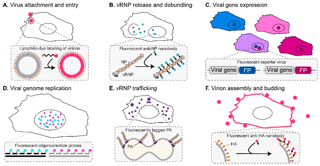

Table 1.
Imaging approaches for studying virus infection: capabilities and limitations.
| Imaging approach | Fixed | Live | Direct readout | Viral infection | Host remodeling or function | Antiviral responses |
Key Interpretive limitation |
| Single-particle virion imaging | ○ | ● | Virion position, movement, membrane fusion, and content release | Attachment, entry, trafficking, and fusion | — | Entry restriction | Detected virions are not necessarily infectious |
| Protein imaging (IF, super-resolution) | ● | — | Selected protein abundance and localization | Viral-protein abundance and localization | Protein and organelle redistribution | Localization of antiviral signaling factors | Does not establish interaction or function |
| RNA imaging (smFISH, multiplexed) | ● | — | RNA abundance, identity, and localization | Viral transcription, genome replication, RNA localization, and genome assembly | Host RNA abundance and localization | Transcription of antiviral genes | Does not directly measure RNA translation, integrity, or function |
| Genetically encoded viral reporters | ○ | ● | Tagged-protein localization or reporter expression | Viral gene expression, protein localization, coinfection, and spread | — | — | Reporter expression may not fully reflect native viral gene expression |
| Nanobody-based imaging | ○ | ● | Fluorescently labeled vRNPs or surface-accessible viral proteins | vRNP trafficking, genome replication, nuclear export, and budding | — | — | Nanobody binding may alter the localization or function of the target protein |
| Functional reporters and biosensors | ○ | ● | Promoter activity, translational activity, membrane potential, or biochemical activity | Viral replication and gene expression | Organelle function and host translation | Antiviral response activation and signaling dynamics | Reporter activity may not fully reflect endogenous biological activity |
| Cargo and tracer assays | ○ | ● | Cargo transport and pathway function | — | Cargo transport and pathway activity | — | Artificial cargo may not faithfully mimic native viral components |
| Tissue-scale imaging | ● | ● | Three-dimensional organization and longitudinal dynamics across multicellular samples | Viral dissemination and spread | Tissue architecture, composition, and function | Intercellular antiviral signaling | Limited molecular and ultrastructural detail |
| Ultrastructural and correlative imaging | ● | — | Membrane topology, structural organization, and molecular–structural correspondence | Replication-compartment architecture, virion structure, and budding | Membrane and organelle remodeling | — | Ultrastructure alone does not reveal molecular composition or function |
Markers: ● primary application; ○ possible or secondary application; — no direct application discussed in this review. Abbreviations: smFISH, single-molecule fluorescence in situ hybridization; vRNP, viral ribonucleoprotein.
Disclaimer/Publisher’s Note: The statements, opinions and data contained in all publications are solely those of the individual author(s) and contributor(s) and not of MDPI and/or the editor(s). MDPI and/or the editor(s) disclaim responsibility for any injury to people or property resulting from any ideas, methods, instructions or products referred to in the content. |
© 2026 by the authors. Licensee MDPI, Basel, Switzerland. This article is an open access article distributed under the terms and conditions of the Creative Commons Attribution (CC BY) license (http://creativecommons.org/licenses/by/4.0/).
Copyright: This open access article is published under a Creative Commons CC BY 4.0 license, which permit the free download, distribution, and reuse, provided that the author and preprint are cited in any reuse.