

Submitted:
17 August 2026
Posted:
18 August 2026
You are already at the latest version
Abstract
Severe traumatic brain injury is a leading cause of death and disability worldwide. The optimal role of invasive intracranial pressure and cerebral perfusion pressure monitoring compared with non‑invasive clinical and imaging‑based management remains uncertain, particularly in low‑resource settings. The objective of this study was to design and evaluate new diagnostic and treatment algorithms for adult patients with severe TBI. A quasi‑experimental study was carried out between January 2010 and December 2024. Patients were assigned to two groups: Group I received continuous invasive ICP and CPP monitoring together with clinical and CT monitoring; Group II received only clinical and CT monitoring. The primary outcome was functional status at one-year post‑discharge according to the Glasgow Outcome Scale (GOS). A total of 250 patients were included: 145 (58%) in Group I and 105 (42%) in Group II. In Group I, the most frequent outcome was GOS V (good recovery), whereas in Group II, GOS I (death) predominated. Satisfactory recovery was achieved in 104/145 patients (71.72%) in Group I vs. 65/105 (61.90%) in Group II. Mortality was 39/145 (26.9%) in Group I vs. 44/105 (41.9%) in Group II (difference 15.0 percentage points; p ≤ 0.05). Continuous invasive ICP and CPP monitoring, was associated with significantly lower mortality and higher rates of satisfactory recovery compared with clinical and imaging‑based management alone. These findings support the use of invasive neuromonitoring where resources permit, while the non‑invasive algorithm may serve as an alternative in low‑resource settings.
Keywords:
traumatic brain injury
; intracranial pressure monitoring
; cerebral perfusion pressure
; neuromonitoring
; treatment algorithm
; low‑income country
1. Introduction
Traumatic brain injury (TBI) is a global health problem. It has been called the "silent epidemic" and is considered to have caused more deaths worldwide than all the major epidemics that have plagued humanity throughout history. The first described cases of head trauma surgery are found in the Edwin Smith Papyrus, written around 1700 BC [1].
It is estimated that between 50 and 60 million people suffer from TBI each year, with an approximate cost to the global economy of US $400 billion. In Europe, more than 2 million cases are admitted annually for this reason, of whom around 82,000 die and many others are left with severe sequelae. Therefore, TBI should be considered in two dimensions: as an acute condition and as a chronic disease that can generate severe sequelae requiring prolonged treatment and follow-up [2].
In the United States, TBI is associated with an estimated 4.8 million emergency department (ED) visits, resulting in more than 214,000 hospitalizations and 69,000 people died [3]. In Cuba, there are no mortality data specifically for TBI, but the overall accident mortality rate was 8.3/100,000 in 2022 and 8.4/100,000 in 2023 [4].
TBI has been the leading cause of consultation at the neurotrauma center of the "Roberto Rodríguez" General Teaching Hospital of Moron, Ciego de Avila, Cuba, for the past 20 years. In the second half of the 1990s, mortality from severe TBI was estimated at around 80%. Treatment for these patients was not yet standardized, which prompted the neurotrauma care team to design and readjust treatment algorithms according to the knowledge available at that time both nationally and internationally. These algorithms were characterized by the introduction of multimodal neuromonitoring to guide stepwise treatment aimed at controlling intracranial pressure (ICP) and improving cerebral perfusion pressure (CPP).
In 2015, the lead author of this work participated in a consensus meeting in Buenos Aires, Argentina, attended by neurosurgeons and intensivists from across Latin America, along with representatives from the Brain Trauma Foundation (BTF). The purpose of the meeting was to establish treatment algorithms for patients without ICP monitoring, guided solely by clinical presentation and CT scans. This was necessary due to the high number of hospitals in low-resource countries treating these patients without neuromonitoring. This meeting was crucial because it allowed us to adapt the treatment algorithms to our specific circumstances for patients who, for any reason, could not benefit from neuromonitoring [5].
Elevated ICP is one of the most common causes of death and disability following severe TBI. It is a clinical condition that can cause brainstem compression and compromise cerebral circulation; therefore, monitoring this parameter became a reasonable strategy to understand its behavior during the clinical course of patients with severe TBI and to identify events of intracranial hypertension (ICH). However, the efficacy of ICP-monitoring-guided treatment to improve prognosis has not been rigorously evaluated, and guidelines for the management of severe TBI have documented insufficient evidence related to this issue [6,7,8]. Furthermore, a clear consensus has not been reached, and there is considerable variation in the practice of TBI management worldwide [9]. A Cuban survey published in 2004 included data from 17 hospitals that treated patients with severe TBI, examining the use of ICP monitoring. It reported that only eight centers used neuromonitoring to guide their treatment protocol, and five had never used it [10].
The Neurosurgery and Adult Intensive Care Unit departments of the "Roberto Rodríguez" General Teaching Hospital are leaders in Cuba in the use of ICP, CPP, and other intracranial parameter monitoring for the treatment of patients with severe TBI.
This work is the result of several research projects, the most recent of which was associated with a national health program: Program No. 2: "Organization, Efficiency and Quality of Services". Project Code: 1902052. Title: "Neuromonitoring in patients with severe traumatic brain injury", which was successfully implemented and completed.
The overall objective of this study was to design and evaluate new diagnostic and treatment algorithms for adult patients with severe TBI in a low-income country using invasive and non-invasive monitoring of ICP and CPP, as well as non-invasive neuromonitoring based solely on clinical and imaging assessment.
The specific objectives were to compare the impact of diagnostic methods and treatment protocols guided by these algorithms on ICP, CPP, neurological worsening, imaging findings at admission and during follow-up, the intensity level of therapy used, complications, and outcomes in both groups.
2. Patients and Methods
2.1. Study Design and Population
A quasi-experimental study was conducted including all patients admitted to the Intensive Care Unit (ICU) of the "Roberto Rodríguez" General Teaching Hospital in Moron, Ciego de Ávila, Cuba, with a diagnosis of severe traumatic brain injury (TBI) between January 2010 and December 2024.
Patients were randomly assigned to one of two groups based on the monitoring modality used. Group I included patients who received clinical and CT monitoring together with continuous invasive ICP and CPP monitoring. Patients who received only clinical and CT monitoring were assigned to Group II. The clinical status on admission was assessed using the Glasgow Coma Scale (GCS).
Inclusion criteria were: age 18 to 60 years; baseline GCS score > 4 and < 9, or deterioration to a GCS score ≤ 8 within 48 hours of injury; an abnormal admission CT scan, or a normal CT scan associated with at least two of the following: abnormal posturing, mean systemic arterial pressure (MSAP) ≤ 90 mmHg, and age > 40 years; initiation of monitoring and treatment within 12 hours of trauma; and absence of associated trauma.
Exclusion criteria were: GCS score of 3 with bilateral fixed and dilated pupils; a non-survivable injury; presence of associated trauma or coagulopathy (prothrombin time > 12.2 seconds, platelet count < 100 × 10³/μl); use of anticoagulants or antiplatelet agents; hepatic cirrhosis; and lack of 6-month follow-up.
2.2. Pre-Experimental Evaluation of the Algorithms
The designed algorithms were presented to the neurosurgery and adult intensive care teams for expert review using the nominal group technique.
2.3. Nominal Group Structure
The nominal group consisted of 17 specialists with substantial experience in the management of severe TBI, working in the neurosurgery and adult intensive care services of the "Roberto Rodríguez" General Teaching Hospital.
To characterise the group, each participant completed a self-assessment of their expertise in TBI diagnosis and treatment. The following coefficients were calculated for each participant: Knowledge Coefficient (Kc), Argumentation Coefficient (Ka), Competence Coefficient (Kcp).
2.4. Nominal Group Procedure
1. The lead author convened a meeting with the nominal group members, during which the diagnostic and treatment algorithms for severe TBI were presented.
2. An orderly discussion was held, with some members participating virtually and others in person. Questions arising from the presentation were addressed and clarified. Participants were given 20 minutes to complete forms providing their opinions on the relevance and feasibility of applying the algorithms.
3. Different proposals were presented, and opinions were collected.
Consensus was reached on limiting the use of hyperventilation to a window of 6 to 12 hours.
4. At the end of the discussion, 100% of the nominal group members considered the algorithms appropriate and recommended initiating their application.
2.5. Delphi Method for the Treatment Algorithm in Group II (Clinical and Imaging Monitoring Only)
The treatment algorithm for Group II was developed at the Buenos Aires Consensus Meeting in 2015. A Delphi method was applied to over 50 neurosurgeons and neurointensivists from Latin America, including the lead author of this study, who represented Cuba at that meeting. The CREVICE protocol [5], which was subsequently applied to our Group II, was developed at this meeting, thus representing a technology transfer.
The proposed algorithms were submitted for evaluation and approval to the scientific council of the "Roberto Rodríguez" General Teaching Hospital and have been presented at national and international scientific meetings. These algorithms have undergone both external and internal validation.
2.6. Description of the Quasi-Experiment
The design and application of the algorithms are based on a series of general measures required to maintain intracranial homeostasis. These include appropriate patient positioning; optimisation of the internal environment (normovolaemia, normocapnia, normoglycaemia, normoxaemia, normothermia, normonatraemia); and ensuring cerebral oxygenation through mechanical ventilation, among other measures [11].
All cases in the study were treated in the ICU with either continuous invasive or non-invasive ICP monitoring.
2.6.1. Group I (Invasive Monitoring)
Patients assigned to Group I, were treated according to the algorithm shown in Figure 1. An external ventricular drain (EVD) was placed via a catheter inserted into the frontal horn of the most affected lateral ventricle. This catheter was connected to a fluid-column system and a transducer that converted mechanical energy into electrical energy, displaying the ICP value on a monitor. The system was also used therapeutically for cerebrospinal fluid (CSF) drainage. CPP was calculated as MAP- ICP.
The therapeutic targets were ICP ≤ 15 mmHg and CPP > 60 mmHg. CSF drainage was the first-line treatment in this group. Additional therapies were initiated when ICP was ≥15 mmHg, a threshold slightly lower than that proposed in major severe TBI guidelines [6,7,8]. The administration of hyperosmolar agents and mild-to-moderate hyperventilation was guided by jugular venous oxygen saturation (SjO2) measured via a catheter placed in the bulb of the internal jugular vein.
Patients with a hyperaemic pattern were treated with hyperventilation, while those with an ischaemic pattern received mannitol. Decompressive craniectomy (DC) was performed in patients with ICP ≥20 mmHg for more than 15 minutes refractory to medical therapy, following an emergency CT scan showing midline shift > 5 mm, absent or compressed cisterns, and a Marshall grade ≥ III. If ICP and CPP remained normal for 24 hours, the EVD was removed, and general supportive care was continued with gradual de-escalation of ICP-control measures.
2.6.2. Group II (Non-Invasive Monitoring)
Patients assigned to Group II were managed according to the algorithm shown in Figure 2. After evacuation of an intracranial mass, or in the absence of lesions requiring surgery, patients were treated with head elevation to 20°, haemodynamic stabilisation, intermittent intravenous boluses of 20% mannitol (0.25 g/kg/dose every 4 hours), controlled mild hyperventilation (PaCO2 30–35 mmHg), and muscle relaxation when indicated. DC was performed in patients with neurological worsening refractory to medical treatment, manifesting as postural or pupillary abnormalities, and associated with sequential CT scans showing imaging deterioration with midline shift ≥ 5 mm and/or effacement of the perimesencephalic cisterns. Barbiturates were used only after DC in patients with clinically and radiologically refractory intracranial hypertension (ICH). Monitoring was non-invasive, with ICP and CPP estimated via transcranial Doppler (TCD) and/or optic nerve sheath diameter (ONSD). Transcraniectomy ultrasound was also used in patients with a bone window.
2.6.3. Non-Invasive Monitoring Methods (Both Groups)
Non-invasive methods used in both groups included ONSD measurement, with ICP estimated using the formula: (ONSD in mm × 5.69) - 8.23, where 3 mm ONSD corresponds to 8 mmHg, 4 mm = 14 mmHg, 5 mm = 20 mmHg, 6 mm = 25 mmHg, 7 mm = 31 mmHg, and 9 mm = 37 mmHg. ICP was also estimated using TCD according to the formula: ICP = MAP – CPP, where CPP = MAP × (Vd/Vm)⁻¹ + 14. Additionally, the formula ICP = 10.927 × PI – 1.28 was used. The pulsatility index (PI) was calculated as (Vs – Vd) / Vm. Patients received treatment for intracranial hypertension whenever ICP exceeded 15 mmHg.
2.6.4. Clinical Criteria for Suspecting ICH (Group II) (CREVICE Protocol [5])
For patients undergoing clinical and imaging monitoring (Group II), clinical criteria, divided into major and minor, were used to suspect ICH. Treatment was initiated upon the presence of one major criterion or two minor criteria.
Major criteria:
1. Compressed cisterns (Marshall Class III) on CT
2. Midline shift ≥ 5 mm (Marshall Class IV) on CT
3. Non-evacuated mass-effect lesion on CT
Minor criteria:
1. Motor component score ≤ 4 on GCS
2. Pupillary asymmetry
3. Abnormal pupillary reactivity
4. Diffuse axonal injury, Marshall Class II (cisterns present with shift 0–5 mm, or focal hyperdense or mixed-density lesions ≤ 25 cm³)
If ICH is not suspected, the patient is observed and general supportive measures are continued. If ICH is suspected, the patient remains intubated and mechanically ventilated with baseline sedation, and first-line treatment measures are initiated (Table 1). A MSAP≥ 90 mmHg must be achieved and maintained.
2.7. Therapy De-Escalation
If the clinical examination or CT scan shows stability or acceptable improvement, therapy reduction is considered according to clinical judgment and a decision-support matrix (Table 2) based on a three-colour traffic-light system: Red = therapy reduction not recommended; Green = therapy can be reduced; Yellow = therapy reduction is doubtful or risky. Frequent neurological examinations by the same medical team member are required under this scheme [5].
2.8. Imaging Protocol
All patients underwent CT scans at baseline, at 72 hours, and at 5 days. Emergency CT was performed in patients with ICP ≥20 mmHg refractory to medical treatment for 15 minutes, and in patients diagnosed with neurological worsening. The admission CT scan was also used to measure bilateral ONSD, which was compared with invasively or non-invasively measured ICP values.
2.9. Definition of Neurological Worsening [5]
Neurological worsening was defined as the appearance of any of the following physical examination findings:
1. Decrease of more than one point on the GCS motor component
2. New loss of pupillary reactivity
3. New pupillary asymmetry > 2 mm difference or bilateral mydriasis
4. New motor focal deficit
5. Signs of cerebral herniation syndrome (e.g., Cushing’s triad).
CT images were classified according to the Marshall system. Standard supportive care, including cardiovascular and respiratory monitoring, mechanical ventilation, sedation, and analgesia, was provided to all patients.
2.10. Outcome Measures and Statistical Analysis
The following primary outcome measures were recorded: age, time from trauma to surgery or treatment initiation, clinical examination findings, side and location of intracranial lesions, number of affected lobes, morbidity, and mortality.
Secondary outcomes included mortality and functional outcome assessed using the Glasgow Outcome Scale (GOS) at one-year post-discharge. Outcomes were dichotomised as favourable (GOS III, IV, V) and unfavourable (GOS I, II).
The chi-square test was used to assess associations between categorical variables. The Mann–Whitney U test was used to compare medians of quantitative variables that did not follow a normal distribution. For comparisons among more than two groups, the Kruskal-Wallis test was used for non-normally distributed data.
3. Results
3.1. Patient Population
The sample consisted of 250 patients, of whom 145 (58%) were assigned to Group I and 105 (42%) to Group II. The median age of all patients was 53.2 years.
3.2. Admission GCS Score and Relationship with GOS
The GCS score at admission and its relationship with the Glasgow Outcome Scale (GOS) showed the distribution of patients across GCS categories was highly similar between the two groups, as confirmed by the Mann-Whitney U test (U = -0.961, p = 0.336), indicating no significant difference between groups. The most frequent GCS score category in both groups was 6–7 points: 70 patients (48.27%) in Group I and 45 (42.9%) in Group II.
In Group I, among the 42 patients (28.97%) admitted with a GCS score of 8, 38 (90.48%) survived, 25 (59.52%) achieved a GOS of V, and 4 (9.52%) died. Among the 70 patients with GCS scores of 6–7, 54 (77.14%) survived, 22 (31.42%) achieved GOS V, and 16 (22.86%) died.
The worst outcomes in Group I were observed in patients admitted with GCS scores of 4–5. Of the 33 patients (22.76%) in this category, 19 (57.57%) died, and only 6 (18.18%) achieved GOS V.
In Group II, 26 patients (24.76%) were admitted with a GCS score of 8; of these, 20 (76.92%) survived, 10 (38.46%) achieved GOS V, and 6 (23.07%) died. Among the 45 patients with GCS scores of 6–7, 32 (71.11%) survived, none achieved GOS V, and 13 (28.88%) died. Among the 34 patients (32.38%) admitted with GCS scores of 4–5, 25 (73.52%) died and only 9 (26.47%) survived.
3.3. Admission CT Findings
Admission CT findings are summarised in Table 3. The most common finding in both groups was a non-evacuated mass-effect lesion, present in 62 patients (42.76%) in Group I and 63 patients (60%) in Group II.
3.4. Monitoring Parameters
3.4.1. Group I (Invasive Monitoring)
ICP < 20 mmHg after treatment implementation: 77 patients (53.10%). Of these, 72 (93.50%) survived with satisfactory recovery (p< 0.001), and 5 (6.49%) died.
ICP 20–25 mmHg: 35 patients (24.14%). Of these, 22 (62.86%) survived, and 13 (37.14%) died.
ICP > 25 mmHg: 33 patients (22.76%). Of these, 10 (30.30%) had satisfactory survival, and 21 (63.33%) died.
3.4.2. Group II (Non-Invasive Monitoring)
Estimated ICP < 20 mmHg: 49 patients (46.66%). Of these, 41 (83.67%) survived with satisfactory recovery (p< 0.001), and 8 (16.32%) died, a negative difference of 9.83% compared to Group I.
Estimated ICP 20–25 mmHg: 26 patients (24.76%). Of these, 14 (53.85%) survived, and 12 (46.15%) died, a negative difference of 9.01% compared to Group I.
Estimated ICP > 25 mmHg: 30 patients (28.57%). Of these, 24 (80%) died, 16.67% more than in Group I.
3.5. Cerebral Perfusion Pressure (CPP)
3.5.1. Group I
CPP showed a strong association with mortality when values were < 60 mmHg. All 35 patients (24.13%) in this category died (p < 0.001). Among the 86 patients (59.31%) with CPP between 60 and 70 mmHg, 84 (97.67%) survived with satisfactory recovery (p< 0.001). Among the 22 patients (15.17%) with CPP > 70 mmHg, 20 (90.90%) survived satisfactorily, and 2 (9.09%) died.
3.5.2. Group II
CPP also showed a close relationship with mortality when values were < 60 mmHg. Of the 42 patients (40%) with CPP < 60 mmHg, 36 (85.71%) died, and the remaining 10% had unfavourable outcomes. Among the 43 patients (40.95%) with CPP 60–70 mmHg, 39 (90.70%) had satisfactory outcomes and 4 (9.30%) died. Among the 20 patients (17.04%) with CPP > 70 mmHg, 16 (80%) recovered satisfactorily and 4 (20%) died.
3.5.3. Clinical and Imaging Evolution in Group II
In Group II, 59 patients (56.19%) showed improvement on clinical examination and sequential CT scans; of these, 27 (84.37%) had a satisfactory recovery, and only 5 (15.62%) died. In 45 cases (46.82%), clinical examination and imaging remained unchanged from admission; of these, 22 (48.89%) had a satisfactory recovery and 21 (46.66%) died. In 28 cases (26.66%), clinical examination and sequential CT scans showed worsening; of these, 22 (78.57%) had an unsatisfactory recovery, 18 (81.81%) died, and only 10 (45.45%) survived of whom 4 (18.18%) remained in a persistent vegetative state.
3.5.4. Median ICP and GOS in Group I
The relationship between median ICP and GOS was also analysed in Group I. For GOS category II, the median could not be calculated due to the presence of only two patients. The medians for GOS grades III, IV, and V were very similar (15.3, 14.1, and 14.5, respectively), whereas the median for GOS grade I (30.8) was double that of the other categories.
Scheffé’s post-hoc test showed that the median for GOS grade I was significantly different from the medians for grades III, IV, and V, and that these grades did not differ significantly from each other. These findings demonstrate a relationship between elevated ICP values and mortality.
The mean duration of ICP monitoring was 4.44 ± 6.67 days. Complications related to the monitoring system were few: 116 patients (80%) had no complications; 15 (10.34%) had system malfunction; minor bleeding along the intracranial catheter tract was observed in 10 patients (6.90%); and only 3 patients (2.07%) developed a system infection.
3.6. Optic Nerve Sheath Diameter (ONSD) and ICP
The relationship between ONSD measured on the initial CT scan in the emergency department and ICP values in both groups is shown in Figure 3. A close correlation was observed between both variables in both groups, with a directly proportional relationship: lower ICP was associated with lower ONSD.
3.7. Neurological Worsening
The diagnosis of neurological worsening in both groups is shown in Figure 4. In Group I, neurological worsening was present in 31 cases (21.38%), with no significant differences between the diagnostic components. In Group II, it was diagnosed in 48 patients (45.71%), also with no significant differences between components.
3.8. Therapeutic Measures
The therapeutic measures used in both groups are shown in Figure 5. Analgesia and sedation were used in all patients in both groups. CSF drainage was performed exclusively in Group I. In Group II, 100% of patients received osmotic agents (cerebral dehydrating agents). Although these agents were used in 100 patients (68.97%) in Group I, the doses were reduced in this group because their use was intermittent in a significant number of patients in response to rapid ICP control.
3.9. Intensity of Therapy
A high intensity level of therapy predominated in both groups: 98 patients (67.59%) in Group I and 68 (64.76%) in Group II, with no significant differences between groups.
3.10. Complications
Medical complications predominated in both groups. In Group I, there were 351 medical complications, with a higher frequency of electrolyte imbalances 101 (28.77%) and healthcare-associated infections 112 (31.91%), compared to 218 surgical complications. The most frequent surgical complication was neuromonitoring system dysfunction 55 (25.23%). In Group II, there were 319 medical complications, the most frequent being electrolyte imbalances 92 (28.84%), compared to 147 surgical complications, among which neurological worsening was the most prominent 48 (32.65%).
3.11. Functional Outcomes (GOS)
The distribution of GOS outcomes in both groups is shown in Table 4. In Group I, the most frequent outcome was GOS V, whereas in Group II, GOS I predominated.
The difference between the groups in terms of satisfactory recovery (GOS III–V) was 9.82% (Group I: 104 patients, 71.72%; Group II: 65 patients, 61.90%).
The Mann-Whitney U test revealed a significant association between continuous ICP monitoring and achieving GOS V (p ≤ 0.05).
4. Discussion
Continuous ICP monitoring has been increasingly used in neurosurgical and neurocritical care practice over the past four decades and is now a routine procedure worldwide in patients with severe TBI. It is generally considered the cornerstone of care in these patients and is recommended in all modern guidelines for the treatment of TBI [3,4]. However, some studies have found no association, or even a negative association, between monitoring-based treatment and prognosis [6,10,12].
Since the studies by Guillaume and Janny in the early 1950s [13,14], it has been known that, in many cases of TBI, the clinical state is not a completely reliable indicator of the behaviour of intracranial parameters such as ICP and CPP, especially when the patient is under sedation or neuromuscular blockade with mechanical ventilation.
The prognostic value of CT in severe TBI has been studied [15,16]. The value of basal cistern status as an indicator of elevated ICP has been confirmed in numerous studies. Lobato et al. (2005) [16] reported that more than 50% of patients with severe TBI who showed initial diffuse axonal injury of types I–II developed new changes on CT, and almost 50% developed intracranial hypertension during the acute post-traumatic period; however, a discrepancy between CT changes and ICP monitoring was observed in 30.3% of cases.
CT scanning is considered the standard examination for the diagnosis of intracranial lesions, which may appear either on admission or during the clinical course. Sequential CT is an essential test, as its findings facilitate the follow-up of intracranial lesions and guide the surgeon to take the appropriate action at the precise moment [15].
In this study, while ICP and CPP monitoring were the main variables measured invasively in Group I and estimated non-invasively in Group II, all patients benefited from CT imaging, especially those with elevated ICP values. In Group II, analysis of CT imaging showed a high correlation between clinical course and imaging pattern, demonstrating the value of this technique for diagnosing the presence or persistence of intracranial hypertension.
Currently, several reports recommend the use of clinical and imaging monitoring as a replacement for invasive monitoring of ICP and other derived intracranial parameters, arguing that there is not full acceptance of its benefits by neurosurgeons and intensivists, in addition to the lack of sufficient scientific evidence to justify its advantages over non-invasive methods [6,8,15,17].
Recently, the relationship between the threshold values of intracranial hypertension tolerated by the brain and the duration of exposure has been studied [18]. These studies have found tolerable pressure thresholds lower than those considered to date, and have shown that short events of intracranial hypertension have a worse effect on the brain than medium or long exposure periods. Our group used lower ICP thresholds for treatment initiation than those recommended by the SIBICC study [19], and we maintained similar values to those used by this group of experts for CPP thresholds. We believe that further studies will be required to determine the most appropriate ICP threshold for these patients. This finding justifies the use of continuous neuromonitoring as the only way to monitor the behaviour of ICP and other derived intracranial parameters, which are of vital importance for guiding treatment in patients under sedation or neuromuscular blockade, where intracranial hypertension events might otherwise go unnoticed, with their well-known consequences.
Our position is that all monitoring methods should be complementary, and that the information obtained from each does not replace the others. Our team considers that the information provided by continuous monitoring of ICP, CPP, and other intracranial parameters is of great value in the treatment of these patients, as is the information obtained from clinical examination and serial CT imaging, a view shared by other authors [6,8,20,21,22,23].
The inclusion of neurological worsening criteria in the algorithms was crucial for ordering sequential imaging studies to detect the appearance of new focal lesions or the worsening of existing ones, thus enabling therapeutic escalation. In Group I, ICP and CPP monitoring allowed for the detection and prevention of these clinical signs in some patients, which was not possible in Group II.
The adaptation of the work algorithms for both study groups to the local context of the province was very important. The modification regarding the timing of primary DC, using it as one of the first measures for ICP control in both groups, was due to the high number of patients with Marshall grade IV imaging on admission or the presence of focal lesions with associated mass effect that required surgical evacuation. This evacuation was performed through the corridor created by the DC, thus considerably avoiding prolonged exposure of the injured brain to intracranial hypertension.
Furthermore, the use of DC has had a positive effect on intracranial compartment syndrome and intracranial compliance. DC transforms the cranial cavity from a "closed box" to an "open box", subjecting its contents to varying internal pressures that are transmitted from one compartment to another. These pressures include CSF pressure, pressure from the oedematous brain, and the presence of space-occupying lesion debris, creating an outward pressure cone. External pressures, such as atmospheric pressure (1 atmosphere = 14.7 PSI = 1033 cmH₂O), also exert an inward pressure cone. Therefore, this procedure achieves pressure equilibrium across the different compartments [24].
Regarding the well-known compartmentalisation of the cranial cavity by the falx cerebri and the tentorium, it is now recognised that ICP is not necessarily equal in each compartment, and pressure cones and vectors can be generated from one compartment to another [11]. In the dimension of the disease, we could call this phenomenon "intracranial compartment syndrome" [25,26], similar to what occurs in the abdominal cavity or the lower limbs. The requirements for developing a compartment syndrome within an anatomical cavity are: (1) the cavity must be inextensible above a certain pressure limit; (2) its compliance must be close to zero when its contents are compressed to a critical level [25,26]. The cranial cavity meets these requirements.
Our philosophy for the quantitative interpretation of ICP has been modified from some years ago. Intracranial hypertension is a multifactorial complication that has a very important influence on outcomes, but it is not the only factor [11]. It is only a component, as other authors have pointed out [11]. Furthermore, the brain cannot be viewed in isolation from the rest of the body. The relationship between the thorax, abdomen, and cranial cavity, with their compartments and different physical characteristics among their contents, has been recognised for several years [11].
Our team agrees with other authors that, rather than focusing on an isolated ICP number, it is more important to identify early the cause of the elevation and to thoroughly evaluate the trends from both a qualitative and quantitative perspective, an approach that is consistent with current therapeutic positions [11].
This approach influenced two essential aspects: the doses of osmotic agents were reduced in Group I, and the intensity of therapy was increased in both patient groups, in accordance with our team's aggressive treatment approach for refractory intracranial hypertension. The flexible use of treatment levels was another innovative feature of the study; the measures were designed so that their application could be adjusted according to the patient's needs, which aligns with contemporary philosophy in this area [11].
The design and application of both working algorithms ensured that none of our patients received empirical treatment for their severe TBI. The application of clinical suspicion criteria for intracranial hypertension in Group II facilitated the ordering of confirmatory complementary examinations, such as sequential CT scans. These CT scans allowed for the evaluation of intracranial midline structures, the status of the basal cisterns, and the sequential measurement of the ONSD, a significant estimator of ICP. This variable, along with the use of transcranial Doppler to estimate ICP and CPP, demonstrated numerous advantages in this study group.
The use of a decision-making matrix [5] in Group II was a novelty in the study. It facilitated, based on the patient's clinical data and imaging findings, making the most appropriate decision regarding escalation or de-escalation of therapeutic measures and therapy intensity.
When analysing the results, we observed that mortality in Group I was 39 cases (26.9%) and in Group II was 44 patients (41.9%), representing a difference of 15.0 percentage points in mortality between the groups and a 9.8 percentage point difference in satisfactory outcomes (71.7% Group I vs. 61.9% Group II), both favouring Group I. Overall mortality in the study was 83 patients (33.2%). The observed absolute risk reduction of 15 percentage points in mortality with invasive monitoring is comparable to that reported in the BEST TRIP trial subgroup analyses [27], although direct comparisons are limited by differences in case mix and resource availability.
Limitations
This study has several limitations. First, the quasi-experimental design and the lack of true randomisation introduce potential selection bias, despite the use of random assignment based on monitoring modality. Second, the study was conducted at a single centre, which may limit generalisability to other settings with different resources and patient populations. Third, the long study period (2010–2024) may have been influenced by changes in standard care over time. Fourth, the non-invasive ICP estimation methods used in Group II have not been validated against invasive measurements to the same standard as ventricular catheters. Fifth, the sample size was not calculated a priori based on a predetermined effect size, which may have resulted in insufficient power for some secondary analyses. Finally, the five-year follow-up period, while a strength, also introduced potential loss to follow-up, although exclusion of patients without follow-up was predefined.
The main conclusions of the study can be stated as follows:
In this quasi-experimental study of 250 patients with severe TBI, invasive ICP/CPP monitoring was associated with significantly lower mortality (26.9% vs. 41.9%) and higher rates of satisfactory recovery (71.7% vs. 61.9%) compared to clinical and imaging monitoring alone. Continuous monitoring of ICP and CPP has a positive influence on outcomes in patients with severe TBI.
The design and application of the workflow algorithms ensure a systematic approach for patients undergoing invasive neuromonitoring and for those undergoing non-invasive neuromonitoring. This represents a strength for the medical care of these patients and can be adapted to the available technological resources and the level of training of specialists in various hospitals with neurosurgery services in the country.
Novel non-invasive neuromonitoring methods were applied, including ICP and CPP estimation using transcranial Doppler, ONSD measurement, and trans-craniotomy ultrasound.
ICP interpretation shifted from a purely quantitative approach to a flexible, mixed-methods approach.
Intracranial hypertension was considered part of a complex pathophysiological phenomenon, not an isolated element, and was considered part of the three-compartment syndrome (brain-thorax-abdomen).
The inclusion of clinical criteria for intracranial hypertension and neurological worsening was a significant benefit for patients, particularly in Group II, where continuous ICP and CPP monitoring was unavailable and where the diagnosis of new intracranial lesions risked being delayed.
The inclusion of the concept of intracranial compartment syndrome and its relationship to intracranial compliance, influenced by the use of DC, facilitated the reinterpretation of an old pathophysiological concept within a new framework.
Author Contributions
Angel Jesús Lacerda Gallardo MD Ph.D. Conceptualization, Investigation, Formal analysis, Methodology, interpretation of data, Project Administration, Writing – Original Draft Preparation, Supervision. Daysi Abreu Pérez MD Ph.D. Formal analysis, Methodology, Supervision, Investigation, Writing – Original Draft Preparation. Miguel de Jesús Mazorra Pazos MD. Investigation, Data curation, interpretation of data, Visualization, approved the submitted version. Carlos Rojas Borroto MD. Investigation, interpretation of data, approved the submitted version. Julio Antonio Díaz Agramonte MD. Investigation, Data curation, interpretation of data, Visualization, approved the submitted version. José Antonio Gálvez MD. Draft reviewer, Language quality editor.
Funding
The authors state that the research did not use any funds.
Institutional Review Board Statement
The study was conducted in accordance with the Declaration of Helsinki, and the protocol was approved by the Ethics Committee of “Roberto Rodriguez” General Teaching Hospital of Moron, Ciego de Avila, Cuba. Project identification code: RRGTH-01-2020 on 3/1/2020.
Informed Consent Statement
Informed consent was obtained from all subjects involved in the study.
Data Availability Statement
Patient data are available in the authors' private database.
Conflicts of Interest
The authors declare no conflicts of interest.
References
- Kshettry, V.R.; Mindea, S.A.; Batjer, H.H. The management of cranial injuries in antiquity and beyond. Neurosurg. Focus 2007, 23, E8. [Google Scholar] [CrossRef] [PubMed]
- Maas, A.I.; Menon, D.K.; Manley, G.T.; et al. Traumatic brain injury: progress and challenges in prevention, clinical care, and research. Lancet Neurol. 2022, 21, 1004–1060. [Google Scholar] [CrossRef]
- Korley, F.K.; Kelen, G.D.; Jones, C.M.; Diaz-Arrastia, R. Emergency department evaluation of traumatic brain injury in the United States, 2009-2010. J. Head. Trauma Rehabil. 2016, 31, 379–387. [Google Scholar] [CrossRef] [PubMed]
- Cuba. Ministerio de Salud Pública. Dirección Nacional de Registros Médicos y Estadísticas de Salud. Anuario estadístico de salud 2023; MINSAP: La Habana, 2024.
- Chesnut, R.M.; Temkin, N.; Videtta, W.; Petroni, G.; Lujan, S.; Pridgeon, J.; et al. Consensus-based management protocol (CREVICE Protocol) for the treatment of severe traumatic brain injury based on imaging and clinical examination for use when intracranial pressure monitoring is not employed. J. Neurotrauma 2020, 37, 1291–1299. [Google Scholar] [CrossRef] [PubMed]
- Carney, N.; Totten, A.M.; O'Reilly, C.; Ullman, J.S.; Hawryluk, G.W.J.; Bell, M.J.; et al. Guidelines for the management of severe traumatic brain injury, fourth edition. Neurosurgery 2017, 80, 6–15. [Google Scholar] [CrossRef] [PubMed]
- Bratton, S.L.; Chesnut, R.M.; Ghajar, J. Guidelines for the management of severe traumatic brain injury. J. Neurotrauma 2007, 24, S1–S106. [Google Scholar] [CrossRef] [PubMed]
- Alvarado-Dyer, R.; Aguilera, S.; Chesnut, R.M.; Videtta, W.; Fischer, D.; Jibaja, M.; et al. Managing severe traumatic brain injury across resource settings: Latin American perspectives. Neurocrit Care 2023, 38, 229–234. [Google Scholar] [CrossRef] [PubMed]
- Wijayatilake, D.S.; Talati, C.; Panchatsharam, S. The monitoring and management of severe traumatic brain injury in the United Kingdom: is there a consensus? A national survey. J. Neurosurg. Anesthesiol. 2015, 27, 336–344. [Google Scholar] [CrossRef] [PubMed]
- López, H.J.; Varela Hernández, A.; Soler Morejón, C.; Vega Basulto, S.; Lacerda Gallardo, A. Estado actual del manejo del traumatismo craneoencefálico grave en los hospitales de atención al adulto en Cuba. Rev. Cub Med. Int. Emerg. 2004, 3, 11–23. [Google Scholar]
- Godoy, D.A.; Badenes, R.; Murillo-Cabezas, F. Diez mandamientos fisiológicos a lograr durante el traumatismo craneoencefálico grave. Rev. Esp. Anestesiol. Reanim. 2021, 68, 280–292. [Google Scholar] [CrossRef] [PubMed]
- Cremer, O.L.; van Dijk, G.W.; van Wensen, E.; et al. Effect of intracranial pressure monitoring and targeted intensive care on functional outcome after severe head injury. Crit. Care Med. 2005, 33, 2207–2213. [Google Scholar] [CrossRef]
- Shafi, S.; Diaz-Arrastia, R.; Madden, C.; Gentilello, L. Intracranial pressure monitoring in brain-injured patients is associated with worsening of survival. J. Trauma. 2008, 64, 335–340. [Google Scholar] [CrossRef]
- Tang, A.; Pandit, V.; Fennell, V.; Jones, T.; Joseph, B.; O'Keeffe, T.; et al. Intracranial pressure monitor in patients with traumatic brain injury. J. Surg. Res. 2015, 194, 565–570. [Google Scholar] [CrossRef] [PubMed]
- Chesnut, R.M. Medical management of severe head injury: present and future. New Horiz. 1995, 3, 581–593. [Google Scholar] [PubMed]
- Lobato, R.D.; Allen, J.F.; Pérez-Núñez, A.; Alday, R.; Gómez, P.A.; Pascual, B.; et al. Value of serial CT scanning and intracranial pressure monitoring for detecting new intracranial mass effect in severe head injury patients showing lesions type I-II in the initial CT scan. Neurocirugía 2005, 16, 217–234. [Google Scholar] [CrossRef]
- Dong-Seong, S.; Sun-Chul, H.; Bum-Tae, K.; Je Hoon, J.; Soo-Bin, I.; Won-Han, S. Serial brain CT scans in severe head injury without intracranial pressure monitoring. Korean J. Neurotrauma 2014, 10, 26–30. [Google Scholar] [CrossRef]
- Åkerlund, C.A.; Donnelly, J.; Zeiler, F.A.; Helbok, R.; Holst, A.; CabeleiraM; et al. Impact of duration and magnitude of raised intracranial pressure on outcome after severe traumatic brain injury: a CENTER-TBI high resolution group study. PLoS ONE 2020, 15, e0243427. [Google Scholar] [CrossRef] [PubMed]
- Hawryluk, G.W.J.; Aguilera, S.; Buki, A.; Bulger, E.; Citerio, G.; et al. A management algorithm for patients with intracranial pressure monitoring: the Seattle International Severe Traumatic Brain Injury Consensus Conference (SIBICC). Intensive Care Med. 2019, 45, 1783–1794. [Google Scholar] [CrossRef] [PubMed]
- Lazaridis, C.; Yang, M.; DeSantis, S.M.; Luo, S.; Robertson, C.S. Predictors of intensive care unit length of stay and intracranial pressure in severe traumatic brain injury. J. Crit. Care 2015, 30, 1258–1262. [Google Scholar] [CrossRef] [PubMed]
- Luca, L.; Florin Rogobete, A.; Horea Bedreag, O.; Sarandan, M.; Cradigati, C.A.; Papurica, M. Intracranial pressure monitoring as a part of multimodal monitoring management of patients with critical polytrauma: correlation between optimised intensive therapy according to intracranial pressure parameters and clinical picture. Turk. J. Anaesth. Reanim. 2015, 43, 412–417. [Google Scholar] [CrossRef]
- Han, J.; Yang, S.; Zhang, C.; Zhao, M.; Li, A. Impact of intracranial pressure monitoring on prognosis of patients with severe traumatic brain injury: a PRISMA systematic review and meta-analysis. Medicine 2016, 95, e3582. [Google Scholar] [CrossRef] [PubMed]
- Vrettou, C.S.; Mentzelopoulos, S.D. Second- and third-tier therapies for severe traumatic brain injury. J. Clin. Med. 2022, 11, 4790. [Google Scholar] [CrossRef] [PubMed]
- Vásquez, S.; Mendoza Mantilla, J.; Suárez, M.N.; Bustamante, L.A.; Guzman, J.; Rubiano, A.M. Decompressive craniectomy in the ICU: usefulness of transcranial Doppler (TCD/TCCS) in the monitoring of hemodynamic changes. In Neurosonology in Critical Care; Rodríguez, C.N., Baracchini, C., Mejía Mantilla, J.H., Czosnyka, M., Suárez, J.I., Csiba, L., Pupo, C., Bartels, E., Eds.; Springer: Cham, 2022; pp. 905–919. [Google Scholar] [CrossRef]
- Tonetti, T.; Biondini, S.; Minardi, F.; Rossi, S.; Picetti, E. Definition and pathomechanism of the intracranial compartment syndrome. In Compartment Syndrome. In Hot Topics in Acute Care Surgery and Trauma; Coccolini, F., Malbrain, M.L., Kirkpatrick, A.W., Gamberini, E., Eds.; Springer: Cham, 2021; pp. 7–16. [Google Scholar] [CrossRef]
- Brasil, S.; Patriota, G.C.; Godoy, D.A.; Paranhos, J.L.; Rubiano, A.M.; Paiva, W.S. Monro-Kellie 4.0: moving from intracranial pressure to intracranial dynamics. Crit. Care 2025, 29, 229. [Google Scholar] [CrossRef] [PubMed]
- Chesnut, R.M.; Temkin, N.; Carney, N.; Dikmen, S.; Rondina, C.; Videtta, W.; et al. A Trial of Intracranial-Pressure Monitoring in Traumatic Brain Injury. N Engl. J. Med. 2012, 367, 2471–2481. [Google Scholar] [CrossRef] [PubMed]
Figure 1.
Algorithm for the management of severe traumatic brain injury with ICP monitoring.
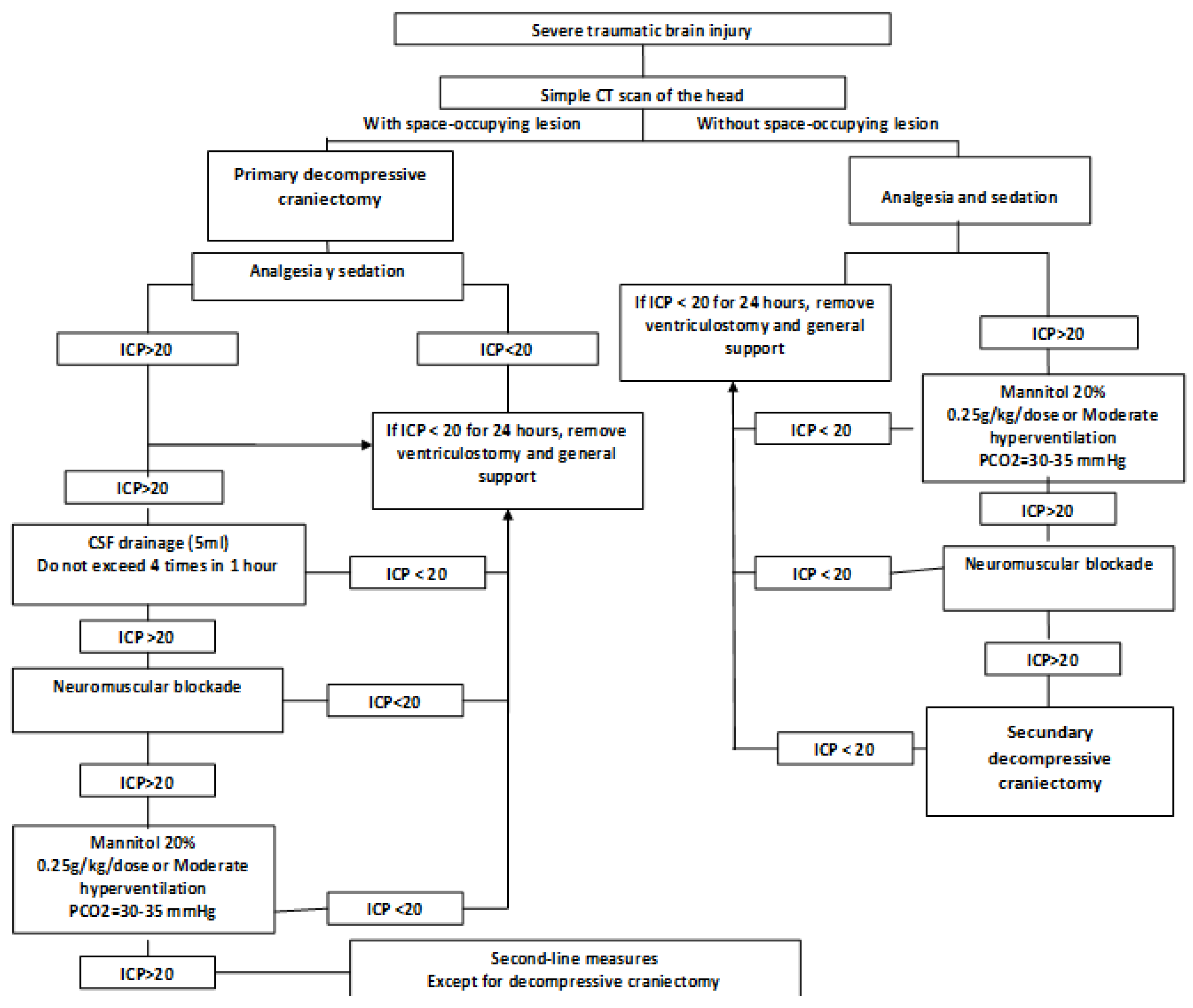
Figure 2.
Algorithm of treatment escalation in patients with clinical and imaging monitoring.
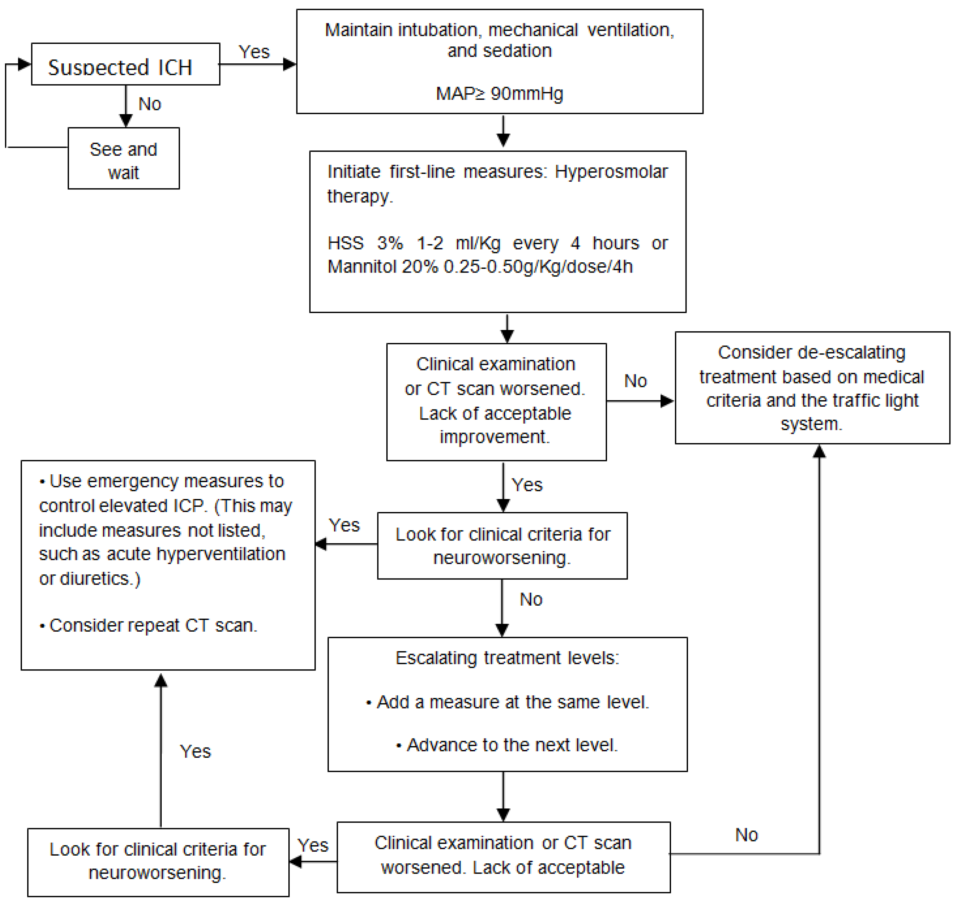
Figure 3.
Relationship between the optic nerve sheath diameters measured on CT scan at admission and ICP values.
Figure 3.
Relationship between the optic nerve sheath diameters measured on CT scan at admission and ICP values.
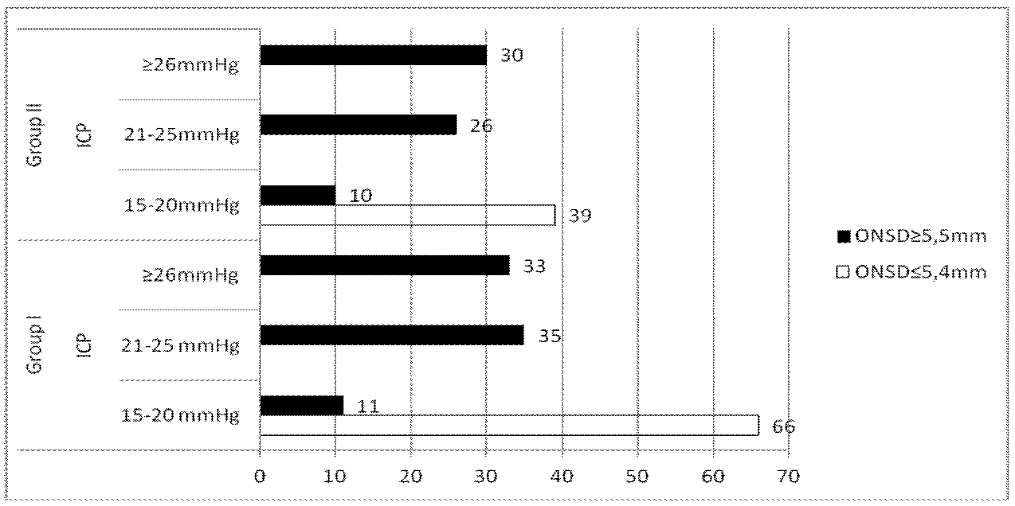
Figure 4.
Diagnosis of neuroworsening.
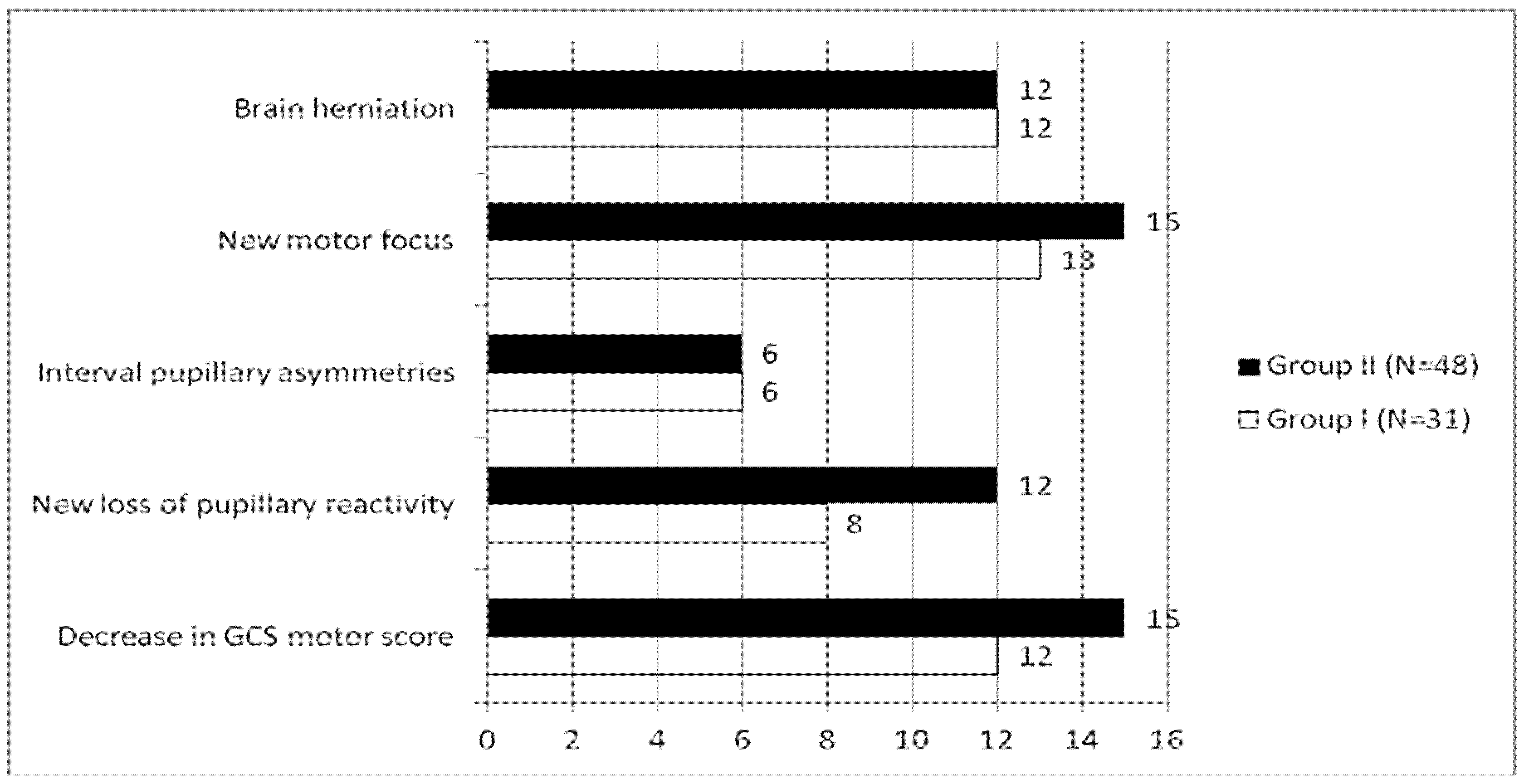
Figure 5.
Therapeutic measures used.
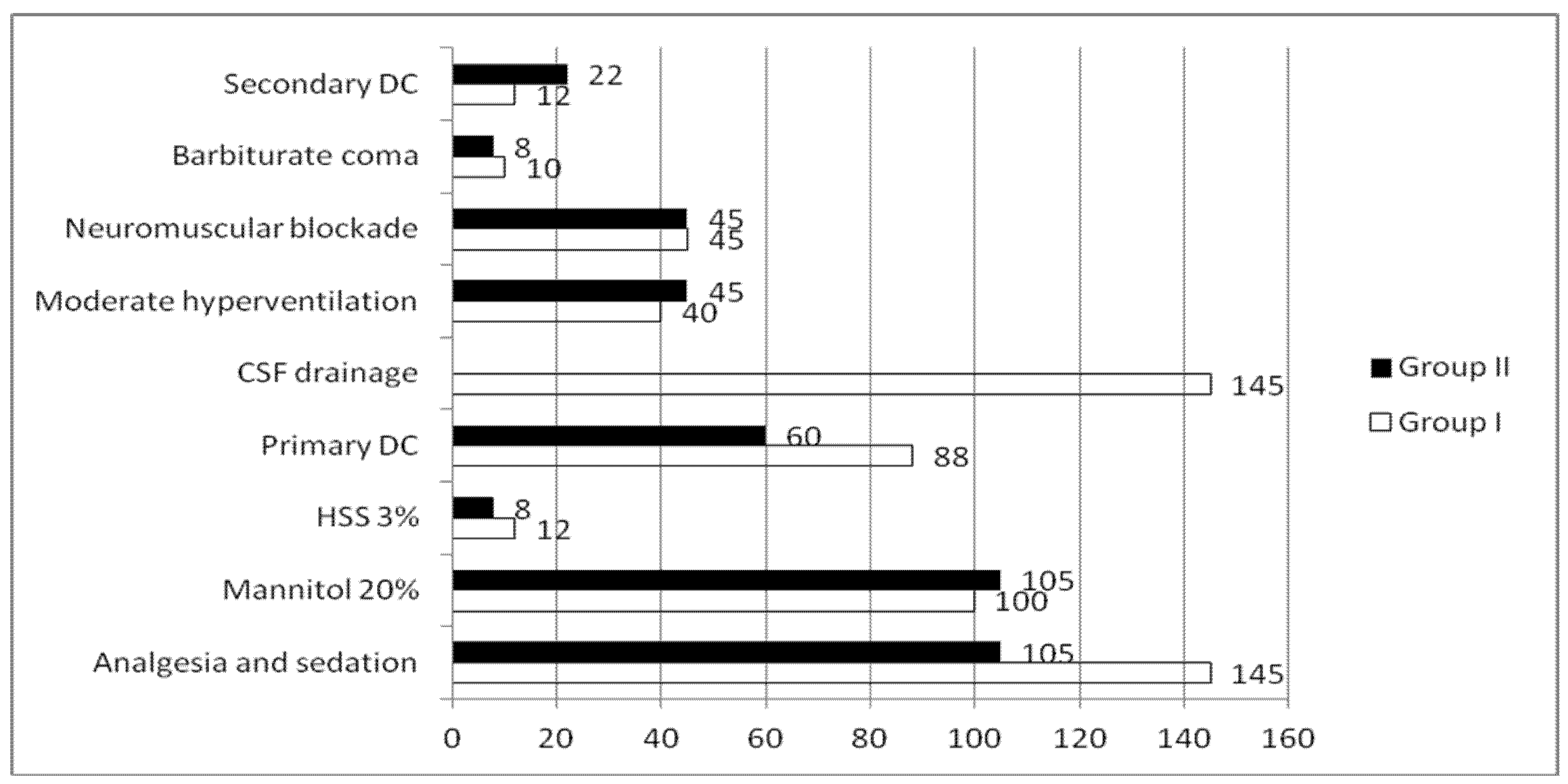

Table 1.
Treatment levels for ICH.
| Level 1 |
|
| Level 2 |
|
| Level 3 |
|
Table 2.
Decision support matrix.
|
Marshall classification on the most recent CT scan DL= Diffuse injury. LME= Evacuated mass injury. |
Motor GCS and pupils NP= Normal pupils AP= Abnormal Pupils without worsening since the trauma |
||||||||
| GCS-M 6 | GCS-M 5 | GCS-M 4 | GCS-M 1-3 | ||||||
| NP | AP | NP | AP | NP | AP | NP | AP | ||
| During the first 24 hours | DI 1-2 | ||||||||
| EMI/DI 1-2 | |||||||||
| DI 3 | |||||||||
| EMI/DI 3 | |||||||||
| NP | AP | NP | AP | NP | AP | NP | AP | ||
| Around 48 hours | DI 1-2 | ||||||||
| EMI/DI 1-2 | |||||||||
| DI 3 | |||||||||
| EMI/DI 3 | |||||||||
| NP | AP | NP | AP | NP | AP | NP | AP | ||
| Around 72 hours | DI 1-2 | ||||||||
| EMI/DI 1-2 | |||||||||
| DI 3 | |||||||||
| EMI/DI 3 | |||||||||
| NP | AP | NP | AP | NP | AP | NP | AP | ||
| After 72 hours | DI 1-2 | ||||||||
| EMI/DI 1-2 | |||||||||
| DI 3 | |||||||||
| EMI/DI 3 | |||||||||
Table 3.
Characteristic of images acquired on admission.
|
Classification of CT images on admission |
Group I | Group II | Total | |||
| No | % | No | % | No | % | |
| DAL Grade II | 18 | 12,41 | 0 | 0 | 18 | 7,2 |
| DAL Grade III | 31 | 21,38 | 18 | 17,14 | 49 | 19,6 |
| DAL Grade IV | 34 | 23,45 | 24 | 22,86 | 58 | 23,2 |
| Unevacuated mass effect injury | 62 | 42,76 | 63 | 60 | 125 | 50 |
| Injury with evacuated mass effect | - | - | - | - | - | - |
| Total | 145 | 100 | 105 | 100 | 250 | 100 |
DAL (Diffuse Axonal Lession), Chi-squared of Pearson (Statistical exact of Fisher): 7,004; p = 0,133 (p > 0,05).
Table 4.
Results in both groups according to the Glasgow Outcome Scale.
|
Glasgow Outcome Scale (GOS) results At six months |
Grupo I | Grupo II | Total | |||
| No | % | No | % | No | % | |
| Grade I | 39 | 26,89 | 44 | 41,90 | 83 | 33,2 |
| Grade II | 2 | 1,38 | 6 | 5,71 | 8 | 3,2 |
| Grade III | 14 | 9,66 | 21 | 20 | 35 | 14 |
| Grade IV | 37 | 25,51 | 24 | 22,87 | 61 | 24,4 |
| Grade V | 53 | 36,56 | 10 | 9,52 | 63 | 25,2 |
| Total | 145 | 100 | 105 | 100 | 250 | 100 |
Disclaimer/Publisher’s Note: The statements, opinions and data contained in all publications are solely those of the individual author(s) and contributor(s) and not of MDPI and/or the editor(s). MDPI and/or the editor(s) disclaim responsibility for any injury to people or property resulting from any ideas, methods, instructions or products referred to in the content. |
© 2026 by the authors. Licensee MDPI, Basel, Switzerland. This article is an open access article distributed under the terms and conditions of the Creative Commons Attribution (CC BY) license (http://creativecommons.org/licenses/by/4.0/).
Copyright: This open access article is published under a Creative Commons CC BY 4.0 license, which permit the free download, distribution, and reuse, provided that the author and preprint are cited in any reuse.