

Submitted:
17 August 2026
Posted:
18 August 2026
You are already at the latest version
Abstract
Aging is an extensive biological process characterized by morphological and functional alterations at different biological scales, resulting in a systematic decline in biological functions ultimately leading to death. Overall, two main types of theories have been proposed: damage-based theories and programmatic theories. We propose a third option, framed in the cognitive perspective on multiscale biological systems. Here, we review the different theories of aging, organize them in a conceptual hierarchy, and integrate our new model within the existing frameworks: aging as a consequence of loss of goal-directedness (in anatomical space). In this model, aging is driven by a dynamical systems-level disruption of homeostatic alignment within cellular collectives that build and repair a healthy body during development and maturation. In this model, morphogenesis is a homeostatic (goal-seeking) process; left with no goals after the developmental phase has been completed, aging can occur in the absence of damage via the functional disbanding of body components from their original aligned goal state. We suggest a roadmap with important implications for regenerative medicine and aging.
Keywords:
aging
; longevity
; bioelectricity
; goal-directedness
1. Introduction
Aging is an extensive biological process characterized by morphological and functional alterations at different biological levels, resulting in a systematic decline in biological functions ultimately leading to death [1,2,3]. Genetic inheritance, environmental conditions, and lifestyle can influence the aging process [4]. Several diseases are related to aging, as the resistance to noise and damage decreases, including cardiovascular diseases, cancer, neurogenerative conditions, diabetes, and immune-related diseases with inflammation, altering significantly the quality of life [5,6] and limiting our potential for growth that may require more than the evolutionarily standard human life span [7].
The current aging literature provides a good understanding of the currently-detectable hallmarks of aging [6]; however, which are consequences and which are drivers of the aging process is still unclear [8]. Moreover, researchers are not really in agreement on a definition of aging [9] or its ultimate cause. Overall, two main types of theories have been proposed: damage accumulation-based theories [5,6,10] and evolutionary programmatic theories [1,11,12]. Over the past century, aging theories have consistently highlighted whichever biological subsystem gained scientific prominence during that particular period. This pattern has resulted in distinct theoretical frameworks that emphasize individual components—whether genetic elements like telomeres, mechanisms controlling gene expression, immune system components, reactive oxygen molecules, or epigenetics to name a few [13]. Most of the major theories are supported by good scientific evidence in the literature; however, they tend to focus on a particular sub-component of the general physiology, illustrating that it is key for the healthy functioning of the biological system and that its dysfunction can lead to aging. But there is still no unified approach explaining why all the critical components of the biological system at different scales degrade with time after development.
Here, we attempt a conceptual mindmap of this diverse and important field. We also propose a third option, framed in the perspective of biological systems composed of nested layers of competent subsystems with agendas [14,15,16,17]: aging as loss of goal-directedness. On this model, aging is first and foremost a failure mode unique to aligned collectives with goal-directed behavioral capabilities at multiple scales. One such collective is groups of cells during morphogenesis, whose activities are functionally held together by a large-scale goal state (the species-specific target morphology), which the group pursues in the space of anatomical possibilities [16,18]. What does such a system do, long after the body is constructed? We propose that after the developmental phase has been completed, cells begin to lose collective alignment. On this view, aging, like cancer [19,20], is a failure of the fundamental driver of multicellularity (shared homeostatic setpoints). The ultimate driver of aging is thus fundamentally a problem that belongs to cybernetics or cognitive science [13,21], not physics (as in damage theories) nor evolutionary biology (as in programmatic theories). It predicts that aging phenomena will occur even in the absence of accumulated damage or evolutionary forces [22]. We add therefore a third theoretical aging category grounded in biological systems seen as collective intelligence [22,23,24].
Here, we attempt to synthesize a simplified conceptual map of the field to emphasize distinct proposed drivers and their numerous sequalae, and discuss implications of the various theories for biomedical approaches to longevity.
2. Theories of Aging
Damage-Based Theories
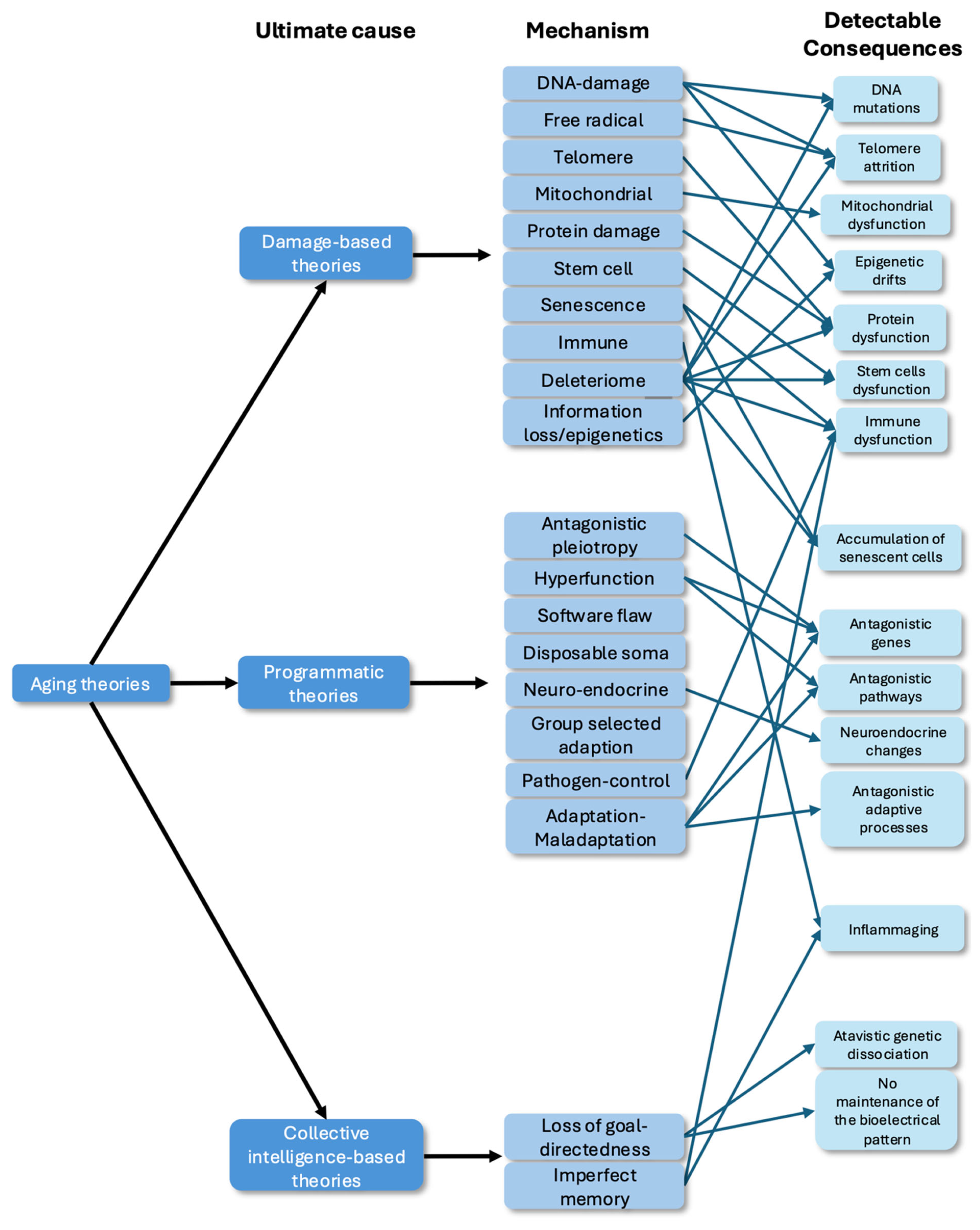
Damage-based theories propose that aging arises from the accumulation of entropic damage, which affect essential molecular and cellular components such as the genome, telomeres, mitochondria, and proteins. This process would be caused by the inherent imperfections in cellular repair mechanisms [5,6,10]. On this view, aging is the result of inescapable rising disorder – in other words, its origin lies in physics, not biology. This family of theories comprises several different versions, focusing on different aspects of aging and on different levels of biological complexity:
-
Molecule level:
- o
- o
- Telomere theory: the aging process is driven by the progressive accumulation of mutations that limit cellular division capacity, mainly due to the gradual shortening of telomeres during each cycle of cell division [27].
- o
- Protein damage theory/Error catastrophe theory: it suggests that that aging results from the accumulation of errors in protein synthesis, leading to a cascade of cellular dysfunction and the eventual failure of cellular systems. This theory posits that once error levels exceed a critical threshold, they trigger a rapid and irreversible decline in cellular function, causing aging [28].
- o
- Mitochondrial theory of aging: it is an extension of the free radical theory of aging and suggests that as mitochondria are involved in regulating programmed cell death, but their functions are especially vulnerable to damage from free radicals, this sensitivity leads to genetic mutations, which in turn increase free-radical production, perpetuating a self-amplifying cycle of cellular damage [29].
- o
- Free radical theory: it posits that molecular damage results from the metabolic reactivity of partially reduced molecular oxygen (ROS). These ROS cause oxidative damage to lipids, proteins, and DNA [30].
- o
- The deleteriome theory of aging: it suggests that endogenous damage is a by-product of imperfect metabolic processes and results from most if not all biological functions [3] at all levels, from molecular, cells, organs, and systems. This leads to damage accumulation and ultimately aging.
-
Cell-level:
- o
- Stem cell theory: aging is due to the depletion and/or dysfunction of stem cells with age.
- o
- Cellular senescence theory: aging is caused by non-dividing senescent cells that accumulate with age.
- o
- Immune system theory: it posits that aging is due to immune system dysfunction and higher inflammation with age [31].
-
Informational layers:
- o
- Loss of bioelectrical morphostatic information: aging is caused by the alteration of the bioelectric pattern in charge of the maintenance of the anatomy or the inability of the cells to understand and integrates the bioelectrical pattern [23].
- o
(Quasi) Programmatic-Based Theories
In contrast to physics-focused theories that emphasize inevitable wear-and-tear damage at different levels, programmatic theories suggest that aging is genetically or evolutionarily predetermined, placing evolutionary forces as the ultimate cause of aging [11,35,36]. Quasi-programmed theories focus on development, aging is here an unintended consequence of developmental and growth programs running in later life; selection is weak against these harmful late effects. These theories hold that aging is not inevitable, but specific aspects of evolution have favor it (or favored development). Under this category, we can find:
-
Evolutionary trade-off ones:
- o
- Disposable soma theory: it posits that organisms allocate limited resources between reproduction and maintenance of the body (soma). Since resources are finite, investing heavily in reproduction reduces the resources available for cellular repair [37]. Here, aging is a product of an evolutionary trade-off.
- o
- o
- Hyperfunction theory: aging is a result of normal growth and developmental pathways that continue excessively or inappropriately beyond their intended period, causing harmful activity (hyper- or hypofunction) in cells and tissues [39].
- o
- Software design flaw theory: it posits that aging results from pre-existing flaws or limitations in the organism’s genetic program—akin to a “bug” or imperfect design in the biological “software”—which becomes apparent as the organism ages [11]. Another possibility is evolutionary commitment to specific mechanisms which cause aging as a side-effect but cannot be improved by evolutionary search due to important dependencies.
- o
- Group selected adaptation: aging evolved to control or limit the death rate over time helping stabilize population dynamics to respect ecological limits and prevent extinctions [40].
- o
- The adaptation/maladaption theory: it proposes that in aging adaptation processes such as adaptive transcription, epigenetic remodeling, and metabolic plasticity drive dysfunction themselves over time (maladaptation) and thereby cause aging-related disorders [41].
Figure 1.
The theoretical landscape of aging. Tree of aging theories by categories from ultimate causes to biomedical interventions. They are classified in 3 larger categories: damage-based, programmatic and collective intelligence-based theories.
Figure 1.
The theoretical landscape of aging. Tree of aging theories by categories from ultimate causes to biomedical interventions. They are classified in 3 larger categories: damage-based, programmatic and collective intelligence-based theories.
Specific systems evolution targeted:
- Neuro-endocrine theory: aging is due to the modulation of cell cycle signaling by reproductive hormones [42].
- Pathogen-control theory: aging evolved to control or limit the spread and impact of pathogens within a species. The mechanism has been proposed to be the immune system [43,44,45,46] but in general is compatible with other means of removing old (and thus presumed infected) individuals from the population.
Goal-Directedness-Based Theory
“Cancer is no more a disease of cells than a traffic jam is a disease of cars. A lifetime of study of the internal-combustion engine would not help anyone understand our traffic problems.” -- D. W. Smithers
Whereas the above theories place the ultimate cause of aging in physics and evolutionary biology respectively, we propose a set of hypotheses which propose aging to be fundamentally a problem of cybernetics (or cognitive science): a misalignment of components within a goal-seeking system once its goals have been met. In other words, nothing needs to be wrong at the hardware level, in the same sense that psychological disorders can arise without organic disease. Imagine a scenario in which a human, a dog, and a snake are put into an ideal environment devoid of any source of infection or molecular damage (a kind of perfect afterlife perhaps, with no entropy), and in which all somatic aging processes are halted. While we don’t know what would happen, one could imagine that the snake would be fine forever, and perhaps the dog would as well – happily chasing rabbits and sleeping in the sunny grass for trillions of years. But, does it seem likely that a human mind could remain sane for enormous lengths of time? If not, it would have to be caused by a psychological dynamic – a failure mode that isn’t due to molecular entropic forces or tissue degradation but is uniquely characteristic of advanced cybernetic systems that become unstable after the have achieved their primary goal but do not have new ones to pursue.
It is tempting to hypothesize that homeodynamic systems operating in other spaces (not just the familiar brain-based cognition driving behavior in 3D space [47]) may be susceptible to this problem as well. Morphogenesis is precisely such a system, as many of its instances implement a goal-seeking pattern homeostasis process that minimizes error relative to a setpoint (an anatomical state) via growth, remodeling, apoptosis, and other effector processes in embryogenesis and regeneration [47]. Such setpoints can be stored (as in the brain) in tissue-level bioelectric patterns [16] but many kinds of biomechanical or biochemical encodings exist as well. In this sense, the departure from coherent functional anatomy in cancer is also a disorder of collective alignment: cells disconnecting from information networks that allow the group to pursue large (organ-level) setpoints [48,49,50], and reverting to ancient tiny goals focused on metabolic and physiological states at their small scale. Such disconnection and its downstream consequences can be caused by molecular (genetic or otherwise) damage, but also by physiological stressors and other dynamics [51,52,53,54,55,56]. We suggest there are important parallels at the intersection of aging, cancer, and cognitive science broadly conceived [57].
Goal-directedness [58] (used here in the minimal, cybernetic sense of error minimization, not in the full-blown metacognitive sense of an intelligent agent knowing their goals [59,60,61,62]) refers to the fact that in multiscale biological systems, higher levels of organization work to achieve homeostatic, homeodynamic, and allostatic setpoints in spaces that are inaccessible to their parts. For example, while individual cells manage goals [59,60,61,62]in metabolic and physiological state space, groups of cells work towards implementing and maintaining complex 3-dimensional states in anatomical morphospace [63,64,65]. Regeneration and maintenance of anatomical form by morphogenetic processes is a closed-loop process that implements error-reduction with respect to a stored goal state. This requires alignment of components, which can break down, as occurs in the case of cancer [19,20]. While such a breakdown of cooperativity can have many causes (including the ones discussed above), multiscale architectures can generate unique disease conditions that are not due to problems with any of their parts. Traffic jams are a good example of unique dynamics arising at higher levels of organization [66,67,68,69,70]. We propose that one such unique disease state can occur in goal-seeking systems that have completed their goal, and in the absence of a new motivating driver, begin to disband (i.e., no longer prevent their semi-autonomous parts – cells – from becoming misaligned) [23]. Specifically, we propose that aging can be caused by cellular collectives having completed their journey in anatomical space and having no new setpoint to implement. Under this general framework, we can place:
- Loss of goal-directedness theory: aging is caused by the loss of goal-directedness after the primary morphogenetic setpoints are achieved during development. This leads to a failure of alignment of cell behavior toward large-scale anatomical setpoint patterns, and thus degradation of order at all scales [22,57,71,72].
- Imperfect memory theory: cells lose the memory of resilient behavior under stress leading to general dysfunction [13]. We add that relevant memories may be of molecular states, physiological states, transcriptional states, or even second-order memories of policies (resilient ways to deal with inputs), not just static setpoint patterns.
The imperfect memory [13] and loss of goal-directedness are very close, while both are part of the collective intelligence category view on biological systems emphasize two different aspects of aging. The imperfect memory theory states that aging and death result from the accumulation of imperfect cellular memories of appropriate behavior under stress due to the inherent ambiguity of biological information [13]. Over time, these imperfect memories accumulate and overwhelm the flexibility cells require to respond adaptively to environmental stresses, leading to a gradual breakdown in cellular information assessment and cell-cell communication. This approach is very similar to the loss of morphostatic information one as bioelectrical patterns can be interpreted as anatomical memories [16,73]. The loss of goal-directedness one suggests that this is the absence of anatomical goal that produces multiscale breakdown and aging. While both frameworks use the cognitive lens on biological systems and frame aging as a defect of information processing, they differ in the cognitive and scale implementations. The former focus on the accumulation of imperfect cellular memories (which is also close to the accumulation of damage reframed in an information-theoretic lens), the latter focus on the lack of anatomical goal, the absence of maintenance/regenerative instructions, causing aging even in the absence of accumulated damage in a multi-competency architecture.
3. Anti-Aging Interventions
The current scientific landscape of anti-aging interventions targets mostly the hallmarks of aging [6] and integrates very different biomedical interventions including chemical-based, epigenetic, blood-based, senolytics and lifestyle approaches (see Table 1). While sustained caloric restriction and mTOR inhibitors like rapamycin remain the best-evidenced interventions for modulating aging biology in existing human data [74,75], the field is increasingly pivoting toward more transformative modalities. Partial epigenetic reprogramming has emerged as the most funded approach, driven by multibillion-dollar investments in ventures like Altos Labs, with recent data demonstrating its ability to safely reset epigenetic clocks and improve tissue function in non-human primates and mice without stripping cell identity [76]. However, despite this potential, clinical translation is severely constrained by fundamental safety and efficacy hurdles. Partial reprogramming strategies are constrained by toxicity: continuous or high-level expression of OSKM factors drives rapid dedifferentiation and loss of cellular identity, causing teratomas [77]. This has been overcome in several models by using transient, low-dose OSKM factors which preserves identity and avoids teratomas while enabling rejuvenation [78,79]. However, systemic induction of OSKM factors has been shown to precipitate acute hepatic and intestinal failure, leading to premature death [80]. Myc-sparing expression (OSK) has also been used in vivo: in mouse retinal ganglion cells, OSK restored youthful DNA-methylation and transcriptional profiles, promoted axon regeneration, and improved visual function [81].
Concurrently, exosome-based interventions are developing as a cell-free alternative to paracrine rejuvenation; extracellular vesicles derived from young plasma or mesenchymal stem cells have demonstrated the capacity to transfer bioactive miRNAs that suppress the senescence-associated secretory phenotype (SASP) and restore mitochondrial homeostasis in aged tissues [82,83].
One important question in the context of anti-aging interventions that needs to be answered is to determine if (and what) combination(s) (number and category) of established intervention(s) act synergistically for cumulative benefit that will impart the desired health and or lifespan enhancements. For example, combining Yamanaka factors + Senolytics produced synergistic effects in flies in terms of substantially longer mean and maximum lifespan, while each treatment alone had limited benefits- OKSM alone offered maximum lifespan (rejuvenated stem cells), while Sen expression alone (removed senescent cells) increased mean lifespan but had no effect on maximum lifespan. Another study in flies with different genetic backgrounds [84], reported on achieving exceptional lifespan extension in flies (D. melanogaster) using multitude of potential anti- aging approaches such as dietary restriction, co-administration of berberine, fucoxanthin, and rapamycin, photo-deprivation, and low-temperature conditions. The strongest effect was observed when all of the mentioned factors were combined, producing a 122% increase in median lifespan and a 124% increase in maximum lifespan in long-lived mutant E(z)/w males. Authors noted that some of the factors investigated when used singly and in combination, impacted the lifespan negatively, while some did not offer any advantage over the control group (effects were sex dependent as well). The study concluded that even if some of the factors did not impart any significant effect when tested alone, their combined action seemed to offer lifespan enhancements, indicating these interventions probably acted synergistically. The geroprotective effects of these interventions were shown to affect the evolutionarily conserved longevity molecular pathways, mTOR signaling modulation, significant increase in foxo and Akt1 gene expression, decrease in Hsp70 gene expression and autophagic mechanisms.
Transient expression of OSKM was shown to be beneficial in flies, while long term or sustained expression was detrimental [85]. The OSKM factors are believed to work by reversing the loss of epigenetic information (an “information theory of aging”) and rejuvenating mitochondrial function. Related to enhanced mitochondrial function as an anti-aging approach is the approach of initiating ‘mitophagy’ (autophagy of mitochondria specifically) by exercise, natural compounds such as Urolithin A and EGCG and/or supplements containing these compounds, with Rapamycin also showing beneficial effect. It will be interesting to test the combination of these factors for their cumulative or synergistic effects as noted above for other such interventions.
One major limitation in the studies we have reviewed here is that the reported human data is largely of healthspan increase rather than lifespan longevity, studies of which are available in other model systems. However, as the research progresses faster and the attention along with the funding this field is getting, we will begin to see real human data in near future. A few encouraging results have been reported, particularly for Rapamycin. These include PEARL trial (NCT04488601) that established safety and tolerance of low dose Rapamycin in healthy aged cohort (50-85 years), resulting in significantly increased lean tissue mass and bone density, pain reduction and improved overall emotional and physical health [86]. Other studies are investigating effect of low dose Rapamycin on enhancing ovarian function and IVF success (VIBRANT trial, NCT05836025), another study demonstrated increased in vitro fertilization (IVF) rates after repeatedly failed cycles, better quality blastocysts resulting in more successful live births [87].
However, demonstrating actual rejuvenation at organismal level in mammals has not been achieved yet. But we can mention that very recently, it has been reported in a preprint that by rejuvenating CD4+ T-cells and reinjecting them in mice could extend lifespan with a 65% increase creating ‘rivers of telomerase’ (Treatment Median 43–45 months vs. Control Median 26–28 months) [88]. Some mice reached twice the normal lifespan of a mice (~60 months) which is a huge gain. Indeed, the gold standard with rapamycin or caloric restriction typically delivers typically delivers a 15–25% median extension [89,90]. If true and this result can be translated to humans, this would be the first intervention that can really delay aging past the current known limits of human lifespan.
4. Unconventional Aging Mechanisms: Bioelectric Patterns and Phylogenetic Drift
Two new recent mechanisms have been proposed to function during the process of aging. The first is progressive degradation of bioelectrical pattern information, and the loss of alignment of cellular transcriptomes with respect to phylogenetic age. Both of these are most clearly connected to the memory loss theories, and in general the goal-directedness root cause model, because those focus on information and the biophysical encodings of homeostatic goal states. But, both are compatible with damage and programmatic causes as well, as they can be due to numerous upstream events.
The Bioelectrical Pattern as the Encoded Morphogenetic Goal
Developmental bioelectricity is increasingly recognized as a global coordinator of growth and form and the encoding of anatomical setpoints [91,92,93,94]. Indeed, through the voltage dynamics governed by ion channels and pumps, cells execute complex computational operations that regulate their behavior [73]. Evolution recognized early—dating back to the emergence of bacterial biofilms [95,96,97] — that bioelectric networks provide an efficient medium for integrating information across spatial and temporal scales, enabling coordinated activity among individual cellular units towards higher large-scale tasks. This principle underlies the organization of both nervous systems and artificial computers, which similarly exploit electrical signaling for large-scale information processing tasks, probably the same reason brains and computers exploit it [98].
Bioelectric signaling dynamics govern a wide range of developmental and regenerative processes, including wound repair, neural circuit formation, organogenesis, and body axis specification [99]. Recent work has demonstrated that modulation of bioelectrical patterns offers a high-level control interface for morphogenesis and regenerative medicine [73,99,100]. Instead of micromanaging molecular events, bioengineers can exploit simple electrical cues to trigger complex, self-limiting cascades of tissue development. Practical strategies span from guiding cell migration through electrotaxis during healing [101,102] to manipulating ion channels with “morphoceuticals” compounds that set bioelectric prepatterns for appendage or organ induction [100]. For example, in vertebrates, craniofacial morphogenesis is guided by distinct bioelectric gradients that spatially organize gene expression, thereby defining the positions of the eyes, mouth, and other facial structures [94,103,104,105]. Experimental alteration of these electrical patterns through targeted ion channel misexpression, as well as genetic channelopathies (reviewed in [106]), can reproduce morphogenetic programs in non-native regions, inducing the formation of complete ectopic organs—such as eyes developing on the gut of tadpoles [107]. These findings reveal that bioelectric cues can unlock latent developmental programs within tissues that are normally non-competent for this specific organogenesis, such as inducing new growth of whole multi-tissue appendages [108,109,110,111,112,113].
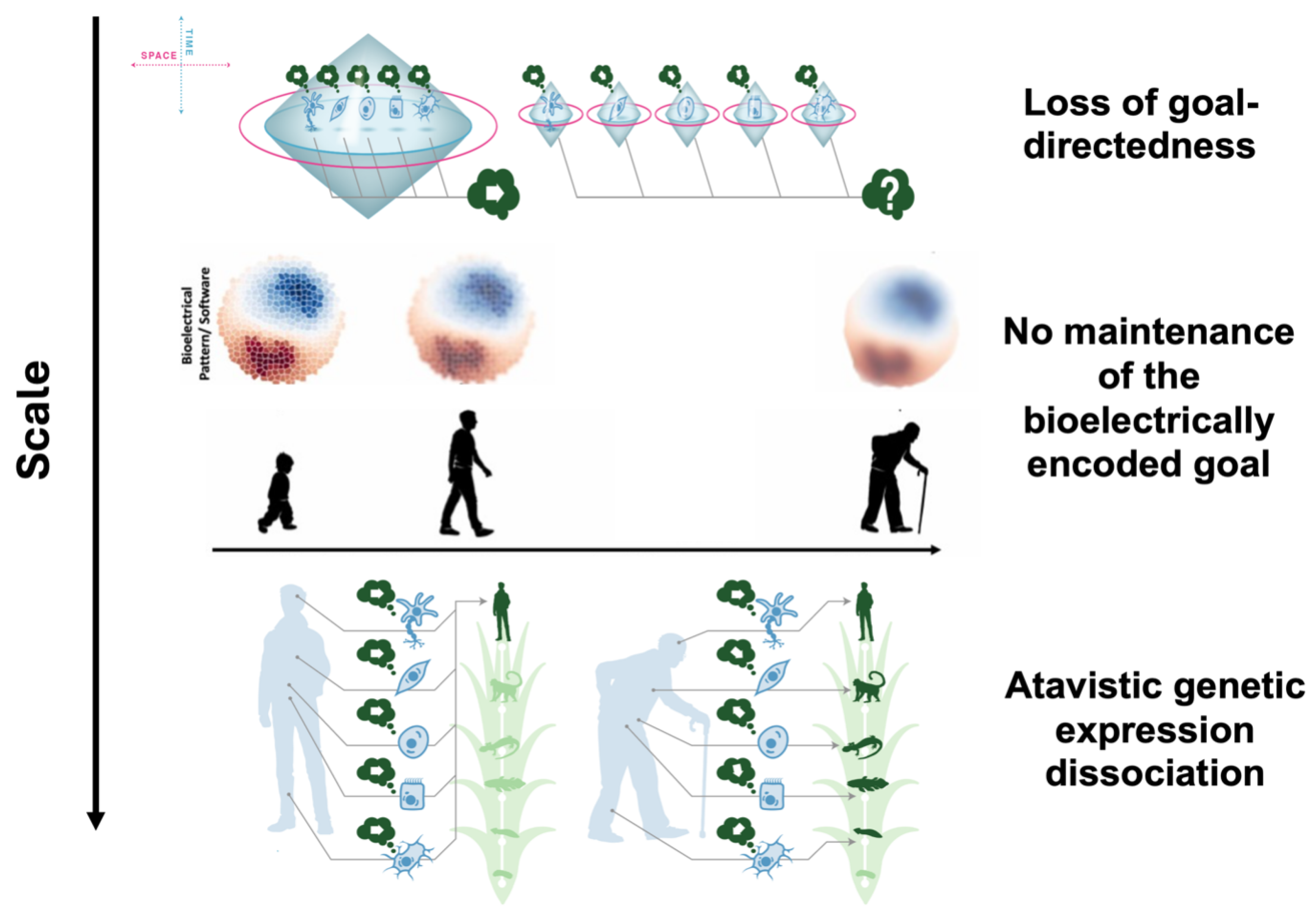
The stopping of the maintenance of the bioelectrical pattern and the associated loss of bioelectrical morphostatic information is a possible mechanistic substrate for the loss of goal-directedness at the tissue and organ levels. Voltage gradients and ion channel networks encode and maintain anatomical setpoints—the information that tells cells and tissues what to grow. The decay of these bioelectric patterns with age leads to a loss of anatomical homeostasis, causing tissues to drift structurally and functionally. These bioelectric circuits, a crucial part of the “physiological software of life” [23], serve as an informational layer that enables large-scale anatomical patterning, working synergistically with genetic hardware. When the precision of these bioelectric patterns degrades or cells lose their ability to sense them, tissues drift away from optimal morphologies, leading to organ dysfunction, disease, and death. Bioelectric signals provide real-time feedback and coordination between cells, tissues, and organs. This layer can encode critical processes such as regeneration, developmental patterning, and cancer suppression [16,20,23,73,99]. When the maintenance of these bioelectric prepatterns is stopped, leading their corruption over time, it is hypothesized that it drives anatomical and functional aging, with channelopathies emerging as a unifying mechanism for many age-related diseases [23,114,115].
Bioelectricity interfaces with established molecular hallmarks of aging. Evidence and plausible mechanistic links are described between ion channel dysfunction and age-dependent degenerative phenomena—including for example senescence, stem cell exhaustion, inflammation (“inflammaging”), loss of proteostasis, and altered nutrient sensing [23,116].. Notably, the bioelectric layer both influences and is influenced by other molecular processes, suggesting reciprocal causality and bidirectional feedback within aging phenotypes [23,116]. In exploring the consequences of the impairment of coordinating and setpoint-encoding information, we reasoned that detectable loss of alignment would be observed among downstream molecular properties regulated by large-scale information systems.
Transcriptional Level Loss of Morphogenetic Control: The Atavistic Genetic Expression Dissociation (AGED)
In searching for signs of misalignment across cells as a function of aging, normally observed at the spatial level, we sought to examine the temporal dimension, and in particular include scales well beyond the single organism lifespan. We thus found a transcriptional atavistic dissociation that operates at the genomic and cellular level during aging [24]: a progressive increase in the disagreement among cells of their evolutionary age. The “Atavistic Genetic Expression Dissociation (AGED)” data reveal that aging is not merely a uniform reversion to ancient gene expression like in cancer [117,118], but fundamentally a process of dissociation and loss of multicellular coordination across tissues and transcriptional programs at the evolutionary scale. This dissociation manifests as a heterogeneous, tissue-specific shift and dispersion of gene expression profiles: instead of all tissues reverting to the same ancestry, each tissue can regress along different evolutionary trajectories [24]. This has been found for two multi-tissue aging databases covering skin, ovarian, immune, senescent and mesenchymal-senescent cells, but interestingly not for brain cells and mesenchymal stem cells. It suggests that cells are lost in phylogenetic space, and tissues lose a unified transcriptional identity and regress in divergent ways. This is related to the loss of identity we can find too during aging [33,119]. When crucial information, especially patterns that serve as instructive anatomical setpoints, degrade within the multi-scale competency architecture of a metazoan body, the normal coordination of morphogenesis can slide backwards, allowing the cells to follow their own goals in their problem space, leading to the atavistic dissociation [24] and/or cancer [63].
Figure 2.
Multiscale breakdown of morphogenetic control of form. The loss of goal-directedness theory is the most general one. When the cells have an anatomical goal, they are aligned and behave towards that goal creating a larger cognitive light cone. When cells lack the higher goal, they are misaligned, desynchronize and follow their own cellular goals. By going down in scale, we can find two possible mechanistic implementations: the loss of bioelectrical morphostatic information and downstream at genetic level, the atavistic genetic expression dissociation (cancer is another possible cellular goal) where cells are lost in phylogenetic space.
Figure 2.
Multiscale breakdown of morphogenetic control of form. The loss of goal-directedness theory is the most general one. When the cells have an anatomical goal, they are aligned and behave towards that goal creating a larger cognitive light cone. When cells lack the higher goal, they are misaligned, desynchronize and follow their own cellular goals. By going down in scale, we can find two possible mechanistic implementations: the loss of bioelectrical morphostatic information and downstream at genetic level, the atavistic genetic expression dissociation (cancer is another possible cellular goal) where cells are lost in phylogenetic space.
5. Future Roadmap for Therapeutics
Organisms such as planaria, or hydra, as well as the continued youthfulness of our germline, serve as existence proofs demonstrating that practically infinite maintenance and amortality are possible. There are life forms that resolve the aging problem at the systems level. It is important to complement existing focus [120,121] on molecular- and cell-level therapeutics with novel approaches that target emerging information-level factors in health and disease [122,123,124,125,126,127,128,129].
Our framework has several implications for roadmaps to longevity. First, robust mechanisms for maintaining and implementing morphogenetic setpoint states (i.e., a robust regenerative response) should be associated with minimal aging and cancer. With respect to cancer, it has long been known that high regenerative capacity (the ability to re-implement anatomical structure with high fidelity) prevents the breakdown into autonomous, lower-level cellular goals like tumorigenesis [20] – well-documented among species with continuous, highly controlled growth and rare cancer incidence [130,131,132,133,134,135,136]. Moreover, evidence from diverse taxa illustrates that some species—such as lobsters, planaria, Greenland sharks, long-lived trees, and fungi—demonstrate negligible senescence while demonstrating sustained regenerative or growth capacities throughout their lifespans [130,137,138,139,140,141]. Thus, we propose that the fundamental way to address aging is the same as that for birth defects, traumatic injury, and cancer: as fundamentally disorders of collectivity, all of these maladies will be addressed by definitive progress in regenerative medicine targeting native morphogenetic programs [142,143,144]. Asexual planaria may be immortal precisely because they can avoid the loss of goal-directedness: every two weeks, they motivate their anatomical homeostatic systems by fissioning: each fragment must re-scale (and thus re-write, sharpen, and renew) the bioelectric and biochemical setpoints, and commit to implementing the goal state. Planaria are never bored of their embodiment, and this confers immortality despite their extremely noisy genome [145,146]. It is not yet known how much or how little repair is needed to refresh the morphogenetic system – does longevity require massive regenerative response as in planaria? Would extremely long lifespans require actual modification of anatomy (a caterpillar-butterfly kind of event, currently entertained on the domain of future-looking transhumanism)? The benefits of exercise (small-scale microtrauma [147]), and the immortality in Hydra (budding, not whole-animal regeneration) provide reason to be optimistic [20,130,148].
A roadmap is emerging. By focusing on reimposing large-scale (whole anatomy) morphogenetic goal, it may be possible to engineer organismal rejuvenation. The regenerative experiment in [22] suggests that giving an embryonic environment to the cells after the loss of one organ during aging may re-active developmental/regenerative pathways, much as embryonic environments have been shown to normalize cancer [149,150,151,152,153]. This is similar to regeneration observations with Yamanaka factors during partial cell reprogramming [154]. Likewise, new ways to detect disorganization at the system level (e.g., the atavistic phylostratigraphy signature discussed above), and non-invasive detection of bioelectric pattern anomalies, as for cancer [155], suggest novel monitoring modalities complementing epigenetic and transcriptional clocks [156,157].
The bioelectric theory in particular suggests an interesting intervention path to longevity: refreshing the bioelectric patterns. A proof of concept of this already exists, in the field of birth defects in the Xenopus model. A number of teratogenic interventions exert their effects by degrading the native bioelectric patterns that establish the correct structure of the brain, face, heart, and gut. Remarkably, whether induced chemically or genetically (Notch mutation), these defects can be over-ridden by a single ion channel activation (via pharmacological or mRNA-induced means), resulting in a sharpening of the bioelectric prepatterns and the return of the anatomical, gene expression, and behavioral endpoints to wild-type despite the continued presence of the teratogenic agent [110,111,158]. Thus, in at least some cases, hardware defects can be repaired “in software” via physiological stimuli induced by a computational platform-selected electroceutical [100,159,160]. This potentially bodes well for such intervention strategies even if there is considerable contribution to aging from low-level molecular noise and damage, but will require advances in the emerging computational methods to infer bioelectric interventions [161].
Atavistic genetic expression dissociation with age suggests that expressing evolutionarily-younger genes may relocate cells lost in phylogenetic space too allowing the cells to re-align on higher morphogenetic goals. Similarly, recent findins of inter-embryo communication that improves morphogenetic robustness [162] could expand existing work on parabiosis [163] and perhaps simulate the effect without needing to connect blood streams.
Finally, it is important to take even further the idea of top-down control over micromanagement. Where the bioelectric pattern renewal path seeks to re-write global setpoints, not micromanage cells or even genes, this can be taken further. Our “age-evidencing” hypothesis is that tissue-level age is not a physical or even biochemical parameter but the output result of cellular computations that integrate current state and past history of inputs [164,165]. It has already been noticed that when an organism is convinced to be older/younger [166], it behaves as such, including metrics at the cellular level, similarly to heterochronic parabiosis [167]. More recently, wild-type biobots made from un-modified adult epithelial cells from human patients experienced a rejuvenation event during the morphogenesis of their new form [168], which included a reversal of the epigenetic clock but not expression of reprogramming factors. This could be capitalized upon with stimuli that further push back cells’ estimates of their own age toward embryonic states without genetic induction of dangerous single genes, and may be related to similar rejuvenation events recently discovered during embryogenesis [169].
6. Conclusion
Aging is a complex field that has been addressed at multiple scales of spatial and temporal dynamics. We propose that the best hope we have of addressing this system-level limitation on our potential is to identify top-level mechanisms and policies which enable the Ship of Theseus that is our body to form and function in the first place. The future is likely not just micromanagement of molecular pathways but communication with the regulatory logic of complex bodies [17,22,170,171,172,173,174]. The loss of goal-directedness theory of aging suggests a specific roadmap: strengthen and perhaps re-write the pattern memories of healthy organs. Interfaces for such interventions include bioelectric, biochemical, biomechanical, and perhaps optical [175]. Future medicine of aging will merge with that of cancer and regenerative medicine aimed at large-scale restoration, likely drawing on all of the existing theories and approaches for a multi-faceted approach to tame this global, but ultimately surpassable, aspect of our current embodiment [176].

Table 1.
Anti-aging treatments, targets, and their impact on aging.
| Name of the intervention | Mechanistic Target | Rationale as anti-aging agent | Results/Observations/Outcome | Species/model | References |
|---|---|---|---|---|---|
| Cell reprogramming (partial/transient)/Stem cell rejuvenation | |||||
|
Yamanaka factors set of four transcription factors- Oct4, Sox2, Klf4 ± c-Myc (OSK or OSKM), ± senolytic peptide (Sen) ± senolytics dasatinib and quercetin (D+Q) |
The four TFs work mechanistically by transient cellular reprogramming and stem cell rejuvenation. Senescent cells have persistently active mTOR pathway, leading to continuous secretion of proinflammatory cytokines and impaired tissue homeostasis. Synergetic combination of transient cellular reprogramming via OSKM factors and targeted removal of senescent cells by senolytic agents achieved lifespan and healthspan benefits. In flies, lifespan extension seemed closely related to preservation of Intestinal Stem Cells (ISCs) in older flies (12 weeks) compared to control groups |
Epigenetic dysregulation is amongst the underlying basis of aging that can be attenuated/reversed by epigenetic remodeling or cellular reprogramming. Approach is shown to extend lifespan, Improve healthspan (the duration of healthy life) by ameliorating age related diseases and reverse age-related changes in organs and tissues |
Lifespan extension (mean and max) in m/f flies using short term pulsed expression of the four Yamanaka factors (102→122 days) + Sen (synergistic effect, 102→ 135 days) and Yamanaka TFs ± (D+Q); ~30% reduction in the initial mortality rate in flies reversal of vision loss in an aged glaucoma mouse model by ectopic expression of OSK genes in retinal ganglion cells which restored younger DNA methylation patterns and promoted axon regeneration after injury inducible systemic OSK -AAV in 124-week-old male mice extended the median remaining lifespan by 109% (133 → 142.5 days) over WT controls and enhanced health parameters such as frailty score (health span improvement). transient expression of OSKM factors attenuated cellular and physiological hallmarks of aging and extended lifespan in a mouse model of premature aging. Expression of OSKM in vivo improved recovery from metabolic disease and muscle injury in older wild-type mice exogenous OSK expression in human keratinocytes lead to significant reduction in epigenetic markers of age suggesting a potential rewiring of genetic networks to a younger healthier state |
flies (Drosophila) mouse human keratinocytes Worms (C.elegans) |
[81,85,177,178,179] |
| Optimized Yamanaka factors (OSK) in conjunction with TERT (Telomerase Reverse Transcriptase) gene therapy | TERT mediated extension of telomeres that shorten with age; OSK expression lead to stem cell rejuvenation | Combined gene therapy proposed as a promising therapeutic strategy for extending lifespan and addressing aging-related diseases | In hu-MRC-5 cells, upregulated expression of youth-related genes- OCT4, SOX2, Klf4, Nanog, c-Myc, and TERT and the expression of aging-related genes- p16, p21, ZSCAN4, ATF3, and MMP13; the pro-inflammatory cytokine IL-6 and senescence-associated genes were significantly reduced |
Human embryonic lung MRC-5 fibroblasts | [180] |
|
Chemical cocktail induced partial reprogramming seven-compound (7c) - CHIR99021, DZNep, Forskolin, TTNPB, Valproic acid (VPA), Repsox, and Tranylcypromine (TCP) or two-compound (2c) reprogramming cocktail- Repsox and TCP |
Chemical reprogramming modulated the underlying mechanisms and molecular hallmarks of aging such as improved age-associated DNA damage, epigenetic alterations, and induction of a unique transcriptomic profile enriched for developmental processes in aged human fibroblasts in vitro |
Extension of both healthspan and lifespan via reversal of multiple aging hallmarks and induction towards a younger state in human fibroblasts |
Dampening of aging phenotypes in dermal fibroblasts such as senescence, heterochromatin loss, genomic instability, and oxidative stress (ROS) with 7c cocktail, while 2c mixture retained rejuvenation effects Median lifespan extension in worms by >42% (19 → 27 days), with improved stress resistance, thermotolerance, reproductive and healthspan markers with 2c cocktail |
Worms (C.elegans) Human fibroblasts and keratinocytes |
[181] |
| Blood/Stem Cell mediated rejuvenation | |||||
| Heterochronic parabiosis (HPB)/Heterochronic blood exchange (HBE) Plasmapheresis or therapeutic plasma exchange (TPE) or plasma dilution Umbilical Cord (UC) Plasma transfusions |
Inflammatory pathways modulated by the young plasma protein fraction included the PI3K-AkT, cytokine-cytokine receptor interaction, JAK-STAT, neurotrophin, NF-kappa B, and MAPK pathways TLR-4 was the node of the age-specific TPE-mediated rejuvenation of canonical signaling networks Decreased entropy in the methylation data, significant correlation between DNAm-based age acceleration and entropy-- decreased entropy may implicate the rejuvenation of the epigenetic landscape |
The younger plasma infusion in elderly subjects influenced a range of biological processes associated with aging and age-related diseases (such as cancer, CVD), via regulation of inflammatory pathways and maybe particularly of interest for patients at risk of impaired and longer recovery time. These ‘younger blood factors’ may help dampen the inflammaging-associated inflammatory responses mediated by the adaptive immune system cells. TPE-mediated recalibration of signaling pathways to younger state lead to improved homeostatic status of multiple organ systems. Plasma concentrate contains youth-promoting factors in the secretome of UC, administration of which resulted in nominally significant age-reversal effect revealed by DNA methylation clock DNAmGrimAge, an established robust epigenetic predictor of morbidity and mortality risk in humans |
Infusion of GRF6021, an FDA approved 5% plasma protein fraction derived from younger donors (avg age~ 35y) to older patients (avg age~ 72 y) resulted in pathway and cell type specific anti-inflammatory immune-modulation in response to surgical injury; proteomics analysis and mass cytometry showed modulating effects on adaptive immune cell signaling- MAPK and JAK/STAT3 signaling responses in CD4 + and CD8 + T cells and regulatory T cell subsets were attenuated, while IkB (negative regulator of NFkB) was elevated In a limited pilot study in humans, longitudinal effects of repeated TPE on systemic aging markers, a reduction in inflammaging and rejuvenation of age-specific systemic proteome was shown- diminished markers of neurodegeneration (TDP43) and cancer (increased P21 and p53), reduced senescence in PBMCs, lower DNA damage, decreased apoptotic inhibitors and improved myeloid/lymphoid homeostasis Human umbilical cord plasma concentrate injected into elderly human subjects (mean age 74) and effect on different biomarkers, including epigenetic age, clinical biomarkers of organ dysfunction, mitochondrial DNA copy number (mtDNA-CN), and leukocyte telomere length were measured. Improved kidney function biomarkers were observed, telomere length and mtDNA-CN were not significantly affected, reduced DNA methylation-based GrimAge by an average of 0.82 years |
Human subjects Human subjects Human subjects |
[182] [183] [184] |
| HPB | Upregulated pathways for hematopoiesis, cytokine production, and stem cell population maintenance; restoration of cell-cell communications with cytokine/receptor interactions, chemotaxis, and leukocyte proliferation pathways enrichment On the pathway level, global rescue of genes encoding electron transport chain (ETC) subunits indicated a mitochondrial function in parabiosis-mediated rejuvenation |
Systemic revitalization of hematopoiesis by ‘young blood’ factors and restoration of dysregulated cytokine-mediated intercellular communication leading to younger transcriptional regulatory patterns Younger blood transfusion induced cellular and molecular programs that instructed systemic revitalization to re-establish youthful transcription/regulatory phenotypes |
HP mediated rejuvenation of lymphopoiesis decline in aged HSCs, rejuvenated senile adult stem cells and their niches across tissues, restoration of youthful transcriptomes, reduced senescence-associated β-galactosidase (SA-β-gal)-positive cells in the spleen, skin, liver, and brain, decreased apoptotic cells in the spleen, skin, liver, and skeletal muscle single-cell RNA sequencing on 20 organs to reveal cell-type-specific responses to young and aged blood in heterochronic parabiosis revealed that ‘aged’ blood reduced global gene expression, and ‘young’ blood restored it in select cell types (adipose mesenchymal stromal cells, hematopoietic stem cells and hepatocytes) |
mouse mouse |
[167] [185] |
| HPB Longer term HPB |
Pro-aging factors in aged blood such as the chemokine CCL11/eotaxin and the inflammation related protein β2-microglobulin reduce neurogenesis, IL-6 and TGF-β impair hematopoiesis and the complement factor C1q compromises muscle repair; changed mitochondrial genes expression patterns involved in the electron transport chain (ETC) Attenuation of inflammatory and interferon gamma (IFN-gamma) response gene sets, reduced pro-inflammatory phenotype; Gene expression changes mimicked longevity intervention signatures (such as median/ maximum lifespan and caloric restriction and growth hormone deficiency signatures) |
restoration of regenerative capacity of old muscle with both parabiosis experiments (extrinsic effect) and transplantation of ex vivo-rejuvenated SCs into old animals (intrinsic effect) long-term HPB acted on global scale affecting gene expression profiles, tissue methylation and systemic epigenomic remodeling leading to lifespan and healthspan extension |
heterochronic parabiosis experiments followed by single-cell transcriptomics showed the soluble factors present in the blood of young mice restored renewal and repair capacity of muscles in old mice; ‘young’ blood factors mediated rejuvenation and restoration of age-associated reduction in general gene expression, specifically the mitochondrial genes extended HPB (3 mo) resulted in sustained and systemic multi-omic rejuvenation effect, altered phenotype of older mice to intermediate (between old and young), reduced epigenetic age of blood and liver based on various clock models (RRBS based clocks such as Meer and Thompson multi-tissue methylation clocks, Petkovich blood clock, a maximum likelihood-based single-cell clock, array-specific epigenetic clocks- Universal, mouse blood/liver), a 6-week extension in median lifespan and a 2-week extension in maximum lifespan in old mice (compared to their isochronic controls) was observed. |
Mouse mouse |
[6,186,187] [188] |
| HBE- circulating Extracellular vesicles (EVs) | EVs improved muscle regeneration in a Klotho mRNA-dependent manner—a longevity protein at the intersection of Ca2+/K+ ion channel activity, mitochondrial function, and aging Mechanistic investigations reveal that young sEVs stimulate PGC-1α expression in vitro and in vivo through their miRNA cargoes, thereby improving mitochondrial functions and mitigating mitochondrial deficits in aged tissues |
young MuSCs are abundant in Klotho transcripts compared to aged MuSCs, loss of Klotho contributes to impaired MuSC mitochondrial ultrastructure and function over time, which can be reversed by exposure to young circulating EVs substantial alterations in the proteomes of aged tissues after young sEV treatment that were closely associated with metabolic processes. young sEVs reversed degenerative changes and age-related dysfunction, at least in part, by stimulating PGC-1α expression and enhancing mitochondrial energy metabolism |
Klotho mRNA within EVs contributed to functional skeletal muscle regeneration (in vivo) and enhanced mitochondrial function in MuSC progeny (in vitro) small extracellular vesicles (sEVs) from the plasma of young mice counteract pre-existing aging at molecular, mitochondrial, cellular and physiological levels Intravenous injection of young sEVs into aged mice extended their lifespan, mitigated senescent phenotypes and ameliorated age-associated functional declines in multiple tissues |
mouse | [189] [82] |
| VEGF | reduced VEGF signaling and increased production of soluble VEGFR1 (sVEGFR1) due to an age-related shift in alternative splicing of VEGFR1 mRNA acting as a VEGF trap |
vascular theory of aging- age related decrease of vascular function is a driver of systemic organismal aging and an upstream driver of multiorgan malfunctioning, hence mitigating it can potentially lead to comprehensive geroprotection | using a transgenic VEGF gain-of-function system or AAV–assisted VEGF transduction enhanced VEGF levels leading to a more youthful VEGF signaling, protection from age-related capillary loss, compromised perfusion, and reduced tissue oxygenation. Aging hallmarks such as mitochondrial dysfunction, compromised metabolic flexibility, endothelial cell senescence, and inflammaging were alleviated in VEGF-treated mice, which had an extended health span, as reflected by reduced abdominal fat accumulation, reduced liver steatosis, reduced muscle and bone loss (sarcopenia, osteoporosis) and reduced burden of spontaneous tumors |
mouse | [190] |
| Extracellular Matrix (ECM) | In human fibroblasts, ‘young’ ECM mediated rejuvenation involved Ku-SIRT1 interaction, which are part of cell’s DNA damage and genome stability maintenance package In C. elegans- proposed mechanism for increase in longevity involved pathway enrichment for chondroitin and heparan sulfate biogenesis and TGFβ pathway as predicted drug-protein targets |
most of the established pro-longevity compounds shown to extend lifespan in model organisms also alter extracellular matrix gene expression or the ’matrisome’, ECM from young human fibroblasts induced a youthful state in aged senescent cells C. elegans-- restored age-related decline of collagen homeostasis with increased lifespan, measured by collagen biosynthesis. |
hu fibroblasts-- cell morphology, phenotypic restoration was assessed by resumption of proliferative potential, growth factor responsiveness, reduction of intracellular reactive oxygen species levels, recovery of mitochondrial membrane potential, and increased telomere length ECM compounds such as chondroitin sulfate and hyaluronic acid restore the age-related decline of collagen and increase lifespan in nematodes; In mice, deletion of chondroitin 6-sulfotransferase results in an abnormal ECM in the brain, early memory loss, and accelerated brain aging |
C. elegans human cells mouse |
[6,191,192] |
| Senolytics | |||||
| Fisetin (3,3’,4’,7-tetrahydroxyflavone) |
Direct antioxidant activity via enhanced glutathione levels, inhibited Lipoxygenases decreasing the production of pro-inflammatory factors/ eicosanoids; lifespan extension in worms mediated by DAF-16-induced stress response and autophagy |
Mitigation of age-related accumulation of oxidative damage (free radical theory of aging), anti-inflammatory, and anti-tumor effect neuroprotective, restored tissue homeostasis in animal models | Reduced senescence markers in murine and human adipose tissues, reduced the cellular ROS levels and increased the resistance to oxidative stress and extended median and maximum lifespan in worms, improved high-glucose diet induced reduction in motility, reduced Amyloid beta-induced toxicity, inhibited degeneration of dopaminergic neurons in a PD model | C.elegans murine and human fibroblasts |
[193,194,195] |
| Dasatinib and Quercetin (D/Q) and Navitoclax |
Pan-TK and BCL2 family of proteins- BCLXL, BCL2 and BCLW; improved α-Klotho levels |
elimination of senescent cells, α-Klotho is a geroprotective protein and declined levels play a role in the pathophysiology of multiple diseases and age-related phenotypes. Pre-clinical evidence suggests that boosting α-Klotho holds therapeutic potential |
increased α-Klotho in IPF, a disease linked to cellular senescence, reduction and clearance of senescent cells and SASP factors, induction of apoptosis of senescent cell-associated phenotypes (SCAPs), therapeutic activity in a wide range of murine disease models associated with senescence |
mouse human primary cells Human/clinical trials |
[6,195,196,197] |
| Senomorphs- Novel/Natural Molecules/Supplements | |||||
| Resveratrol (3, 5, 4′-trihydroxy-trans-stilbene) |
Allosteric activators of SIRT family of proteins (SIRT1), CR mimetic | multi-effector anti-aging benefits across species | improved adipose insulin signaling in middle-aged mice on a high-calorie diet leading to better health/survival and insulin sensitivity, increased mitochondrial number, and improved motor functions; in yeast- stimulated SIRT1, increased DNA stability and extended lifespan by 70% ; in worms and fruit flies- extended lifespan without affecting reproduction; in NHPs- suppressed body mass gain in obesity model, reversed age-associated proinflammatory phenotypes in vascular smooth muscle cells, prevented age-related DNA and RNA oxidative damage, reduced mitochondrial loss and promoted autophagy in skeletal muscle and enhanced cognitive performances; humans- improved glucose metabolism and cardiovascular disease markers, reduced oxidative stress, inflammation, and cell death, neuroprotective, antidiabetic, antibacterial and anti-aging effects and delayed cognitive decline in Alzheimer’s disease patients |
yeast monkey/NHPs mice flies Hu |
[198,199,200,201,202,203,204] |
| Alpha-ketoglutarate (KGA) | Different mechanisms across species, such as inhibiting mTOR and ATP-synthase, modulating DNA and histone demethylation and reducing ROS formation; flies--Activation of AMPK signaling, inhibition of mTOR pathway and ATP-synthase worms- Inhibition of ATP-synthase; mice--Enhancement of IL-10 production; humans- Rejuvant® supplement lead to reduction of DNA methylation |
KGA plays a crucial role in the cell energy metabolism, amino acid/protein synthesis, epigenetic regulation, stemness and differentiation, fertility and reproductive health, and cancer cell behaviors | reduced cellular senescence, protected against telomerase deficiency, suppressed mitochondrial dysfunction, and decreased both DNA damage and inflammation. AKG extended longevity in C. elegans by ∼50% and delayed age-related phenotypes and reduced oxygen consumption; mice- lifespan extension up to 10% in female and 5% in male flies- lifespan extension up to 23% in female and 2.8% in male; human- AKG has beneficial effects on skin hydration and barrier function in human; in clinical trials- Rejuvant® supplementation lead to lifespan extension of up to 6.98 years in females and 8.44 years in males (biological age), improved functioning of bone, muscle, pancreas, brain, fat, gut, and immune system was observed indicating an overall increase in health span |
mouse human pig fly worms |
[205,206] |
| Taurine (2-aminoethanesulfonic acid) |
Taurine proposed to modulate nutrient sensing and proteostasis pathways, with enhanced (LC3A/B), an autophagy marker, suppression of senescence, suppressed adverse consequences of telomerase deficiency, affects epigenetic changes via DNA methylation patterns |
in humans, lower circulating taurine and its metabolites are associated with multiple age-associated pathologies, and their abundance increases after an exercise regimen. higher blood taurine and hypotaurine levels were associated with lower body mass index (BMI) and waist-to-hip ratio (WHR), and less abdominal fat, lower prevalence of type 2 diabetes, lower glucose levels and lower levels of the inflammation marker, C-reactive protein (CRP) |
Taurine supplementation increased the median lifespan in mice by 10 -12%, and life expectancy at 28 months increased by 18- 25%; increased muscle mass endurance, coordination, and strength in taurine-treated female mice; enhanced cognitive function- reduced depression-like behavior and anxiety, and enhanced exploratory behavior and memory; increased lifespan in worms by 10-23% ; improved health parameters in middle-aged non-human primates |
mouse monkey worms human |
[207] |
| NAD | NAD+/Sirtuin pathway modulated longevity through activation of mitochondrial UPR and FOXO signaling, NAD precursor NR activated the UPRmt and the prohibitin signaling pathway in inhibition of MuSC senescence; sirtuin protein sir-2.1, activated by NAD+, induced the UPR(mt), leading to the nuclear translocation and activation of the FOXO transcription factor DAF-16, which promoted antioxidant protection and extends lifespan |
strategies that conserve, replenish cellular NAD(+) may reprogram dysfunctional stem cells to enhance the life span in mammals |
in aging mice, NR/NAD treatment protected from muscle degeneration, loss of muscle stem cells, prevented MuSC senescence in the mdx (C57BL/10ScSn-Dmd(mdx)/J) mouse model of muscular dystrophy, was protective of neural and melanocyte stem cells leading to the extended life span of the NR-treated animals. In worms, enhancing NAD+ availability extended lifespan acting through sir-2.1 proteins In humans- NAD/NR exerted a lipid-lowering effect and is a treatment option for dyslipidemia. It acts as a ligand for the G–protein-coupled receptor GPR109a, which inhibits hepatosteatosis and the progression of atherosclerosis and other inflammatory conditions |
mouse human C. elegans |
[208,209,210,211] |
| Oxytocin | Improved myogenic progenitor cell proliferation via activation of the MAPK/ERK pathway; effects of OXT in chronic isolation related cellular aging in voles are mediated by reduction in elevated glucocorticoids |
combats or prevent skeletal muscle aging/sarcopenia, and mediates the regenerative potential of muscle stem cells |
systemic administration of oxytocin improved muscle regeneration while inhibition of oxytocin signaling in young animals reduced muscle regeneration; Oxytocin treatment diminished the increased glucocorticoid levels, oxidative damage, telomere degradation and anhedonia after six weeks of chronic isolation in female prairie voles |
mouse female prairie voles |
[212,213,214] |
| Epitalon aka Epithalon or Epithalone, (tetrapeptide, Ala-Glu-Asp-Gly (AEDG)) |
Specific and nonspecific mechanisms including direct influence on melatonin synthesis, influencing the mRNA levels of interleukin-2, and enhancing the activity of various enzymes, including AChE, BuChE, and telomerase | potential geroprotective and neuroendocrine effects of Epitalon, resulting from its antioxidant, neuro-protective, and antimutagenic effects. Shown to lead to reactivation of telomerase gene in somatic cells with the possibility of prolonging life span of cell populations/organism |
clinical trials showed decreased (2-fold effect), functional age and cardiovascular aging with enhanced exercise tolerance in long-term treatment in elderly patients with accelerated aging. Elongates telomeres and induces telomerase activity in telomerase-negative human fetal fibroblast cultures |
human |
[215,216,217] |
|
Drugs/Medicinal interventions/Pathway Modulators (Inhibitors/Activators) | |||||
| Rapamycin/Rapalogs | primarily exerts effect by Inhibiting m/TOR pathway, mimics CR-initiated nutrient scarcity, affects cellular shift from anabolic processes to maintenance and repair pathways, autophagy induction, enhanced proteostasis and translation fidelity |
geroprotective, affects multi-modal mechanisms associated with lifespan extension such as induction of autophagy, modulating proteostasis (see MoA) |
decreased mTOR signaling extends lifespan by up to 20% in yeast, 19% in worms, 24% in flies, and 60% in mice; randomized controlled trials showed reversal of immunosenescence and enhanced immune response following flu vaccines in older adults |
mouse human |
[75,89,218,219,220] |
| Metformin | AMPK/LKB1 pathway activator, upregulation of pro-longevity signaling in Nrf2-GPx7 mediated antioxidant response | GP×7 expression levels are decreased in senescent human cells, and GPx7 depletion results in premature cellular senescence; metformin-Nrf2-GPx7 pathway delays aging in worms, geroprotective effects on NHP neurons via the activation of Nrf2/anti-oxidant pathway | worms- extended median lifespan and promoted youthful mobility; monkey- decelerated aging in males with reduction of aging markers, neuroprotective effect, with~6-year regression in brain aging, reduced oxidative damage and chronic inflammation; humans- exerted effects by modulating multiple pro-aging pathways such as metabolic pathways, collagen trimerization and extracellular matrix (ECM) remodeling, adipose tissue and fatty acid metabolism, mitochondria, and the MutS genes (DNA mismatch repair) |
C. elegans Cynomolgus monkey human cells |
[221,222,223,224,225] |
| Sodium-glucose cotransporter 2 inhibitors (SGLT2-i such as dapagliflozin, canagliflozin, empagliflozin) |
modulation of major nutrient-sensing pathways-upregulation of the energy deprivation sensors AMPK by shifting the AMP/ATP balance in favor of AMP and SIRT1, inhibition of the nutrient sensors mTOR and insulin/IGF1, and modulate the closely linked hypoxia-inducible factor (HIF)-2α/HIF-1α pathways; induces metabolic shift at the systemic level, promoting ketones and fatty acids utilization as energy source, lowers glucose by promoting its elimination through urine; CR mimetic |
shown to ameliorate inflammaging in clinical trials, effects of SGLT-2i proposed to be mediated by their ability to attenuate the drivers of aging | Cardio and reno-protective effect in clinical trials against aging related major adverse cardiovascular (CV) events, heart failure, CV-related death, all-cause mortality, and renal outcomes in patients with or without diabetes; increased mean lifespan mice fed on a long-term high-fat diet with reduction of inflammation, oxidative stress, and cellular senescence in visceral adipose tissue. |
human mouse |
[226,227,228,229] |
| Glucagon-like peptide 1 receptor agonists (GLP-1 RAs) (albiglutide, dulaglutide, liraglutide, semaglutide, exenatide and lixisenatide |
pleiotropic mechanisms of action- protects against cell apoptosis via the PI3K/Akt/mTOR/redox signaling pathway, protects beta cells against apoptosis and prevents beta-cell glucolipotoxicity via AMPK/mTOR signaling, enhances the DNA repair through the stimulation of APE1 expression, protects against apoptosis and inhibits ROS production via Sirt1 pathway, Liraglutide ameliorates mitochondrial dysfunction via AMP/PKA pathway; modulates cellular senescence through AMPK/SIRT1/FOXO3a pathway |
protective against major hallmarks of aging such as oxidative stress, cellular senescence and chronic inflammation and related diseases | Cardio and neuro and reno-protective effect due to strong hypoglycemic action, lowered lipid levels, maintained blood pressure; increased pancreatic insulin, GLUT2, and glucokinase mRNA, improved insulin response and glucose clearance in old rats, ameliorated muscle wasting, skeletal muscle atrophy and enhanced myogenesis, cardio, neuro and muscle protective roles demonstrated in rodent models |
human rodents |
[230,231,232,233,234,235] |
| Sapanisertib | mTORC1/2 dual inhibitor | attenuated chronological aging in aged zebrafish and replicative senescence in HFF fibroblasts, potential for treating premature aging and promoting healthy aging | exerted superior effect in ameliorating cellular senescence compared to rapamycin in Zebrafish wrnKO, effect on aging phenotypes- inhibited SA-β-Gal activity (senescence), reduced the p21 and p53, improved swimming activity (primary indicator of healthspan); increased the proliferative capacity of the intestine and kidney marrow in aged zebrafish; increased the cell proliferation rate of HFF cells, reduced SA-β-Gal staining |
Zebrafish, human cells—replicative senescent HFF |
[236] |
| Trametinib | reversibly inhibits the MEK1 and MEK2 kinases within the MAPK pathway; increases translation accuracy |
suppression of the insulin–IGF–mTORC1–Ras network ameliorates aging in animals | extended lifespan in flies; improved translation fidelity, geroprotector in mice, extended lifespan in both sexes in mice |
fly mouse |
[218,237] |
| Proteostasis modulator RPS23-K60R mutant | enhances translational fidelity and improves protein homeostasis and promotes damaged protein degradation |
part of the ribosomal ‘accuracy center’, leads to proteostasis, one of the major hallmarks of aging | genetic manipulation of the ribosomal protein RPS23 to improve the accuracy of RNA-to-protein translation extended lifespan across organisms, and imparted heat shock resistance | yeast flies worms |
[6,218] |
| Life Style/Diet Interventions | |||||
| Calorie (CR) and Dietary restriction (DR)/ Intermittent Fasting (IF) | Beneficial effects are primarily mediated by increased activity of SIRT family of proteins, leading to modulated transcriptional programs implicated in mitochondrial metabolism, anti-inflammatory responses, and longevity, deletion of SIRT1 prevents longevity extension by CR in budding yeast, worms, and flies; Another proposed mechanism is by reduction of PLA2G7 levels mediate the positive effects of caloric restriction (CR), such as improved metabolic health, reduced inflammation and longer health span; In mice and rhesus monkeys, age-related methylation drift correlated with lifespan and CR attenuated negative impacts of it; Nutrient sensing pathways respond to reduced glucose levels and increase AMPK activity with decreased mTORC1 signaling Rats on IF/EOD exhibited slower growth rate, reduced weight and retarded development, perhaps trade off for the enhanced lifespan |
over-expression of SIRT1 or SIRT6 in mice mimics physiological effects observed in CR and exercise, and extends lifespan, SIRT proteins protected against obesity, cancer, neurodegeneration, frailty, and a range of cardiometabolic conditions in monkeys and in humans; reduction of PLA2G7 levels may lead to immunometabolic effects of CR and could potentially be modulated to lower inflammation and extend the health span Reduced feeding frequency directly improved lifespan in rats. |
CR delayed aging, improved metabolic profile and lowered age-related morbidity risk in Rh monkeys by >two-fold; 22-30 years-old rhesus monkeys exposed to 30% caloric restriction showed attenuation of age-related methylation drift (blood methylation age ~ 7 years younger than chronologic age) compared to ad libitum-fed controls, more pronounced effects were seen in 2.7-3.2-year-old mice exposed to 40% caloric restriction. The effects of caloric restriction on DNA methylation were detectable across different tissues and correlated with gene expression; intermittent fasting regimens have been reported to extend Drosophila lifespan by 15%–20%; In humans- fasting and CR/DR shown to have strong associations with decreased rates of age-related disease and mortality in longitudinal studies, and from observational studies and randomized clinical trials, analyses of healthy humans aged 25–45 years of age, with sustained CR for two years showed enhanced thymic function, reduction of ectopic lipid, increased functional thymic volume, and increase in recent thymic emigrants in CD4 and CD8 T cells in blood. In rats, IF (every-other-day/EOD) resulted in reduced tumor formation, better long-term activity levels and significant lifespan enhancement, upto 83% (`124 weeks vs 103 weeks in ad libitum diet group). DR shown to increase median and maximum lifespan in rats (average increase in median lifespan by 14–45% and maximum lifespan of 32.3%) and mice (average increase in median lifespan by 4–27% and maximum lifespan of ~18%) |
NHPs rodents flies yeast Hu Rat mouse |
[90,200,202,238,239,240,241,242,243] |
| Amino Acids/Protein restriction | protein restriction (PR) and reduction in amino acids such as Methionine and Arginine and the branched-chain amino acids impact the activity of AKT, FOXO, mTOR, NAD+, AMPK and fibroblast growth factor 21 (FGF21), the key components of geroprotective signaling pathways |
nutrient sensing pathways are affected (MoA) that promote healthy longevity |
Human clinical trials--- PR reduces weight and adipose mass, and improve blood glucose regulation, in overweight individuals and people with type 2 diabetes; restriction of protein has been shown to extend the lifespan in flies and rodents |
human flies rodents |
[6,242] |
| Specialized Diets, Exercise, Meditation and Social Interactions | |||||
| Mediterranean | Cardio-protective and myo-protective effects by yet to characterize mechanisms, the diet components of unsaturated fat, polyphenols and phytosterols (EVOO, nuts, vegetables, fatty fish) activate the AMPK and sirtuin pathways, while mTOR is inhibited and autophagy is stimulated (EVOO increases mRNA levels of the autophagy marker LC3 in older rats); reduction of LDL-C levels by inhibition of cholesterol intestinal absorption and by blocking proprotein convertase subtilisin/kexin-9 (PCSK9). Role in improving mitochondrial metabolism; improvement of immune system dysregulation by micronutrients and ω-3 fatty acids (EPA and DHA), beneficial effects on microbiota diversity, activity and composition |
cardio-protective and myo-protective effects prevent an excessive reduction of muscle mass and strength related to aging, hence exert positive effect on healthspan | myoprotective mechanism involved in ameliorating muscle ageing and sarcopenia, with higher bone mineral density; Cardioprotective effect shown in long-term clinical trials with reduction of cardiovascular disease (CVD), favorable effects on insulin sensitivity, blood pressure, lipid profiles, lipoprotein particles, oxidative stress, inflammation, and carotid atherosclerosis, lower inflammatory response |
rodents human |
[6,244,245] |
| Ketogenic diet | Pleiotropic mode of action, mediated via formation of ketone bodies such as 3-hydroxybutyrate which may influence aging by acting as an energy substrate and alternative to glucose, through protein acetylation or β-hydroxybutyrylation, or via inhibition of histone deacetylase. It induces vasodilatation and activates immune responses acting on GTP protein coupled receptor 109A (GPR109A), with direct inhibition of the NLRP3 inflammasome |
β-hydroxybutyrate inhibits the NLRP3 inflammasome and a ketogenic diet mitigates age-related increases in pro-inflammatory cytokines; this inhibition of inflammation may play a role in the extension of healthspan and longevity with a ketogenic diet | 13.6% increase in median lifespan in mice with a significant decrease in histiocytic sarcomas, improvement in motor function and memory Studies of mice on an intermittent ketogenic diet fed every other week showed improvement in healthspan in aged animals | Mouse |
[6,242] |
| Exercise | Multiple pathways affected, signaling cascades include IGF1, IL-6/JAK/STAT3, hippo and SIRT1 pathways; Hippo signaling through YAP and Taz has been demonstrated to play a role in satellite cell activation and proliferation; increased levels of taurine and taurine-related metabolites might mediate some of the health benefits of exercise. |
beneficial impact on age related muscular impairment. Exercise triggered increased levels of taurine metabolites in blood lead to antiaging effects of exercise. Mammalian SIRT1, SIRT3 and SIRT6 proposed to mediate the positive effects of exercise, while their genetic ablation prevents the benefits |
exercise shown to impart multiple benefits to overall health, including improved bone and muscle strength, offset obesity, improved cardiovascular, cognitive and immune system functions; human strength training (single/long-term) sessions resulted in a marked induction of muscle satellite stem cell activation and proliferation, activation of the muscle stem cell pool, increased mitochondrial numbers and quality, enhanced innervation by neuromuscular junctions, stimulate fibroapidogenic progenitors, (FAPs), and stimulate satellite cells to activate muscle repair and growth, leading to enhanced muscle strength and functioning. |
mouse rat human |
[195,207,246,247,248,249] |
| Mind-body therapies (MBT) tai chi, qigong, meditation, mindfulness, or yoga |
stress management and reduced inflammation via reduced signaling of the proinflammatory NF-κB pathway and increased anti-inflammatory GR signaling |
shown to be effective in improving sleep, reducing stress and improving quality of life, with possible impact on inflammation | 26 randomized controlled trials showed the effects of mind-body therapies (MBTs) on circulating, cellular, and genomic markers of inflammation with mixed effects on circulating inflammatory markers, such as CRP and IL-6, decreased expression of inflammation-related genes and reduced signaling through the proinflammatory transcription factor NF-κB | human | [250] |
| Family/Relationships and Social Interactions | the hypothalamic neuropeptide oxytocin (OXT) has been proposed to play an important role; chronic isolation leads to elevated glucocorticoids, which can affect cellular mechanisms of aging, including increased levels of oxidative stress and shortened telomere lengths |
poor social support leads to accelerated aging and is associated with reduced telomere length. Happy relationships, intimacy and family/social support is associated with increased immunity, better overall health, and a longer lifespan | Oxytocin treatment diminished the increased glucocorticoid levels, oxidative damage, telomere degradation and anhedonia after six weeks of chronic isolation | female prairie voles humans |
[213,214] |
| Several clinical trials are in progress to measure benefits/impacts of multi-modal anti-aging interventions. | [230,251,252,253] | ||||
| Mind-body therapies (MBT) tai chi, qigong, meditation, mindfulness, or yoga |
stress management and reduced inflammation via reduced signaling of the proinflammatory NF-κB pathway and increased anti-inflammatory GR signaling |
shown to be effective in improving sleep, reducing stress and improving quality of life, with possible impact on inflammation | 26 randomized controlled trials showed the effects of mind-body therapies (MBTs) on circulating, cellular, and genomic markers of inflammation with mixed effects on circulating inflammatory markers, such as CRP and IL-6, decreased expression of inflammation-related genes and reduced signaling through the proinflammatory transcription factor NF-κB | human | [250] |
| Family/Relationships and Social Interactions | the hypothalamic neuropeptide oxytocin (OXT) has been proposed to play an important role; chronic isolation leads to elevated glucocorticoids, which can affect cellular mechanisms of aging, including increased levels of oxidative stress and shortened telomere lengths |
poor social support leads to accelerated aging and is associated with reduced telomere length. Happy relationships, intimacy and family/social support is associated with increased immunity, better overall health, and a longer lifespan | Oxytocin treatment diminished the increased glucocorticoid levels, oxidative damage, telomere degradation and anhedonia after six weeks of chronic isolation | female prairie voles humans |
[213,214] |
| Several clinical trials are in progress to measure benefits/impacts of multi-modal anti-aging interventions. | [230,251,252,253] | ||||
| Experimental anti-aging treatments | |||||
| Rejuvenating CD4+ T-cells | CD4+ T-cells | youthful immune signaling improve tissue maintenance and resilience by delivering ‘rivers of telomerase’ | transferring young or metabolically reprogrammed CD4+ T cells into aged mice triggered telomere-river production, and isolated rivers could be transplanted to reproduce the rejuvenation effect. | rodents |
[88] |
Funding
This research was funded at Tufts under a Sponsored Research Agreement with Astonishing Labs. M.L. is a co-founder and shareholder of Astonishing Labs. Astonishing Labs has certain rights to any inventions associated with this research.
Acknowledgments
We gratefully acknowledge support for this work provided through a sponsored research agreement with Astonishing Labs.
Abbreviations
OSKM- Oct4, Sox2, Klf4, and c-Myc; RRBS- Reduced Representation Bisulfite Sequencing; AAV- adeno-associated viruses; TERT- telomerase reverse transcriptase; HSPCs- hematopoietic stem and progenitor cells; MuSCs -Muscle stem cells; Ku-SIRT1- Ku70 protein and Sirtuin 1; PD-Parkinson’s disease; LC3A/B- Autophagy marker Light Chain 3 (LC3); APE1- apurinic/apyrimidinic endonuclease 1; cAMP- the cyclic AMP; mTOR- Mammalian target of the rapamycin; adenosine monophosphate-activated protein kinase (AMPK); glutathione peroxidase 7 (GPx7); nuclear factor erythroid 2-related factor 2 (Nrf2); LKB1 (Liver Kinase B1); Non-human primate (NHP); Werner syndrome RecQ-like helicase knockout (wrnKO); HFF (human foreskin fibroblasts); PARP1 (Poly(ADP-ribose) polymerase 1); TK- Tyrosine Kinases; BCL2- B-cell lymphoma 2; (idiopathic pulmonary fibrosis (IPF); senescence-associated secretory phenotype (SASP); CR- calorie restriction; DR- dietary restriction; SIRT1- Sirtuin 1; MAPK- Mitogen-Activated Protein Kinases; ERK- extracellular signal-regulated kinase 1/2; NAD- nicotinamide adenine dinucleotide; NR- Nicotinamide Riboside; UPR(mt)- mitochondrial unfolded protein response; VEGF- Vascular Endothelial Growth Factor; ECM- extra cellular matrix; HFF- Human Foreskin Fibroblasts; PLA2G7- Platelet-Activating Factor Acetylhydrolase; NLRP3- Node-like receptor protein 3; EVOO- extra virgin olive oil; EPA- eicosapentaenoic acid; DHA- docosahexaenoic acid; AChE- Acetylcholinesterase and BuChE – butyrylcholinesterase; GR- glucocorticoid receptor.
References
- de Magalhães, J.P., The biology of ageing: a primer. An introduction to gerontology, 2011. 21(47): p. 1.
- Austad, S.N. and J.M. Hoffman, Is antagonistic pleiotropy ubiquitous in aging biology? Evol Med Public Health, 2018. 2018(1): p. 287-294. [CrossRef]
- Gladyshev, V.N., Aging: progressive decline in fitness due to the rising deleteriome adjusted by genetic, environmental, and stochastic processes. Aging Cell, 2016. 15(4): p. 594-602. [CrossRef]
- Partridge, L., J. Deelen, and P.E. Slagboom, Facing up to the global challenges of ageing. Nature, 2018. 561(7721): p. 45-56.
- Hayflick, L., Biological aging is no longer an unsolved problem. Ann N Y Acad Sci, 2007. 1100(1): p. 1-13. [CrossRef]
- Lopez-Otin, C., et al., Hallmarks of aging: An expanding universe. Cell, 2023. 186(2): p. 243-278. [CrossRef]
- Pio-Lopez, L. and M. Levin, Multi-Scale Longevity: Defeating Aging from Cells to Embodied Human Minds, and the Future of the Species, in Frontiers of Longevity Science (in press). 2026, Springer.
- de Magalhaes, J.P., Distinguishing between driver and passenger mechanisms of aging. Nat Genet, 2024. 56(2): p. 204-211.
- Gladyshev, V.N., et al., Disagreement on foundational principles of biological aging. PNAS Nexus, 2024. 3(12): p. pgae499. [CrossRef]
- Gladyshev, V.N., et al., Molecular Damage in Aging. Nat Aging, 2021. 1(12): p. 1096-1106. [CrossRef]
- de Magalhaes, J.P., Ageing as a software design flaw. Genome Biol, 2023. 24(1): p. 51.
- Kirkwood, T.B. and S. Melov, On the programmed/non-programmed nature of ageing within the life history. Curr Biol, 2011. 21(18): p. R701-7. [CrossRef]
- Miller, W.B., Jr., et al., Why death and aging ? All memories are imperfect. Prog Biophys Mol Biol, 2024. 187: p. 21-35.
- Levin, M., Technological Approach to Mind Everywhere: An Experimentally-Grounded Framework for Understanding Diverse Bodies and Minds. Front Syst Neurosci, 2022. 16: p. 768201. [CrossRef]
- Manicka, S. and M. Levin, The Cognitive Lens: a primer on conceptual tools for analysing information processing in developmental and regenerative morphogenesis. Philos Trans R Soc Lond B Biol Sci, 2019. 374(1774): p. 20180369. [CrossRef]
- Levin, M., Bioelectric networks: the cognitive glue enabling evolutionary scaling from physiology to mind. Anim Cogn, 2023. 26(6): p. 1865-1891. [CrossRef]
- Levin, M., The Multiscale Wisdom of the Body: Collective Intelligence as a Tractable Interface for Next-Generation Biomedicine. Bioessays, 2025. 47(3): p. e202400196. [CrossRef]
- Pio-Lopez, L., et al., The scaling of goals from cellular to anatomical homeostasis: an evolutionary simulation, experiment and analysis. Interface Focus, 2023. 13(3): p. 20220072. [CrossRef]
- Moore, D., S.I. Walker, and M. Levin, Cancer as a disorder of patterning information: computational and biophysical perspectives on the cancer problem. Convergent Science Physical Oncology, 2017. 3: p. 043001. [CrossRef]
- Levin, M., Bioelectrical approaches to cancer as a problem of the scaling of the cellular self. Prog Biophys Mol Biol, 2021. 165: p. 102-113. [CrossRef]
- Levin, M., Self-Improvising Memory: A Perspective on Memories as Agential, Dynamically Reinterpreting Cognitive Glue. Entropy (Basel), 2024. 26(6). [CrossRef]
- Pio-Lopez, L., B. Hartl, and M. Levin, Aging as a Loss of Goal-Directedness: An Evolutionary Simulation and Analysis Unifying Regeneration with Anatomical Rejuvenation. Adv Sci (Weinh), 2025. 12(46): p. e09872. [CrossRef]
- Pio-Lopez, L. and M. Levin, Aging as a loss of morphostatic information: A developmental bioelectricity perspective. Ageing Res Rev, 2024. 97: p. 102310. [CrossRef]
- Pio-Lopez, L. and M. Levin, Atavistic Genetic Expression Dissociation (AGED) during aging: meta-phylostratigraphic evidence of cellular-and tissue-levels phylogenetic dissociation. Aging Cell, 2025. [CrossRef]
- Kirkwood, T.B., DNA, mutations and aging. Mutat Res, 1989. 219(1): p. 1-7. [CrossRef]
- Medawar, P.B., An unsolved problem of biology. 1952.
- Razgonova, M.P., et al., Telomerase and telomeres in aging theory and chronographic aging theory (Review). Mol Med Rep, 2020. 22(3): p. 1679-1694. [CrossRef]
- Goel, N.S. and M. Ycas, The error catastrophe hypothesis and aging. J Math Biol, 1976. 3(2): p. 121-47. [CrossRef]
- Bratic, A. and N.G. Larsson, The role of mitochondria in aging. J Clin Invest, 2013. 123(3): p. 951-7.
- Speakman, J.R. and C. Selman, The free-radical damage theory: Accumulating evidence against a simple link of oxidative stress to ageing and lifespan. Bioessays, 2011. 33(4): p. 255-9. [CrossRef]
- Walford, R.L., The immunologic theory of aging. Immunological reviews, 1969. 2(1): p. 171-171.
- Lu, Y.R., X. Tian, and D.A. Sinclair, The Information Theory of Aging. Nat Aging, 2023. 3(12): p. 1486-1499.
- Yang, J.H., et al., Loss of epigenetic information as a cause of mammalian aging. Cell, 2023. 186(2): p. 305-326 e27. [CrossRef]
- de Magalhaes, J.P. and A. Ocampo, Cellular reprogramming and the rise of rejuvenation biotech. Trends Biotechnol, 2022. 40(6): p. 639-642.
- Gems, D., The hyperfunction theory: An emerging paradigm for the biology of aging. Ageing Res Rev, 2022. 74: p. 101557. [CrossRef]
- Skulachev, M.V. and V.P. Skulachev, New data on programmed aging—slow phenoptosis. Biochemistry (Moscow), 2014. 79: p. 977-993. [CrossRef]
- Kirkwood, T.B.L., The disposable soma theory. The evolution of senescence in the tree of life, 2017. 552: p. 23-39.
- Bartke, A., L.Y. Sun, and V. Longo, Somatotropic signaling: trade-offs between growth, reproductive development, and longevity. Physiol Rev, 2013. 93(2): p. 571-98. [CrossRef]
- Blagosklonny, M.V., Aging and immortality: quasi-programmed senescence and its pharmacologic inhibition. Cell Cycle, 2006. 5(18): p. 2087-102. [CrossRef]
- Mitteldorf, J., Aging is a group-selected adaptation: theory, evidence, and medical implications. 2017: CRC Press.
- Lissek, T., Aging as a Consequence of the Adaptation-Maladaptation Dilemma. Adv Biol (Weinh), 2024. 8(4): p. e2300654. [CrossRef]
- Atwood, C.S. and R.L. Bowen, The reproductive-cell cycle theory of aging: an update. Exp Gerontol, 2011. 46(2-3): p. 100-7. [CrossRef]
- Lidsky, P.V. and R. Andino, Epidemics as an adaptive driving force determining lifespan setpoints. Proc Natl Acad Sci U S A, 2020. 117(30): p. 17937-17948. [CrossRef]
- Lidsky, P.V. and R. Andino, Could aging evolve as a pathogen control strategy? Trends Ecol Evol, 2022. 37(12): p. 1046-1057. [CrossRef]
- Lidsky, P.V., J. Yuan, and R. Andino, Reconsidering life history theory amid infectious diseases. Trends Ecol Evol, 2023. 38(8): p. 699-700. [CrossRef]
- Lidsky, P.V., et al., Is Aging an Inevitable Characteristic of Organic Life or an Evolutionary Adaptation? Biochemistry (Mosc), 2022. 87(12): p. 1413-1445. [CrossRef]
- Fields, C. and M. Levin, Competency in Navigating Arbitrary Spaces as an Invariant for Analyzing Cognition in Diverse Embodiments. Entropy (Basel), 2022. 24(6). [CrossRef]
- Levin, M., The Computational Boundary of a “Self”: Developmental Bioelectricity Drives Multicellularity and Scale-Free Cognition. Frontiers in Psychology, 2019. 10(2688): p. 2688. [CrossRef]
- Waddington, C.H., Cancer and the theory of organisers. Nature, 1935. 135(606): p. 606-608.
- Rubin, H., Cancer as a dynamic developmental disorder. Cancer Res, 1985. 45(7): p. 2935-42.
- Plankar, M., I. Jerman, and R. Krasovec, On the origin of cancer: Can we ignore coherence? Prog Biophys Mol Biol, 2011. 106(2): p. 380-90.
- Huang, S. and D.E. Ingber, A non-genetic basis for cancer progression and metastasis: self-organizing attractors in cell regulatory networks. Breast Dis, 2006. 26: p. 27-54. [CrossRef]
- Gatenby, R.A. and J. Brown, Mutations, evolution and the central role of a self-defined fitness function in the initiation and progression of cancer. Biochim Biophys Acta, 2017. 1867(2): p. 162-166. [CrossRef]
- Sonnenschein, C., et al., Competing views on cancer. Journal of biosciences, 2014. 39(2): p. 281-302. [CrossRef]
- Soto, A.M. and C. Sonnenschein, The tissue organization field theory of cancer: a testable replacement for the somatic mutation theory. BioEssays, 2011. 33(5): p. 332-40. [CrossRef]
- Tarin, D., Role of the host stroma in cancer and its therapeutic significance. Cancer metastasis reviews, 2013. 32(3-4): p. 553-66. [CrossRef]
- Rubin, H., Ordered heterogeneity and its decline in cancer and aging. Advances in cancer research, 2007. 98: p. 117-47.
- McMillen, P. and M. Levin, Collective intelligence: A unifying concept for integrating biology across scales and substrates. Commun Biol, 2024. 7(1): p. 378. [CrossRef]
- Heylighen, F., The meaning and origin of goal-directedness: a dynamical systems perspective. Biological Journal of the Linnean Society, 2022. in press. [CrossRef]
- Busseniers, E., T. Veloz, and F. Heylighen, Goal Directedness, Chemical Organizations, and Cybernetic Mechanisms. Entropy (Basel), 2021. 23(8). [CrossRef]
- Heylighen, F., Self-organization in Communicating Groups: The Emergence of Coordination, Shared References and Collective Intelligence. Complexity Perspectives on Language, Communication and Society, 2013: p. 117-149.
- Rosenblueth, A., N. Wiener, and J. Bigelow, Behavior, purpose, and teleology. Philosophy of Science, 1943. 10: p. 18-24. [CrossRef]
- Levin, M., The Computational Boundary of a “Self”: Developmental Bioelectricity Drives Multicellularity and Scale-Free Cognition. Front Psychol, 2019. 10(2688): p. 2688. [CrossRef]
- Rasskin-Gutman, D. and J.C. Izpisua-Belmonte, Theoretical morphology of developmental asymmetries. Bioessays, 2004. 26(4): p. 405-12. [CrossRef]
- Stone, J.R., The spirit of D’arcy Thompson dwells in empirical morphospace. Math Biosci, 1997. 142(1): p. 13-30. [CrossRef]
- Smithers, D.W., An attack on cytologism. Lancet, 1962. 1(7228): p. 493-9. [CrossRef]
- Versypt, A.N.F., Multiscale modeling in disease. Current opinion in systems biology, 2021. 27: p. 100340. [CrossRef]
- Goh, K.I., et al., The human disease network. Proc Natl Acad Sci U S A, 2007. 104(21): p. 8685-90. [CrossRef]
- Menche, J., et al., Disease networks. Uncovering disease-disease relationships through the incomplete interactome. Science, 2015. 347(6224): p. 1257601. [CrossRef]
- Nagel, K. and M. Paczuski, Emergent traffic jams. Phys Rev E Stat Phys Plasmas Fluids Relat Interdiscip Topics, 1995. 51(4): p. 2909-2918. [CrossRef]
- Rubin, H., What keeps cells in tissues behaving normally in the face of myriad mutations? BioEssays, 2006. 28(5): p. 515-24. [CrossRef]
- Rubin, H., M. Chow, and A. Yao, Cellular aging, destabilization, and cancer. Proc Natl Acad Sci U S A, 1996. 93(5): p. 1825-30. [CrossRef]
- Levin, M., Bioelectric signaling: Reprogrammable circuits underlying embryogenesis, regeneration, and cancer. Cell, 2021. 184(8): p. 1971-1989. [CrossRef]
- Belsky, D.W., et al., Change in the rate of biological aging in response to caloric restriction: CALERIE biobank analysis. The Journals of Gerontology: Series A, 2018. 73(1): p. 4-10. [CrossRef]
- Lee, D.J.W., A. Hodzic Kuerec, and A.B. Maier, Targeting ageing with rapamycin and its derivatives in humans: a systematic review. Lancet Healthy Longev, 2024. 5(2): p. e152-e162. [CrossRef]
- Browder, K.C., et al., In vivo partial reprogramming alters age-associated molecular changes during physiological aging in mice. Nat Aging, 2022. 2(3): p. 243-253. [CrossRef]
- Abad, M., et al., Reprogramming in vivo produces teratomas and iPS cells with totipotency features. Nature, 2013. 502(7471): p. 340-5. [CrossRef]
- Chondronasiou, D., et al., Multi-omic rejuvenation of naturally aged tissues by a single cycle of transient reprogramming. Aging Cell, 2022. 21(3): p. e13578. [CrossRef]
- de Lazaro, I., G. Cossu, and K. Kostarelos, Transient transcription factor (OSKM) expression is key towards clinical translation of in vivo cell reprogramming. EMBO Mol Med, 2017. 9(6): p. 733-736. [CrossRef]
- Parras, A., et al., In vivo reprogramming leads to premature death linked to hepatic and intestinal failure. Nat Aging, 2023. 3(12): p. 1509-1520. [CrossRef]
- Lu, Y., et al., Reprogramming to recover youthful epigenetic information and restore vision. Nature, 2020. 588(7836): p. 124-129. [CrossRef]
- Chen, X., et al., Small extracellular vesicles from young plasma reverse age-related functional declines by improving mitochondrial energy metabolism. Nat Aging, 2024. 4(6): p. 814-838.
- Yoon, H., et al., Extracellular vesicle as therapeutic agents in anti-aging: Mechanistic insights and future potential. J Control Release, 2025. 383: p. 113796. [CrossRef]
- Shaposhnikov, M.V., et al., Molecular mechanisms of exceptional lifespan increase of Drosophila melanogaster with different genotypes after combinations of pro-longevity interventions. Commun Biol, 2022. 5(1): p. 566. [CrossRef]
- Kaur, P., et al., Combining stem cell rejuvenation and senescence targeting to synergistically extend lifespan. Aging (Albany NY), 2022. 14(20): p. 8270-8291. [CrossRef]
- Moel, M., et al., Influence of rapamycin on safety and healthspan metrics after one year: PEARL trial results. Aging (Albany NY), 2025. 17(4): p. 908-936. [CrossRef]
- Li, J., et al., Ribosome dysregulation and intervention in age-related infertility. Cell Rep Med, 2025. 6(11): p. 102424. [CrossRef]
- Lanna, A., et al., CD4+ T cells confer transplantable rejuvenation via Rivers of telomeres. bioRxiv, 2025: p. 2025-11.
- Harrison, D.E., et al., Rapamycin fed late in life extends lifespan in genetically heterogeneous mice. Nature, 2009. 460(7253): p. 392-5. [CrossRef]
- Swindell, W.R., Dietary restriction in rats and mice: a meta-analysis and review of the evidence for genotype-dependent effects on lifespan. Ageing Res Rev, 2012. 11(2): p. 254-70. [CrossRef]
- Levin, M., Bioelectric signaling: Reprogrammable circuits underlying embryogenesis, regeneration, and cancer. Cell, 2021. 184(4): p. 1971-1989. [CrossRef]
- Silic, M.R. and G. Zhang, Bioelectricity in Developmental Patterning and Size Control: Evidence and Genetically Encoded Tools in the Zebrafish Model. Cells, 2023. 12(8). [CrossRef]
- Harris, M.P., Bioelectric signaling as a unique regulator of development and regeneration. Development, 2021. 148(10). [CrossRef]
- Bates, E., Ion Channels in Development and Cancer. Annu Rev Cell Dev Biol, 2015. 31: p. 231-47. [CrossRef]
- Prindle, A., et al., Ion channels enable electrical communication in bacterial communities. Nature, 2015. 527(7576): p. 59-63. [CrossRef]
- Martinez-Corral, R., et al., Metabolic basis of brain-like electrical signalling in bacterial communities. Philos Trans R Soc Lond B Biol Sci, 2019. 374(1774): p. 20180382. [CrossRef]
- Yang, C.Y., et al., Encoding Membrane-Potential-Based Memory within a Microbial Community. Cell Syst, 2020. 10(5): p. 417-423 e3. [CrossRef]
- Fields, C., J. Bischof, and M. Levin, Morphological Coordination: A Common Ancestral Function Unifying Neural and Non-Neural Signaling. Physiology (Bethesda), 2020. 35(1): p. 16-30. [CrossRef]
- Levin, M., G. Pezzulo, and J.M. Finkelstein, Endogenous Bioelectric Signaling Networks: Exploiting Voltage Gradients for Control of Growth and Form. Annu Rev Biomed Eng, 2017. 19(1): p. 353-387. [CrossRef]
- Pio-Lopez, L. and M. Levin, Morphoceuticals: Perspectives for discovery of drugs targeting anatomical control mechanisms in regenerative medicine, cancer and aging. Drug Discov Today, 2023. 28(6): p. 103585. [CrossRef]
- Feng, J.F., et al., Electrical Guidance of Human Stem Cells in the Rat Brain. Stem Cell Reports, 2017. 9(1): p. 177-189. [CrossRef]
- Zajdel, T.J., et al., SCHEEPDOG: Programming Electric Cues to Dynamically Herd Large-Scale Cell Migration. Cell Syst, 2020. 10(6): p. 506-514 e3. [CrossRef]
- Adams, D.S., et al., Bioelectric signalling via potassium channels: a mechanism for craniofacial dysmorphogenesis in KCNJ2-associated Andersen-Tawil Syndrome. J Physiol, 2016. 594(12): p. 3245-3270. [CrossRef]
- Belus, M.T., et al., Kir2.1 is important for efficient BMP signaling in mammalian face development. Dev Biol, 2018. 444 Suppl 1: p. S297-S307. [CrossRef]
- Dahal, G.R., et al., An inwardly rectifying K+ channel is required for patterning. Development, 2012. 139(19): p. 3653-64. [CrossRef]
- Srivastava, P., et al., A Meta-Analysis of Bioelectric Data in Cancer, Embryogenesis, and Regeneration. Bioelectricity, 2020. in press(1): p. 42-67. [CrossRef]
- Pai, V.P., et al., Transmembrane voltage potential controls embryonic eye patterning in Xenopus laevis. Development, 2012. 139(2): p. 313-23.
- Adams, D.S., A.S. Tseng, and M. Levin, Light-activation of the Archaerhodopsin H(+)-pump reverses age-dependent loss of vertebrate regeneration: sparking system-level controls in vivo. Biology open, 2013. 2(3): p. 306-13. [CrossRef]
- Tseng, A.S., et al., Induction of vertebrate regeneration by a transient sodium current. J Neurosci, 2010. 30(39): p. 13192-200. [CrossRef]
- Pai, V.P., et al., HCN2 Rescues brain defects by enforcing endogenous voltage pre-patterns. Nature Communications, 2018. 9(1): p. 998. [CrossRef]
- Pai, V.P., et al., Endogenous Gradients of Resting Potential Instructively Pattern Embryonic Neural Tissue via Notch Signaling and Regulation of Proliferation. The Journal of Neuroscience, 2015. 35(10): p. 4366-85. [CrossRef]
- Durant, F., et al., The Role of Early Bioelectric Signals in the Regeneration of Planarian Anterior/Posterior Polarity. Biophys J, 2019. 116(5): p. 948-961. [CrossRef]
- Durant, F., et al., Long-Term, Stochastic Editing of Regenerative Anatomy via Targeting Endogenous Bioelectric Gradients. Biophys J, 2017. 112(10): p. 2231-2243. [CrossRef]
- Kapsetaki, S.E., et al., The Bioelectrics of Immortality and Mortality in Cold-Sensitive Hydra oligactis. Bioelectricity, 2025. 7(3): p. 166-179. [CrossRef]
- Chen, D., et al., Fibroblast bioelectric signaling drives hair growth. Cell, 2025. 188(19): p. 5175-5193 e21. [CrossRef]
- Venkatachalam, K., Regulation of Aging and Longevity by Ion Channels and Transporters. Cells, 2022. 11(7): p. 1180. [CrossRef]
- Davies, P.C. and C.H. Lineweaver, Cancer tumors as Metazoa 1.0: tapping genes of ancient ancestors. Phys Biol, 2011. 8(1): p. 015001. [CrossRef]
- Lineweaver, C.H. and P.C.W. Davies, Comparison of the atavistic model of cancer to somatic mutation theory: Phylostratigraphic analyses support the atavistic model, in The Physics of Cancer: Research Advances. 2021, World Scientific. p. 243-261.
- Izgi, H., et al., Inter-tissue convergence of gene expression during ageing suggests age-related loss of tissue and cellular identity. Elife, 2022. 11: p. e68048. [CrossRef]
- Ji, S., et al., Cellular rejuvenation: molecular mechanisms and potential therapeutic interventions for diseases. Signal transduction and targeted therapy, 2023. 8(1): p. 116. [CrossRef]
- Guo, J., et al., Aging and aging-related diseases: from molecular mechanisms to interventions and treatments. Signal Transduct Target Ther, 2022. 7(1): p. 391. [CrossRef]
- Veres, T., et al., Cellular forgetting, desensitisation, stress and ageing in signalling networks. When do cells refuse to learn more? Cell Mol Life Sci, 2024. 81(1): p. 97. [CrossRef]
- Csermely, P., et al., Learning of Signaling Networks: Molecular Mechanisms. Trends Biochem Sci, 2020. 45(4): p. 284-294. [CrossRef]
- Perez-Lopez, A.R., et al., Targets of drugs are generally, and targets of drugs having side effects are specifically good spreaders of human interactome perturbations. Sci Rep, 2015. 5: p. 10182. [CrossRef]
- Kovacs, I.A., R. Mizsei, and P. Csermely, A unified data representation theory for network visualization, ordering and coarse-graining. Sci Rep, 2015. 5: p. 13786. [CrossRef]
- Gyurko, M.D., et al., Multitarget network strategies to influence memory and forgetting: the Ras/MAPK pathway as a novel option. Mini Rev Med Chem, 2015. 15(8): p. 696-704. [CrossRef]
- Lagasse, E. and M. Levin, Future medicine: from molecular pathways to the collective intelligence of the body. Trends Mol Med, 2023. [CrossRef]
- Levin, M., The Multiscale Wisdom of the Body: Collective Intelligence as a Tractable Interface for Next-Generation Biomedicine. Bioessays, 2024: p. e202400196. [CrossRef]
- Mathews, J., et al., Cellular signaling pathways as plastic, proto-cognitive systems: Implications for biomedicine. Patterns (N Y), 2023. 4(5): p. 100737. [CrossRef]
- Edwards, J.E., et al., Advancing research for the management of long-lived species: a case study on the Greenland shark. Frontiers in Marine Science, 2019. 6: p. 87. [CrossRef]
- Oviedo, N.J. and W.S. Beane, Regeneration: The origin of cancer or a possible cure? Semin Cell Dev Biol, 2009. 20(5): p. 557-64.
- Brockes, J.P., Regeneration and cancer. Biochimica et Biophysica Acta, 1998. 1377(1): p. M1-11.
- Del Rio-Tsonis, K. and P.A. Tsonis, Amphibian tissue regeneration - a model for cancer regulation. International Journal of Oncology, 1992. 1: p. 161-164. [CrossRef]
- Tsonis, P.A., Limb Regeneration in Newts with Spontaneous Skin-Cancer. Canadian Journal of Zoology-Revue Canadienne De Zoologie, 1984. 62(12): p. 2681-2685. [CrossRef]
- Wolsky, A., Regeneration and cancer. Growth, 1978. 42(4): p. 425-6.
- Donaldson, D.J. and J.M. Mason, Cancer-related aspects of regeneration research: a review. Growth, 1975. 39(4): p. 475-96.
- Adams, D.S., et al., Bioelectric signalling via potassium channels: a mechanism for craniofacial dysmorphogenesis in KCNJ2-associated Andersen-Tawil Syndrome. J Physiol, 2016. 594(12): p. 3245-70. [CrossRef]
- Munne-Bosch, S., Limits to Tree Growth and Longevity. Trends Plant Sci, 2018. 23(11): p. 985-993. [CrossRef]
- Osiewacz, H.D., Genes, mitochondria and aging in filamentous fungi. Ageing Res Rev, 2002. 1(3): p. 425-42. [CrossRef]
- Sahu, S., A. Dattani, and A.A. Aboobaker. Secrets from immortal worms: what can we learn about biological ageing from the planarian model system? : Elsevier. [CrossRef]
- Sheehy, M.R.J., et al., New perspectives on the growth and longevity of the European lobster (Homarus gammarus). Canadian Journal of Fisheries and Aquatic Sciences, 1999. 56(10): p. 1904-1915.
- McLaughlin, K.A. and M. Levin, Bioelectric signaling in regeneration: Mechanisms of ionic controls of growth and form. Dev Biol, 2018. 433(2): p. 177-189. [CrossRef]
- Whited, J.L. and M. Levin, Bioelectrical controls of morphogenesis: from ancient mechanisms of cell coordination to biomedical opportunities. Curr Opin Genet Dev, 2019. 57: p. 61-69. [CrossRef]
- Davidian, D. and M. Levin, Inducing Vertebrate Limb Regeneration: A Review of Past Advances and Future Outlook. Cold Spring Harb Perspect Biol, 2022. 14(4). [CrossRef]
- Guo, L., et al., An adaptable chromosome preparation methodology for use in invertebrate research organisms. BMC Biology, 2018. 16(1): p. 25. [CrossRef]
- Nishimura, O., et al., Unusually Large Number of Mutations in Asexually Reproducing Clonal Planarian Dugesia japonica. PLoS One, 2015. 10(11): p. e0143525. [CrossRef]
- Kawamura, T., et al., Exercise as a geroprotector: focusing on epigenetic aging. Aging (Albany NY), 2025. 17(7): p. 1583-1589. [CrossRef]
- Oviedo, N.J. and W.S. Beane. Regeneration: The origin of cancer or a possible cure? : Elsevier. [CrossRef]
- Telerman, A., R. Amson, and M.J. Hendrix, Tumor reversion holds promise. Oncotarget, 2010. 1(4): p. 233-4.
- Costa, F.F., et al., Epigenetically reprogramming metastatic tumor cells with an embryonic microenvironment. Epigenomics, 2009. 1(2): p. 387-98. [CrossRef]
- Kasemeier-Kulesa, J.C., et al., Reprogramming multipotent tumor cells with the embryonic neural crest microenvironment. Dev Dyn, 2008. 237(10): p. 2657-66. [CrossRef]
- Illmensee, K. and B. Mintz, Totipotency and normal differentiation of single teratocarcinoma cells cloned by injection into blastocysts. Proc Natl Acad Sci U S A, 1976. 73(2): p. 549-53. [CrossRef]
- Mintz, B. and K. Illmensee, Normal genetically mosaic mice produced from malignant teratocarcinoma cells. Proc Natl Acad Sci U S A, 1975. 72(9): p. 3585-9. [CrossRef]
- Paine, P.T., A. Nguyen, and A. Ocampo, Partial cellular reprogramming: A deep dive into an emerging rejuvenation technology. Aging Cell, 2024. 23(2): p. e14039. [CrossRef]
- Chernet, B.T. and M. Levin, Transmembrane voltage potential is an essential cellular parameter for the detection and control of tumor development in a Xenopus model. Disease models & mechanisms, 2013. 6(3): p. 595-607. [CrossRef]
- Huang, Y., et al., Unraveling aging from transcriptomics. Trends Genet, 2025. 41(3): p. 218-235.
- Johnstone, S.E., et al., Epigenetic clocks, aging, and cancer. Science, 2022. 378(6626): p. 1276-1277. [CrossRef]
- Pai, V.P. and M. Levin, HCN2 channel-induced rescue of brain, eye, heart and gut teratogenesis caused by nicotine, ethanol and aberrant notch signalling. Wound Repair Regen, 2022. [CrossRef]
- Churchill, C.D.M., et al., EDEn – Electroceutical Design Environment: An Ion Channel Database with Small Molecule Modulators and Tissue Expression Information. iScience, 2018. 11: p. 42-56.
- Balasubramanian, S., et al., Electroceuticals: emerging applications beyond the nervous system and excitable tissues. Trends Pharmacol Sci, 2024. 45(5): p. 391-394. [CrossRef]
- Pietak, A. and M. Levin, Exploring Instructive Physiological Signaling with the Bioelectric Tissue Simulation Engine (BETSE). Frontiers in Bioengineering and Biotechnology, 2016. 4: p. 55. [CrossRef]
- Tung, A., et al., Embryos assist morphogenesis of others through calcium and ATP signaling mechanisms in collective teratogen resistance. Nat Commun, 2024. 15(1): p. 535. [CrossRef]
- Lagunas-Rangel, F.A., Aging insights from heterochronic parabiosis models. NPJ Aging, 2024. 10(1): p. 38. [CrossRef]
- Kuchling, F., et al., Morphogenesis as Bayesian inference: A variational approach to pattern formation and control in complex biological systems. Phys Life Rev, 2020. 33: p. 88-108. [CrossRef]
- Friston, K., et al., Knowing one’s place: a free-energy approach to pattern regulation. J R Soc Interface, 2015. 12(105). [CrossRef]
- Langer, E.J., Counterclockwise. 2009: Random House Digital, Inc.
- Ma, S., et al., Heterochronic parabiosis induces stem cell revitalization and systemic rejuvenation across aged tissues. Cell Stem Cell, 2022. 29(6): p. 990-1005 e10. [CrossRef]
- Gumuskaya, G., et al., The Morphological, Behavioral, and Transcriptomic Life Cycle of Anthrobots. Adv Sci (Weinh), 2025. 12(31): p. e2409330. [CrossRef]
- Gladyshev, V.N., The Ground Zero of Organismal Life and Aging. Trends Mol Med, 2021. 27(1): p. 11-19. [CrossRef]
- Levin, M., Darwin’s agential materials: evolutionary implications of multiscale competency in developmental biology. Cell Mol Life Sci, 2023. 80(6): p. 142. [CrossRef]
- Power, D.A., et al., What can ecosystems learn? Expanding evolutionary ecology with learning theory. Biol Direct, 2015. 10(1): p. 69. [CrossRef]
- Goldsmith, T.C., Aging, evolvability, and the individual benefit requirement; medical implications of aging theory controversies. J Theor Biol, 2008. 252(4): p. 764-8. [CrossRef]
- Roget, T., et al., A scenario for an evolutionary selection of ageing. Elife, 2024. 13: p. RP92914.
- Szilagyi, A., et al., Directional selection coupled with kin selection favors the establishment of senescence. BMC Biol, 2023. 21(1): p. 230. [CrossRef]
- Wijk, R.V. and E.P. Wijk, An introduction to human biophoton emission. Forsch Komplementarmed Klass Naturheilkd, 2005. 12(2): p. 77-83.
- Lagasse, E. and M. Levin, Future medicine: from molecular pathways to the collective intelligence of the body. Trends Mol Med, 2023. 29(9): p. 687-710. [CrossRef]
- Tolwinski, N.S., et al., Partial Reprogramming Is Conserved from Insect to Mammal. Cells, 2026. 15(2). [CrossRef]
- Macip, C.C., et al., Gene Therapy-Mediated Partial Reprogramming Extends Lifespan and Reverses Age-Related Changes in Aged Mice. Cell Reprogram, 2024. 26(1): p. 24-32. [CrossRef]
- Ocampo, A., et al., In Vivo Amelioration of Age-Associated Hallmarks by Partial Reprogramming. Cell, 2016. 167(7): p. 1719-1733 e12. [CrossRef]
- Jiang, M., Q. Xu, and Z. Wu, Optimized Yamanaka factors combined with TERT gene therapy for enhanced anti-aging effects. Genes & Diseases, 2025: p. 101669. [CrossRef]
- Schoenfeldt, L., et al., Chemical reprogramming ameliorates cellular hallmarks of aging and extends lifespan. EMBO Mol Med, 2025. 17(8): p. 2071-2094. [CrossRef]
- Gaudilliere, B., et al., Infusion of young donor plasma components in older patients modifies the immune and inflammatory response to surgical tissue injury: a randomized clinical trial. J Transl Med, 2025. 23(1): p. 183. [CrossRef]
- Kim, D., et al., Old plasma dilution reduces human biological age: a clinical study. Geroscience, 2022. 44(6): p. 2701-2720. [CrossRef]
- Clement, J., et al., Umbilical cord plasma concentrate has beneficial effects on DNA methylation GrimAge and human clinical biomarkers. Aging Cell, 2022. 21(10): p. e13696. [CrossRef]
- Palovics, R., et al., Molecular hallmarks of heterochronic parabiosis at single-cell resolution. Nature, 2022. 603(7900): p. 309-314. [CrossRef]
- Villeda, S.A., et al., Young blood reverses age-related impairments in cognitive function and synaptic plasticity in mice. Nat Med, 2014. 20(6): p. 659-63. [CrossRef]
- Bengal, E., et al., Rejuvenating stem cells to restore muscle regeneration in aging. F1000Res, 2017. 6: p. 76. [CrossRef]
- Zhang, B., et al., Multi-omic rejuvenation and life span extension on exposure to youthful circulation. Nat Aging, 2023. 3(8): p. 948-964.
- Sahu, A., et al., Regulation of aged skeletal muscle regeneration by circulating extracellular vesicles. Nat Aging, 2021. 1(12): p. 1148-1161. [CrossRef]
- Grunewald, M., et al., Counteracting age-related VEGF signaling insufficiency promotes healthy aging and extends life span. Science, 2021. 373(6554). [CrossRef]
- Statzer, C., et al., Youthful and age-related matreotypes predict drugs promoting longevity. Aging Cell, 2021. 20(9): p. e13441. [CrossRef]
- Choi, H.R., et al., Restoration of senescent human diploid fibroblasts by modulation of the extracellular matrix. Aging Cell, 2011. 10(1): p. 148-57. [CrossRef]
- Park, S., B.K. Kim, and S.K. Park, Effects of Fisetin, a Plant-Derived Flavonoid, on Response to Oxidative Stress, Aging, and Age-Related Diseases in Caenorhabditis elegans. Pharmaceuticals (Basel), 2022. 15(12).
- Yousefzadeh, M.J., et al., Fisetin is a senotherapeutic that extends health and lifespan. EBioMedicine, 2018. 36: p. 18-28. [CrossRef]
- Li, X., et al., Inflammation and aging: signaling pathways and intervention therapies. Signal Transduct Target Ther, 2023. 8(1): p. 239. [CrossRef]
- Zhu, Y., et al., Orally-active, clinically-translatable senolytics restore alpha-Klotho in mice and humans. EBioMedicine, 2022. 77: p. 103912. [CrossRef]
- Lelarge, V., et al., Senolytics: from pharmacological inhibitors to immunotherapies, a promising future for patients’ treatment. NPJ Aging, 2024. 10(1): p. 12. [CrossRef]
- Brown, K., et al., Resveratrol for the Management of Human Health: How Far Have We Come? A Systematic Review of Resveratrol Clinical Trials to Highlight Gaps and Opportunities. Int J Mol Sci, 2024. 25(2). [CrossRef]
- Hyatt, J.K., R. de Cabo, and J.A. Mattison, Resveratrol Blunts Mitochondrial Loss in Slow and Mixed Skeletal Muscle Phenotypes of Non-Human Primates following a Long-Term High Fat/Sugar Diet. J Diet Suppl, 2023. 20(4): p. 563-581. [CrossRef]
- Marchal, J., et al., Calorie restriction and resveratrol supplementation prevent age-related DNA and RNA oxidative damage in a non-human primate. Exp Gerontol, 2013. 48(9): p. 992-1000. [CrossRef]
- Baur, J.A., et al., Resveratrol improves health and survival of mice on a high-calorie diet. Nature, 2006. 444(7117): p. 337-42. [CrossRef]
- Rogina, B. and H.A. Tissenbaum, SIRT1, resveratrol and aging. Front Genet, 2024. 15: p. 1393181. [CrossRef]
- Timmers, S., et al., Calorie restriction-like effects of 30 days of resveratrol supplementation on energy metabolism and metabolic profile in obese humans. Cell Metab, 2011. 14(5): p. 612-22. [CrossRef]
- Lagouge, M., et al., Resveratrol improves mitochondrial function and protects against metabolic disease by activating SIRT1 and PGC-1alpha. Cell, 2006. 127(6): p. 1109-22. [CrossRef]
- Demidenko, O., et al., Rejuvant(R), a potential life-extending compound formulation with alpha-ketoglutarate and vitamins, conferred an average 8 year reduction in biological aging, after an average of 7 months of use, in the TruAge DNA methylation test. Aging (Albany NY), 2021. 13(22): p. 24485-24499. [CrossRef]
- Naeini, S.H., et al., Alpha-ketoglutarate as a potent regulator for lifespan and healthspan: Evidences and perspectives. Exp Gerontol, 2023. 175: p. 112154. [CrossRef]
- Singh, P., et al., Taurine deficiency as a driver of aging. Science, 2023. 380(6649): p. eabn9257. [CrossRef]
- Mouchiroud, L., et al., The NAD(+)/Sirtuin Pathway Modulates Longevity through Activation of Mitochondrial UPR and FOXO Signaling. Cell, 2013. 154(2): p. 430-41. [CrossRef]
- Iqbal, T. and T. Nakagawa, The therapeutic perspective of NAD(+) precursors in age-related diseases. Biochem Biophys Res Commun, 2024. 702: p. 149590. [CrossRef]
- Zhang, H., et al., NAD(+) repletion improves mitochondrial and stem cell function and enhances life span in mice. Science, 2016. 352(6292): p. 1436-43. [CrossRef]
- Ryall, J.G., et al., The NAD(+)-dependent SIRT1 deacetylase translates a metabolic switch into regulatory epigenetics in skeletal muscle stem cells. Cell Stem Cell, 2015. 16(2): p. 171-83. [CrossRef]
- Elabd, C., et al., Oxytocin is an age-specific circulating hormone that is necessary for muscle maintenance and regeneration. Nat Commun, 2014. 5: p. 4082. [CrossRef]
- Benameur, T., M.A. Panaro, and C. Porro, The antiaging role of oxytocin. Neural Regen Res, 2021. 16(12): p. 2413-2414. [CrossRef]
- Stevenson, J.R., et al., Oxytocin administration prevents cellular aging caused by social isolation. Psychoneuroendocrinology, 2019. 103: p. 52-60. [CrossRef]
- Araj, S.K., et al., Overview of Epitalon-Highly Bioactive Pineal Tetrapeptide with Promising Properties. Int J Mol Sci, 2025. 26(6). [CrossRef]
- Korkushko, O.V., et al., Geroprotective effect of epithalamine (pineal gland peptide preparation) in elderly subjects with accelerated aging. Bull Exp Biol Med, 2006. 142(3): p. 356-9. [CrossRef]
- Khavinson, V., I.E. Bondarev, and A.A. Butyugov, Epithalon peptide induces telomerase activity and telomere elongation in human somatic cells. Bull Exp Biol Med, 2003. 135(6): p. 590-2. [CrossRef]
- Martinez-Miguel, V.E., et al., Increased fidelity of protein synthesis extends lifespan. Cell Metab, 2021. 33(11): p. 2288-2300 e12. [CrossRef]
- Roark, K.M. and P.H. Iffland, 2nd, Rapamycin for longevity: the pros, the cons, and future perspectives. Front Aging, 2025. 6: p. 1628187. [CrossRef]
- Sharp, Z.D. and R. Strong, Rapamycin, the only drug that has been consistently demonstrated to increase mammalian longevity. An update. Exp Gerontol, 2023. 176: p. 112166. [CrossRef]
- Fang, J., et al., Metformin alleviates human cellular aging by upregulating the endoplasmic reticulum glutathione peroxidase 7. Aging Cell, 2018. 17(4): p. e12765. [CrossRef]
- Mohammed, I., et al., A Critical Review of the Evidence That Metformin Is a Putative Anti-Aging Drug That Enhances Healthspan and Extends Lifespan. Front Endocrinol (Lausanne), 2021. 12: p. 718942. [CrossRef]
- Yang, Y., et al., Metformin decelerates aging clock in male monkeys. Cell, 2024. 187(22): p. 6358-6378 e29. [CrossRef]
- Onken, B. and M. Driscoll, Metformin induces a dietary restriction-like state and the oxidative stress response to extend C. elegans Healthspan via AMPK, LKB1, and SKN-1. PLoS One, 2010. 5(1): p. e8758. [CrossRef]
- Ivimey-Cook, E.R., Z. Sultanova, and A.A. Maklakov, Rapamycin, Not Metformin, Mirrors Dietary Restriction-Driven Lifespan Extension in Vertebrates: A Meta-Analysis. Aging Cell, 2025. 24(9): p. e70131.
- La Grotta, R., et al., Repurposing SGLT-2 Inhibitors to Target Aging: Available Evidence and Molecular Mechanisms. Int J Mol Sci, 2022. 23(20).
- Hoong, C.W.S. and M.W.J. Chua, SGLT2 Inhibitors as Calorie Restriction Mimetics: Insights on Longevity Pathways and Age-Related Diseases. Endocrinology, 2021. 162(8). [CrossRef]
- Sugizaki, T., et al., Treatment of diabetic mice with the SGLT2 inhibitor TA-1887 antagonizes diabetic cachexia and decreases mortality. NPJ Aging Mech Dis, 2017. 3: p. 12. [CrossRef]
- Yesilyurt-Dirican, Z.E., et al., SGLT2 inhibitors as a novel senotherapeutic approach. NPJ Aging, 2025. 11(1): p. 35. [CrossRef]
- Peng, W., et al., Novel Insights into the Roles and Mechanisms of GLP-1 Receptor Agonists against Aging-Related Diseases. Aging Dis, 2022. 13(2): p. 468-490. [CrossRef]
- Kimura, R., et al., Glucagon-like peptide-1 (GLP-1) protects against methylglyoxal-induced PC12 cell apoptosis through the PI3K/Akt/mTOR/GCLc/redox signaling pathway. Neuroscience, 2009. 162(4): p. 1212-9. [CrossRef]
- Miao, X.Y., et al., The human glucagon-like peptide-1 analogue liraglutide regulates pancreatic beta-cell proliferation and apoptosis via an AMPK/mTOR/P70S6K signaling pathway. Peptides, 2013. 39: p. 71-9. [CrossRef]
- Wang, T., et al., Targeting cellular senescence prevents glucocorticoid-induced bone loss through modulation of the DPP4-GLP-1 axis. Signal Transduct Target Ther, 2021. 6(1): p. 143. [CrossRef]
- Kreiner, F.F., et al., Glucagon-like peptide-1 receptor agonists to expand the healthy lifespan: Current and future potentials. Aging Cell, 2023. 22(5): p. e13818. [CrossRef]
- Chavda, V.P., et al., Unlocking longevity with GLP-1: A key to turn back the clock? Maturitas, 2024. 186: p. 108028. [CrossRef]
- Ma, J., et al., Establishment and application of a zebrafish model of Werner syndrome identifies sapanisertib as a potential antiaging drug. Proc Natl Acad Sci U S A, 2025. 122(5): p. e2413719122. [CrossRef]
- Gkioni, L., et al., The geroprotectors trametinib and rapamycin combine additively to extend mouse healthspan and lifespan. Nat Aging, 2025. 5(7): p. 1249-1265. [CrossRef]
- Spadaro, O., et al., Caloric restriction in humans reveals immunometabolic regulators of health span. Science, 2022. 375(6581): p. 671-677. [CrossRef]
- Maegawa, S., et al., Caloric restriction delays age-related methylation drift. Nat Commun, 2017. 8(1): p. 539. [CrossRef]
- Mattison, J.A., et al., Caloric restriction improves health and survival of rhesus monkeys. Nat Commun, 2017. 8: p. 14063. [CrossRef]
- Green, C.L., D.W. Lamming, and L. Fontana, Molecular mechanisms of dietary restriction promoting health and longevity. Nat Rev Mol Cell Biol, 2022. 23(1): p. 56-73. [CrossRef]
- Mihaylova, M.M., et al., When a calorie is not just a calorie: Diet quality and timing as mediators of metabolism and healthy aging. Cell Metab, 2023. 35(7): p. 1114-1131. [CrossRef]
- Goodrick, C.L., et al., Effects of intermittent feeding upon growth and life span in rats. Gerontology, 1982. 28(4): p. 233-41. [CrossRef]
- Andreo-Lopez, M.C., et al., Influence of the Mediterranean Diet on Healthy Aging. Int J Mol Sci, 2023. 24(5). [CrossRef]
- Mazza, E., et al., Mediterranean Diet In Healthy Aging. J Nutr Health Aging, 2021. 25(9): p. 1076-1083. [CrossRef]
- Lambert, C.P. and W.J. Evans, Effects of aging and resistance exercise on determinants of muscle strength. J Am Aging Assoc, 2002. 25(2): p. 73-8. [CrossRef]
- Chen, W., D. Datzkiw, and M.A. Rudnicki, Satellite cells in ageing: use it or lose it. Open Biol, 2020. 10(5): p. 200048. [CrossRef]
- Judson, R.N., et al., The Hippo pathway member Yap plays a key role in influencing fate decisions in muscle satellite cells. J Cell Sci, 2012. 125(Pt 24): p. 6009-19. [CrossRef]
- Park, G.H., et al., Novel TAZ modulators enhance myogenic differentiation and muscle regeneration. Br J Pharmacol, 2014. 171(17): p. 4051-61. [CrossRef]
- Bower, J.E. and M.R. Irwin, Mind-body therapies and control of inflammatory biology: A descriptive review. Brain Behav Immun, 2016. 51: p. 1-11. [CrossRef]
- Nielsen, J.L., D. Bakula, and M. Scheibye-Knudsen, Clinical Trials Targeting Aging. Front Aging, 2022. 3: p. 820215.
- Zhang, Z., et al., A new clinical age of aging research. Trends Endocrinol Metab, 2025. 36(5): p. 440-458. [CrossRef]
- LaFountain, R. Top 10 Longevity & Anti-Aging Breakthroughs of 2025: What the Year Taught Us About Extending Human Healthspan. 2025; Available from: https://www.gethealthspan.com/research/article/top-ten-longevity-anti-aging-breakthroughs-of-2025?srsltid=AfmBOoqs7ky4LhfuBqI9fNQxLsslu621kBjUT4C054TPxGYyyp4hFIWF&zp_type=article&zp_slug=top-ten-longevity-anti-aging-breakthroughs-of-2025.
Disclaimer/Publisher’s Note: The statements, opinions and data contained in all publications are solely those of the individual author(s) and contributor(s) and not of MDPI and/or the editor(s). MDPI and/or the editor(s) disclaim responsibility for any injury to people or property resulting from any ideas, methods, instructions or products referred to in the content. |
© 2026 by the authors. Licensee MDPI, Basel, Switzerland. This article is an open access article distributed under the terms and conditions of the Creative Commons Attribution (CC BY) license (http://creativecommons.org/licenses/by/4.0/).
Copyright: This open access article is published under a Creative Commons CC BY 4.0 license, which permit the free download, distribution, and reuse, provided that the author and preprint are cited in any reuse.