

Submitted:
14 August 2026
Posted:
18 August 2026
You are already at the latest version
Abstract
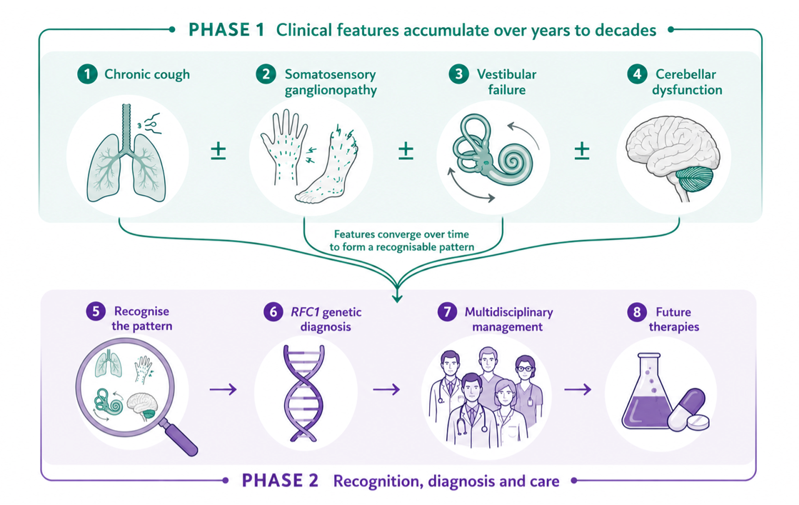
Cerebellar ataxia, neuropathy and vestibular areflexia syndrome (CANVAS) and the wider family of RFC1-related diseases are among the most common causes of adult-onset hereditary ataxia, yet diagnosis is frequently delayed by decades. This Patient Perspective combines the historical evolution of clinical understanding with a first-person patient account to illustrate why the disorder is so often missed, and to offer a structured, clinically usable approach to recognition, investigation and multidisciplinary management. We argue that no single symptom is diagnostic; rather, the diagnosis emerges from recognising phenotypic patterns, which in the case of CANVAS are sensory ganglionopathy, bilateral vestibular hypofunction and cerebellar impairment. This triad may accumulate over many years and is often attributed by patients and clinicians alike to unrelated, more common conditions. A chronic cough generally predates the onset of the other features. Practical diagnostic pearls, a stepwise investigation algorithm and a multidisciplinary management framework are presented alongside the patient’s own account of a three-decade diagnostic journey, to help shorten the path to diagnosis for future patients.
Keywords:
CANVAS
; RFC1
; cerebellar ataxia
; bilateral vestibulopathy
; sensory ganglionopathy
; chronic cough
; diagnostic delay
; patient perspective
1. Plain Language Summary
CANVAS is a slowly progressive neurological condition caused by changes in a gene called RFC1. It affects three parts of the body’s balance mechanisms, the balance organs of the inner ear, the coordination centre of the brain, and information from the feet on position and touch, that travels back to the brain. The brain combines this information to keep us balanced and to keep our vision steady when we move. Because these systems normally back one another up, problems can stay hidden for years, and the first sign is often something that seems unrelated, for example a long-standing dry cough that can begin decades before any balance problem. Symptoms then build up so gradually, and seem so ordinary, that patients and doctors tend to explain each one on its own rather than seeing them as a single condition. This often leads to many years of tests and doctor’s visits before the diagnosis is made. In this article, a patient describes her own thirty-year journey, from an unexplained cough, to numbness and difficulty reading signs while walking, to the eventual diagnosis. Alongside her account, we explain the clues that should prompt clinicians to consider RFC1-related disease sooner, how it is confirmed with brain, nerve, balance and genetic tests. There is no cure yet, but earlier diagnosis spares unnecessary tests, guides management and family planning, and opens the door to research.
2. Introduction
“For many years I believed I had a collection of unrelated problems. It never occurred to me that they were all manifestations of a single disease.”
This observation captures a material challenge in recognising complex diseases such as cerebellar ataxia, neuropathy and bilateral vestibular areflexia (CANVAS). CANVAS is now known to be one of an increasing number of RFC1-related disorders [1,2,3]. Even in the case of CANVAS, patients do not necessarily present initially with the classical triad of cerebellar ataxia, bilateral vestibular hypofunction and somatosensory ganglionopathy [2]. Symptoms may emerge insidiously over decades, potentially affecting disparate neurological systems at different times. Chronic cough not uncommonly precedes imbalance by decades [4], somatosensory symptoms may initially appear trivial, and oscillopsia for example may be attributed to deteriorating eyesight [5,6]. Both patients and clinicians are therefore at risk of attempting to explain each symptom individually rather than recognising the gradual evolution of a multifocal neurological disease.
This pattern of presentation contributes directly to the prolonged diagnostic delay that continues to characterise RFC1-related disease [7]. Patients may consult respiratory physicians for chronic cough, orthopaedic surgeons for gait disturbance, ophthalmologists for visual symptoms, otolaryngologists for dizziness and neurologists for neuropathy before the relationship between these apparently unrelated difficulties becomes apparent. In practice, the diagnostic odyssey often spans multiple specialties [7], with each clinician reasonably focused on the organ system that is more obviously affected which lies within their own expertise. The cumulative effect is a fragmented record of isolated encounters rather than a single coherent narrative, and it is precisely this fragmentation that the clinician must learn to minimise.
The history of this disorder mirrors the experience of many patients. The syndrome now recognised as CANVAS was not defined until the early twenty-first century, when careful clinical observation identified the characteristic combination of cerebellar dysfunction, somatosensory ganglionopathy and bilateral vestibular failure [6,8]. Subsequent advances in vestibular physiology, quantitative eye movement assessment and neurophysiology established that these seemingly disparate abnormalities represented a single clinicopathological entity [5,9,10]. The discovery in 2019 that biallelic intronic AAGGG repeat expansions in gene encoding Replication Factor C Subunit 1 (RFC1) cause CANVAS transformed understanding of the disease, providing a molecular explanation for one of the most common causes of adult-onset hereditary ataxia [1,11]. Importantly, it also revealed that the clinical spectrum extends well beyond the classical CANVAS phenotype to encompass isolated cerebellar ataxia, chronic refractory cough and a range of overlapping neurological presentations [2,4,12].
Although molecular diagnosis has become increasingly accessible, careful clinical phenotyping remains fundamental: genetic testing confirms the diagnosis, but less commonly suggests it. Recognition still depends upon identifying a distinctive pattern of progressive dysfunction affecting the somatosensory, vestibular and cerebellar systems. No individual symptom is diagnostic. Rather, the diagnosis emerges when the apparently disconnected features of a patient’s history are viewed collectively. This process may be confounded by a misunderstanding around the specificity of various signs and symptoms; including the assumption that dizziness reflects vestibular dysfunction when in fact it is a not uncommon feature of cerebellar disease [13], or that a broad based gait may be seen in cerebellar, vestibular or somatosensory disease. Similarly, motion-induced oscillopsia does not differentiate between vestibular and cerebellar impairment [5]. Genetic confirmation remains mandatory, as this triad of abnormalities may be variably seen in other inherited diseases, including SCA27B, SCA3 and Friedreich ataxia [14,15,16].
This distinction is, however, important because RFC1-related disease is fundamentally different from many inherited ataxias. The disability experienced by patients is not simply the consequence of cerebellar degeneration. Instead, it reflects progressive failure of the three principal systems responsible for maintaining balance, coordination and stable vision: somatosensory, vestibular and cerebellar function. Whilst not exclusively characteristic, the interaction between these systems explains many of the characteristic features of the disorder, including severe imbalance in darkness, marked motion-induced oscillopsia, impaired spatial orientation and yet, a surprising degree of compensation. The latter possibly attributable to the relatively slow tempo of progression in CANVAS. This apparent paradox, profound physiological deficits accompanied by a degree of preserved everyday function, at least early in the disease, is itself an important clinical clue, and one that can mislead the unwary examiner into underestimating the severity of underlying dysfunction.
Traditional review articles have understandably focused on the rapidly expanding genetic and phenotypic spectrum of RFC1-related disease. While these have greatly improved scientific understanding, they often fail to capture the experience of patients and the practical clinical lessons that emerge from their diagnostic journey. Conversely, patient narratives alone rarely explain why apparently minor symptoms such as chronic cough or difficulty reading street signs while walking, should immediately prompt consideration of a multisystem neurological disorder.
In this perspective, we combine a patient’s experience with the evolving history of CANVAS and other RFC1-related disease to illustrate how the disorder unfolds over time. Rather than providing an exhaustive review of the literature, we focus on the practical clinical observations that shorten diagnostic delay and improve patient care. The historical evolution of scientific understanding is woven into the patient’s own journey, highlighting how advances in clinical observation, vestibular physiology and molecular genetics have progressively transformed one of neurology’s most challenging diagnoses. The remainder of this paper is organised around the practical questions a clinician must answer in sequence: when should the diagnosis be suspected; how is it confirmed; and how should the patient then be managed..
3. Patient’s Journey
3.1. A Cough That Seemed to Mean Nothing
I am writing to share a patient perspective that may assist clinicians in recognizing the often subtle and protracted presentation of CANVAS (Cerebellar Ataxia, Neuropathy, and Vestibular Areflexia Syndrome) associated with RFC1 repeat expansion disorder. When I look back over the past three decades, what strikes me most is not how dramatic my symptoms were, but how ordinary they seemed at the time. Each new problem appeared sufficiently common to have its own explanation. None seemed serious enough to suggest a progressive neurological disease and, more importantly, none appeared to be related to the others.
The first symptom was a persistent dry cough that developed in the early 1990s. Initially it was little more than an irritation. It would appear unpredictably, often in prolonged bouts that were socially embarrassing but never severe enough to interfere substantially with my daily life. Like many patients with chronic cough, I sought medical advice on several occasions. Various explanations were proposed, including allergy, upper airway irritation and gastro-oesophageal reflux. None proved convincing, but equally there was nothing to suggest that the cough might represent the earliest manifestation of a neurological disorder.
In retrospect, this chronology is entirely consistent with what is now recognised about CANVAS. Chronic cough frequently precedes neurological symptoms by many years and, in some patients, by several decades [4]. Yet its significance is almost impossible to appreciate prospectively because chronic cough is so common in the general population. The lesson is therefore not that every patient with chronic cough requires neurological investigation, but that a remote history of otherwise unexplained chronic cough should assume new importance once somatosensory, vestibular or cerebellar symptoms emerge.
For many years nothing else appeared to happen. Life continued normally. Looking back, I suspect this prolonged asymptomatic interval contributed as much to the eventual diagnostic delay as the rarity of the disorder itself. By the time neurological symptoms appeared, the cough had become such an accepted part of everyday life that neither I nor my doctors considered it relevant.
The first unmistakable neurological symptoms emerged more than twenty years later. Around 2014 I noticed intermittent tingling affecting my feet. Initially the sensation was mild and transient, but over time it became persistent and gradually extended proximally towards my knees. At about the same time I realised that facial sensation had subtly changed. On several occasions I became aware that food remained around my mouth after meals because I had not felt it. These symptoms were sufficiently mild that they seemed more curious than concerning, yet together they represented the first clear evidence of a developing somatosensory ganglionopathy.
Walking also began to change, although so gradually that I barely noticed it. Narrow footpaths demanded increasing concentration. Climbing stairs while carrying a basket of washing became unexpectedly awkward because I could no longer divide my attention between the object I was carrying and the position of my feet. Reading street signs while walking became surprisingly difficult because the words appeared to bounce with each step. I assumed I needed stronger glasses.
Only much later did I learn that this symptom had a name — motion-induced oscillopsia. Like many patients with this symptom, I had interpreted a neurological symptom as a visual problem.
3.2. Recognising the Pattern in Hindsight: The Clinical Phenotype of RFC1-Related Disease
The patient’s account illustrates a principle that is central to recognising RFC1-related disease in practice: the diagnosis is rarely suggested by a single abnormality, but by the accumulation, over years, of findings in three interacting domains (somatosensory, vestibular and cerebellar), frequently preceded by a remote history of chronic cough [2,4]. Each domain, considered alone, is common and non-specific. It is the combination, and particularly the way these features accumulate over a long and otherwise unremarkable interval, that should prompt consideration of the diagnosis.
Somatosensory involvement reflects a non-length-dependent somatosensory ganglionopathy, affecting the dorsal root and cranial sensory ganglia rather than the nerve itself [10,17]. Patients typically describe patchy paraesthesia, numbness or a sense of imbalance that is disproportionately worse in the dark or with the eyes closed. Facial numbness, or the unnoticed retention of food around the mouth, as described here, may indicate trigeminal ganglion involvement and is easily overlooked unless specifically sought [2].
Vestibular involvement is characteristically bilateral and symmetrical and profoundly affected, distinguishing it from unilateral vestibular disorders such as vestibular neuritis [18,19]. Because the vestibulopathy is bilateral, patients rarely experience vertigo per se, recalling that vertigo of a vestibular origin is generated by asymmetric vestibular output. Instead, the dominant symptoms are gait unsteadiness and motion-induced oscillopsia, a perceived jumping or bouncing of the visual scene during head movement, because the vestibulo-ocular reflex can no longer stabilise gaze [5]. This is often first noticed, as in this account, as difficulty reading text or signage while walking, and is very frequently misattributed to a refractive error rather than a vestibular one (or in the case of RFC1-related disease related to both vestibular and cerebellar dysfunction).
Cerebellar involvement contributes gait and limb ataxia, dysarthria and characteristic ocular motor abnormalities, including gaze-evoked nystagmus, impaired smooth pursuit and dysmetric saccades, and the resulting spontaneous and motion-induced oscillopsia [8]. Cerebellar signs may be relatively mild early in the disease, and brain imaging can be deceptively unremarkable, particularly in the first years after neurological symptoms appear [2]. Clinicians should not be reassured by a normal or near-normal MRI in a patient with a compatible history and examination.
A further, frequently overlooked, domain is chronic cough, which precedes neurological symptoms in a substantial proportion of patients, sometimes by several decades, as in the case presented here [4]. The mechanism is thought to relate to somatosensory ganglionopathy affecting the vagal ganglia, producing an irritable, hypersensitive cough reflex [20]. Because chronic cough is extremely common in the general population and usually benign, its neurological significance is often undetectable prospectively; it only becomes meaningful retrospectively, once other features of the syndrome emerge. Autonomic symptoms, including constipation, urinary urgency and orthostatic intolerance, may also occur but are often not volunteered unless specifically asked about [2]. Table 1 summarises the principal clinical manifestations across each affected system, together with the specific clues that help distinguish RFC1-related disease from more common alternative explanations for each symptom in isolation.
3.3. Why Patients Become Disabled: Convergent Failure of Three Systems
A useful conceptual model is that disability in RFC1-related disease arises from the interaction of the somatosensory, vestibular and cerebellar dysfunction rather than from failure of any single system [2]. Under normal circumstances, these three systems are usually involved in compensatory mechanisms: vision and proprioception can partially substitute for vestibular loss, and vestibular input can partially substitute for proprioceptive loss. When all three systems degrade simultaneously, however, this redundancy is lost. The result is a disproportionate, and sometimes surprising, degree of instability, particularly in situations that remove visual compensation — walking in the dark, at night, or on uneven ground.
This explains several otherwise puzzling clinical observations [13]. Patients often function reasonably well in well-lit, familiar environments, where vision compensates for the failing vestibular and proprioceptive systems, yet report dramatic worsening in the dark, in unfamiliar surroundings, or when vision is otherwise unavailable, such as when closing the eyes to wash the face. Similarly, patients frequently describe a paradoxical worsening of imbalance when attention is divided, such as carrying an object while climbing stairs, because postural control that would normally be automatic has become dependent on conscious visual and cognitive monitoring.
3.4. A Practical Diagnostic Approach
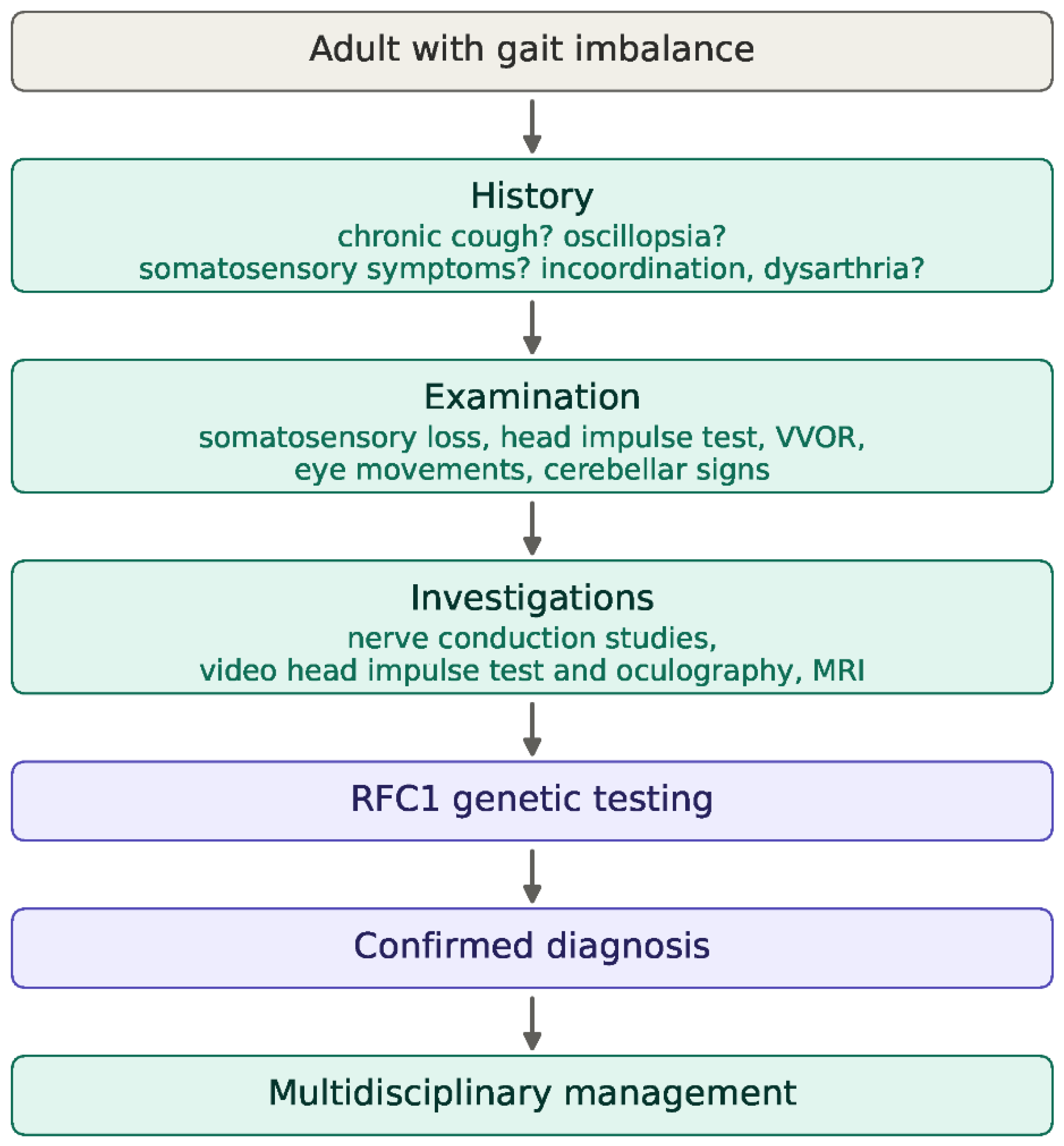
Given the insidious, potentially multi-focal neurological evolution of RFC1-related disease, a structured but pragmatic diagnostic pathway is more useful than reliance on a single discriminating test. The pathway set out in Figure 1 begins, deliberately, with history rather than investigation, because the key diagnostic clues, chronic cough, oscillopsia, darkness-dependent imbalance and patchy somatosensory symptoms, are elicited by targeted questioning in the first instance.
3.4.1. History
Four questions are disproportionately useful. First, has the patient ever experienced a chronic, otherwise unexplained non-productive cough, even many years earlier? [4] Second, does the patient have difficulty reading signs, text messages or faces while walking or moving, a simple screen for motion-induced oscillopsia? [5] Third, is imbalance substantially worse in the dark, in the shower with the eyes closed, or at night? [2] Fourth, are there patchy somatosensory symptoms, including altered facial sensation or unnoticed food around the mouth, limb numbness or neuropathic pain [10]? A positive answer to any of these, particularly in combination, should prompt a focused examination directed at the somatosensory, vestibular and cerebellar systems. It should be noted however that while useful, none of these factors are specific for CANVAS/RFC1-related disease. The features that, in combination, should raise suspicion, spanning history, examination and disease course, and the reason each is discriminating, are summarised in Table 2.
3.4.2. Examination
Ocular motor examination should assess visual pursuit, gaze-evoked nystagmus and saccade to target metrics. The bedside head impulse test is one of the highest-yield components of the examination: bilateral corrective catch-up saccades in vestibular hypofunction are one of the most sensitive findings in all medical examination [9,21]. Examination should assess light touch, pin prick, vibration and joint position sense, specifically looking for patchy loss and so, testing should be continued even if a ‘sensory level’ is thought to be identified as it may represent a patch of retained sensation. Reliance on the sensory component of the examination is not advisable as it is the least robust component of the neurological bedside examination, and both false negative and positive results are found when compared to nerve conduction studies [17]. It may be worthwhile cautioning against relying on the utility of a positive Romberg sign. Although originally described as a test of sensory loss, the Romberg’s test may be positive in any combination of cerebellar, vestibular and/or sensory loss [13].
3.4.3. Investigations
Nerve conduction studies demonstrate a somatosensory ganglionopathy, with absent or markedly reduced somatosensory responses and relatively preserved motor conduction, in a non-length dependent fashion, a pattern that should prompt consideration of a ganglionopathy rather than a length-dependent peripheral neuropathy [10,17]. Less common sensory and/or motor abnormalities have been identified in other RFC1-related phenotypes [2]. Video head impulse testing provides objective, quantitative confirmation of bilateral reduction in vestibulo-ocular reflex gain [9]. Video-oculography interrogating eye movements including the VVOR are of particular diagnostic value [5]. Brain MRI may show cerebellar atrophy but particularly early in the disease course may be normal and a normal scan should not be used to exclude the diagnosis [2]. Because this triad of somatosensory, vestibular and cerebellar involvement is not unique to RFC1-related disease and can be seen, in varying combinations, in other late-onset inherited ataxias, genetic confirmation remains essential; the conditions that most often enter the differential, and the features that help separate them from RFC1-related disease, are summarised in Table 3.
4. Genetic Confirmation
In most patients, RFC1-related disease is caused by a biallelic intronic repeat expansion. The reference (AAAAG)₁₁ configuration in intron 2 is replaced on both alleles by a large (AAGGG)ₙ expansion, typically several hundred to a few thousand repeats [1,11]. Non-canonical pathogenic motifs such as ACAGG, described in Asia-Pacific families, are increasingly recognised [22,23], and a minority of patients carry one expanded allele together with a loss of function RFC1 variant in trans [24]. A motif- and expansion-aware testing strategy is therefore preferable to one assuming a single canonical repeat, and carriers of a heterozygous pathogenic (AAGGG)ₙ expansion should subsequently be screened for loss of function alleles.
Inheritance is autosomal recessive, with heterozygous carrier frequencies ranging from 0.7% to 6.5% across populations [3]. Because onset is late and the phenotype may go unrecognised, patients frequently have no affected relatives, so a negative family history does not argue against the diagnosis. Gene panels, exome sequencing and conventional short-read genome variant-calling are not designed to genotype large or complex repeats, so standard diagnostic testing pipelines do not report the expansion [27], which is deep intronic and often very large. Locus-specific testing is therefore required. Flanking (across-the-repeat) PCR infers a pathogenic allele from the absence of a normal-sized amplicon, but cannot size large expansions and cannot reliably distinguish a true biallelic expansion from failure of one allele to amplify (allelic dropout). Repeat-primed PCR detects a given repeat motif from its characteristic sawtooth ladder, but is motif-specific and can yield ambiguous or false-negative results when a non-canonical motif is present [22,23]. Long-read sequencing increasingly complements or replaces PCR-based testing, potentially resolving repeat length, motif composition and interruptions in one assay [28]. Genotype–phenotype correlation remains limited: the same biallelic AAGGG expansion underlies presentations ranging from isolated chronic cough or isolated sensory neuronopathy to the full CANVAS triad and a broader multisystemic spectrum, so genotype cannot yet predict phenotype or prognosis [2].
Taken together, the recognition, examination, investigation and confirmation steps set out above are distilled into a set of practical clinical pearls (Table 4).
6. Historical Evolution of Clinical and Scientific Understanding
The patient’s own three-decade journey mirrors, almost exactly, the trajectory by which medical understanding of this disorder developed. Isolated clinical observations of patients with an unusual combination of imbalance, somatosensory loss and bilateral vestibular failure accumulated through the 1990s and 2000s, without a unifying explanation. The formal characterisation of CANVAS as a distinct clinical syndrome occurred in 2011 [6,8,34]. Over the subsequent decade, refinements in vestibular physiology, video head impulse testing and quantitative ocular motor assessment strengthened bedside and laboratory diagnostic confidence [5,9,19], well before the underlying genetic cause was known.
The identification of biallelic RFC1 repeat expansions in 2019 [1,11] represented a turning point comparable to the discovery of the expanded triplet repeat in Friedreich ataxia a generation earlier: a single molecular finding that explained a syndrome previously defined only by its clinical features [16]. In the years since, the phenotypic spectrum recognised to be associated with RFC1 has expanded considerably beyond classical CANVAS, to include isolated chronic cough [4], isolated bilateral cerebellar ataxia [12] and broader multifocal neurological phenotypes, some of which overlap with other late-onset ataxias including idiopathic late onset cerebellar ataxia (ILOCA) and idiopathic cerebellar ataxia and bilateral vestibulopathy (iCABV) [35].
7. Looking Forward: From Recognition to Precision Medicine
The journey presented here spans more than two decades. During that period the understanding of RFC1-related disease evolved from isolated clinical observations to recognition of a distinctive neurological syndrome and, ultimately, to identification of its molecular basis. Few neurological disorders have undergone such rapid conceptual transformation. Yet the greatest challenge has not changed. Patients continue to experience prolonged delays before receiving a diagnosis, despite the availability of highly accurate molecular testing [7].
This apparent paradox reflects an important principle in modern neurology. Technological advances do not replace clinical reasoning; they increase its value. The discovery of the repeat expansion in RFC1 has undoubtedly simplified diagnostic confirmation, but molecular testing remains dependent upon recognising the phenotype in the first place. For this reason, detailed clinical observation continues to underpin effective neurological practice, even within an era increasingly dominated by genomic medicine.
The expanding phenotypic spectrum of RFC1-related disease also challenges traditional approaches to neurological classification. Historically, disorders were categorised according to the system appearing most obviously affected: hereditary ataxias, peripheral neuropathies or vestibular disorders. RFC1-related disease does not fit comfortably within these boundaries. Patients may initially present to respiratory physicians with chronic cough, to otolaryngologists with imbalance, to ophthalmologists because of oscillopsia or to neurologists because of somatosensory ganglionopathy. The disease therefore illustrates the limitations of considering neurological systems independently when, in reality, they function as components of an integrated network responsible for maintaining posture, gaze and coordinated movement.
Recognising this broader concept has implications extending beyond RFC1-related disease itself. Many neurodegenerative disorders involve multiple interacting neural systems, yet clinical practice often fragments their assessment according to subspecialty interests. The patient’s journey described here demonstrates the importance of reconstructing symptoms longitudinally rather than considering each consultation in isolation. A history obtained over several decades may reveal a coherent disease process that is invisible when individual symptoms are viewed independently.
The next phase of research will increasingly focus on understanding disease mechanisms rather than simply defining phenotypes. Although the pathogenic repeat expansion has been identified [1,11], the biological consequences of RFC1 dysfunction remain incompletely understood. Unusually for a repeat-expansion disorder, the pathogenic expansion has not been shown to reduce RFC1 expression or to generate the RNA foci or repeat-associated non-AUG translation products seen in comparable diseases, and non-pathogenic expansions of similar size occur in healthy individuals [3]. The possibility that the repeat motif, rather than its length, drives pathogenicity is also supported by patient-derived induced pluripotent stem cell neuronal models [36]. Whether the disease arises through a conditional loss of function or a motif-dependent toxic mechanism therefore remains unresolved [37]. Why somatosensory and cranial ganglia (including vestibular, facial and trigeminal), demonstrate such striking vulnerability, why chronic cough precedes neurological symptoms by many years [4] and why clinical severity varies substantially between individuals remain unanswered questions [2]. Addressing these issues will require integration of molecular biology, disease modelling, neuropathology, neurophysiology and careful longitudinal clinical phenotyping.
Progress towards disease-modifying therapy will depend upon equally rigorous natural history studies [2]. Reliable biomarkers capable of detecting progression over relatively short time intervals remain a priority [38]. Quantitative vestibular assessment [9], instrumented gait analysis, wearable sensor technology, digital speech analysis and objective ocular motor measurements [5,39,40] all offer considerable promise. Importantly, many of these approaches directly reflect the physiological systems responsible for disability and may therefore prove more sensitive than conventional clinical rating scales, as has already been demonstrated for quantitative neuroimaging in RFC1-related disease and for digital gait measures in other hereditary ataxias [40,41]. Development of such biomarkers is essential for future therapeutic trials.
At the same time, advances in genomic technology continue to reshape diagnosis. Because the expansion can be detected in standard short-read whole-genome and even whole-exome data using dedicated repeat-detection algorithms, the increasing use of genome sequencing in routine practice raises the prospect of identifying RFC1-related disease opportunistically. This has the potential to shorten the diagnostic delay that has characterised the condition [3,27]. Long-read sequencing is already providing greater resolution of repeat structure than conventional repeat-primed PCR-based testing [22,28] and resolving repeat motif and interruption structure rather than length alone may help clarify the molecular determinants of the phenotype [1,3]. Targeted long-read approaches can also genotype many repeat-expansion loci in parallel in a single assay, offering a unified route through the genetically heterogeneous late-onset ataxias rather than sequential single-gene tests [28]. The discovery of a second, unrelated late-onset ataxia repeat-expansion disorder, the intronic FGF14 GAA expansion underlying spinocerebellar ataxia 27B, illustrates how rapidly this landscape is evolving [14,42] and how careful genotype–phenotype correlation across these overlapping disorders will remain essential. These technologies will almost certainly reveal additional pathogenic repeat configurations and improve understanding of repeat instability, although their widespread clinical implementation will require careful validation [22].
For patients, however, the most immediate priority remains considerably simpler. Earlier diagnosis provides reassurance, avoids unnecessary investigations, facilitates appropriate rehabilitation, symptom management, family planning (including the option of pre-implantation diagnosis) and enables future care planning [7,29]. It also allows participation in research at a time when therapeutic development is accelerating rapidly. As the field moves towards interventional studies, reducing diagnostic delay will become increasingly important, ensuring that patients are identified before advanced disability has developed.
Every clinician who encounters adults with progressive gait imbalance, cerebellar impairment and/or somatosensory loss, or otherwise unexplained chronic cough can shorten what has historically been a prolonged diagnostic journey.
8. Conclusions
The patient’s experience illustrates both the evolution of RFC1-related disease and the evolution of neurological understanding itself. Symptoms accumulated gradually over many years, yet each appeared sufficiently common to encourage an isolated explanation. Only when chronic cough, somatosensory ganglionopathy, bilateral vestibular dysfunction and cerebellar impairment were viewed collectively did the diagnosis become apparent. The history of scientific discovery followed a remarkably similar trajectory, progressing from recognition of individual clinical observations to definition of CANVAS as a distinct syndrome and finally to identification of the underlying RFC1 repeat expansion [1,6,8,11].
The principal lesson is therefore one of clinical pattern recognition. RFC1-related disease should be considered whenever there is a history of progressive imbalance, particularly when symptoms evolve over many years. Careful neurological examination, supported by neurophysiology, formal vestibular and oculomotor assessment and targeted molecular testing, remains the most effective diagnostic strategy [2,9,17].
Accurate diagnosis has immediate practical value even in the absence of a disease-modifying therapy. It facilitates multidisciplinary rehabilitation, guides genetic counselling, reduces unnecessary investigations and enables participation in the rapidly expanding international research effort directed towards understanding disease mechanisms and developing disease-modifying therapies [29].
Perhaps the most enduring message comes from the patient rather than the clinician. Looking backwards, the diagnosis seems almost obvious because every important clue was present. Looking forwards, the challenge is to recognise those same clues while the disease is still unfolding. If greater awareness of RFC1-related disease allows clinicians to identify this characteristic pattern earlier, future patients may spend fewer years searching for an explanation and more years benefiting from informed multidisciplinary care and, ultimately, effective targeted therapies.
Author Contributions
KB, KB and NP provided the lived-experience account. KB and NP conceived and drafted the article. PJL and DS contributed the genetic, clinical and scientific content.
Informed Consent Statement
All authors provided written Consent to Publish, and all authors approved the final version. The Royal Children’s Hospital Human Research Ethics Committee (HREC #28097) approved the study.
Data Availability Statement
All data associated with this manuscript are available within the document.
Acknowledgments
Citation checking, formatting for this manuscript and graphical abstract generation were AI-assisted (Claude Opus 4.8).
Conflicts of Interest
The authors declare no competing interests.
References
- Cortese, A.; Simone, R.; Sullivan, R.; Vandrovcova, J.; Tariq, H.; Yan, Y.W.; et al. Biallelic expansion of an intronic repeat in RFC1 is a common cause of late-onset ataxia. Nat. Genet. 2019, 51(4), 649–58. [Google Scholar] [CrossRef] [PubMed]
- Traschutz, A.; Cortese, A.; Reich, S.; Dominik, N.; Faber, J.; Jacobi, H.; et al. Natural History, Phenotypic Spectrum, and Discriminative Features of Multisystemic RFC1 Disease. Neurology 2021, 96(9), e1369–e82. [Google Scholar] [CrossRef] [PubMed]
- Davies, K.; Szmulewicz, D.J.; Corben, L.A.; Delatycki, M.; Lockhart, P.J. RFC1-Related Disease: Molecular and Clinical Insights. Neurol. Genet. 2022, 8(5), e200016. [Google Scholar] [PubMed]
- Garvey, A.; Melville, I.Z.; Scriba, C.K.; Yong, V.; Rodrigues, M.; Kao, J.; et al. Nerve ultrasound, neuronopathy and cough predict sensory neuropathy patients with RFC1 expansions. Brain Commun. 2025, 7(6), fcaf434. [Google Scholar] [CrossRef] [PubMed]
- Halmagyi, G.M.; Kumar, K.; McGarvie, L.A. The visually enhanced vestibulo-ocular reflex in CANVAS. J. Neurol. 2022, 269(1), 490–2. [Google Scholar] [CrossRef] [PubMed]
- Szmulewicz, D.J.; Waterston, J.A.; MacDougall, H.G.; Mossman, S.; Chancellor, A.M.; McLean, C.A.; et al. Cerebellar ataxia, neuropathy, vestibular areflexia syndrome (CANVAS): a review of the clinical features and video-oculographic diagnosis. Ann. N Y Acad. Sci. 2011, 1233, 139–47. [Google Scholar] [CrossRef] [PubMed]
- Faye, F.; Crocione, C.; Anido de Pena, R.; Bellagambi, S.; Escati Penaloza, L.; Hunter, A.; et al. Time to diagnosis and determinants of diagnostic delays of people living with a rare disease: results of a Rare Barometer retrospective patient survey. Eur. J. Hum. Genet. 2024, 32(9), 1116–26. [Google Scholar] [CrossRef] [PubMed]
- Szmulewicz, D.J.; Roberts, L.; McLean, C.A.; MacDougall, H.G.; Halmagyi, G.M.; Storey, E. Proposed diagnostic criteria for cerebellar ataxia with neuropathy and vestibular areflexia syndrome (CANVAS). Neurol. Clin. Pract. 2016, 6(1), 61–8. [Google Scholar] [CrossRef] [PubMed]
- MacDougall, H.G.; Weber, K.P.; McGarvie, L.A.; Halmagyi, G.M.; Curthoys, I.S. The video head impulse test: diagnostic accuracy in peripheral vestibulopathy. Neurology 2009, 73(14), 1134–41. [Google Scholar] [PubMed]
- Szmulewicz, D.J.; McLean, C.A.; Rodriguez, M.L.; Chancellor, A.M.; Mossman, S.; Lamont, D.; et al. Dorsal root ganglionopathy is responsible for the sensory impairment in CANVAS. Neurology 2014, 82(16), 1410–5. [Google Scholar] [CrossRef] [PubMed]
- Rafehi, H.; Szmulewicz, D.J.; Bennett, M.F.; Sobreira, N.L.M.; Pope, K.; Smith, K.R.; et al. Bioinformatics-Based Identification of Expanded Repeats: A Non-reference Intronic Pentamer Expansion in RFC1 Causes CANVAS. Am. J. Hum. Genet. 2019, 105(1), 151–65. [Google Scholar] [CrossRef] [PubMed]
- Montaut, S.; Diedhiou, N.; Fahrer, P.; Marelli, C.; Lhermitte, B.; Robelin, L.; et al. Biallelic RFC1-expansion in a French multicentric sporadic ataxia cohort. J. Neurol. 2021, 268(9), 3337–43. [Google Scholar] [CrossRef] [PubMed]
- Feil, K.; Strobl, R.; Schindler, A.; Krafczyk, S.; Goldschagg, N.; Frenzel, C.; et al. What Is Behind Cerebellar Vertigo and Dizziness? Cerebellum 2019, 18(3), 320–32. [Google Scholar] [CrossRef] [PubMed]
- Rafehi, H.; Read, J.; Szmulewicz, D.J.; Davies, K.C.; Snell, P.; Fearnley, L.G.; et al. An intronic GAA repeat expansion in FGF14 causes the autosomal-dominant adult-onset ataxia SCA50/ATX-FGF14. Am. J. Hum. Genet. 2023, 110(1), 105–19. [Google Scholar] [CrossRef] [PubMed]
- Paulson, H.; Shakkottai, V. Spinocerebellar Ataxia Type 3; Adam, M.P., Bick, S., Mirzaa, G.M., Pagon, R.A., Wallace, S.E., Amemiya, A., Eds.; GeneReviews((R)): Seattle (WA), 1993. [Google Scholar]
- Corben, L.A.; Collins, V.; Milne, S.; Farmer, J.; Musheno, A.; Lynch, D.; et al. Clinical management guidelines for Friedreich ataxia: best practice in rare diseases. Orphanet J. Rare Dis. 2022, 17(1), 415. [Google Scholar] [CrossRef] [PubMed]
- Gwathmey, K.G. Sensory neuronopathies. Muscle Nerve 2016, 53(1), 8–19. [Google Scholar] [CrossRef] [PubMed]
- Ishai, R.; Seyyedi, M.; Chancellor, A.M.; McLean, C.A.; Rodriguez, M.L.; Halmagyi, G.M.; et al. The Pathology of the Vestibular System in CANVAS. Otol. Neurotol. 2021, 42(3), e332–e40. [Google Scholar] [CrossRef] [PubMed]
- Strupp, M.; Kim, J.S.; Murofushi, T.; Straumann, D.; Jen, J.C.; Rosengren, S.M.; et al. Bilateral vestibulopathy: Diagnostic criteria Consensus document of the Classification Committee of the Barany Society. J. Vestib. Res. 2017, 27(4), 177–89. [Google Scholar] [CrossRef] [PubMed]
- Guilleminault, L.; Mazzone, S.B.; Chazelas, P.; Frachet, S.; Lia, A.S.; Magy, L. Cerebellar ataxia, neuropathy and vestibular areflexia syndrome: a neurogenic cough prototype. ERJ Open Res. 2024, 10(4). [Google Scholar] [CrossRef] [PubMed]
- Halmagyi, G.M.; Curthoys, I.S. A clinical sign of canal paresis. Arch. Neurol. 1988, 45(7), 737–9. [Google Scholar] [CrossRef] [PubMed]
- Scriba, C.K.; Stevanovski, I.; Chintalaphani, S.R.; Gamaarachchi, H.; Ghaoui, R.; Ghia, D.; et al. RFC1 in an Australasian neurological disease cohort: extending the genetic heterogeneity and implications for diagnostics. Brain Commun. 2023, 5(4), fcad208. [Google Scholar] [CrossRef] [PubMed]
- Davies, K.C.; Rafehi, H.; Fearnley, L.G.; Snell, P.; Gillies, G.; Field, T.A.; et al. Comprehensive Characterisation of the RFC1 Repeat in an Australian Cohort. Cerebellum 2025, 24(4), 111. [Google Scholar] [CrossRef] [PubMed]
- Davies, K.C.; Fearnley, L.G.; Snell, P.; Bourke, D.; Mossman, S.; Kyne, K.; et al. A multi-exon RFC1 deletion in a case of CANVAS: expanding the genetic mechanism of disease. J. Neurol. 2024, 271(12), 7622–7. [Google Scholar] [CrossRef] [PubMed]
- Stankovic, I.; Fanciulli, A.; Sidoroff, V.; Wenning, G.K. A Review on the Clinical Diagnosis of Multiple System Atrophy. Cerebellum 2023, 22(5), 825–39. [Google Scholar] [CrossRef] [PubMed]
- Sinnreich, M.; Klein, C.J.; Daube, J.R.; Engelstad, J.; Spinner, R.J.; Dyck, P.J. Chronic immune sensory polyradiculopathy: a possibly treatable sensory ataxia. Neurology 2004, 63(9), 1662–9. [Google Scholar] [PubMed]
- Chintalaphani, S.R.; Pineda, S.S.; Deveson, I.W.; Kumar, K.R. An update on the neurological short tandem repeat expansion disorders and the emergence of long-read sequencing diagnostics. Acta Neuropathol. Commun. 2021, 9(1), 98. [Google Scholar] [CrossRef] [PubMed]
- Stevanovski, I.; Chintalaphani, S.R.; Gamaarachchi, H.; Ferguson, J.M.; Pineda, S.S.; Scriba, C.K.; et al. Comprehensive genetic diagnosis of tandem repeat expansion disorders with programmable targeted nanopore sequencing. Sci. Adv. 2022, 8(9), eabm5386. [Google Scholar] [CrossRef] [PubMed]
- Cortese, A.; Reilly, M.M.; Houlden, H. RFC1 CANVAS / Spectrum Disorder; Adam, M.P., Bick, S., Mirzaa, G.M., Pagon, R.A., Wallace, S.E., Amemiya, A., Eds.; GeneReviews((R)): Seattle (WA), 1993. [Google Scholar]
- Meldrum, D.; Jahn, K. Gaze stabilisation exercises in vestibular rehabilitation: review of the evidence and recent clinical advances. J. Neurol. 2019, 266 (Suppl 1), 11–8. [Google Scholar] [CrossRef] [PubMed]
- Vogel, A.P.; Keage, M.J.; Johansson, K.; Schalling, E. Treatment for dysphagia (swallowing difficulties) in hereditary ataxia. Cochrane Database Syst. Rev. 2015, 2015(11), CD010169. [Google Scholar] [CrossRef] [PubMed]
- de Silva, R.N.; Vallortigara, J.; Greenfield, J.; Hunt, B.; Giunti, P.; Hadjivassiliou, M. Diagnosis and management of progressive ataxia in adults. Pract. Neurol. 2019, 19(3), 196–207. [Google Scholar] [CrossRef] [PubMed]
- Mastammanavar, V.S.; Kamble, N.; Yadav, R.; M, N.; Jain, S.; Kumar, K.; et al. Non-motor symptoms in patients with autosomal dominant spinocerebellar ataxia. Acta Neurol. Scand. 2020, 142(4), 368–76. [Google Scholar] [CrossRef] [PubMed]
- Szmulewicz, D.J.; Waterston, J.A.; Halmagyi, G.M.; Mossman, S.; Chancellor, A.M.; McLean, C.A.; et al. Sensory neuropathy as part of the cerebellar ataxia neuropathy vestibular areflexia syndrome. Neurology 2011, 76(22), 1903–10. [Google Scholar] [CrossRef] [PubMed]
- Szmulewicz, D.J. Combined central and peripheral degenerative vestibular disorders: CANVAS, idiopathic cerebellar ataxia with bilateral vestibulopathy (CABV) and other differential diagnoses of the CABV phenotype. Curr. Otorhinolaryngol. Rep. 2017, 5(3), 167–74. [Google Scholar] [CrossRef]
- Maltby, C.J.; Krans, A.; Grudzien, S.J.; Palacios, Y.; Muinos, J.; Suarez, A.; et al. AAGGG repeat expansions trigger RFC1-independent synaptic dysregulation in human CANVAS neurons. Sci. Adv. 2024, 10(36), eadn2321. [Google Scholar] [CrossRef] [PubMed]
- Benkirane, M.; Da Cunha, D.; Marelli, C.; Larrieu, L.; Renaud, M.; Varilh, J.; et al. RFC1 nonsense and frameshift variants cause CANVAS: clues for an unsolved pathophysiology. Brain 2022, 145(11), 3770–5. [Google Scholar] [CrossRef] [PubMed]
- Klockgether, T.; Ashizawa, T.; Brais, B.; Chuang, R.; Durr, A.; Fogel, B.; et al. Paving the Way Toward Meaningful Trials in Ataxias: An Ataxia Global Initiative Perspective. Mov. Disord. 2022, 37(6), 1125–30. [Google Scholar] [CrossRef] [PubMed]
- Di Rauso, G.; Castellucci, A.; Cavallieri, F.; Tozzi, A.; Fioravanti, V.; Monfrini, E.; et al. Speech, Gait, and Vestibular Function in Cerebellar Ataxia with Neuropathy and Vestibular Areflexia Syndrome. Brain Sci. 2023, 13(10). [Google Scholar] [CrossRef] [PubMed]
- Ilg, W.; Muller, B.; Faber, J.; van Gaalen, J.; Hengel, H.; Vogt, I.R.; et al. Digital Gait Biomarkers Allow to Capture 1-Year Longitudinal Change in Spinocerebellar Ataxia Type 3. Mov. Disord. 2022, 37(11), 2295–301. [Google Scholar] [CrossRef] [PubMed]
- Lobo, C.C.; Rezende, T.J.R.; Pimentel-Silva, L.R.; Jarola, G.M.; Santos, N.B.S.; da Silva Schmitt, G.; et al. Longitudinal Evaluation of Ataxia and Brain Structural Changes in RFC1-Related Disorder. Mov. Disord. Clin. Pract. 2025, 12(12), 2311–6. [Google Scholar] [CrossRef] [PubMed]
- Pellerin, D.; Danzi, M.C.; Wilke, C.; Renaud, M.; Fazal, S.; Dicaire, M.J.; et al. Deep Intronic FGF14 GAA Repeat Expansion in Late-Onset Cerebellar Ataxia. N Engl. J. Med. 2023, 388(2), 128–41. [Google Scholar] [CrossRef] [PubMed]
Figure 1.
Diagnostic pathway for RFC1-related disease (CANVAS). A phenotype-directed algorithm in which targeted history, examination and investigations precede confirmatory RFC1 repeat-expansion testing, leading to a confirmed diagnosis and multidisciplinary management. VVOR, visually enhanced vestibulo-ocular reflex.
Figure 1.
Diagnostic pathway for RFC1-related disease (CANVAS). A phenotype-directed algorithm in which targeted history, examination and investigations precede confirmatory RFC1 repeat-expansion testing, leading to a confirmed diagnosis and multidisciplinary management. VVOR, visually enhanced vestibulo-ocular reflex.

| System | Common manifestations | Clinical clues |
| Somatosensory | Paraesthesia, neuropathic pain, impaired proprioception, somatosensory ataxia | Non-length-dependent somatosensory loss |
| Vestibular | Motion-induced oscillopsia, unsteadiness, | Visual degradation whilst in motion; positive head impulse test |
| Cerebellar | Gait ataxia, limb ataxia, dysarthria, gaze-evoked nystagmus, impaired visual pursuit, dysmetric saccades to target, oscillopsia | Progressive imbalance despite relatively mild MRI changes early in disease; non-balance related features ie. those not related to sensory or vestibular dysfunction |
| Autonomic | Urinary urgency, erectile dysfunction, orthostatic intolerance, altered perspiration | Frequently overlooked unless specifically questioned |
| Non-neurological | Chronic non-productive cough | Often precedes neurological manifestations by years or decades |
| Clinical feature | Why it matters |
| Chronic non-productive cough | Often predates neurological disease by decades |
| Progressive somatosensory ganglionopathy | Particularly non-length-dependent involvement |
| Oscillopsia | Absent in pure somatosensory disease |
| Appendicular ataxia, central oculomotor abnormalities, cerebellar dysarthria | Absent in somatosensory and/or vestibular disease |
| Marked deterioration in darkness | Indicates significant dependence on visual input because cerebellar, vestibular and somatosensory systems are impaired |
| Bilateral abnormal head impulse test | Most sensitive bedside test of vestibular hypofunction |
| Abnormal VVOR | Associated with combined bilateral vestibular and cerebellar dysfunction |
| Slowly progressive adult-onset ataxia | Particularly when accompanied by somatosensory and/or vestibular features |
| Family history may be absent | Recessive inheritance not uncommonly obscures genetic diagnosis |
Table 3.
Differential Diagnosis.
| Disorder | Distinguishing features |
| RFC1-related disease | Somatosensory ± motor abnormalities on NCS ± bilateral vestibular failure ± cerebellar dysfunction ± chronic cough [2] |
| CANVAS | Classical triad of cerebellar, vestibular and somatosensory loss generally preceded by chronic cough [6,8] |
| Spinocerebellar ataxias | Dominant inheritance; variable phenotypes within and between diseases [15] |
| Friedreich ataxia | Generally childhood onset; possible cardiomyopathy; possible diabetes; possible auditory and/or optic neuropathy [16] |
| Multiple system atrophy (cerebellar type) | Relatively rapid progression; severe autonomic failure; characteristic MRI findings may be present [25] |
| Chronic immune somatosensory ganglionopathy | Often subacute onset; inflammatory or autoimmune markers; absence of cerebellar and generally absent vestibular involvement [26] |
| Bilateral vestibulopathy | Absence of somatosensory and cerebellar signs [19] |
Table 4.
Clinical Pearls.
| Pearl | Why it matters |
| Don’t think “ataxia”—think “multifocal balance disorder.” | Explains the phenotypes more accurately and supports a more inclusive diagnostic process [2] |
| Ask specifically about chronic cough. | Patients rarely volunteer its significance [4] |
| Examine eye movements carefully. | Ocular motor findings often precede obvious gait ataxia [5,8] |
| Test the bedside head impulse reflex. | Quickly and specifically identifies vestibular involvement [9,19] |
| Confirm somatosensory ganglionopathy with nerve conduction studies. | One of the strongest objective diagnostic clues to the CANVAS phenotype in particular [10,17] |
| Where available use genetics to confirm the diagnosis. | Phenotypes may be suggestive but are not specific [1,29] |
Disclaimer/Publisher’s Note: The statements, opinions and data contained in all publications are solely those of the individual author(s) and contributor(s) and not of MDPI and/or the editor(s). MDPI and/or the editor(s) disclaim responsibility for any injury to people or property resulting from any ideas, methods, instructions or products referred to in the content. |
© 2026 by the authors. Licensee MDPI, Basel, Switzerland. This article is an open access article distributed under the terms and conditions of the Creative Commons Attribution (CC BY) license (http://creativecommons.org/licenses/by/4.0/).
Copyright: This open access article is published under a Creative Commons CC BY 4.0 license, which permit the free download, distribution, and reuse, provided that the author and preprint are cited in any reuse.