

Submitted:
14 August 2026
Posted:
18 August 2026
You are already at the latest version
Abstract
Background/Objectives: Myeloproliferative neoplasms are a group of clonal myeloid malignancies that affect bone marrow and include polycythemia vera (PV), essential thrombocythemia (ET) and primary myelofibrosis (PMF). PV is caused in most of patients by the JAK2-V617F mutation and is characterized at phenotypic level by overproduction and accumulation of red blood cells. PV is associated with significant morbidity, including risk of thrombotic events and of hematologic evolution (myelofibrotic or leukemic transformation) and reduced survival. In addition to JAK2-V617F mutation, PV patients display additional molecular abnormalities. The aim of this study is to review recent studies investigating molecular abnormalities observed in PV. Methods: An extensive search of the most recent literature was performed, selecting and critically analyzing the most relevant studies. Results: The studies carried out in the last years have provided an extensive molecular characterization of PV, showing its heterogeneity, characterized in many patients by the presence of additional cytogenetic and non-driver gene mutations that contribute to the disease development and evolution. Conclusions: PV is a complex disease that needs to be carefully characterized at molecular level at diagnosis, to be monitored in time to predict the risk for thrombotic complications and hematologic evolution and to receive an adequate treatment.
Keywords:
myeloproliferative neoplasms
; polycythemia vera
; JAK2
; next generation sequencing
; thrombosis
; clonal hematopoiesis
1. Introduction
Myeloproliferative neoplasms (MPNs) are malignant clonal diseases of the hematopoietic system, characterized by the excessive production of mature blood cells. Concerning BCR-ABL negative MPNs, three main types of MPN exist, polycythemia vera (PV), essential thrombocytosis (ET) and myelofibrosis (MF). These three MPNs are caused by gain-of-function mutations in one of three different genes, JAK2, CALR and MPL. Particularly, JAK2-V617F is present in > 98% of PV patients and in about half of ET and MF patients [1,2,3]. The JAK2 mutation occurs in >95% of PV patients. Approximately half of these patients develop homozygosity for the JAK2 mutation, due to uniparental disomy on chromosome 9p, which is associated to a higher disease burden. The remaining 2-5% of PV patients carry mutations located to exon 12 of the JAK2 gene.
PV was first described by a French physician, Louis Henri Vaquez in 1892. The disease is characterized by overproduction of red blood cells (RBCs). The typical natural history of PV progresses through distinct phases; a preclinical phase initiated by the occurrent of a JAK2-V617F mutation at the level of an hemopoietic stem cell; this initial step is followed by a phase of expansion of the mutated clone which requires several decades and leads to the development of the clinically detectable PV disease; a chronic clinical phase, whose duration may arrive up to 30-35 years, if not interrupted by life-threating thrombotic events; in 10-20% of patients, the disease may undergo an hematologic transformation leading to myelofibrosis or acute leukemia transformation (Figure 1).
The fundamental mechanism through which the JAK2-V617F mutation causes an oncogenetic program involves a structural and biochemical cascade that determines the shift of JAK2 protein in a constitutively active state. The JAK2-V617F mutation consists in a single nucleotide substitution (G→T) at position 1849 replacing the amino acid valine (V) with phenylalanine (F) at codon 617 [1,2,3]. This mutation determines a loss of autoinhibition: normal JAK2 protein contains an active tyrosine kinase domain (JH1) and an adjacent, catalytically inactive pseudo kinase domain (JH2); the JH2 domain acts as an autoinhibitory brake, keeping baseline kinase activity low in the absence of stimulation by cytokines; the JAK2-V617 mutation destabilizes the JH2 domain breaking the autoinhibitory activity over the kinase domain JH1 [1,2,3]. At structural level, the mutant phenylalanine at position 617 causes strong π-π ring stacking interactions with other aromatic residues (such as F594 and F595 in the αC helix), thus stabilizing an active dimer conformation [4]. The functional consequence of the loss of autoinhibitory control of JAK2 activity is that JAK2 is constitutively activated and no longer strictly requires the binding of an extracellular ligand, such as erythropoietin (Epo) or thrombopoietin (Tpo) to initiate cell signaling [1,2,3]. The constitutive JAK2 activation determines the phosphorylation of its targets: particularly, hyperactive JAK2 continuously phosphorylates the cytoplasmic tails of cytokine receptors, with consequent recruitment and activation of signaling cascades, such as STAT, PI3K/AKT and RAS/MAPK. The continuous JAK-STAT pathway activation promotes uncontrolled proliferation and survival of myeloid and megakaryocyte progenitor cells; MAPK and PI3K/AKT pathway further stimulate cell cycle progression and promote cell survival by inhibiting apoptosis [5].
The molecular and biochemical mechanisms triggered by JAK2-V617F are responsible for several important clinical consequences that characterize MPN diseases. A first direct consequence clinically relevant is that JAK2-V617F-mutated stem cells gain a marked survival and proliferation advantage, leading to the abnormal accumulation of RBCs or thrombocytes. A second clinically relevant consequence is that mutant cell produce abnormal levels of proinflammatory cytokines (IL-6, TNF-α, IFN-α), contributing to the generation of an inflammatory microenvironment in the bone marrow that sustain tumor growth and symptom burden (fatigue, weight loss, bone marrow fibrosis) [6]. Furthermore, the JAK2-V617F mutation promotes thrombotic and vascular defects related to multiple mechanisms, involving increased clothing activity due to hyperactive platelets, hyperviscosity related in PV to overabundance of RBCs and to endothelial dysfunction promoting abnormal interaction between RBCs and the inner lining of blood vessels.
JAK2-V617F is a disease-driver in that is a causative event that can alone initiate, promote and maintain MPN disease without the need for additional cooperating mutations [7]. However, JAK2-V617F is also a clonal driver, in that it can be a marker of clonal hematopoiesis (CH), corresponding to a premalignant condition. In fact, Cordua et al. evaluated the frequency of individuals with JAK2-V617F mutations in a cohort of 19,558 individuals of the Danish population using a highly sensitive PCR method with a variant allele frequency (VAF) of 0.01%; on 19,958 probands, 613 (3.1%) carried JAK2-V617F and only 16 cases of previously unrecognized MPN were detected [8]. However, the large majority of individuals positive for JAK2-V617F mutation did not fulfill diagnostic criteria for MPN [8]. The rate of evolution from JAK2-V617F CH to MPN remains to be determined; however, it was evident that individuals with JAK2-V617F CH with a VAF>2% or individuals showing a clear increase of JAK2-V617F VAF in the follow-up display a high rate of conversion from CH to MPN [9]. A longitudinal study of blood donors who progressed to MPN suggested that the time required for the conversion to MPN can be estimated in the range of 5-15 years [10]. Whole genome sequencing studies of single cell colonies was applied for a reconstruction of the phylogenic and timing of acquisition of the JAK2-V617F mutation during the development of MPNs. These studies reached the conclusion that the JAK2-V617F mutation occurred during decades before the diagnosis of MPN [11,12].
The conversion of CH to MPN is a multi-step process. While the acquisition of driver mutations, such as JAK2-V617 initiates CH, several biological, environmental and genetic factors favor the expansion of these mutant stem cells into clinical MPN.
Among the genetic factors, some factors are of hereditary origin (germline) and play a role determining the individuals more prone to convert from CH to MPN. Among these hereditary factors, one is represented by familial predisposition: in fact, individuals with first-degree relatives who have an MPN disease have 5-7 times higher risk of acquiring the disease themselves [13]. JAK2 46/1 haplotype is a strong hereditary factor risk of MPN, increasing 2 times the risk of developing MPN and representing a significant fraction of the genetic predisposition to MPN [13]. Some hereditary variants, such as those involving CHEK2 and GFI1B loci favor the CH to MPN conversion by increasing the fitness of the mutated stem cells, affecting their proliferation and their self-renewal and function [14]. Inherited variations in the TERT gene also increase predisposition to MPN. Finally, certain inherited immune system factors and germline variants modulate the hematopoietic microenvironment, contributing to whether mutated stem cells are recognized and eliminated by the body, or allowed to expand into an MPN.
A second genetic mechanism is represented by non-driver gene mutations cooperating with drive gene mutation and favoring the expansion of mutant JAK2-V617 stem cells. In fact, NGS studies have shown that PV at diagnosis exhibits a mean number of 6.5 somatic gene mutations, although a part of these mutations are “passenger” mutations. Additional non-driver mutations may be acquired through two different cellular steps: either after the initial acquisition of JAK2-V617 driver mutation by the acquisition of additional mutations in a branching evolution process defining clones that diverge at several steps or before JAK2-V617 driver mutation since the driver mutation is acquired in a preleukemic clone (CH) that already carries a mutation in genes such as TET2, DNMT3A, ASXL1, SRSF2, SF3B1 [15]. Non-driver mutations mainly pertain to three groups: epigenetic regulation, RNA splicing and tumor suppression. Non-driver gene mutations follow a temporal hierarchy during clonal evolution of PV. Early events, mainly involve epigenetic regulators such as TET2 and DNMT3A, and contribute to destabilize the genome and to promote self-renewal of mutant HSCs; intermediate-stage mutations, mainly represented by ASXL1 or SRSF2 or SF3B1, contribute to the development of MPN clone and influence the phenotype of MPN cells; late mutational events, mainly represented by TP53 or RAS mutations, impair DNA repair mechanism or activate MAPK (PI3K signaling), favoring the leukemic transformation [15].
Inflammatory mechanisms are important modulators of expansion of mutated hematopoietic stem cells. Experimental models of MPN disease in mice have supported an important role of inflammatory mechanisms in early stages of MPN development. Combining JAK2-V617-induced MPN and knockout of either IL-1β or IL-1R1, Rai et al. showed that both IL-1β and IL-1R1 are required for optimal MPN disease initiation and expansion; furthermore, in vivo inhibition of IL-1β using a neutralizing antibody reduced disease initiation in JAK2-V617-induced MPN mice [16]. In these mice, megakaryocytes and monocytes of JAK2-V617-expressing mice produce more IL-1β than in in WT-JAK2 mice [16].
A recent study showed that two healthy lifestyle behaviors, such as uninterrupted sleep and exercise, exert a significant role in JAK2-V617 CH clone expansion [17]. These studies were based on the analysis of JAK2-V617-mutant mice. Thus, in atherogenic mice with JAK2-V617 or TET2 mutation, sleep or exercise reduce CH clone expansion [17]. In CH JAK2-V617 mutation, sleep or exercise reduce clone expansion by reprogramming mutant, but not concomitant WT, HSCs and HPCs towards antiproliferative phenotype by reducing bone marrow macrophage-HPCs IL-1β signaling [17]. In JAK2-V617-mutant mice sleep and exercise reduce atherosclerosis development through an inhibitory activity on the inflammasome activity of arterial macrophages [17].
Finally, a last mechanism that may contribute to favor clonal MPN expansion is represented by immune system evasion; particularly, clonal evolution into MPN is markedly favored when mutated cells acquire mechanisms to evade immunosurveillance, allowing them to expand without being suppressed by T cells or natural killer (NK) cells.
2. JAK2-V617 Allelic Burden
The JAK2-V617 variant allele frequency (VAF), also known as allelic burden, measures the percentage of blood cells carrying gene mutations (JAK2-V617) that causes PV. The evaluation of JAK2-V617 VAF at diagnosis and during the course of disease is important because in PV patients, a VAF >50% is generally associated with a higher risk of thrombosis, larger splenomegaly, a potential progression to myelofibrosis. In PV patients, a low VAF (<10%) can be observed in early stages or as an incidental finding in the context of detection of CH; a moderate VAF (10-50%) is typically observed at the time of PV diagnosis; a high VAF (>50%) is usually observed in patients with larger disease duration and implies that the mutated cells become dominant over the normal. Stem cell fitness in PV is correlated with JAK2-V617 VAF in HSC and progenitor cell compartments, with higher levels in these compartments being associated with higher fitness levels [18]. However, evaluation of JAK2-V617 VAF in peripheral blood is an inappropriate marker in PV for evaluating HSC fitness. However, in spite these limitations, at clinical level a high JAK2-V617 VAF in PB is an established risk factor for vascular complications and for progression to post-PV myelofibrosis [19,20]. A PB JAK2-V617 VAF >50% is an increased risk factor for venous thrombosis even after adjusting for prior events, WBC, and age [21]. Guglielmelli et al., through the study of a large cohort of 576 PV patients, provided evidence that JAK2-V617 VAF >50% was a strong predictor of venous thrombosis; this conclusion was observed both in low-risk and high-risk PV patients [22]. Importantly, this study showed that JAK2-V617F VAF>50% represents an increased risk for venous thrombosis but not for arterial thrombosis; in these patients, the presence of diabetes, hyperlipidemia and previous arterial thrombosis are independent risk factors for arterial thrombosis.
A meta-analysis, including 21 studies, explored the association of JAK2-V617F VAF with clinical correlates in PV patients [23]. This analysis showed that patients who had higher JAK2-V617F VAF had a significantly increased tendency for developing pruritus, splenomegaly, thrombosis, myelofibrosis, and AML [23]. Furthermore, leukocyte and hematocrit counts were significantly higher in patients with higher JAK2-V617F VAF [23].
3. Cytogenetic Abnormalities in PV
Approximately 15-20% PV patients at diagnosis exhibit abnormal karyotypes, with the frequency rising over time and during fibrotic or leukemic transformation. The most common chromosomal alterations observed in PV are trisomy 9 (+9), loss of the Y chromosome in males (-Y), deletion of the long arm of chromosome 20 (del(20q)) and trisomy 8 (+8). In addition to canonical driving JAK2-V617F mutation, karyotypic abnormalities may represent a molecular mechanism triggering progression of the disease clone.
Many studies have investigated the role of chromosomal abnormalities (CA) in PV progression. An initial study by Swolin and coworkers reported a longitudinal study in 64 PV patients: at initial observation, 11 (17%) patients had chromosomal abnormalities, and 20 additional patients developed CAs during the course of disease; an abnormal karyotype (AK) was observed in 71% to 80% of patients after development of myeloid metaplasia, myelofibrosis or leukemia [24]. Patients treated with myelosuppressive agents showed a significantly greater risk of CAs that did patients who had been phlebotomized [24]. Surprisingly, in this study no consistent relationship was observed between the initial presence of CAs and the development of myelofibrosis or leukemia [24]. A similar conclusion was reached in another early study by Gangat et al. reporting CAs in 15 of PV patients at diagnosis: -Y in 7% of male patients and other chromosomal abnormalities (+8, +9, del(20q)) in 11% of patients [25]. Age but not JAK2-V617F allele burden and fibrotic or leukemic transformation were associated with CAs [25]. In 2013, a large international study on 1545 PV patients provided evidence that older age, AC at diagnosis and leukocytes >15x109/L are associated with reduced survival and increased risk of leukemic transformation [26].
In 2017, Tang et al. provided an extensive analysis of characteristics and clinical significance of CAs in a large cohort of PV patients, including 271 patients in polycythemic phase (PP), 112 post-PP myelofibrosis (PPP), 11 in accelerated phase and 28 in blast phase [27]. The frequency of CAs greatly changed in these various stages of PV: 20% in PP, 45% in post-PP MF, 90% in accelerated/blastic phase [27]. Similarly, also the types of CAa changed in the different stages of PV: single abnormalities including 20(q), +9 and +8 being the most frequent CAs observed in PP patients; singe CAs were less frequent MF patients, with rare-absent +8 and +9 and with the presence of 24% of complex karyotype (CK) (with chromosome 5, 7 and 17 abnormalities); single CAs were virtually absent in accelerated/blastic phase, with predominant CK (69%) with its typical CAs [27]. (Figure 2)
Patients in chronic PP received one or two or more evaluations for cytogenetic profile: (i) 146 PP patients with multiple BM evaluations, showed disease progression to post-PP MF (31 patients) and to accelerated/blastic phase (14 patients): patients with AK had a higher risk of disease progression compared to those without CAs; (ii) 59 PP patients had BM evaluation only at diagnosis and patients with AK had a higher frequency of transformation (60% vs 10%) and a shorter transformation survival (101 months vs undefined) compared with patients with normal karyotype; (iii) in 76 patients with post-PP MF, the presence of CA shortened transformation-free survival but did not modify the risk of transformation compared to patients without CAs [27]. The presence of AK had a negative impact in OS in both PP and post-PP MF patients; concerning the presence of single CA abnormalities, only del(20q) had a negative impact on OS in PP patients; the presence of CK in both PP and post-PP MF had a markedly negative effect on OS [27]. The acquisition of additional CAs was more pronounced among patients with AK compared to those with normal karyotype and was associated with increased tendency to disease progression [27]. According to all the data obtained, prognostic risk evaluation of cytogenetic abnormalities was elaborated, with the identification of three risk groups with distinctly different OS profiles: low-risk group, including patients with normal karyotype and sole +8, +9 abnormalities; intermediate-risk, including patients with sole del(20q) and double chromosome abnormalities; high-risk, including patients with CK [27].
In 2017, Barraco et al. provided a more extensive analysis of CAs in 196 PV patients: 19% had AK, 4% for unfavorable karyotype, 17% for sole abnormalities and 0% for complex karyotype [28]. In univariate analysis, AK adversely affected OS, LFS and MFFS (myelofibrosis-free survival), but not TFS (thrombosis-free survival); inferior outcome was observed also for unfavorable karyotype [28]. On multivariate analysis for OS, which included age, leukocyte count, thrombosis and adverse mutations, the detrimental effect of AK and loss of Y remained significant [28].
Iftikhar et al. reported an extensive characterization of the CAs observed in 669 PV patients: 436 evaluated within 1 year from diagnosis; 116 within 1-10 years, 47 after more than 10 years and 70 at post MF or leukemia transformation (LT) [29]. The frequency of CAs progressively increased in these patients with disease duration from 15% to 28% 49% and 50-67%, respectively in the four groups of patients above described [29]. (Figure 2) In patients evaluated within 1 year from diagnosis, -7 (3%), +9 (3%), del(20q) (2%), +8 (1%) and ≥2 abnormalities were the most frequent CAs; the frequency of del(20q) and single abnormalities excluding del(20q), +8, +9. -Y, and ≥2 abnormalities including CK markedly increased with longer disease duration [29]. The analysis of patients studied 2-3 times for their cytogenetic profile post-diagnosis provided evidence of clonal evolution both during chronic and transformation phases [29]. AK correlated with older age, lower platelet count and grade ≥2 reticulin fibrosis. At median follow-up of 7.4 years, 37% of death, 11% of fibrotic transformation and 3% of leukemic transformation were documented [29]. In univariate and multivariable analyses, AK was associated with a reduced OS and with an increased risk of developing post-PV MF and LT [29].
Tefferi and coworkers explored the cytogenetic clonal evolution in a cohort of 648 patients with MPN (9% with ET, 26% with PV and 28% with PMF) explored with serial BM biopsies [30]. Baseline karyotype was abnormal in 9% of ET patients, 14% PV patients and 40% of PMF patients; new cytogenetic abnormalities were detected in 14% of patients with ET, 25% with PV and 32% with PMF [30]. A change in karyotype from normal to abnormal, in the absence of overt disease transformation into fibrotic or blast phase disease, showed a trend for adverse survival in both ET and PV patients and a reduced survival in PMF patients [30].
As above discussed, in PV an acquired trisomy 8 is one of the most recurrent numerical chromosomal abnormalities, being observed in 1-3% of patients at diagnosis. An additional chromosome 8 copy can be acquired during disease course, an event that can be associated with adverse outcomes. Trisomy 8 is frequently observed in cases of PV that have transformed to MF or AML. Trisomy 8 in myeloid neoplasms is known to drive the overexpression of the MYC gene, located at 8q24. A recent study showed that trisomy 8 causes MYC overexpression and may contribute to drive a peculiar subtype of MPN in triple-negative MF patients [31].
As above discussed, chromosome 9 trisomy or partial gains on the short arm 9p is a recurrent CA in PV, observed in 2-3% of cases at diagnosis. Trisomy alone is not conclusively recognized as an independent prognostic factor in PV patients in the early stages. Trisomy 9 results in three copies of all gene loci in the affected chromosome arm; for patients with the JAK2-V617F mutation, this frequently translates to having two mutated JAK2 alleles and one unmutated allele, thus accelerating oncogenic signaling. Trisomy 9 acts as a disease modifier in JAK2-V617F patients; in fact, CD34+ cells isolated from JAK2-V617F patients with trisomy 9 displayed increased clonogenicity, generating in vitro an increased number of immature colonies [32]. Furthermore, JAK2-V617F-mutant patients showed increased PD-L1 expression, a condition that fosters T-cell exhaustion and favors growth of the malignant clone through immune evasion [32]. At the level of chromosome 9, another frequent chromosome abnormality is represented by chromosome 9p loss of heterozygosity (9p-LOH). The JAK2-V617F mutation occurs in a single allele present on chromosome 9; during cell division, a mitotic recombination can occur, resulting in a LOH, where the cell retains two copies of chromosome 9, but both copies carry the mutant JAK2 allele, not visible on standard cytogenetic analysis because the total number of chromosomes remains unchanged, but detectable using SNP array technology.
Recent studies suggest that 9p LOH plays a role as factor favoring PV transformation to MF. In a large genomic characterization of 2035 MPN patients, including 356 PV patients, Grinfeld at al. reported the classification of 7 genomic groups of patients subdivided according to the type of driver mutations; two groups were characterized by JAK2-V617F mutations and were subdivided into a group with homozygous JAK2 mutation (due to 9p LOH) and a group with heterozygous JAK2 mutation [33]. In the group of homozygous JAK2-V617F mutation PV were the most frequent patients, while ET patients were rare; in contrast, in the group of heterozygous JAK2-V617F patients both ET and PV patients were the most frequent [33]. Among PV patients about half of patients were homozygous and half heterozygous for JAK2-V617F mutation. The group of JAK2-V617F homozygous PV patients displayed a higher rate of MF and leukemic transformation compared to the JAK2-V617F heterozygous patients [33]. Oh et al. have evaluated the role of JAK2-V617F and 9p-LOH in the transformation to MF in PV patients, in the context of the REVEAL study [34]. 454 patients were explored in this study: 62% had 9p-LOH, 3% had trisomy 9 and 30% had no chromosome 9 abnormalities; a JAK2-V617F >50% was predictive of 9p-LOH in 100% of cases; however, a 9p-LOH was not necessarily associated with JAK2-V617F VAF >50%; 9p-LOH but not trisomy 9 was predictive of MF transformation [34]. A JAK2-V617F VAF >75% was highly predictive of MF transformation [34].
As above discussed, a 20q deletion (del(20q)) is a common cytogenetic abnormality in PV patients, occurring in about 8-10% of cases at diagnosis. It arises in early HSCs and is linked to older age and increased marrow fibrosis. The deletion usually involves the long arm of chromosome 20 and occurs as an interstitial deletion, leading to the loss of potential tumor suppressor genes that confer a proliferative advantage to myeloid cells. Its presence correlates with a higher risk of developing MF.
4. Non-Driver Mutations in PV
About 50% of PV patients carry additional non-driver mutations; these mutations do not cause the development of PV, but influence the risk of thrombosis, disease severity and like hood of progression to MF or leukemia. PV patients may acquire additional mutations in genes regulating epigenetics, RNA splicing and DNA repair.
A key study in 2016 reported the results of targeted deep sequencing in a cohort of 316 Mayo Clinic patients with PV (133 patients) or ET (183 patients) with a long-term evaluation of their survival [35]. As expected, 98% of PV patients had canonical JAK2 driver mutation, while ET patients had JAK2 (52%), CALR (26%) or MPL (4%) driver mutations [35]. The mutational profile of non-driver gene mutations was similar in PV and ET patients; only SF3B1, TP53 and DNAMT3A were more frequent among ET than PV patients, while SH2B3 mutations were more frequent in PV than in ET patients [35]. (Figure 3)
In both groups of patients, TET2 and ASXL1 were the most recurrent non-driver mutations; in ET patients, ASXL1 co-segregated with EZH2, IDH2 and RUNX1 mutations and in PV with IDH2 and KIT mutations; TET2 in PV co-segregated with SH2B3 [35]. Some mutations displayed remarkable phenotypic associations: TET2 mutations were associated with thrombosis in ET patients; in ET patients, TET2 and SFB1 mutations associated with older age; ASXL1 and SH2B3 mutations with palpable splenomegaly in ET and PV patients [35]. The analysis of prognostic impact showed that in PV patients adverse mutations included ASXL1, SRSF2, and IDH2: these mutations with a combined prevalence of 15%, were associated with inferior OS (7.7 vs 16.9 years) and the effect was independent of conventional prognostic models [35].
Morishita and coworkers analyzed non-driver gene mutations in a group of 246 PV and 579 ET patients and confirmed that the mutational profile was highly comparable in the two groups of patients [36]. Furthermore, as well as in the study of Tefferi et al. [35], they observed a higher frequency of SF3B1 mutations in ET than in PV patients (2.6% vs 1.1%, respectively) and a trend toward higher ASXL1 mutational frequency in PV than in ET patients (6.8% vs 3.8%, respectively) [36]. In both PV and ET patients, ASXL1 mutations were found as a risk factor for leukemic/myelofibrotic transformation [36].
Senin and coworkers have explored non-driver mutations in a cohort of 100 patients with JAK2-V617F-mutated PV or ET with long-term molecular follow-up [36]. The most frequently mutated genes were TET2, DNMT3A, TP53 and ASXL1; the VAF of most of mutated genes expanded during the follow-up, with a significant increase being observed for TET2 and ASXL1 [37]. The presence of non-driver mutations at diagnosis was associated with an increased risk for acquiring new genetic events [37]. Patients with additional mutations at first evaluation post-diagnosis had a higher probability of developing cytopenia under hydroxyurea cytoreductive therapy; patients with ASXL1, TP53, SRSF2, IDH1/2 and RUNX1 have an increased probability of leukemic transformation [37]. The paired analysis of non-driver gene mutations in JAK2-V617Fmutated PV and ET patients before and after MF or leukemic transformation showed that MF transformation did not significantly change the mutational profiles of non-driver mutations, while the leukemic transformation was associated with a significant increase in TET2, TP53, RUNX1, SF3B1, SETBP1, SH2B3, CBL and IDH1/2 mutations clustered with normal karyotype [37].
The inclusion of information relative to non-driver mutations enhanced the prognostic value of international systems for PV ET evaluation [38]. Particularly, for PV, age >67 years, leukocytosis > 15x109/l, thrombosis history and prevalence of SRSF2 mutations predicted reduced survival [37]. Kandula and coworkers explored a group of 151 PV patients and observed non-driver mutations in 53% of cases, mainly involving ASXL1, SRSF2, U2AF1 and IDH2 [39]. MF transformation in these PV patients was affected by JAK2-V617F VAF and by the presence of coexisting non-driver mutations [39].
The comparison of the mutational profile of non-driver gene mutations in PV patients with normal karyotype or with abnormal karyotype showed some remarkable difference in that SFRSF2, IDH2 and SEBP1 mutations clustered with normal karyotype [29]. (Figure 3)
A recent study reported an extensive characterization of non-driver mutations in a cohort of 439 PV patients. Non-driver mutations were detected in 53.3% of patients, of whom 13.4% had two mutations and 9.3% three or more mutations [40]. The most frequently detected mutations involved TET2, ASXL1, NF-E2, PPM1D (mostly truncating mutations), DNMT3A, CBL, SRSF2, IDH1/2, SF3B1 and TP53 (mostly missense mutations) [40]. The VAF was variable for different mutated genes, being low for DNMT3A and NF-E2 mutations, but usually high for IDH2 and SRSF2; TET2 and ASXL1 had a bimodal VAF distribution with a group at low VAF and a group with VAF around 40% [39]. Phenotypic associations showed that TET2, DNMT3A, ASXL1 SRSF2 mutations were associated with older age; ASXL1, SRSF” and IDH1/2 mutations were associated with higher monocyte counts [40]. A Bayesan network analysis showed that ASXL1 mutations represent a central node in molecular landscape, being isolated in only 22% of cases and in the rest of cases associated with mutations in TET2, EZH2, SRSF2 or IDH1/2 genes [40]. TET2 (missense and truncating mutations), SRFSF2, IDH1/2, EZH2 and ASXL1 mutations were associated with reduced OS; NFE2, SRSF2, EZH2 mutations and SRFSF2, IDH1/2 mutations were associated with secondary myelofibrosis and AML transformation, respectively [40]. Furthermore, the total number (2 or more) of non-driver gene mutations had a negative impact on OS and transformation-free survival [40]. According to these observations, a genomic classification was developed predicting OS and TFS better than other scoring systems adopted for PV patients [40].
Mora and coworkers recently evaluated the genetic alterations in PV and ET patients who have developed secondary myelofibrosis, in the context of the MYSEC (Myelofibrosis secondary to PV and ET) study [41]. The study was based on the evaluation of 644 patients with secondary MF by NGS; 429 (66.6%) of these patients had at least one non-driver gene mutation; TET2, ASXL1 and DNMT3A were the genes most recurrently mutated [41]. Some gene mutation profiles were associated with OS: U2AF1, TP53 or SRFSF2 variants (UTS, mOS 4.1 years), ASXL1 mutations without UTS (mOS 8.4 years) [41]. By integrating these genetic signatures, a MYSEC-Prognostic Model was developed, based on some independent predictors of survival: hemoglobin level <11g/dl, circulating blasts>3%, platelets <150x109/l, age, ASXL1 without UTS and any UTS mutation [41]. The MYSEC-PM classified these patients in four groups: low (mOS 18 years), intermediate-1 (8.8 years), intermediate-2 (4.6 years) and high-risk (1.9 years) [41]. The MYSEC-PM could be further implemented by cytogenetic profile [41].
Rana and coworkers explored the mutational landscape in a large cohort of PV patients (319 patients), subdivided into three groups: group A at diagnosis or in chronic phase (85%); group B at the time of fibrotic transformation (12%) and group C at the time of leukemic transformation (3%) [42]. Mutational frequencies of TP53/SRSF2/IDH1/U2AF1 were markedly different in patients of group A (2%/4%/2%/0.4%), group B (8%/0%/0%/5%) and group C (50%/25%/17%/8%) [42].
A detailed analysis of the group A patients showed: ASXL1 mutations were associated with younger age and leukocytosis; TP53 mutations were associated with leukocytosis; mutation co-segregation was apparent between ASXL1 and IDH2 or SRSF2, SRSF2 and IDH2, TP53 and NRAS; multivariable analysis showed that mutations in SRSF2, IDH2, ASXL1, leukocyte count ≥15x109/l and advanced age as risk factors for OS [42]. Median OS in the absence or in the presence of any adverse mutation (SRSF2, ASXL1, IDH2) was 17.8 vs 8.8 years, respectively [42]. The number of non-JAK2 mutations was significant in predicting outcome in univariate but not multivariable analysis [42].
As above discussed, ASXL1 mutations are usually observed in 5-15% of PV patients at diagnosis. When present, ASXL1 mutations are associated with increased risk of thrombosis, enhanced risk of thrombosis, enhanced bone marrow inflammation, and shorter survival. Acquisition of ASXL1 mutations favors progression of MF. In a large cohort of 1004 PV patients, Zhang et al. showed that the presence of ASXL1 mutations increases the risk of developing thrombosis [43]. Diaz et al. observed that the increased thrombotic risk was particularly evident in patients with ASXL1 mutations associated with TET2 but not with DNMT3A mutations [44]. The presence of ASXL1 mutations accelerates bone marrow fibrosis through a biochemical pathway involving EGR1-TNFα-mediated neoplastic fibrocyte generation in MPN [45].
It is of interest to note that the transcription factor NFE2 is frequently altered in PV patients. In fact, NFE2 is a downstream target of activated JAK2 and its expression in increased in PV through a stimulatory effect mediated by JAK2-V617F [46]. Furthermore, NFE2 mutations are observed in 7.3% of PV patients [46]. While NFE2 overexpression is a primary driver event contributing to expansion of erythroid cells in PV, somatic NFE2 mutations are a secondary genetic event. These mutations, insertions or deletions causing frameshifts, confer a greater proliferative advantage to cells already expressing mutant JAK2 [46]. NFE2 mutations in PV are associated with a reduced OS and an increased risk of leukemic transformation [46].
5. Risk of PV Hematological Evolution
In PV it is of fundamental importance to identify patients who have a greater risk of disease hematological evolution, in terms of myelofibrotic or leukemic transformation.
5.1. Myelofibrotic Transformation
Myelofibrotic transformation of PV, known as post-PV MF, occurs in approximately 10-20% of patients. Key factors driving secondary MF include advanced age, prolonged disease duration, elevated WBC counts, a higher JAK2-V617F burden, some non-driver mutations and an unstable or complex karyotype. The progression of PV to MF is characterized by the bone marrow becoming increasing scarred and fibrotic, which frequently results in declining blood cell counts and an enlarged spleen.
A persistently high or progressively increasing burden of the JAK2-V617F mutations (JAK2-V617F VAF>50%) is one of the strongest predictors of fibrotic progression. The mechanism of MF triggered by higher JAK2-V617F VAF are related to a greater overactivation of the JAK-Stat signaling pathway, resulting in higher WBC counts and increased secretion of inflammatory cytokines that trigger bone marrow fibrosis. As PV disease progresses, patients often transition from being heterozygous (VAF near 50%) to homozygous (VAF near 100%) for JAK2-V617F mutation, due to 9p-LOH. The Prospective Observational Study of Patients with Polycythemia Vear in US Practice Trial (REVAEL) investigated the clonal architecture and molecular mechanisms predicting transformation to MF in PV patients [47]. 1880 of the 2510 patients enrolled in REVEAL study had a detailed molecular analysis; 114 of these 1880 patients had MF transformation during the study period [47]. The clinical characteristics of patients at enrollment were similar in the transformed and non-transformed groups. The transformed and non-transformed groups differed for some genetic features: median JAK2-V617F VAF was significantly higher in the transformed vs non-transformed groups (84.2% vs 57.4%, respectively); a higher number of combined non-driver mutations was observed in the transformed vs non-transformed group: particularly, SF3B1, IDH-1/2, EZH2 and TP53 mutations were enriched in the transformed group [47]. Furthermore, the VAF of non-driver mutations trended higher in the transformed compared to the non-transformed group [47]. Analysis of CNV provided evidence that inflammatory pathway genes could play a relevant role in MF transformation in PV patients [47]. In a more recent analysis, the longitudinal molecular changes observed in 41 of these 114 PV patients who underwent MF transformation were reported [48]. Heterogeneous and distinct molecular patterns were observed in these patients. 9p-LOH was observed in 85% of these 41 patients at enrollment which correlated with higher JAK2-V617F VAF. Of 6 patients without 9p-LOH at enrollment, 5 acquired 9p-LOH at MF transformation [48]. JAK2-V617F increased >5% in 17 patients, remained stable in 13 patients, and decreased >5% in 11 patients at transformation [48]. Additional non-driver gene mutations were identified in 18/41 patients [48]. It is important ro note that in this study among four clinical factors identified as possible predictors of disease progression, history of thrombotic events, high WBC, low hematocrit level and age, high WBC count resulted to be the most significant predictor of MF transformation [48].
The classical clinical prognostic factors described in PV, such as older age, history of thrombosis, constitutional symptoms (fatigue, sweating or weight loss) were associated with decreased overall survival, but not with the risk of hematologic transformation. The identification of PV patients at high risk of hematologic transformation requires the study of molecular markers. In this context, the study of Monsier et al. showed that NFE2, SRSF2, EZH2 and the total number of non-driver mutations (two or more in the same patient) were associated with secondary MF, while SRSF2, IDH1/2 and the total number of non-driver mutations were associated with AML transformation [40]. According to these findings, t was proposed a molecular prognostic signature based on the definition of three groups: a PV-HMR (High molecular risk) signature englobing patients with mutations in SRSF2, IDH1/2, EZH2 and/or NFE2, or more than one additional non-driver mutation or presence of at least one non-9p CNV; patients with TET2 mutations with a VAF >5% were considered at intermediate risk; other genetic profiles were considered at low risk [40]. Using a multistate modeling it was provided evidence that PV-HMR groups and age at diagnosis were associated with an independent and significant higher risk of hematologic transformation and risk of death without hematologic transformation [40].
5.2. Leukemic Transformation
Evolution to secondary AML is observed in PV and ET patients in 2 to 5% of patients at 15 years post-diagnosis. Primary risk factors of leukemic transformation are traditionally considered older age (>60 years), longer disease duration, elevated WBC counts, abnormal chromosomal profiles and specific high-risk mutations. Historical exposure to alkylating agents and sequential use of multiple myelosuppressive drugs also elevate this risk. The prognosis of AML post-PV or post-ET is poor and is usually associated with a mOS of few months.
As above discussed, the mutational profile of non-driver mutations of post-PV AMLs differs from that observed in chronic phase [40]. Importantly, the frequency of many mutated genes differs in post-PV AML compared to that typically observed in de novo AML for a markedly lower frequency of FLT3 and NPM1 mutations and a markedly higher frequency of TP53, ASXL1, TET2 and RUNX1 mutations [49].
The interval between diagnosis and leukemic progression is highly variable from one patient to another, ranging from few years to more than 20 years. Few studies have characterized the mutational landscape of PV and ET patients post-AML transformation and have compared it to that observed during the chronic phase. In this context, a key study by Puque Paz and coworkers reported a detailed molecular characterization of 49 PV and ET patients during chronic phase and post-leukemic transformation. At leukemic transformation, the patients exhibited a median number of 4 additional non-driver mutations, undetectable during the chronic phase [50]. The most frequently mutated genes both in PV and ET patients were TP53, TET2, RUNX1, ASXL1, and EZH2 [50]. The timing of leukemic transformation was very different and therefore these patients were subdivided into three groups: a group with short-term, a group with intermediate-term and a group with long-term transformation time. These three groups consistently differed in the profile of acquisition of new mutations [50]. Thus, short-term transformations were characterized by mutations in RUNX1, IDH1/2, U2AF1, DNMT3A and TET2 genes, while the long-term transformations were enriched in TP53, NRAS, ASXL1 and BCORL1 gene mutations [50]. (Figure 4) These observations have suggested that leukemic transformation of PV and ET may be driven by distinct time-dependent molecular mechanisms [50].
As above discussed, the transformation of PV to sAML is associated with the acquisition of additional chromosomal abnormalities, such as complex karyotype and specific abnormalities, most notable involving chromosems 1, 5, 7, 8, 9, 17 and 20 [27,29]. Key cytogenetic findings during leukemic transformation include: complex karyotype, defined as ≥3 chromosomal abnormalities, is frequently observed in these patients and indicates genomic instability; chromosome 17p deletions (loss of the short arm of chromosome 17), often associated with TP53 mutations; chromosome 5 and 7 deletions, the classic markers of sAML and myelodysplastic syndromes.
The treatment of patients with post-PV AML is particularly challenging for the consistent resistance to all the standard treatments used for de novo or s-AML or MPN patients. Interestingly, recent studies have shown that the gene encoding the anti-apoptotic BCL-XL protein is expressed at higher levels in post-MPN AML than in de novo AML, thus suggesting a rationale for targeting BCL-XL in these patients [51,52]. In line with this suggestion, targeting of BCL-XL together with JAK2 targeting resulted in a significant antileukemic effect in primary post-MPN leukemic cells and in mouse models of JAK2-mutated post-MPN AML [51,52].
5.3. TP53-Mutant PV
TP53 mutations are observed in a minority of PV patients.
Using ultradeep NGS analysis showed the presence of low-burden TP53 mutations (0.2% to 11.6% VAF) was detectable in 26.8% of patients; the TP53 mutations were strongly associated with age [53]. In the majority of patients, these mutations may persist at low levels for years without immediate risk of progression.
Famoud et al. explored TP53 mutations in a large cohort of MPN patients in chronic phase and in leukemic/blastic phase [54]. In patients in chronic phase, TP53 mutations were observed in 6%, 13% and 15% of PV, ET and PMF patients, respectively, while TP53 mutations were detected in 66% of patients in leukemic/blastic phase [54]. Furthermore, TP53-mutant VAF was significantly higher in patients with leukemic phase compared to those in chronic phase [54].
Rolles and coworkers have explored a group of 1540 MPN patients and 111 had mutations in the TP53 gene (32% with PV, 39% with ET and 25% with PMF) [55]. 67% of mutant TP53 patients were defined by single-hit status (monoallelic disease) and 33% by multi-hit status (biallelic disease) [55]. sAML developed in 1% of patients with a recent diagnosis of TP53-WT PV/ET, 6% of patients with PMF/TP53-WT/prefibrotic MF/secondary myelofibrosis and in 23% of patients with TP53-mutant MPN [55]. Among patients diagnosed with pV or ET, those with TP53 mutations had a significantly shorter mOS and mLFS than those without TP53 mutations (for OS, 16.8 years vs 29.8 years; for LFS, 16.3 years vs 29.0 years, respectively) [55]. Mos and mLFS were significantly shorter in TP53-mutant patients with multi-hit TP53 allelic status compared to those with mono-hit TP53 allelic status (for OS, 0.8 years vs 3.2 years; for LFS, 0.7 yearsvs 2.9 years, respectively) [55]. In multivariable analysis, presence of fibrosis in bone marrow and multi-hit mutation status were associated with worse OS [55].
6. Risk of Secondary Non-Hematologic Malignancies in PV
PV patients have a high rate of secondary hematologic and nonhematologic cancers. The incidence of secondary cancers is increased by about 30 to 60% in patients with PV compared with the general population, with reported incidence of PV-associated secondary malignancies ranging from 1.6 to 3.0 per person-years [56,57]. Particularly, an increased incidence of skin, lung, kidney, and thyroid cancers was observed in patients with PV older than 60 years; however, the risk of other solid tumors such as colon, breast and prostate cancer did not significantly differ from that observed in the general population [58].
In a population-based study of nearly 10,000 Swedish patients with MPNs, including about 4200 patients with PV, the risk of developing secondary non-hematologic malignancies was 60% higher than that observed in matched controls (standardized incidence ratio 3.3); non-melanoma skin cancers, including SCC, BCC and Mekel cell carcinogenesis, were the most frequently reported malignancies (HR 2.8) [56]. In a cohort of 3941 patients with PV in the Unites States, the cumulative incidence of other primary malignancies was estimated to be 7.9% and 13.1% at 5 and 10 years, respectively, corresponding to an approximated standardized incidence ration increase of 30% compared to that of the general population [59]. In a large real-world study based on the analysis of the data reported in Optimum Market Clarity electronic health record data on >20,000 PV US patients, 28% of patients developed secondary malignancies over 4.3 years of follow-up (9.6 cancers per 100 patients-years); the most common secondary malignancies were skin cancers (basal cell carcinoma, squamous cell carcinoma, and melanoma) [60]. The strongest risk factors for occurrence of skin cancer included older age, previous history of skin cancer, and use of cytoreductive medications, such as hydroxyurea [60]. Loscocco and coworkers reported the analysis of 1,968 MPN patients (1,001 PV and 967 with ET) with a median follow-up of 11.2 years. A history of another cancer was observed in 30% of patients and included cancers before/at diagnosis (15%) or after (20%) MPN diagnosis; patients who developed secondary cancer were significantly older at diagnosis compared with those who did not; secondary cancer was more frequent among patients with PV compared with ET patients (58% vs 49%, respectively); cardiovascular risk factors, such as hypertension and hyperlipidemia, were more frequent in patients with secondary cancer; patients who developed secondary cancer had higher leukocyte counts and absolute neutrophil counts [57].
The pathogenic mechanisms underlying the higher risk of developing secondary cancer in PV relaim in part unclear and it was suggested that an intrinsic tendency toward malignancy, genetic predisposition, the effect of cytotoxic anti-neoplastic therapies, immune dysfunction and/or chronic inflammation could play a relevant role [61].
7. Thromboembolic Risk in PV
Thromboembolic events represent a consistent risk for PV patients and are a major cause of early mortality in these patients. The incidence of thrombotic events is age dependent. PV patients leaved untreated display a high rate of death for thromboembolic events.
Thromboembolic events occurring after PV diagnosis may play a role in the risk of mortality, disease progression and occurrence of second cancer during follow-up [61,62,63,64,65]. A recent study evaluated the impact of thrombosis on disease progression, cancer and mortality in two cohorts of 3074 ET and 2302 PV patients [66]. In the PV cohort, 318 thromboembolic events were observed (181 arterial thrombosis and 137 venous thrombosis); patients displaying arterial thrombosis had significantly shorter OS compared to non-thrombosis patients, while no differences were observed according to venous thrombosis [60]. Similar findings were observed for ET patients. There was no significant association between thrombosis and progression to MF for both PV and ET patients [66]. In PV patients, thrombotic events were significantly associated with increased probability of secondary cancer: particularly, arterial thrombosis patients had a higher probability of secondary cancer, while venous thrombosis patients had a trend towards higher probability of secondary cancer [66].
Many studies have attempted to identify patients who have a major risk for venous or arterial thrombosis and among these, high JAK2-V617F burden emerged as a major determinant of the risk to develop venous thrombosis in PV patients [21,22]. Arterial thrombosis in PV patients seems to be more related to atherosclerosis and cardiovascular morbidities [21,22]. The causative link between high JAK2-V617F VAF and venous thrombosis seems to be related to the effects of mutant JAK2 on blood hyperviscosity and on endothelial dysfunction [21,22].
On the other hand, various non-driver mutations such as ASXL1, TET2, DNMT3A seems to contribute to increase the risk of venous thrombosis. Pasquer et al. have developed two prognostic models, one for arterial thrombosis (ARTS) and the other for venous thrombosis (VETS) which considered a high JAK2-V617F allele VAF >50% and prior venous thrombosis to define the risk of venous thrombosis and prior arterial thrombosis, age >60 years, cardiovascular risk factors and the presence of TET2 and DNMT3A mutations to define the risk of arterial thrombosis [67]. The ARTS performed better than two-tiered risk stratification, while the discrimination potential of VETS was limited and comparable to that of two-tiered conventional risk stratification [67].
Recently, the European Leukemia Net (ELN) introduced a framework based on clinical signs and symptoms, including leukocytosis, splenomegaly, inadequate control, cardiovascular risk factors and severe pruritus which showed predictive value for thrombosis across all risk groups, including low-risk patients [68]. A low but consistent risk of thrombotic events was observed also in low-risk polycythemia vera patients. In a recent study, based on the analysis of 437 LR-PV patients with a follow-up of 8.6 years, 17.4% of thrombotic events were observed (11.7% arterial and 5.7% venous). In these patients, thrombotic events are mainly driven by cardiovascular risk factors; cytoreductive therapy was associated with reduced thrombotic risk [69].
Recent studies have better defined that cardiovascular risk factors predict worse survival and thrombosis in PV patients but not progression to MF or to AML [70]. The QRISK3 score, a system used in the general UK population to measure the cardiovascular risk incorporating various clinical and demographic variables, such as age, history of hypertension, diabetes mellitus 2, atrial fibrillation, severe mental illness, high BMI, to predict the risk of developing thrombotic events in PV and ET patients [71]. Media QRISK3 scores at diagnosis were higher in conventional high-risk PV and ET patients; during the follow-up of these patients a QRISK3 score of >7.5% further stratified individuals at high risk of thrombotic events both in low-risk and high-risk groups of patients [71]. Importantly, cytoreductive therapy instead of active surveillance in patients with QRISK scores >7.5% significantly reduced the risk of developing thrombotic events [71].
8. Conclusions
Studies carried out in the last years have provided a detailed molecular characterization of PV patients, supporting their consistent clinical and molecular heterogeneity. Patients with PV need to be diagnosed and evaluated for their clinical and genomic profiles to define as well as possible their risk of thromboembolic events and of hematologic evolution. Each PV patient at diagnosis should be evaluated for clinical parameters (with particular emphasis to hematologic and cardiovascular evaluations), for genomic profile, including analysis of the presence of JAK2-V617F mutant with VAF evaluation, non-driver mutations by targeted NGS and chromosomal abnormalities by standard cytogenetic analysis; this evaluation will provide criteria to elaborate an individual profile of risk of thromboembolic events arterial and venous and of hematologic evolution and to define also personalized therapeutic options optimized to each patient [72].
Supplementary Materials
None.
Author Contributions
The single author of this manuscript has contributed to Ita preparation and editing.
Funding
This research received no external funding.
Institutional Review Board Statement
Not applicable.
Informed Consent Statement
Not applicable.
Data Availability Statement
Not applicable.
Acknowledgments
The authors have reviewed and edited the output and take full responsibility for the content of this publication.
Conflicts of Interest
The authors declare no conflicts of interest.
References
- James, C.; Ugo, V.; Le Couedic, J.P.; Starkl, J.; Delhommeau, F.; Lacat, C.; Garçon, L.; Raslova, H.; Berger, R.; Benacceur-Griscelli, A.; et al. A unique clonal JAK2 mutation leading to constitutive signallin causes polycythemia vera. Nature 2005, 434, 1144–1148. [Google Scholar] [CrossRef] [PubMed]
- Kralovics, R.; Passamonti, F.; Buser, A.S.; Teo, S.S.; Tiedt, R.; Passweg, J.R.; Tichelli, A.; Cazzola, M.; Skoda, R.C.; et al. A-gain-of-function mutation of JAK2 in myeloproferative disorders. N Engl. J. Med. 2005, 352, 1779–1790. [Google Scholar] [PubMed]
- Levine, R.T.; Waldeigh, M.; Cools, J.; Ebert, B.L.; Weirining, G.; Huntley, B.; Beggon, T.J.; Wloreska, I.; Clark, J.J.; Moore, S.; et al. Activating mutation in the tyrosine kinase JAK2 in polycythemia vera, and myeloid metaplasia with myelofibrosis. Cancer Cell 2005, 7, 387–397. [Google Scholar] [CrossRef] [PubMed]
- Guanasambandan, K.; Magis, A.; Sayeski, P.P. The constitutive activation of Jak2-V617F is mediated by a π stacking mechanism involving the Phe595 and Phe617. Biochemistry 2010, 49, 9972–9984. [Google Scholar] [CrossRef] [PubMed]
- Ma, X.; Zhou, Z.; Gu, S.; Guo, Y.; Zhou, T.; Shao, R.; Yan, J.; Chen, W.; Shi, X. Advances in the diagnosis and treatment of myeloproliferative neoplasms (MPNs). Cancers 2025, 17, 3142. [Google Scholar] [CrossRef] [PubMed]
- Van Egeren, D.; Kamaz, B.; Liu, S.; Nguyen, M.; Reilly, C.; Kalyva, M.; DeAngelo, D.J.; Galinsky, I.; Wladeigh, M.; Winer, E.S.; et al. Transcriptional differences between JAK2-V617F and wild-type bone marrow cells in myeloproferative neoplasm patients. Exp. Hematol. 2022, 107, 14–19. [Google Scholar] [CrossRef] [PubMed]
- Loque Paz, D.; Kralovics, R.; Skoda, R.C. Genetic basis and molecular profiling in myeloprolifetarive neoplasms. Blood 2023, 141, 1909–1921. [Google Scholar] [CrossRef] [PubMed]
- Cordua, S.; Kjaer, L.; Skov, V.; Pallisgaard, N.; Hasselblach, H.C. Prevalence and phenotypes of JAK2 C617F and calreticulin mutations in a Danish general population. Blood 2019, 134, 469–479. [Google Scholar] [CrossRef] [PubMed]
- Cordua, S.; Kjaer, L.; Skov, V.; Pallisgaard, N.; Kefala, M.; Gsedrum, L.; Hasselbalch, H.C.; Ellervit, C. Early detection of myeloproliferative neoplasms in a Danish general population study. Leukemia 2021, 35, 2706–2709. [Google Scholar] [CrossRef] [PubMed]
- McKerrell, T.; Park, N.; Chi, J.; Collard, G.; Moreno, T.; Proustingl, H.; Dias, J.; Geramison, P.; Melorothoiu, K.; Prodocoipiou, C.; et al. JAK2 V617F hematopoietic clones are present several yers prior to MPN diagnosis and follow different expansion kinetics. Blood Adv. 2017, 1, 968–971. [Google Scholar] [CrossRef] [PubMed]
- Van Egeren, D.; Escabi, J.; Nguyen, M.; Liu, S.; Reilly, C.R.; Patel, S.; Kamar, B.; Kalyva, M.; De Angelo, D.J.; Galinsky, I. Reconstructing the lineage histories and differentiation trajectories of individual cancer cells in myeloproliferative neoplasms. Cell Stem Cell 2021, 28, 514–523. [Google Scholar] [CrossRef] [PubMed]
- Williams, N.; Lee, J.; Mitchell, E.; Moore, L.; Baxter, E.J.; Hewinson, J.; Dawson, K.J.; Menzies, A.; Godfrey, A.l.; Green, A.R.; et al. Life histories of myeloproliferative neoplasms inferred from phylogenies. Nature 2022, 602, 162–168. [Google Scholar] [CrossRef] [PubMed]
- Hormoz, S.; Sankaran, V.G.; Mullally, A. Evolution of myeloproliferative neoplasms from normal blood stem cells. Haematologica 2025, 110, 840–852. [Google Scholar] [CrossRef] [PubMed]
- Bao, E.L.; Nandakumar, S.; Liao, X.; Bick, A.; Karjalainen, J.; Tabaka, M.; Gan, O.I.; Havulinna, A.; Kiiskinen, T.; Lareau, C.; et al. Inherited myeloproliferative neoplasms risk impacts hematopoietic stem cells. Nature 2020, 386, 769–775. [Google Scholar] [CrossRef] [PubMed]
- Maslah, N.; Benajiba, L.; Giraudier, S.; Kiladjian, J.J.; Cassinat, B. Clonal architecture in myeloproliferative neoplasms: froma driver mutation to a complex heterogeneous mutational and phenotypic landscape. Leukemia 2023, 37, 957–963. [Google Scholar] [CrossRef] [PubMed]
- Rai, S.; Zhang, Y.; Grockwiack, E.; Kimmerlin, Q.; Hansen, N.; Stoll, C.B.; Usart, M.; Luque Paz, D.; Heo-Shen, H.; Zhu, Y.; et al. IL-1β promotes MPN disease initiation by favoring early clonal expansion of JAK2-mutant hematopoietic stme cells. Blood Adv. 2024, 8, 1234–1240. [Google Scholar] [CrossRef] [PubMed]
- Gerhardt, T.; Jacob, W.; Gaebel, L.; Heiser, M.; Wolfram, C.; Huyhn, P.; Nakao, T.; Gindri Dos Santos, B.; Toh, P.; Douglas, A.; et al. Mutation-dependent responses to sleep and exercise in clonal hematopoiesis. Nature 2026. [Google Scholar] [CrossRef] [PubMed]
- Abu-Zeinhal, G.; Di Giandomenico, S.; Choi, D.; Cruz, T.; Erdos, K.; Tailor, E., III; Ritchie, E.K.; Siver, R.T.; Scandura, J.M. Hematopoietic fitness of JAK2V617F myeloproliferative neoplasms is linked to clinical outcome. Blood Adv. 2022, 6, 5477–5481. [Google Scholar] [CrossRef] [PubMed]
- Vannucchi, A.M.; Antonioli, E.; Guglielmelli, P.; Longo, C.; Pancrazzi, A.; Ponziani, V.; Bogani, C.; Rossi Ferrini, P.; Rambaldi, A.; Guerini, V.; et al. Prospective identification of high-risk polycythemia vera patients based on JAK2V617F allele burden. Leukemia 2007, 21, 1952–1959. [Google Scholar] [CrossRef] [PubMed]
- Passamonti, F.; Rumi, E.; Pietra, D. A prospective study of 338 patients with polycythemia vera: the impact of JKA2 (V617F) allele burden and leukocytosis on fibrotic or leukemic disease transformation and vascular complications. Leukemia 2010, 24, 1574–1579. [Google Scholar] [CrossRef] [PubMed]
- Soudet, S.; Le Roy, G.; Cadet, E.; Michuad, A.; Morel, P.; Marolleau, J.P.; Sevestre, M.A. JAK2 allele burden is correlated with a risk of venous but not arterial thrombosis. Thromb. Res. 2022, 211, 1–5. [Google Scholar] [CrossRef] [PubMed]
- Guglielminelli, P.; Loscocco, G.; Mannarelli, C.; Rossi, E.; Mannelli, F.; Ramundo, F.; Coltro, G.; Betti, S.; Maccari, C.; Ceglie, S.; et al. JAK2V617F variant. Blood Cancer J. 2021, 11, 199. [Google Scholar] [CrossRef] [PubMed]
- Chen, C.C.; Chen, J.L.; Lin, A.J.H.; Yu, L.H.L.; Hou, H.A. Association of JAK2V617F allele burden and clinical correlates in polycythemia vera: a systematic review and meta-analysis. Ann. Hematol. 2024, 103, 1947–1965. [Google Scholar] [CrossRef] [PubMed]
- Swolin, B.; Weinfeld, A.; Westin, J. A prospective long-term study in polycythemia vera in relation to treatment and clinical course. Blood 1988, 72, 386–395. [Google Scholar] [CrossRef]
- Gangat, N.; Strand, J.; Lasho, T.L.; Finke, C.M.; Knudson, R.A.; Pardanani, A.; Li, C.Y.; Ketterling, R.D.; Tefferi, A. Cytogenetic studies at diagnosis in polycythemia vera: clinical and JAK2V617F allele burden correlates. Eur. J. Hematol. 2008, 80, 197–200. [Google Scholar] [CrossRef] [PubMed]
- Tefferi, A.; Rumi, E.; Finazzi, G.; Gisslinger, H.; Vannucchi, A.M.; Rodeghiero, F.; Randi, M.L.; Vaidya, R.; Cazzola, M.; Rambaldi, A.; et al. Survival and prognosis among 1545 patients with contemporary polycythemia vera: an international study. Leukemia 2013, 27, 1874–1881. [Google Scholar] [CrossRef] [PubMed]
- Tang, G.; Lopez, J.E.H.; Wang, S.A.; Wu, S.; Ma, J.; Pierce, S.; Zuo, W.; Ciarballo-Zerate, A.A.; Yin, C.C.; Yang, Z.; et al. Characteristics and clinical significance of cytogenetic abnormalities in polycythemia vera. Haematologica 2017, 102, 1511–1519. [Google Scholar] [CrossRef] [PubMed]
- Barraco, D.; Cerquozzi, D.; Hanson, C.A.; Ketterling, R.P.; Pardanani, A.D.; Gangat, N.; Tefferi, A. Cytogenetic findings in WHO-defined polycythemia vera and their prognostic relevance. Brit J. Haematol. 2018, 182, 427–456. [Google Scholar] [CrossRef] [PubMed]
- Itikhar, M.; Rana, M.; Jaddon, Y.; Abdelmagid, M.; Reichard, K.; Meodza, C.Z.; Pardanani, A.; Tefferi, A.; Gangat, N. Cytogenetic abnormalities in polycythemia vera: phenotypic correlates and prognostic relevance for 669 informative cases. Haematologica 2025, 110, 2091–2101. [Google Scholar] [CrossRef] [PubMed]
- Tefferi, A.; Nicolosi, M.; Penna, D.; Vallapureddy, R.; Rousan, C.A.; Pardanani, A.; Gangat, N.; Ketterling, R.P. Cytogenetic clonal evolution in myeloproliferative neoplasms: context and prognostic impact among 648 patients with serial bone marrow biopsies. Leukemia 2019, 33, 2522–2553. [Google Scholar] [CrossRef] [PubMed]
- Vincelette, N.D.; Yu, X.; Kuykendall, A.T.; Moon, J.; Su, S.; Cheng, C.H.; Sammut, R.; Razabdouski, T.N.; Nguyen, H.V.; Eksioglu, E.A.; et al. Trisomy 8 defines s distinct subtype of myeloproliferative neoplasms driven in the MYC-Alarmin axis. Blood Cancer Discov. 2024, 5, 276–297. [Google Scholar] [CrossRef] [PubMed]
- Carretta, C.; Parenti, S.; Bertesi, M.; Rontauroli, S.; Baddi, F.; Tavernari, L.; Genovese, E.; Malerba, E.; Papa, E.; Sperduti, S.; et al. Chromosome 9p trisomy increases stem cell clonogenic potential and fosters T-cell exhaustion in JAK2-mutant myeloproliferative neoplasms. Leukemia 2024, 38, 2171–2182. [Google Scholar] [CrossRef] [PubMed]
- Grinfeld, J.; Ngalia, J.; Baxter, E.J.; Wedge, D.C.; Angelopoulos, N.; Cantrill, R.; Godfrey, A.L.; Papaemmanuil, E.; Gundem, G.; MacLean, C.; et al. Classification and personalized prognosis in myeloproliferative neoplasms. N Engl. J. Med. 2018, 379, 1416–1430. [Google Scholar] [CrossRef] [PubMed]
- Oh, S.; Zwicker, J.; Kim, T.; Crowgey, E.; Hamer-Maanson, J.E.; Schaffer, M.; Timmers, C.; Braunstein, E.; Grumwald, M. JAK2 V617 VAF and presence of copy neutral-LOH at chromosome 9p (CHR9P) predicts transformation to myelofibrosis (MF) in patients with polycythemia vera (PV) enrolled in REVEAL. In EHA Library; 4160226; p. PF819.
- Tefferi, A.; Lasho, T.L.; Guglielminelli, P.; Finke, C.M.; Rutunno, G.; Elala, Y.; Pacilli, A.; Hanson, C.A.; Pancrazzi, A.; Katterling, R.P.; et al. Targeted deep sequencing in polycythemia vera and essential thrombocythemia. Blood Adv. 2016, 1, 21–30. [Google Scholar] [CrossRef] [PubMed]
- Morishita, S.; Hashimoto, Y.; Furuya, C.; Edahiro, Y.; Shirane, S.; Irano, T.; Ando, M.; Araki, M.; Komatsu, N. Non-driver gene mutation analysis in a large cohort of polycythemia vera and essential thrombocythemia. Eur. J. Haematol. 2023, 110, 131–136. [Google Scholar] [CrossRef] [PubMed]
- Senin, A.; Fernandez-Rodriguez, C.; Bellosillo, B.; Camacho, L.; Langaron, R.; Angona, A.; Besses, C.; Alvarez-Larràn, A. Non-driver mutations in patients with JAK2-V617F-mutated polycythemia vera or essential thrombocythemia with long-term molecular foolow-up. Ann. Hematol. 2018, 97, 443–451. [Google Scholar] [CrossRef] [PubMed]
- Tefferi, A.; Guglielminelli, P.; Lasho, T.L.; Coltro, G.; Finke, C.M.; Loscocco, G.G.; Sordi, B.; Szuber, N.; Rotunno, G.; Pacilli, A.; et al. Mutation-enhanced international prognostic systems for essential thrombocythemia and polycythemia vea. Br. J. Haematol. 2020, 189, 291–302. [Google Scholar] [PubMed]
- Kandula, Z.; Janowski, M.; Wieckowska, B.; Paczkowska, E.; Lewandowski, K. JAK2V617F variant allele frequency, non-driver mutations, single-nucleotide variants and polycythemia vera outcome. J. Cancer Res. Clin. Oncol. 2023, 149, 4789–4803. [Google Scholar] [CrossRef] [PubMed]
- Mansier, O.; Lippert, E.; Benajiba, L.; Ranta, D.; Girodon, F.; Ianotto, J.C.; Chauveau, A.; Roy, L.; Boyer, F.; Médiaville, C.; et al. A molecular signature predicts hematologic evolution in polycythemia vera patients. Leukemia 2025, 39, 1937–1947. [Google Scholar] [CrossRef] [PubMed]
- Mora, B.; Palandri, F.; Gulglielmelli, P.; Kuykendall, A.; Maffioli, M.; Iurlo, A.; De Stefano, V.; Salmoiraghi, S.; Devos, T.; Itri, F.; et al. Genomic profiling for decision-making in post polycythemia vera and post-essential thrombocythemia myelofibrosis. Blood 2026. [Google Scholar] [CrossRef] [PubMed]
- Rana, M.; Iftikar, M.; Jadoon, Y.; Abdelmagid, M.; Viswanata, D.S.; He, R.; Reichard, K.K.; Pardanani, A.:D.; Gangat, N.; Tefferi, A. The mutational landscape in polythemia vera: phenotype, genotype, and prognostic correlates. Am. J. Hematol. 2025, 100, 1343–1353. [Google Scholar] [CrossRef] [PubMed]
- Zhang, Y.; Teng, G.; Du, C.; Wang, Y.; Hu, N.; Han, Y.; Ma, J.; Gao, S.; Xiang, G.; Saho, Z.; et al. ASXL1 is a risk factor for thrombosis in polycythemia vera. Blood 2022, 140, 12167–12168. [Google Scholar] [CrossRef]
- Segura-Diaz, A.; Stuckey, R.; Florido, Y.; Sobas, M.; Alvarez-Larran, A.; Ferrer-Marin, F.; Perez-Encinas, M.; Carreno-Terragona, G.; Fox, M.L.; Tazon Vega, B.; et al. DNMT3a/TET2/ASXL1 mutations are an age-independent thrombotic risk factor in polycythemia vera patients: an observational study. Thromb. Hemostas 2024, 124, 669–675. [Google Scholar] [CrossRef] [PubMed]
- Shi, Z.; Liu, J.; Zhao, Y.; Yang, L.; Cai, Y.; Zhang, P.; Xu, Z.; Qin, T.; Qu, S.; Pan, L.; et al. DNMT3a/TET2/ASXL1 mutations are an age-independent thrombotic risk factor in polycythemia vera patients: an observational study. Haematologica 2023, 108, 1359–1368. [Google Scholar] [PubMed]
- Marcault, C.; Zhao, L.P.; Maslah, N.; Verger, E.; De Oliveira, R.D.; Soret-Dulphy, J.; Cazaux, M.; Gauthier, N.; Roux, B.; Clappier, E.; et al. Impact of NFE2 mutations on AML transformation and overall survival in patients with myeloproliferative neoplasms. Blood 2021, 138, 2142–2148. [Google Scholar] [CrossRef] [PubMed]
- Oh, S.; Zwicker, J.I.; Patel, K.; Crowgey, E.; Timmers, C.; Feldman, P.; Hamer-Meansson, J.E.; Bhatt, V.; Braunstein, E.M.; Grumwald, M. Molecular predictors of disease progression to myelofibrosis (MF) in patients with polycythemia vera (PV) enrolled in Revaeal. Blood 2024, 144, 3145–3146. [Google Scholar] [CrossRef]
- Oh, S.; Grumwald, M.; Kim, T.; Crowgey, E.; Hamer-Meansson, J.E.; Naim, A.; Bhatt, V.; Zwicker, J. Longitudinal genomic shifts associated with disease transformation in patients with polycythemia vera (PV) enrolled in REVEAL. Blood 2025, 144, 3749–3750. [Google Scholar] [CrossRef]
- Dunbar, A.J.; Rampal, R.K.; Levine, R. Leukemia secondary to myeloproliferative neoplasms. Blood 2020, 136, 61–70. [Google Scholar] [CrossRef] [PubMed]
- Luque Paz, D.; Jouanneau-Courville, R.; Riou, J.; Ianotto, J.C.; Boyer, F.; Chauveau, A.; Renard, M.; Chamel, J.C.; Cayssials, E.; Gallego-Hernanz, M.P.; et al. Leukemic evolution of polycythemia vera and essential thrombocythemia: genomic profiles predict time to transformation. Blood Adv. 2020, 4, 4887–4897. [Google Scholar] [CrossRef] [PubMed]
- Wang, Z.; Skwarska, A.; Poigailwar, G.; Chaudry, S.; Rodriguez-Meira, A.; Sui, P.; Olivier, E.; Jia, Y.; Gupta, V.; Fiskus, W.; et al. Efficacy of a novel BCL-XL degrader, DT2216, in preclinical models of JAK2-mutated post-MPN AML. Blood 146, 341–350. [CrossRef] [PubMed]
- Wu, C.; Wang, Y.; Zhang, Q.; Li, C.; Chen, Y.; Wu, Y. BCL-XL drives fibrotic and leukemic progression in myeloproliferative neoplasms. Fron Immunol. 2026, 17, 1818806. [Google Scholar] [CrossRef] [PubMed]
- Kubesova, B.; Pavlova, S.; Malcokova, J.; Kabathova, J.; Radova, L.; Tom, N.; Tichy, B.; Plevova, K.; Kantonova, B.; Fiedorova, K.; et al. Low-burden TP53 mutations in chronic phase myeloproliferative neoplasms: association with age, hydroxyurea administration, disease type and jak2 mutational status. Leukemia 2018, 32, 450–461. [Google Scholar] [CrossRef] [PubMed]
- Famoud, N.; Famulare, C.; Papaemmanuil, E.; McGoven, E.; Medina, J.; Arango Ossa, J.E.; Rampal, R.; Li, B.; Levine, R.; Mascarenhas, J.; et al. Landscape of TP53 mutations in MPN. Blood 2019, 134, 1681. [Google Scholar] [CrossRef]
- Rolles, B.; Martins de Olivear Filho, C.; Fergusson, N.; Bewersdorf, J.; Keating, J.; Perkins, C.; Schwede, M.; England, J.; Luskin, M.R.; DeAngelo, D.J.; et al. Risk stratification of patients with TP53-mutated myeloproliferative neoplasms. Leukemia 2026, 40, 773–782. [Google Scholar] [CrossRef] [PubMed]
- Landtblom, A.R.; Bower, H.; Andersson, T.M.L.; Dickman, P.W.; Samuelsson, J.; Bjorkholm, M.; Kristinsson, S.Y.; Hulcrantz, M. Second malignancies in patients with myeloproliferative neoplasms: a population-based cohort study of 9379 patients. Leukemia 2018, 32, 2203–2210. [Google Scholar] [CrossRef] [PubMed]
- Loscocco, G.G.; Aperna, F.; Iftikhar, M.; Faldu, P.; Rana, M.S.; Singh, A.; Barbui, T.; Guglielmelli, P.; Vannucchi, A.; Pardanani, A.; et al. Cancer history and second cancer risk in polycythemias vera and essential thrombocythemia. Blood Cancer J. 2026. [Google Scholar] [CrossRef] [PubMed]
- Baribrond, M.; Frederiksen, H. Risks of solid malignancies in patients with myeloid neoplasms: clinical implications. Cancers 2020, 12, 1–18. [Google Scholar] [CrossRef] [PubMed]
- Khanal, N.; Giri, S.; Upadhyay, S.; Shostrom, V.K.; Pthak, R.; Bhatt, V.R. Risk of secondary primary malignancies and survival of adult patients with polycythemia vera: a United States population-based retrospective study. Leuk. Lymphoma 2016, 57, 129–133. [Google Scholar] [CrossRef] [PubMed]
- Pemmaraju, N.; Markova, A.; Masarova, L.; Reaven, N.; Funk, S.; Ferguson, T.W.; Bellamy, L.; Dinh, P.; Oyela, P.; Valone, F.; et al. Prevalence, incidence, and risk factors of secondary malignancies in patients with polycythemia vera: real-world study. Blood Adv. 2026, 10, 4860–4871. [Google Scholar] [CrossRef] [PubMed]
- Barbui, T.; Ghirardi, A.; Masciulli, A.; Palandri, F.; Vianelli, N.; De Stefano, V.; Betti, S.; Di Veroli, A.; Iurlo, A.; Cattaneo, D.; et al. Second cancer in Philadelphia negative myeloproliferative neoplasms (MPN-K). A nested case-control study. Leukemia 2019, 33, 1996–2005. [Google Scholar] [CrossRef] [PubMed]
- Barbui, T.; Carrobbio, A.; Thiele, J.; Gangat, N.; Rumi, E.; Rambaldi, A. IWG-MRT group. The impact of thrombosis on probabilities of death and disease progression in polycythemia vera: a multistate transition analysis of 1,545 patients. Blood Cancer J. 2023, 13, 187. [Google Scholar] [CrossRef] [PubMed]
- Barbui, T.; Ghirardi, A.; Carrobbio, A.; De Stefano, V.; Rambaldi, A.; Tefferi, A. Thrombosis in myeloproliferative neoplasms: a viewpoint on its impact on myelofibrosis, mortality, and solid tumors. Blood Cancer J. 2024, 14, 188. [Google Scholar] [CrossRef] [PubMed]
- De Stefano, V.; Gherardi, A.; Masciulli, A.; Carobbio, A.; Palandri, F.; Vianelli, N. Arterial thrombosis in Philadelphia-negative myeloproliferative neoplasms predicts second cancer. A case-sontrol studu. Blood 2020, 135, 181–186. [Google Scholar] [CrossRef] [PubMed]
- Ghirardi, A.; Carobbio, A.; Gluglielmelli, P.; Rambaldi, A.; De Stefano, V.; Vannucchi, A.M. Age-stratified analysis reveals arterial thrombosis as a predictor for gender-related second cances in myeloproliferative neoplasms: a case-control study. Blood Cancer J. 2024, 14, 68. [Google Scholar] [CrossRef] [PubMed]
- Angona, A.; Cuevas, B.; Perez-Encinas, M.; Ferrer-Marin, F.; Senin, A.; Arellano-Rodrigo, E.; Hernandez-Boluda, J.C.; Vazquez, M.; Caballero-Navarro, G.; Gomez-Cesares, M.T.; et al. Impact of thrombosis on disease progression, cancer and mortality in patients with essential thrombocythemia and polycythemia vera. Blood Cancer J. 2026, 16, 70. [Google Scholar] [CrossRef] [PubMed]
- Pasquer, H.; De Oliveira, R.D.; Vasseur, L.; Soret-Dulphy, J.; Maslah, N.; Zhao, L.P.; Marcault, C.; Cazaux, M.; Guathier, N.; Verger, E.; et al. Distinct clinic-molecular arterial and venous thrombosis scores for myeloproliferative neoplasms risk stratification. Leukemia 2024, 38, 326–339. [Google Scholar] [CrossRef] [PubMed]
- Palandri, F.; Breccia, M.; Elli, E.M.; Latagliata, R.; Benevolo, G.; Morsia, E.; Tribelli, M.; Cavazzini, F.; D’Addio, A.; Tieghi, A.; et al. Impact of ELN clinical signs and symptoms on the thrombotic risk in polycythemia vera patients treated with frontline hydroxyurea. Leukemia 2025, 39, 1928–1936. [Google Scholar] [CrossRef] [PubMed]
- Cohen, I.; Rozental, A.; Abulafia, A.S. Thrombosis in low-risk polycythemia vera: insights from a large real-world cohort. Hematolol Oncol. 2026, 44, e70200. [Google Scholar] [CrossRef] [PubMed]
- How, J.; Leiva, O.; Redd, R.; Mameth, A.E.; DeAngelo, D.J.; Dieli-Conwright, C.M.; El-Jawahri, A.; Kamaz, B.; Kim, C.; Lindsley, R.C.; et al. Cardiovascular risk factors in myeloproliferative neoplasms: associations with survival and thrombotic outcomes. Blood Vessel. Thromb. 2025, 2, 10051. [Google Scholar] [CrossRef] [PubMed]
- Duminuco, A.; Vaghela, R.; Virdee, S.; Woodley, C.; Asirvatham, S.; Curto-Garcia, N.; Sriskandarajah, P.; O’Sullivan, J.; De Lavallade, H.; Radia, D.; et al. QRISK3 score is predictive of thrombotic risk in patients with myeloproliferative neoplasms. Leukemia 2025, 39, 2384–2390. [Google Scholar] [CrossRef] [PubMed]
- Palandri, F.; Sartor, C.; Pileri, A.; Addimanda, O.; Gagliardi, C.; Sartori, M.; Benni, M.; Giannella, M.; Lo Dato, F.; Sabattini, E.; et al. Modern and multidisciplinary care in polycythemia vera. Ann. Hematol. 2026, 105, 251. [Google Scholar] [CrossRef] [PubMed]
Figure 1.
Typical natural history of PV subdivided into three main phases: an initial phase characterized by a mutagenic event occurring in HSCs and causing the occurrence of a JAK2-V617F mutation; the expansion of the JAK2-V617F-mutant clone is favored by inflammation, the occurrence of non-driver mutations and in some instances by hereditary factors and determines the progressive development of a clinically detectable PV, mainly characterized by overproduction of RBCs; the chronic phase of PV may have a long duration up to 30-35 years; in 10-20% of patients, PV may undergo an hematologic transformation to myelofibrosis or to acute myeloid leukemia.
Figure 1.
Typical natural history of PV subdivided into three main phases: an initial phase characterized by a mutagenic event occurring in HSCs and causing the occurrence of a JAK2-V617F mutation; the expansion of the JAK2-V617F-mutant clone is favored by inflammation, the occurrence of non-driver mutations and in some instances by hereditary factors and determines the progressive development of a clinically detectable PV, mainly characterized by overproduction of RBCs; the chronic phase of PV may have a long duration up to 30-35 years; in 10-20% of patients, PV may undergo an hematologic transformation to myelofibrosis or to acute myeloid leukemia.
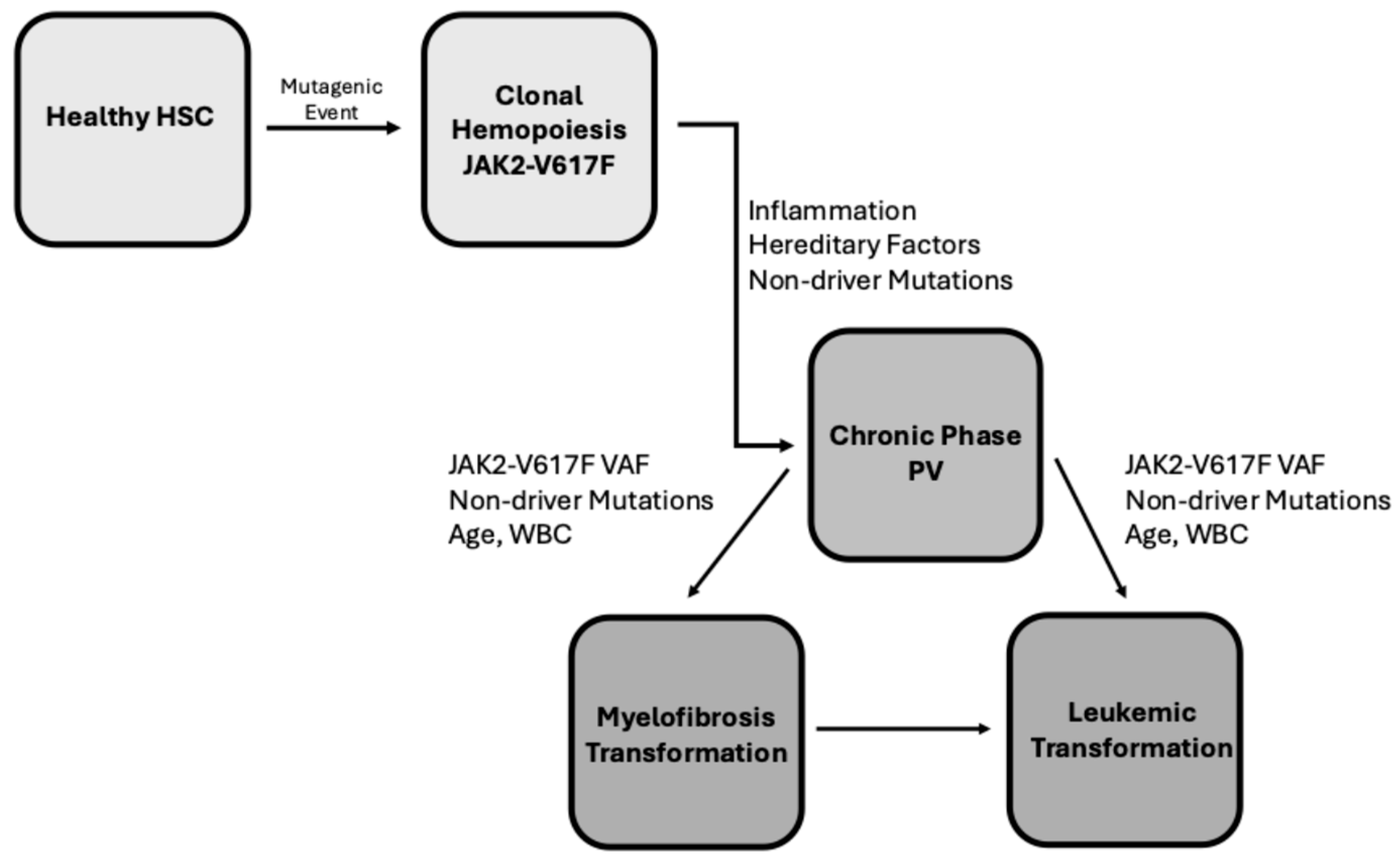
Figure 2.
Chromosomal abnormalities in PV. Top Panel: Chromosomal abnormalities in PV patients at different stages of disease: chronic phase, post-MF and post-leukemic transformation. Original data are reported in Tang et al. 2018 [27]. Bottom Panel: Chromosomal abnormalities in PV patients explored in the chronic phase either within 1 year (1yr) or 1-10 years (1-10 yr) or >10 years (>10 yr) from diagnosis or post MF/AML transformation. Original data reported in Iftikhar et al., 2025 [29]. Abbreviations: NK (normal karyotype), AK (abnormal karyotype), SA (single chromosomal abnormality), DA (double chromosomal abnormalities) and CK (complex karyotype).
Figure 2.
Chromosomal abnormalities in PV. Top Panel: Chromosomal abnormalities in PV patients at different stages of disease: chronic phase, post-MF and post-leukemic transformation. Original data are reported in Tang et al. 2018 [27]. Bottom Panel: Chromosomal abnormalities in PV patients explored in the chronic phase either within 1 year (1yr) or 1-10 years (1-10 yr) or >10 years (>10 yr) from diagnosis or post MF/AML transformation. Original data reported in Iftikhar et al., 2025 [29]. Abbreviations: NK (normal karyotype), AK (abnormal karyotype), SA (single chromosomal abnormality), DA (double chromosomal abnormalities) and CK (complex karyotype).
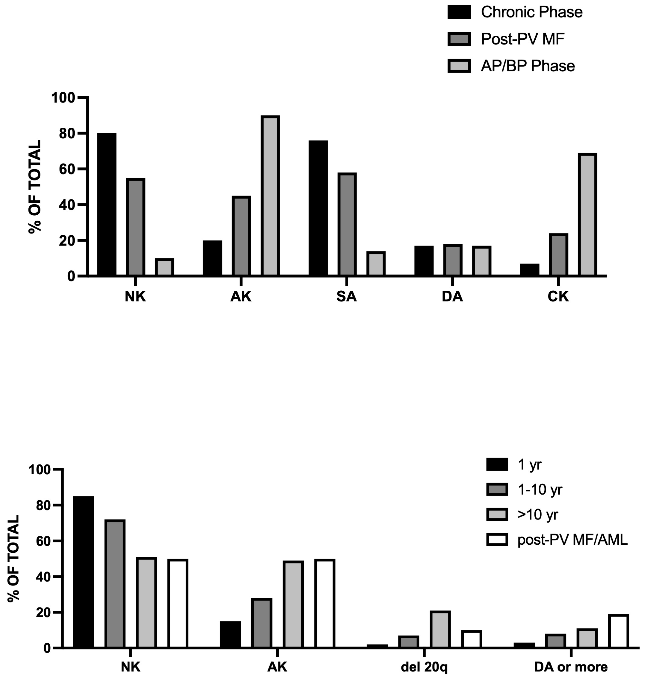
Figure 3.
Non-driver gene mutations in PV and ET patients. Top Panel: Comparison of the frequency of the main non-driver gene mutations in PV and ET patients. Original data are reported in Tefferi et al. 2016 [35]. Bottom Panel: Comparison of the frequency of the most recurrent non-driver gene mutations in PV patients with normal (NK) or abnormal karyotype (AK). Original data are reported in Iftikhar et al., 2025 [29].
Figure 3.
Non-driver gene mutations in PV and ET patients. Top Panel: Comparison of the frequency of the main non-driver gene mutations in PV and ET patients. Original data are reported in Tefferi et al. 2016 [35]. Bottom Panel: Comparison of the frequency of the most recurrent non-driver gene mutations in PV patients with normal (NK) or abnormal karyotype (AK). Original data are reported in Iftikhar et al., 2025 [29].
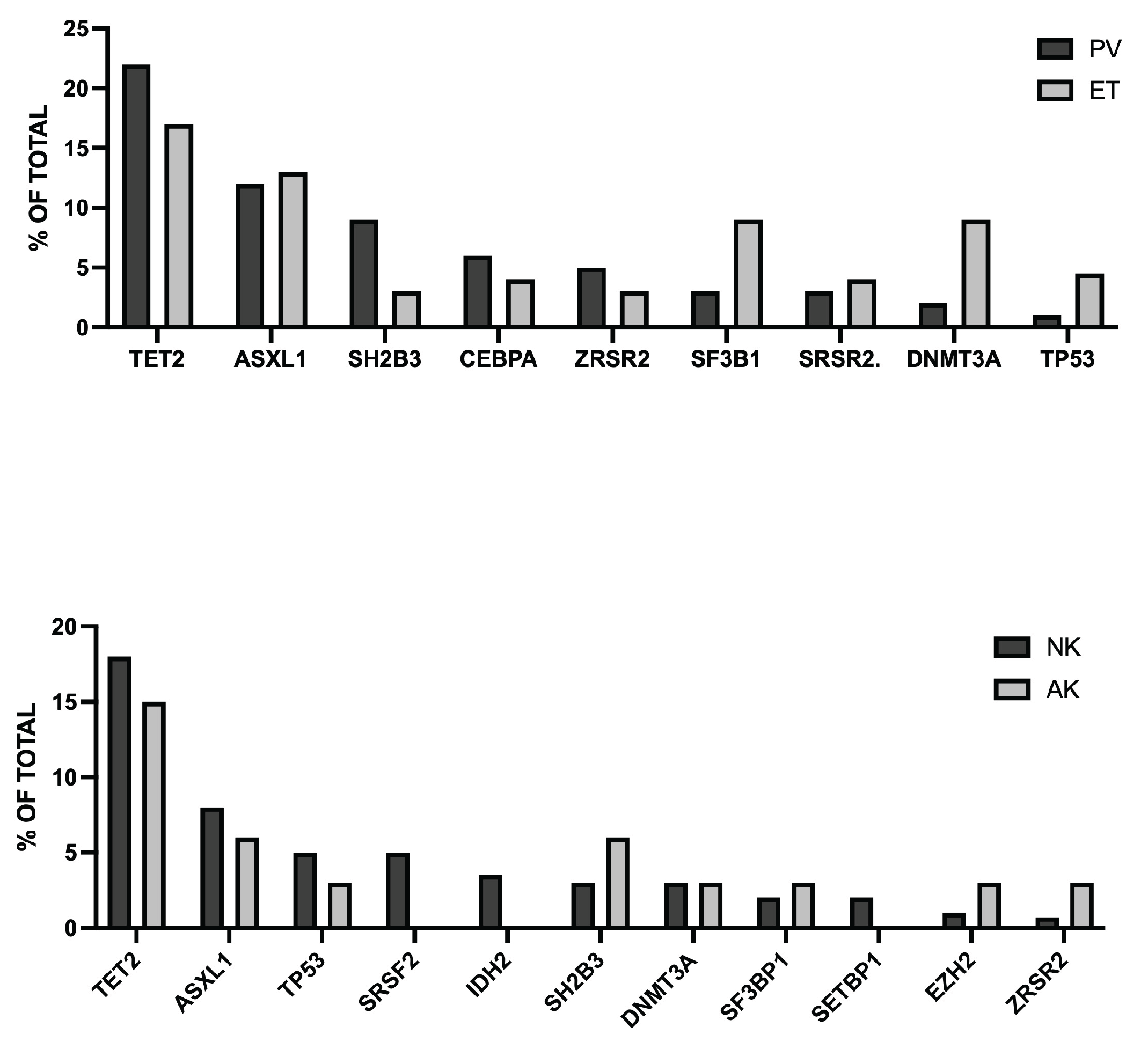
Figure 4.
Top Panel: Profile of non-driver mutations in PV patients during chronic disease (CD) or after MF (MF-T) or leukemic transformation (AML-T). Original data are reported by Rana and coworkers, 2025 [42]. Bottom Panel: Additional non-driver mutations acquired after AML transformation in PV patient exhibiting a short-term (ST-T), intermediate-term (INT-T) or long-term (LT-T) leukemic transformation. Original data reported in.
Figure 4.
Top Panel: Profile of non-driver mutations in PV patients during chronic disease (CD) or after MF (MF-T) or leukemic transformation (AML-T). Original data are reported by Rana and coworkers, 2025 [42]. Bottom Panel: Additional non-driver mutations acquired after AML transformation in PV patient exhibiting a short-term (ST-T), intermediate-term (INT-T) or long-term (LT-T) leukemic transformation. Original data reported in.
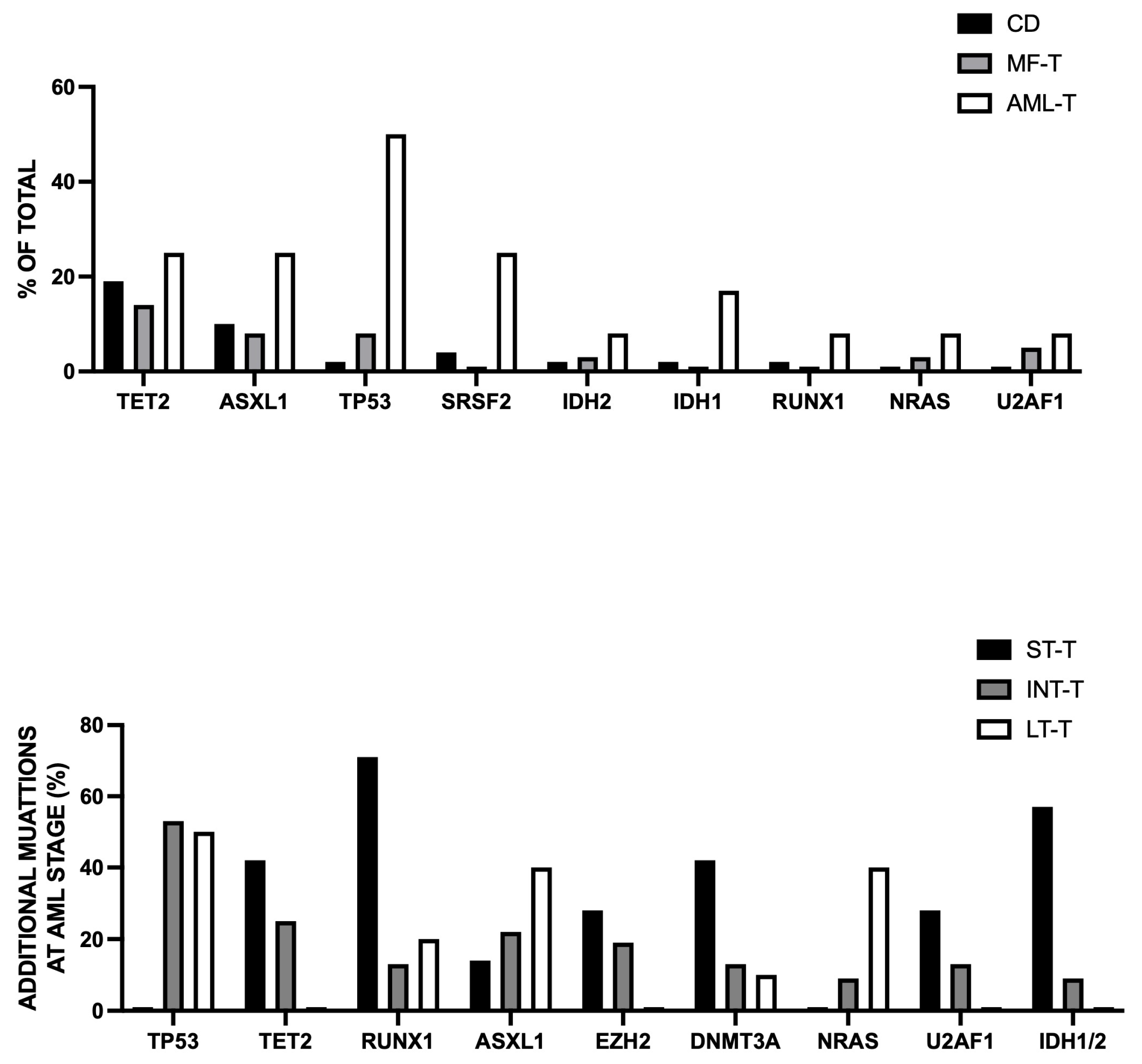
Disclaimer/Publisher’s Note: The statements, opinions and data contained in all publications are solely those of the individual author(s) and contributor(s) and not of MDPI and/or the editor(s). MDPI and/or the editor(s) disclaim responsibility for any injury to people or property resulting from any ideas, methods, instructions or products referred to in the content. |
© 2026 by the authors. Licensee MDPI, Basel, Switzerland. This article is an open access article distributed under the terms and conditions of the Creative Commons Attribution (CC BY) license (http://creativecommons.org/licenses/by/4.0/).
Copyright: This open access article is published under a Creative Commons CC BY 4.0 license, which permit the free download, distribution, and reuse, provided that the author and preprint are cited in any reuse.