
Submitted:
14 August 2026
Posted:
17 August 2026
You are already at the latest version
Abstract
Background: Persistent postural-perceptual dizziness (PPPD) is a chronic functional neuro-otologic disorder marked by persistent dizziness, unsteadiness, and visual dependence. Its electrophysiological mechanisms remain unclear. This study examined resting-state EEG spectral power and functional connectivity in PPPD and their associations with postural stability and clinical symptoms. Methods: Forty patients with PPPD and 40 age- and sex-matched healthy controls were studied with an eyes-closed resting state EEG recording. Static postural stability was measured with Pro-Kin system. Spectral power was calculated for delta, theta, alpha, beta, and gamma bands. Functional connectivity was assessed using weighted phase lag index. Results: Static posturography showed impaired postural stability in patients with PPPD compared with healthy controls,especially under eyes-closed conditions. Spectral power analysis showed increased theta-band relative power over the frontal and right parieto-occipital regions. Connectivity analysis showed increased prefrontal–temporal and decreased temporal–occipital connectivity in the theta band, increased fronto-parieto-occipital and decreased parietal -temporal connectivity in the alpha band, and decreased cerebellar and temporal, central connectivity in the gamma band. Frontal spectral power was positively correlated with HAMA scores in the PPPD group. Conclusions: PPPD was associated with frequency-specific alterations in resting-state cortical oscillations and functional connectivity involving cognitive-affective, visual, and sensorimotor networks. Resting-state EEG may help characterize network-level abnormalities in PPPD.
Keywords:
PPPD
; electroencephalography
; resting-state
; neural oscillations
; functional connectivity
1. Introduction
Persistent postural-perceptual dizziness (PPPD) is a chronic functional neuro-otologic disorder characterized by persistent dizziness, non-spinning vertigo, or unsteadiness lasting for at least 3 months and exacerbated by upright posture, active or passive motion, and exposure to moving or complex visual stimuli [1]. It is increasingly recognized as a common cause of chronic vestibular symptoms in neurology and dizziness clinics, and may impose a substantial burden on daily activities, emotional well-being, and health-related quality of life [2,3,4]. Despite standardized diagnostic criteria, PPPD remains clinically challenging because vestibular tests and conventional neuroimaging are often non-specific, whereas symptoms are frequently accompanied by anxiety, depression, visual dependence, and impaired postural control [5,6]. Consequently, objective neurophysiological markers that reflect the underlying network dysfunction are still needed.
Current evidence suggests that PPPD is not solely attributable to peripheral vestibular dysfunction, but may involve maladaptive interactions among vestibular, visual, somatosensory, postural, and affective systems [7,8]. Structural and functional neuroimaging studies have reported abnormalities in multimodal vestibular regions, visual cortices, insular networks, frontal regulatory regions, and cerebellar–sensorimotor circuits [9,10,11,12]. Network-level alterations have also been described in patients with phobic postural vertigo and PPPD, supporting the view that chronic dizziness may be associated with altered cortical integration rather than isolated vestibular end-organ dysfunction [13]. However, most available studies have relied on MRI-based measures, which provide limited temporal resolution for characterizing neural oscillatory dynamics. Although clinical and neurophysiological studies have begun to explore electrophysiological features in PPPD, resting-state EEG evidence remains limited, particularly regarding frequency-specific power changes and phase-based functional connectivity patterns [14]. In addition, few studies have integrated EEG indices with quantitative posturography in the same cohort [15].
The present study was designed to investigate resting-state EEG abnormalities in patients with PPPD using spectral power analysis and weighted phase lag index-based functional connectivity. We hypothesize that PPPD would be associated with frequency-specific alterations in cortical oscillatory activity and functional connectivity involving prefrontal, visual, temporal, cerebellar, and sensorimotor regions. We further propose that these EEG alterations would be related to dizziness-related disability and postural instability. To test these hypotheses, patients with PPPD and age- and sex-matched healthy controls were assessed using clinical scales, static posturography, and 71-channel resting-state EEG. This study aimed to clarify whether resting-state electrophysiological markers may help characterize the neural network alterations associated with PPPD and provide a basis for future mechanistic and longitudinal investigations.
2. Materials and Methods
2.1. Participants
This study enrolled 40 patients with persistent postural-perceptual dizziness (PPPD) who were recruited from the outpatient and inpatient services of Nanjing Brain Hospital between April 2025 and April 2026. The PPPD group included 15 men and 25 women, with a mean age of 58.58 ± 9.76 years. All patients met the 2017 Bárány Society diagnostic criteria for PPPD. The inclusion criteria were as follows: (1) age between 30 and 75 years; (2) absence of other neurological disorders, including traumatic brain injury, intracranial tumors, or cerebrovascular disease; (3) no use of medications that could affect resting-state brain activity, such as antidepressants or anxiolytics, within 4 weeks before enrollment.
Forty age- and sex-matched healthy controls were recruited. Control participants had no history of otologic, neurological, or psychiatric disorders and had not used medications known to affect resting-state brain activity. The study protocol was approved by the Ethics Committee of Nanjing Medical University Affiliated Brain Hospital(Approval No: IRB-AF40-1.0). Written informed consent was obtained from all participants before study participation.
2.2. Clinical Assessment
Demographic and clinical data were collected before EEG acquisition. The recorded variables included sex, age, and disease duration. Dizziness severity and balance-related symptoms were assessed using the Visual Analog Scale (VAS), Dizziness Handicap Inventory (DHI) [16,17], and Activities-specific Balance Confidence Scale (ABC) [18]. Psychologic symptoms were evaluated using the Hamilton Anxiety Rating Scale (HAMA) and Hamilton Depression Rating Scale (HAMD).
2.3. Static Posturography
Static postural stability was assessed using the Pro-Kin visual feedback balance system (PK254; TecnoBody S.R.L., Bergamo, Italy) [19,20]. Participants were instructed to stand comfortably on the force platform in a standardized position, with their arms placed alongside the body and their gaze fixed on a target located directly in front of them. Each participant completed two 30-second trials under eyes-open (EO) and eyes-closed (EC) conditions. Center-of-pressure (COP) data were recorded using pressure-sensitive sensors. The following posturographic parameters were analyzed: COP trajectory length, ellipse sway area, and mean sway velocity in the anterior–posterior and medio-lateral directions. Greater trajectory length, larger sway area, and higher sway velocity were interpreted as indicators of poorer postural stability. See Figure 1 for further details on static posturography.
2.4. EEG Acquisition
Resting-state EEG was recorded using a 71-channel whole-head EEG cap system (BoruiKang Technologies, Changzhou, China) [21]. The electrode montage covered conventional cortical regions and cerebellar electrodes, including PO9, PO10, O9, O10, Iz, CBz, CB1, and CB2. Electrode placement followed the international 10–10 system. The reference electrode was positioned at CPz, and the ground electrode was positioned at AFz.
EEG signals were sampled at 1000 Hz. Electrode impedance was maintained below 20 kΩ throughout acquisition. Recordings were performed during the daytime in a quiet environment. Participants were asked to wash their scalp before testing and were screened to ensure the absence of hunger, hypoglycemia, fatigue, or other discomfort. During recording, participants sat quietly with their eyes closed, remained awake, relaxed their facial and body muscles, and minimized horizontal and vertical eye movements. A 10-minute eyes-closed resting-state EEG recording was obtained from each participant.
2.5. Spectral Power Analysis
Spectral power analysis was performed in MATLAB [22]. The Welch method was applied to resting-state EEG data using a Hamming window, with 50% overlap between adjacent segments. The segment length was set to one-eighth of the total data length. Absolute power was calculated for the following frequency bands: delta, 1–4 Hz; theta, 4–8 Hz; alpha, 8–13 Hz; beta, 13–30 Hz; and gamma, 30–50 Hz. Relative power was then calculated for each frequency band. Cluster-based permutation testing was used to assess between-group differences in spectral power while controlling for multiple comparisons across electrodes.
2.6. Functional Connectivity Analysis
Functional connectivity was assessed using the weighted phase lag index (wPLI), which estimates phase synchronization between EEG signals while reducing the influence of volume conduction [23,24]. For each participant, 67 × 67 functional connectivity matrices were constructed for the delta, theta, alpha, beta, and gamma frequency bands. Mean wPLI values were then calculated within each frequency band and compared between groups.
2.7. Statistical Analysis
Statistical analyses were performed using SPSS version 25.0 (SPSS Inc., Chicago, IL, USA) and MATLAB. Normality and homogeneity of variance were examined before group comparisons. Continuous clinical variables were compared using independent-samples t tests when parametric assumptions were satisfied. For EEG spectral power and functional connectivity analyses, between-group differences were assessed across frequency bands. Cluster-based permutation statistics were applied to spectral power analyses to control for multiple comparisons across EEG electrodes. For Functional connectivity, Network-Based Statistics (NBS) were applied to compare wPLI matrices between PPPD groups and healthy controls [25]. To control for potential confounding effects of affective symptoms, group comparisons of EEG spectral power and functional connectivity were reanalyzed using analysis of covariance (ANCOVA), with HAMA and HAMD scores as covariates. Within the PPPD group, correlations between EEG indices and clinical, posturographic, and psychological variables were evaluated using Pearson or Spearman correlation analysis, depending on data distribution. All tests were two-sided, and statistical significance was set at P < 0.05.
3. Results
3.1. Clinical Characteristics
Demographic and clinical characteristics of the subjects are summarized in Table 1. No significant between-group differences were observed in age and sex distribution. Compared with healthy controls, patients with PPPD had significantly higher HAMA and HAMD scores, indicating greater anxiety and depressive symptom burden.
3.2. Static Postural Stability
Static posturography showed impaired postural stability in patients with PPPD compared with healthy controls. Detailed statistical analyses of all parameters were shown in Table 2. Under the eyes-open condition, patients with PPPD showed significantly greater medio-lateral COP displacement (EO-x, P = 0.0013), anterior–posterior COP displacement (EO-y, P = 0.0058), and sway area (EO-area, P = 0.0242). COP trajectory length under the eyes-open condition showed an increasing trend but did not reach statistical significance (EO-length, P = 0.0558).
Under the eyes-closed condition, patients with PPPD showed significantly greater medio-lateral displacement (EC-x, P = 0.0201), anterior–posterior displacement (EC-y, P = 0.0011), sway area (EC-area, P = 0.0070), and trajectory length (EC-length, P = 0.0199) than healthy controls. These findings indicate that postural control was impaired in PPPD and became more unstable when visual input was removed.
3.3. Resting-State EEG Spectral Power
Cluster-based permutation analysis revealed significant between-group differences in theta-band relative power(Figure 2). Compared with healthy controls, patients with PPPD showed increased theta-band power over the frontal region (electrodes :AF3,F1,F3) and right parieto-occipital region (electrodes:PO7,PO9), after controlling for psychiatric comorbidities. No significant between-group differences were detected in delta, alpha, beta, or gamma power.
3.4. Functional Connectivity
wPLI-based functional connectivity analysis showed frequency-specific alteration in patients with PPPD compare to healthy controls (Figure 3). After controlling for psychiatric comorbidities, temporal–occipital connectivity was decreased, whereas prefrontal–temporal connectivity was increased in the theta band. Connectivity among frontal, parietal, and occipital regions was increased, and parietal -temporal connectivity was decreased in the alpha band. Cerebellar and temporal, central connectivity was decreased in the gamma band. No significant differences were observed in cerebellar-related connectivity in the remaining frequency bands.
3.5. Correlation Analysis
Correlation analyses were performed within the PPPD group (Figure 4). Frontal spectral power was positively correlated with HAMA scores (r = 0.332, P =0.036). DHI scores were negatively correlated with ABC scores (r = −0.441, P =0.004). In addition, frontal spectral power was significant correlated with occipital spectral power (r = 0.930, P < 0.001). Age, disease duration, dizziness or balance assessment indices, and EEG spectral power showed no significant correlations in the remaining analyses.
4. Discussion
In this study, PPPD was associated with frequency-specific resting-state EEG alterations, including increased theta-band relative power over frontal and right parieto-occipital regions, altered theta-, alpha-, and gamma-band functional connectivity, impaired postural stability, and a positive association between frontal spectral power and anxiety severity. These findings support the presence of altered electrophysiological patterns involving cognitive-affective, visual, and sensorimotor systems in PPPD, while requiring confirmation in larger longitudinal cohorts.
Theta oscillations have been implicated in long-range communication involved in cognitive control and contextual integration, particularly in fronto-temporal and fronto-parietal systems [26]. The increased theta-band activity over frontal regions and increased prefrontal–temporal connectivity may represent an electrophysiological correlate of altered attentional control, body vigilance, and affective modulation rather than a disease-specific biomarker. This interpretation is consistent with recent conceptualizations of PPPD as a functional neuro-otologic disorder in which persistent symptoms are maintained by maladaptive interactions among postural control, spatial orientation, sensory reweighting, and threat-related processes [5,6]. The positive association between frontal spectral power and HAMA scores further suggests that frontal oscillatory activity may be linked to anxiety burden in PPPD. Affective symptoms, particularly anxiety and depression, are highly prevalent in patients with PPPD and are increasingly recognized as important modulators of both symptom severity and central nervous system functioning. Recent clinical studies have shown that anxiety-related hypervigilance, fear of movement, and maladaptive threat monitoring may contribute to persistent dizziness and postural instability in PPPD, forming part of a self-reinforcing perceptual–affective loop rather than representing purely comorbid psychiatric conditions [27,28,29,30]. In the present study, ANCOVA results demonstrated that after controlling for HAMA and HAMD scores, several EEG abnormalities—particularly posterior theta power and specific theta/alpha/gamma connectivity alterations remained significant. This supports the notion that emotional symptoms may act as modulators rather than sole drivers of PPPD-related neural dysfunction.
The occipital and right parieto-occipital theta-band findings are also consistent with previous neuroimaging evidence implicating visual and posterior cortical regions in PPPD. Li et al. reported altered spontaneous activity in the right precuneus and cuneus in patients with PPPD, indicating that posterior cortical regions involved in visuospatial processing may contribute to the pathophysiology of this disorder [10]. In the present study, increased theta power in the occipital/parieto-occipital region and decreased temporal–occipital connectivity may indicate altered top-down modulation of visual processing during rest. This interpretation is also consistent with studies of visually induced dizziness, in which altered functional brain connectivity has been observed in networks related to visual motion perception and multisensory integration [31,32]. Nevertheless, because EEG source localization was not performed, the anatomical interpretation of scalp-level occipital and parieto-occipital signals should remain cautious.
The decreased theta-band temporal–occipital connectivity and alpha-band parietal-temporal connectivity may reflect impaired integration between vestibular-related, auditory/temporal, visual, and higher-order regulatory systems [33,34]. Previous fMRI work in chronic subjective dizziness demonstrated altered activity in vestibular and insular systems during vestibular stimulation, suggesting that abnormal processing within vestibular–interoceptive networks may contribute to chronic dizziness symptoms [35]. Moreover, Li et al. reported altered intra- and inter-network functional connectivity in PPPD, including abnormal interactions between visual, sensorimotor, and default-mode-related networks [9].The present EEG findings extend these observations by showing that such network alterations may be frequency-specific and detectable in resting-state electrophysiological connectivity.
The increased fronto-parieto-occipital connectivity in alpha-band may be related to enhanced visual–spatial monitoring or increased reliance on visual information for postural control [32,36,37]. This interpretation is supported by the posturographic findings: patients with PPPD showed significantly greater COP displacement and sway area than controls, and instability became more evident when visual input was removed under eye-closed conditions. Similar behavioral observations have been reported in patients with phobic postural vertigo, in whom gait and postural control were influenced by fear of falling, attention, and visual input [38]. Experimental work has also shown that visual motion can modulate postural sway, supporting the role of visual input in balance control [39]. Together with the present EEG findings, these data suggest that excessive visual weighting and altered cortical control of sensory integration may participate in PPPD-related postural instability. However, the present study did not directly manipulate visual motion or vestibular stimulation; therefore, visual dependence was inferred from postural performance rather than experimentally tested.
In the gamma band, decreased connectivity between cerebellar and temporal, central regions may be clinically relevant because cerebellar and sensorimotor networks are important for postural control, movement calibration, and vestibular–motor integration. Prior multimodal imaging work in phobic postural vertigo reported cortical and cerebellar alterations, suggesting that distributed motor and vestibular networks may be involved in functional dizziness syndromes [12]. In addition, structural connectome studies have demonstrated that the vestibular cortical network is widely distributed and includes multimodal sensory and motor-related regions [40]. The decreased cerebellar and temporal,central connectivity observed here may reflect reduced coupling between cerebellar and sensorimotor systems. However, his finding should be interpreted cautiously, gamma-band EEG is susceptible to muscle and other non-neural artifacts, and scalp-level EEG has limited ability to localize cerebellar generators.
The main contribution of this study is that regional spectral power, wPLI-based connectivity, posturography, and psychological measures were examined within the same cohort. This multimodal design provides preliminary evidence that resting-state EEG alterations in PPPD are related to clinically relevant features, including postural instability and anxiety symptoms. These findings indicate that PPPD is associated with selective, frequency-dependent reorganization of functional brain networks involving posterior sensory regions, frontal control systems, and cerebellar–sensorimotor circuits. This pattern aligns with contemporary models of PPPD emphasizing abnormal sensory reweighting and predictive processing dysfunction across distributed cortical and subcortical networks [5,6].
Several limitations should be acknowledged. First, the sample size was modest, with 40 patients and 40 controls, which may limit statistical power and the generalizability of the findings. Second, this was a single-center study, and potential center-specific recruitment or assessment bias cannot be excluded. Third, the cross-sectional design precluded causal inference regarding whether the observed EEG abnormalities represent predisposing factors, compensatory changes, or consequences of persistent dizziness. Fourth, no intervention or longitudinal follow-up was included; therefore, the sensitivity of the identified EEG markers to treatment response remains unknown. Fifth, EEG was recorded only in the eye-closed resting state, and task-based EEG during visual motion, vestibular stimulation, or postural challenge was not performed. Sixth, scalp-level EEG has limited spatial resolution, and the present findings should not be interpreted as precise localization of deep or cerebellar generators without source-level validation.
5. Conclusions
Patients with PPPD showed increased theta-band power over frontal and parieto-occipital regions, frequency-specific alterations in functional connectivity, impaired postural stability, and an association between frontal electrophysiological activity and anxiety severity. These findings suggest that PPPD may involve abnormal resting-state interactions among cognitive-affective, visual, vestibular-related, and sensorimotor systems. Future studies should use larger multicenter cohorts, longitudinal designs, source-localized EEG or multimodal EEG–fMRI approaches, and task paradigms involving visual motion or vestibular stimulation. Interventional studies, including vestibular rehabilitation, cognitive-behavioral therapy, neuromodulation, or combined treatment strategies, are also needed to determine whether these electrophysiological alterations are modifiable and whether they can serve as biomarkers for prognosis or treatment response.
Author Contributions
Conceptualization, J.S. and F.L.; methodology, J.S. and F.L.; software, C.Z.; validation, F.L., and C.Z.; formal analysis, F.L.; investigation, F.L.; resources, L.T. and S.C.; data curation, X.L.; writing—original draft preparation, F.L.; writing—review and editing, F.L.; visualization, B.S.; supervision, J.W.; project administration, Y.W.; funding acquisition, J.S. All authors have read and agreed to the published version of the manuscript.
Funding
This work was supported by the National Natural Science Foundation of China, Grant/Award Numbers: 82571639.
Institutional Review Board Statement
The study was conducted in accordance with the Declaration of Helsinki, and approved by the Medical Research Ethics Committee of Nanjing Brain Hospital affiliated with Nanjing Medical University (Approval No: IRB-AF40-1.0) in 27 April 2025.
Informed Consent Statement
Informed consent was obtained from all subjects involved in the study.
Data Availability Statement
Due to the privacy protection of sensitive information, we cannot disclose our data.
Acknowledgments
The authors sincerely thank all participants who contributed to this study. We also acknowledge the assistance of the clinical and technical staff involved in participant recruitment, clinical assessment, EEG recording, and posturographic measurements. Their support was essential for the completion of this research.
Conflicts of Interest
The authors declare no conflicts of interest.
Abbreviations
The following abbreviations are used in this manuscript:
| PPPD | Persistent Postural-Perceptual Dizziness |
| EEG | Electroencephalography |
| rs-EEG | Resting-State Electroencephalography |
| FC | Functional Connectivity |
| wPLI | Weighted Phase Lag Index |
| DHI | Dizziness Handicap Inventory |
| ABC | Activities-specific Balance Confidence Scale |
References
- Staab, J.P.; Eckhardt-Henn, A.; Horii, A.; Jacob, R.; Strupp, M.; Brandt, T.; Bronstein, A. Diagnostic criteria for persistent postural-perceptual dizziness (PPPD): Consensus document of the committee for the Classification of Vestibular Disorders of the Bárány Society. J. Vestib. Res. Equilib. Orientat. 2017, 27, 191–208. [Google Scholar] [CrossRef] [PubMed]
- Popkirov, S.; Staab, J.P.; Stone, J. Persistent postural-perceptual dizziness (PPPD): a common, characteristic and treatable cause of chronic dizziness. Pract. Neurol. 2018, 18, 5–13. [Google Scholar] [CrossRef] [PubMed]
- Steensnaes, M.H.; Knapstad, M.K.; Goplen, F.K.; Berge, J.E. Persistent Postural-Perceptual Dizziness (PPPD) and quality of life: a cross-sectional study. Eur. Arch. Oto-Rhino-Laryngol. Off. J. Eur. Fed. Oto-Rhino-Laryngol. Soc. 2023, 280, 5285–5292. [Google Scholar] [CrossRef] [PubMed]
- Staab, J.P. Persistent Postural-Perceptual Dizziness. Semin. Neurol. 2020, 40, 130–137. [Google Scholar] [CrossRef] [PubMed]
- Staab, J.P. Persistent Postural-Perceptual Dizziness: Review and Update on Key Mechanisms of the Most Common Functional Neuro-otologic Disorder. Neurol. Clin. 2023, 41, 647–664. [Google Scholar] [CrossRef] [PubMed]
- Yagi, C.; Kimura, A.; Horii, A. Persistent postural-perceptual dizziness: A functional neuro-otologic disorder. Auris Nasus Larynx 2024, 51, 588–598. [Google Scholar] [CrossRef] [PubMed]
- Castro, P.; Bancroft, M.J.; Arshad, Q.; Kaski, D. Persistent Postural-Perceptual Dizziness (PPPD) from Brain Imaging to Behaviour and Perception. Brain Sci. 2022, 12. [Google Scholar] [CrossRef] [PubMed]
- Indovina, I.; Passamonti, L.; Mucci, V.; Chiarella, G.; Lacquaniti, F.; Staab, J.P. Brain Correlates of Persistent Postural-Perceptual Dizziness: A Review of Neuroimaging Studies. J. Clin. Med. 2021, 10. [Google Scholar] [CrossRef] [PubMed]
- Li, K.; Si, L.; Cui, B.; Ling, X.; Shen, B.; Yang, X. Altered intra- and inter-network functional connectivity in patients with persistent postural-perceptual dizziness. NeuroImage. Clin. 2020, 26, 102216. [Google Scholar] [CrossRef] [PubMed]
- Li, K.; Si, L.; Cui, B.; Ling, X.; Shen, B.; Yang, X. Altered spontaneous functional activity of the right precuneus and cuneus in patients with persistent postural-perceptual dizziness. Brain Imaging Behav. 2020, 14, 2176–2186. [Google Scholar] [CrossRef] [PubMed]
- Nigro, S.; Indovina, I.; Riccelli, R.; Chiarella, G.; Petrolo, C.; Lacquaniti, F.; Staab, J.P.; Passamonti, L. Reduced cortical folding in multi-modal vestibular regions in persistent postural perceptual dizziness. Brain Imaging Behav. 2019, 13, 798–809. [Google Scholar] [CrossRef] [PubMed]
- Popp, P.; Zu Eulenburg, P.; Stephan, T.; Bögle, R.; Habs, M.; Henningsen, P.; Feuerecker, R.; Dieterich, M. Cortical alterations in phobic postural vertigo - a multimodal imaging approach. Ann. Clin. Transl. Neurol. 2018, 5, 717–729. [Google Scholar] [CrossRef] [PubMed]
- Huber, J.; Flanagin, V.L.; Popp, P.; Zu Eulenburg, P.; Dieterich, M. Network changes in patients with phobic postural vertigo. Brain Behav. 2020, 10, e01622. [Google Scholar] [CrossRef] [PubMed]
- Adamec, I.; Juren Meaški, S.; Krbot Skorić, M.; Jažić, K.; Crnošija, L.; Milivojević, I.; Habek, M. Persistent postural-perceptual dizziness: Clinical and neurophysiological study. J. Clin. Neurosci. Off. J. Neurosurg. Soc. Australas. 2020, 72, 26–30. [Google Scholar] [CrossRef] [PubMed]
- Qin, C.; Zhang, R.; Yan, Z. Research Progress on the Potential Pathogenesis of Persistent Postural-Perceptual Dizziness. Brain Behav. 2025, 15, e70229. [Google Scholar] [CrossRef] [PubMed]
- Jacobson, G.P.; Newman, C.W. The development of the Dizziness Handicap Inventory. Arch. Otolaryngol.--Head. Neck Surg. 1990, 116, 424–427. [Google Scholar] [CrossRef] [PubMed]
- Van De Wyngaerde, K.M.; Lee, M.K.; Jacobson, G.P.; Pasupathy, K.; Romero-Brufau, S.; McCaslin, D.L. The Component Structure of the Dizziness Handicap Inventory (DHI): A Reappraisal. Otol. Neurotol. Off. Publ. Am. Otol. Soc. Am. Neurotol. Soc. [and] Eur. Acad. Otol. Neurotol. 2019, 40, 1217–1223. [Google Scholar] [CrossRef] [PubMed]
- Montilla-Ibáñez, A.; Martínez-Amat, A.; Lomas-Vega, R.; Cruz-Díaz, D.; Torre-Cruz, M.J.; Casuso-Pérez, R.; Hita-Contreras, F. The Activities-specific Balance Confidence scale: reliability and validity in Spanish patients with vestibular disorders. Disabil. Rehabil. 2017, 39, 697–703. [Google Scholar] [CrossRef] [PubMed]
- Wu, C.; YunYang; Jin, W.; Cao, R.; Lu, J.; Qian, K.; Xu, G. The application of computerized quadrato motor training in enhancing balance and executive performance in stroke patients. Sci. Rep. 2025, 15, 18850. [Google Scholar] [CrossRef] [PubMed]
- Zhai, X.; Wu, Q.; Li, X.; Xu, Q.; Zhang, Y.; Fan, S.; Zhang, L.Q.; Pan, Y. Effects of Robot-Aided Rehabilitation on the Ankle Joint Properties and Balance Function in Stroke Survivors: A Randomized Controlled Trial. Front. Neurol. 2021, 12, 719305. [Google Scholar] [CrossRef] [PubMed]
- Song, B.; Tian, M.; Wang, T.; Wang, X.; Ye, X.; Yao, Q.; Shi, J.; Yin, K. Effects of Cerebellar Repetitive Transcranial Magnetic Stimulation at Different Frequencies on Working Memory: An EEG Study. CNS Neurosci. Ther. 2025, 31, e70491. [Google Scholar] [CrossRef] [PubMed]
- Delorme, A.; Makeig, S. EEGLAB: an open source toolbox for analysis of single-trial EEG dynamics including independent component analysis. J. Neurosci. Methods 2004, 134, 9–21. [Google Scholar] [CrossRef] [PubMed]
- Imperatori, L.S.; Betta, M.; Cecchetti, L.; Canales-Johnson, A.; Ricciardi, E.; Siclari, F.; Pietrini, P.; Chennu, S.; Bernardi, G. EEG functional connectivity metrics wPLI and wSMI account for distinct types of brain functional interactions. Sci. Rep. 2019, 9, 8894. [Google Scholar] [CrossRef] [PubMed]
- Vinck, M.; Oostenveld, R.; van Wingerden, M.; Battaglia, F.; Pennartz, C.M. An improved index of phase-synchronization for electrophysiological data in the presence of volume-conduction, noise and sample-size bias. NeuroImage 2011, 55, 1548–1565. [Google Scholar] [CrossRef] [PubMed]
- Zalesky, A.; Fornito, A.; Bullmore, E.T. Network-based statistic: identifying differences in brain networks. NeuroImage 2010, 53, 1197–1207. [Google Scholar] [CrossRef] [PubMed]
- Cavanagh, J.F.; Frank, M.J. Frontal theta as a mechanism for cognitive control. Trends Cogn. Sci. 2014, 18, 414–421. [Google Scholar] [CrossRef] [PubMed]
- Alahmari, K.A.; Alshehri, S. Evaluating the efficacy of vestibular rehabilitation therapy on quality of life in persistent postural-perceptual dizziness: the role of anxiety and depression in treatment outcomes. Front. Neurol. 2025, 16, 1524324. [Google Scholar] [CrossRef] [PubMed]
- Ibrahim, N.M.K.; Hazza, N.M.A.; Yaseen, D.M.; Galal, E.M. Effect of vestibular rehabilitation games in patients with persistent postural perceptual dizziness and its relation to anxiety and depression: prospective study. Eur. Arch. Oto-Rhino-Laryngol. Off. J. Eur. Fed. Oto-Rhino-Laryngol. Soc. 2024, 281, 2861–2869. [Google Scholar] [CrossRef] [PubMed]
- Jáuregui-Renaud, K.; Cabrera-Pereyra, R.; Miguel-Puga, J.A.; Alcántara-Thome, M. Graviception Uncertainty, Spatial Anxiety, and Derealization in Patients with Persistent Postural-Perceptual Dizziness. J. Clin. Med. 2024, 13. [Google Scholar] [CrossRef] [PubMed]
- Maywald, M.; Pogarell, O.; Chrobok, A.; Levai, S.; Keeser, D.; Tschentscher, N.; Rauchmann, B.S.; Stöcklein, S.; Ertl-Wagner, B.; Papazov, B.; et al. Diagnostics and Group Therapy in Patients with Persistent Postural-Perceptual Dizziness and Anxiety Disorder: Biomarkers and Neurofunctional Correlates of Underlying Treatment Effects. Diagnostics 2025, 15. [Google Scholar] [CrossRef] [PubMed]
- Van Ombergen, A.; Heine, L.; Jillings, S.; Roberts, R.E.; Jeurissen, B.; Van Rompaey, V.; Mucci, V.; Vanhecke, S.; Sijbers, J.; Vanhevel, F.; et al. Altered functional brain connectivity in patients with visually induced dizziness. NeuroImage. Clin. 2017, 14, 538–545. [Google Scholar] [CrossRef] [PubMed]
- Liu, Y.; Peng, X.; Lin, C.; Liu, D.; Sun, Y.; Huang, F.; Liu, T.; Xiao, L.; Wei, X.; Wang, K.; et al. Fractional Amplitude of Low-Frequency Fluctuation and Voxel-Mirrored Homotopic Connectivity in Patients with Persistent Postural-Perceptual Dizziness: Resting-State Functional Magnetic Resonance Imaging Study. Brain Connect. 2024, 14, 274–283. [Google Scholar] [CrossRef] [PubMed]
- Lee, J.O.; Lee, E.S.; Kim, J.S.; Lee, Y.B.; Jeong, Y.; Choi, B.S.; Kim, J.H.; Staab, J.P. Altered brain function in persistent postural perceptual dizziness: A study on resting state functional connectivity. Hum. Brain Mapp. 2018, 39, 3340–3353. [Google Scholar] [CrossRef] [PubMed]
- Li, K.; Ling, X.; Zhao, J.; Wang, Z.; Yang, X. Abnormal neural circuits and altered brain network topological properties in patients with persistent postural-perceptual dizziness. Commun. Biol. 2025, 8, 122. [Google Scholar] [CrossRef] [PubMed]
- Indovina, I.; Riccelli, R.; Chiarella, G.; Petrolo, C.; Augimeri, A.; Giofrè, L.; Lacquaniti, F.; Staab, J.P.; Passamonti, L. Role of the Insula and Vestibular System in Patients with Chronic Subjective Dizziness: An fMRI Study Using Sound-Evoked Vestibular Stimulation. Front. Behav. Neurosci. 2015, 9, 334. [Google Scholar] [CrossRef] [PubMed]
- Indovina, I.; Riccelli, R.; Staab, J.P.; Lacquaniti, F.; Passamonti, L. Personality traits modulate subcortical and cortical vestibular and anxiety responses to sound-evoked otolithic receptor stimulation. J. Psychosom. Res. 2014, 77, 391–400. [Google Scholar] [CrossRef] [PubMed]
- Storm, R.; Krause, J.; Blüm, S.K.; Wrobel, V.; Frings, A.; Helmchen, C.; Sprenger, A. Visual and vestibular motion perception in persistent postural-perceptual dizziness (PPPD). J. Neurol. 2024, 271, 3227–3238. [Google Scholar] [CrossRef] [PubMed]
- Schniepp, R.; Wuehr, M.; Huth, S.; Pradhan, C.; Brandt, T.; Jahn, K. Gait characteristics of patients with phobic postural vertigo: effects of fear of falling, attention, and visual input. J. Neurol. 2014, 261, 738–746. [Google Scholar] [CrossRef] [PubMed]
- Balestrucci, P.; Daprati, E.; Lacquaniti, F.; Maffei, V. Effects of visual motion consistent or inconsistent with gravity on postural sway. Exp. Brain Res. 2017, 235, 1999–2010. [Google Scholar] [CrossRef] [PubMed]
- Indovina, I.; Bosco, G.; Riccelli, R.; Maffei, V.; Lacquaniti, F.; Passamonti, L.; Toschi, N. Structural connectome and connectivity lateralization of the multimodal vestibular cortical network. NeuroImage 2020, 222, 117247. [Google Scholar] [CrossRef] [PubMed]
Figure 1.
Balance assessment using Pro-Kin visual feedback balance system. (A) Participants stood on a force platform with feet positioned shoulder-width apart and viewed a monitor at eye level.(B) The platform recorded center-of-pressure (COP) data during participants standing with eyes open (EO) and eyes closed (EC).(C) Representative COP sway trajectories in a patient with persistent postural-perceptual dizziness (PPPD) and a healthy control. PPPD showed larger sway area and greater excursions compared with the control.
Figure 1.
Balance assessment using Pro-Kin visual feedback balance system. (A) Participants stood on a force platform with feet positioned shoulder-width apart and viewed a monitor at eye level.(B) The platform recorded center-of-pressure (COP) data during participants standing with eyes open (EO) and eyes closed (EC).(C) Representative COP sway trajectories in a patient with persistent postural-perceptual dizziness (PPPD) and a healthy control. PPPD showed larger sway area and greater excursions compared with the control.
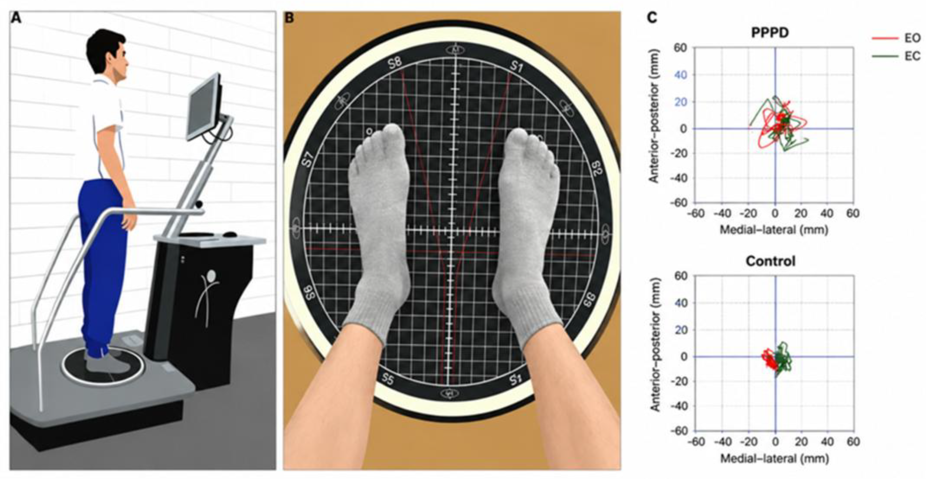
Figure 2.
Topographic maps of t-values for EEG power spectrum differences between PPPD patients and healthy controls across five frequency bands. The t-value topographies illustrate the statistical differences in power spectral density (PSD) between the PPPD group and healthy controls in the delta (0.5–4 Hz), theta (4–8 Hz), alpha (8–13 Hz), beta (13–30 Hz), and gamma (30–50 Hz) bands. Black asterisks denote scalp regions showing statistically significant between-group differences (p < 0.05, corrected).
Figure 2.
Topographic maps of t-values for EEG power spectrum differences between PPPD patients and healthy controls across five frequency bands. The t-value topographies illustrate the statistical differences in power spectral density (PSD) between the PPPD group and healthy controls in the delta (0.5–4 Hz), theta (4–8 Hz), alpha (8–13 Hz), beta (13–30 Hz), and gamma (30–50 Hz) bands. Black asterisks denote scalp regions showing statistically significant between-group differences (p < 0.05, corrected).
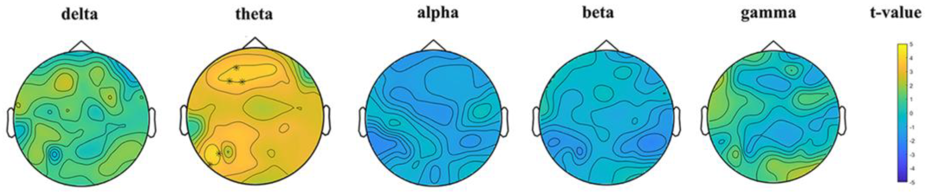
Figure 3.
Functional connectivity differences between PPPD patients and healthy controls across frequency bands. Significant between-group differences in functional connectivity are shown for the theta (A), alpha (B), and gamma (C) frequency bands. Red lines indicate significantly increased functional connectivity in patients with PPPD compare to healthy controls, while blue lines indicate significantly decreased functional connectivity. Labeled nodes denote the electrode sites involved in the significant connections. Electrode abbreviations: AF: anterior frontal; F: frontal; FC : frontocentral; C: central; CP: centroparietal; P: parietal; PO: parieto-occipital; O: occipital; T: temporal; TP: temporoparietal; CB: cerebellar.
Figure 3.
Functional connectivity differences between PPPD patients and healthy controls across frequency bands. Significant between-group differences in functional connectivity are shown for the theta (A), alpha (B), and gamma (C) frequency bands. Red lines indicate significantly increased functional connectivity in patients with PPPD compare to healthy controls, while blue lines indicate significantly decreased functional connectivity. Labeled nodes denote the electrode sites involved in the significant connections. Electrode abbreviations: AF: anterior frontal; F: frontal; FC : frontocentral; C: central; CP: centroparietal; P: parietal; PO: parieto-occipital; O: occipital; T: temporal; TP: temporoparietal; CB: cerebellar.
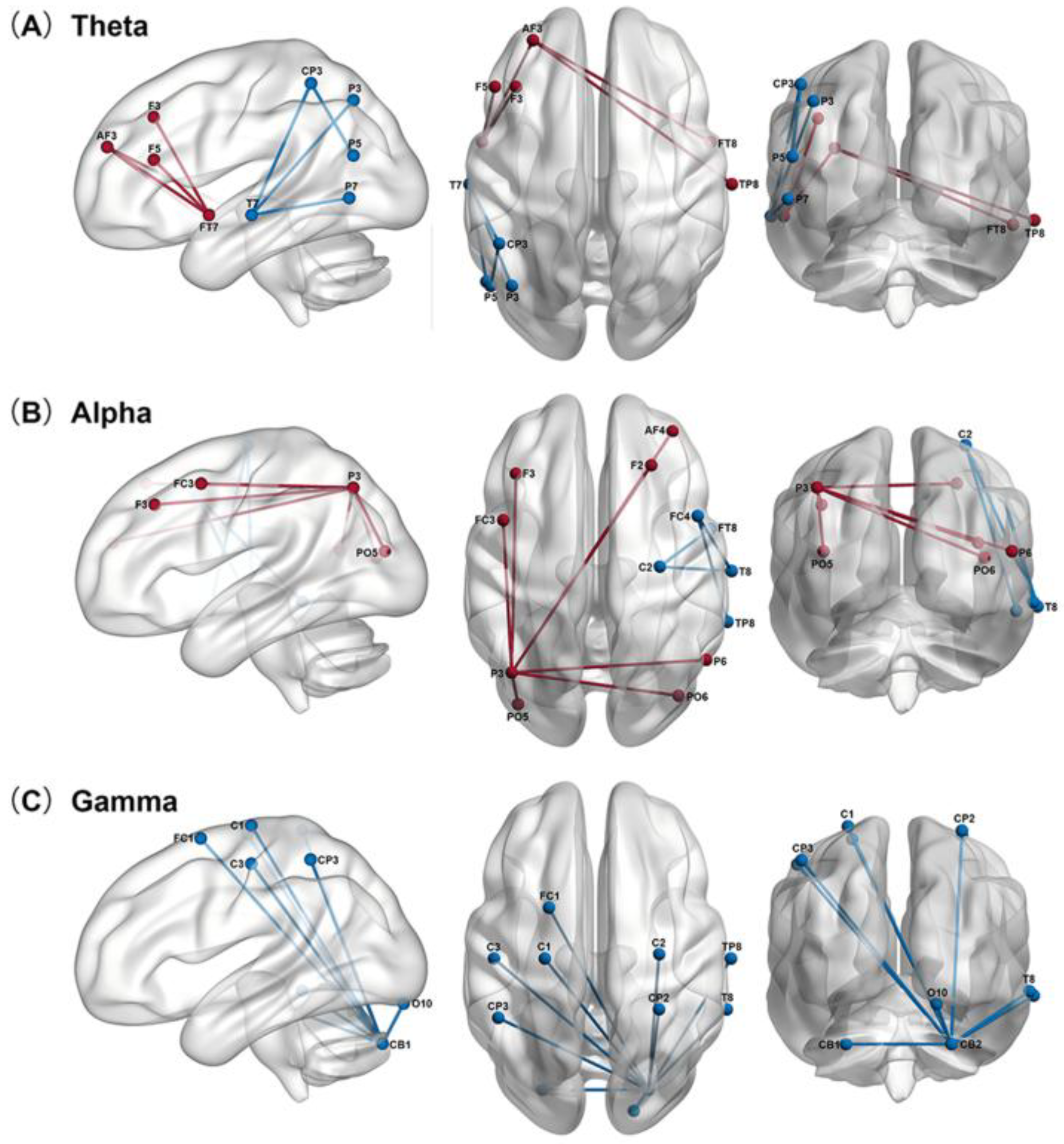
Figure 4.
Correlation analysis between clinical assessments and EEG spectral power in patients with PPPD (N=40). Red indicates positive correlation, blue indicates negative correlation. Asterisks indicate statistical significance: *p<0.05, **p<0.01, ***p<0.001.
Figure 4.
Correlation analysis between clinical assessments and EEG spectral power in patients with PPPD (N=40). Red indicates positive correlation, blue indicates negative correlation. Asterisks indicate statistical significance: *p<0.05, **p<0.01, ***p<0.001.
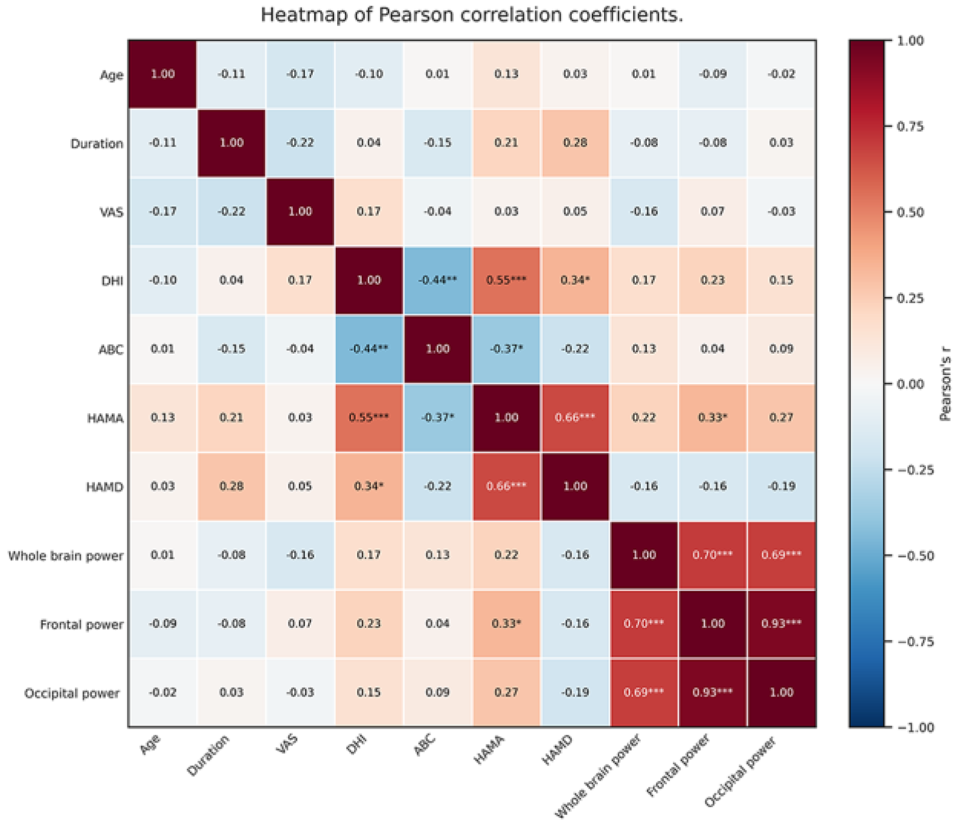

Table 1.
Clinical Characteristics and Evaluation of Subjects.
| PPPD | Control | P | |
| Female/male | 25/15 | 25/15 | NS |
| Age(year) | 58.58±9.76 | 58.12±9.23 | NS |
| Duration (month) | 24.13±22.33 | - | |
| VAS | 4.88±1.40 | - | |
| DHI | 49.05±15.58 | - | |
| ABC | 59.20±17.93 | - | |
| HAMA | 14.90±4.65 | 3.72±2.67 | <0.001 |
| HAMD | 10.75±3.96 | 2.88±2.14 | <0.001 |
VAS: Visual Analog Scale; DHI: Dizziness Handicap inventory; ABC: Activities Specific Balance Confidence Scale; HAMA: Hamilton Anxiety Rating Scale; HAMD : Hamilton Depression Rating Scale; NS: No significant.
Table 2.
Comparison of postural sway parameters between PPPD patients and healthy controls.
| Parameter | PPPD | Control | P-value |
| EO | |||
| AP average sway speed (mm/s) | 10.18 ± 2.58 | 8.38 ± 2.44 | 0.0013 |
| ML average sway speed (mm/s) | 7.98 ± 2.24 | 6.68 ± 1.49 | 0.0058 |
| Trajectory area (mm2) | 414.25 ± 109.73 | 361.08 ± 96.69 | 0.0242 |
| Trajectory length (mm) | 382.58 ± 108.32 | 339.82 ± 87.49 | 0.0558 |
| EC | |||
| AP average sway speed (mm/s) | 16.42 ± 5.14 | 14.05 ± 3.67 | 0.0201 |
| ML average sway speed (mm/s) | 11.58 ± 3.66 | 9.25 ± 2.61 | 0.0011 |
| Trajectory area (mm2) | 813.62 ± 262.50 | 695.35 ± 214.29 | 0.0070 |
| Trajectory length (mm) | 749.22 ± 242.36 | 656.82 ± 199.41 | 0.0199 |
Data are presented as mean ± standard deviation. PPPD: persistent postural-perceptual dizziness; EO: Eyes open condition; EC: Eyes closed condition; AP: anterior-posterior direction; ML: medial-lateral direction. Intergroup comparisons were performed using independent-samples t-test.
Disclaimer/Publisher’s Note: The statements, opinions and data contained in all publications are solely those of the individual author(s) and contributor(s) and not of MDPI and/or the editor(s). MDPI and/or the editor(s) disclaim responsibility for any injury to people or property resulting from any ideas, methods, instructions or products referred to in the content. |
© 2026 by the authors. Licensee MDPI, Basel, Switzerland. This article is an open access article distributed under the terms and conditions of the Creative Commons Attribution (CC BY) license (http://creativecommons.org/licenses/by/4.0/).
Copyright: This open access article is published under a Creative Commons CC BY 4.0 license, which permit the free download, distribution, and reuse, provided that the author and preprint are cited in any reuse.