

Submitted:
14 August 2026
Posted:
17 August 2026
You are already at the latest version
Abstract
The surgical procedure currently known as arthroereisis has been performed for decades as a treatment for recurrent adult-acquired and pediatric flexible talotarsal deformity, yet its name has never been formally challenged despite being etymologically and anatomically inaccurate. The term derives from the Greek arthro (joint) and ereisis (to lift or support), implying joint-level action that does not occur — the sinus tarsi implant acts not on a joint surface but on the talar body itself, physically elevating and realigning the talus as a whole bone and producing a cascade of pantalar corrections across the subtalar, talonavicular, calcaneocuboid, and talocrural joints simultaneously. Radiographic evidence from unilateral cases demonstrates measurable talar elevation of 1.8 cm between treated and untreated limbs on weight-bearing views, confirming that the corrective effect is osseous, not articular. This article proposes the adoption of the term osteoereisis — from the Greek osteo (bone) and ereisis (to lift or support) — as an anatomically precise replacement that correctly identifies the talus as the primary object of surgical effect and more fully reflects the pantalar scope of the correction achieved. The author invites collegial discussion of this proposal within the podiatric and foot and ankle surgical community.
Keywords:
osteoereisis
; talotarsal stabilization
; nomenclature
; subtalar implant
; talus
; pantalar correction
; talotarsal deformity
; flatfoot
; podiatric surgery
; arthroereisis
1. Introduction
Medical nomenclature carries weight far beyond mere labeling. The names we assign to surgical procedures shape how clinicians conceptualize the mechanism of action, how educators teach the procedure to residents, how researchers categorize outcomes in the literature, and how patients and insurers understand what is being done. When a name is inaccurate, it introduces conceptual error at every level of the chain — from surgical planning to informed consent.
The procedure known as arthroereisis has been performed in its modern form since the 1970s and 1980s, most commonly using a cone-shaped, cylindrical, or mushroom-capped implant inserted into the sinus tarsi — the lateral anatomical corridor between the talus and calcaneus. Over the decades, the procedure has been refined, with various implant designs introduced, and its indications have expanded from pediatric flexible flatfoot to adult-acquired flatfoot deformity, posterior tibial tendon dysfunction, and associated conditions [1,2]. It is today one of the most commonly performed podiatric surgical interventions for flatfoot correction worldwide.
Yet despite this clinical ubiquity, the foundational name of the procedure has never been formally challenged. The word arthroereisis, as this article will demonstrate, is derived from Greek roots that describe an action on a joint — and that description is simply incorrect. The procedure does not act on a joint, does not stabilize a joint surface, and does not produce its corrective effect through joint mechanics alone. It acts on the talus as a whole bone, repositioning it in space and producing a cascade of corrections across multiple talocrural and midtarsal joints simultaneously [3,4].
This article proposes the term osteoereisis as a replacement — a name that is etymologically sound, anatomically precise, and clinically comprehensive.
2. Etymology and the Problem with “Arthroereisis”
The term arthroereisis is a compound derived from two Greek roots: arthro (ἄρθρο), meaning joint or pertaining to a joint or articulation; and ereisis (ἔρεισις), meaning to lift, to prop, to support, or to push upward. Taken together, arthroereisis literally means to lift or support a joint, implying that the procedure exerts its effect at the level of a joint [5].
However, this framework is fundamentally incorrect. The implant placed during this procedure is inserted into the sinus tarsi — a canal-like space between the talus superiorly and the calcaneus inferiorly. The sinus tarsi has no articular cartilage and is not a joint [6]. The device does not enter a joint space. It does not contact articular cartilage under normal circumstances. Instead, it acts as a mechanical buttress against the lateral process of the talus, physically preventing the talus from excessive plantar flexion and medial rotation [7].
The effect is not articular; it is osseous. The talus itself is repositioned — lifted off its pathological plantarflexed and adducted orientation — by the force of the implant acting against the talar body and lateral process. What moves is not a joint; what moves is a bone.
3. Anatomical Reality: What the Procedure Actually Does
In flatfoot deformity — whether pediatric flexible flatfoot or adult-acquired flatfoot — the fundamental pathological event is the loss of normal talar position [8]. The talus undergoes plantarflexion, medial rotation, and anterior translation relative to the calcaneus. This talar malposition is the axis around which the entire deformity is organized.
When an implant is placed in the sinus tarsi, it engages the lateral talar process and acts as a physical buttress, preventing the talus from continuing its pathological descent. The talus is mechanically elevated — lifted — back toward its corrected anatomical orientation [7]. This is a bone-level event, not a joint-level event. The downstream effects of this talar repositioning affect multiple joint levels simultaneously:
Subtalar joint (talocalcaneal joint): Restoration of normal talocalcaneal angle and hindfoot alignment, with significant radiographic improvement in calcaneal pitch documented across multiple outcomes series [9,10].
Talonavicular joint: As the talus derotates and rises, the navicular is drawn back into proper alignment against the talar head, restoring the talonavicular coverage angle and correcting forefoot abduction [9,11].
Calcaneocuboid joint: Correction of calcaneal eversion and abduction at the midtarsal joint complex restores the lateral column to proper length and alignment.
Ankle mortise (talocrural joint): Talar derotation reduces abnormal forces on the ankle mortise, improving the mechanical relationship between the talus and the tibial plafond.
Medial longitudinal arch: With the talus repositioned, the spring ligament and plantar fascia are placed under appropriate tension. Finite element analysis confirms ligament strain relief following implant placement [12].
This multi-level, multi-joint correction is precisely what the term pantalar implies — the procedure produces corrections that span the full talar articulation complex. No single joint is targeted in isolation. The talus as a whole bone is repositioned, and the entire foot reorganizes around it.
It is important to emphasize that the osseous elevation produced by osteoereisis is not limited to the talus alone. As the talus rises and derotates, it carries with it the entire medial column of the foot in a sequential chain of osseous realignment. The navicular, which articulates directly with the talar head at the talonavicular joint, is elevated and repositioned simultaneously with the talus. The medial cuneiform and first metatarsal — the distal components of the medial column — follow in a cascading chain reaction of realignment. The entire medial longitudinal arch is therefore restored not through soft tissue tension alone, but through the osseous repositioning of multiple bones in sequence, all driven by the primary elevation and derotation of the talus [9,11]. This multi-osseous correction further reinforces the appropriateness of the term osteoereisis — a bone-level intervention — over the joint-centric designation arthroereisis.
Furthermore, the corrective effect of osteoereisis extends beyond the foot itself and propagates up the entire lower extremity kinetic chain. The subtalar joint functions as a torque converter between the foot and the proximal lower extremity, such that 1 degree of rearfoot inversion or eversion produces a corresponding 1 degree of tibial internal or external rotation [19]. In the flatfoot deformity state, the talus assumes a pathologically plantarflexed and internally rotated position, which forces the tibia and fibula complex into an abnormal varum rotational alignment at the ankle joint — a deviation from the physiologically normal tibiotalar relationship characterized by approximately 15 degrees of natural valgus angulation. Excessive subtalar pronation has been well documented to cause tibial and femoral internal rotation through the kinetic chain mechanism when the foot is fixed on the ground, producing dynamic knee valgus and patellofemoral dysfunction [19,20,21]. As the talus is elevated and derotated by the osteoereisis implant, the tibiotalar articulation is restored toward its natural valgus orientation, correcting the abnormal internal rotational torque transmitted to the tibia and fibula complex. This tibiotalar realignment in turn reduces abnormal rotational and compressive forces transmitted proximally to the knee joint, affecting patellofemoral tracking, tibial torsion mechanics, and ultimately hip and pelvic alignment [20,21]. Osteoereisis therefore functions not merely as a foot correction but as a kinetic chain realignment procedure, with measurable biomechanical consequences that extend from the sinus tarsi to the knee and beyond. No joint procedure — no arthroereisis — could accurately describe an intervention of this osseous and systemic scope.
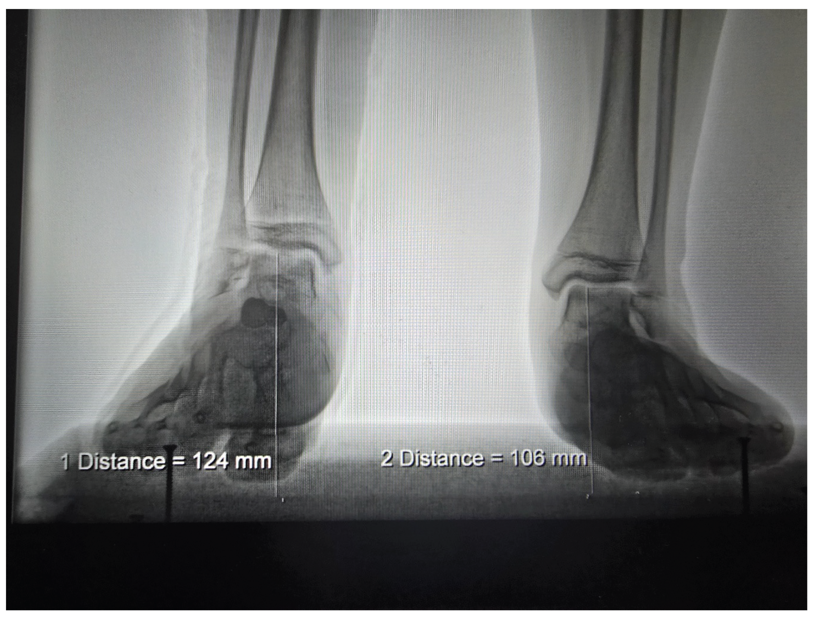
Bilateral weight-bearing lateral radiographic comparison in a representative case demonstrates measurable talar height of 106 mm on the osteoereisis-treated right limb versus 124 mm on the untreated left limb — a difference of 18 mm (1.8 cm) — confirming bone-level talar elevation consistent with the osseous corrective mechanism proposed herein (Figure 1).
4. The Proposed Term: Osteoereisis
We propose the adoption of the term Osteoereisis (pronounced os-tee-oh-eh-RAY-sis) as the replacement for arthroereisis. The term is constructed from osteo (ὀστέο), meaning bone or pertaining to bone, and ereisis (ἔρεισις), meaning to lift, to prop, to support, or to reposition. Osteoereisis therefore means: to lift, support, or reposition a bone — precisely what the procedure accomplishes.
The term carries several advantages over its predecessor:
Etymological precision: Osteoereisis is constructed from the same classical Greek tradition as arthroereisis, fitting naturally within the existing vocabulary of orthopedic and podiatric surgery [13].
Mechanistic accuracy: The term correctly identifies the bone — the talus — as the primary object of surgical effect.
Scope recognition: By naming a bone-level intervention, osteoereisis implicitly acknowledges the pantalar scope of the correction.
Educational clarity: Trainees learning the procedure under the name osteoereisis will immediately understand that the implant acts on the talar body, not on articular cartilage.
Research classification: A precise name will improve literature indexing and outcomes research consistency [14,15].
In formal academic and research contexts where the multi-joint corrective scope warrants emphasis, the full phrase Pantalar Talar Osteoereisis may be used. In clinical practice and general surgical literature, Osteoereisis is the preferred short form.
5. Historical Context and the Persistence of Misnomers in Surgery
The movement to replace eponymous and anatomically imprecise surgical names with descriptive terms has gained significant traction since the International Congress of Anatomy formalized standardized nomenclature, subsequently revised as the Terminologia Anatomica [13,15]. The case of arthroereisis is distinct from an eponym: it is not named after a person, but its descriptive content is simply wrong. This may be more insidious than an eponym, because it gives the false impression of being informative while actually misdirecting clinical understanding [14].
Multiple published biomechanical analyses confirm that the sinus tarsi implant acts by restricting talar descent and rotation, not by acting on articular surfaces [7,12]. The literature has consistently described the correct mechanism while simultaneously using the incorrect name — a contradiction this proposal seeks to resolve.
6. Clinical and Educational Implications
Adoption of the term osteoereisis would have meaningful practical effects across podiatric medicine and surgery. In surgical education, the name immediately communicates that the procedure targets the talus as a bone, naturally prompting instructors to teach the pantalar scope of the correction from the outset. In patient communication, the term — explained as a procedure that lifts and repositions the ankle bone — is more accurate and intuitive, supporting superior informed consent. In outcomes research, consistent nomenclature would facilitate more reliable identification and classification of the procedure in databases, coding systems, and systematic reviews [16].
7. Discussion
We are fully aware that arthroereisis is deeply embedded in the podiatric surgical literature, in training curricula, in device marketing, and in the vocabulary of practicing surgeons. We anticipate that some colleagues will view this proposal as disruptive. We respectfully submit that the standard for surgical nomenclature should be anatomical accuracy rather than historical inertia.
We do not propose the elimination of existing literature. Studies published under the term arthroereisis retain their validity and contributions. We propose only that going forward, the procedure be referred to by the name that accurately describes what it does. A transitional period using the formulation osteoereisis (formerly arthroereisis) would facilitate continuity of literature indexing and reader comprehension.
We also note that the implant-based sinus tarsi intervention is not the only technique described under the arthroereisis umbrella — bone staples, extra-osseous screws, and other devices have been grouped under the same term [17]. The name osteoereisis accommodates all of these techniques equally, as each acts to reposition the talus as a bone, regardless of the specific implant design.
8. Conclusions
The surgical procedure currently known as arthroereisis is misnamed. Its name implies joint-level action that does not occur; it fails to reflect the bone-level repositioning of the talus that is the actual mechanism of correction; and it conceals the pantalar scope of the deformity correction the procedure achieves.
The adoption of the term Osteoereisis — from the Greek osteo (bone) and ereisis (to lift or support) — is supported by anatomical, etymological, biomechanical, and radiographic evidence. The name describes what the procedure does, at the correct anatomical level of analysis, and positions the podiatric surgical community to communicate, teach, and research this intervention with the precision it deserves. Nomenclature in medicine is not merely administrative. It is the architecture of clinical thought.
Author Contributions
Conceptualization, writing — original draft preparation, writing — review and editing: E.C.D. The author has read and agreed to the published version of the manuscript.
Funding
This research received no external funding.
Institutional Review Board Statement
Not applicable. This article does not report on research involving human participants or animals.
Informed Consent Statement
Written patient consent for publication of the radiographic image included in this article (Figure 1) has been obtained from the patient and is on file with the corresponding author.
Data Availability Statement
No new data were created or analyzed in this study. Data sharing is not applicable to this article.
Conflicts of Interest
The author serves as part-time Chief Medical Officer (CMO) of Gramedica Corporation, the manufacturer of the HyProCure sinus tarsi implant, a device used in the surgical procedure discussed in this article. This relationship is disclosed in the interest of full transparency. The author declares that this affiliation did not influence the nomenclature proposal presented, which is based solely on etymological, anatomical, and biomechanical analysis.
References
- Chambers, E.F. Pes planovalgus in children. J. Bone Jt. Surg. Am. 1946, 28, 108. [Google Scholar]
- Metcalfe, S.A.; Bowling, F.L.; Reeves, N.D. Subtalar joint arthroereisis in the management of pediatric flexible flatfoot: a critical review of the literature. Foot Ankle Int. 2011, 32, 1127–1139. [Google Scholar] [CrossRef] [PubMed]
- Lelievre, J. The valgus foot: current concepts and correction. Clin. Orthop. Relat. Res. 1970, 70, 43–55. [Google Scholar] [CrossRef]
- Subotnick, S.I. The subtalar joint lateral extra-articular arthroereisis: a preliminary report. J. Am. Podiatry Assoc. 1974, 64, 701–711. [Google Scholar] [CrossRef] [PubMed]
- Haraldsson, S. On the correction of the hypermobile flatfoot in children. Acta Orthop. Scand. 1965, 35, 390–398. [Google Scholar]
- Graham, M.E. Dispelling the misconceptions about subtalar arthroereisis. Podiatry Today 2011. [Google Scholar]
- Vogler, H.W. Subtalar joint blocking operations for pathological pronation syndromes. In Comprehensive Textbook of Foot Surgery; McGlamry, E.D., Ed.; Williams & Wilkins: Baltimore, MD, USA, 1987; pp. 466–482. [Google Scholar]
- Sabry, A.; Dreyer, M. Arthroereisis. In StatPearls [Internet]; StatPearls Publishing: Treasure Island, FL, USA, 2024. [Google Scholar] [PubMed]
- Zhu, Y.; Chen, W.; Cao, S.; et al. Mid-term results of subtalar arthroereisis with Talar-Fit implant in pediatric flexible flatfoot. Orthop. Surg. 2021, 13, 175–184. [Google Scholar] [CrossRef] [PubMed]
- Needleman, R.L. A surgical approach for flexible flatfeet in adults including a subtalar arthroereisis with the MBA sinus tarsi implant. J. Foot Ankle Surg. 2006, 45, 57–66. [Google Scholar]
- Sinha, S.; Song, H.R.; Kim, H.T.; et al. Imaging findings of arthroereisis in planovalgus feet. Skelet. Radiol. 2017, 46, 349–357. [Google Scholar]
- Tan, S.S.; Chen, J.Y.; Koo, K.; et al. Finite element analysis of subtalar joint arthroereisis on adult-acquired flexible flatfoot deformity. J. Orthop. Transl. 2021, 28, 33–40. [Google Scholar] [CrossRef] [PubMed]
- Kachlik, D.; Baca, V.; Bozdechova, I.; Cech, P.; Musil, V. Anatomical terminology and nomenclature: past, present and highlights. Surg. Radiol. Anat. 2008, 30, 459–466. [Google Scholar] [CrossRef] [PubMed]
- Dolan, R.T.; Butler, J.S.; Moriarty, P.; Kaar, T.K. The status of medical eponyms: advantages and disadvantages. Surgeon 2014, 12, 226–230. [Google Scholar]
- Perkins, J.A.; Keith, J.L. Perceptions of journal editors on the use of eponyms in anatomical publishing. Ann. Anat. 2024, 256, 152322. [Google Scholar]
- Husain, Z.S.; Fallat, L.M. Biomechanical analysis of Maxwell-Brancheau arthroereisis implants. J. Foot Ankle Surg. 2002, 41, 352–358. [Google Scholar] [CrossRef] [PubMed]
- Scharer, B.M.; Black, B.E.; Sockrider, N. Treatment of painful pediatric flatfoot with Maxwell-Brancheau subtalar arthroereisis implant. Foot Ankle Spec. 2010, 3, 67–72. [Google Scholar] [CrossRef] [PubMed]
- Arangio, G.A.; Reinert, K.L.; Salathe, E.P. A biomechanical model of the effect of subtalar arthroereisis on the adult flexible flat foot. Clin. Biomech. 2004, 19, 847–852. [Google Scholar] [CrossRef] [PubMed]
- Norkin, C.C.; Levangie, P.K. Joint Structure and Function: A Comprehensive Analysis, The subtalar joint couples foot motion to tibial rotation at a 1:1 ratio in the transverse plane, 2nd ed.; F.A. Davis Company: Philadelphia, PA, USA, 1992. [Google Scholar]
- Tiberio, D. The effect of excessive subtalar joint pronation on patellofemoral mechanics: a theoretical model. J. Orthop. Sports Phys. Ther. 1987, 9, 160–165. [Google Scholar] [CrossRef] [PubMed]
- Cheung, R.T.H.; Ng, G.Y.F.; Chen, B.F.C. Association of footwear with patellofemoral pain syndrome in runners Foot pronation causes knee valgus alignment by causing tibial and femoral internal rotation with the kinetic chain mechanism when the foot is fixed on the ground. Sports Med. 2006, 36, 199–205. [Google Scholar] [PubMed]
Figure 1.
Bilateral weight-bearing lateral radiographs of a representative patient following unilateral osteoereisis of the right foot. Annotated PACS measurements demonstrate talar height of 106 mm on the treated right limb and 124 mm on the untreated left limb, confirming an 18 mm (1.8 cm) elevation of the talus as a whole bone following sinus tarsi implantation. This bone-level positional change — not a joint-level correction — is the anatomical basis for the proposed term osteoereisis. Written patient consent for publication of this image has been obtained and is on file.
Figure 1.
Bilateral weight-bearing lateral radiographs of a representative patient following unilateral osteoereisis of the right foot. Annotated PACS measurements demonstrate talar height of 106 mm on the treated right limb and 124 mm on the untreated left limb, confirming an 18 mm (1.8 cm) elevation of the talus as a whole bone following sinus tarsi implantation. This bone-level positional change — not a joint-level correction — is the anatomical basis for the proposed term osteoereisis. Written patient consent for publication of this image has been obtained and is on file.
Disclaimer/Publisher’s Note: The statements, opinions and data contained in all publications are solely those of the individual author(s) and contributor(s) and not of MDPI and/or the editor(s). MDPI and/or the editor(s) disclaim responsibility for any injury to people or property resulting from any ideas, methods, instructions or products referred to in the content. |
© 2026 by the authors. Licensee MDPI, Basel, Switzerland. This article is an open access article distributed under the terms and conditions of the Creative Commons Attribution (CC BY) license (http://creativecommons.org/licenses/by/4.0/).
Copyright: This open access article is published under a Creative Commons CC BY 4.0 license, which permit the free download, distribution, and reuse, provided that the author and preprint are cited in any reuse.