

Submitted:
13 August 2026
Posted:
14 August 2026
You are already at the latest version
Abstract
Pulmonary hypertension (PH) is a heterogeneous clinical syndrome in which similar haemodynamic abnormalities may arise from distinct vascular, cardiac, pulmonary, thromboembolic, and molecular mechanisms. This complexity limits the ability of conventional classifications and risk scores to fully capture individual disease trajectories and treatment responses. Artificial intelligence (AI) offers a framework for integrating clinical data, electrocardiography, multimodal imaging, invasive haemodynamics, biomarkers, and multi-omics information across the PH care pathway. This review summarises current applications of machine learning and deep learning in early detection, diagnostic referral, right ventricular and pulmonary vascular phenotyping, molecular endotyping, risk stratification, and therapeutic decision support. Available studies show promising results for AI-assisted electrocardiographic screening, automated echocardiographic and cardiac magnetic resonance analysis, CT-based phenotyping, and multimodal prognostic modelling. Multi-omics approaches may further identify immune, transcriptomic, proteomic, metabolic, and genetic signatures relevant to biological endotyping and biomarker-driven trial enrichment. However, most evidence remains retrospective, derives from selected referral populations, and lacks robust external or prospective validation. No AI-based model currently supports routine drug selection or autonomous clinical decision-making. Future progress will require harmonised multicentre datasets and standardised acquisition protocols, transparent and interpretable models, and prospective studies demonstrating meaningful clinical benefit. AI should therefore be viewed as an emerging decision-support tool that may strengthen precision medicine in PH while complementing clinical expertise across diagnosis, phenotyping, risk assessment, and therapeutic stratification pathways.
Keywords:
pulmonary hypertension
; pulmonary arterial hypertension
; artificial intelligence
; machine learning
; multi-omics
; molecular endotyping
; right ventricular phenotyping
; precision medicine
; risk stratification
; therapeutic stratification.
1. Introduction
Pulmonary hypertension (PH) is simultaneously a haemodynamic abnormality, a family of mechanistically distinct diseases, and a common complication of prevalent cardiac and pulmonary disorders [1,2,3]. It represents a haemodynamic final common pathway produced by divergent vascular, cardiac, pulmonary, thromboembolic, and systemic disorders [2,3]. This mismatch - one pressure abnormality but multiple biological causes - makes PH an archetypal precision-medicine problem, in which the task is not merely to detect elevated pressure but to identify the mechanism, phenotype, and trajectory that matter for the individual patient [3,4]. The contemporary definition has moved the diagnostic boundary closer to the upper limit of normal: PH is present when mean pulmonary arterial pressure (mPAP) is >20 mmHg at rest on right-heart catheterisation [1,5,6]. Pre-capillary PH requires pulmonary arterial wedge pressure (PAWP) ≤15 mmHg and pulmonary vascular resistance (PVR) >2 Wood units (WU); isolated post-capillary PH is defined by PAWP >15 mmHg and PVR ≤2 WU; and combined post- and pre-capillary PH by PAWP >15 mmHg and PVR >2 WU [1,5]. Exercise PH is defined by an mPAP/cardiac-output slope >3 mmHg/L/min from rest to exercise [1]. These haemodynamic categories sit alongside the five-group clinical classification: pulmonary arterial hypertension (PAH; group 1), PH associated with left-heart disease (group 2), lung disease and/or hypoxia (group 3), pulmonary-artery obstruction, principally chronic thromboembolic disease (group 4), and unclear or multifactorial mechanisms (group 5) [1,2,5]. The classification remains clinically useful, but its borders are increasingly porous in older, multimorbid patients [3,4].
The scale of the problem is easily underestimated because PAH - the best-characterised therapeutic subgroup - is rare, whereas PH as a whole is not [2]. PH affects at least 1% of the global population, with groups 2 and 3 accounting for most cases and prevalence rising sharply with age [2,7]. Registry data place PAH incidence at approximately 2.5-7.5 cases per million per year and prevalence at 15-50 per million, although estimates vary with ascertainment [7,8]. In interstitial lung disease, a large meta-analysis found a pooled PH prevalence of 36% by right-heart catheterisation and 34% by echocardiography; PH associated with interstitial lung disease was linked to greater symptom burden and worse prognosis [9,10]. Across aetiologies, progressive pulmonary vascular load drives RV remodelling and failure, a major proximate cause of death, while repeated investigations, specialist care, hospitalisation, and advanced therapies generate a burden that increases with severity [2,11]. Yet patients often enter this pathway late. In the REVEAL registry, the mean interval from symptom onset to diagnostic catheterisation was 2.8 years, reflecting non-specific dyspnoea, fragmented testing, and delayed specialist recognition [12]. Current recommendations therefore endorse fast-track referral to a PH centre whenever severe pulmonary vascular disease or right-heart failure is suspected [1,6]. Lower haemodynamic thresholds may expose disease earlier, but they also sharpen the phenotyping problem: in EVIDENCE-PAH UK, most patients with mPAP 21-24 mmHg had underlying heart or lung disease, and mildly abnormal mPAP or PVR predicted poorer survival [13].
This heterogeneity reveals the ceiling of conventional risk assessment [3,4]. Scores combining functional class, exercise capacity, biomarkers, and haemodynamics estimate outcomes in established PAH, but compress complex biology into population-level probabilities and do not identify molecular endotypes, mixed group 2/3-like phenotypes, or treatment-responsive mechanisms [3,4,5]. In this context, AI may serve as an integrative layer across longitudinal electronic health records, electrocardiography, imaging, invasive haemodynamics, biomarkers, genomics, and other omics [4,14]. An NHS England model used pre-diagnostic healthcare-use patterns from 709 patients with idiopathic PAH and more than 2.8 million controls to identify a disease-enriched population, although its low sensitivity and cross-validation-only design preclude clinical deployment [15]. In 256 newly diagnosed patients with PH, machine learning of three-dimensional CMR motion improved one-year survival discrimination beyond conventional markers [16]. Unsupervised analysis of circulating immune proteins identified four PAH clusters with distinct survival, illustrating how omics may uncover endotypes that conventional diagnostic labels miss [17]. The current evidence nevertheless remains predominantly retrospective, single-centre, or internally validated. The central objective is therefore to convert pattern recognition into externally validated, calibrated, explainable, and prospectively tested tools that shorten referral, deepen phenotyping, and improve clinical decisions without amplifying bias. The proposed integration of AI across the PH care pathway is summarised in Figure 1.
2. Artificial Intelligence in Pulmonary Hypertension: Concepts, Models, and Data Sources
The integration of AI into pulmonary vascular medicine offers computational methods capable of extracting complex, non-linear relationships from high-dimensional data [18,19]. Machine learning (ML), a major branch of AI, encompasses supervised methods trained on labelled outcomes and unsupervised methods designed to identify latent structure without predefined labels. In PH, supervised learning may support classification, estimation of key haemodynamic parameters, or prediction of clinical outcomes, whereas unsupervised learning may reveal phenotypic or molecular clusters that are not captured by the conventional clinical classification [4,20].
Common supervised approaches include penalised regression, such as least absolute shrinkage and selection operator and ridge regression, and non-parametric ensemble methods, including random forests and gradient-boosting algorithms. Deep learning uses multilayer neural networks to derive hierarchical representations directly from raw data. Convolutional neural networks are particularly suited to spatial and temporal signals and have been applied to ECG waveforms, echocardiographic cine loops, CMR, and CT, including automated cardiac-chamber segmentation and extraction of RV features [21,22,23]. Model complexity should, however, be matched to sample size and the intended clinical task; highly flexible algorithms can magnify overfitting when applied to small, selected PH cohorts.
Unsupervised techniques, including k-means, partitioning around medoids, consensus clustering, hierarchical clustering, and latent-class analysis, group patients according to similarity across clinical, haemodynamic, imaging, or molecular variables. These methods can support hypothesis-generating endotype discovery, but a data-derived cluster should not be regarded as a biological endotype unless it is reproducible, mechanistically coherent, temporally stable, and clinically informative [17,20,24].
The clinical validity of any AI model depends on the depth, scale, representativeness, and quality of its underwlying data. Relevant sources include structured and unstructured electronic health records, ECG, echocardiography, CMR, CT or CT pulmonary angiography, right-heart catheterisation waveforms and derived haemodynamics, laboratory biomarkers, multi-omics profiles, and wearable or remote-monitoring signals. Each modality provides a different view of the disease: electronic records capture longitudinal healthcare trajectories; ECG offers low-cost scalable screening; imaging characterises RV adaptation and pulmonary vascular remodelling; catheterisation provides the invasive physiological reference standard; and omics data resolve molecular heterogeneity [14,15,17]. The principal data modalities, computational tasks, and potential clinical outputs are summarised in Table 1.
3. AI for Early Detection, Screening, and Diagnostic Referral
Early PH signals are often subtle, dispersed across routine investigations, and attributed to more common causes of breathlessness. AI may help integrate these otherwise overlooked signals and prompt earlier, targeted investigation.
The standard 12-lead electrocardiogram is an attractive entry point because it is inexpensive, widely available, and often recorded repeatedly before PH is suspected. Deep-learning models, including convolutional neural networks, can analyse raw voltage–time waveforms and detect patterns beyond conventional signs of right ventricular hypertrophy or strain. The PH Early Detection Algorithm (PH-EDA), developed from Mayo Clinic ECG data and externally validated at Vanderbilt University Medical Center, achieved AUCs of 0.92 and 0.88, respectively. Its performance persisted for up to five years before diagnosis, with AUCs remaining at or above 0.79 and 0.73, respectively [25]. A separate University of California, San Francisco model achieved an AUC of 0.89 for overall PH and also identified pre-capillary PH, PAH, and group 3 PH, with AUCs of 0.91, 0.88, and 0.80. Applied to earlier ECGs, it retained an AUC of at least 0.79 up to two years before diagnosis [26]. In an earlier two-centre cohort, an AI-ECG algorithm achieved internal and external validation AUCs of 0.859 and 0.902. Among 2,939 individuals without PH on baseline echocardiography, those classified as high risk developed echocardiographic PH more often than low-risk individuals (31.5% vs 5.9%; p < 0.001) [27].
Echocardiography remains the principal bridge between suspicion and invasive haemodynamic confirmation, although its performance depends on image quality, reader expertise, and a measurable tricuspid regurgitation velocity (TRV). In 7,853 patients who underwent echocardiography and right heart catheterisation within seven days, a gradient-boosting model integrating 19 echocardiographic features detected PH with an AUC of 0.83 and 88% sensitivity without requiring TRV [28]. This is important because an absent or inadequate tricuspid regurgitation signal should not be interpreted as evidence against PH. In 1,031 patients referred for suspected PH, automated TRV assessment matched manual diagnostic discrimination (AUC 0.88) and yielded a value in 87% of studies [29]. Fully automated deep-learning analysis has also generated TRV, right atrial area, and tricuspid annular plane systolic excursion measurements with minimal bias relative to core-laboratory readings. However, in patients with milder haemodynamic abnormalities, automated TRV showed modest discrimination and did not outperform clinical interpretation [30]. Automation may improve consistency and completeness, but not eliminate uncertainty near the diagnostic threshold.
The clinical value of AI may depend on pre-test probability. In systemic sclerosis, a random-forest model integrating pulmonary function, ECG, echocardiographic, and CT variables identified catheterisation-confirmed PH with an AUC of 0.92, 95% sensitivity, and 80% specificity; pulmonary artery diameter and diffusing capacity were the strongest predictors [31]. At the population level, a model based on five years of National Health Service resource-use data compared 709 patients with idiopathic PAH with more than 2.8 million controls and identified a disease-enriched subgroup, although sensitivity was only 14.1% at the selected rare-disease screening threshold [15]. These strategies are complementary: intensive multimodal assessment in high-risk populations and broad, low-cost case finding across electronic health records.
Beyond detection, AI may support aetiological phenotyping across left-heart, pulmonary parenchymal, and chronic thromboembolic disease. In an externally validated cohort of patients with PAH or PH due to left-heart disease, a random-forest model identified PH-LHD with 64% sensitivity at 100% specificity, potentially helping expert centres establish a left-heart phenotype non-invasively while directing uncertain cases towards evaluation for pre-capillary disease [32]. Explainable three-dimensional CT frameworks can quantify emphysema, ground-glass opacity, reticulation, and honeycombing, objectively representing parenchymal disease relevant to group 3 PH [33]. In chronic thromboembolic disease, AI-assisted CTPA segmentation quantified pulmonary vascular volume, tortuosity, and fractal dimension. Arterial tortuosity increased from controls to chronic thromboembolic disease without PH and CTEPH, although discrimination between the latter groups was modest and the evidence retrospective [34]. At the accessible end of the pathway, deep learning applied to chest radiographs showed AUCs of 0.872 internally and 0.811 in a small external cohort for catheterisation-confirmed PH [35], whereas a digital-stethoscope algorithm achieved a cross-validated AUC of 0.79 for elevated echocardiographic pulmonary artery systolic pressure [36].
The goal is not an algorithm-generated diagnosis, but an earlier, better-calibrated decision to escalate: from an abnormal AI-ECG or electronic-record signal to structured echocardiography, and from intermediate or high echocardiographic probability to expedited expert-centre evaluation and right heart catheterisation. Current studies do not show that deployment shortens diagnostic delay or improves outcomes. Algorithms must therefore be prospectively tested in their intended populations, calibrated to local prevalence, and evaluated for false reassurance, referral burden, equity, clinical utility, and net benefit. AI may widen the diagnostic doorway, but expert phenotyping must still determine who should pass through it.
4. AI-Based Imaging Phenotyping of the Right Heart-Pulmonary Vascular Unit
The right heart-pulmonary vascular unit is central to the pathophysiology and prognosis of pulmonary hypertension (PH). Structural adaptation of the right ventricle (RV) to increased pulmonary vascular load largely determines symptoms, functional capacity, and survival. Recent advances in artificial intelligence (AI), machine learning (ML), and deep learning (DL) have expanded the role of cardiovascular imaging beyond conventional visual assessment, enabling automated quantification of cardiac and pulmonary vascular remodelling and facilitating the identification of novel imaging phenotypes.
4.1. Echocardiography
Echocardiography remains the recommended first-line imaging modality for patients with suspected PH and represents the initial step in current diagnostic algorithms [5]. It provides non-invasive assessment of pulmonary pressures, RV morphology and function, and the cardiac consequences of elevated pulmonary vascular resistance. Key echocardiographic parameters include RV dimensions, tricuspid annular plane systolic excursion (TAPSE), fractional area change (FAC), RV free-wall longitudinal strain, and indices of RV-pulmonary artery (RV-PA) coupling, all of which have demonstrated diagnostic and prognostic significance in PH [37,38,39].
Despite its widespread availability, echocardiography is limited by operator dependence, variability in image quality, and imperfect correlation with invasive haemodynamic measurements obtained during right heart catheterisation (RHC) [14]. AI-based image analysis has the potential to address some of these limitations by improving reproducibility and enabling automated extraction of quantitative parameters.
One of the earliest applications of AI in PH imaging has been the automated segmentation of right heart structures. Cervantes-Guzman et al. developed a deep learning segmentation framework based on the U-Net architecture that improved delineation of the right-sided chambers and demonstrated high agreement with expert annotations [22]. Such approaches may facilitate standardised assessment of RV remodelling while reducing interobserver variability.
Machine learning models have also improved diagnostic performance compared with conventional echocardiographic evaluation. In a cohort of patients undergoing both echocardiography and RHC, ML-based analysis achieved excellent discrimination between PH and non-PH patients and outperformed standard echocardiographic assessment [40]. Similarly, another study demonstrated that integrating multiple echocardiographic and clinical variables improved classification of patients into non-PH, pre-capillary PH, and post-capillary PH categories compared with guideline-based approaches [41].
Among echocardiographic biomarkers, RV free-wall longitudinal strain (RVFWLS) has emerged as one of the most sensitive indicators of early RV dysfunction. Unlike conventional indices such as TAPSE and FAC, RV strain can detect subtle abnormalities in myocardial deformation before overt impairment of global systolic function becomes apparent. Several studies have shown significant associations between impaired RV strain, invasive haemodynamic severity, exercise limitation, clinical worsening, and mortality [42,43,44,45]. AI-enhanced speckle-tracking echocardiography may further increase the clinical utility of RV strain by improving tracking accuracy, reducing operator dependency, and enabling automated analysis of large imaging datasets.
Despite these promising developments, several challenges remain. Variability in image acquisition, vendor-specific strain software, and the absence of universally accepted cut-off values continue to limit the standardisation of AI-enhanced echocardiographic biomarkers. Furthermore, most available studies have been conducted in relatively small cohorts, highlighting the need for larger prospective validation studies before widespread clinical implementation.
Overall, AI-based echocardiographic phenotyping may provide a more comprehensive characterisation of RV adaptation and dysfunction, potentially improving diagnostic accuracy and risk assessment beyond what conventional imaging parameters alone can provide.
4.2. Cardiac Magnetic Resonance
Cardiac magnetic resonance (CMR) is the reference standard for non-invasive assessment of RV structure and function. It provides highly reproducible measurements of RV volumes, ejection fraction, ventricular mass, and interventricular interactions while also allowing evaluation of pulmonary arterial remodelling [46]. Importantly, CMR-derived RV parameters are among the strongest imaging predictors of outcome in PH [47,48,49].
The application of AI to CMR has primarily focused on automated image analysis and extraction of quantitative imaging biomarkers. By integrating anatomical and physiological parameters, AI-enhanced CMR has demonstrated high diagnostic accuracy for identifying PH and characterising disease severity. In one representative study, the combination of MRI-derived structural features and computational physiological metrics improved diagnostic performance and highlighted the potential of advanced imaging approaches to complement invasive assessment [50].
Radiomics and ML techniques have further expanded the information obtainable from CMR. Quantitative features extracted from both RV and left ventricular myocardium can identify subtle patterns of remodelling associated with PH and have demonstrated excellent diagnostic performance, even in patients with relatively preserved ventricular function [51]. These findings support the concept that AI can uncover imaging biomarkers that are not readily apparent through conventional image interpretation.
AI-derived CMR biomarkers also show considerable prognostic potential. ML models based on three-dimensional RV motion analysis have identified specific contraction abnormalities associated with adverse outcomes and improved survival prediction beyond traditional imaging metrics [16]. Furthermore, automated CMR analysis has demonstrated strong correlations with invasive haemodynamic measurements, including mean pulmonary artery pressure and pulmonary vascular resistance, while maintaining high reproducibility across different centres and imaging platforms [23].
Parameters such as RV ejection fraction, RV end-diastolic volume, ventricular mass index, right atrial enlargement, and interventricular septal displacement consistently emerge as important markers of disease severity and prognosis [47,48,49]. Consequently, AI-enhanced CMR may facilitate more comprehensive and reproducible characterisation of RV adaptation and disease progression.
Nevertheless, important barriers remain. Most AI-based CMR models have been developed and validated in specialised referral centres, and their performance in broader real-world populations remains uncertain. In addition, differences in acquisition protocols, scanner vendors, and segmentation methodologies may affect model generalisability. Future studies should prioritise external validation and assessment of incremental value over established CMR risk markers.
4.3. Computed Tomography and Radiomics
Computed tomography (CT) plays an important role in the evaluation of PH by providing a detailed assessment of pulmonary vascular anatomy, cardiac remodelling, and associated lung disease [46,52]. It is particularly valuable for identifying the underlying aetiology of PH and remains central to the diagnosis and therapeutic planning of chronic thromboembolic pulmonary hypertension (CTEPH) [52].
Traditional CT interpretation relies largely on visual assessment and a limited number of anatomical measurements, such as pulmonary artery diameter and the pulmonary artery-to-aorta ratio. Recent advances in quantitative CT and radiomics have enabled the automated extraction of high-dimensional imaging features that describe pulmonary vascular morphology, cardiac remodelling, and lung parenchymal abnormalities [53].
Machine learning algorithms can quantify vascular calibre, branching patterns, vessel tortuosity, and vascular pruning, generating objective markers of pulmonary vascular disease burden that correlate with invasive haemodynamic severity [54,55,56]. Similarly, radiomic analysis of lung parenchyma may improve characterisation of coexisting pulmonary disorders and support the emerging concept of CT-derived phenotyping in pulmonary vascular disease [57,58].
In patients with CTEPH, AI-assisted CT analysis may facilitate automated detection of chronic thromboembolic lesions and more precise assessment of vascular obstruction. Emerging technologies such as dual-energy CT and iodine perfusion mapping further enhance the ability to evaluate pulmonary vascular anatomy and regional perfusion simultaneously, potentially improving disease characterisation and treatment planning.
However, radiomics studies remain particularly vulnerable to variability in image acquisition, reconstruction algorithms, and feature extraction methods. The lack of standardised radiomic pipelines currently limits reproducibility across institutions and represents a major obstacle to clinical translation.
4.4. Integration of Imaging, Haemodynamic, and Clinical Data
The greatest potential of AI-based imaging phenotyping lies in integrating imaging features with invasive haemodynamics, biomarkers, and clinical variables. Contemporary ML models increasingly combine imaging-derived parameters with mean pulmonary artery pressure (mPAP), pulmonary vascular resistance (PVR), right atrial pressure (RAP), cardiac index, mixed venous oxygen saturation, functional status, and laboratory biomarkers.
Such multimodal frameworks provide a more comprehensive characterisation of the right heart-pulmonary vascular unit, capturing the complex interaction between pulmonary vascular remodelling and RV adaptation. Recent studies suggest that integrated models outperform isolated imaging parameters and traditional risk assessment approaches, supporting the development of AI-driven phenotyping frameworks for diagnosis, risk stratification, and personalised treatment selection [54,59].
Although the results are encouraging, most available studies remain retrospective, frequently involve limited sample sizes, and often lack independent external validation. Demonstrating that AI-driven phenotyping improves clinical decision-making and patient outcomes beyond current guideline-based approaches remains a critical unmet need. Future research should therefore focus on prospective multicentre evaluation, model interpretability, calibration, and assessment of real-world clinical utility before routine implementation can be recommended.
Nevertheless, AI-driven imaging phenotyping represents one of the most promising applications of precision medicine in PH and may substantially improve the evaluation and management of affected patients.
5. AI, Multi-Omics, and Molecular Endotyping in Pulmonary Hypertension
5.1. Molecular Heterogeneity as the Biological Basis for Endotyping
Pulmonary hypertension (PH) is defined haemodynamically, but its biological substrate is markedly heterogeneous. This distinction is essential for precision medicine: patients who share similar pulmonary pressures and functional limitation may have different initiating mechanisms, vascular cell states, inflammatory profiles, metabolic adaptations, and patterns of right ventricular (RV) response. Most molecular and multi-omics evidence currently derives from pulmonary arterial hypertension (PAH), particularly idiopathic and heritable PAH; therefore, extrapolation to PH associated with left heart disease, lung disease, or chronic thromboembolic obstruction should remain cautious. Within PAH itself, the conventional clinical classification captures aetiology but only partially reflects the molecular processes that determine disease penetrance, progression, and treatment response.
Genomic studies established impaired bone morphogenetic protein receptor type 2 (BMPR2) signalling as a central susceptibility mechanism in heritable PAH [60]. Subsequent large-scale sequencing expanded the genetic architecture to include rare variants in genes involved in transforming growth factor-β (TGF-β) signalling, endothelial integrity, ion-channel function, transcriptional regulation, and pulmonary vascular development, including ACVRL1, ENG, SMAD9, GDF2, SOX17, TBX4, KDR, ATP13A3, and AQP1 [61]. Genome-wide association analyses further identified common variation near SOX17 and within the HLA region, linking pulmonary vascular development and immune regulation to both disease susceptibility and outcome [62]. However, incomplete penetrance among mutation carriers demonstrates that genotype alone is insufficient to define the disease state. Sex, age, hormonal exposure, inflammation, environmental stressors, somatic events, and epigenetic regulation probably interact with inherited susceptibility to determine whether and when clinically manifest PAH develops.
At pathway level, PAH is characterised by an imbalance between growth-restrictive BMP-SMAD1/5/9 signalling and proliferative TGF-β/activin-SMAD2/3 signalling. Experimental work has shown that endothelial activin A can promote BMPR2 internalisation and degradation, thereby amplifying endothelial dysfunction [63]. Conversely, ligand trapping with ACTRIIA-Fc rebalanced activin/growth differentiation factor signalling against BMP signalling, reduced vascular-cell proliferation, and reversed pulmonary vascular remodelling in experimental models [64]. These findings illustrate how molecular endotyping may identify disease-driving axes that are not evident from haemodynamics alone. Nevertheless, the BMPR2-activin axis is only one component of a broader network that includes reduced nitric oxide and prostacyclin signalling, enhanced endothelin activity, mitochondrial dysfunction, oxidative stress, dysregulated iron handling, extracellular-matrix remodelling, and immune-cell activation.
Non-coding RNAs add a further regulatory layer. Reduced miR-204 expression in pulmonary artery smooth muscle cells promotes a proliferative and apoptosis-resistant phenotype through STAT3-NFAT signalling [65]. Network-based analyses identified miR-21 as an integrator of multiple pathogenic pathways, including hypoxia and BMPR2-related signalling [66], whereas loss of endothelial miR-124 shifts pyruvate kinase splicing towards PKM2 and supports glycolytic, proliferative metabolism [67]. These observations support a model in which molecular heterogeneity arises not from a single dominant lesion, but from interacting genetic, epigenetic, transcriptional, inflammatory, and metabolic programmes. An endotype should therefore denote a reproducible, biologically coherent mechanism with potential prognostic or therapeutic relevance, rather than a statistically convenient cluster without mechanistic validation.
5.2. Omics Layers and Biological Resolution
Each omics layer interrogates a different level of PAH biology. Genomics identifies inherited or acquired susceptibility but is relatively static. Bulk transcriptomics measures active gene-expression programmes, although signals may be confounded by differences in circulating-cell composition or tissue sampling. Proteomics is closer to functional biology and can capture secreted mediators, extracellular-matrix turnover, inflammation, and cardiac stress. Metabolomics and lipidomics provide a dynamic readout of cellular bioenergetics and systemic adaptation. MicroRNA and other non-coding RNA profiles reflect post-transcriptional regulation, while single-cell and spatial technologies resolve cell-specific states and cellular interactions that are obscured in bulk tissue.
Circulating proteomics has produced some of the most clinically advanced examples. Aptamer-based profiling identified a nine-protein panel associated with mortality independently of conventional risk markers [68]. A later analysis using a broader proteomic platform derived and replicated a six-protein score that improved long-term risk discrimination beyond N-terminal pro-B-type natriuretic peptide and appeared sensitive to treatment-related change [69]. These studies demonstrate the value of high-dimensional molecular data, but they also highlight an important distinction: a prognostic biomarker panel may improve risk prediction without necessarily defining a causal endotype. Proteins associated with adverse outcome may reflect pulmonary vascular remodelling, RV dysfunction, renal impairment, systemic inflammation, or treatment intensity. Their mechanistic interpretation therefore requires integration with tissue expression, cell-of-origin data, and experimental validation.
Metabolomic studies consistently indicate altered bioenergetics in PAH. Plasma profiling has linked disease outcome to modified transfer-RNA nucleosides, altered glycolysis, fatty-acid oxidation, and mitochondrial pathways [70]. More recent analyses identified metabolite signatures associated with RV dilation, disease severity, and mortality, including changes in lipid, amino-acid, and nucleotide metabolism [71]. Metabolomics is particularly attractive for longitudinal monitoring because it responds rapidly to physiological and therapeutic perturbations. However, it is also vulnerable to diet, fasting status, renal and hepatic function, microbiome composition, concomitant medication, and sample-processing conditions. Robust clinical translation therefore requires standardised pre-analytical procedures and external validation across geographically and phenotypically diverse cohorts.
Single-cell transcriptomics and spatially resolved profiling have substantially refined the cellular map of PAH. Human lung studies have identified disease-associated transcriptional programmes in endothelial cells, pericytes, smooth muscle cells, fibroblasts, and macrophages, including pathways related to extracellular-matrix organisation, angiogenesis, inflammation, and cell survival [72]. Single-cell analysis of pulmonary artery endothelial cells has further revealed endothelial subpopulations and patient-specific activation states [73]. Cross-species computational integration of single-cell datasets from experimental models with human PAH has been used to prioritise conserved pathways and candidate drugs [74]. Spatial single-cell imaging has shown that inflammatory subsets are not randomly distributed but are anatomically linked to vascular lesions, with monocyte-derived dendritic cells, neutrophils, and specific T-cell populations associated with more severe vasculopathy [75]. These technologies move the field from average tissue expression towards cell-state and cell-neighbourhood biology, which is more compatible with mechanistic endotyping.
The RV should be considered an additional molecular compartment rather than a passive consequence of pulmonary vascular disease. RV adaptation varies substantially among patients exposed to comparable afterload. Human tissue, imaging, and metabolic studies have shown suppressed fatty-acid oxidation, myocardial lipid accumulation, and lipotoxic intermediates in heritable and non-heritable PAH [76,77]. High-dimensional plasma proteomics has also identified extracellular-matrix, inflammatory, and metabolic signatures associated with RV dilation, natriuretic peptide levels, and mortality, with supporting evidence from RV tissue datasets [78]. An integrated molecular model of PH should therefore distinguish pulmonary vascular endotypes from RV-adaptation endotypes, while recognising that the two compartments interact through haemodynamic load, neurohormonal activation, inflammation, and systemic metabolism.
5.3. Artificial Intelligence for Molecular Endotype Discovery
Artificial intelligence (AI) and machine-learning methods are required because multi-omics datasets contain thousands of correlated variables, nonlinear interactions, and substantially more features than patients. Supervised models can select molecular features that predict a predefined outcome, such as mortality, clinical worsening, or therapeutic response. Unsupervised approaches - including consensus clustering, hierarchical clustering, latent-class models, non-negative matrix factorisation, and network-based community detection - can identify previously unrecognised patient groups without imposing conventional clinical labels. More recent multimodal strategies, such as similarity-network fusion, multi-omics factor analysis, graph-based learning, and variational autoencoders, can derive latent representations shared across genomic, transcriptomic, proteomic, metabolomic, imaging, and clinical domains.
Proof-of-concept studies indicate that such approaches can reveal clinically meaningful heterogeneity. Unsupervised analysis of circulating cytokines identified four reproducible immune phenotypes in PAH with distinct inflammatory networks, clinical characteristics, and survival [17]. Whole-blood transcriptomic profiling subsequently identified three major idiopathic PAH subgroups with different biological programmes and clinical features [24]. These studies are important because clusters were derived from molecular data rather than from preselected clinical categories. They also demonstrate that clinically similar patients may occupy different immune or transcriptional states. However, the labels generated by unsupervised learning should not automatically be interpreted as endotypes. Cluster membership may be driven by age, sex, medication, disease severity, sampling site, leukocyte composition, or technical batch. Biological plausibility, temporal stability, replication, and evidence of differential treatment response are required before a molecular cluster can be considered clinically actionable.
Multi-omics integration offers a route to stronger endotypes by requiring concordance across biological levels. For example, a putative inflammatory endotype could be supported by a cytokine pattern, immune-cell transcriptomic state, spatial immune infiltration, and a corresponding clinical trajectory. A metabolic endotype could combine circulating metabolites, RV lipid imaging, tissue expression of fatty-acid oxidation genes, and functional measures of RV reserve. Such cross-layer consistency reduces the risk that a model captures platform-specific noise. It may also improve interpretability by organising thousands of features into pathway-level representations. Network approaches are particularly useful because they can identify central regulators or modules rather than isolated biomarkers, thereby supporting target prioritisation and drug-repurposing analyses.
Methodological rigour is critical. The combination of small cohorts and high-dimensional data creates a substantial risk of overfitting, unstable feature selection, and optimistic performance estimates. Feature selection, normalisation, imputation, and hyperparameter tuning must be performed within resampling loops to prevent information leakage. Cluster stability should be assessed across algorithms and subsamples, and models should undergo external validation in independent cohorts using harmonised assays. Analyses should explicitly address treatment exposure, centre effects, ancestry, comorbidities, and sample timing. Where possible, models should estimate uncertainty and provide interpretable pathway or feature contributions. Longitudinal sampling is also necessary because molecular states may evolve with disease progression or therapy; a dynamic endotype may be more informative than a single baseline assignment.
5.4. Translational Bridge to Biomarker-Guided Therapy and Trial Enrichment
The principal translational objective is not simply to classify patients more accurately, but to connect molecular signatures to decisions. Three applications are particularly relevant. First, diagnostic or referral biomarkers could identify high-risk individuals before overt haemodynamic deterioration. Second, prognostic endotypes could refine follow-up intensity and escalation strategies beyond conventional risk scores. Third, predictive endotypes could identify patients more likely to benefit from a specific intervention. Only the third application directly supports biomarker-guided treatment selection, and it requires demonstration of a treatment-by-biomarker interaction rather than an association with outcome alone.
The activin/BMP pathway provides a useful translational paradigm. Genetic and experimental data identified an imbalance between these signalling systems, and ligand trapping subsequently demonstrated clinical efficacy with sotatercept in treated PAH [64,79]. This progression validates molecularly informed therapeutic development, but it does not yet establish a biomarker-definedw responder population. Current evidence does not support selecting sotatercept or other PAH therapies on the basis of a molecular endotype, and no AI-derived multi-omics classifier is recommended for routine prescribing. Future studies should therefore embed biospecimen collection into randomised trials, define molecular hypotheses prospectively, and test whether baseline or early-change signatures predict differential treatment benefit.
AI-assisted trial enrichment may increase efficiency in a rare and heterogeneous disease. Prognostic enrichment can select patients with a sufficiently high event rate, whereas predictive enrichment can select patients with the target pathway or cellular state most likely to respond. Molecular signatures may also identify early non-responders, support adaptive randomisation, or serve as pharmacodynamic markers. To avoid excluding biologically important minorities, enrichment algorithms should be evaluated across sex, ancestry, age, PAH subtype, and background therapy. They should also be compared against simpler clinical models to demonstrate incremental utility, not merely statistical significance.
Ultimately, clinically useful molecular endotyping in PH will require prospective multicentre biobanks, harmonised sampling, standardised data dictionaries, transparent computational pipelines, and independent replication. Tissue-based discovery should be linked to scalable blood-based or imaging surrogates, because lung or RV tissue is rarely available during routine care. Causal inference and experimental perturbation should be used to distinguish disease drivers from consequences of advanced illness. The most credible near-term strategy is therefore a staged framework: AI-enabled discovery of molecular clusters; biological validation across omics layers and tissues; development of parsimonious biomarkers; and prospective testing of their ability to improve therapeutic allocation or trial design. In this framework, AI is not a substitute for mechanistic investigation, but an integrative tool that can convert molecular complexity into testable, clinically relevant endotypes.
6. AI for Prognosis and Risk Stratification
Accurate risk stratification is central to PAH management because it informs therapeutic escalation, monitoring intensity, transplantation referral, and the interpretation of treatment response [5,80]. Validated tools such as REVEAL 2.0 and REVEAL Lite 2, COMPERA-based models, and the ESC/ERS framework have improved evidence-based prognostication. Nevertheless, conventional point-based systems reduce complex trajectories to a limited set of variables, generally assume relatively simple functional relationships, and may be difficult to apply when data are missing or collected asynchronously [4,5,80].
ML methods can accommodate non-linear interactions, higher-dimensional inputs, and conditional dependencies among variables. The Pulmonary Hypertension Outcomes Risk Assessment (PHORA) model uses a tree-augmented naïve Bayesian network in which clinical, functional, and haemodynamic variables are represented as interdependent nodes. In internal and external evaluations using large PAH registries, the model achieved strong discrimination for one-year survival and was designed to update risk estimates when new information became available [81]. Such models may support dynamic assessment, but discrimination alone is insufficient: calibration, decision-curve analysis, and comparison with contemporary risk tools remain necessary before clinical implementation.
Advanced imaging provides prognostic information that is not fully represented by conventional risk scores. In 256 newly diagnosed patients with PH, supervised analysis of three-dimensional CMR motion identified adverse patterns involving reduced septal, free-wall, and basal longitudinal contraction. Integration of these motion signatures with clinical and haemodynamic variables improved survival discrimination compared with conventional markers alone [16]. Automated CMR measurements have also shown associations with invasive haemodynamics and mortality, supporting reproducible incorporation of RV structure and function into multimodal risk models [23].
Unsupervised ML adds a complementary dimension by identifying latent phenotypes with distinct trajectories. Consensus clustering of circulating immune proteins identified four PAH immune phenotypes with substantially different transplant-free survival [17]. Clustering based on routinely available variables has likewise identified groups with differing mortality profiles [20]. These findings are hypothesis-generating: clusters may reflect disease biology, severity, treatment exposure, comorbidity, or technical artefact, and therefore require independent replication and longitudinal stability assessment.
The most clinically relevant future models will combine serial clinical status, imaging, biomarkers, exercise capacity, and invasive haemodynamics rather than predicting from a single baseline snapshot. Evaluation should include mortality, hospitalisation, clinical worsening, transplantation, and treatment response, while explicitly reporting calibration, uncertainty, subgroup performance, and net clinical benefit. Prospective impact studies must establish whether AI-supported risk estimates change management and improve patient-centred outcomes rather than merely increase statistical accuracy.
7. AI for Therapeutic Stratification and Precision Therapeutics in Pulmonary Hypertension
Pulmonary hypertension (PH), particularly pulmonary arterial hypertension (PAH), is a heterogeneous disease characterised by marked variability in disease progression and treatment response. Current management relies on a multiparametric risk assessment that incorporates clinical, functional, imaging, biomarker, and haemodynamic variables [5,80]. However, these approaches may not fully capture the complexity of disease biology or the interactions among multiple determinants of outcome.
7.1. AI-Based Risk Stratification and Patient Phenotyping
Artificial intelligence (AI) has emerged as a promising tool to address these limitations. Machine learning algorithms can process large and heterogeneous datasets, identifying patterns that are difficult to recognise using conventional statistical approaches. Recent studies have demonstrated the potential of AI-based models to improve prediction of hospitalisation, disease progression, transplantation, and mortality [18,19,82]. Beyond prognostic assessment, AI may also facilitate patient phenotyping. Individuals classified within the same PH subgroup often exhibit distinct clinical trajectories and therapeutic responses. Unsupervised machine learning approaches can identify previously unrecognised phenotypic clusters by integrating demographic, clinical, imaging, haemodynamic, and molecular data [18,19]. These data-driven phenotypes may reflect different biological mechanisms and provide the foundation for precision medicine strategies [83,84].
The growing availability of multimodal datasets further strengthens AI’s role in PH. Clinical characteristics, laboratory biomarkers, echocardiographic parameters, cardiopulmonary exercise testing results, right heart catheterisation measurements, cardiac magnetic resonance imaging, and emerging omics data can all be incorporated into predictive models [18,19,85]. Such integration enables a more comprehensive characterisation of disease complexity than any individual variable alone.
7.2. AI for Therapeutic Decision-Making and Prediction of Treatment Response
While risk stratification represents an important application of AI, its greatest potential may lie in therapeutic decision-making. Current treatment algorithms for PAH rely on risk assessment followed by serial evaluation of treatment response [5]. However, patients with similar baseline characteristics frequently demonstrate markedly different responses to the same therapeutic strategy.
Deep learning models can integrate clinical variables, biomarkers, echocardiographic findings, cardiopulmonary exercise testing results, right heart catheterisation measurements, cardiac magnetic resonance imaging, and molecular signatures within a single predictive framework [18,19,85]. Such multimodal models may identify complex patterns associated with treatment response and individualised therapeutic benefit.
Despite growing interest, current evidence remains largely exploratory. Most published models have been developed using retrospective datasets, relatively small patient cohorts, and expert referral centres, limiting generalisability. Prospective validation remains essential before clinical implementation [18,19,82].
This approach is particularly relevant given the expanding therapeutic landscape of PAH. Endothelin receptor antagonists, phosphodiesterase type-5 inhibitors, prostacyclin pathway therapies, and soluble guanylate cyclase stimulators have substantially improved outcomes, yet predicting individual treatment response remains challenging [5]. Emerging evidence suggests that machine learning models may help identify clinical and biological features associated with therapeutic benefit, potentially reducing the trial-and-error approach that often characterises treatment selection [18,19,82].
The introduction of sotatercept has further reinforced the need for precision therapeutics. By targeting dysregulated activin signalling and pulmonary vascular remodelling, sotatercept represents a novel disease-modifying strategy [79,86]. AI may help identify treatment-responsive phenotypes by integrating clinical, imaging, haemodynamic, and biomarker data [18,83,84,85]. Whether AI-derived phenotypes can predict differential responses to sotatercept remains unknown and represents an important area for future research [79,86].
AI may also facilitate the early identification of treatment non-responders, potentially supporting earlier therapeutic escalation. Such models could inform decisions regarding upfront combination therapy, escalation to triple therapy, initiation of parenteral prostacyclins, introduction of sotatercept, and referral for lung transplantation [5,84]. However, no prospective study has yet demonstrated that AI-guided treatment escalation improves outcomes compared with contemporary guideline-directed management [5,18,19,82].
7.3. Future Perspectives: Precision Therapeutics, Adaptive Monitoring, and Digital Twins
Future developments are likely to expand the role of AI beyond risk prediction and treatment selection. Advances in remote monitoring systems and wearable technologies may enable continuous assessment of disease progression and treatment response through AI-driven analysis of physiological data [85].
AI may also contribute to clinical trial design. Predictive models could facilitate trial enrichment by identifying patients most likely to respond to investigational therapies, thereby improving study efficiency and accelerating drug development [18,84].
Among the most innovative future applications is the concept of digital twins. A digital twin is a virtual representation of an individual patient, generated by integrating clinical, laboratory, imaging, haemodynamic, and molecular information. Such models could simulate disease evolution and estimate the effects of alternative therapeutic strategies before treatment decisions are implemented [83,84,85]. Although digital twins have attracted considerable interest across several medical disciplines, their application in pulmonary hypertension remains largely theoretical. Significant challenges related to data integration, model validation, interpretability, and regulatory approval will need to be addressed before clinical adoption becomes feasible [83,84,85].
Ultimately, AI may evolve from a tool primarily focused on prognostic assessment into a comprehensive therapeutic support system capable of guiding treatment selection, predicting treatment response, monitoring therapeutic effectiveness, and continuously adapting management strategies throughout the disease course [83,84,85].
7.4. Translational Perspective
Artificial intelligence is emerging as a valuable tool for therapeutic stratification in pulmonary hypertension. By integrating multimodal clinical, imaging, haemodynamic, laboratory, and molecular data, AI may improve patient phenotyping, risk assessment, and prediction of treatment response, supporting precision medicine approaches [18,19,82,83,84,85]. Nevertheless, large multicentre datasets, robust external validation, and prospective studies remain necessary before AI-guided therapeutic decision-making can be routinely adopted in clinical practice [18,19,82,83,84,85].
8. Implementation, Explainability, Regulation, and Methodological Standards
Despite promising diagnostic and prognostic performance, translation of AI into routine PH care remains constrained by methodological, regulatory, and infrastructural barriers. The published literature is dominated by retrospective studies from selected expert centres, often with limited sample size, class imbalance, incomplete reporting, and insufficient external validation [18,19]. Models trained in a narrow referral population may fail when applied to community cohorts, different PH groups, new equipment, altered coding systems, or evolving treatment pathways. Development and evaluation must therefore anticipate dataset shift and establish transportability rather than relying on random internal splits.
Explainability is particularly important when predictions influence invasive testing, treatment escalation, or transplantation referral. Complex neural networks and ensemble models may be difficult to interrogate directly, but explainable-AI methods can estimate the contribution of individual variables or spatial regions to a prediction. These explanations should be treated as model diagnostics rather than causal evidence. They require clinical plausibility testing, stability analysis, and assessment of whether they improve clinician understanding without producing false reassurance. Visual localisation in imaging models and feature-attribution methods for clinical models may support this objective, but should complement rather than replace transparent model design and uncertainty reporting [19,23].
Regulatory evaluation should distinguish automated measurement from autonomous diagnosis and adaptive decision support. AI-enabled echocardiographic software can standardise extraction of variables such as tricuspid regurgitation velocity, right atrial area, and RV dimensions, and has demonstrated agreement with expert measurements in PH cohorts [29,30]. Nevertheless, regulatory clearance for a technical function does not establish clinical utility across all PH populations. Post-deployment surveillance, version control, monitoring for performance drift, and clear assignment of professional accountability are required.
Methodological and reporting standards provide a minimum framework for trustworthy evidence. TRIPOD+AI requires transparent description of data sources, participants, outcomes, missing data, model development, hyperparameter tuning, performance, calibration, and fairness [87]. Interventional studies of AI-assisted care should follow CONSORT-AI, while protocols should follow SPIRIT-AI [88,89]. Risk-of-bias assessment, prespecified analysis plans, external validation, and clinically meaningful comparators are essential, particularly when the number of candidate predictors is large relative to the number of events.
Data fragmentation is a structural obstacle in a rare and heterogeneous disease. Federated learning may allow institutions to train shared models without central transfer of raw patient-level data [90]. It does not, however, eliminate differences in acquisition, coding, case mix, or governance, and model updates may themselves create privacy risks. Interoperable data models, harmonised acquisition protocols, robust cybersecurity, and independent auditing are therefore prerequisites for multicentre AI development. A concise overview of the principal methodological and translational barriers is provided across Table 1 and Table 2.
9. Future Directions
The next phase of artificial intelligence (AI) research in pulmonary hypertension (PH) must prioritise clinically transportable systems rather than further proliferation of isolated, retrospective models. Because PH is uncommon and biologically heterogeneous, no single centre is likely to capture the sample size, phenotypic breadth, and event rate required for robust model development. Harmonised multicentre registries should therefore combine standardised clinical data with electrocardiography, imaging, invasive haemodynamics, biospecimens, treatment trajectories, and longitudinal outcomes. Initiatives such as PVDOMICS illustrate the value of deep phenotyping across conventional diagnostic groups and provide a template for linking high-dimensional molecular data to clinically meaningful phenotypes [91]. Federated learning could extend this approach by enabling institutions to train shared models without transferring raw patient-level data [90]. However, it should not be regarded as a complete solution to privacy or governance: heterogeneity in acquisition protocols, coding practices, case mix, and therapeutic pathways may still introduce systematic bias, while model updates can themselves disclose information. Multimodal foundation models represent a second major opportunity. In principle, models pretrained on large and diverse datasets could integrate free-text reports, serial echocardiograms, cardiac magnetic resonance images, computed tomography, right-heart catheterisation waveforms, circulating biomarkers, genomics, and other omics layers within a unified representation [92]. Such systems may support tasks that are currently addressed separately, including diagnostic referral, phenotypic classification, outcome prediction, and identification of treatment-response signatures. For PH, however, scale alone will not guarantee validity. Development should preserve disease-specific physiological constraints, explicitly represent missingness and measurement uncertainty, and undergo evaluation across PH groups, referral settings, demographic strata, and equipment vendors. Particular attention is required to prevent majority phenotypes from obscuring rare but clinically consequential subgroups. Digital twins constitute a more ambitious translational objective. A clinically useful PH twin would be a dynamic, patient-specific model that is repeatedly updated with haemodynamic, imaging, biomarker, functional, and treatment data. It could simulate changes in right ventricular-pulmonary arterial coupling, congestion, exercise capacity, or projected risk under alternative therapeutic strategies. At present, this concept remains largely investigational; many proposed healthcare “digital twins” are static predictive models rather than continuously updated systems with demonstrated counterfactual validity [93]. Progress will require integration of mechanistic cardiopulmonary models with data-driven learning, transparent quantification of uncertainty, and prospective confirmation that simulated treatment effects correspond to observed patient outcomes. Most importantly, evaluation must move beyond discrimination metrics. Prospective studies should test calibration, clinical utility, workflow burden, equity, safety, and effects on referral time, treatment escalation, hospitalisation, quality of life, and survival. Early-stage deployment studies should examine human–AI interaction before cluster-randomised or individually randomised trials compare AI-assisted care with usual practice. Reporting should follow TRIPOD+AI for prediction models and CONSORT-AI or SPIRIT-AI for interventional studies [87,88,89]. Only this staged pathway—from reproducible multicentre development to externally validated and prospectively tested decision support—can establish whether AI improves PH care rather than merely predicting its outcomes.
10. Conclusions
Artificial intelligence (AI) offers a promising framework for addressing the clinical and biological complexity of pulmonary hypertension (PH). By integrating clinical data with electrocardiography, multimodal imaging, invasive haemodynamics, biomarkers, and emerging multi-omics datasets, AI has the potential to support earlier case identification, improve phenotypic characterisation, refine risk assessment, and facilitate the development of tailored management strategies [3,4,14,18,19]. These applications are particularly relevant in PH, where similar haemodynamic profiles may arise from diverse underlying mechanisms and where conventional classification only partially reflects biological heterogeneity [1,3,5].
The available evidence is encouraging but remains at an early stage of clinical translation. Most published studies are retrospective, originate from specialist referral centres, involve relatively small or selected populations, and frequently lack independent external validation or prospective assessment [15,16,17,23,25,26,27,28,29,30,31,32,33,34,35,36,81,82,83,84,85]. Although several AI models have demonstrated promising diagnostic and prognostic performance, improvements in predictive accuracy alone are insufficient to justify routine clinical implementation [18,19]. Future studies should establish calibration across different populations, robustness to dataset shift, fairness, interpretability, and, most importantly, whether AI-assisted care improves clinically meaningful outcomes, including earlier diagnosis, appropriate referral, treatment decisions, quality of life, and survival [18,19,87,88,89,90].
The next stage of AI implementation in pulmonary hypertension will probably rely on the creation of multimodal models that integrate clinical, physiological, imaging, haemodynamic, and molecular data within interpretable analytical frameworks [4,14,17,18,19]. Such approaches may improve disease phenotyping, refine risk stratification, support therapeutic decision-making, and facilitate biomarker-guided clinical trial enrichment by capturing complementary information across multiple biological and clinical domains [17,24,79,83,84,85]. However, subsequent transition into clinical practice will require standardised protocols for data acquisition and creation of harmonised multicentre datasets, transparent model development, rigorous external validation, and prospective studies demonstrating clinical utility beyond improvements in predictive performance alone [18,19,87,88,89,90,91,92].
Currently, AI should be considered a clinical decision-support tool rather than an autonomous diagnostic or therapeutic system. Its implementation will depend not only on algorithmic performance but also on integration into clinical workflows, model interpretability, appropriate regulatory oversight, and evidence that AI-assisted care improves patient-centred outcomes [18,19,87,88,89,90]. If these requirements are met, AI may contribute to the implementation of precision medicine in pulmonary hypertension by complementing, rather than replacing, clinical expertise [3,4,91,92,93].
References
- Kovacs, G.; Bartolome, S.; Denton, C.P.; et al. Definition, classification and diagnosis of pulmonary hypertension. Eur. Respir. J. 2024, 64(4), 2401324. [Google Scholar] [CrossRef] [PubMed]
- Mocumbi, A.; Humber, M.; Saxena, A.; et al. Pulmonary Hypertension. Nat. Rev. Dis. Prim. 2024, 10(1), 1. [Google Scholar] [PubMed]
- Weatherald, J.; Hemnes, A.R.; Maron, B.A.; et al. Phenotypes in pulmonary hypertension. Eur. Respir. J. 2024, 64(3), 2301633. [Google Scholar] [CrossRef] [PubMed]
- Rhodes, C.J.; Sweatt, A.J.; Maron, B.A. Harnessing Big Data to Advance Treatment and Understanding of Pulmonary Hypertension. Circ. Res. 2022, 130(9), 1423–1444. [Google Scholar] [CrossRef] [PubMed]
- Humbert, M.; Kovacs, G.; Hoeper, M.M.; et al. 2022 ESC/ERS Guidelines for the diagnosis and treatment of pulmonary hypertension. Eur. Heart J. 2022, 43(38), 3618–3731. [Google Scholar] [CrossRef] [PubMed]
- Maron, B.A. Revised Definition of Pulmonary Hypertension and Approach to Management: A Clinical Primer. J. Am. Heart Assoc. 2023, 12(8), e029024. [Google Scholar] [CrossRef] [PubMed]
- Hoeper, M.M.; Humber, M.; Souza, R.; et al. A global view of pulmonary hypertension. Lancet Respir. Med. 2016, 4(4), 306–22. [Google Scholar] [CrossRef] [PubMed]
- Peacock, A.J.; Murphy, N.F.; McMurray, J.J.V.; et al. An epidemiological study of pulmonary arterial hypertension. Eur. Respir. J. 2007, 30(1), 104–9. [Google Scholar] [CrossRef] [PubMed]
- Ang, H.L.; Schulte, M.; Chan, R.K.; et al. Pulmonary Hypertension in Interstitial Lung Disease: A Systematic Review and Meta-Analysis. Chest 2024, 166(4), 778–792. [Google Scholar] [PubMed]
- Shlobin, O.A.; Adir, Y.; Barbera, J.A.; et al. Pulmonary hypertension associated with lung diseases. Eur. Respir. J. 2024, 64(4), 2401200. [Google Scholar] [CrossRef] [PubMed]
- Paoletti, M.; Vizza, D.; D’Alto, M.; et al. Economic and Social Burden of Pulmonary Arterial Hypertension in Italy: A Cost-of-Illness Study. Drugs Real World Outcomes 2025, 12(4), 623–630. [Google Scholar] [CrossRef] [PubMed]
- Badesch, D.B.; Raskob, G.E.; Elliott, C.G.; et al. Pulmonary arterial hypertension: baseline characteristics from the REVEAL Registry. Chest 2010, 137(2), 376–87. [Google Scholar] [CrossRef]
- Karia, N.; Howard, L.; Johnson, M.; et al. Predictors of outcomes in mild pulmonary hypertension according to 2022 ESC/ERS Guidelines: the EVIDENCE-PAH UK study. Eur. Heart J. 2023, 44(44), 4678–4691. [Google Scholar] [CrossRef] [PubMed]
- Rajagopal, S.; Bogaard, H.J.; Elbaz, M.S.M.; et al. Emerging multimodality imaging techniques for the pulmonary circulation. Eur. Respir. J. 2024, 64(4), 2401128. [Google Scholar] [CrossRef] [PubMed]
- Kiely, D.G.; Doyle, O.; Drage, E.; et al. Utilising artificial intelligence to determine patients at risk of a rare disease: idiopathic pulmonary arterial hypertension. Pulm. Circ. 2019, 9(4), 2045894019890549. [Google Scholar] [CrossRef] [PubMed]
- Dawes, T.J.W.; de Marvao, A.; Shi, W.; et al. Machine Learning of Three-dimensional Right Ventricular Motion Enables Outcome Prediction in Pulmonary Hypertension: A Cardiac MR Imaging Study. Radiology 2017, 283(2), 381–390. [Google Scholar] [CrossRef] [PubMed]
- Sweatt, A.J.; Hedlin, H.K.; Balasubramanian, V.; et al. Discovery of Distinct Immune Phenotypes Using Machine Learning in Pulmonary Arterial Hypertension. Circ. Res. 2019, 124(6), 904–919. [Google Scholar] [CrossRef] [PubMed]
- Attaripour Esfahani, S.; et al. A Comprehensive Review of Artificial Intelligence Applications in Pulmonary Hypertension. Medicina 2025, 61, 85. [Google Scholar] [CrossRef] [PubMed]
- Kramer, T.; Sun, M.; Sharma, A.; et al. Artificial intelligence in pulmonary hypertension: a systematic review. Pulm. Circ. 2025. [Google Scholar] [CrossRef] [PubMed]
- Tondo, P.; et al. Utility of clustering in mortality risk stratification in pulmonary hypertension. Bioengineering 2025, 12, 408. [Google Scholar] [CrossRef] [PubMed]
- Diller, G.P.; Vidal, M.L.B.; Kempny, A.; et al. A framework of deep learning networks provides expert-level accuracy for the detection and prognostication of pulmonary arterial hypertension. Eur. Heart J. Cardiovasc. Imaging 2022, 23, 1447–1456. [Google Scholar] [CrossRef] [PubMed]
- Cervantes-Guzmán, A.; McPherson, K.; Olveres, J.; Moreno-García, C.F.; Torres Robles, F.; Elyan, E.; Escalante-Ramírez, B. Robust Cardiac Segmentation Corrected with Heuristics. PLoS ONE 2023, 18, e0293560. [Google Scholar] [CrossRef] [PubMed]
- Alabed, S.; Alandejani, F.; Dwivedi, K.; Karunasaagarar, K.; Sharkey, M.; Garg, P.; de Koning, P.J.; Tóth, A.; Shahin, Y.; Johns, C.; et al. Validation of Artificial Intelligence Cardiac MRI Measurements: Relationship to Heart Catheterization and Mortality Prediction. Radiology 2022, 305, 68–79. [Google Scholar] [CrossRef] [PubMed]
- Kariotis, S.; Jammeh, E.; Swietlik, E.M.; et al. Biological heterogeneity in idiopathic pulmonary arterial hypertension identified through unsupervised transcriptomic profiling of whole blood. Nat. Commun. 2021, 12, 7104. [Google Scholar] [CrossRef] [PubMed]
- DuBrock, H.M.; Wagner, T.E.; Carlson, K.; et al. An electrocardiogram-based AI algorithm for early detection of pulmonary hypertension. Eur. Respir. J. 2024, 64, 2400192. [Google Scholar] [CrossRef] [PubMed]
- Aras, M.A.; Abreau, S.; Mills, H.; et al. Electrocardiogram detection of pulmonary hypertension using deep learning. J. Card. Fail. 2023, 29, 1017–1028. [Google Scholar] [CrossRef] [PubMed]
- Kwon, J.M.; Kim, K.; Medina-Inojosa, J.; et al. Artificial intelligence for early prediction of pulmonary hypertension using electrocardiography. J. Heart Lung Transplant. 2020, 39, 805–814. [Google Scholar] [CrossRef] [PubMed]
- Anand, V.; Weston, A.D.; Scott, C.G.; Kane, G.C.; Pellikka, P.A.; Carter, R.E. Machine learning for diagnosis of pulmonary hypertension by echocardiography. Mayo Clin. Proc. 2024, 99, 260–270. [Google Scholar] [CrossRef] [PubMed]
- Salehi, M.; Alabed, S.; Sharkey, M.; et al. Artificial intelligence-based echocardiography assessment to detect pulmonary hypertension. ERJ Open Res. 2025, 11, 00592–2024. [Google Scholar] [CrossRef] [PubMed]
- Celestin, B.; Bagherzadeh, S.; et al. Artificial intelligence-based echocardiography in pulmonary arterial hypertension. Chest 2026, 169, 207–219. [Google Scholar] [CrossRef] [PubMed]
- Lui, J.K.; Gillmeyer, K.R.; Sangani, R.A.; et al. A multimodal prediction model for diagnosing pulmonary hypertension in systemic sclerosis. Arthritis Care Res. 2023, 75, 1462–1468. [Google Scholar] [CrossRef] [PubMed]
- Hardy, L.; et al. Machine learning to differentiate pulmonary hypertension due to left heart disease from pulmonary arterial hypertension. ERJ Open Res. 2023, 9, 00229–2023. [Google Scholar] [CrossRef] [PubMed]
- Mamalakis, M.; Dwivedi, K.; Sharkey, M.; et al. A transparent artificial intelligence framework to assess lung disease in pulmonary hypertension. Sci. Rep. 2023, 13. [Google Scholar] [CrossRef] [PubMed]
- Xu, W.; Xi, L.; Ni, Y.; et al. Artificial intelligence-driven quantitative analysis of CT morphological differences between chronic thromboembolic pulmonary hypertension and chronic thromboembolic disease. 2025. [CrossRef] [PubMed]
- Deep learning-enhanced non-invasive detection of pulmonary hypertension and subtypes via chest radiographs, validated by catheterisation. Chest 2025. [CrossRef] [PubMed]
- Development and evaluation of a deep learning-based pulmonary hypertension screening algorithm using a digital stethoscope. J. Am. Heart Assoc. 2025, 14, e036882. [CrossRef] [PubMed]
- Fine, N.M.; Chen, L.; Bastiansen, P.M.; et al. Outcome prediction by quantitative right ventricular function assessment in pulmonary arterial hypertension. Eur. Heart J. Cardiovasc Imaging 2013. [Google Scholar] [CrossRef] [PubMed]
- Park, J.H.; Negishi, K.; Kwon, D.H.; et al. Right ventricular strain as a predictor of outcomes in pulmonary hypertension. J. Am. Soc. Echocardiogr. 2015. [Google Scholar] [CrossRef] [PubMed]
- Tello, K.; Richter, M.J.; Axmann, J.; et al. Right ventricular-arterial coupling in pulmonary hypertension. Eur. Respir. J. 2019. [Google Scholar] [CrossRef]
- Liao, Z.; Liu, K.; Ding, S.; Zhao, Q.; Jiang, Y.; Wang, L.; Huang, T.; Yang, L.; Luo, D.; Zhang, E.; et al. Automatic Echocardiographic Evaluation of the Probability of Pulmonary Hypertension Using Machine Learning. Pulm. Circ. 2023, 13, e12272. [Google Scholar] [CrossRef] [PubMed]
- Hirata, Y.; Tsuji, T.; Kotoku, J.; Sata, M.; Kusunose, K. Echocardiographic Artificial Intelligence for Pulmonary Hypertension Classification. Heart 2024, 110, 586–593. [Google Scholar] [CrossRef] [PubMed]
- Fukuda, Y.; Tanaka, H.; Sugiyama, D.; et al. Utility of right ventricular strain in pulmonary hypertension. Eur. Respir. J. 2011. [Google Scholar] [CrossRef] [PubMed]
- Li, Y.; Xie, M.; Wang, X.; et al. Right ventricular strain and disease severity in pulmonary hypertension. Echocardiography 2013. [Google Scholar] [CrossRef] [PubMed]
- Badagliacca, R.; Poscia, R.; Pezzuto, B.; et al. Right ventricular remodeling as a prognostic determinant in pulmonary arterial hypertension. J. Heart Lung Transplant. 2015. [Google Scholar] [CrossRef]
- Motoji, Y.; Tanaka, H.; Fukuda, Y.; et al. Prognostic significance of right ventricular strain in pulmonary hypertension. Int. J. Cardiol. 2018. [Google Scholar] [CrossRef]
- Johns, C.S.; Kiely, D.G.; Swift, A.J. Novel Imaging Techniques in Pulmonary Hypertension. Curr. Opin. Cardiol. 2018, 33, 587–593. [Google Scholar] [CrossRef] [PubMed]
- van de Veerdonk, M.C.; Kind, T.; Marcus, J.T.; et al. Progressive right ventricular dysfunction in pulmonary arterial hypertension. J. Am. Coll. Cardiol. 2011. [Google Scholar] [CrossRef]
- Swift, A.J.; Capener, D.; Johns, C.; et al. Prognostic value of cardiovascular magnetic resonance imaging in pulmonary hypertension. JACC Cardiovasc Imaging 2017. [Google Scholar] [CrossRef] [PubMed]
- Lewis, R.A.; Johns, C.S.; Cogliano, M.; et al. Identification of cardiac MRI thresholds for risk stratification in pulmonary arterial hypertension. Eur. Respir. J. 2020. [Google Scholar] [CrossRef] [PubMed]
- Lungu, A.; Swift, A.J.; Capener, D.; Kiely, D.; Hose, R.; Wild, J.M. Diagnosis of Pulmonary Hypertension from Magnetic Resonance Imaging-Based Computational Models and Decision Tree Analysis. Pulm. Circ. 2016, 6, 181–190. [Google Scholar] [CrossRef] [PubMed]
- Priya, S.; Aggarwal, T.; Ward, C.; Bathla, G.; Jacob, M.; Gerke, A.; Hoffman, E.A.; Nagpal, P. Radiomics Side Experiments and DAFIT Approach in Identifying Pulmonary Hypertension Using Cardiac MRI Derived Radiomics Based Machine Learning Models. Sci. Rep. 2021, 11, 12686. [Google Scholar] [CrossRef] [PubMed]
- Remy-Jardin, M.; Ryerson, C.J.; Schiebler, M.L.; Leung, A.N.C.; Wild, J.M.; Hoeper, M.M.; Alderson, P.O.; Goodman, L.R.; Mayo, J.; Haramati, L.B.; et al. Imaging of Pulmonary Hypertension in Adults: A Position Paper from the Fleischner Society. Radiology 2021, 298, 531–549. [Google Scholar] [CrossRef] [PubMed]
- Dwivedi, K.; Sharkey, M.; Condliffe, R.; Uthoff, J.M.; Alabed, S.; Metherall, P.; Lu, H.; Wild, J.M.; Hoffman, E.A.; Swift, A.J.; et al. Pulmonary Hypertension in Association with Lung Disease: Quantitative CT and Artificial Intelligence to the Rescue? State-of-the-Art Review. Diagnostics 2021, 11, 679. [Google Scholar] [CrossRef] [PubMed]
- Rahaghi, F.N.; Ross, J.C.; Agarwal, M.; et al. Pulmonary vascular morphology assessed by quantitative CT. Radiology 2019. [Google Scholar] [CrossRef] [PubMed]
- Shin, S.; King, C.S.; Brown, A.W.; et al. CT vascular measurements and pulmonary hypertension severity. Eur. Respir. J. 2014. [Google Scholar] [CrossRef]
- Matsuoka, S.; Washko, G.R.; Dransfield, M.T.; et al. Quantitative CT assessment of pulmonary vascular remodeling. AJR Am. J. Roentgenol. 2010. [Google Scholar] [CrossRef]
- Chassagnon, G.; Martin, C.; Bennani, S.; et al. Radiomics and CT phenotyping of pulmonary vascular disease. Eur. Respir. J. 2021. [Google Scholar]
- Capener, D.A.; Johns, C.S.; Elliot, C.A.; et al. Artificial intelligence-derived CT biomarkers in pulmonary hypertension. Radiology 2022. [Google Scholar]
- Dawes, T.J.W.; de Marvao, A.; Shi, W.; et al. Machine learning of cardiovascular imaging for phenotype discovery and risk prediction. Circ. Cardiovasc Imaging 2017. [Google Scholar]
- Lane, K.B.; Machado, R.D.; Pauciulo, M.W.; et al. Heterozygous germline mutations in BMPR2, encoding a TGF-beta receptor, cause familial primary pulmonary hypertension. Nat. Genet. 2000, 26, 81–84. [Google Scholar] [CrossRef] [PubMed]
- Graf, S.; Haimel, M.; Bleda, M.; et al. Identification of rare sequence variation underlying heritable pulmonary arterial hypertension. Nat. Commun. 2018, 9, 1416. [Google Scholar] [CrossRef] [PubMed]
- Rhodes, C.J.; Batai, K.; Bleda, M.; et al. Genetic determinants of risk in pulmonary arterial hypertension: International genome-wide association studies and meta-analysis. Lancet Respir. Med. 2019, 7, 227–238. [Google Scholar] [CrossRef] [PubMed]
- Ryanto, G.R.T.; Ikeda, K.; Miyagawa, K.; et al. An endothelial activin A-bone morphogenetic protein receptor type 2 link is overdriven in pulmonary hypertension. Nat. Commun. 2021, 12, 1720. [Google Scholar] [CrossRef] [PubMed]
- Yung, L.M.; Yang, P.; Joshi, S.; et al. ACTRIIA-Fc rebalances activin/GDF versus BMP signaling in pulmonary hypertension. Sci. Transl. Med. 2020, 12, eaaz5660. [Google Scholar] [CrossRef] [PubMed]
- Courboulin, A.; Paulin, R.; Giguere, N.J.; et al. Role for miR-204 in human pulmonary arterial hypertension. J. Exp. Med. 2011, 208, 535–548. [Google Scholar] [CrossRef] [PubMed]
- Parikh, V.N.; Jin, R.C.; Rabello, S.; et al. MicroRNA-21 integrates pathogenic signaling to control pulmonary hypertension: Results of a network bioinformatics approach. Circulation 2012, 125, 1520–1532. [Google Scholar] [CrossRef] [PubMed]
- Caruso, P.; Dunmore, B.J.; Schlosser, K.; et al. Identification of microRNA-124 as a major regulator of enhanced endothelial cell glycolysis in pulmonary arterial hypertension via PTBP1 and pyruvate kinase M2. Circulation 2017, 136, 2451–2467. [Google Scholar] [CrossRef] [PubMed]
- Rhodes, C.J.; Wharton, J.; Ghataorhe, P.; et al. Plasma proteome analysis in patients with pulmonary arterial hypertension: An observational cohort study. Lancet Respir. Med. 2017, 5, 717–726. [Google Scholar] [CrossRef] [PubMed]
- Rhodes, C.J.; Wharton, J.; Swietlik, E.M.; et al. Using the plasma proteome for risk stratifying patients with pulmonary arterial hypertension. Am. J. Respir. Crit. Care Med. 2022, 205, 1102–1111. [Google Scholar] [CrossRef] [PubMed]
- Rhodes, C.J.; Ghataorhe, P.; Wharton, J.; et al. Plasma metabolomics implicates modified transfer RNAs and altered bioenergetics in the outcomes of pulmonary arterial hypertension. Circulation 2017, 135, 460–475. [Google Scholar] [CrossRef] [PubMed]
- Pi, H.; Xia, L.; Ralph, D.D.; et al. Metabolomic signatures associated with pulmonary arterial hypertension outcomes. Circ. Res. 2023, 132, 254–266. [Google Scholar] [CrossRef] [PubMed]
- Saygin, D.; Tabib, T.; Bittar, H.E.T.; et al. Transcriptional profiling of lung cell populations in idiopathic pulmonary arterial hypertension. Pulm. Circ. 2020, 10, 2045894020908782. [Google Scholar] [CrossRef] [PubMed]
- Asosingh, K.; Comhair, S.A.A.; Mavrakis, L.; et al. Single-cell transcriptomic profile of human pulmonary artery endothelial cells in health and pulmonary arterial hypertension. Sci. Rep. 2021, 11, 14714. [Google Scholar] [CrossRef] [PubMed]
- Hong, J.; Arneson, D.; Umar, S.; et al. Single-cell study of two rat models of pulmonary arterial hypertension reveals connections to human pathobiology and drug repositioning. Am. J. Respir. Crit. Care Med. 2021, 203, 1006–1022. [Google Scholar] [CrossRef] [PubMed]
- Ferrian, S.; Cao, A.; McCaffrey, E.F.; et al. Single-cell imaging maps inflammatory cell subsets to pulmonary arterial hypertension vasculopathy. Am. J. Respir. Crit. Care Med. 2024, 209, 206–218. [Google Scholar] [CrossRef] [PubMed]
- Hemnes, A.R.; Brittain, E.L.; Trammell, A.W.; et al. Evidence for right ventricular lipotoxicity in heritable pulmonary arterial hypertension. Am. J. Respir. Crit. Care Med. 2014, 189, 325–334. [Google Scholar] [CrossRef] [PubMed]
- Brittain, E.L.; Talati, M.; Fessel, J.P.; et al. Fatty acid metabolic defects and right ventricular lipotoxicity in human pulmonary arterial hypertension. Circulation 2016, 133, 1936–1944. [Google Scholar] [CrossRef] [PubMed]
- Pi, H.; Xia, L.; Boucherat, O.; et al. Proteomic signatures of right ventricular outcomes in pulmonary arterial hypertension. Circ. Heart Fail. 2024, 17, e012067. [Google Scholar] [CrossRef] [PubMed]
- Hoeper, M.M.; Badesch, D.B.; Ghofrani, H.A.; et al. Phase 3 trial of sotatercept for treatment of pulmonary arterial hypertension. N. Engl. J. Med. 2023, 388, 1478–1490. [Google Scholar] [CrossRef] [PubMed]
- Benza, R.L.; Kanwar, M.K.; Raina, A.; et al. Development and validation of an abridged version of the REVEAL 2.0 risk score calculator, REVEAL Lite 2. Chest 2021, 159, 337–346. [Google Scholar] [CrossRef] [PubMed]
- Benza, R.L.; et al. Risk stratification in pulmonary arterial hypertension using Bayesian analysis. Eur. Respir. J. 2020, 56, 2000008. [Google Scholar] [CrossRef] [PubMed]
- Ahmed, F.; et al. Comparative diagnostic accuracy of artificial intelligence models in pulmonary hypertension risk stratification: a systematic review and meta-analysis. Front. Artif. Intell. 2025. [Google Scholar] [CrossRef] [PubMed]
- Maron, B.A.; Brittain, E.L.; Hess, E.; et al. Pulmonary hypertension phenomics and precision medicine approaches. Circulation 2023. [Google Scholar] [CrossRef]
- McLaughlin, V.V.; Humbert, M. Precision medicine in pulmonary arterial hypertension: current status and future directions. Eur. Respir. J. 2024. [Google Scholar]
- Zeng, Y.; Ling, G.; Zhang, H.; et al. Artificial intelligence-driven multivariate integration for pulmonary arterial pressure prediction in pulmonary hypertension. npj Digit. Med. 2026. [Google Scholar] [CrossRef] [PubMed]
- Humbert, M.; McLaughlin, V.V.; Badesch, D.B.; et al. Sotatercept in Patients with Pulmonary Arterial Hypertension at High Risk for Death (ZENITH Trial). N. Engl. J. Med. 2025. [Google Scholar] [CrossRef] [PubMed]
- Collins, G.S.; Moons, K.G.M.; Dhiman, P.; Riley, R.D.; Beam, A.L.; Van Calster, B.; Ghassemi, M.; Liu, X.; Reitsma, J.B.; van Smeden, M.; et al. TRIPOD+AI Statement: Updated Guidance for Reporting Clinical Prediction Models That Use Regression or Machine Learning Methods. BMJ 2024, 385, e078378. [Google Scholar] [CrossRef] [PubMed]
- Liu, X.; Rivera, S.C.; Moher, D.; Calvert, M.J.; Denniston, A.K. SPIRIT-AI and CONSORT-AI Working Group. Reporting Guidelines for Clinical Trial Reports for Interventions Involving Artificial Intelligence: The CONSORT-AI Extension. BMJ 2020, 370, m3164. [Google Scholar] [CrossRef] [PubMed]
- Rivera, S.C.; Liu, X.; Chan, A.-W.; Denniston, A.K.; Calvert, M.J.; SPIRIT-AI and CONSORT-AI Working Group. Guidelines for Clinical Trial Protocols for Interventions Involving Artificial Intelligence: The SPIRIT-AI Extension. BMJ 2020, 370, m3210. [Google Scholar] [CrossRef] [PubMed]
- Rieke, N.; Hancox, J.; Li, W.; Milletarì, F.; Roth, H.R.; Albarqouni, S.; Bakas, S.; Galtier, M.N.; Landman, B.A.; Maier-Hein, K.; et al. The Future of Digital Health with Federated Learning. npj Digit. Med. 2020, 3, 119. [Google Scholar] [CrossRef] [PubMed]
- Hemnes, A.R.; Beck, G.J.; Newman, J.H.; Abidov, A.; Aldred, M.A.; Barnard, J.; Berman Rosenzweig, E.; Borlaug, B.A.; Chung, W.K.; Comhair, S.A.A.; et al. PVDOMICS: A Multi-Center Study to Improve Understanding of Pulmonary Vascular Disease Through Phenomics. Circ. Res. 2017, 121, 1136–1139. [Google Scholar] [CrossRef] [PubMed]
- Moor, M.; Banerjee, O.; Abad, Z.S.H.; Krumholz, H.M.; Leskovec, J.; Topol, E.J.; Rajpurkar, P. Foundation Models for Generalist Medical Artificial Intelligence. Nature 2023, 616, 259–265. [Google Scholar] [CrossRef] [PubMed]
- Tudor, B.H.; Shargo, R.; Gray, G.M.; Fierstein, J.L.; Kuo, F.H.; Burton, R.; Johnson, J.T.; Scully, B.B.; Asante-Korang, A.; Rehman, M.A.; et al. A Scoping Review of Human Digital Twins in Healthcare Applications and Usage Patterns. npj Digit. Med. 2025, 8, 587. [Google Scholar] [CrossRef] [PubMed]
Figure 1.
Artificial intelligence across the pulmonary hypertension patient journey. The schematic depicts the progression from population-level screening and diagnostic referral to invasive confirmation, multimodal phenotyping, risk assessment, therapeutic stratification, and longitudinal follow-up, with clinician oversight at every stage. This figure was created with the assistance of artificial intelligence and subsequently reviewed and curated by the authors.
Figure 1.
Artificial intelligence across the pulmonary hypertension patient journey. The schematic depicts the progression from population-level screening and diagnostic referral to invasive confirmation, multimodal phenotyping, risk assessment, therapeutic stratification, and longitudinal follow-up, with clinician oversight at every stage. This figure was created with the assistance of artificial intelligence and subsequently reviewed and curated by the authors.
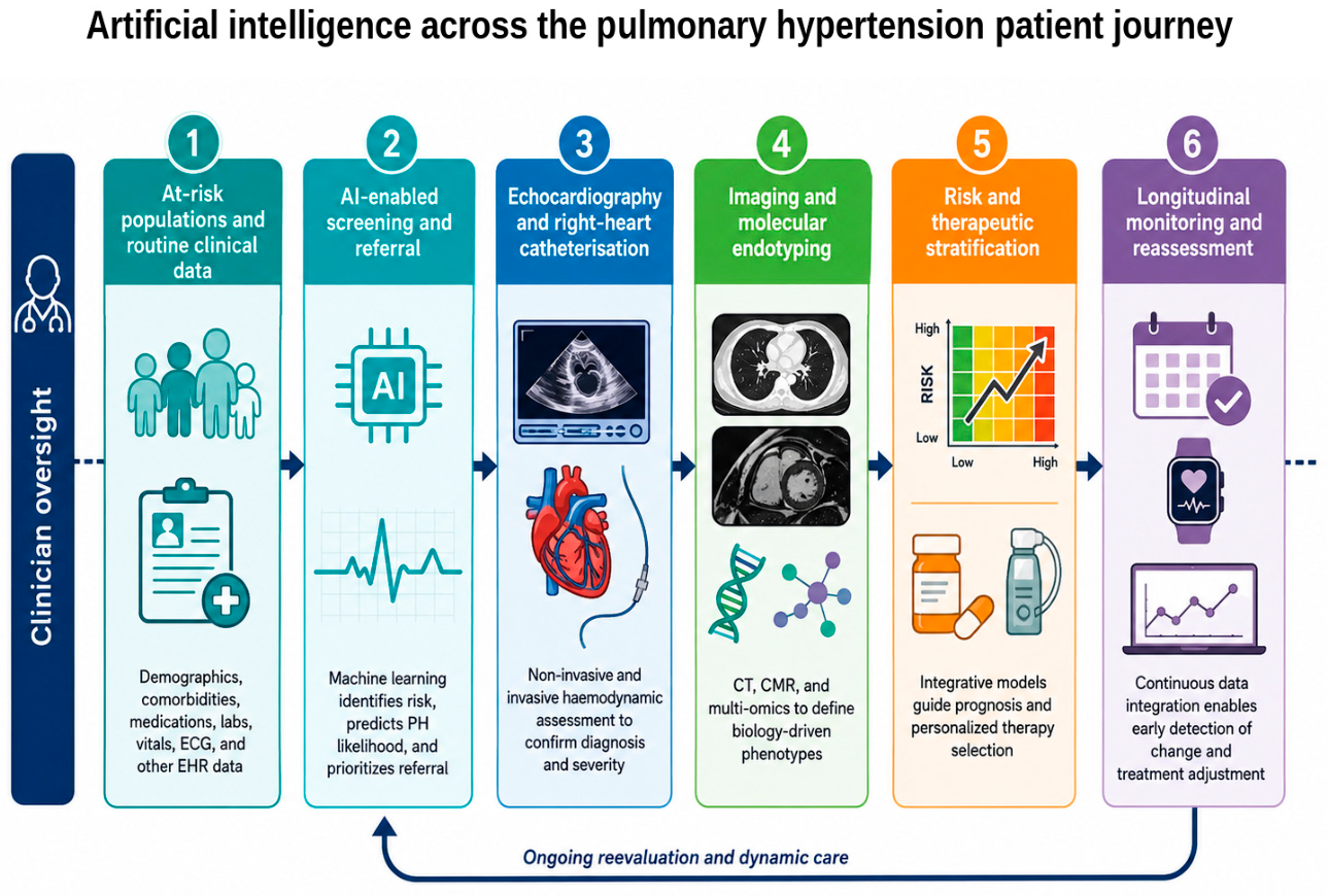
Figure 2.
Multimodal AI architecture for precision management of pulmonary hypertension. The schematic shows how heterogeneous clinical, physiological, imaging, and molecular inputs are harmonised and fused into interpretable outputs for diagnosis, phenotyping, prognosis, and therapeutic stratification, with uncertainty estimates and clinician oversight. This figure was created with the assistance of artificial intelligence and subsequently reviewed and curated by the authors.
Figure 2.
Multimodal AI architecture for precision management of pulmonary hypertension. The schematic shows how heterogeneous clinical, physiological, imaging, and molecular inputs are harmonised and fused into interpretable outputs for diagnosis, phenotyping, prognosis, and therapeutic stratification, with uncertainty estimates and clinician oversight. This figure was created with the assistance of artificial intelligence and subsequently reviewed and curated by the authors.
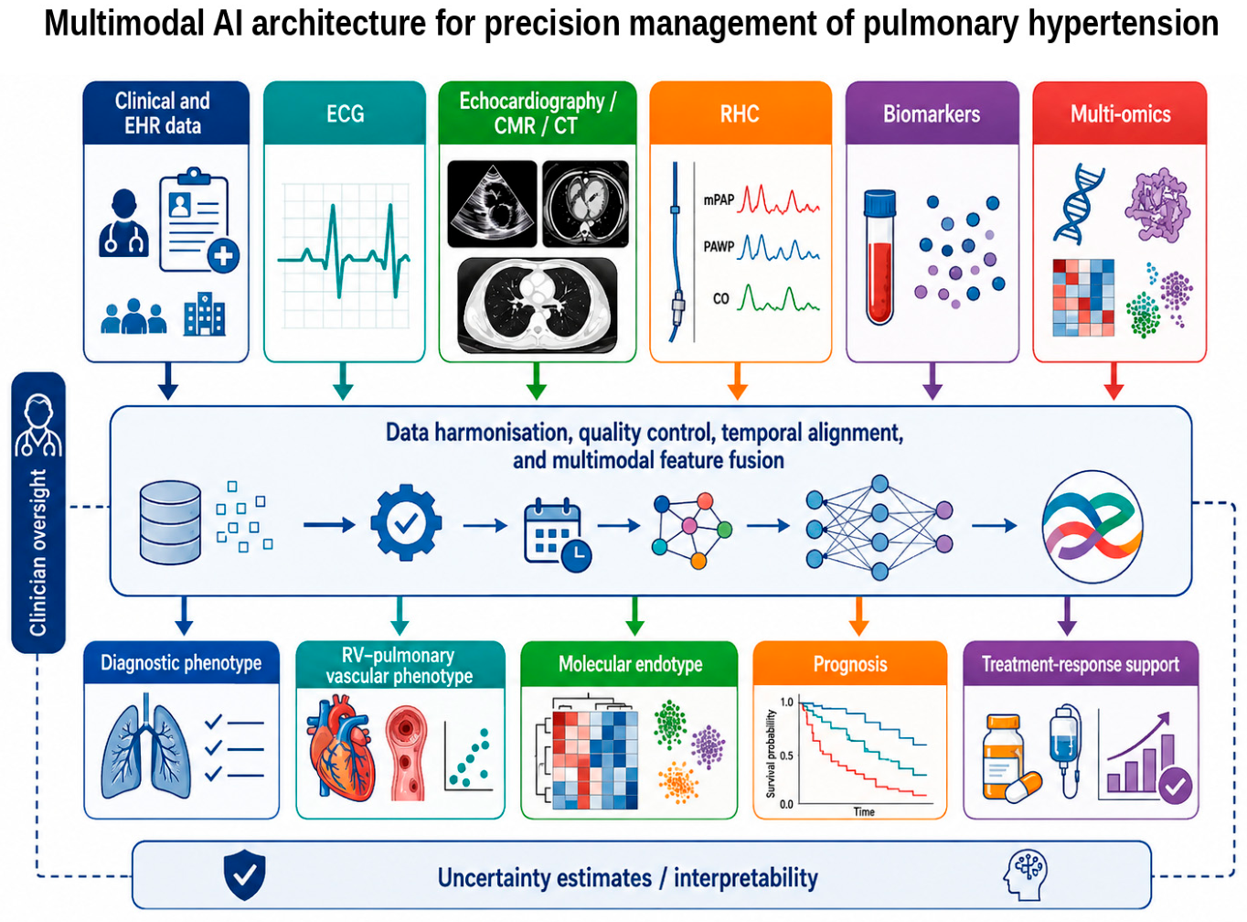

Table 1.
Data modalities and AI tasks in pulmonary hypertension.
| Data source | Representative inputs | AI tasks and potential outputs | Principal limitations |
|---|---|---|---|
| Clinical data and EHRs | Symptoms, diagnoses, medication exposure, healthcare use, laboratory trajectories | Case finding, referral prioritisation, missing-data-aware risk prediction, longitudinal phenotyping | Coding heterogeneity, informative missingness, temporal drift, confounding by healthcare access |
| Electrocardiography | Raw 12-lead waveforms, intervals, rhythm and morphology | Early detection, right-heart stress signatures, phenotyping of pre- versus post-capillary disease | Spectrum bias, device and site effects, limited mechanistic interpretability |
| Echocardiography | TRV, RV dimensions, TAPSE, FAC, strain, RA area, cine loops | Automated measurement, PH probability estimation, RV phenotyping and prognosis | Image quality, vendor dependence, absent TR signal, threshold uncertainty |
| Cardiac magnetic resonance | RV volumes, mass, ejection fraction, tissue features, three-dimensional motion | Automated segmentation, motion phenotyping, haemodynamic estimation and outcome prediction | Referral-centre datasets, acquisition heterogeneity, cost and availability |
| CT/CTPA | Vascular calibre and pruning, perfusion, thromboembolic lesions, parenchymal radiomics | Aetiological classification, CTEPH support, quantitative vascular and lung phenotyping | Radiomic instability, reconstruction dependence, radiation and contrast exposure |
| Right-heart catheterisation | Pressure and flow waveforms, mPAP, PAWP, PVR, RAP, cardiac index, SvO2 | Physiological phenotyping, quality control, trajectory and outcome modelling | Invasive acquisition, waveform artefacts, centre-specific protocols |
| Biomarkers and multi-omics | Proteins, metabolites, RNA, genetic variants, single-cell and spatial data | Molecular endotyping, biomarker panels, target discovery and trial enrichment | High dimensionality, batch effects, limited sample size, uncertain causality |
| Wearables and remote monitoring | Heart rate, activity, oxygen saturation, symptoms and home measurements | Early deterioration alerts, treatment-response monitoring and adaptive follow-up | Adherence, missingness, device variability, alert burden and privacy |
Note: Abbreviations: AI, artificial intelligence; CTEPH, chronic thromboembolic pulmonary hypertension; EHR, electronic health record; FAC, fractional area change; mPAP, mean pulmonary arterial pressure; PAWP, pulmonary arterial wedge pressure; PVR, pulmonary vascular resistance; RA, right atrium; RAP, right atrial pressure; RV, right ventricle; SvO2, mixed venous oxygen saturation; TAPSE, tricuspid annular plane systolic excursion; TR, tricuspid regurgitation; TRV, tricuspid regurgitation velocity.
Table 2.
Multi-omics layers for AI-based molecular endotyping in pulmonary hypertension.
| Omics layer | Biological information | Representative AI/ML task | Potential translational output and current limitations |
|---|---|---|---|
| Genomics and epigenomics | Rare and common variants, regulatory architecture, methylation and chromatin state | Variant prioritisation, polygenic modelling, pathway and network analysis | Susceptibility endotypes and family counselling; incomplete penetrance and ancestry imbalance |
| Bulk transcriptomics | Active gene-expression programmes in blood or tissue | Unsupervised clustering, differential-expression signatures, network modules | Biological subgroups and pathway activity; tissue access and cell-composition confounding |
| Proteomics | Circulating mediators, matrix turnover, inflammation and cardiac stress | Feature selection, latent factors, survival and response models | Parsimonious biomarker panels; platform effects and uncertain cell of origin |
| Metabolomics and lipidomics | Bioenergetics, substrate use, mitochondrial and systemic adaptation | Multivariate signatures, pathway enrichment, longitudinal state modelling | Dynamic disease and RV-adaptation markers; sensitivity to diet, organ function and processing |
| MicroRNA and non-coding RNA | Post-transcriptional regulation and intercellular signalling | Network inference, target prediction and integrated classifiers | Mechanistic biomarkers and therapeutic targets; assay and normalisation variability |
| Single-cell and spatial omics | Cell states, lineage-specific programmes and cellular neighbourhoods | Cell-state discovery, trajectory analysis, spatial graph learning | Mechanistically resolved endotypes and target discovery; high cost, small samples and limited routine scalability |
Note: Abbreviations: AI, artificial intelligence; ML, machine learning; RV, right ventricle.
Disclaimer/Publisher’s Note: The statements, opinions and data contained in all publications are solely those of the individual author(s) and contributor(s) and not of MDPI and/or the editor(s). MDPI and/or the editor(s) disclaim responsibility for any injury to people or property resulting from any ideas, methods, instructions or products referred to in the content. |
© 2026 by the authors. Licensee MDPI, Basel, Switzerland. This article is an open access article distributed under the terms and conditions of the Creative Commons Attribution (CC BY) license (http://creativecommons.org/licenses/by/4.0/).
Copyright: This open access article is published under a Creative Commons CC BY 4.0 license, which permit the free download, distribution, and reuse, provided that the author and preprint are cited in any reuse.