

Submitted:
13 August 2026
Posted:
14 August 2026
You are already at the latest version
Abstract
SHANK3-related disorder within the Phelan-McDermid syndrome spectrum is clinically heterogeneous and commonly includes severe expressive language impairment, hypotonia, motor coordination difficulties, sensory features, sleep disturbance, reduced pain expression, and regression in a subset of affected individuals. However, a potential dystonic contribution to expressive language impairment, orofacial dysfunction, other motor abnormalities, and illness-associated functional deterioration has not been systematically examined. Case Presentation: We report a 7-year-old girl whose early presentation was dominated by severe expressive language delay. Receptive communication, social reciprocity and practical problem-solving appeared substantially stronger than expressive output suggested. Longitudinal features included fine-motor incoordination, toe walking, intermittent atypical hand movements, sensory reactivity, overload-associated dysregulation and prolonged mouthing/oral exploration of objects. During febrile illness in March 2025, she developed painful gait deterioration, continuous toe walking, dystonic hand posturing and worsening orofacial motor function. Low-dose levodopa was initiated because dopa-responsive dystonia or a related dopaminergic disorder was clinically considered. Following initiation of levodopa, improvements were observed across several functional domains that had shown only slow progress or prolonged plateau phases during the preceding years. Short-read whole-genome sequencing identified a heterozygous pathogenic SHANK3 frameshift variant, NM_001372044.2:c.3904dup, with the predicted protein consequence NP_001358973.1:p.(Ala1302fs). With continued levodopa and later trihexyphenidyl, further gains were observed across gait stability, orofacial control, spoken language output, sensory tolerance and functional participation; the previously persistent mouthing/oral exploration was no longer observed after dopaminergic treatment had been established. Conclusion: Across neurodevelopmental disorders of diverse genetic aetiology and across age groups, the co-occurrence of marked expressive language impairment, orofacial dysfunction, and fine- or gross-motor abnormalities should prompt targeted assessment for dystonia or another movement disorder. Such motor manifestations may represent a convergent and potentially modifiable component of the clinical phenotype, including previously unrecognized impairment of speech-motor control. The multidomain improvement observed following initiation of levodopa in this case of SHANK3-related disorder was clinically meaningful and hypothesis-generating and warrants prospective investigation.
Keywords:
SHANK3
; Phelan-McDermid syndrome
; dystonia
; expressive language impairment
; case report
1. Introduction
Severe expressive language impairment in childhood may occur in isolation but can also be part of a broader genetic, neurodevelopmental or neuromotor disorder [1]. This distinction is particularly relevant when restricted spoken output contrasts with stronger receptive communication and everyday problem-solving in familiar settings.
The true prevalence of SHANK3-related Phelan-McDermid syndrome remains uncertain. The condition is likely underdiagnosed because the phenotype is heterogeneous and molecular genetic testing is required to establish the diagnosis [2,3]. Reported features include developmental delay, intellectual disability, severe speech and language impairment, hypotonia, gait and coordination problems, sensory abnormalities, sleep disturbance, autism-spectrum-like behaviours, reduced pain sensitivity and regression in a subset of affected individuals [3,4,5,6,7,8,9].
Motor impairment in SHANK3-related disorder is commonly described as hypotonia, delayed motor development, impaired coordination, abnormal gait or regression [3,6,7,8,9]. Dystonia has received less explicit attention as a potential contributor to expressive language impairment, orofacial dysfunction, fine-motor difficulty, fluctuation or illness-associated deterioration. This case report describes a child whose early clinical presentation was dominated by severe expressive language impairment, followed by infection-associated dystonic motor deterioration and subsequent treatment-associated multidomain improvement.
2. Case Presentation
The patient is currently a 7-year-old girl. Pregnancy, delivery, neonatal adaptation and newborn hearing screening were unremarkable; no organ malformations were known. Clinical examination did not reveal dysmorphic features. Early gross motor milestones, including head control, rolling, sitting and pulling to stand, were achieved within an expected timeframe. Independent standing occurred at approximately 12 months; independent walking emerged about 3 months later than expected from the available motor prerequisites.
From the second year of life onward, marked expressive speech and language impairment became increasingly apparent. First words had emerged, but subsequent spoken output remained clearly restricted. In familiar settings with reduced sensory load, receptive communication, social reciprocity and practical everyday problem-solving appeared closer to age-expected levels. Eye contact, non-verbal interaction and communicative intent were preserved. Under familiar, low-sensory-load conditions the child followed multi-step instructions, solved age-appropriate puzzles, played memory-type matching games, demonstrated quantity comprehension, sorted and matched items according to task rules, retained task-relevant information over short intervals and transferred learned strategies to new tasks. These video-documented observations supported the clinical impression that receptive understanding, visual-spatial reasoning, rule-based learning, short-term task retention and practical problem-solving were substantially stronger than expressive speech output suggested.
Speech and language therapy started before 2 years of age and was later directed towards suspected childhood apraxia of speech and orofacial motor dysfunction. Therapy reports described a complex developmental language disorder. Progress remained slow up to approximately 6 years of age, with repeated plateau phases. By that age, expressive speech was usually limited to two- to three-word utterances and was intelligible mainly to experienced listeners in the family environment and to the long-standing speech-language therapist; four-word utterances were rare, and sentence structure remained unstable and grammatically immature.
In parallel, fine-motor difficulties were apparent in everyday bimanual activities, including cutlery use, fastening zippers, putting on shoes, using scissors, and drawing. Intermittent difficulties with selective voluntary motor control were also observed. Voluntary thumb abduction and tongue protrusion could not be performed reliably on request, whereas spontaneous thumb abduction was observed; these findings were initially interpreted as dyspraxic. Intermittent atypical hand and arm movements were also noted but were not initially considered part of a movement disorder. Other longitudinal features included prolonged mouthing/oral exploration of objects, pronounced sound sensitivity, reduced pain expression, overload-associated emotional and behavioural decompensation, and substantial toe walking. From 2.5 years onward, physiotherapy, occupational therapy with sensory integration, music therapy, and developmental educational support were added.
During a febrile intercurrent illness in March 2025, at the age of 5 years and 11 months, the child developed a marked exacerbation and generalization of the motor phenotype, clinically suggestive of a dystonia-associated movement disorder. Previously intermittent toe walking intensified to continuous toe walking. Walking became markedly painful and restricted; she could walk only a few steps, and descending stairs was no longer possible because of pain. In addition, action-dependent upper-limb dysfunction became evident. When attempting to bring a spoon to the mouth, she could no longer perform a smooth, goal-directed hand-arm movement. Instead, rapid, poorly coordinated and excessive movement components occurred. Recurrent torsional, twisting arm movements were observed; these appeared patterned and not voluntarily controllable. Craniofacially, unilateral mouth-corner elevation and ipsilateral eye squeezing were documented on home video recordings and reviewed during the movement-neurological reassessment. The video material supported the interpretation of action- and stress-dependent dystonic motor involvement, including gait deterioration, torsional upper-limb movements, impaired goal-directed hand-arm control and craniofacial activation. This constellation prompted movement-neurological reappraisal and consideration of a broader dystonia-associated motor disorder.
Because dopa-responsive dystonia or another disorder of dopamine synthesis or metabolism was clinically considered, levodopa treatment was initiated. The preparation contained 100 mg levodopa and 25 mg carbidopa per tablet. Quarter-tablets were used, corresponding to 25 mg levodopa and 6.25 mg carbidopa per dose. During the first week, caregivers and clinicians noted improved gait and orofacial motor control, as well as reduced painful posturing, while the child was receiving 25 mg levodopa twice daily. After 2 weeks, the dosing frequency was increased to three times daily and, after a further 2 weeks, to four times daily, with daytime doses administered approximately 3 hours apart. At the 6-week assessment, she was receiving 25 mg levodopa/6.25 mg carbidopa four times daily, corresponding to 100 mg/day levodopa and 25 mg/day carbidopa, or approximately 5 mg/kg/day levodopa at a body weight of 20 kg. At that time, she was 6 years old, 115 cm tall, and weighed 20 kg.
After titration, the child showed gradual and sustained improvement across motor, orofacial, and speech-language domains during stable levodopa treatment. Walking became less painful and more stable, toe walking decreased, orofacial control improved, and spoken language became more readily accessible. These changes were also independently noted by several treating therapists, who had not been informed that levodopa treatment had been initiated when they reported the improvements.
In summer 2025, after several weeks of stable and well-tolerated levodopa treatment with continued clinical improvement, she developed a severe dystonic exacerbation during a febrile illness. The clinical pattern largely resembled that observed during the March 2025 episode, including marked pain during walking, continuous motor agitation, and recurrent painful, rapid, patterned dystonic movements of the right upper limb, characterized by repeated upward movements of the arm with stereotyped spiral torsion of the wrist and hand. In addition, swallowing difficulty raising concern for aspiration and vomiting with features suggestive of delayed gastric emptying occurred. Given concern that the severe dystonic exacerbation was progressing toward status dystonicus, hospital evaluation included screening for systemic complications and showed no clinical or biochemical evidence of rhabdomyolysis. Management included benzodiazepine treatment, hydration, calorie support, antipyretic treatment, supportive infection management, physical rest, and reduction of environmental stimulation. During the acute episode, the levodopa/carbidopa regimen was increased from 25 mg/6.25 mg four times daily to 25 mg/6.25 mg five times daily, corresponding to a total daily dose of 125 mg levodopa and 31.25 mg carbidopa. This regimen was maintained thereafter. Within 1–2 weeks after resolution of the acute episode, she returned to the functional level that had been achieved before the exacerbation. Gradual improvements across motor, orofacial, and speech-language domains then resumed under continued levodopa treatment and remained evident until another febrile illness occurred approximately 4 months later. During this illness, she developed a painful cervical dystonic episode with early-morning awakening and a fixed cervical tilt consistent with torticollis. Levodopa was increased to 25 mg levodopa/6.25 mg carbidopa six times daily, corresponding to 150 mg/day levodopa and 37.5 mg/day carbidopa, approximately 6.5 mg/kg/day levodopa at 23 kg body weight, and trihexyphenidyl was added. Trihexyphenidyl was initiated at 0.5 mg/day and slowly titrated by 0.5 mg every 2 weeks to 0.5 mg four times daily. The torticollis resolved during treatment. Transient difficulty initiating micturition occurred early during trihexyphenidyl treatment; it resolved spontaneously.
The response supported broad neurogenetic evaluation rather than restriction to classic dopa-responsive dystonia. Short-read whole-genome sequencing with bioinformatic exome/panel analysis identified a heterozygous pathogenic SHANK3 frameshift variant, NM_001372044.2:c.3904dup, with the predicted protein consequence NP_001358973.1:p.(Ala1302fs), establishing SHANK3-related disorder. The variant was interpreted as a frameshift with premature termination and expected loss of function. A genomic copy-number analysis did not identify disease-causing copy-number variants, including in the 22q13 region. Karyotyping showed a normal female karyotype, 46,XX. Targeted review of exome data did not reveal another molecular diagnosis within the evaluated dystonia/dyskinesia, monoaminergic, mitochondrial or selected neurodevelopmental differential-diagnosis spectrum.
At most recent follow-up, maintenance treatment consisted of levodopa/carbidopa 25 mg/6.25 mg six times daily, corresponding to 150 mg/day levodopa and 37.5 mg/day carbidopa, and trihexyphenidyl 0.5 mg four times daily. The regimen had remained stable for approximately 8 months. Medication administration and adherence were documented by the caregivers. Overall, the regimen was well tolerated, as assessed through caregiver reports, clinical follow-up, and the child’s own reports of wellbeing and symptoms. During two subsequent febrile illnesses, the total daily levodopa dose was temporarily increased by 25 mg. Supportive measures were initiated promptly at the first signs of each illness, including physical rest, increased caloric intake, reduced sensory stimulation, and antipyretic and anti-inflammatory treatment as clinically indicated. High-protein meals were avoided around levodopa administration to minimise potential interference with levodopa absorption. Both illnesses resolved without progression to a severe dystonic exacerbation. After resolution of each febrile illness, the levodopa/carbidopa regimen was reduced to the maintenance dose of 25 mg/6.25 mg six times daily.
Compared with the period before the March 2025 deterioration, baseline function was reported to have improved across several domains. Spoken output became more continuous, with a marked increase in vocabulary and spontaneous, grammatically structured sentences exceeding five words and occasionally reaching up to ten words. Articulatory clarity also improved but remained variable; familiar communication partners could generally identify the intended words and sentences, whereas speech was sometimes less readily understood by unfamiliar listeners. Motor gains included improved walking endurance, orofacial control, and gross-motor stability; the child could run faster, walk longer distances, swim independently, and perform sustained, coordinated jumping on a trampoline. Fine-motor function improved, including independent dressing. Continence improved substantially. The previously persistent mouthing/oral exploration of objects was no longer observed after dopaminergic treatment had been established. Mild mouth pulling, brief eye squeezing and occasional hand posturing were usually absent but could re-emerge transiently during marked stress or increased demand.
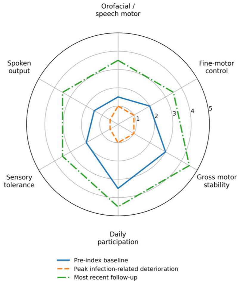
The multidomain functional profiles before the March 2025 deterioration, during the peak infection-associated exacerbations, and at the most recent follow-up are shown in Figure 1.
A chronological timeline of the clinical course, diagnostic evaluation, therapeutic interventions, and reported outcomes is provided in Supplementary Table 2.
3. Focused Literature Review
A focused case-based literature search was conducted using PubMed and Google Scholar through 1 August 2026. Search terms combined “SHANK3”, “Phelan–McDermid syndrome”, and “22q13 deletion syndrome” with terms relating to speech or language impairment, speech-motor or orofacial dysfunction, motor or movement abnormalities, gait, dystonia, abnormal posturing, regression, catatonia, neuropsychiatric deterioration, illness- or fever-associated deterioration, and treatment. Reference lists of relevant reports and reviews were screened for additional publications. Case reports, case series, sibling and family reports were considered when they provided sufficiently detailed patient-level information relevant to one or more of these domains. Reports meeting these criteria and considered relevant to the interpretation of the present case are summarized in Table S1, including genetic findings, relevant clinical manifestations and course, source interpretation, treatment, and reported outcomes [10,11,12,13,14,15,16,17,18,19,20,21,22,23,24,25,26,27,28,29,30,31,32,33,34,35,36,37,38,39,40,41,42].
Across the identified reports, recurrently described features included severe or absent speech, developmental delay or intellectual disability, autism-spectrum features, hypotonia, abnormal gait, regression, catatonia, and neuropsychiatric deterioration. Reports of regression, catatonia-related deterioration, or severe neuropsychiatric decompensation included lithium-associated reversal or stabilization in selected cases [18,36] and improvement following immunomodulatory treatment in a subacute neuropsychiatric case series [12]. Early-onset catatonic manifestations were described in an additional three-patient case study [16]. Motor- and movement-related findings included a parkinsonian-dystonic phenotype in a patient with a pathogenic SHANK3 variant and co-occurring SYNJ1 variants [19], ataxia or progressive lower-extremity spasticity [42], and regression associated with an additional treatable neurological disorder [21]. Marked language impairment was also described in reports involving a SHANK3-disrupting duplication [20].
Taken together, the identified reports show phenotypic overlap with the present case across expressive language impairment, speech-motor and orofacial dysfunction, coordination and gait abnormalities, fine-motor difficulty, atypical posturing, psychomotor agitation, episodic deterioration, and other involuntary movements. However, these features were generally reported and interpreted within separate diagnostic frameworks, including hypotonia, developmental motor delay, regression, catatonia, parkinsonism, or neuropsychiatric deterioration. Within the identified literature, they were not systematically evaluated as potentially interrelated manifestations of a shared dystonia-related motor component. This gap provides the context for the present case, in which febrile illness accentuated previously less conspicuous motor abnormalities and thereby revealed a broader dystonia-associated motor phenotype. The subsequent multidomain improvement after initiation of levodopa further raised the possibility of a clinically modifiable motor contribution.
4. Discussion
The central observation in this case is a longitudinal shift in diagnostic interpretation. For several years, the clinical presentation appeared to be dominated by severe expressive language impairment. As the clinical course evolved, however, febrile illness accentuated previously less conspicuous motor abnormalities and thereby revealed a broader dystonia-associated motor phenotype. Viewed in this context, the speech-motor, orofacial, fine-motor, and gait abnormalities may represent interrelated manifestations of a broader motor-neurodevelopmental phenotype in SHANK3-related disorder.
The phenotypic constellations described across the identified case-based reports provide a plausible basis for this interpretation. Severe language impairment and a range of motor, gait-related, postural, regression-associated, catatonic, and neuropsychiatric features were reported in varying combinations [10,11,12,13,14,15,16,17,18,19,20,21,22,23,24,25,26,27,28,29,30,31,32,33,34,35,36,37,38,39,40,41,42]. Although different diagnostic labels were used, several of these constellations show clinically relevant overlap with the present case. The reported manifestations were generally interpreted within separate frameworks, including hypotonia, developmental motor delay, regression, catatonia, parkinsonism, or an additional neurological disorder [12,16,18,19,21,36,42]. The identified literature did not systematically examine whether speech-motor, orofacial, limb-motor, and gait abnormalities might represent interrelated expressions of an unrecognized dystonia-related motor component.
This distinction is clinically important. Contemporary paediatric dystonia literature conceptualizes dystonia as a disorder of sensorimotor integration within distributed networks [43]. Current recommendations describe acutely worsening dystonia as a clinical continuum that includes pre-status dystonicus and status dystonicus and emphasize prompt recognition, supportive care, dystonia-directed treatment, and identification and minimization of aggravating factors [44]. Intercurrent illness, fever, dehydration, pain, discomfort, reduced intake, and other physiological stressors may precipitate or aggravate dystonic worsening [44]. These principles support the plausibility that febrile illness, pain, reduced intake, dehydration risk, and catabolic stress lowered motor reserve and amplified a pre-existing dystonic component in the present child.
When acute dystonic worsening is accompanied by marked motor agitation, painful patterned posturing, gait deterioration, or bulbar symptoms, its externally observable presentation may resemble behavioural, catatonic, or neuropsychiatric deterioration. Regression, catatonia, and severe neuropsychiatric presentations have been reported in individuals with SHANK3-related disorder and Phelan-McDermid syndrome [12,16,18,36]. In individuals with limited expressive communication, an inability to communicate pain and internal physical distress reliably may further increase the risk of diagnostic misinterpretation. This proposed phenomenological overlap does not imply that previously reported psychiatric or catatonic episodes were dystonic. It supports parallel assessment for dystonia and other movement disorders when acute behavioural or psychomotor deterioration is accompanied by pain, patterned movements or postures, gait deterioration, bulbar dysfunction, illness-associated fluctuation, or other motor signs.
In the present child, pain and physical distress were clearly communicated. The concurrent painful gait deterioration, continuous toe walking, patterned torsional limb movements, impaired goal-directed hand-arm control, and craniofacial activation, together with the close temporal association with febrile illness, supported recognition of the episodes as dystonic exacerbations and argued against a primary behavioural or psychiatric presentation. During the later severe episode, marked motor agitation occurred together with painful torsional arm posturing, swallowing difficulty, and gastrointestinal symptoms. The overall clinical constellation remained consistent with a severe dystonic exacerbation.
This distinction had direct therapeutic implications. Acute management addressed both the dystonic exacerbation and its physiological triggers through hydration, caloric support, antipyretic and supportive infection treatment, physical rest, reduction of environmental stimulation, benzodiazepine treatment during the severe episode, and adjustment of dystonia-directed medication. During two subsequent febrile illnesses, supportive measures were initiated promptly and the total daily levodopa dose was temporarily increased. Both illnesses resolved without progression to another severe dystonic exacerbation, supporting continuation of this individualized illness-related management strategy.
The improvement following initiation of levodopa/carbidopa is clinically notable. Early improvement in gait, orofacial motor control, and painful posturing was observed within the first week of treatment, after several years of very slow developmental progress and repeated plateau phases despite multidisciplinary intervention. Gradual and sustained gains subsequently occurred during continued levodopa treatment across multiple functional domains, including gait, orofacial control, spoken language output, fine-motor function, continence, sensory tolerance, functional participation, and mouthing or oral exploration. Taken together, the rapid onset of improvement after several years of slow progress and repeated plateau phases, the sustained multidomain gains, and the concordant observations by caregivers, clinicians, and treating therapists who were unaware of the medication change when they reported the improvements strongly support a clinically meaningful levodopa-associated response in this child. The initial multidomain response preceded the later addition of trihexyphenidyl, after which further improvement was observed. Because this was an uncontrolled single case without standardized outcome measures, causality cannot be conclusively established, and the observed response cannot yet be generalized to other individuals with SHANK3-related disorder.
The response to levodopa/carbidopa supported consideration of a dopaminergic contribution to the motor phenotype. A 2021 report described a girl with a pathogenic SHANK3 variant and co-occurring SYNJ1 variants who developed early-onset parkinsonism with hypomimia, dysarthric and hypophonic speech, gait impairment, limb and axial dystonia, and later catatonic episodes [19]. Levodopa/carbidopa was associated with rapid improvement in motor function, gait, and speech. Withdrawal of levodopa subsequently worsened the motor symptoms without ameliorating the psychiatric manifestations, whereas reintroduction of levodopa/carbidopa was followed by clear motor improvement. In that report, the parkinsonian-dystonic phenotype and levodopa responsiveness were interpreted primarily in relation to the co-occurring SYNJ1 variants. The case therefore does not establish levodopa responsiveness as a manifestation of SHANK3-related disorder itself. Nevertheless, the concordant improvement across motor, gait, and speech domains, together with deterioration after withdrawal and improvement following reintroduction, provides clinically relevant contextual support for the present observation. Among the reports identified in this focused case-based search, we found no previous case describing levodopa-responsive dystonia in an individual with SHANK3-related disorder without an additional potentially explanatory molecular diagnosis [10,11,12,13,14,15,16,17,18,19,20,21,22,23,24,25,26,27,28,29,30,31,32,33,34,35,36,37,38,39,40,41,42].
Evidence from other paediatric neurological contexts indicates that dopaminergic treatment may be clinically relevant in selected neurodevelopmental and movement-disorder phenotypes. These reports include levodopa-responsive dystonia secondary to CTNNB1 neurodevelopmental disorder and heterogeneous clinical experience with very-low-dose levodopa in paediatric neurological conditions [45,46]. Trihexyphenidyl is commonly used as a symptomatic treatment for paediatric dystonia, although the available evidence remains limited and is derived largely from small, heterogeneous, or non-blinded studies [47,48,49]. These observations provide relevant clinical context but do not constitute a general treatment recommendation for SHANK3-related disorder.
Experimental findings provide additional, although indirect, biological context. Shank3 disruption has been shown to affect the maturation and function of ventral tegmental area social-reward circuits [50]. Shank3B-deficient mice also show reduced neurite outgrowth and arborization in primary neurons derived from dopaminergic brain regions, together with alterations in dopaminergic receptor gene expression [51]. More broadly, dopaminergic perturbation has been implicated in the pathophysiology of several neurodevelopmental disorders [52]. These findings support the biological plausibility of dopaminergic involvement but do not establish a SHANK3-specific dopaminergic or cholinergic therapeutic mechanism. Such a mechanism remains hypothesis-generating.
The genetic findings also require careful interpretation. The identified heterozygous pathogenic SHANK3 frameshift variant, NM_001372044.2:c.3904dup, with the predicted protein consequence NP_001358973.1:p.(Ala1302fs), is consistent with a loss-of-function mechanism. The normal karyotype and absence of disease-causing copy-number variants, including in the 22q13 region, support an intragenic SHANK3-related disorder instead of a terminal 22q13 deletion or ring chromosome 22. Because parental testing was not performed, a de novo origin cannot be considered proven. It is clinically likely given the unremarkable family history and clinically unaffected parents and half-siblings, although germline mosaicism remains a residual consideration.
This report has several limitations. It describes a single individual and is based on longitudinal clinical observation, caregiver report, home-video documentation, and routine clinical records; it did not follow a prospective study protocol. Formal psychometric testing could not be completed because the child did not engage sufficiently with the test tasks and repeatedly focused on competing stimuli. Home-video recordings nevertheless documented task performance and everyday problem-solving under familiar, low-sensory-load conditions.
Treatment-related changes were not assessed using standardized movement, speech, language, sensory, or functional outcome measures. The semi-quantitative ratings presented in Figure 1 are retrospective descriptive estimates, not validated clinical scores. The literature review was focused and phenotype-based but was not conducted as a formal systematic review. Furthermore, incomplete reporting of illness-related triggers, pain, detailed movement phenomenology, and parallel assessment for catatonia and dystonia in previously published cases limits direct comparison with the present case and precludes reliable retrospective reclassification of earlier episodes.
The findings should therefore be interpreted as clinically relevant and hypothesis-generating. They do not establish a generalizable SHANK3-specific treatment strategy. Nevertheless, this case supports targeted movement-disorder assessment in patients with marked expressive language impairment accompanied by orofacial dysfunction, patterned or otherwise atypical posturing, and fine- or gross-motor abnormalities, particularly when symptoms fluctuate or worsen during intercurrent illness. The possible phenomenological overlap between severe dystonic exacerbation and apparent behavioural, catatonic, or neuropsychiatric deterioration further supports parallel neurological and neuropsychiatric assessment and argues against premature attribution to a single diagnostic framework.
This diagnostic principle may be relevant across age groups and across neurodevelopmental disorders of diverse genetic aetiology. Prospective studies are required to determine whether a reproducible and therapeutically modifiable dystonia-related phenotype can be identified in SHANK3-related disorder and other genetically defined neurodevelopmental conditions.
5. Conclusions
This longitudinal case suggests that an apparently language-dominant presentation of SHANK3-related disorder may conceal a broader motor phenotype that becomes more evident under physiological stress. More broadly, irrespective of the underlying genetic or neurodevelopmental diagnosis, the co-occurrence of severe expressive language impairment with fluctuating abnormalities of gait, posture, and limb or orofacial control should prompt active consideration of dystonia, particularly when symptoms worsen during intercurrent illness. In this child, levodopa initiation was associated with early improvement and sustained gains across multiple functional domains, with further progress after the addition of trihexyphenidyl. Although a single uncontrolled observation cannot establish causality or support a general treatment recommendation, recognizing dystonia in such constellations is clinically important because it may reveal a potentially pharmacologically modifiable contributor to disability.
Supplementary Materials
Supplementary Table S1. Published case reports, case series, and family reports relevant to speech and language impairment, movement abnormalities, regression, catatonia, illness-associated deterioration, and treatment response in SHANK3-related disorder. References [10,11,12,13,14,15,16,17,18,19,20,21,22,23,24,25,26,27,28,29,30,31,32,33,34,35,36,37,38,39,40,41,42] are cited in Table S1. Supplementary Table S2. Timeline of the clinical course, diagnostic evaluation, treatment and reported outcomes.
Author Contributions
Ulrike Roser conceptualized the case report, reviewed and synthesized the longitudinal clinical course, developed the clinical interpretation, prepared the figure and drafted the manuscript. Mattias Roser contributed to clinical interpretation and critically revised the manuscript. Both authors read and approved the final manuscript.
Funding Sources
No specific funding was received for this work.
Reporting Guideline
This case report was prepared in accordance with the CARE reporting guidelines. A completed CARE checklist is provided as a separate file.
Patient and Caregiver Perspective
At the time of the first exacerbation in March 2025, before levodopa treatment was initiated, the child could communicate that she was in pain but could not reliably describe where it hurt. During later follow-up while receiving treatment, she was able to communicate her general wellbeing, regularly stated that she felt well, and, when pain occurred, increasingly reported it verbally and named the affected body part. The caregivers perceived the treatment-associated changes as clinically meaningful because improvement occurred after several years of slow progress and plateau phases and affected mobility, orofacial control, spoken communication, sensory tolerance, oral exploration behaviour, and daily participation.
Institutional Review Board Statement
Ethical review and approval were not required for this retrospective single-patient case report, which describes routine clinical care and involved no research-specific intervention or prospective study protocol, in accordance with applicable local regulations.
Informed Consent Statement
Written informed consent for publication of the clinical information was obtained from the patient’s legal guardians.
Data Availability Statement
All relevant de-identified clinical data are included in this article and its supplementary material. Home video recordings were reviewed clinically but are not publicly available because they contain identifiable patient material. Additional de-identified details are available from the corresponding author on reasonable request.
Acknowledgments
We thank the team at the MGZ – Medical Genetics Center, Munich, Germany, for performing the genetic testing and providing the molecular diagnostic interpretation.
Conflicts of Interest Statement
The authors declare no financial or commercial conflicts of interest. The clinical observations and therapeutic interpretation reported in this manuscript were derived from routine clinical care and were not influenced by any external sponsor, commercial entity or product manufacturer.
References
- Kaspi, A.; Hildebrand, M.S.; Jackson, V.E.; et al. Genetic aetiologies for childhood speech disorder: novel pathways co-expressed during brain development. Mol. Psychiatry 2023, 28(4), 1647–1663. [Google Scholar] [CrossRef] [PubMed]
- Orphanet. Phelan-McDermid syndrome. ORPHA. Available online: https://www.orpha.net/en/disease/detail/ (accessed on 28 June 2026).
- Schön, M.; Lapunzina, P.; Nevado, J.; et al. Definition and clinical variability of SHANK3-related Phelan-McDermid syndrome. Eur. J. Med. Genet. 2023, 66(7), 104754. [Google Scholar] [CrossRef] [PubMed]
- Burdeus-Olavarrieta, M.; Nevado, J.; van Weering-Scholten, S.; et al. Consensus recommendations on communication, language and speech in Phelan-McDermid syndrome. Eur. J. Med. Genet. 2023, 66(5), 104745. [Google Scholar] [CrossRef] [PubMed]
- Brignell, A.; Gu, C.; Holm, A.; et al. Speech and language phenotype in Phelan-McDermid (22q13.3) syndrome. Eur. J. Hum. Genet. 2021, 29, 564–574. [Google Scholar] [CrossRef] [PubMed]
- Frank, Y. The neurological manifestations of Phelan-McDermid syndrome. Pediatr. Neurol. 2021, 122, 59–64. [Google Scholar] [CrossRef] [PubMed]
- Srivastava, S.; Sahin, M.; Buxbaum, J.D.; et al. Updated consensus guidelines on the management of Phelan-McDermid syndrome. Am. J. Med. Genet A 2023, 191(8), 2015–2044. [Google Scholar] [CrossRef] [PubMed]
- Chen, L.; Yao, Z.Y.; Wu, X.; et al. Phelan-McDermid syndrome in pediatric patients with novel mutations: genetic and phenotypic analyses. Front Pediatr. 2022, 10, 888001. [Google Scholar] [CrossRef] [PubMed]
- De Rubeis, S.; Siper, P.M.; Durkin, A.; et al. Delineation of the genetic and clinical spectrum of Phelan-McDermid syndrome caused by SHANK3 point mutations. Mol. Autism 2018, 9, 31. [Google Scholar] [CrossRef] [PubMed]
- Accogli, A.; Yang, R.; Blain-Juste, M.E.; et al. SHANK3 mutation and mosaic Turner syndrome in a female patient with intellectual disability and psychiatric features. J. Neuropsychiatry Clin. Neurosci. 2019, 31(3), 272–275. [Google Scholar] [CrossRef] [PubMed]
- Ahn, H.; Seo, G.H.; Keum, C.; et al. Diagnosis of metachromatic leukodystrophy in a patient with regression and Phelan-McDermid syndrome. Brain Dev. 2020, 42(5), 414–417. [Google Scholar] [CrossRef] [PubMed]
- Bey, A.L.; Gorman, M.P.; Gallentine, W.; et al. Subacute neuropsychiatric syndrome in girls with SHANK3 mutations responds to immunomodulation. Pediatrics 2020, 145, e20191490. [Google Scholar] [CrossRef] [PubMed]
- Bisgaard, A.M.; Kirchhoff, M.; Nielsen, J.E.; et al. Chromosomal deletion unmasking a recessive disease: 22q13 deletion syndrome and metachromatic leukodystrophy. Clin. Genet. 2009, 75(2), 175–179. [Google Scholar] [CrossRef] [PubMed]
- Boley, G.; Pierri, J.; Finegold, D.; Pan, L. Evaluation of catatonia in autism and severe depression revealing Phelan-McDermid syndrome and tetrahydrobiopterin deficiency. BMJ Case Rep. 2024, 17(1), e256155. [Google Scholar] [CrossRef] [PubMed]
- Denayer, A.; Van Esch, H.; de Ravel, T.; et al. Neuropsychopathology in 7 patients with the 22q13 deletion syndrome: presence of bipolar disorder and progressive loss of skills. Mol. Syndromol. 2012, 3(1), 14–20. [Google Scholar] [CrossRef] [PubMed]
- Dhossche, D.; de Billy, C.; Laurent-Levinson, C.; Le Normand, M.T.; Recasens, C.; Robel, L.; Philippe, A. Early-onset catatonia associated with SHANK3 mutations: looking at the autism spectrum through the prism of psychomotor phenomena. Front Psychiatry 2023, 14, 1186555. [Google Scholar] [CrossRef] [PubMed]
- Egger, J.I.M.; Zwanenburg, R.J.; van Ravenswaaij-Arts, C.M.A.; Kleefstra, T.; Verhoeven, W.M.A. Neuropsychological phenotype and psychopathology in seven adult patients with Phelan-McDermid syndrome: implications for treatment strategy. Genes Brain Behav. 2016, 15(4), 395–404. [Google Scholar] [CrossRef] [PubMed]
- Egger, J.I.M.; Verhoeven, W.M.A.; Groenendijk-Reijenga, R.; Kant, S.G. Phelan-McDermid syndrome due to SHANK3 mutation in an intellectually disabled adult male: successful treatment with lithium. BMJ Case Rep. 2017, 2017, bcr2017220778. [Google Scholar] [CrossRef] [PubMed]
- Galosi, S.; Martinelli, S.; Pannone, L.; et al. Co-occurring SYNJ1 and SHANK3 variants in a girl with intellectual disability, early-onset parkinsonism and catatonic episodes. Park. Relat. Disord. 2021, 84, 5–7. [Google Scholar] [CrossRef] [PubMed]
- Granocchio, E.; Pollina, E.; De Salvatore, M.; et al. 22q13.33 duplication involving SHANK3 gene: a boy and his mother with persistent language and speech sound disorder. Psychiatr. Genet. 2024, 34, 19–23. [Google Scholar] [CrossRef] [PubMed]
- Jesse, S.; Delling, J.P.; Schön, M.; Boeckers, T.M.; Ludolph, A.; Senel, M. Phelan-McDermid syndrome: multiple sclerosis as a rare but treatable cause for regression - a case report. Int. J. Mol. Sci. 2021, 22, 2311. [Google Scholar] [CrossRef] [PubMed]
- Jungová, P.; Čumová, A.; Kramarová, V.; et al. Phelan-McDermid syndrome in adult patient with atypical bipolar psychosis repeatedly triggered by febrility. Neurocase 2018, 24(4), 227–230. [Google Scholar] [CrossRef] [PubMed]
- Kankuri-Tammilehto, M.; Sauna-Aho, O.; Arvio, M. Neurocognitive follow-up in adult siblings with Phelan-McDermid syndrome due to a novel SHANK3 splicing site mutation. Mol. Genet Genom. Med. 2021, 9(12), e1780. [Google Scholar] [CrossRef] [PubMed]
- Li, L.; Zhang, J.; Shi, X.; Huang, Y.; Chang, X.; Zhang, L. Case report: co-occurring de novo SHANK3 and SRCAP variants in a patient with autoimmune encephalitis and exhibiting Phelan-McDermid syndrome features. Front Genet. 2025, 16, 1699311. [Google Scholar] [CrossRef] [PubMed]
- Lin, S.; Sun, X.; Zhou, Y.; Ding, Z.; Jiang, K.; Feng, J. Phelan-McDermid syndrome in a Chinese pediatric patient: a case report—new heterozygous mutations lead to PMS. Medicine 2025, 104(36), e44114. [Google Scholar] [CrossRef] [PubMed]
- Mallakmir, S.; Chandrasekaran, A.; Phelan, K.; Merchant, R.; Patil, S. Co-occurrence of Phelan-McDermid syndrome and metachromatic leukodystrophy. Int. J. Sci. Rep. 2025, 11(2), 80–84. [Google Scholar] [CrossRef]
- Messias, E.; Kaley, S.N.; McKelvey, K.D. Adult-onset psychosis and clinical genetics: a case of Phelan-McDermid syndrome. J. Neuropsychiatry Clin. Neurosci. 2013, 25(4), E27. [Google Scholar] [CrossRef] [PubMed]
- Mingbunjerdsuk, D.; Wong, M.; Bozarth, X.; Sun, A. Co-occurrence of metachromatic leukodystrophy in Phelan-McDermid syndrome. J. Child Neurol. 2021, 36(2), 148–151. [Google Scholar] [CrossRef] [PubMed]
- Mischel, N.A.; Mooneyham, G.C.; Lau, C.; Van Mater, H.; Weiner, R.D. Non-N-methyl-D-aspartate autoimmune encephalopathy and catatonia treated with electroconvulsive therapy: a pediatric case series and treatment guidelines. Psychosomatics 2020, 61(6), 834–839. [Google Scholar] [CrossRef] [PubMed]
- Moyal, M.; Plaze, M.; Baruchet, A.; et al. Efficacity of tDCS in catatonic patients with Phelan McDermid syndrome, a case series. Brain Stimul. 2022, 15(6), 1432–1434. [Google Scholar] [CrossRef] [PubMed]
- Parellada, M.; Burdeus-Olavarrieta, M.; Fraguas, D.; San José Cáceres, A.; Medrano, C.; Rodríguez, M.D. Long-term treatment with insulin-like growth factor-1 in Phelan-McDermid syndrome: a case report. Rare Dis. Orphan Drugs J. 2024, 3, 14. [Google Scholar] [CrossRef]
- Pasini, A.; D'Agati, E.; Casarelli, L.; Curatolo, P. Dose-dependent effect of risperidone treatment in a case of 22q13.3 deletion syndrome. Brain Dev. 2010, 32(5), 425–427. [Google Scholar] [CrossRef] [PubMed]
- Philippe, A.; Craus, Y.; Rio, M.; et al. Case report: an unexpected link between partial deletion of the SHANK3 gene and Heller’s dementia infantilis, a rare subtype of autism spectrum disorder. BMC Psychiatry 2015, 15, 256. [Google Scholar] [CrossRef] [PubMed]
- Rowland, T.; Pathania, R.; Roy, A. Phelan-McDermid syndrome, bipolar disorder and treatment with lithium. Br. J. Learn Disabil. 2018, 46(3), 202–205. [Google Scholar] [CrossRef]
- Rysstad, A.L.; Kildahl, A.N.; Skavhaug, J.O.; Dønnum, M.S.; Helverschou, S.B. Case study: organizing outpatient pharmacological treatment of bipolar disorder in autism, intellectual disability and Phelan-McDermid syndrome (22q13.3 deletion syndrome). Int. J. Dev. Disabil. 2022, 68(3), 378–387. [Google Scholar] [CrossRef] [PubMed]
- Serret, S.; Thümmler, S.; Dor, E.; et al. Lithium as a rescue therapy for regression and catatonia features in two SHANK3 patients with autism spectrum disorder: case reports. BMC Psychiatry 2015, 15, 107. [Google Scholar] [CrossRef] [PubMed]
- Skavås, M.R.; Rognlid, M.; Kildahl, A.N. Case study: bipolar disorder and catatonia in an adult autistic male with intellectual disability and Phelan-McDermid syndrome (22q13.33 deletion syndrome): psychopharmacological treatment and symptom trajectories. Int. J. Dev. Disabil. 2026, 72(3), 640–651. [Google Scholar] [CrossRef]
- Sun, Y.Y.; Xia, Y.; Zhi, Q.N.; Liu, X.Y. Diagnosis and treatment of bipolar disorder in Phelan-McDermid syndrome: a case report and review of literature. World J. Psychiatry 2025, 15(2), 101948. [Google Scholar] [CrossRef] [PubMed]
- Verhoeven, W.M.A.; Egger, J.I.M.; Willemsen, M.H.; de Leijer, G.J.M.; Kleefstra, T. Phelan-McDermid syndrome in two adult brothers: atypical bipolar disorder as its psychopathological phenotype? Neuropsychiatr. Dis. Treat. 2012, 8, 175–179. [Google Scholar] [CrossRef] [PubMed]
- Vucurovic, K.; Landais, E.; Delahaigue, C.; et al. Bipolar affective disorder and early dementia onset in a male patient with SHANK3 deletion. Eur. J. Med. Genet. 2012, 55(11), 625–629. [Google Scholar] [CrossRef] [PubMed]
- Xie, R.J.; Li, T.X.; Sun, C.; et al. A case report of Phelan-McDermid syndrome: preliminary results of the treatment with growth hormone therapy. Ital. J. Pediatr. 2021, 47, 49. [Google Scholar] [CrossRef] [PubMed]
- Zhu, W.; Li, J.; Chen, S.; et al. Two de novo novel mutations in one SHANK3 allele in a patient with autism and moderate intellectual disability. Am. J. Med. Genet A 2018, 176, 973–979. [Google Scholar] [CrossRef] [PubMed]
- McClelland, V.M.; Lin, J.P. Sensorimotor integration in childhood dystonia and dystonic cerebral palsy: a developmental perspective. Front Neurol. 2021, 12, 668081. [Google Scholar] [CrossRef] [PubMed]
- Vogt, L.M.; Yang, K.; Tse, G.; et al. Recommendations for the management of initial and refractory pediatric status dystonicus. Mov. Disord. 2024, 39, 1435–1445. [Google Scholar] [CrossRef] [PubMed]
- Algethami, H.; Lim, W.K.; Chitayat, D.; Tein, I.; Fasano, A.; Gorodetsky, C. Levodopa-responsive dystonia secondary to CTNNB1 neurodevelopmental disorder. Mov. Disord. Clin. Pract. 2025, 12(11), 2012–2014. [Google Scholar] [CrossRef] [PubMed]
- Hoshino, K.; Hayashi, M.; Ishizaki, A.; Kimura, K.; Kubota, M.; Nezu, A.; Yasuhara, A. Very-low-dose levodopa therapy for pediatric neurological disorders: a preliminary questionnaire in Japan. Front Pediatr. 2021, 9, 569594. [Google Scholar] [CrossRef] [PubMed]
- Gorodetsky, C.; Fasano, A. Approach to the treatment of pediatric dystonia. Dystonia 2022, 1, 10287. [Google Scholar] [CrossRef]
- Carranza-del Rio, J.; Clegg, N.J.; Moore, A.; Delgado, M.R. Use of trihexyphenidyl in children with cerebral palsy. Pediatr. Neurol. 2011, 44, 202–206. [Google Scholar] [CrossRef] [PubMed]
- Panda, P.K.; Moirangthem, V.; Tomar, A.; Neyaz, O.; Sharawat, I.K. Efficacy of oral trihexyphenidyl plus clonazepam versus trihexyphenidyl for the treatment of dystonia in children with dystonic cerebral palsy: an open-label randomized controlled trial. Pediatr. Neurol. 2024, 158, 35–40. [Google Scholar] [CrossRef] [PubMed]
- Bariselli, S.; Tzanoulinou, S.; Glangetas, C.; et al. SHANK3 controls maturation of social reward circuits in the VTA. Nat. Neurosci. 2016, 19, 926–934. [Google Scholar] [CrossRef] [PubMed]
- Bacova, Z.; Havranek, T.; Mihalj, D.; et al. Reduced neurite arborization in primary dopaminergic neurons in autism-like Shank3B-deficient mice. Mol. Neurobiol. 2025, 62, 5838–5849. [Google Scholar] [CrossRef] [PubMed]
- Ijomone, O.K.; Oria, R.S.; Ijomone, O.M.; Aschner, M.; Bornhorst, J. Dopaminergic perturbation in the aetiology of neurodevelopmental disorders. Mol. Neurobiol. 2025, 62, 2420–2434. [Google Scholar] [CrossRef] [PubMed]
Figure 1.
Longitudinal multi-domain clinical profile. Semi-quantitative radar plot based on longitudinal clinical observation and caregiver-supported real-world functioning. Ratings range from 0 to 5, with higher scores indicating better function. The three profiles represent pre-index baseline, peak infection-related deterioration and most recent follow-up. Ratings are retrospective descriptive estimates assigned by the authors from longitudinal clinical observation, caregiver-supported real-world functioning and routine documentation; the figure is not based on a validated clinical scale.
Figure 1.
Longitudinal multi-domain clinical profile. Semi-quantitative radar plot based on longitudinal clinical observation and caregiver-supported real-world functioning. Ratings range from 0 to 5, with higher scores indicating better function. The three profiles represent pre-index baseline, peak infection-related deterioration and most recent follow-up. Ratings are retrospective descriptive estimates assigned by the authors from longitudinal clinical observation, caregiver-supported real-world functioning and routine documentation; the figure is not based on a validated clinical scale.
Disclaimer/Publisher’s Note: The statements, opinions and data contained in all publications are solely those of the individual author(s) and contributor(s) and not of MDPI and/or the editor(s). MDPI and/or the editor(s) disclaim responsibility for any injury to people or property resulting from any ideas, methods, instructions or products referred to in the content. |
© 2026 by the authors. Licensee MDPI, Basel, Switzerland. This article is an open access article distributed under the terms and conditions of the Creative Commons Attribution (CC BY) license (http://creativecommons.org/licenses/by/4.0/).
Copyright: This open access article is published under a Creative Commons CC BY 4.0 license, which permit the free download, distribution, and reuse, provided that the author and preprint are cited in any reuse.