

Submitted:
13 August 2026
Posted:
14 August 2026
You are already at the latest version
Abstract
Background: Fibromyalgia syndrome (FMS) is a chronic pain disorder characterized by widespread musculoskeletal pain, fatigue, sleep disturbances, cognitive dysfunction, and psychological symptoms. Despite its considerable prevalence and clinical burden, the biological mechanisms underlying FMS remain incompletely understood, and no validated biomarkers are currently available for its diagnosis, prognosis, or clinical monitoring. Objective: This narrative review aimed to synthesize and critically examine the available evidence on inflammatory, immune-related, and neurobiological biomarkers associated with FMS. Methods: A narrative review of the scientific literature was conducted. Relevant studies published between 1999 and 2026 were identified through searches of PubMed, Scopus, and Web of Science using terms related to fibromyalgia, biological biomarkers, inflammation, immune dysregulation, neuroinflammation, neuroplasticity, and neural injury. Original studies examining molecular, inflammatory, immune-related, or neurobiological biomarkers in individuals with FMS were considered. The available evidence was narratively synthesized according to the biological functions of the biomarkers, the biological samples analyzed, their associations with clinical manifestations, and their potential diagnostic, prognostic, or therapeutic relevance. Results: The literature has examined a wide range of biomarkers in blood, serum, plasma, saliva, cerebrospinal fluid, skin biopsies, and peripheral blood cells. Findings regarding classical inflammatory biomarkers, including C-reactive protein, tumor necrosis factor-α, interleukins, and chemokines, have been heterogeneous and, in some cases, contradictory. Nevertheless, several emerging biomarkers, such as triggering receptor expressed on myeloid cells-1, colony-stimulating factor-1, interleukin-31, interleukin-33, glial fibrillary acidic protein, and neurofilament light chain, have shown potentially relevant alterations in individuals with FMS. Biomarkers associated with neuroplasticity, glial activation, and neural injury, including brain-derived neurotrophic factor and matrix metalloproteinases, have also displayed abnormal concentrations, although the evidence remains inconsistent. Several biomarkers have been associated with pain severity, fatigue, sleep disturbances, cognitive impairment, anxiety, and depression, suggesting relationships between biological alterations and the heterogeneous clinical manifestations of FMS. Conclusions: The available evidence supports the involvement of inflammatory, immune, and neurobiological processes in the pathophysiology of FMS. However, methodological variability, small and heterogeneous samples, differences in biological specimens and analytical procedures, and the limited replication of individual biomarkers currently preclude their clinical application. FMS should therefore be understood as a multifactorial and heterogeneous condition involving complex interactions among immune dysregulation, neuroinflammation, glial activation, and maladaptive neuroplasticity. Future longitudinal and multicenter studies integrating inflammatory, immune-related, and neurobiological biomarkers with detailed clinical phenotyping are needed to identify clinically meaningful subgroups and facilitate the development of personalized diagnostic and therapeutic strategies.
Keywords:
biological biomarker
; inflammatory biomarker
; neurological biomarker
; fibromyalgia syndrome
1. Introduction
Fibromyalgia syndrome (FMS) is a chronic, multifaceted pain disorder characterized by persistent and widespread musculoskeletal pain that predominantly affects women. In addition to pain, individuals with FMS commonly experience fatigue, morning stiffness, non-restorative sleep, cognitive dysfunction, and mood disturbances, with symptom severity and clinical presentation often fluctuating throughout the course of the disease [1,2,3]. A hallmark of FMS is nociplastic pain, which results from altered nociceptive processing in the central nervous system (CNS) in the absence of clear evidence of ongoing tissue damage or inflammation [4].
Pain is the predominant and most disabling symptom experienced by individuals with FMS, substantially impairing functional capacity and health-related quality of life [5]. Beyond pain intensity, multiple factors contribute to disease burden including adverse effects of pharmacological treatment, fear-avoidance behaviors, perceived functional limitations, chronic stress, pain catastrophizing, and underlying biochemical or structural alterations [6,7]. This broad spectrum of interacting biological, psychological, and behavioral factors contributes to the marked clinical heterogeneity of FMS, making its characterization and management particularly challenging [2,7]. Consistent with this heterogeneity, several studies have identified distinct FMS phenotypes through exploratory cluster analyses based on patient-reported outcomes and biological markers, suggesting the existence of clinically meaningful subgroups that may differ in symptom severity, underlying mechanisms, and treatment response [3]. This heterogeneity has prompted increasing efforts to identify objective markers capable of characterizing distinct FMS phenotypes and guiding individualized management.
The etiology and pathophysiology of FMS remains incompletely understood and are though to involve a complex interplay of biological, neurological, endocrine, immune, and psychosocial factors. Proposed mechanism included neuroendocrine and hormonal dysregulation, alterations in regional cerebral blood flow, metabolic and immune abnormalities, and neurogenic inflammation triggered by immune activation in response to infectious agents, environmental exposures, or psychological stress [1,8,9]. Increasing evidence also suggests that persistent neuroimmune interactions, involving both peripheral and central immune cells as well as pro-inflammatory cytokines, may contribute to central sensitization and abnormal pain processing, two key mechanisms underlying nociplastic pain in FMS [7,10].
Current evidence of FMS emphasizes altered central pain processing as a key mechanism underlying the development and maintenance of nociplastic pain. Nevertheless, accumulating evidence suggests that inflammatory and neuroimmune processes may also contribute to FMS pathogenesis, supporting the notion that the disorder extends beyond abnormal nociceptive processing and may involve systemic dysregulation [2,11,12]. In addition, alterations in several neurobiological pathways involved in endogenous pain modulation have been implicated in the pathophysiology of FMS and may contribute to the amplification and persistence of pain symptoms [12,13].
The absence of validated laboratory biomarkers in FMS poses significant challenges for diagnosis, patient stratification, and the identification of underlying disease mechanism. Although the pathophysiological processes involved in FMS remain incompletely understood, emerging evidence suggests that immunological markers may contribute to improving diagnosis accuracy and provide insights into the biological pathways implicated in the disorder [1,14,15]. In parallel, the limited effectiveness of conventional analgesics and the risk of adverse effects associated with long-term use continue to hinder the management of chronic pain in FMS, highlighting the urgent need for mechanism-based therapeutic strategies and more personalized approaches to care [14,16].
The diagnosis of FMS remains challenging, and many cases go undetected due to the absence of specific biomarkers, reliance on self-reported measures, and the heterogeneous symptom profile, which often overlaps with that of other disorders [2,7,17]. To date, no validated biomarkers are available for the diagnosis of FMS, which continues to rely primarily on clinical assessment [7,18]. Differential diagnosis represents an additional challenge, as FMS frequently coexists with other conditions sharing similar clinical manifestations and biological features [19]. Consequently, there is an urgent need to further elucidate the underlying pathogenic mechanism and identify reliable disease biomarkers.
Previous studies have suggested that inflammation, particularly neuroinflammation, and autoimmune mechanism contribute to the pathogenesis of FMS. Furthermore, maladaptive neuroplasticity mediated by multiple factors appears to play a key role in the pathophysiology of FMS, contributing to the progression and persistence of clinical manifestations of this chronic pain syndrome (6-8,20).
Despite the growing body of evidence on molecular inflammatory, immune-related, and neurobiological biomarkers in FMS, no previous reviews have comprehensively summarized these findings. Therefore, this scoping review aims to map and synthesize the available evidence on molecular inflammatory, immune-related, and neurobiological biomarkers measured in human biological samples and to explore their potential role in the pathophysiology of FMS
2. Materials and Methods
The review protocol was preregistered on the Open Science Framework (OSF). Registration Link: https://osf.io/7jfp5/overview?view_only=e10a054245ab4e58b4e36d34e537d39d. A literature search was conducted in PubMed/MEDLINE, Scopus, and WEB of Science to identify original studies evaluating molecular inflammatory, immune-related, and neurobiological biomarkers in FMS. The search included articles published up to June 17, 2026, without date restrictions, using combinations of keywords related to fibromyalgia and inflammatory, immune-related, and neurobiological biomarkers.
This review focused on studies involving adults diagnosed with FMS according to established diagnostic criteria that evaluated molecular biomarkers in human biological samples, including serum, plasma, whole blood, cerebrospinal fluid, saliva, skin biopsy specimens, and peripheral blood cells. Studies focusing exclusively on neuroimaging or neurophysiological techniques, genetic or epigenetic analyses, metabolomics, untargeted proteomics, endocrine or metabolic biomarkers, neurotransmitters or neuropeptides, intracellular signaling pathways, or therapeutic outcomes without biomarker assessment were considered beyond the scope of this review. Reviews, conference abstracts, editorials, letters, and case reports were not considered.
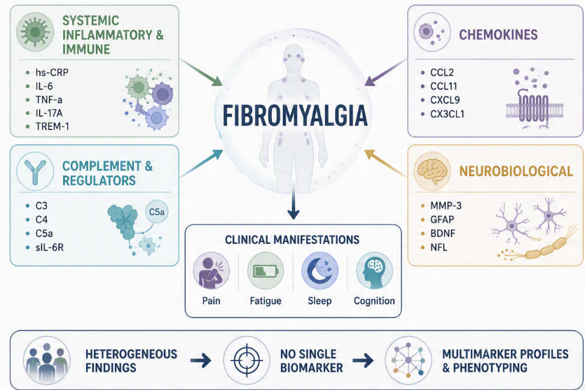
The identified evidence was synthesized narratively, with emphasis on the biological relevance and consistency of the reported findings. To facilitate interpretation, biomarkers were organized into four main categories according to their biological function: systemic inflammatory and immune-related markers, chemokines and mediators of cellular recruitment, complement system components and soluble inflammatory regulators, and neurobiological biomarker associated with neural damage and plasticity.
3. Results
3.1. Molecular Inflammatory, Immune-Related, and Neurobiological Biomarkers in Fibromyalgia
The characteristics of the studies included in this review are summarized in Table 1. Overall, the reviewed literature comprised 34 original studies involving 1,682 individuals with FMS and 1,193 healthy controls. Most studies were published between 1999 and 2026, predominantly included women, and employed a cross-sectional case-control design.
Blood was the most frequently analyzed biological sample, and the American College of Rheumatology (ACR) 1990 criteria were the most commonly applied for the diagnosis of FMS, followed by the ACR 2016 and ACR 2010 criteria. The studies were conducted across Europe, the Americas, and Asia, with Brazil and Turkey contributing the largest number of publications. Detailed information regarding study design, participant characteristics, diagnostic criteria, biological samples, and evaluated biomarkers is provided in Table 1.
3.1.1. Systemic Inflammatory and Immune-Related Biomarkers
The main finding regarding systemic inflammatory and immune-related biomarkers are summarized in Table 2.1 and Table 2.2. Overall, the available evidence is highly heterogeneous, with conflicting findings reported for most biomarkers. Consistent alterations were observed only for TREM-1, CSF-1, IL-31, IL-33, hs-CRP, an IL-18, although evidence for TREM-1 [26], CSF-1 [17], IL-31 [20], and IL-33 [20] was limited to single studies, whereas hs-CRP [9,10,29] and IL-18 [23,25] were assessed in three and two studies, respectively.
Among acute-phase proteins, elevated hs-CRP concentrations were consistently reported across studies [9,10,29]. Increased hs-CRP levels remained significant after adjustment for potential confounding factors, including body mass index, and were associated with higher abnormality rates in individuals with FMS compared with healthy controls [9]. Moreover, hs-CRP concentrations correlated with pro-inflammatory cytokines, including IL-6 and IL-8, and differed across clinical phenotypes, with patients presenting more severe forms of FMS exhibiting higher levels than those with milder disease [3,9,10]. In contrast, evidence regarding CRP was less consistent, with one study reporting increased concentrations [26] and two reporting no significant differences between patients with FMS and healthy controls [1,11]. Although association between CRP concentrations and pain severity lost significance after adjustment for age, sex, and body mass index [10], higher CRP levels were associated with poorer sleep quality and greater symptom severity in some cohorts [1,11,15]. Taken together, these findings suggests that hs-CRPS may represent a more consistent indicator of low-grade systemic inflammation than CRP in FMS.
Pro-inflammatory cytokines were the most extensively investigated biomarkers; however, the available evidence remains inconsistent. IL-6, the most frequently evaluated cytokine [32], showed increased concentrations in several studies [1,4,20,23,35], whereas others found no significant differences between patients with FMS and healthy controls [9,21,22,30,32,33,37,38,40]. Similar discrepancies were observed for TNF-α, which was elevated in seven studies [1,17,20,23,24,37,40] but unchanged in four [21,22,30,38], and for IL-8, which showed increased concentrations in seven studies [17,19,22,23,30,35,40] but no differences in four others [5,9,21,38]. Overall, these findings do not support a consistent pattern of cytokine dysregulation across patients with FMS.
Beyond between-group comparisons, several studies examined the relationship between inflammatory biomarkers and disease severity. Increased IL-6 and TNF-α concentrations were associated with higher Fibromyalgia Impact Questionnaire (FIQ) scores [1,4,22,31,35], with IL-6 emerging as one of the strongest predictors of disease impact [1]. Weak but significant positive correlations were also observed between IL-6 levels and pain intensity, whereas TNF-α concentration correlated positively with Visual Analogue Scale (VAS) scores [1,4]. Likewise, elevated IL-8 concentrations were associated with greater symptom burden, showing positive correlations with both pain intensity and FIQ scores [17,19,23,31,35,40].
Evidence regarding IL-1 cytokine family was similarly heterogeneous. IL-1β concentrations were increased in two studies [23,25], unchanged in one [16], and reduced in another [30]. Likewise, studies evaluating IL-1 reporting conflicting results, with increased [24], decreased [22], or unchanged concentrations [21]. IL-2 levels were elevated in two studies [24,37] and unchanged in one [30].
IL-17A has only recently been investigated in FMS. One study reported increased circulating concentrations taken together with positive correlations with TNF-α and other inflammatory cytokines [37], whereas another found no significant differences between patients and healthy controls [30].
Anti-inflammatory cytokines also showed inconsistent findings. IL-4 concentrations were increased in one study [37] and unchanged in two [30,40]. Similarly, IL-10 levels were elevated in two studies [22,38], reduced in two [5,24], and unchanged in six [19,21,23,30,37,40]. Notably, one study reported significantly lower circulating IL-10 concentrations in patients with FMS than in healthy controls, supporting the hypothesis of impaired anti-inflammatory regulation in a subset of patients [5].
Finally, emerging immune-related biomarkers, including TREM-1, CSF-1, IL-31, and IL-33, demonstrated consistent alterations in FMS, although the available evidence remains limited because these findings were derived from only a small number of studies. Notably, increased TREM-1 concentrations were significantly associated with several clinical manifestations of FMS, including pain, disease severity, sleep disturbance, depression, anxiety, and fatigue [26]. Overall, these emerging biomarkers represent promising candidates for future research, although larger and well-designed studies are needed to confirm their clinical relevance.
3.1.2. Chemokines and Mediators of Cellular Recruitment
The main findings regarding chemokines and mediators of cellular recruitment are summarized in Table 3. Overall, the available evidence remains limited, as most biomarkers within this category have been evaluated in a single study. The investigated mediators included CC chemokines, such as CCL2 (MCP-1) [30,41], CCL3 [17], CCL11 (eotaxin) [28,30], CCL16 (HCC4) [28], CCL17 (TARC) [28], CCL18 (PARC) [28], and CCL22 (MDC) [28], as well as CXC chemokines, including CXCL9 (MIG) [28], CXCL11 (I-TAC) [28], and CX3CL1 (fractalkine) [28].
Among the evaluated chemokines, only CCL2 (MCP-1) [30,41] and eotaxin [28,30] were investigated in more than one study. Evidence for both biomarkers was inconsistent, with one study reporting increased concentrations [28,41] and another finding no significant differences between patients with FMs and healthy controls [30].
Most of the remaining chemokines showed increased circulating concentrations in patients with FMS, including CCL3 [17], CCL17 (TARC) [28], CCL22 (MDC) [28], CXCL9 (MIG) [28], and CXCL11 (I-TARC) [28]. In contrast, no significant differences were observed for CCL16 (HCC4) [28] and CCL18 (PARC) [28], whereas CX3CL1 (fractalkine) [28] was the only chemokine reported to be decreased in patients with FMS.
Taken together, the available evidence suggests a potential involvement of chemokines in the inflammatory processes associated with FMS. However, the limited number of studies, the lack of replication, and the inconsistent findings across the few biomarkers evaluated preclude definitive conclusions regarding their role in disease pathophysiology. Further studies are needed to clarify the contribution of these mediators to immune cell recruitment and chronic pain mechanism in FMS.
3.1.3. Complement System Components and Soluble Inflammatory Regulators
3.1.3.1. Complement System Components
The main findings regarding complement system components are summarized in Table 4. Overall, evidence supporting the involvement of the complement cascade in FMS remains limited, as each biomarker has been evaluated in a single study. Increased circulating concentrations were reported for CH50 [10] and C5a [11], whereas no significant differences were observed for C3 and C4 [10].
Notably, elevated CH50 concentrations remained significant after adjustment for body mass index [10] and were positively correlated with pain intensity, fatigue, and anxiety scores in patients with FMS. Similarly, increased circulating C5a levels were observed in patients with FMS compared with healthy controls and were significantly associated with cold pain threshold (CPT) and warm pain threshold (WPT) [11].
Taken together, these preliminary findings suggest a possible contribution of complement activation to pain processing and symptom severity in FMS. However, the available evidence remains insufficient to establish the role of complement system dysregulation in disease pathophysiology because all findings are derived from isolated studies. Further research is needed to confirm these observations and clarify the clinical relevance of complement-related biomarkers in FMS.
3.1.3.2. Soluble Inflammatory Regulators
The main findings regarding soluble inflammatory regulators are summarized in Table 5. Overall, evidence remains limited, as each biomarker has been evaluated in a single study. Increased circulating concentrations were reported for IL-6R [1] and sgp130 [33], whereas no significant differences between patients with FMS and healthy controls were observed for sIL-6R and sIL-1RA [33].
No significant associations were identified between age or sex and circulating levels of IL-6, sIL-6R, sgp130, and sIL-1RA [1,33]. Although serum concentrations of sIL-6R and sIL-1RA did not differ significantly between patients with FMS and healthy controls overall [33], individuals with FMS presenting depressive symptoms exhibited significantly higher levels of both biomarkers. Accordingly, multiple regression analyses identified depressive symptom severity, assessed using the Hamilton Depression Rating Scale (HDRS), as the strongest predictor of circulating sIL-6R and sIL-1RA concentrations [1,33].
Serum sgp130 concentrations were significantly increased in patients with FMS and positively correlated with stiffness scores. Likewise, IL-6R concentrations were positively correlated with FIQ scores, suggesting that alterations in IL-6 signaling pathways may contribute to disease severity [1,33].
Notably, receiver operating characteristics (ROC) analyses demonstrated a high diagnostic performance for IL-6R, with an area under the curve (AUC) of 0.9511. Comparable findings were reported for IL-6 (AUC= 0.9530) and TNF-α (AUC= 0.9717), supporting the potential diagnostic value of inflammatory mediators in FMS [1,32,33].
Taken together, these findings suggest that soluble inflammatory regulators, particularly those involved in IL-6 signaling, may contribute to symptom severity and could represent promising diagnostic biomarkers in FMS. Nevertheless, the available evidence is based on a limited number of studies and lacks independent replication, highlighting the need for further validation before these biomarkers can be translated into clinical practice.
3.2. Neurobiological Biomarkers Associated with Neural Damage and Plasticity
The main findings regarding neurobiological biomarkers associated with neural damage and plasticity are summarized in Table 6. The evaluated biomarkers included matrix metalloproteinases (MMP-3 and MMP-10), high mobility group box 1 protein (HMGB1), glial fibrillary acidic protein (GFAP), brain-derived neurotrophic factor (BDNF), neurofilament light chain (NFL), S100 calcium-binding protein B (S100B), and nerve growth factor (NGF).
Most neurobiological biomarkers have been evaluated in only a single study. Increased circulating concentrations were reported for MMP-3 [1], MMP-10 [17], GFAP [34], and S100B [6], whereas HMGB1concentrations did not differ between patients with FMS and healthy controls [36]. In contrast, NGF levels reduced in patients with FMS [30].
BDNF was the most extensively investigated neurobiological biomarker, having been evaluated in five studies [5,16,18,30,38]. Nevertheless, the available evidence remains inconsistent. Increased concentrations were reported in three studies [5,6,30], one study found no significant differences [38], and another reported reduced BDNF levels in patients with FMS [18]. Notably, male patients exhibited significantly lower BDNF concentrations than female patients (p<.0001) [18], while circulating BDNF levels were negatively correlated with hunger scores (r= -0.52) [6].
NFL was assessed in two studies [27,39], both reported increased circulating concentrations in patients with FMS compared with healthy controls. Elevated NFL levels were associated with fewer hours of sleep and poorer performance on working memory tasks [39], although no significant correlations were observed with neuropathic pain scores assessed using the pain DETECT questionnaire [27].
Regarding glial and neuroinflammatory biomarkers, increased GFAP concentrations were associated with several clinical parameters [34], whereas S100B levels showed positive correlations with hunger scores (r= 0.463) [6].
Taken together, the available evidence suggests that biomarkers related to neuronal injury, glial activation, and neuroplasticity may contribute to the pathophysiology of FMS. However, with the exception of BDNF and NFL, most biomarkers have only been evaluated in isolated studies, and the marked heterogeneity observed, particularly for BDNF, limits definitive conclusions. Further well-designed studies are needed to clarify their biological and clinical relevance in FMS.
4. Discussion
4.1. Principal’s Findings
This narrative review synthesized the available evidence on molecular inflammatory, immune-related, and neurobiological biomarkers in FMS. Overall, the reviewed literature was characterized by substantial heterogeneity in sample characteristics, biological samples, diagnostic criteria, and biomarkers assessed. Most studies employed cross-sectional case-control designs and focused on peripheral blood samples, limiting the ability to establish temporal or causal relationships between biomarker alterations and FMS manifestations.
The available evidence supports the involvement of inflammatory, immune, and neurobiological mechanism in the pathophysiology of FMS, although no consistent biomarker profile has yet emerged. Among inflammatory biomarkers, the most frequently investigated cytokines – including IL-6, TNF-α, IL-8, IL-10, and IL-1β – showed heterogeneous and, in some cases, conflicting findings. Similarly, evidence regarding chemokines and complement components remains limited, as most biomarkers have been evaluated in only one or two studies. Nevertheless, emerging biomarkers such as TREM-1, CSF-1, IL-31, and IL-33 consistently showed increased concentrations in patients with FMS [17,20,26], although these findings have not yet been independently replicated.
Neurobiological biomarkers showed similarly diverse patterns. Whereas BDNF findings were inconsistent, NFL, GFAP, MMP-3, and MMP-10 displayed more consistent alterations [1,17,27,34], supporting a potential contribution of neuronal injury, glial dysfunction, and altered neuroplasticity to the development and maintenance of FMS symptoms. Importantly, several biomarkers were associated with clinical manifestations, including pain, fatigue, sleep disturbances, anxiety, depression, cognitive impairment, and disease impact. Some inflammatory mediators, particularly IL-6R, IL-6, and TNF-α, also showed promising diagnostic performance; however, the available evidence remains insufficient to support their clinical implementation [1,4,37,40].
Taken together, the evidence suggests that FMS cannot be explained by isolated inflammatory abnormalities but rather reflects a complex interplay between immune dysregulation, neuroinflammation, and altered neural plasticity. However, the limited replications of individual biomarker findings and the methodological heterogeneity of the available literature currently preclude the identification of a reliable biological signature. Future longitudinal and multicenter studies should therefore prioritize standardized biomarker assessment and the identification of biologically and clinically meaningful FMS phenotypes.
4.2. Inflammation and Immune Dysregulation in FMS
The present findings support the involvement of low-grade inflammatory and immune dysregulation in FMS, although the heterogeneous results across biomarkers suggests that this process is unlikely to be characterized by a uniform inflammatory profile. Alterations in cytokines, chemokines, complement components, and immune regulatory proteins collectively point toward interactions between peripheral immune mechanisms and neuroimmune pathways rather than isolated abnormalities in individual biomarkers [9,10,26].
Among acute-phase proteins, increased hs-CRP concentrations were consistently observed in individuals with FMS [9,10,29]. However, this association may be partly explained by confounding factors, including body mass index, medical comorbidities, mood disturbances, and sleep impairment [3,29,42]. In particular, obesity represent a pro-inflammatory state capable of promoting cytokine release and increasing circulating hs-CRP levels [9,42]. Thus, although hs-CRP may reflect inflammatory alterations in FMS, its specificity as a disease-related biomarker remains uncertain.
Pro-inflammatory cytokines showed a more heterogeneous pattern. TNF-α, IL-6, IL-8, and IL-17A were elevated in several studies, whereas others reported no significant differences between patients with FMS and healthy controls. IL-17A and TNF-α, may contribute to sustained inflammatory signaling through interactions among immune cell populations [37,43], while TNF-α has also been implicated in the sensitization of nociceptive pathways through peripheral and central mechanisms [44,45]. Similarly, IL-6 is involved in a bidirectional communication between the immune and nervous systems and has been associated with central sensitization, fatigue, depression, and pain severity in FMS [1,2,4,46]. Nevertheless, the inconsistent findings across studies preclude establishing a direct relationship between circulating cytokine concentrations and FMS pathophysiology.
The diagnostic performance observed for IL-6R is particularly noteworthy. Soluble IL-6 signaling may be relevant to chronic inflammatory and painful conditions because interactions among IL-6, its soluble receptor, and gp130 can amplify downstream signaling [33,47,48]. Thus, alterations in IL-6 trans-signaling may provide a more informative perspective on immune dysregulation than isolated measurements of circulating IL-6. However, the diagnostic potential of IL-6R requires independent replication before its clinical relevance can be established.
Evidence of altered anti-inflammatory regulation was also observed. Reduced IL-10 concentrations were reported in some studies [5,24], supporting the possibility that impaired anti-inflammatory responses may contribute to the persistent nociceptive sensitization in FMS [7,40]. However, the conflicting findings across studies suggests that this imbalance may characterize only specific patient subgroups rather than FMS as a whole.
Chemokines may provide further insight into the interaction between immune activation and cellular recruitment. Despite the limited number of available studies, several chemokines showed increased concentrations in patients with FMS. In one study, all patients exhibited serum MIG and MDC concentrations above the mean values observed in healthy controls [28,41]. Increased MIG and TARC concentrations may also be related to the elevated IFN-γ levels reported in some patients, suggesting coordinated inflammatory signaling rather than isolated alterations in individual chemokines [28]. Nevertheless, the limited replication of these findings prevents firm conclusions regarding their contribution to FMS pathophysiology.
Additional evidence of immune dysregulation comes from studies of the complement system. Increased concentrations of CH50 and C5a may indicate complement activation in FMS and potentially reflect chronic inflammatory processes [10]. CH50 was associated with fatigue and trait anxiety, whereas C5a correlated with thermal pain thresholds, suggesting that complement-related mechanism may extend beyond systemic inflammation and interact with symptom expression. Alterations in complement proteins have also been described in psychiatric disorders, including depression, anxiety, and post-traumatic stress disorder, raising the possibility of shared neuroimmune mechanism [10].
Among emerging inflammatory biomarkers, TREM-1 deserved special attention. TREM-1 is expressed by immune and neuroimmune cells and amplifies inflammatory responses through interaction with Toll-like receptors [7,26]. Increased TREM-1 concentrations were associated with pain intensity, disease severity, sleep disturbances, fatigue, anxiety, depression, and inflammatory markers such as CRP [26]. Because TREM-1 activation promotes the release of pro-inflammatory cytokines and chemokines, it may represent a potential link between immune activation and the broad symptom profile observed in FMS [26,49]. However, these findings currently rely on limited evidence and require replication.
Overall, the available evidence suggests that immune dysregulation in FMS is unlikely to result from isolated alterations in individual biomarkers. Rather, the observed pattern points toward interactions among inflammatory mediators, immune regulatory pathways, stress-response systems, and central pain processing. This broader network perspective may help explain both the heterogeneity of biomarker findings and the clinical variability characteristic of FMS. interactions among inflammatory mediators, neuroimmune pathways, stress-response systems, and central-pain processing mechanism.
4.3. Neurobiological Alterations and Maladaptive Neuroplasticity
Beyond immune dysregulation, the present review identified alterations in biomarkers related to neuronal injury, glial function, neuroplasticity, and neuroimmune communication, supporting the view that FMS pathophysiology extends beyond systemic inflammation. Although FMS is not typically characterized by overt tissue damage, increasing evidence suggests that mechanism involving central sensitization, altered neuronal excitability, and persistent pain may contribute to symptom development and maintenance.
Among the biomarkers evaluated, NFL emerged as one of the most consistent patterns, with both available studies reporting increased concentrations in patients with FMS [27,39]. NFL is a cytoskeletal protein released following axonal injury and is an established blood-based marker of neural damage in disorders affecting the central and peripheral nervous systems [27,50]. Its elevation in FMs may therefore indicate subtle neuronal alterations, although the clinical significance of these findings remains unclear [27,39]. Moreover, higher NFL concentrations were associated with shorter sleep duration and poorer working-memory performance, suggesting a potential relationship between neuronal alterations, cognitive symptoms, and sleep disturbances in FMS [39]. These findings are also consistent with the possibility that neurobiological alterations may be influenced by immune-mediated mechanism [27,51,52].
Further evidence of neuroimmune involvement comes from GFAP, whose increased concentrations in patients with FMS may reflect altered glial activity [12,34]. Glial activation has been associated with the release of pronociceptive mediators, including cytokines, chemokines, and growth factors, which can contribute to neuronal hyperexcitability and pain hypersensitivity [12,34]. This is particularly relevant to chronic pain, as glial signaling has been implicated in the maintenance of persistent and neuropathic pain states [53].
BDNF, the most extensively investigated neurobiological biomarker, showed considerably more heterogeneous findings. Although several studies reported increased serum concentrations in patients with FMS [5,6,30], others found no differences or reduced levels, particularly among male patients [18]. BDNF modulates synaptic plasticity and is expressed by nociceptive sensory neurons, suggesting a potential role in the development and maintenance of inflammatory pain hypersensitivity [5,18,60]. However, its precise contribution to FMS remains uncertain because both pro-nociceptive and anti-nociceptive effects have been described [5,18]. Similarly, increased concentrations of S100B, a marker associated with glial and neuroinflammatory processes, provide further evidence of altered neuroimmune signaling in FMS [6].
Matrix metalloproteinases may also contribute to these processes. MMP-3 and MMP-10 were elevated in patients with FMS [1,10,17]. Although MMP-3 is traditionally associated with extracellular matrix remodeling and joint pathology, matrix metalloproteinases also participate in inflammatory signaling and tissue remodeling, potentially linking peripheral inflammatory processes with alterations in nociceptive and neuroimmune pathways. However, the limited evidence available for these biomarkers prevents determining their specific contribution to FMS pathophysiology.
Taken together, the available evidence supports a potential role for neuronal, glial, and neuroplastic mechanism in FMS, complementing the evidence of peripheral immune dysregulation. Rather than representing independent processes, these alterations may interact through bidirectional neuroimmune signaling and contribute to persistent pain and the heterogeneous clinical manifestations of MFS. Nevertheless, the limited number of studies and the inconsistent findings for several biomarkers, particularly BDNF, preclude the identification of a specific neurobiological signature.
4.4. Diagnostic Implications and Clinical Utility
The lack of objective biomarkers for the diagnosis and monitoring of FMS remains an important challenge in clinical practice [1]. Although FMS is not characterized by a single inflammatory abnormality, the available evidence suggests that alterations at the interface between immune and nervous systems may provide clinically relevant information regarding symptom burden and disease heterogeneity [2,7,9].
Several biomarkers, including IL-6R, IL-6, TNF-α, CH50, GFAP, and NFL, were associated with clinical manifestations such as pain, fatigue, sleep disturbances, cognitive impairment, and psychological symptoms [1,10,30,34]. In particular, the diagnostic performance reported for IL-6R, IL-6, and TNF-α suggest potential value for distinguishing patients with FMS from healthy controls. However, these findings should be interpreted cautiously because they are based on a limited number of studies and have not yet been independently replicated. Accordingly, none of these biomarkers can currently be considered sufficiently validated for routine diagnosis or monitoring of FMS.
The heterogeneity observed across biomarkers also supports the view that FMS is unlikely to represent a biologically homogeneous condition. Rather than relying on a single biomarker, future research should investigate multimarker panels integrating inflammatory, immune-related, neurobiological, and potentially clinical measures. Such approaches may help identify biologically and clinically meaningful phenotypes, improve patient stratification, and ultimately support more personalized diagnostic and therapeutic strategies.
Overall, the clinical value of molecular biomarkers in FMS may lie not in replacing clinical assessment but in complementing it by providing objective information about underlying biological processes and clinically relevant patient subgroups.
4.5. Strengths and Limitations
This review has several strengths. To our knowledge, it provides one of the most comprehensive synthesis of the available evidence on molecular inflammatory, immune-related, and neurobiological biomarkers in FMS. The inclusion of a broad range of biomarkers and biological matrices allowed consideration of multiple biological processes potentially involved in FMS pathophysiology. In addition, the organization of the evidence according to biological function facilitated the identification of consistent findings, area of uncertainty, and potential directions for future research.
Several limitations should also be acknowledged. First, most of the available studies employed cross-sectional case–control designs, limiting causal inference and preventing conclusions regarding the temporal relationship between biomarker alterations and clinical manifestations. Second, substantial heterogeneity was observed across studies in terms of diagnostic criteria, sample size, biological specimens, and biomarkers assessed, which limits direct comparison and generalizability. Moreover, many biomarkers were investigated in only one or two studies, and several findings therefore lack independent replication. Potential confounding factors, including body mass index, sleep disturbances, psychological symptoms, and medical comorbidities, may also have influenced biomarker concentrations and contribute to the observed variability.
Future research should prioritize longitudinal and multicenter studies using standardized diagnostic criteria, biological sampler procedures, and analytical methods. Greater attention to potential confounders and clinically relevant patient characteristics will also be necessary to determine whether multimarker profiles can improve biological phenotyping and support the clinical application of biomarkers in FMS.
5. Conclusions
The available evidence suggests that FMS involves a complex interplay among inflammatory, immune, and neurobiological mechanism rather than a single pathophysiological process. Although several biomarkers showed promising associations with clinical manifestations and disease severity, the heterogeneity and limited replication of the available findings currently preclude the identification of a reliable biomarker for diagnosis or clinical monitoring.
Overall, these findings reinforce the multifactorial nature of FMS and support the involvement of bidirectional interactions between the immune and nervous systems in symptom development and maintenance. Future longitudinal and multicenter studies using standardized methodologies and multimarker approaches may help identify biologically and clinically meaningful FMS phenotypes and determine whether such profiles can contribute to more accurate diagnosis, patient stratification, and personalized therapeutic strategies.
Author Contributions
B.M-G.: Conceptualization; Formal analysis; Methodology; Visualization; Roles/Writing - original draft; Writing - review & editing. J.A.C-R.: Conceptualization; Formal analysis; Resources; Project administration; Supervision; Validation; Visualization; Writing - review & editing. G.A.R.P.: Conceptualization; Formal analysis; Resources; Project administration; Supervision; Validation; Visualization; Writing - review & editing. C.M.G-S.: Conceptualization; Formal analysis; Resources; Project administration; Supervision; Validation; Visualization; Writing - review & editing.
Funding
This research received no external funding.
Institutional Review Board Statement
Not applicable.
Informed Consent Statement
Not applicable.
Data Availability Statement
The data that support the findings of this study are available on request from the corresponding author.
Conflicts of Interest
The authors declare no conflicts of interest.
References
- Gumus, A.; Erdogan, F.; Durak, S.; Karamese, M.; Zeybek, U. Investigation of MMP-3, IL-6, IL-6R, and TNF-alpha levels in fibromyalgia patients presenting to a physical medicine and rehabilitation clinic. J Clin Med. 2026;15:4107. [CrossRef]
- Häuser, W.; Perrot, S.; Clauw, D.J.; Fitzcharles, M.A. Unravelling fibromyalgia—steps toward individualized management. J Pain. 2018;19:125-134. [CrossRef]
- Pérez-Aranda, A.; Andrés-Rodríguez, L.; Feliu-Soler, A.; Núñez, C.; Stephan-Otto, C.; Pastor-Mira, M.A.; López-Roig, S.; Peñacoba, C.; Calandre, E.P.; Slim, M.; Salgueiro, M.; Feixas, G.; Luciano, J.V. Clustering a large Spanish sample of patients with fibromyalgia using the Fibromyalgia Impact Questionnaire–Revised: differences in clinical outcomes, economic costs, inflammatory markers, and gray matter volumes. Pain. 2019;160(4):908-921. [CrossRef]
- Baris, E.; Topaloglu, I.; Akalin, E.; et al. Serum choline, leptin and interleukin-6 levels in fibromyalgia syndrome-induced pain: a case-control study. BMC Musculoskelet Disord. 2025;26:97. [CrossRef]
- Salgado, A.S.I.; Takemoto, M.H.; Souza, C.F.T.C.; et al. Gentle touch therapy, pain relief and neuroplasticity at baseline in fibromyalgia syndrome: a randomized, multicenter trial with six-month follow-up. J Clin Med. 2022;11:4898. [CrossRef]
- Elkfury, J.L.; Antunes, L.C.; Angoleri, L.D.M.; et al. Dysfunctional eating behavior in fibromyalgia and its association with serum biomarkers of brain plasticity (BDNF and S100B): an exploratory study. Arch Endocrinol Metab. 2021;65(6):733-742. [CrossRef]
- Sarzi-Puttini, P.; Giorgi, V.; Marotto, D.; Atzeni, F. Fibromyalgia: an update on clinical characteristics, aetiopathogenesis and treatment. Nat Rev Rheumatol. 2020;16(11):645-660. [CrossRef]
- Siracusa, R.; Di Paola, R.; Cuzzocrea, S.; Impellizzeri, D. Fibromyalgia: pathogenesis, mechanisms, diagnosis and treatment options update. Int J Mol Sci. 2021;22:3891. [CrossRef]
- Xiao, Y.; Haynes, W.L.; Michalek, J.E.; Russell, I.J. Elevated serum high-sensitivity C-reactive protein levels in fibromyalgia syndrome patients correlate with body mass index, interleukin-6, interleukin-8, erythrocyte sedimentation rate. Rheumatol Int. 2013;33:1259-1264. [CrossRef]
- Izuno, S.; Hosoi, M.; Anno, K.; Kato, T.A.; Sudo, N.; Yoshihara, K. Association of high complement and low immunoglobulins with the clinical symptoms of patients with fibromyalgia. BioPsychoSocial Med. 2025;19:1. [CrossRef]
- Fujimoto, K.; Anno, K.; Tanaka, Y.; Murakami, M.; Inamine, S.; Kato, T.A.; Sudo, N.; Hosoi, M. C5a in the peripheral plasma of female fibromyalgia patients is elevated but not related to pain sensitivity as in healthy controls. Sci Rep. 2025;15:17387. [CrossRef]
- Ji, R.R.; Nackley, A.; Huh, Y.; Terrando, N.; Maixner, W. Neuroinflammation and central sensitization in chronic and widespread pain. Anesthesiology. 2018;129:343-366. [CrossRef]
- Martins, D.F.; Viseux, F.J.; Salm, D.C.; et al. The role of the vagus nerve in fibromyalgia syndrome. Neurosci Biobehav Rev. 2021;131:1136-1149. [CrossRef]
- Jurado-Priego, L.N.; Cueto-Ureña, C.; Ramírez-Expósito, M.J.; Martínez-Martos, J.M. Fibromyalgia: a review of the pathophysiological mechanisms and multidisciplinary treatment strategies. Biomedicines. 2024;12:1543. [CrossRef]
- O’Mahony, L.F.; Srivastava, A.; Mehta, P.; Ciurtin, C. Is fibromyalgia associated with a unique cytokine profile? A systematic review and meta-analysis. Rheumatology (Oxford). 2021;60:2602-2614. [CrossRef]
- Barut, Z.; Karataş, Ö.; Akdeniz, F.T.; Çırçırlı, B.; Demir, S.; İşbir, T. The association between miRNA-223-3p levels and pain severity in fibromyalgia syndrome: a molecular approach. Int J Mol Sci. 2026;27:176. [CrossRef]
- Fineschi, S.; Klar, J.; Ayoola Gustafsson, K.; Jonsson, K.; Karlsson, B.; Dahl, N. Inflammation and interferon signatures in peripheral B-lymphocytes and sera of individuals with fibromyalgia. Front Immunol. 2022;13:874490. [CrossRef]
- Iannuccelli, C.; Lucchino, B.; Gioia, C.; et al. Gender influence on clinical manifestations, depressive symptoms and brain-derived neurotrophic factor (BDNF) serum levels in patients affected by fibromyalgia. Clin Rheumatol. 2022;41:2171-2178. [CrossRef]
- Otero, E.; Gálvez, I.; Ortega, E.; Hinchado, M.D. Influence of chronic fatigue syndrome codiagnosis on the relationship between perceived and objective psychoneuro-immunoendocrine disorders in women with fibromyalgia. Biomedicines. 2023;11:1488. [CrossRef]
- Theoharides, T.C.; Tsilioni, I.; Arbetman, L.; Panagiotidou, S.; Stewart, J.M.; Gleason, R.M.; et al. Neuropeptides CRH, SP, HK-1, and inflammatory cytokines IL-6 and TNF are increased in serum of patients with fibromyalgia syndrome, implicating mast cells. J Pharmacol Exp Ther. 2016;356(3):664-672. [CrossRef]
- Bazzichi, L.; Giacomelli, C.; De Feo, F.; et al. Antipolymer antibody in Italian fibromyalgic patients. Arthritis Res Ther. 2007;9(4):R86. [CrossRef]
- Bazzichi, L.; Rossi, A.; Massimetti, G.; et al. Cytokine patterns in fibromyalgia and their correlation with clinical manifestations. Clin Exp Rheumatol. 2007;25(2):225-230.
- Bote, M.E.; García, J.J.; Hinchado, M.D.; Ortega, E. Inflammatory/stress feedback dysregulation in women with fibromyalgia. Neuroimmunomodulation. 2012;19:343-351. [CrossRef]
- Brol, A.M.; Zardo, V.; Barbieri, R.; et al. IL-10 cytokine as potential biomarkers in women with fibromyalgia who underwent to a short balneotherapy treatment. Braz Arch Biol Technol. 2020;63:e20180734. [CrossRef]
- Cordero, M.D.; Alcocer-Gómez, E.; Culic, O.; et al. NLRP3 inflammasome is activated in fibromyalgia: the effect of coenzyme Q10. Antioxid Redox Signal. 2014;20(8):1169-1180. [CrossRef]
- Aydogan Baykara, R.; Kıran, T.R.; Otlu, Ö.; Erdem, M.; Pıhtılı Taş, N. Could TREM-1 be a novel marker in the diagnosis of fibromyalgia? A cross-sectional study. Medicine (Baltimore). 2024;103(28):e38808. [CrossRef]
- Fundaun, J.; Kelleher, E.; Coxon, L. et al. Serum neurofilament light chain in fibromyalgia: comparative evidence of neuronal injury across chronic pain conditions. Pain Rep. 2026;11:e1423. [CrossRef]
- García, J.J.; Cidoncha, A.; Bote, M.E.; Hinchado, M.D.; Ortega, E. Altered profile of chemokines in fibromyalgia patients. Ann Clin Biochem. 2014;51(5):576-581. [CrossRef]
- Groven, N.; Fors, E.A.; Klæbo Reitan, S. Patients with fibromyalgia and chronic fatigue syndrome show increased hsCRP compared to healthy controls. Brain Behav Immun Health. 2020;9:100153. [CrossRef]
- Jablochkova, A.; Bäckryd, E.; Kosek, E.; et al. Unaltered low nerve growth factor and high brain-derived neurotrophic factor levels in plasma from patients with fibromyalgia after a 15-week progressive resistance exercise. J Rehabil Med. 2019;51(10):779-787. [CrossRef]
- Kutu, F.C.; Özdolap, Ş.; Sarıkaya, S. Pro-inflammatory cytokines and oxidized low-density-lipoprotein in patients with fibromyalgia. Arch Rheumatol. 2019;34(2):123-129. [CrossRef]
- Loçasso, F.A.; Filho, H.A.; Alvarenga, RMP.; et al. Assessing the impact of IL-6 and serotonin on pain and symptomatology in fibromyalgia: an exploratory clinical study. J Pers Med. 2024;14:886. [CrossRef]
- Maes, M.; Libbrecht, I.; Van Hunsel, F.; Lin, A-H.; De Clerck, L.; Stevens, W.; Kenis, G.; de Jongh, R.; Bosmans, E.; Neels, H. The immune-inflammatory pathophysiology of fibromyalgia: increased serum soluble gp130, the common signal transducer protein of various neurotrophic cytokines. Psychoneuroendocrinology. 1999;24:371-383.
- Massó, F.; Martínez-Martínez, L.A.; Amezcua-Guerra, L.M.; Mercado, F.; Almanza, A.; Martínez-Lavín, M. Glial fibrillary acidic protein in fibromyalgia: its serum levels and antibodies. Adv Rheumatol. 2026;66:42. [CrossRef]
- Mendieta, D.; De la Cruz-Aguilera, D.L.; Barrera-Villalpando, M.I.; et al. IL-8 and IL-6 primarily mediate the inflammatory response in fibromyalgia patients. J Neuroimmunol. 2016;290:22-25. [CrossRef]
- Oktayoğlu, P.; Tahtaşız, M.; Bozkurt, M.; et al. Serum levels of high mobility group box 1 protein and its association with quality of life and psychological and functional status in patients with fibromyalgia. Clin Rheumatol. 2013;32(11):1671-1675. [CrossRef]
- Pernambuco, A.P.; Schetino, L.P.; Alvim, C.C.; et al. Increased levels of IL-17A in patients with fibromyalgia. Clin Exp Rheumatol. 2013;31(6 Suppl 79):S60-S63.
- Ranzolin, A.; Duarte, ALB.; Bredemeier, M.; et al. Evaluation of cytokines, oxidative stress markers and brain-derived neurotrophic factor in patients with fibromyalgia: a controlled cross-sectional study. BMC Musculoskelet Disord. 2016;17:239. [CrossRef]
- Ruggieri, M.; Paparella, G.; Clemente, L.; Libro, G.; Gargano, C.D.; De Tommaso, M. Plasma neurofilament light chain in fibromyalgia: a case-control study exploring correlation with clinical and cognitive features. Eur J Pain. 2025;29:e4752. [CrossRef]
- Wang, H.; Moser, M.; Schiltenwolf, M.; Buchner, M. Circulating cytokine levels compared to pain in patients with fibromyalgia: a prospective longitudinal study over 6 months. J Rheumatol. 2008;35(7):1366-1370.
- Zhao, Y.C.; Hu, T.; Chen, Y.; Du, K.T. Elevated serum levels of monocyte chemotactic protein-1/chemokine C-C motif ligand 2 are linked to disease severity in patients with fibromyalgia syndrome. Pain Res Manag. 2020;2020:8857686. [CrossRef]
- Feinberg, T.; Sambamoorthi, U.; Lilly, C.; Innes, K.K. Potential mediators between fibromyalgia and C-reactive protein: results from a large U.S. community survey. BMC Musculoskelet Disord. 2017;18:294.
- Pichavant, M.; Goya, S.; Meyer, E.H.; et al. Ozone exposure in a mouse model induces airway hyperreactivity that requires the presence of natural killer T cells and IL-17. J Exp Med. 2008;205:385-393.
- Sluka, K.A.; Clauw, D.J. Neurobiology of fibromyalgia and chronic widespread pain. Neuroscience. 2016;338:114-129. [CrossRef]
- Tanaka, T.; Narazaki, M.; Kishimoto, T. IL-6 in inflammation, immunity, and disease. Cold Spring Harb Perspect Biol. 2014;6:a016295. [CrossRef]
- Üçeyler, N.; Häuser, W.; Sommer, C. Cytokines in fibromyalgia syndrome: a systematic review and meta-analysis. BMC Musculoskelet Disord. 2011;12:245. [CrossRef]
- Bock, G.R.; Marsh, J.; Widdows, K. Polyfunctional cytokines: IL-6 and LIF. In: Ciba Foundation Symposium 167. Chichester, UK: Wiley; 1992.
- Rose-John S. IL-6 trans-signaling via the soluble IL-6 receptor: importance for the pro-inflammatory activities of IL-6. Int J Biol Sci. 2012;8:1237-1247. [CrossRef]
- Bouchon, A.; Dietrich, J.; Colonna, M. Cutting edge: inflammatory responses can be triggered by TREM-1, a novel receptor expressed on neutrophils and monocytes. J Immunol. 2000;164:4991-4995. [CrossRef]
- Gaetani, L.; Blennow, K.; Calabresi, P.; Di Filippo, M.; Parnetti, L.; Zetterberg, H. Neurofilament light chain as a biomarker in neurological disorders. J Neurol Neurosurg Psychiatry. 2019;90:870-881. [CrossRef]
- Khalil, M.; Teunissen, C.E.; Lehmann, S.; et al. Neurofilaments as biomarkers in neurological disorders—towards clinical application. Nat Rev Neurol. 2024;20:269-287. [CrossRef]
- Krock, E.; Morado-Urbina, C.E.; Menezes, J.; et al. Fibromyalgia patients with elevated levels of anti-satellite glia cell immunoglobulin G antibodies present with more severe symptoms. Pain. 2023;164:1828-1840. [CrossRef]
- Kim, D.S.; Figueroa, K.W.; Li, K.W.; Boroujerdi, A.; Yolo, T.; Luo, Z.D. Profiling of dynamically changed gene expression in dorsal root ganglia post peripheral nerve injury and a critical role of injury-induced glial fibrillary acidic protein in maintenance of pain behaviors. Pain. 2009;143:114-122. [CrossRef]
- Merighi, A.; Salio, C.; Ghirri, A.; et al. BDNF as a pain modulator. Prog Neurobiol. 2008;85(3):297-317. [CrossRef]

Table 1.
Characteristics of the Studies Included in This Review.
| STUDY | COUNTRY | STUDY DESIGN | SAMPLE SIZE | DIAGNOSTIC CRITERIA | SAMPLE TYPE |
|---|---|---|---|---|---|
| 1 | TURKEY | CROSS-SECTIONAL CASE-CONTROL STUDY | n FM= 82 / n HC= 82 | ACR CRITERIA (2016) | VENOUS BLOOD SAMPLES |
| 4 | TURKEY | CROSS-SECTIONAL CASE-CONTROL STUDY | n FM= 38/ n HC= 38 | ACR CRITERIA (2010) | FASTING BLOOD SAMPLES |
| 5 | BRAZIL | RCT (DOUBLE-BLIND, MULTICENTER) | n FM= 64/ n HC= 18 | ACR CRITERIA (2016) | BLOOD SAMPLES |
| 6 | BRAZIL | CROSS SECTIONAL CASE-CONTROL STUDY | n FM= 17/ nHC= 10 | ACR CRITERIA (2010) | BLOOD SAMPLES |
| 9 | USA | CROSS-SECTIONAL CASE-CONTROL STUDY | n FM= 105/ n HC= 61 | ACR CRITERIA (1990) | VENOUS BLOOD SAMPLES |
| 10 | JAPAN | CROSS SECTIONAL CASE-CONTROL STUDY | n FM= 41/ n HC= 30 | ACR CRITERIA (2010) | PERIPHERAL BLOOD SAMPLES |
| 11 | JAPAN | CROSS-SECTIONAL CASE-CONTROL STUDY | n FM= 30/ n HC= 29 | ACR CRITERIA (2010, 2016) | PERIPHERAL VENOUS BLOOD |
| 16 | TURKEY | CROSS-SECTIONAL CASE-CONTROL PILOT | n FM= 18/ n HC= 18 | ACR CRITERIA (2016) | PERIPHERAL BLOOD SAMPLES |
| 17 | SWEDEN | CROSS SECTIONAL CASE-CONTROL STUDY | n FM= 30/ n HC= 24 | ACR CRITERIA (2010, 2016) | BLOOD SAMPLE |
| 18 | ITALY | CROSS SECTIONAL CASE-CONTROL STUDY | n FM= 40/ n HC= 40 | ACR CRITERIA (2016) | VENOUS BLOOD SAMPLE |
| 19 | SPAIN | CROSS SECTIONAL CASE-CONTROL STUDY | n FM= 17/ n HC= 11 | ACR CRITERIA (2010) | BLOOD SAMPLES |
| 20 | USA | CROSS SECTIONAL CASE-CONTROL STUDY | n FM= 84/ n HC= 20 | ACR CRITERIA (1990) | BLOOD SAMPLES |
| 25 | ITALY | CROSS SECTIONAL CASE-CONTROL STUDY | n FM= 285/ n HC= 100 | ACR CRITERIA (1990) | SERUM SAMPLES |
| 26 | ITALY | CROSS SECTIONAL CASE-CONTROL STUDY | n FM= 80/ n HC= 45 | ACR CRITERIA (1990) | BLOOD SAMPLES |
| 27 | SPAIN | CROSS SECTIONAL CASE-CONTROL STUDY | n FM= 25/ n HC= 20 | ACR CRITERIA (1990) | PERIPHERAL BLOOD SAMPLES |
| 28 | BRAZIL | PROSPECTIVE INTERVENTIONAL STUDY | n FM= 11/ n HC= 11 | ACR CRITERIA (1990) | SALIVA AND BLOOD SAMPLES |
| 29 | SPAIN | CROSS SECTIONAL CASE-CONTROL STUDY | n FM= 30/ n HC= 20 | ACR CRITERIA (1990) | PERIPHERAL BLOOD SAMPLES |
| 30 | TURKEY | CROSS SECTIONAL CASE-CONTROL STUDY | n FM= 45/ n HC= 46 | ACR CRITERIA (2016) | BLOOD SAMPLES |
| 31 | USA | SECONDARY STUDY | n FM= 60/ n HC= 30 | ACR CRITERIA (2016) | BLOOD SAMPLES |
| 32 | ISRAEL | CROSS SECTIONAL CASE-CONTROL STUDY | n FM= 50/ n HC= 15 | ACR CRITERIA (2010) | BLOOD SAMPLES |
| 33 | SPAIN | CROSS SECTIONAL CASE-CONTROL STUDY | n FM= 17/ n HC= 10 | ACR CRITERIA (2010) | PLASMA CONCENTRATION |
| 34 | NORWAY | CROSS SECTIONAL CASE-CONTROL STUDY | n FM= 58/ n HC= 53 | ACR CRITERIA (1990) | BLOOD SAMPLES |
| 35 | SWEDEN | SUB-STUDY OF RANDOMIZED CONTROLLED | n FM= 15/ n HC= 25 | ACR CRITERIA (1990) | PLASMA SAMPLES |
| 36 | TURKEY | CROSS SECTIONAL CASE-CONTROL STUDY | n FM= 48/ n HC= 43 | ACR CRITERIA (2010) | BLOOD SAMPLES |
| 37 | BRAZIL | CROSS SECTIONAL CASE-CONTROL STUDY | n FM= 27/ n HC= 16 | ACR CRITERIA (2016) | PERIPHERAL BLOOD SAMPLES |
| 38 | BELGIUM | CROSS SECTIONAL CASE-CONTROL STUDY | n FM= 21/ n HC= 33 | ACR CRITERIA (1990) | BLOOD SAMPLES |
| 39 | MEXICO | CROSS SECTIONAL CASE-CONTROL STUDY | n FM= 47/ n HC= 31 | ACR CRITERIA (2016) | BLOOD SAMPLES |
| 40 | MEXICO | CROSS SECTIONAL CASE-CONTROL STUDY | n FM= 15/ n HC= 14 | ACR CRITERIA (2010) | PERIPHERAL BLOOD SAMPLES |
| 41 | TURKEY | CROSS SECTIONAL CASE-CONTROL STUDY | n FM= 29/ n HC= 29 | ACR CRITERIA (1990) | SERUM SAMPLES |
| 42 | BRAZIL | CROSS SECTIONAL CASE-CONTROL STUDY | n FM= 58/ n HC= 39 | ACR CRITERIA (1990) | BLOOD SAMPLES |
| 43 | BRAZIL | CROSS SECTIONAL CASE-CONTROL STUDY | n FM= 69/ n HC= 61 | ACR CRITERIA (1990,2010) | BLOOD SAMPLES |
| 44 | ITALY | CROSS SECTIONAL CASE-CONTROL STUDY | n FM= 33/ n HC= 22 | ACR CRITERIA (2016) | BLOOD SAMPLES |
| 45 | GERMANY | PROSPECTIVE LONGITUDINAL STUDY | n FM= 20/ n HC= 80 | ACR CRITERIA (1990) | STANDARD BLOOD SAMPLE |
| 46 | CHINA | CROSS SECTIONAL CASE-CONTROL STUDY | n FM= 79/ n HC= 75 | ACR CRITERIA (1990) | BLOOD SAMPLES |
*Abbreviations: ACR: American College of Rheumatology; FM: Fibromyalgia; HC: Healthy Controls; USA: United States of America.
Table 2.1.
Systemic inflammatory and immune-related biomarkers.
| ANTI-INFLAMMATORY CYTOKINES | EMERGING IMMUNE-RELATED BIOMARKERS | |||
|---|---|---|---|---|
| STUDY | IL-4 | IL-10 | TREM-1 | CSF-1 |
| 5 | ↓** | |||
| 17 | ↑* | |||
| 19 | = | |||
| 25 | = | |||
| 26 | ↑*** | |||
| 27 | = | |||
| 28 | ↓** | |||
| 30 | ↑*** | |||
| 35 | = | = | ||
| 42 | ↑*** | = | ||
| 43 | ↑*** | |||
| 45 | = | = | ||
Biomarker nomenclatura was harmonized across studies. Blank cells indicate that the biomarker was not assessed; ↑: significantly increased in individuals with FMS compared with controls; ↓: significantly decreased in individuals with FMS compared with controls; =: no significant differences between groups; *p < 0.05; **p < 0.01; ***p < 0.001. *Abbreviations: CRP: C-reactive protein; CSF-1: colony-stimulating factor 1; hs-CRP: high-sensitivity C-reactive protein; IL: interleukin; INF-γ: interferon gamma; TNF-α: tumor necrosis factor alpha; TREM-1: triggering receptor expressed on myeloid cells.
Table 2.2.
Acute Phase Proteins and Pro-Inflammatory Citokynes.
| ACUTE PHASE PROTEINS AND PRO-INFLAMMATORY CYTOKINES | |||||||||||||
|---|---|---|---|---|---|---|---|---|---|---|---|---|---|
| STUDY | hs-CRP | CRP | TNF-α | IFN-γ | IL-1β | IL-1 | IL-2 | IL-6 | IL-8 | IL-17A | IL-18 | IL-31 | IL-33 |
| 1 | = | ↑*** | ↑*** | ||||||||||
| 4 | ↑*** | ||||||||||||
| 5 | = | ||||||||||||
| 9 | ↑** | = | = | ||||||||||
| 10 | ↑* | ||||||||||||
| 11 | = | ||||||||||||
| 16 | = | ||||||||||||
| 17 | ↑* | ↑* | |||||||||||
| 19 | ↑* | ||||||||||||
| 20 | ↑** | ↑* | ↓*** | ↓* | |||||||||
| 25 | = | = | = | = | |||||||||
| 26 | = | ↓*** | = | ↑* | |||||||||
| 27 | ↑** | ↑* | ↑* | ↑*** | ↑* | ||||||||
| 28 | ↑* | ↑* | ↑* | ||||||||||
| 29 | ↑* | ↑* | |||||||||||
| 30 | ↑*** | ||||||||||||
| 34 | ↑*** | ||||||||||||
| 35 | = | = | ↓** | = | = | ↑* | = | ||||||
| 37 | = | ||||||||||||
| 38 | = | ||||||||||||
| 40 | ↑** | ↑*** | |||||||||||
| 42 | ↑*** | ↑*** | ↑*** | = | ↑*** | ||||||||
| 43 | = | = | = | ||||||||||
| 45 | ↑*** | = | ↑*** | ||||||||||
Table 3.
Chemokines and mediators of cellular recruitment.
| CC CHEMOKINES | CXC CHEMOKINES | OTHER | ||||||||||
|---|---|---|---|---|---|---|---|---|---|---|---|---|
| STUDY | CCL2 (MCP-1) | CCL3 |
CCL11 (Eotaxin) |
CCL16 (HCC4) | CCL17 (TARC) | CCL18 (PARC) | CCL22 (MDC) | CXCL9 (MIG) |
CXCL11 (I-TAC) |
CX3CL1 (Fractalkine) |
Eotaxin-2 | |
| 17 | ↑* | |||||||||||
| 32 | ↑* | |||||||||||
| 33 | ↑* | = | ↑*** | = | ↑** | ↑*** | ↑** | ↓*** | ||||
| 35 | = | = | ||||||||||
| 46 | ↑*** | |||||||||||
Biomarker nomenclatura was harmonized across studies. Blank cells indicate that the biomarker was not assessed; ↑: significantly increased in individuals with FMS compared with controls; ↓: significantly decreased in individuals with FMS compared with controls; =: no significant differences between groups; *p < 0.05; **p < 0.01; ***p < 0.001. *Abbreviations: CCL, CC: chemokine ligand; CXCL, CXC: chemokine ligand; CX3CL1: fractalkine; Eotaxin (CCL11): eosinophil chemotactic protein; Eotaxin-2: eosinophil chemotactic protein 2; HCC4: hemofiltrate CC chemokine 4; I-TAC: interferon-inducible T-cell alpha chemoattractant; MCP-1: Monocyte chemoattractant protein-1; MDC: macrophage-derived chemokine; MIG: monokine induced by interferon gamma; PARC: pulmonary and activation-regulated chemokine; TARC: thymus and activation-regulated chemokine.
Table 4.
Complement system components.
| COMPLEMENT SYSTEM COMPONENTS | ||||
|---|---|---|---|---|
| STUDY | CH50 | C3 | C4 | C5a |
| 10 | ↑*** | = | = | |
| 11 | ↑** | |||
Biomarker nomenclatura was harmonized across studies. Blank cells indicate that the biomarker was not assessed; ↑: significantly increased in individuals with FMS compared with controls; ↓: significantly decreased in individuals with FMS compared with controls; =: no significant differences between groups; *p < 0.05; **p < 0.01; ***p < 0.001. *Abbreviations: C: complement component; CH50: total hemolytic complement activity.
Table 5.
Soluble inflammatory regulators.
| SOLUBLE INFLAMMATORY REGULATORS | ||||
|---|---|---|---|---|
| STUDY | IL-6R | sIL-6R | sIL-1RA | sgp130 |
| 1 | ↑*** | |||
| 38 | = | = | ↑*** | |
Biomarker nomenclatura was harmonized across studies. Blank cells indicate that the biomarker was not assessed; ↑: significantly increased in individuals with FMS compared with controls; ↓: significantly decreased in individuals with FMS compared with controls; =: no significant differences between groups; *p < 0.05; **p < 0.01; ***p < 0.001. *Abbreviations: sgp130: soluble glycoprotein 130; IL-6R: interleukin-6-receptor; sIL-6R: soluble interleukin-6 receptor; sIL-1RA: soluble interleukin-1 receptor antagonist.
Table 6.
Neurobiological biomarkers associated with neural damage and plasticity.
| NEUROBIOLOGICAL BIOMARKERS ASSOCIATED WITH NEURAL DAMAGE AND PLASTICITY | ||||||||
|---|---|---|---|---|---|---|---|---|
| STUDY | MMP-3 | MMP-10 | HMGB1 | GFAP | BDNF | NFL | S100B | NGF |
| 1 | ↑*** | |||||||
| 5 | ↑* | |||||||
| 6 | ↑* | ↑* | ||||||
| 17 | ↑* | |||||||
| 18 | ↓*** | |||||||
| 31 | ↑* | |||||||
| 35 | ↑*** | ↓*** | ||||||
| 39 | ↑* | |||||||
| 41 | = | |||||||
| 43 | = | |||||||
| 44 | ↑*** | |||||||
Biomarker nomenclatura was harmonized across studies. Blank cells indicate that the biomarker was not assessed; ↑: significantly increased in individuals with FMS compared with controls; ↓: significantly decreased in individuals with FMS compared with controls; =: no significant differences between groups; *p < 0.05; **p < 0.01; ***p < 0.001. *Abbreviations: BDNF: brain-derived neurotrophic factor; GFAP: glial fibrillary acidic protein; HMGB1: high mobility group box 1 protein; MMP: matrix metalloproteinase; NFL: neurofilament light chain; NGF: nerve growth factor; S100B: S100 calcium-binding protein B.
Disclaimer/Publisher’s Note: The statements, opinions and data contained in all publications are solely those of the individual author(s) and contributor(s) and not of MDPI and/or the editor(s). MDPI and/or the editor(s) disclaim responsibility for any injury to people or property resulting from any ideas, methods, instructions or products referred to in the content. |
© 2026 by the authors. Licensee MDPI, Basel, Switzerland. This article is an open access article distributed under the terms and conditions of the Creative Commons Attribution (CC BY) license (http://creativecommons.org/licenses/by/4.0/).
Copyright: This open access article is published under a Creative Commons CC BY 4.0 license, which permit the free download, distribution, and reuse, provided that the author and preprint are cited in any reuse.