

Submitted:
13 August 2026
Posted:
14 August 2026
You are already at the latest version
Abstract
This was an interventional consecutive case series including 63 patients (70 eyes) who underwent enhancement of uveoscleral outflow by rerouting aqueous humor (AH) flow from the anterior chamber (AC) to the suprachoroidal space (SCS) without detaching the ciliary body from the scleral spur. Long-term outcomes were evaluated retrospectively. Inclusion required a minimum follow-up of 6 months, with data collected longitudinally for up to 36 months after baseline. In addition to evaluating the safety and efficacy of the applied technique, the role of the scleral, episcleral and conjunctival lymphatic vessels (CLVs) in suprachoroidal drainage was assessed clinically and using optical coherence tomography (OCT) technologies. The primary outcome was the proportion of eyes with a final IOP < 18 mm Hg and a > 20% reduction in IOP with the same or fewer medications compared to baseline. This outcome was achieved in 90.1, 88.3, 90.4, 93.5, 96.2 and 100% of cases at months 6, 12, 18, 24, 30 and 36, respectively. No bleb formation occurred after surgery in any of the cases. Intra- and postoperative complications were rare and easily manageable. Transparent CLVs arising directly from the sclera and far from the surgical site were visible on biomicroscopy in the superior bulbar conjunctiva in 52.9% of eyes (37/70). Long-lasting CLVs were related to higher values of intraocular pressure (IOP). In comparison to the preoperative evaluation, when no CLVs were identified on OCT, they were identified in 94.6% of eyes after surgery. Long-term CLV’s identification on OCT was related to a long-term decrease in IOP and less additional hypotensive medication. The technique was safe and effective in achieving a long-term reduction in IOP and decreasing the medication burden. A novel SCS–scleral–episcleral–CLV outflow route was identified for suprachoroidal drainage.
Keywords:
open-angle glaucoma
; collagen implant
; glaucoma surgery
; suprachoroidal drainage
; non-trabecular outflow pathway
; conjunctival lymphatic vessels
; SCS–scleral–episcleral–CLV route
1. Introduction
At present, in the management of open-angle glaucoma (OAG), increased intraocular pressure (IOP) is the only factor affected by IOP-lowering medications and laser or surgical techniques. Aqueous humor (AH) outflows from the anterior chamber (AC) via two main pathways: trabecular (conventional) and non-trabecular (nonconventional) [1].
Despite the vast potential to lower IOP in glaucoma patients, the non-trabecular outflow pathway is the least investigated area to date. Cyclodialysis (detachment of the ciliary body from the scleral spur to create a cyclodialysis cleft), first performed by Heine L. in 1905, was the most common surgical procedure for treating glaucoma before trabeculectomy [2]. The surgery decreased IOP significantly, but the long-term results were unpredictable, and the cyclodialysis cleft had a tendency to close. Various implants and scaffolds to keep the cleft open and operational have been proposed and implemented with limited success [3,4,5,6,7]. Detaching the ciliary body from the scleral spur is a traumatic procedure that could lead to inflammation and fibrosis at the surgical site and may be a logical explanation for cleft closure. This calls for the development of surgical techniques that enable rerouting of AH flow from the AC to the SCS exclusively without cyclodialysis while preventing AH leakage to the subconjunctival space.
A two-stage approach was developed to achieve these objectives [8]. First, an intrascleral chamber (ISC) was created by excising a block of deep scleral tissue, along with the external wall of Schlemm’s canal (SC). To avoid filtration in the early postoperative period, no window was made in Descemet’s membrane. A biodegradable collagen implant was placed with one end in the ISC and the other in the SCS to function as a spacer and conduit. Postoperatively, Nd:YAG laser trabeculotomy (YAG-LT) was performed to allow AH to flow from the AC to the intrascleral pool and from there to the SCS. This technique proved to be highly efficient in lowering IOP without forming a filtration bleb (FB), which was confirmed by optical coherence tomography (OCT). In early cases, interesting observations were made during postoperative follow-ups. On slit lamp examination, in nearly 50% of cases, various forms and shapes of transparent conjunctival lymphatic vessels (CLVs) became visible in the superior bulbar conjunctiva. The efficacy and safety of the proposed technique were evaluated in a consecutive case series consisting of 38 patients, in which the same results were achieved [9]. This led to a hypothesis that cyclodialysis-independent enhancement of the non-trabecular outflow pathway resulted in the activation of not only the uveoscleral, uveovortex and uveolymphatic routes but also a novel pathway: the SCS–scleral–episcleral–CLV route. Restoration of a functional lymphatic system resulted in a significant decrease in IOP. This interventional consecutive case series reports long-term clinical outcomes of the proposed technique to support this hypothesis.
2. Materials and Methods
This is a clinical, interventional, non-comparative consecutive case series. In this retrospective cohort study, the medical records of ninety-eight patients (104 eyes) who underwent surgery from March 1, 2020, to March 31, 2025, were selected and analyzed. Sixty-three patients (70 eyes) fulfilled the inclusion criteria and were included in this study. A standalone procedure was performed in 22 eyes (31.4%) and in combination with cataract surgery in 48 eyes (68.6%). One surgeon (VK) conducted all the operations.
The patients included were those who provided informed consent and had OAG in phakic eyes with or without visually significant cataract or with pseudophakia, medically uncontrolled IOP, non-compliance with prescribed hypotensive medication, and decompensated IOP after previous glaucoma surgeries. Inclusion required a minimum follow-up of 6 months, with data collected longitudinally for up to 36 months after baseline.
The exclusion criteria were narrow-angle or closed-angle glaucoma, acute attack of glaucoma, neovascular glaucoma, congenital glaucoma, phacolytic or phacomorphic glaucoma, rubeosis or angle abnormalities, any significant ocular comorbid disease, inability to discontinue use of blood thinners, history of uveitis or infection within 90 days before screening in either eye or ocular pathology that may interfere with accurate IOP measurements.
Before surgery, a comprehensive ophthalmological examination was conducted, including a visual acuity (VA) assessment using Snellen’s chart (values converted to logMAR for analysis purposes), tonometry, perimetry, ophthalmoscopy and optical coherence tomography (OCT) using a SOCT Copernicus Revo 80 (OPTOPOL Technology Sp.z.o.o., Zawiercie, Poland). On OCT, if the native lens condition permitted, glaucoma analysis (RNFL, ONH morphology, DDLS, Ganglion analysis as RNFL + GCL + IP and GCL + IPL, OU, and hemisphere asymmetry) was also carried out. Every patient underwent OCT of the superior bulbar conjunctiva to identify CLVs.
No preoperative change was made in the patient’s ocular hypotensive medication regimen. Antibacterial (sol. Levofloxacin 0.5%, 1–2 drops 3 times a day) and anti-inflammatory (sol. bromfenac 0.09%, once a day) medications were prescribed for a period of three days before surgery. Glaucoma medications were counted as two separate medications.
The study protocol was approved by the research Ethics Committee of the Medical Institute of the Patrice Lumumba People’s Friendship University of Russia, Moscow, and complied with the principles of the Declaration of Helsinki. Written informed consent was obtained from all patients included in the study.
2.1. Surgical Technique
The surgical technique has been described in detail elsewhere [8,9]. Retrobulbar anesthesia was preferred. A traction suture was applied at the limbus at either the 12-00 or 18-00 o’clock position. In standalone cases, before attempting glaucoma surgery, a 1.5 to 2.0 mm wide clear corneal incision was made, and the AC was partially filled with a cohesive viscoelastic device (1-1.4% hyaluronic acid solution). Peripheral iridotomy was performed using capsulorhexis needle with the Oertli CataRhex 3 apparatus (supplementary material: video S1). The viscoelastic device was then irrigated out, and the incision was hydrated. After fornix-based conjunctival peritomy, a 4 × 4 mm 1/3 thick limbal-based superficial scleral flap was fashioned. Parallel to the limbus and 2, 3 and 4 mm away from it, three transversal incisions up to the uveal tissue were made in the deep scleral layers to divide them into three parts. In the distal part, deep scleral tissue was excised along with the external wall of the SC, leaving a small amount of sclera over the ciliary body. No window was created in Descemet’s membrane. Part of the juxtacanalicular connective tissue was stripped from the inner wall of the SC and removed. Under the middle and proximal parts of the sclera, the uvea was detached from the overlying sclera using a thin, blunt spatula. Using forceps, a strip of biodegradable collagen implant (MakMedi glaucoma drainage, Russia) was inserted into the cleft with its anterior end in the ISC to act as a spacer and conduit. The proximal scleral part was completely excised, exposing a wider uveal surface area for contact with AH (Figure 1). At the end, the superficial scleral flap was replaced and sutured watertight with five interrupted 10–0 nylon sutures. The conjunctiva was then sutured back to the limbus (supplementary material: video S2).
A two-site approach was employed for combined surgery. First, cataract surgery was performed. After intraocular lens (IOL) implantation, the viscoelastic device was irrigated out, and the pupil was constricted by irrigating the AC with 0.01% carbachol solution (sol. Miochol 0.01%, Appasamy ocular devices, India). The AC was then partially refilled with the viscoelastic device, and a peripheral iridotomy was made using capsulorhexis needle with the CataRhex 3 apparatus (Oertli instruments, Switzerland). The viscoelastic device was removed from the AC by irrigation and aspiration, and an interrupted suture was placed on the main incision. Glaucoma surgery was then attempted at 12:00. In patients who had undergone previous glaucoma procedures, the surgery was performed in areas close to the previous surgical site so as not to disturb it.
Postoperatively, in some cases, it was difficult to identify the exact location of the surgical site on gonioscopy. To solve this problem, the technique was modified slightly. Two small pieces (0.5cm each) of 4-0 or 5-0 polypropylene suture were inserted into both open ends of the SC (supplementary material: video S3). This helped to identify the surgical site during gonioscopy (Figure 2a, b). The ‘scleral bridge’ was further thinned out, leaving about 25% of sclera over the uveal tissue. This increased the contact surface area of uvea with AH.
The primary outcome was the proportion of eyes with a final IOP < 18 mm Hg and a > 20% reduction in IOP with the same or fewer medications compared to baseline. Secondary outcomes were the mean changes in IOP at different postoperative follow-ups, and changes in the number of IOP-lowering medications compared with baseline.
Regarding safety outcomes, eyes with intraoperative and postoperative complications, those requiring further glaucoma surgery, and eyes with a loss of 2 or more lines of vision were reported.
A standard postoperative protocol was followed, including the instillation of topical dexamethasone 0.1% (administered 3 times daily for 1 week, reduced to twice daily for the 2nd week and further reduced to once daily for the 3rd week) and antibiotic eye drops (sol. Moxifloxacin 0.5% or sol. Levofloxacin 0.5%, 3 times daily for one week). Patients were evaluated the next day and based upon their IOP level, were instructed to either continue or discontinue the instillation of hypotensive medications. Conjunctival sutures were removed on days 7 –10 after surgery.
Postoperative assessment included VA assessment, tonometry, biomicroscopy, ophthalmoscopy, gonioscopy and OCT evaluation of the surgical site and areas adjacent to it. Wherever possible, findings were documented via photography and videography. Follow-up visits were scheduled for day one, weekly for the 1st month, monthly for up to 3 months, every 3-4 months for the 1st year and every 6 months thereafter. Patients with suboptimal IOP control required more frequent visits. In cases of high IOP, YAG-LT was performed using a slit lamp-mounted Nd:YAG laser (Optotek Medical optoYAG, Slovenia) and a single mirror laser gonio lens (Ocular Latina Gonio Laser Lens, Ocular Instruments, Bellevue, WA, USA). The procedure was performed no earlier than 7-10 days after surgery. This period was needed for the conjunctiva to heal completely, with the underlying scleral tissue preventing any leakage of AH from the ISC to the subconjunctival space. Usually, 3 or 5 millijoules of energy were used, and one or more openings were created in the trabecular meshwork. In a successful trabeculotomy, pulsatile movement of AH passing through was detected. After the procedure, patients were advised to use non-steroidal anti-inflammatory eye drops (Bromfenac ophthalmic solution 0.09%) once daily for days 7 to 10. If LT was performed within month 1 after surgery, for analysis purposes, the first postoperative day was the day following the LT. Patients with IOP >18mm Hg even after YAG-LT were started on antiglaucoma medications. Those with uncontrolled IOP, despite YAG-LT and maximal therapy, were considered for additional glaucoma surgery.
During OCT, patients were asked to look down, and the upper lid was retracted to expose the surgical site. Five horizontal and five vertical Raster scans 1 mm apart were obtained. The scans were assessed qualitatively. The scan was considered good-quality if the scleral flap, collagen implant, scleral lake, internal fluid-filled cavities and AH drainage routes were identifiable. FBs were defined as the internal fluid-filled cavities with significantly low-reflective fluid-filled spaces adjacent to the scleral flap. The hyporeflective area was delineated by the hyperreflective conjunctiva and Tenon’s capsule in the bleb. Microcyst was defined as a small round hyporeflective space more than 10 μm in diameter in the bleb wall (in the epithelial layer) [10]. Lymphatic vessels were defined as hyporeflective spaces with characteristic bicuspid valves in their lumen.
Success was defined as a final IOP < 18 mm Hg with an IOP reduction > 20% in patients on the same number of hypotensive medications or less and without severe loss of vision (loss of more than 2 lines in Snellen’s VA chart). Failure was defined as an IOP level measured above the upper limit or below the lower limit on two consecutive visits and in the case of a need for further glaucoma intervention other than YAG-LT or iridotomy and the loss of 2 or more Snellen’s VA lines. The date of failure was the midpoint between the last day of success and the first day of failure. Hypotony was defined as having IOP of less than 5 mmHg with persistent macula folds, edema or choroidal effusion observed on dilated fundus examination and OCT.
The ophthalmic collagen implants (MakMedi, Moscow, Russia) were made from a natural biopolymer—the connective tissue collagen of farm animals (made from the sclera of swine eyes). The implant used in this study is a certified medical item registered with the competent authorities of the country in which the study was conducted. They are ophthalmic collagen implants meant for use in glaucoma surgery and are permitted for use in human beings (Registration № RU № FC 01032006/3759-06 dated 23.10.2006). They are commercially available in different shapes and sizes; for this study, rectangular implants measuring 0.1 × 2.0 × 5.0–6.0 mm were selected. The implants have a unique layered-cell structure with large interlayer spaces. When immersed in fluid, they swell and increase in thickness while the frontal dimensions remain practically unchanged. According to the manufacturer, when implanted in eye tissue, the implant is slowly resorbed by tissue fluids over time.
2.2. Statistics
Continuous variables were described as means with standard deviations (SDs). Categorical variables were described with the frequency as a percentage. A paired t-test procedure was employed to determine the significance of the mean change in IOP and in the number of glaucoma medications from the baseline to different timepoints. The success of treatment was expressed as a Kaplan–Meier curve. p-values below 0.05 were considered statistically significant. SPSS Statistics (IBM) 22.0.0.0 software and Microsoft Office 365 Excel were used for statistical processing.
3. Results
Demographic and baseline clinical characteristics are summarized in Table 1.
YAG-LT was performed in 51 eyes (72.9%). In most cases (43 eyes, 84.4%), the procedure was needed within one month after surgery. Between months 1 and 3, the procedure was performed in another four eyes (7.8%), and in another four eyes (7.8%) after 6 months. One procedure was sufficient to lower the IOP in 36 eyes (70.6%). In 15 eyes (29.4%), the procedure was repeated more than one time; of these eyes, the procedure was performed two times in 11 eyes (21.6%), three times in 2 eyes (3.9%) and four times in another 2 eyes (3.9%). Visually detectable fluid movement, including tiny pigment particles, from the AC to the LT opening immediately after LT was observed in 35 eyes (68.6%). Some blood oozed from the SC in seven cases, resulting in hyphema in one case. The blood from the AC dissolved spontaneously without any special treatment. YAG-LT resulted in a decrease in the IOP to the target level in 49 eyes (96.1%). The need for YAG-LT was greater in eyes that had undergone previous filtering surgery. Out of 18 such eyes, the procedure was required in 15 (83.3%) (1 time in 9 eyes, 2 times in 5 eyes and 3 times in 1 eye).
3.1. Efficacy
IOP reductions at different follow-up intervals and changes in IOP after surgery (mean IOP decrease and percentage reduction in IOP in comparison to baseline at different postoperative intervals) are presented in Table 2 and Table 3 and in Figure 3.
The primary outcome was achieved in 90.1, 88.3, 90.4, 93.5, 96.2 and 100% of cases at months 6, 12, 18, 24, 30 and 36, respectively.
In total, 32 eyes (45.7%) did not receive any IOP-lowering eye drops after surgery. In 38 eyes (54.3%), the surgery was effective for a certain period of time, after which additional hypotensive medications were required to lower the IOP to the target level.
Complete success was achieved in 32 eyes (45.7%), and partial success was achieved in 21 eyes (30.0%). Outcomes were unsatisfactory for seventeen eyes (24.3%), out of which seven (41.2%, 7/17) were declared failure cases within one month after surgery. In contrast, in three, one, one, three, one and one eye, the hypotensive effect of the surgery lasted for 3, 6, 9, 12, 18 and 30 months, respectively, after which additional medication was prescribed to lower the IOP. In the eyes that had previously undergone glaucoma surgery (18 eyes), complete success was achieved in 44.4% and partial in 55.6%.
The success of the treatment, expressed using a Kaplan–Meier curve, is illustrated in Figure 4.
Kaplan–Meier survival curves after surgery in the standalone surgery and combined surgery groups are presented in Figure 5.
3.2. Safety
Most of the difficulties encountered during surgery were related to cataract surgery for intumescent cataracts and in eyes with pseudo-exfoliation syndrome, where the pupils were rigid and resistant to dilation, necessitating the use of different pupil-dilating devices and manipulations. In all cases with previously unsuccessful filtration surgeries, the surgeon faced difficulties in dissecting the conjunctival and scleral flaps. There was no difficulty with inserting the CI into the suprachoroidal tunnel in any case.
Postoperatively, one patient had a spontaneous retinal hemorrhage unrelated to the surgery, which resolved after one month with vision restoration. In another case, blood oozed from the SC after YAG-LT, with the patient developing hyphema; this resolved spontaneously within week one ((supplementary material: video S4). This patient had a history of diabetes. There were no cases of hypotony, shallow AC or choroidal effusion during the follow-up period.
The median VA (logMar) before surgery was 0.6 [0.2;1.9]. Changes in VA after surgery over time are presented in Table 6.
3.3. Biomicroscopic Visualization of CLVs After Surgery
On slit lamp examination, some conjunctival swelling at the surgical site due to surgical trauma was noticed in all cases in the first few days after surgery. Except for one case in which a bleb formed, lasting for a period of one week, no blebs were identified. Transparent CLVs appeared in different parts of the superior bulbar conjunctiva at different postoperative intervals in 37 eyes (52.9%, 37/70) (Table 7 and Table 8). The preferred site for CLVs was the superior nasal quadrant, where they were visualized 46 times, followed by the superior temporal quadrant at 17 times.
The lymphatic nature of these transparent vessels was confirmed in all cases by OCT. They had bicuspid valve-like structures in their lumen (Figure 6a-d, Figure 7 a-b).
As these vessels were transparent, some maneuvers were required to visualize them. Moving the upper lid margin over the conjunctiva sometimes helped in detecting the hidden CLVs, as shown in Figure 8 a-c, Figure 9 a-c and Figure 10 a-d (supplementary material: video S5).
Out of 51 eyes in which YAG-LT was performed, CLVs appeared immediately after the procedure in 6 (Figure 11 1a-c, 2a-c, 3a-c) (supplementary material: figure S11, video S6). CLVs appeared later in other eyes.
In most cases (31/37 eyes; 83.8%), these vessels appeared within 1 month after surgery or YAG-LT.
In terms of duration, in 73% of cases (27/37 eyes), these vessels lasted for a maximum period of <1 month.
It was observed that, as a rule, in eyes in which CLVs became visible after surgery without LT and disappeared over time, they reappeared in the same place following YAG-LT. This is clearly shown in Figure 12.
It was common for CLVs to arise directly from the sclera in areas far from the surgical site, with no connection to the surgical site (Figure 6 b, 7a, 11 2a, 3a; Figure 12 1c, 2c, 3c, 13a).
No pattern in their appearance was identified. Sometimes they appeared running parallel to the limbus (Figure 6b and Figure 8a-c, 12 1c–3c, 13a) and other times perpendicular to it; in some cases, a plexus was observed (Figure 8 a-c, 10 c, d), and in other cases, they remained as isolated 1 or 2 vessels (supplementary material: video S7).
In some cases, especially eyes in which YAG-LT was performed when the IOP was high, it was possible to differentiate between layers of CLVs (superficial and deep conjunctival lymphatic vessels) immediately or after some period after the procedure (Figure 13 b) (supplementary material: video S8).
The numbers and sizes of the biomicroscopic visible CLVs were related to the IOP level. A relationship between biomicroscopic visualization of CLV and an unsatisfactory hypotensive effect after surgery was established. Out of 17 cases considered to have unsatisfactory results, there were 10 in which CLVs were visualized using a slit lamp microscope, indicating a rate of 58.8%. Another relationship exists between the biomicroscopic visualization of CLVs and the need for additional hypotensive medications to control IOP. Among the eyes in need of additional postoperative medications (38 eyes), there were 24 (63.2%, 24/38) with CLVs; in other words, out of 37 eyes with biomicroscopic visible CLVs, 24 required additional medication, making it 64.9%.
3.4. OCT Identification of CLVs After Surgery
Analysis of OCT evaluation of the superior bulbar conjunctiva, which was part of the standard protocol for pre- and postoperative evaluations of the patients, showed that in a real-world scenario, it was not possible to perform OCT in every case at each postoperative follow-up interval. Patients were scheduled for OCT examinations at week 1 and months 1, 3, 6, 9, 12, 18, 24, 30 and 36. Out of 595 OCT investigations that should have been performed according to protocol, only 209 were possible, resulting in 35.1% coverage (Table 9). There were 14 patients in whom postoperative OCT evaluation was not performed at all due to a variety of unavoidable obstacles. In the other 56 eyes, OCT was performed once only in 15 (26.8%) cases, twice in 7 cases (12.5%), three times in 14 cases (25%) and more than three times in 20 cases (35.7%). Timewise, OCT was performed at week 1 in 24 cases (42.9%, 24/56) and at month 3 in 60.7% (34/56) of cases. For months 12, 24 and 30, OCT was performed in 37.5% (21/56), 45.2% (14/31) and 38.5% (10/26) of cases, respectively. For analysis purposes, 56 eyes that underwent postoperative OCT were considered.
No bleb formation occurred in any of the cases (Figure 14). In one case, a bleb cavity was diagnosed by OCT and lasted for a short period of 1 week.
Before surgery, no CLVs were identified on OCT in the superior bulbar conjunctiva in any of the cases. After surgery, CLVs were identified on OCT in 53 cases (94.6%, 53/56). There was no report of identified CLVs in the records of the other three cases.
Results of the postoperative OCT evaluation and the distribution of identified CLVs in the superior bulbar conjunctiva at different follow-up intervals are presented in Table 9.
Location-wise, in 22.5% of cases, CLVs were identified in all investigated areas, followed by simultaneous identification at the superior temporal and medial quadrants in 17.2% of eyes. In 16.8% of cases, the CLVs were only identified in the superior medial quadrant and in 10.5%, only in the superior temporal quadrant. Identification of CLVs at the surgical site only was observed in 9.6% of cases.
Out of 32 cases having achieved complete success, CLVs on OCT were identified in 25 cases (78.1%), whereas in 7 cases (21.9%), there were no CLVs identified (p<0.0001). In cases with partial success (38 eyes), CLVs were identified in 12 cases (31.6%), whereas in 10 cases (26.3%) they were absent. In the other 10 cases (26.3%), CLVs were identified in the early postoperative period but disappeared afterwards, indicating a nonfunctioning conjunctival lymphatic system. In another six cases (15.8%) the CLVs appeared after a certain follow-up period, indicating that some time was needed before the lymphatic system became operational.
3.5. Analysis of Cases with Unsatisfactory Results
Seventeen cases (24.3%) had unsatisfactory outcomes. Seven (41.2%) were declared failures within 1 month after surgery, and three cases each at 3 and 12 months were also considered failures. At each of the 6-, 9-, 18- and 30-month follow-ups, one case had an unsatisfactory result. Among these cases, >40% (7/17) of patients were suffering from advanced glaucoma, and nearly 65% (11/17) were taking >3 classes of hypotensive medications. Four patients (23.5%) had previously been operated on for glaucoma. In nearly 60% of cases (10/17), CLVs were observed biomicroscopically.
3.6. Subgroup Analysis: Standalone Group Versus Combined Surgery Group
Gender, age, laterality, latest follow-up, mean preoperative IOP, medication burden and VA were the same in both groups (Table 1). In the standalone group, YAG-LT was required in most eyes—20 (90.9%)—whereas in the combined surgery group, this procedure was needed in 31 eyes only (64.6%) (p=0.034). The number of eyes requiring YAG-LT more than once was the same in both groups. IOP reductions at different follow-up intervals were also the same in both groups, except at 18 and 24 months. The mean IOP (SD) reductions at months 3, 6, 12, 18, 24 and 30 were -17.3 (10.3) and -13.9 (8.3) (p=0.187), -16.8 (10.8) and -13.2 (8.0) (p=0.175), -14.9 (12.4) and -12.0 (8.1) (p=0.366), -18.2 (10.7) and -11.2 (6.1) (p=0.048), -20.6 (10.7) and -12.8 (7.9) (p=0.052) for the standalone surgery group and combined surgery group, respectively. In comparison to the baseline IOP values, the IOP reduction was statistically significant (p<0.0001) in both groups at all follow-up intervals. Percentagewise, the IOP reduction was also the same in both groups except at months 18 and 24, where the IOP reduction was significantly greater in the standalone surgery group. The difference in the number of cases with complete success was statistically insignificant in both groups except for the month 36. All patients in the standalone surgery group (7 cases) who had 36 months of follow-up needed additional medications, whereas in the combined surgery group, the percentage of such cases was 60% (9/15). Throughout the follow-up period, the need for additional hypotensive medication was the same in both surgery groups, which was also true for patients who underwent a second glaucoma surgery.
There were few complications in both groups, and they were related to the YAG-LT procedure only.
In the standalone surgery group, the median VA changed from the baseline 0.7[0.2;1.8] to 0.6[0.1;1.2], 0.5[0.2;1.7], 0.4[0.2;1.7], 0.5[0.2;1.7], 0.4[0;0.6] and 0[0;0.8] at months 6, 18, 24, 30 and 36, respectively. In the combined surgery group, the median VA changed from the baseline 0.5[0.2;1.7] to 0.1[0;0.4], 0.1[0;0.3], 0.1[0;0.2], 0[0;0.2], 0[0;0.1] and 0[0;0.1] at months 6, 18, 24, 30 and 36, respectively (Table 6b). In the combined surgery group, VA deteriorated over time in three cases due to the development of secondary cataract. YAG laser capsulotomy restored vision.
Out of 17 patients with unsatisfactory results, 6 (27.3%, 6/22) were in the standalone surgery subgroup, and 11 (22.9%, 11/48) were in the combined surgery subgroup (Table 7). The difference between subgroups was statistically insignificant (P= 0.534; Chi-square test).
3.7. Case Reports
Case 1. This clinical case demonstrates the role of lymphatics in suprachoroidal drainage to decrease IOP. A 54-year-old female patient suffering from glaucoma in her left eye since 2022, with an IOP of 27 mm Hg, and on three classes of hypotensive medication, was operated upon on March 23, 2023, applying the technique described above. The patient had an intolerance to most IOP-lowering medications. Postoperatively, the patient’s IOP remained below 13 mm Hg for a period of 10 months without medication and without YAG-LT, after which the patient started having episodes of increased IOP. To lower the IOP, YAG-LT was performed, after which IOP normalized and remained in the range of 8 to 13 mm Hg. Immediately after LT, a few CLVs appeared in the superior conjunctiva near the surgical field, which were identifiable biomicroscopically. These vessels disappeared after 2 days. Further follow-up of the patient revealed several episodes of increases in IOP, which were associated with oppositional occlusion of the trabeclotomy foramen by the iris root. Instillation of 1% pilocarpine normalized the IOP to single digits, but the patient showed pronounced intolerance to pilocarpine. Replacement of the native lens with an IOL was proposed in order to provide an uninterrupted outflow of AH through the trabeculotomy. This was discussed with the patient, and written consent was obtained from her. Before phacoemulsification, the possible AH outflow pathway was studied. The AH in the AC was replaced with an aqueous tracer dye (0.08% solution of trypan blue). The procedure was performed with an operating microscope capable of video recording. Simultaneous video recording of the superior bulbar conjunctiva at the surgical and nearby sites was performed. The obtained results are presented in a series of snaps made from a video clip of the procedure (Figure 15 a-l). Using “Movavi Video Editor 2024” software, the video clip was edited using the timer application. The video clip is provided as supplementary material Video S9.
Case 2. An in vivo investigation of CLV distribution at the time of glaucoma surgery revealed the existence of a close network consisting of CLVs and episcleral and scleral lymphatic vessels interlacing with each other. Earlier, Gusev AM discovered, in the limbal area, a connection between conjunctival lymphatic capillaries and episcleral lymphatic capillaries [11]. To study the lymphatic system of the conjunctiva, the author used an interstitial injection of a modified Gerota mass, which is a suspension to be injected into the lymphatic and blood vessels of a cadaver while preparing anatomical specimens. In our investigation, trypan blue dye was injected into the conjunctival tissue with the help of a 34G injection needle, with the hope that some dye would enter some of the lymphatic vessels. The needle directly punctured a lymph vessel, and the slow injection of dye manifested in the development of a whole lymph tree demonstrating the connections between different layers of the CLVs of the conjunctival lymphatic system. The flow pattern of the dye and the relationship of CLVs with episcleral and scleral lymphatics are shown in Figure 16 a-j. A video clip of the investigation is provided as supplementary material Video S10.
4. Discussion
In nature, the AH flows in a closed system with a bulk flow mechanism. It flows out of the eye primarily through the trabecular (conventional) outflow pathway, which includes the trabecular meshwork, SC, collector channels, aqueous veins and episcleral veins. This outflow pathway is IOP-dependent [1].
Bill A. and colleagues were the first to discover another outflow pathway, which they termed the ‘unconventional route’. Authors perfused radiolabeled molecules of various sizes through the ACs of various animal eyes (monkeys, rabbits and cats) to study different routes by which the fluid left the eye [12,13,14,15,16,17,18]. They found that only about 80% of the tracer left the eye through the trabecular outflow pathway. The remaining tracer accumulated in the ciliary body, choroid, and sclera and based upon his findings, proposed that there exists another route for AH to flow from the AC through the uvea and into the sclera by way of the choroid and suprachoroid [14,15,17,18].
Bill A. and Phillips C.I. [19] confirmed the occurrence of uveoscleral drainage of AH, which had been demonstrated previously in animal eyes, particularly in monkeys, in human eyes [16].
The non-trabecular outflow pathway consists of the ciliary muscle, supraciliary and SCS. The supraciliary space is located anteriorly between the outer surface of the ciliary body and the internal surface of the sclera. The SCS is located posteriorly between the choroid and the internal surface of the sclera. AH passes from the AC through the most posterior aspects of the uveal meshwork, enters the open spaces between longitudinal aspects of the ciliary muscle and then enters the SCS [20,21,22].
Morphologically, the SCS is not simply a space. Karlova and others [23] perfused 36 human cadaver eyeballs using a 5% suspension of India ink in a balanced saline solution and discovered a system of suprachoroidal plates ensuring a unidirectional movement of fluid from the posterior edge of the ciliary muscle to the equatorial region and further to the posterior pole of the eye. The reverse flow of fluid (from the equator to the limbus) caused the suprachoroidal plates to rise, making it impossible for the fluid to move anteriorly. As per the authors’ description, the trabecular apparatus is a unique structure with a complex spatial organization that facilitates fluid flow 1) through the layers of the trabeculae to the SC and 2) along the layers of uveal trabeculae into the longitudinal spaces between the bundles of the ciliary muscle (uveal tract), forming the uveal or uveoscleral outflow. From the ciliary muscle, fluid moves to the SCS, where the valvular structure of the suprachoroid provides unidirectional movement from the place of its exit to the paravascular spaces of the sclera in the posterior segment of the eye.
Krohn J, Bertelsen T. [24] carried out a study to visualize the SCS and to study the morphology of possible uveoscleral drainage routes in the human eye. The authors injected Indian-ink-stained gelatine directly into the SCSs of seven human donor eyes. The quadrants in which gelatine appeared in the episcleral venous network were cut in their entirety in consecutive sections and examined by light microscopy. They observed gelatine in the connective tissue surrounding the scleral vessels and nerves and in fine, endothelium-lined channels at the inner aspect of the anterior sclera, which originated at the inner surface of the sclera, close behind the scleral spur, and communicated with the intrascleral venous plexus. The authors concluded that fluid drains from the SCS through the perivascular and perineural spaces of the scleral blood vessels and nerves. This study also indicated that there are preformed channels at the inner aspect of the anterior sclera capable of draining fluid directly into scleral veins.
The same authors confirmed their findings using a corrosion casting technique. Batson’s mixture No. 17 (methyl methacrylate) was injected through a sclerotomy into the SCSs of human cadaver eyes. Following polymerization of the injected mixture, the surrounding tissue was dissolved with 10% natrium hydroxide and macroscopic and scanning electron microscopic examination were carried out. The authors observed transscleral drainage of resin and found different types of branches derived from the outer (scleral) surface of the casts. Some of these branches corresponded to the perivascular spaces of both ciliary vessels and vortex veins. In addition, the authors found branches that likely represented channels derived directly from the SCS, communicating with the intrascleral venous plexus [25].
Bill A. ligated the large cervical lymph vessels and all other connections between the cervical and thoracic lymph vessels near the thoracic aperture of a rabbit [26]. After ligating the lymph vessels, the cervical lymph vessels on the head side of the ligations were opened to allow free outflow of lymph. Then, 2μl of the labeled albumin solution was injected into the SCS. Based upon the results of the experiments, the author concluded that the albumin left the SCS by bulk flow and that >85% passed out through the sclera and was collected by lymph vessels. The author also noticed that disconnecting the cervical lymph paths from the large veins did not wholly prevent the passage of labeled albumin into the general circulation, and about 15% still reached the blood.
The non-trabecular outflow is controlled by the resistance imparted by the ciliary muscle bundles and the connective tissue of the ciliary body. The flow through this pathway is pressure-independent. The pressure in the suprauveal and SCS is a little lower than the IOP, creating a pressure gradient. An increase in pressure in the AC has little effect on the pressure gradient [27]. This pathway can be turned into a pressure-dependent pathway bypassing the ciliary muscle, as in cyclodialysis. When the uveo-scleral route is turned into a pressure-dependent pathway, the decrease in IOP is very significant, and the postoperative IOP can reach the low teens or single digits [28].
AH leaves the SCS by uveo-scleral, uveovortex and uveo-lymphatic routes [13,29,30]. To study the possible routes by which labeled albumin introduced into the SCS leaves the eye, Bill A. injected albumin labeled with Evans blue and 131I into the SCSs of rabbits and investigated tissues at different intervals after the injection, observing that after 60 minutes, > 60% of the labeled material had left the eye and a considerable part of the albumin entered the conjunctiva and left by way of the conjunctival lymphatics, draining into regional lymph nodes which, in turn, drained into the superficial cervical lymph vessels [13]. The author concluded that labeled albumin injected into the SCS penetrated the sclera through the perivascular spaces and most likely also through other pathways, finally moving into the conjunctiva where it was collected by conjunctival lymph vessels.
Reports confirmed that uveoscleral flow depends upon scleral permeability [1].
Barany E. [29], Pederson JE et al. [31], and Sherman SH et al. [32] proposed that a fraction of fluid in the SCS is absorbed osmotically by the choroid and passes into the vortex veins (the uveovortex pathway). The outflow is driven by a large colloidal osmotic gradient.
Yucel Y. et al. [30,33] described and proved the presence of a uveolymphatic pathway for AH drainage using specific lymphatic markers such as podoplanin, a transmembrane mucin-type glycoprotein, and lymphatic vessel endothelial hyaluronan receptor-1 (LYVE-1). The authors confirmed the presence of lymphatic endothelium in the human ciliary body. Later, these findings were confirmed by Oatts and colleagues by comparing in vitro and in vivo the uveoscleral outflow capability of two different suprachoroidal shunts [34].
Considering the suitability of the supraciliary space for decreasing IOP, surgeons have tried various techniques to exploit this pathway in the management of glaucoma. These surgeries, which are intended to enhance AH flow through the non-trabecular outflow pathway, offer many advantages, one of which is their blebless nature. It is known that one of the biggest limitations of traditional filtering glaucoma surgeries is blebs, which are related to poor cosmesis, bleb leakage, a lifetime risk for endophthalmitis, and an unpredictable wound healing response [35]. In contrast, blebless surgery reduces the risk of a flat AC and minimizes tissue trauma. Further, the absence of bleb leakage minimizes costs related to secondary surgical intervention [35,36,37]. Rerouting the AH from the AC directly to the supraciliary or SCS significantly reduces inflammation and fibrosis and consequently reduces the risk of surgical failure [34]. Another advantage is that the supraciliary space offers the surgeon two surgical approaches: ab externo and ab interno.
Various attempts have been made to exploit the uveoscelral outflow pathway in lowering IOP in glaucoma patients. SOLX Gold Shunt™ [36,37,38,39], polypropylene Aquashunt™ [34] and the STARflo™ [40] devices have been developed and popularized as blebless glaucoma surgeries to enhance the non-trabecular outflow pathway without cyclodialysis. These surgeries are variants of penetrating glaucoma surgeries. At the time of surgery, one end of the device is inserted into the AC and the other end is inserted into the SCS. Melamed et al. [38] examined thirty-eight patients with uncontrolled IOP who underwent SOLX Gold Shunt implantation with a mean follow-up period of 11.7 months. Using 20 MHz ultrasound biomicroscopy, the authors noted a spongy appearance of the sclera above the plate of the device. A mean IOP reduction of 9 mmHg was observed, going from 27.6 mmHg (baseline) to 18.2 mmHg (last follow-up). The mean number of anti-glaucoma medications also decreased from 2.0 at baseline to 1.5 at the last follow-up. Total surgical success was achieved in 79% of patients, while complete success was achieved in only 13.2%. Figus et al., in 2011 [36,39], studied the long-term efficiency and safety of this device in fifty-five patients with refractory glaucoma and a history of previous failed glaucoma surgery. At twenty-four months, complete success (defined as an IOP < 21mmHg together with a 33% reduction from baseline without any anti-glaucoma medication use) was achieved in 5.5% of the patients (3 eyes), and qualified success was achieved in 67.3% of the patients (37 eyes). Skaat [37] compared the safety and efficacy of the Gold shunt and the Ahmed® glaucoma valve. No statistically significant difference was observed in terms of IOP reduction or number of medications. In terms of safety, they reported no major operative or postoperative complications. Several studies have demonstrated that fibroblastic activation is one of the main causes of failure of these surgeries [34,41,42,43].
Several authors have attempted to improve the outcome of deep sclerectomy (DS) by combining it with an improvement in supraciliary outflow [36,44,45,46,47,48,49,50,51]. The results obtained thus far are encouraging but not definitive. DS is a non-penetrating filtering surgery. In this type of surgery, the AH leaks to the subconjunctival space to form a bleb. Perhaps the presence of a conjunctival bleb as the main filtration site for the AH did not open this pathway completely, thus affecting the final results. In our opinion, this observation is important and indicates that in order to exploit the non-trabecular outflow pathway, it is necessary to prevent leakage of AH into the subconjunctival space. Our technique fulfills both these requirements. Watertight suturing of the superficial scleral flap and the allocation of appropriate time for complete healing of the conjunctiva with the underlying tissues prevent any leakage of AH into the subconjunctival space. The collagen implant placed in the ISC and the suprachoroidal tunnel acts not only as a spacer but also as a conduit for AH. Once the conjunctiva is completely healed, the ISC is separated from the AC only by the thin inner wall of the SC. After YAG-LT, the AH flow is rerouted from the AC to the SCS exclusively.
The authors of various recent studies have published encouraging results of DS augmented with suprachoroidal drainage to increase its long-term efficacy [52,53,54,55]. Zavgorodnyaya N.G. et al. [52] carried out cyclodialysis ab interno and implanted a nondegradable biocollagen implant in the suprachoroidal cleft as a spacer to improve the results of surgical treatment of glaucoma. Surgeons operated upon 86 patients (90 eyes) and achieved significant long-term reductions in IOP. Karlova reported nearly identical favorable long-term results of non-penetrating sinusotomy in lowering IOP [23]. The authors operated upon 105 patients using a collagen implant to enhance uveoscleral outflow. Pershin KB et al. [51] compared the long-term results of surgical treatment of glaucoma by non-penetrating DS with and without suprachoroidal drainage. The authors used a nondegradable biocollagen implant. They found that hypotensive efficacy and safety were the same across groups, except that the frequency of laser goniopuncture of Descemet’s membrane was significantly lower in the group with suprachoroidal drainage.
Different biomaterials (sclera [6,7], collagen implants (Ologen and others) [4,56], synthetic materials (Teflon [57,58,59], polyethylene, or silicone, silicone elastomer (Silastic) [60], hydrogel [61], metallic devices [3,62]) have been proposed and popularized to maintain the cyclodialysis cleft. These implants can be installed by either an ab externo or ab interno approach. The results of implanting these devices were not encouraging and did not offer any additional advantage.
The role of lymphatic drainage from functional blebs after penetrating, non-penetrating and MIGS surgeries has been determined in animals and normal subjects [63,64,65,66,67,68,69,70]. Recently, it was proved that in addition to the blood circulation system, lymphatics play a significant role in the drainage of trabecular outflow [71]. Until now, it was believed that there is no direct connection between the SC, an important structure of the trabecular outflow pathway, and lymphatic vessels. Using high-resolution three-dimensional (3D) imaging technology to examine intact eyeballs, Yang Y et al. [71] reported the discovery of a new structure, which they named the ‘lymphatic bridge’, that directly connects the SC to the limbal and conjunctival lymphatic pathway. The authors also confirmed the existence of a new conjunctival lymphatic outflow pathway for AH from the AC. They performed a functional study of AC dye injection (Texas Red™ Dextran) and live imaging of AH outflow in mice using an advanced live imaging system and detected a fluorescent signal inside the Prox-1+ CLVs, which in turn challenged the traditional view of the conventional outflow pathway from the SC into the episcleral veins only, which are blood vessels. The entire conjunctival outflow pathway (SC–lymphatic bridge–limbal–CLV) was Prox-1 positive. Starting from the lymphatic bridge, all structures involved in this new pathway also expressed LYVE-1, indicating a lymphatic nature. The results of this study emphasize that the lymphatic system plays a more significant role in AH drainage than previously recognized.
Under normal conditions, the non-trabecular outflow accounts for less than 20% of the total AH outflow. The condition changes once the resistance of the ciliary muscle is bypassed and there is a multifold increase in uveoscleral outflow. It remains uncertain how nature copes with the excess fluid accumulating in the SCS. The usual pathways are inefficient at draining excessive fluid, and it becomes necessary to activate some of the hidden compensatory mechanisms to draw out this excessive fluid.
Except for a few reports on the role of lymphatics in suprachoroidal drainage to maintain post-surgical hydrodynamics [8,9,30], this question has not received proper attention. Sauntharrajan B et al. [72] used near-infrared (NIR) fluorescence imaging to detect the NIR fluorescent tracer CF770/BSA 20 minutes after suprachoroidal injection in albino mice. Suprachoroidally injected tracer was detected within podoplanin-positive CLVs in the nasal quadrant of the ipsilateral eye, with no signal observed in contralateral control eyes. The authors report that these findings indicate that CLVs participate in distal drainage of tracer from the SCS.
An interesting observation was made by Khoo et al [70]. While conducting a study to identify the etiology of persistent hypotony, these authors discovered ‘lymphatic overdrain’ as a cause of post-trabeculectomy hypotony. Khoo investigated 10 subjects with hypotony. He irrigated the ACs of these patients with a 0.1% solution of trypan blue dye and categorized the pattern of dye flow in the AC. In seven subjects with confirmed cyclodialysis clefts on gonioscopy, a preferential flow of the dye to the cleft region was noticed. Investigations of the other two subjects with post-trabeculectomy hypotony resulted in the discovery of lymphatic overdrain. In these two hypotonus subjects, the authors observed rapid, extensive lymphatic staining extending over six clock hours without visible bleb formation. The authors named it ‘lymphatic overdrain’. Both patients were female and had an IOP of 1 mmHg after glaucoma surgery. They both suffered from hypotonous maculopathy, and neither had a visible trabeculectomy bleb on standard slit lamp examination. One patient had been hypotonus for nine months, and the other for five weeks. After the dye study, the patient had undergone surgery involving dissection of the conjunctiva surrounding the lymphatic origin and dye pooling. A diathermy was applied to the area of lymphatic overdrain, with subsequent conjunctival closure. In the other patient, direct diathermy was applied to the conjunctiva surrounding the origin of the lymphatic drainage from the sclera. In both patients, the IOP normalized after surgery, with improved VA. The authors suggested that in these patients, a direct lymphatic connection to the AH entering the sclera was made beneath the surgically created scleral flap of the trabeculectomy. The lymphatic connections were responsible for removing the AH faster than it could enter the subconjunctival space and hence interrupt the formation of a subconjunctival bleb. The diathermy treatment broke the connection between lymphatics and intrascleral AH and was successful in these two patients.
In our report, sufficient clinical observations supported by OCT investigations regarding the role of scleral, episcleral and CLVs in suprachoroidal drainage are provided. Among other characteristics, it was observed that the CLVs appeared directly from the sclera at sites far from the operation site without having any relation to the surgical site. This confirms that the AH did not accumulate in the SCS at the surgical site but spread to other areas. Reaching the scleral areas rich with lymphatics, the AH leaves the SCS and passes into them and, from there, through episcleral lymphatic vessels, reaches the CLVs. There may be another explanation for this event. During surgery, some diathermy was applied to the upper layers of the sclera to make the surgical field bloodless. This could result in damage to the lymphatic vessels of this area, causing AH to seek other sites to exit the SCS.
In the present case series, clinical observations and OCT evaluation of the surgical sites confirmed the absence of FBs in all cases, indicating that applying the proposed technique enables most of the AH to flow from the AC to the SCS without being diverted to the subconjunctival space. The role of lymphatics in suprachoroidal drainage and lowering the IOP is supported by the results obtained. In more than fifty percent of cases, previously non-visible CLVs became visible biomicroscopically, indicating their involvement in AH drainage. The time of their appearance, along with their duration, number, shape, and size, differed from patient to patient. In certain cases, they appeared after surgery and in others, immediately after YAG-LT, especially in cases with a higher postoperative IOP. Observations indicate that to activate this pathway, it is essential that LT is performed only when the patient’s IOP is more than the target IOP. Our findings support the observations of Benedict O. [65], who pointed out that if after trabeculectomy the tension is too low at the surgical site, in most cases, a FB will form. If the IOP is higher than the episcleral vein pressure, new vessels (including lymphatic vessels) may develop and drain the AH from the scleral fistula, providing a good pressure-regulating effect without a bleb.
A reason for the appearance of biomicroscopic CLV in some cases, especially in patients who have undergone YAG-LT, was proposed in our previous report [9]. The results of this study further support the earlier proposed hypothesis. In living beings, the lymphatic vasculature plays an important role in regulating interstitial fluid homeostasis, as these vessels drain excess interstitial fluid. Extracellular fluid, leukocytes, proteins, and metabolites penetrate the blind permeable lymphatic capillaries to form lymph. In nature, the conjunctiva, episclera and sclera have plenty of ‘sleeping’ lymphatic vessels. Following surgery via the proposed technique and after LT, the AH enters the ISC and accumulated there under pressure, which is more than the pressure in episcleral veins. From there, AH enters the SCS, which has been made easy by implanting a CI with one end in the ISC and the other end in the suprachoroidal tunnel. The CI serves as not only a spacer but also as a conduit. As there is no leakage to the subconjunctival space, AH enters the SCS only. From there, through scleral microchannels and lymphatics, the AH leaves the SCS and enters the episcleral and CLVs. With an excessive amount of AH in their lumen, the lymphatic vessels and capillaries swell. They remain in this state if the forward lymph flow is not adequate or resistance is encountered on the flow path. The bulging of vessels with excessive AH makes them visible on slit lamp examination. With the onset of normal AH flow via these vessels, the LV became less prominent and reduced in size and number, as shown in Figure 8 a-c and Figure 9 b-d. At this stage, they are not visible on biomicroscopy, but they are identifiable on OCT. This was demonstrated in our previous reports and is proven in this report. The results obtained from this study demonstrate that the continuous flow of AH from the AC to the SCS activates a novel outflow pathway, the SCS–scleral–episcleral–CLV, to drain excess fluid from the SCS.
In this series, a biodegradable CI was used as a spacer and conduit. Many other authors used intrascleral implantation of biocompatible CI to enhance the success rate of DS and to lessen the effect of fibrosis at the surgical site [46,73,74]. These implants maintain the space in the scleral bed, provide support for the AH elimination route and act like a sponge, carrying the liquid by capillary action. Mitwally et al. [50] demonstrated a greater IOP-lowering effect of DS with suprachoroidal CI than with DS and intrascleral implantation. The role of the non-absorbable collagen implant (Xenoplast, Dubna-Biofarm, Moscow, Russia) in the SCS in decreasing IOP was studied by Shradka A.S. et al. [4]. According to these authors, Xenoplast may be implanted via an ab externo or ab interno approach. The main drawback of this implant is its coarse surface, which makes implantation traumatic to the uveal tissue, leading to fibrosis and blockage of the cyclodialysis tunnel. The implant used in the present study is thin and stiff in its dry state, with smooth surfaces, so implantation is easy and atraumatic. Notably, in the proposed technique, exposure of the uveal tissue surface for AH resorption is maximized by also incising a strip of deep scleral layers posterior to the scleral bridge. The CI placement specified for this technique plays an important role in activating the lymphatic outflow.
The proposed technique has certain advantages. All surgical steps are performed without perforating the eyeball; hence, all complications related to the sudden decrease in IOP are either minimized or completely avoided. In the proposed technique, the enhanced AH outflow occurs through the SCS–scleral–episcleral–CLV route, resembling a closed outflow system. This outflow is independent of FBs. There is no need to use antifibrotic agents to salvage and increase the lifespan of FBs or to perform needling or revision of failed blebs.
This technique has certain drawbacks as well. First, it is an ab externo procedure that requires extensive tissue dissection, resulting in surgical trauma and prolongation of surgery time. Second, it is a two-stage surgery and necessitates close monitoring of patients in the early postoperative period. Third, there is a possibility that the trabeculotomy opening will be blocked by iris tissue, resulting in an increase in IOP. A pre-, peri- or postoperative peripheral iridotomy adjacent to the area of proposed intervention avoids this complication.
A shortcoming of this study is that it is a consecutive case series without a control arm. Randomized, controlled, and comparative studies with larger groups are required to confirm the efficacy and safety of this technique.
5. Conclusions
The long-term, real-world clinical outcomes of using the proposed technique enable us to conclude that the technique is safe and effective in decreasing IOP in the long term and decreasing the medication burden; it is also bleb independent. A novel outflow route—SCS–scleral–episcleral–CLV—was identified for suprachoroidal drainage.
6. Patents
- Method to activate the uveoscleral outflow pathway of aqueous humor. Patent of the Russian Federation for invention № 2712640, granted on 30.01.2020.
- Combined method to activate different mechanisms of aqueous humor outflow in glaucoma. Patent of the Russian Federation for invention № 2766730, granted on 15.03.2022.
- Surgical method for simultaneous activation of different aqueous humor outflow pathways in glaucoma. Patent of the Russian Federation for invention № 2782126 granted on 21.10.2022.
- Method to treat open angle glaucoma. Patent of the Russian Federation for invention № 2830376, granted on 18.11.2024.
- Method to activate the conventional and non-conventional outflow pathways of aqueous humor in glaucoma. Patent of the Russian Federation for invention № 2833766, granted on 28.01.2025.
Author Contributions
Conceptualization, V.K.; data curation, V.K., Z.S.R., A.I.B., A.S.S.S.; investigation, V.K., Z.S.R, A.I.B., and A.S.S.S.; methodology, V.K., A.I.B., and A.S.S.S.; project administration, V.K., M.A.F., G.N.D.; supervision, V.K., G.N.D., and M.A.F.; validation, A.I.B. and G.N.D.; writing—original draft, V.K.; writing—review and editing, V.K.; funding acquisition, not applicable. All authors have read and agreed to the published version of the manuscript.
Funding
This research received no external funding.
Institutional Review Board Statement
The study was conducted according to the guidelines of the Declaration of Helsinki and approved by the Ethics Committee of the Medical Institute of RUDN University (protocol code 16 dated 17 November 2016).
Informed Consent Statement
Informed consent was obtained from all subjects involved in the study.
Data Availability Statement
The data presented in this study are available on request from the corresponding author. The data are not publicly available due to case histories of patients.
Conflicts of Interest
The authors declare no conflicts of interest.
References
- Johnson, M.; McLaren, J.W.; Overby, D.R. Unconventional aqueous humor outflow: A review. Exp. Eye Res. 2017, 158, 94–111. [Google Scholar] [CrossRef] [PubMed]
- Heine, L. Die cyclodialyse, eine neue glaukomoperation. Dtsch. Med. Wochenschr. (in German). 1905, 31, 824–826. [Google Scholar] [CrossRef]
- Kumar, V.; Frolov, M.A.; Dushina, G.N.; Shradqa, A.S.; Bezzabotnov, A.I. Obratnyĭ meridional’nyĭ tsiklodializ ab interno s vvedeniem v supratsiliarnoe prostranstvo metallicheskogo implantata pri glaukome razlichnoĭ étiologii [Reverse meridional cyclodialysis ab interno with implantation of a metallic implant in patients with glaucoma of different etiology]. Vestn. Oftalmol. (in Russ). 2019, 135(3), 10–19. [Google Scholar] [CrossRef] [PubMed]
- Shradqa, A.S.; Kumar, V.; Frolov, M.A.; Dushina, G.N.; Bezzabotnov, A.I.; Abu Zaalan, K.A. Cyclodialysis ab externo with implantation of a collagen implant in surgical management of glaucoma. Vestnikrgmu in Russ.. 2019, 5, 101–108. [Google Scholar] [CrossRef]
- Portney, G.L. Silicone elastomer implantation cyclodialysis. A negative report. Arch. Ophthalmol. 1973, 89(1), 10–2. [Google Scholar] [CrossRef] [PubMed]
- Nesterov, A.P.; Kolesnikova, L.N. Implantation of a scleral strip into the supraciliary space and cyclodialysis in glaucoma. Acta Ophthalmol. (Copenh) 1978, 56(5), 697–704. [Google Scholar] [CrossRef] [PubMed]
- Ianchulev, T.; Weinreb, R.N.; Kamthan, G.; Calvo, E.; Pamnani, R.; Ahmed, I.K. Biotissue stent for supraciliary outflow in open-angle glaucoma patients: surgical procedure and first clinical results of an aqueous drainage biostent. Br. J. Ophthalmol. 2024, 108(2), 217–222. [Google Scholar] [CrossRef] [PubMed]
- Kumar, V.; Abu Zaalan, K.A.; Bezzabotnov, A.I.; Dushina, G.N.; Shradqa, A.S.S.; Rustamova, Z.S.; Frolov, M.A. Bleb-Independent Glaucoma Surgery to Activate the Uveolymphatic Route of Non-Trabecular Aqueous Humor Outflow: Short-Term Clinical and OCT Results. Vision 2022, 6(1), 4. [Google Scholar] [CrossRef] [PubMed]
- Kumar, V.; Bezzabotnov, A.I.; Rustamova, Z.S.; Dushina, G.N.; Abu Zaalan, K.A.; Shradqa, A.S.S.; Frolov, M.A. Clinical and Optical Coherence Tomography Evidence of Aqueous Humor Flow from the Suprachoroidal Space to Conjunctival Lymphatics. Vision 2023, 7(3), 59. [Google Scholar] [CrossRef] [PubMed]
- Gusev, A.M. Lymphatic vessels of the human conjunctiva. Arch. Anat. Histol. Embryol. (in Russ). 1963, XLV (11), 81–87. [Google Scholar]
- Bill, A. The drainage of blood from the uvea and the elimination of aqueous humour in rabbits. Exp. Eye Res. 1962, 1, 200–5. [Google Scholar] [CrossRef] [PubMed]
- Bill, A. The drainage of albumin from the uvea. Exp. Eye Res. 1964, 3, 179–87. [Google Scholar] [CrossRef] [PubMed]
- Bill, A.; Hellsing, K. Production and drainage of aqueous humor in the cynomolgus monkey (Macaca irus). Invest Ophthalmol. 1965, 4(5), 920–6. [Google Scholar] [PubMed]
- Bill, A. The aqueous humor drainage mechanism in the cynomolgus monkey (Macaca irus) with evidence for unconventional routes. Invest Ophthalmol. 1965, 4(5), 911–9. [Google Scholar] [PubMed]
- Bill, A. Conventional and uveo-scleral drainage of aqueous humour in the cynomolgus monkey (Macaca irus) at normal and high intraocular pressures. Exp. Eye Res. 1966, 5(1), 45–54. [Google Scholar] [CrossRef] [PubMed]
- Bill, A. The routes for bulk drainage of aqueous humour in rabbits with and without cyclodialysis. Doc. Ophthalmol. 1966, 20, 157–69. [Google Scholar] [CrossRef] [PubMed]
- Bill, A. The routes for bulk drainage of aqueous humour in the vervet monkey (Cercopithecus ethiops). Exp. Eye Res. 1966, 5(1), 55–7. [Google Scholar] [CrossRef] [PubMed]
- Bill, A.; Phillips, C.I. Uveoscleral drainage of aqueous humor in human eyes. Exp. Eye Res. 1971, 312, 275–281. [Google Scholar] [CrossRef] [PubMed]
- Inomata, H.; Bill, A.; Smelser, G.K. Unconventional routes of aqueous humor outflow in Cynomolgus monkey (Macaca irus). Am. J. Ophthalmol. 1972, 73(6), 893–907. [Google Scholar] [CrossRef] [PubMed]
- Tripathi, R.C. Uveoscleral drainage of aqueous humour. Exp. Eye Res. 1977, 25 Suppl, 305–8. [Google Scholar] [CrossRef] [PubMed]
- Lindsey, J.D.; Weinreb, R.N. Identification of the mouse uveoscleral outflow pathway using fluorescent dextran. Invest Ophthalmol. Vis. Sci. 2002, 43(7), 2201–5. [Google Scholar] [PubMed]
- Karlova, E.V. Experimental studies of the uveoscleral way of intraocular fluid outflow. Pac. med. journ. (in Russ). 2014, (4), 12–17. [Google Scholar]
- Krohn, J.; Bertelsen, T. Light microscopy of uveoscleral drainage routes after gelatine injections into the suprachoroidal space. Acta Ophthalmol. Scand. 1998, 76(5), 521–7. [Google Scholar] [CrossRef] [PubMed]
- Krohn, J.; Bertelsen, T. Corrosion casts of the suprachoroidal space and uveoscleral drainage routes in the human eye. Acta Ophthalmol. Scand. 1997, 75(1), 32–5. [Google Scholar] [CrossRef] [PubMed]
- Bill, A. Movement of albumin and dextran through the sclera. Arch. Ophthalmol. 1965, 74, 248–52. [Google Scholar] [CrossRef] [PubMed]
- Emi, K.; Pederson, J.E.; Toris, C.B. Hydrostatic pressure of the suprachoroidal space. Invest Ophthalmol. Vis. Sci. 1989, 30(2), 233–8. [Google Scholar] [PubMed]
- Suguro, K.; Toris, C.B.; Pederson, J.E. Uveoscleral outflow following cyclodialysis in the monkey eye using a fluorescent tracer. Invest Ophthalmol. Vis. Sci. 1985, 26(6), 810–3. [Google Scholar] [PubMed]
- Barany, E. Pseudofacility and uveoscleral outflow routes. In Glaucoma, Tutzing Symposium, Basel Unconventional aqueous humor outflow: A review. Exp. Eye Res.; Leydhecker, W., Karger, S., Eds.; 1967; Volume 158, pp. 94–111. [Google Scholar] [CrossRef] [PubMed]
- Yücel, Y.H.; Johnston, M.G.; Ly, T.; Patel, M.; Drake, B.; Gümüş, E.; Fraenkl, S.A.; Moore, S.; Tobbia, D.; Armstrong, D.; Horvath, E.; Gupta, N. Identification of lymphatics in the ciliary body of the human eye: a novel “uveolymphatic” outflow pathway. Exp. Eye Res. Epub. 2009, 89(5), 810–9. [Google Scholar] [CrossRef] [PubMed]
- Pederson, J.E.; Gaasterland, D.E.; MacLellan, H.M. Uveoscleral aqueous outflow in the rhesus monkey: importance of uveal reabsorption. Invest Ophthalmol. Vis. Sci. 1977, 16(11), 1008–7. [Google Scholar] [PubMed]
- Sherman, S.H.; Green, K.; Laties, A.M. The fate of anterior chamber fluorescein in the monkey eye. 1. The anterior chamber outflow pathways. Exp. Eye Res. 1978, 27(2), 159–73. [Google Scholar] [CrossRef] [PubMed]
- Yucel, Y.; Gupta, N. Lymphatic drainage from the eye: A new target for therapy. Prog. Brain Res. 2015, 220, 185–198. [Google Scholar] [CrossRef] [PubMed]
- Oatts, J.T.; Zhang, Z.; Tseng, H.; Shields, M.B.; Sinard, J.H.; Loewen, N.A. In vitro and in vivo comparison of two suprachoroidal shunts. Invest Ophthalmol. Vis. Sci. 2013, 54(8), 5416–23. [Google Scholar] [CrossRef] [PubMed]
- Kim, E.A.; Law, S.K.; Coleman, A.L.; Nouri-Mahdavi, K.; Giaconi, J.A.; Yu, F.; Lee, J.W.; Caprioli, J. Long-Term Bleb-Related Infections After Trabeculectomy: Incidence, Risk Factors, and Influence of Bleb Revision. Am. J. Ophthalmol. 2015, 159(6), 1082–91. [Google Scholar] [CrossRef] [PubMed]
- Figus, M.; Bartolomei, M.P.; Lazzeri, S.; Nardi, M. Very deep sclerectomy. J. Glaucoma 2011, 20(1), 67; author reply 67–8. [Google Scholar] [CrossRef] [PubMed]
- Skaat, A.; Sagiv, O.; Kinori, M.; Ben Simon, G.J.; Goldenfeld, M.; Melamed, S. Gold Micro-Shunt Implants Versus Ahmed Glaucoma Valve: Long-term Outcomes of a Prospective Randomized Clinical Trial. J. Glaucoma 2016, 25(2), 155–61. [Google Scholar] [CrossRef] [PubMed]
- Melamed, S.; Ben Simon, G.J.; Goldenfeld, M.; Simon, G. Efficacy and safety of gold micro shunt implantation to the supraciliary space in patients with glaucoma: a pilot study. Arch. Ophthalmol. 2009, 127(3), 264–9. [Google Scholar] [CrossRef] [PubMed]
- Figus, M.; Loiudice, P.; Passani, A.; Perciballi, L.; Agnifili, L.; Nardi, M.; Posarelli, C. Long-term outcome of supraciliary gold micro shunt in refractory glaucoma. Acta Ophthalmol. 2022, 100(3), e753–e759. [Google Scholar] [CrossRef] [PubMed]
- König, S.; Hirneiß, C.W. STARflo – ein suprachoroidales Drainageimplantat in der Glaukomchirurgie [STARflo-a suprachoroidal drainage implant in glaucoma surgery]. Ophthalmologe (in German). 2018, 115(8), 670–675. [Google Scholar] [CrossRef] [PubMed]
- Mastropasqua, L.; Agnifili, L.; Ciancaglini, M.; Nubile, M.; Carpineto, P.; Fasanella, V.; Figus, M.; Lazzeri, S.; Nardi, M. In vivo analysis of conjunctiva in gold micro shunt implantation for glaucoma. Br. J. Ophthalmol. 2010, 94(12), 1592–6. [Google Scholar] [CrossRef] [PubMed]
- Agnifili, L.; Costagliola, C.; Figus, M.; Iezzi, G.; Piattelli, A.; Carpineto, P.; Mastropasqua, R.; Nardi, M.; Mastropasqua, L. Histological findings of failed gold micro shunts in primary open-angle glaucoma. Graefes Arch. Clin. Exp. Ophthalmol. 2012, 250(1), 143–9. [Google Scholar] [CrossRef] [PubMed]
- Rękas, M.; Pawlik, B.; Grala, B.; Kozłowski, W. Clinical and morphological evaluation of gold micro shunt after unsuccessful surgical treatment of patients with primary open-angle glaucoma. Eye (Lond) 2013, 27(10), 1214–7. [Google Scholar] [CrossRef] [PubMed]
- Shaarawy, T.; Nguyen, C.; Schnyder, C.; Mermoud, A. Comparative study between deep sclerectomy with and without collagen implant: long term follow up. Br. J. Ophthalmol. 2004, 88(1), 95–8. [Google Scholar] [CrossRef] [PubMed]
- Chihara, E.; Hayashi, K. Effect of a Fenestration Between an Intrascleral Lake and Supraciliary Space on Deep Sclerectomy. J. Glaucoma 2016, 25(4), e299-307. [Google Scholar] [CrossRef] [PubMed]
- Mansouri, K.; Tran, H.V.; Ravinet, E.; Mermoud, A. Comparing deep sclerectomy with collagen implant to the new method of very deep sclerectomy with collagen implant: a single-masked randomized controlled trial. J. Glaucoma 2010, 19(1), 24–30. [Google Scholar] [CrossRef] [PubMed]
- Muñoz, G. Nonstitch suprachoroidal technique for T-flux implantation in deep sclerectomy. J. Glaucoma 2009, 18(3), 262–4. [Google Scholar] [CrossRef] [PubMed]
- Ozdamar, A.; Aras, C.; Karacorlu, M. Suprachoroidal seton implantation in refractory glaucoma: a novel surgical technique. J. Glaucoma 2003, 12(4), 354–9. [Google Scholar] [CrossRef] [PubMed]
- Emami-Naeini, P.; Yiu, G. Medical and Surgical Applications for the Suprachoroidal Space. Int. Ophthalmol Clin. 2019, 59(1), 195–207. [Google Scholar] [CrossRef] [PubMed]
- Mitwally, R.; Aref, A.; Shaarawy, T. Deep Sclerectomy With Intrascleral Versus Suprachoroidal Collagen Implant: A Randomized Control Trial. J. Glaucoma 2021, 30(2), 124–128. [Google Scholar] [CrossRef] [PubMed]
- Pershin, K.B.; Pashinova, N.F.; Tsygankov, A.Y.; Kosova, I.V.; Solovyova, G.M. Non-penetrating deep sclerectomy and implantation of collagen drainage in the surgical treatment of glaucoma. Natl. J. Glaucoma (In Russian) 2022, 21, 42–50. [Google Scholar] [CrossRef]
- Zavgorodnaya, N.G.; Gaidarzhi, T.P. Surgical activation of natural uveoscleral outflow in the treatment of patients with primary and secondary glaucoma. Pathologia (In Russ.). 2014, 1(30), 51–55. [Google Scholar]
- Kumar, V.; Abu Zaalan, K.A.; Frolov, M.A.; Shradka, A.S.; et al. Activation of uveolymphatic outflow pathway after non-penetrating glaucoma surgery without filtering bleb: case reports. RMJ Clin. Ophthalmol. (In Russ.). 2021, 21(2), 108–115. [Google Scholar] [CrossRef]
- Gigon, A.; Shaarawy, T. The Suprachoroidal route in glaucoma surgery. J. Curr. Glaucoma Pract. 2016, 10(1), 13–20. [Google Scholar] [CrossRef] [PubMed]
- Zolotarev, A.V.; Karlova, E.V.; Pavlov, D.V.; Starcev, I.S. Suprachoroidea: structural features and role in uveoscleral outflow. Bashkortostan Med. J. (In Russ.). 2016, 11(1), 121–123. [Google Scholar]
- Dada, T.; Sharma, R.; Sinha, G.; Angmo, D.; Temkar, S. Cyclodialysis-enhanced trabeculectomy with triple Ologen implantation. Eur. J. Ophthalmol. 2016, 26(1), 95–7. [Google Scholar] [CrossRef] [PubMed]
- Gills, J.P. Cyclodialysis implants in human eyes. Am. J. Ophthalmol. 1966, 61 5 Pt 1, 841–6. [Google Scholar] [CrossRef] [PubMed]
- Gills, J.P., Jr. Cyclodialysis implants. South Med. J. 1967, 60(7), 692–5. [Google Scholar] [CrossRef] [PubMed]
- Pinnas, G.; Boniuk, M. Cyclodialysis with teflon tube implants. Am. J. Ophthalmol. 1969, 68(5), 879–83. [Google Scholar] [CrossRef] [PubMed]
- Portney, G.L. Silicone elastomer implantation cyclodialysis. A negative report. Arch. Ophthalmol. 1973, 89(1), 10–2. [Google Scholar] [CrossRef] [PubMed]
- Bakharev, A.V.; Fedorov, A.A.; Batmanov, IuE. [Comparative experimental morphological study of the impact of various drainages for cyclodialysis to the adjacent tissues]. Vestn. Oftalmol. (in Russ). 2008, 124(2), 44–6. [Google Scholar] [PubMed]
- Eusebio, J. Modifications of cyclodialysis and the implantation of metallic magnesium in the eye in the treatment of glaucoma. J. Philipp. Med. Assoc. 1947, 23(9), 393–7. [Google Scholar] [PubMed]
- Yu, D.Y.; Morgan, W.H.; Sun, X.; Su, E.N.; Cringle, S.J.; Yu, P.K.; House, P.; Guo, W.; Yu, X. The critical role of the conjunctiva in glaucoma filtration surgery. Prog. Retin Eye Res. 2009, 28(5), 303–28. [Google Scholar] [CrossRef] [PubMed]
- Freitas-Neto, C.A.; Costa, R.A.; Kombo, N.; Freitas, T.; Oréfice, J.L.; Oréfice, F.; Foster, C.S. Subconjunctival indocyanine green identifies lymphatic vessels. JAMA Ophthalmol. 2015, 133(1), 102–4. [Google Scholar] [CrossRef] [PubMed]
- Benedikt, O. Die Darstellung des Kammerwasserabflusses normaler und glaukomkranker menschlicher Augen durch Füllung der Vorderkammer mit Fluorescein [Demonstration of aqueous outflow patterns of normal and glaucomatous human eyes through the injection of fluorescein solution in the anterior chamber (author’s transl)]. In Albrecht Von Graefes Arch Klin Exp Ophthalmol.; German, 1976; Volume 199, 1, pp. 45–67. [Google Scholar] [CrossRef] [PubMed]
- Akiyama, G.; Saraswathy, S.; Bogarin, T.; Pan, X.; Barron, E.; Wong, T.T.; Kaneko, M.K.; Kato, Y.; Hong, Y.; Huang, A.S. Functional, structural, and molecular identification of lymphatic outflow from subconjunctival blebs. Exp. Eye Res. 2020, 196, 108049. [Google Scholar] [CrossRef] [PubMed]
- Lee, J.Y.; Heilweil, G.; Le, P.; Saraswathy, S.; Hong, Y.K.; Girkin, C.A.; Huang, A.S. Structural Confirmation of Lymphatic Outflow from Subconjunctival Blebs of Live Human Subjects. Ophthalmol. Sci. 2021, 1(4), 100080. [Google Scholar] [CrossRef] [PubMed]
- Ng, H.K.; Yu, P.; Yu, D.Y.; Morgan, W. Prominent lymphatic vessel in a functioning bleb after repeat XEN gel stent implantation surgery in pseudoexfoliative glaucoma: A case report. Asian J. Ophthalmol. 2021, 18, 92–97. [Google Scholar]
- Lee, J.Y.; Strohmaier, C.A.; Akiyama, G.; Saraswathy, S.; Yoo, C.; Kim, Y.Y.; Hong, Y.K.; Huang, A.S. Bleb-related Porcine Lymphatic Outflow is Greater from Subconjunctival compared to Subtenon Blebs. J. Curr. Glaucoma Pract. 2022, 16(3), 144–151. [Google Scholar] [CrossRef] [PubMed]
- Khoo, Y.J.; Abdullah, A.A.H.; Yu, D.Y.; Morgan, W.H. Use of trypan blue as an aqueous tracer dye to investigate hypotony where cyclodialysis cleft is suspected. Clin. Exp. Ophthalmol. 2019, 47(7), 904–908. [Google Scholar] [CrossRef] [PubMed]
- Yang, Y.; Shi, M.; Li, G.; Shen, L.; Chen, L. Novel discovery of a lymphatic bridge connecting Schlemm’s canal to limbal and conjunctival lymphatic pathway. Ocul. Surf. 2023, 29, 272–278. [Google Scholar] [CrossRef] [PubMed]
- Sauntharrajan, B.; Gupta, N.; Zhou, X.; Gümeler, E.; Stanisz, G.; Lam, W.W.; Koletar, M.; Yücel, Y.H. Identification and Mapping of a Posterior Ocular Lymphatic Outflow (POLO) Pathway Through Choroidal Lymphatics. Transl. Vis. Sci. Technol. 2026, 15(7), 23. [Google Scholar] [CrossRef] [PubMed]
- Mesci, C.; Erbil, H.H.; Karakurt, Y.; Akçakaya, A.A. Deep sclerectomy augmented with combination of absorbable biosynthetic sodium hyaluronate scleral implant and mitomycin C or with mitomycin C versus trabeculectomy: long-term results. Clin. Exp. Ophthalmol. 2012, 40(4), 197–207. [Google Scholar] [CrossRef] [PubMed]
- Loscos-Arenas, J.; Parera-Arranz, A.; Romera-Romera, P.; Castellvi-Manent, J.; et al. Deep Sclerectomy with a new nonabsorbable Uveoscleral implant (Esnoper-clip): 1-year outcomes. J. Glaucoma 2016, 24(6), 421–425. [Google Scholar]
Figure 1.
A case of combined surgery. After phacoemulsification and intraocular lens implantation, an interrupted suture was placed on the phaco incision. The figure shows the superficial scleral flap (SSF); exposed inner wall of Schlemm’s canal (SC); a strip of transparent biodegradable collagen implant (CI) lying on the remaining sclera over the uvea, with its anterior end 1mm away from the inner wall of Schlemm’s canal by and the other end inserted into the suprachoroidal tunnel; the proximal part of the deep scleral layers after complete excision of the deep scleral layers, exposing a wider uveal surface area; and the implant for future contact with AH (white star with black borders). The CI is held in place by the middle part of ‘the scleral bridge’ (SB) of the sclera, without any suture fixation. .
Figure 1.
A case of combined surgery. After phacoemulsification and intraocular lens implantation, an interrupted suture was placed on the phaco incision. The figure shows the superficial scleral flap (SSF); exposed inner wall of Schlemm’s canal (SC); a strip of transparent biodegradable collagen implant (CI) lying on the remaining sclera over the uvea, with its anterior end 1mm away from the inner wall of Schlemm’s canal by and the other end inserted into the suprachoroidal tunnel; the proximal part of the deep scleral layers after complete excision of the deep scleral layers, exposing a wider uveal surface area; and the implant for future contact with AH (white star with black borders). The CI is held in place by the middle part of ‘the scleral bridge’ (SB) of the sclera, without any suture fixation. .
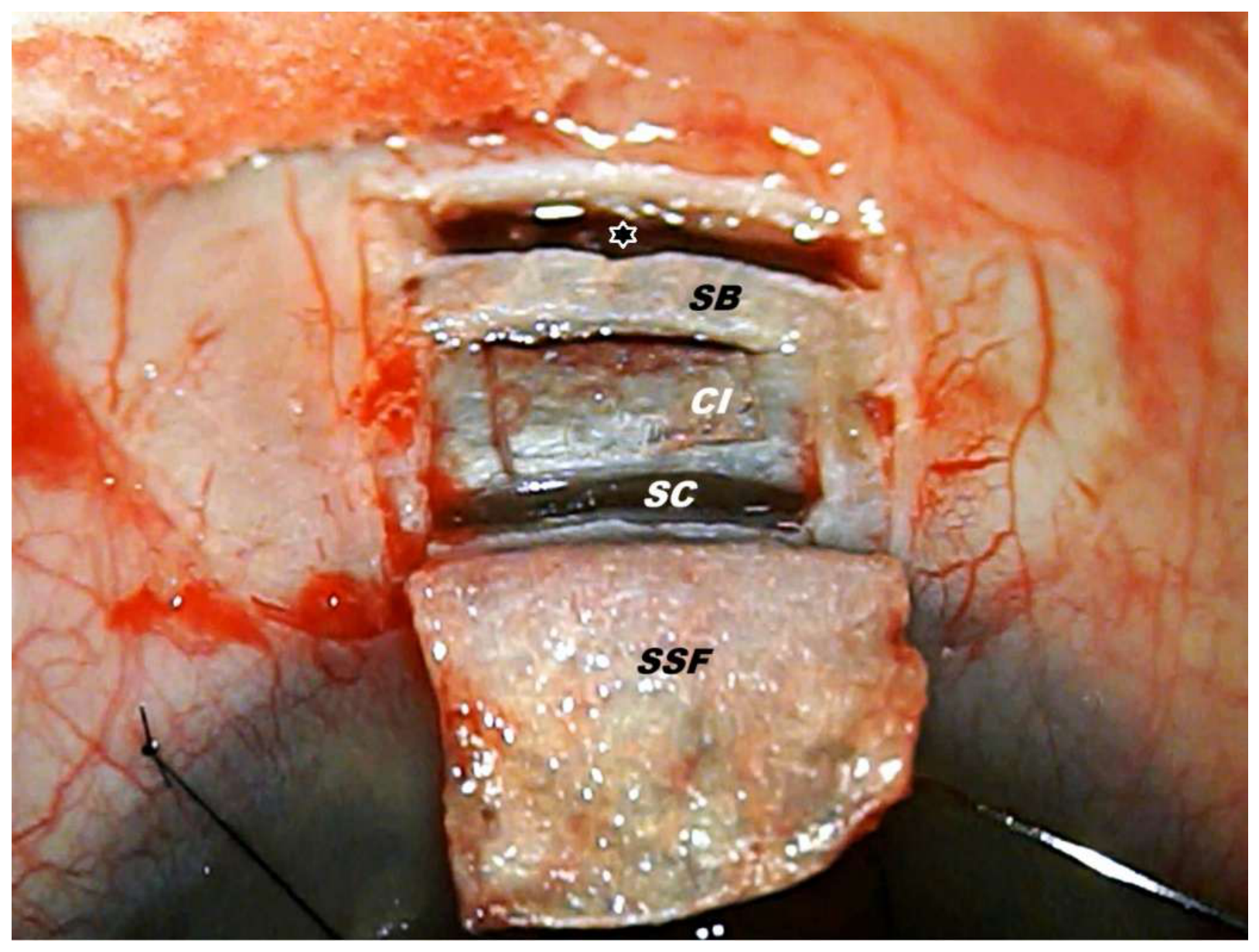
Figure 2.
Insertion of two small pieces of 4-0 polypropelene suture in the lumen of Schlemm’s canal (SC): a—showing inserted into both open ends of SC two small pieces (0.5cm) of 4-0 polyproplene suture (white arrows with black borders); b—gonioscopy view of the anterior angle with pieces of polyporpelene suture (white and black arrows) in the lumen of SC. The area between pieces is the surgical site where YAG-LT was performed safely and effectively using minimum energy.
Figure 2.
Insertion of two small pieces of 4-0 polypropelene suture in the lumen of Schlemm’s canal (SC): a—showing inserted into both open ends of SC two small pieces (0.5cm) of 4-0 polyproplene suture (white arrows with black borders); b—gonioscopy view of the anterior angle with pieces of polyporpelene suture (white and black arrows) in the lumen of SC. The area between pieces is the surgical site where YAG-LT was performed safely and effectively using minimum energy.
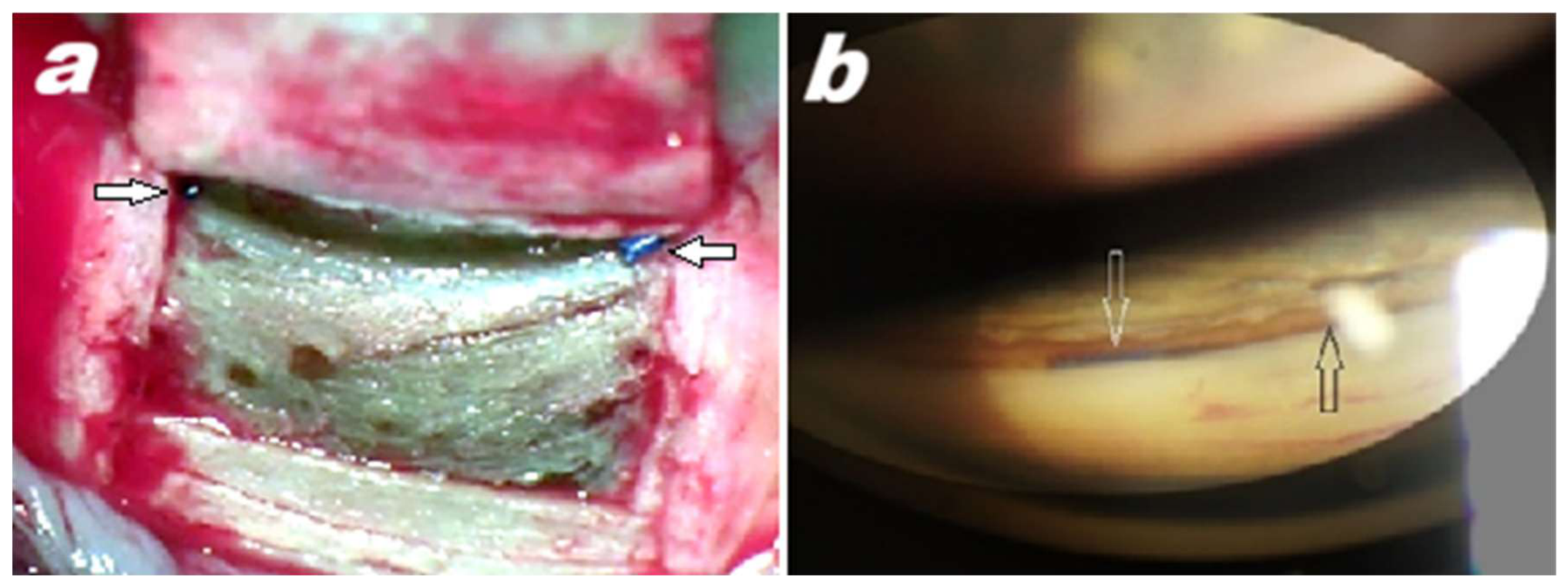
Figure 3.
Linear curve showing mean IOP values at different postoperative intervals. Vertical solid lines demonstrate 95% confidence interval values.
Figure 3.
Linear curve showing mean IOP values at different postoperative intervals. Vertical solid lines demonstrate 95% confidence interval values.
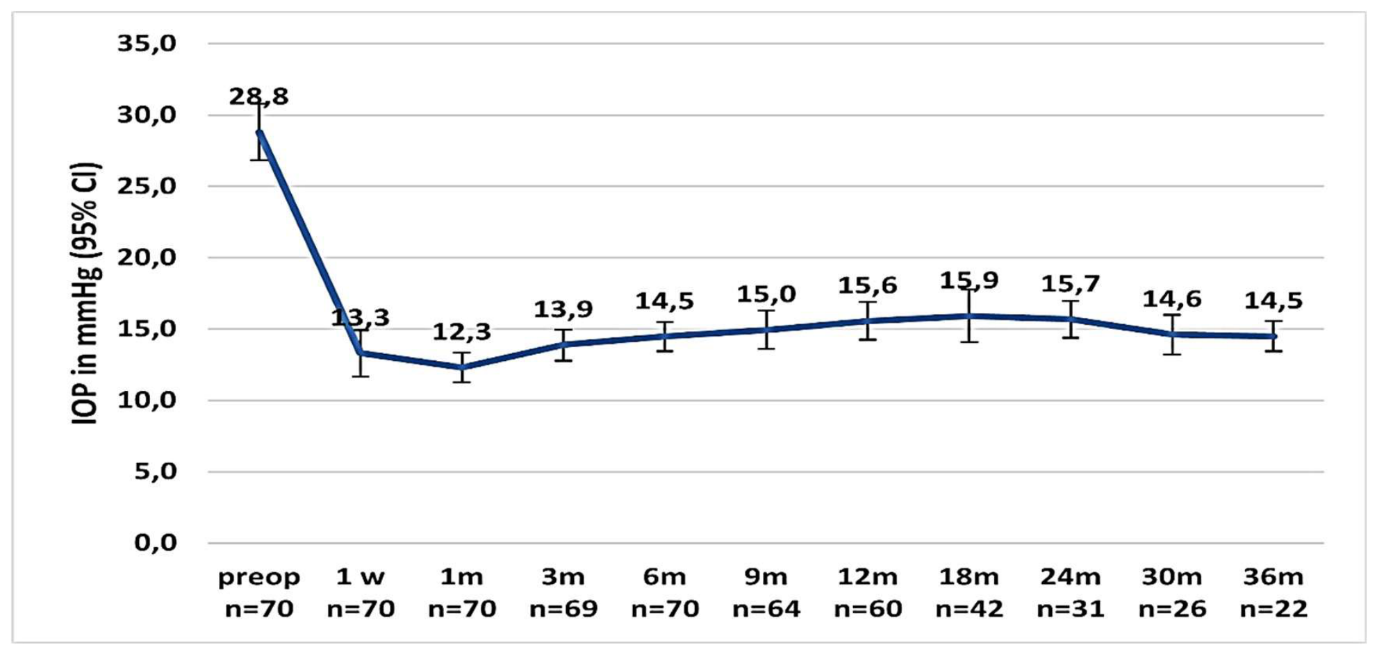
Figure 4.
Kaplan–Meier survival curve after surgery.
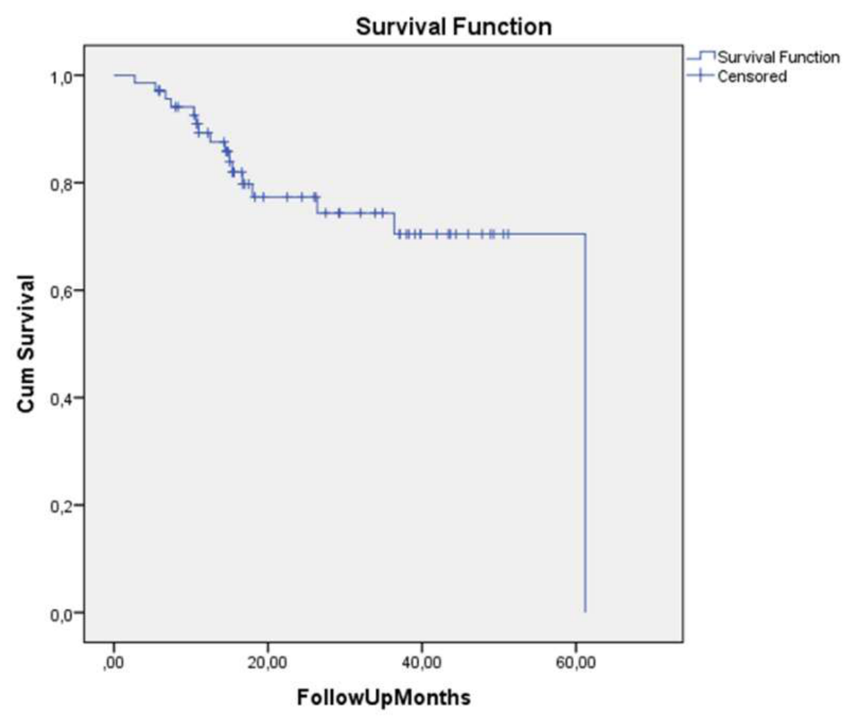
Figure 5.
The Kaplan–Meier survival curve after surgery for the standalone surgery subgroup (blue curve) and the combined surgery subgroup (green curve).
Figure 5.
The Kaplan–Meier survival curve after surgery for the standalone surgery subgroup (blue curve) and the combined surgery subgroup (green curve).
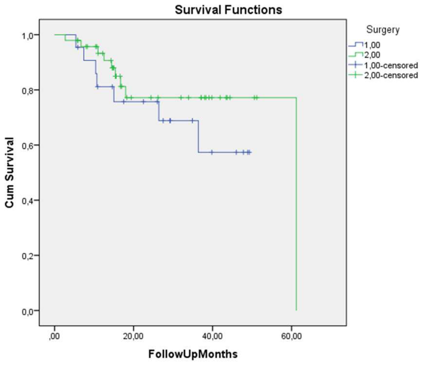
Figure 6.
Biomicroscopic visualization of CLVs and OCT confirmation of their lymphatic nature. Slit lamp front views of the surgical site and superior temporal quadrant of the conjunctiva of the left eye and OCT scans of the mentioned areas; 3 months after surgery. a—At the surgical site (black star with white borders), no CLVs are seen. b—A plexus of CLVs (indigo arrows with white borders) arising directly from the sclera, with no visible connection with the surgical site, appeared in the superior temporal quadrant; c—OCT scan of the surgical site showing CLVs (indigo arrows with white borders), which were not visible biomicroscopically; d—OCT scan of the conjunctival area with biomicroscopically visible CLVs showing lymphatic vessels (indigo arrows with white borders) with bicuspid valvelike structures in their lumens (white arrow with red borders).
Figure 6.
Biomicroscopic visualization of CLVs and OCT confirmation of their lymphatic nature. Slit lamp front views of the surgical site and superior temporal quadrant of the conjunctiva of the left eye and OCT scans of the mentioned areas; 3 months after surgery. a—At the surgical site (black star with white borders), no CLVs are seen. b—A plexus of CLVs (indigo arrows with white borders) arising directly from the sclera, with no visible connection with the surgical site, appeared in the superior temporal quadrant; c—OCT scan of the surgical site showing CLVs (indigo arrows with white borders), which were not visible biomicroscopically; d—OCT scan of the conjunctival area with biomicroscopically visible CLVs showing lymphatic vessels (indigo arrows with white borders) with bicuspid valvelike structures in their lumens (white arrow with red borders).
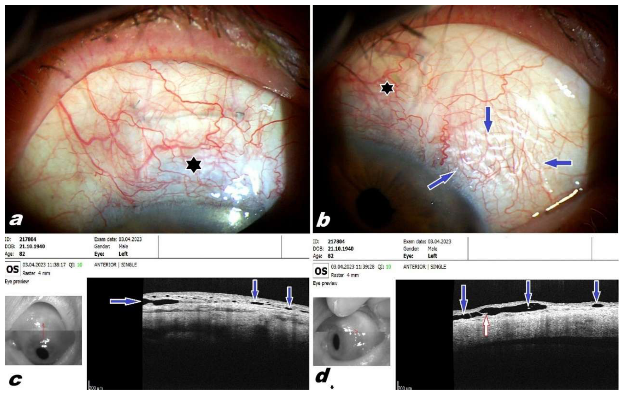
Figure 7.
Biomicroscopic visualization of CLVs and OCT confirmation of their lymphatic nature (another case). Left eye of a 69-year-old female patient, 1 month after surgery. a—Slit lamp front view of the superior medial quadrant of the conjunctiva, showing a plexus of transparent CLVs (indigo arrows with white borders) arising directly from the sclera at a site far away from the surgical site; b—OCT scan of the area showing CLVs (indigo arrows) with bicuspid valvelike structures in their lumen (white arrows with red borders), with the red arrow indicating the scan direction. .
Figure 7.
Biomicroscopic visualization of CLVs and OCT confirmation of their lymphatic nature (another case). Left eye of a 69-year-old female patient, 1 month after surgery. a—Slit lamp front view of the superior medial quadrant of the conjunctiva, showing a plexus of transparent CLVs (indigo arrows with white borders) arising directly from the sclera at a site far away from the surgical site; b—OCT scan of the area showing CLVs (indigo arrows) with bicuspid valvelike structures in their lumen (white arrows with red borders), with the red arrow indicating the scan direction. .
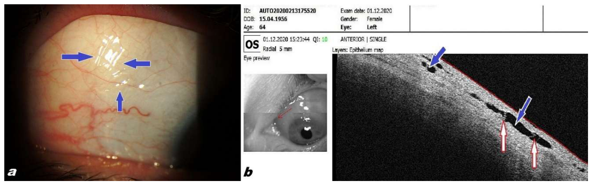
Figure 8.
Visualization of CLVs by moving eye lid margin over conjunctiva applying resistance to lymph flow. Left eye of an 82-year-old male patient, 3 months after surgery. IOP without hypotensive medication, 8 mmHg. a—Front view of the superior temporal quadrant of the bulbar conjunctiva showing transparent CLVs (indigo arrow with white borders) near the limbus, arising directly from the sclera, without connection with the surgical site and running parallel to the limbus. b—CLVs (indigo arrows with white borders) were more prominent once the lymph flow was temporarily obstructed by the upper lid margin. The lid margin was temporarily withdrawn to take a photograph (a snap taken from a video clip). c—The appearance of the CLVs changed as soon as the pressure on them was released by lifting the lid margin.
Figure 8.
Visualization of CLVs by moving eye lid margin over conjunctiva applying resistance to lymph flow. Left eye of an 82-year-old male patient, 3 months after surgery. IOP without hypotensive medication, 8 mmHg. a—Front view of the superior temporal quadrant of the bulbar conjunctiva showing transparent CLVs (indigo arrow with white borders) near the limbus, arising directly from the sclera, without connection with the surgical site and running parallel to the limbus. b—CLVs (indigo arrows with white borders) were more prominent once the lymph flow was temporarily obstructed by the upper lid margin. The lid margin was temporarily withdrawn to take a photograph (a snap taken from a video clip). c—The appearance of the CLVs changed as soon as the pressure on them was released by lifting the lid margin.
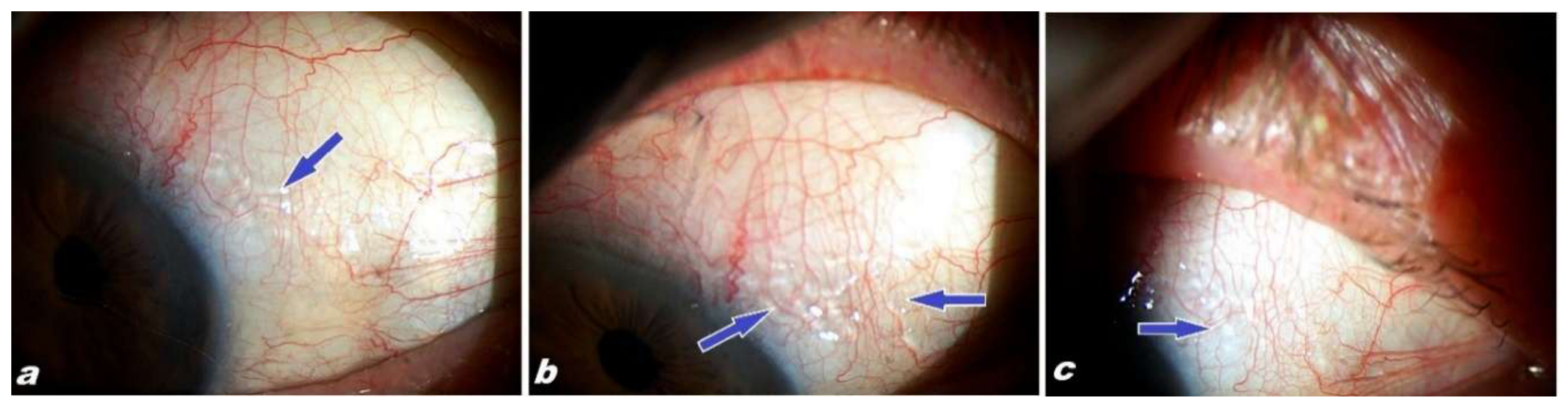
Figure 9.
Visualization of CLVs by moving eye lid margin over conjunctiva applying resistance to lymph flow (another case). Slit lamp front view of the superior bulbar conjunctiva of the left eye of a patient showing CLVs 2 years after surgery. a—Superior temporal quadrant of the conjunctiva, with one small visible CLV (indigo arrow with white borders). b—CLVs became prominent once the lymph flow was obstructed by the upper lid margin (indigo arrows with white borders). c—The CLVs became less prominent as soon as the pressure on them was released (indigo arrow with white borders).
Figure 9.
Visualization of CLVs by moving eye lid margin over conjunctiva applying resistance to lymph flow (another case). Slit lamp front view of the superior bulbar conjunctiva of the left eye of a patient showing CLVs 2 years after surgery. a—Superior temporal quadrant of the conjunctiva, with one small visible CLV (indigo arrow with white borders). b—CLVs became prominent once the lymph flow was obstructed by the upper lid margin (indigo arrows with white borders). c—The CLVs became less prominent as soon as the pressure on them was released (indigo arrow with white borders).
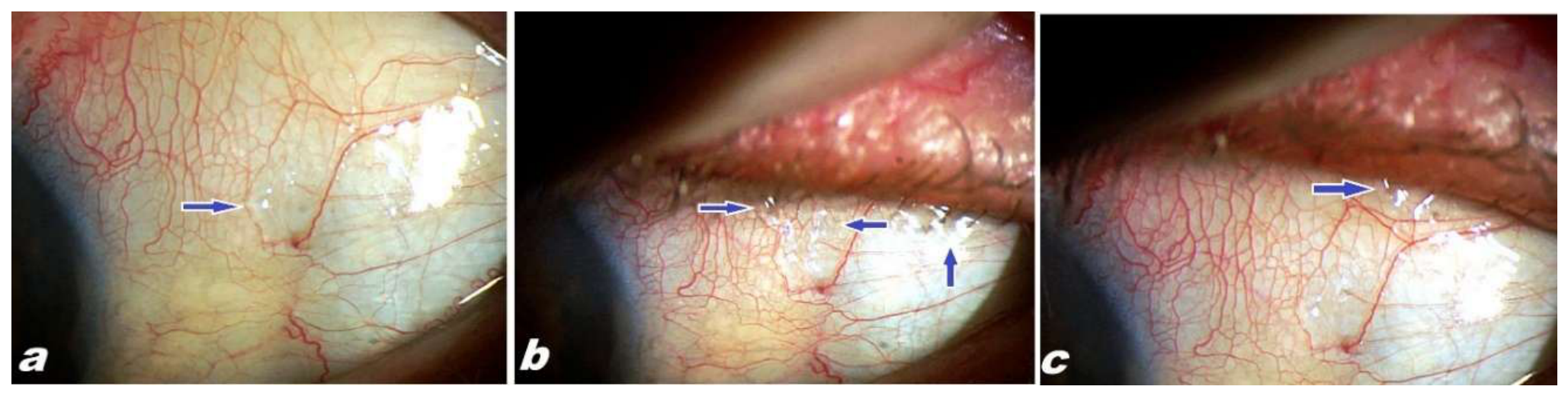
Figure 10.
Visualization of CLVs by moving eye lid margin over conjunctiva applying resistance to lymph flow (another case). Left eye of an 88-year-old male patient 15 days after surgery. a—Slit lamp front view of the surgical site (black star with white borders) showing the absence of blebs and CLVs, with some noticeable edema after surgery. b—Slit lamp front view of the temporal quadrant of the eye globe showing one transparent lymphatic vessel (indigo arrow with white borders) appearing in the conjunctiva as some pressure was applied on it by moving lid margin over it. c—Development of a CLV plexus (indigo arrows with white borders) after sweeping movements of the lid margin on the conjunctiva. d—As soon as the lid movements ceased, the CLVs (indigo arrow with white borders) became less prominent.
Figure 10.
Visualization of CLVs by moving eye lid margin over conjunctiva applying resistance to lymph flow (another case). Left eye of an 88-year-old male patient 15 days after surgery. a—Slit lamp front view of the surgical site (black star with white borders) showing the absence of blebs and CLVs, with some noticeable edema after surgery. b—Slit lamp front view of the temporal quadrant of the eye globe showing one transparent lymphatic vessel (indigo arrow with white borders) appearing in the conjunctiva as some pressure was applied on it by moving lid margin over it. c—Development of a CLV plexus (indigo arrows with white borders) after sweeping movements of the lid margin on the conjunctiva. d—As soon as the lid movements ceased, the CLVs (indigo arrow with white borders) became less prominent.
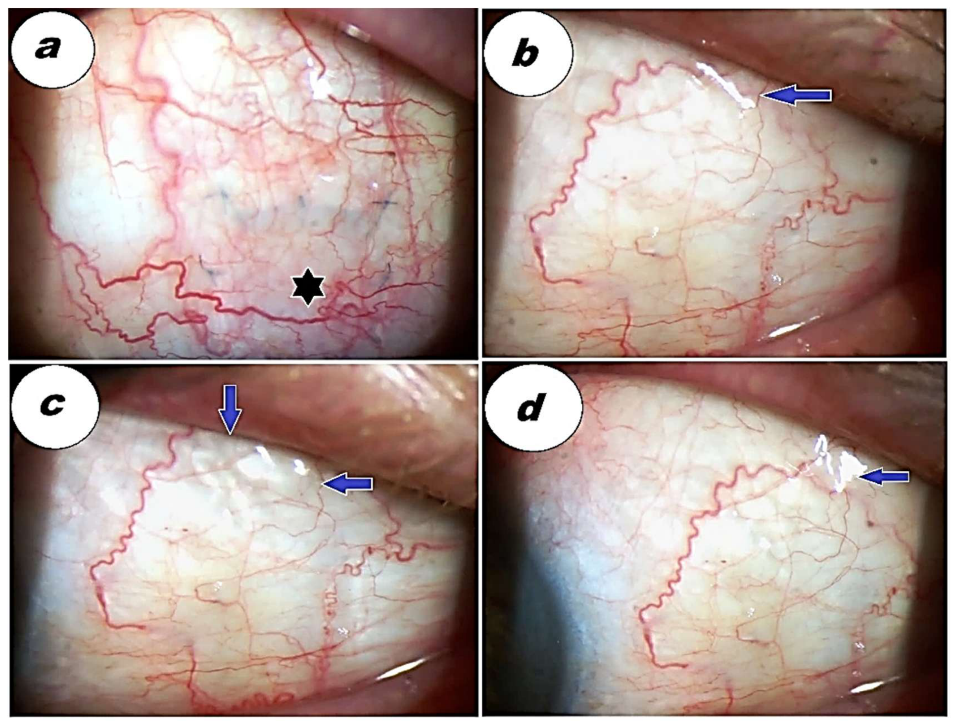
Figure 11.
The appearance of CLVs after YAG-LT in the temporal quadrant of the superior bulbar conjunctiva in the pseudophakic right eye of a 74-year-old female patient who had undergone glaucoma surgery 2 weeks before. 1—Slit lamp view before YAG-LT: a—superior temporal quadrant, b—surgical site, c—superior medial quadrant. 2—Slit lamp view immediately after YAG-LT; a—a transparent LV arising directly from the sclera (indigo arrow with white borders) in the temporal quadrant; b, c—unchanged surgical site and medial quadrant. 3—Slit lamp view 10 minutes after YAG-LT; a—more prominent CLVs in the temporal quadrant at the same site as in Figure 2, a; the surgical site (b) and medial quadrant (c) are unchanged.
Figure 11.
The appearance of CLVs after YAG-LT in the temporal quadrant of the superior bulbar conjunctiva in the pseudophakic right eye of a 74-year-old female patient who had undergone glaucoma surgery 2 weeks before. 1—Slit lamp view before YAG-LT: a—superior temporal quadrant, b—surgical site, c—superior medial quadrant. 2—Slit lamp view immediately after YAG-LT; a—a transparent LV arising directly from the sclera (indigo arrow with white borders) in the temporal quadrant; b, c—unchanged surgical site and medial quadrant. 3—Slit lamp view 10 minutes after YAG-LT; a—more prominent CLVs in the temporal quadrant at the same site as in Figure 2, a; the surgical site (b) and medial quadrant (c) are unchanged.
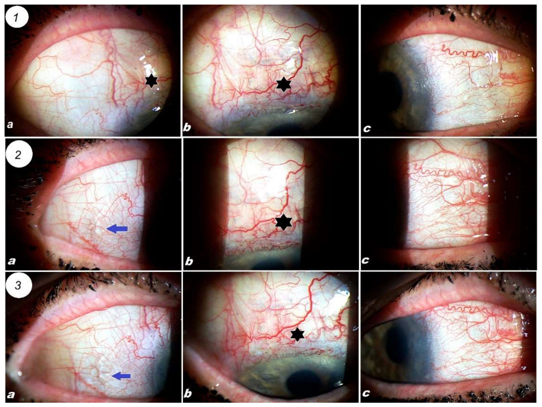
Figure 12.
Postoperative visualization of CLVs without LT and their reappearance in the same place after YAG-LT. Slit lamp views of the right eye of a 61-year-old male patient, after surgery. 1—One week after surgery, IOP on 3 classes of IOP-lowering medication, - 16 mm Hg; a—slit lamp view of the superior temporal quadrant showing the absence of CLVs; b—surgical site (black star), showing the absence of blebs and CLVs; c—slit lamp view of the medial quadrant of superior bulbar conjunctiva and area of medial rectus muscle insertion, showing 2-3 prominent CLVs (indigo arrows with white borders) running parallel to the limbus, arising directly from the sclera near the medial rectus muscle insertion. 2—Slit lamp view two weeks after surgery and before YAG-LT; IOP on 3 classes of IOP-lowering medication, - 22 mm Hg. a, b—temporal superior quadrant and surgical site without any change; c—CLVs reduced in size (indigo arrows with white borders) at the superior medial quadrant and medial rectus muscle insertion site. 3—Slit lamp views 1 week after YAG-LT; a, b—showing temporal superior quadrant and surgical site without CLVs and blebs; c—showing a well-developed plexus of prominent CLVs bulging with transparent fluid (indigo arrows with white borders) arising directly from the sclera at the same place as in Figure 1 c.
Figure 12.
Postoperative visualization of CLVs without LT and their reappearance in the same place after YAG-LT. Slit lamp views of the right eye of a 61-year-old male patient, after surgery. 1—One week after surgery, IOP on 3 classes of IOP-lowering medication, - 16 mm Hg; a—slit lamp view of the superior temporal quadrant showing the absence of CLVs; b—surgical site (black star), showing the absence of blebs and CLVs; c—slit lamp view of the medial quadrant of superior bulbar conjunctiva and area of medial rectus muscle insertion, showing 2-3 prominent CLVs (indigo arrows with white borders) running parallel to the limbus, arising directly from the sclera near the medial rectus muscle insertion. 2—Slit lamp view two weeks after surgery and before YAG-LT; IOP on 3 classes of IOP-lowering medication, - 22 mm Hg. a, b—temporal superior quadrant and surgical site without any change; c—CLVs reduced in size (indigo arrows with white borders) at the superior medial quadrant and medial rectus muscle insertion site. 3—Slit lamp views 1 week after YAG-LT; a, b—showing temporal superior quadrant and surgical site without CLVs and blebs; c—showing a well-developed plexus of prominent CLVs bulging with transparent fluid (indigo arrows with white borders) arising directly from the sclera at the same place as in Figure 1 c.
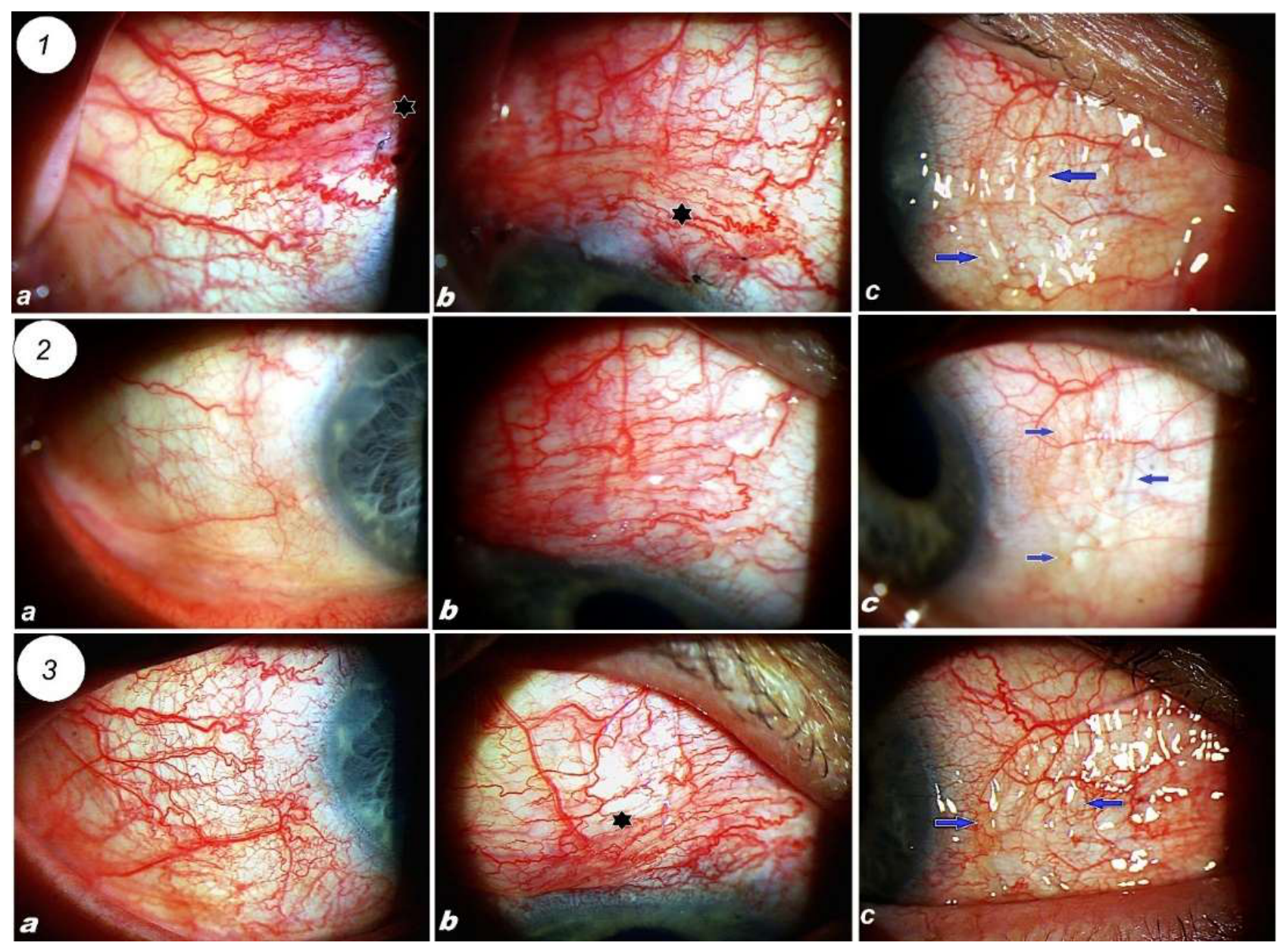
Figure 13.
Slit lamp front views of the superior conjunctiva of 3 different patients who underwent YAG-LT for high postoperative IOP, showing the development of CLVs belonging to different layers of the conjunctival lymphatic system with no relation to conjunctival blood vessels. a—Development of a deep CLV (lilac arrow with white borders), running parallel to the limbus at a distance from it, with a dull appearance and conjunctival blood vessels running over it. b—The development of a plexus of superficial (indigo arrow with white borders) and deep CLVs (lilac arrows with white borders). The superficial CLVs have sharp contours, prominent and bright in appearance, and conjunctival blood vessels are running under them. In contrast, deeper CLVs have blurred contours and dull appearance and color (lilac arrows); the superficial vessels arise from deep vessels and are their continuation. c—View of the superior temporal quadrant of the conjunctiva (the figure is taken from Figure 9 c), showing the development of a plexus consisting of transparent superficial (indigo color with white borders) and deep (lilac arrows with white borders) CLVs with nearly the same characteristics as in Figure 13 b.
Figure 13.
Slit lamp front views of the superior conjunctiva of 3 different patients who underwent YAG-LT for high postoperative IOP, showing the development of CLVs belonging to different layers of the conjunctival lymphatic system with no relation to conjunctival blood vessels. a—Development of a deep CLV (lilac arrow with white borders), running parallel to the limbus at a distance from it, with a dull appearance and conjunctival blood vessels running over it. b—The development of a plexus of superficial (indigo arrow with white borders) and deep CLVs (lilac arrows with white borders). The superficial CLVs have sharp contours, prominent and bright in appearance, and conjunctival blood vessels are running under them. In contrast, deeper CLVs have blurred contours and dull appearance and color (lilac arrows); the superficial vessels arise from deep vessels and are their continuation. c—View of the superior temporal quadrant of the conjunctiva (the figure is taken from Figure 9 c), showing the development of a plexus consisting of transparent superficial (indigo color with white borders) and deep (lilac arrows with white borders) CLVs with nearly the same characteristics as in Figure 13 b.
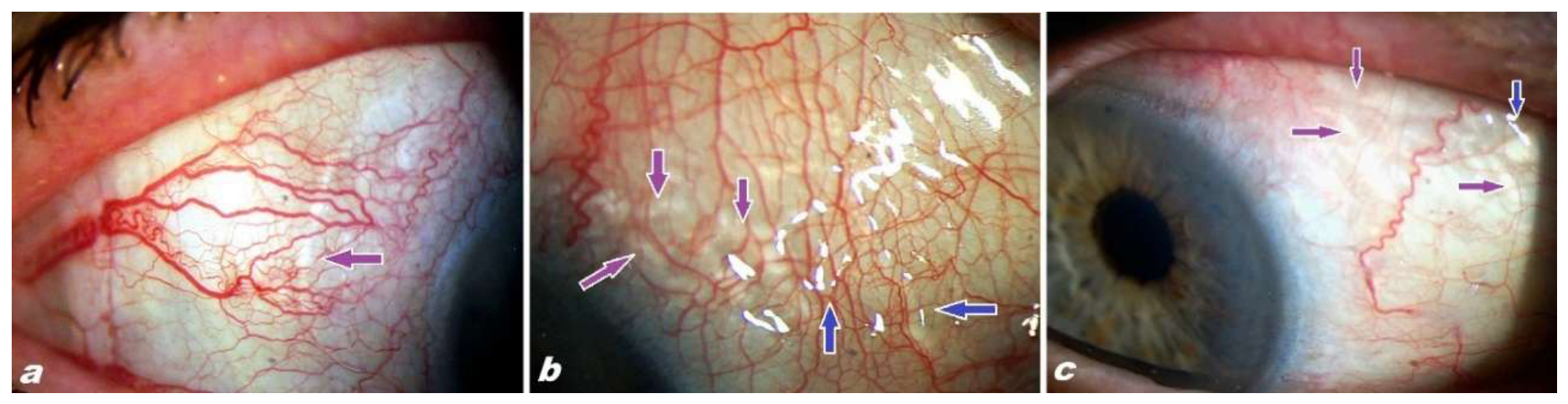
Figure 14.
OCT scan of the surgical site of the left eye of a 68-year-old female patient 1 month after surgery. The left figure shows a front view of the surgical site, with the red arrow indicating the scanned area. The OCT scan on the right side demonstrates the absence of blebs. The collagen implant is covered by the superficial scleral flap (black arrow with white borders). The yellow arrow with white borders points to the conjunctiva. The sclera is marked by a red star with white borders. .
Figure 14.
OCT scan of the surgical site of the left eye of a 68-year-old female patient 1 month after surgery. The left figure shows a front view of the surgical site, with the red arrow indicating the scanned area. The OCT scan on the right side demonstrates the absence of blebs. The collagen implant is covered by the superficial scleral flap (black arrow with white borders). The yellow arrow with white borders points to the conjunctiva. The sclera is marked by a red star with white borders. .
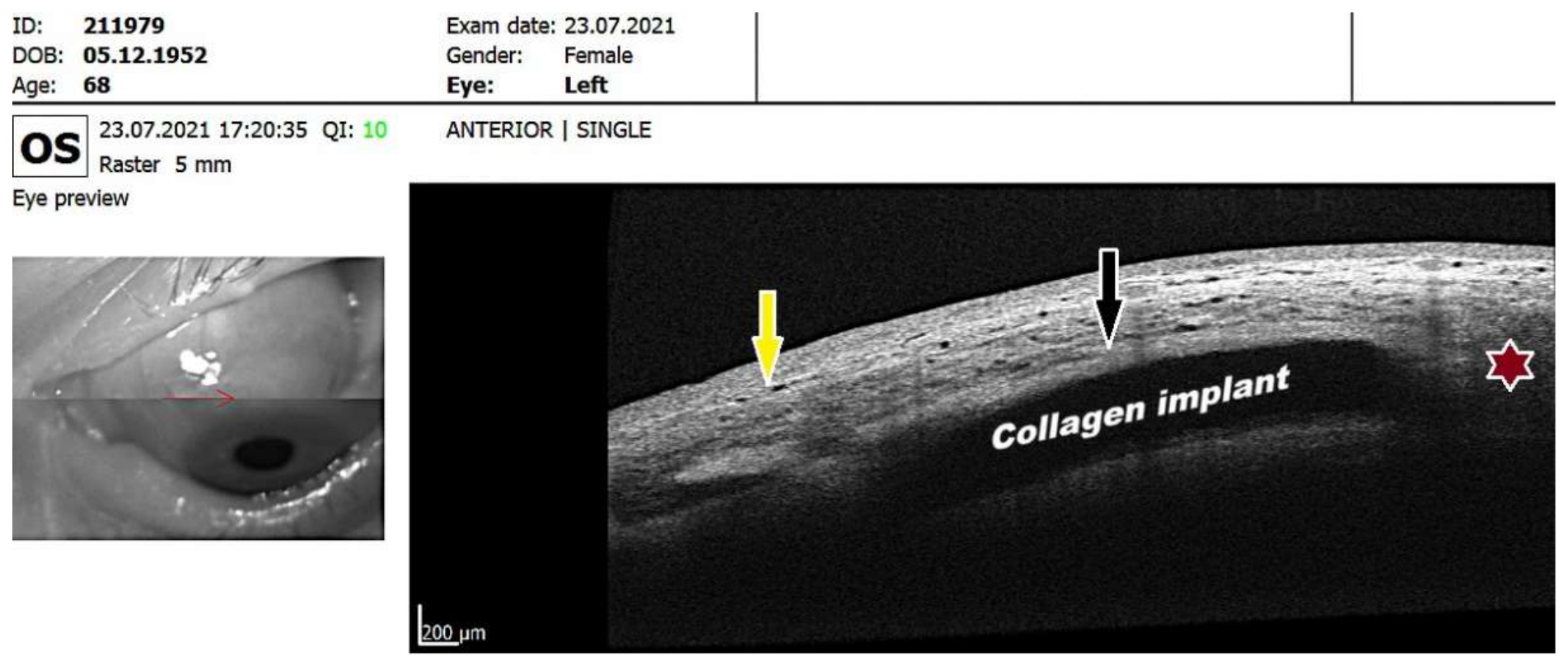
Figure 15.
Demonstrating the role of lymphatics in suprachoroidal drainage using a trypan blue aqueous tracer dye to investigate the possible AH outflow route after surgery. a—Slit lamp view of the surgical site before cataract surgery in the left eye of the patient showing an absence of blebs at the surgical site (black star); a white arrow with black borders indicates the upper border of the superficial scleral flap. b—Operating microscope view of the eye globe at the start of the study showing an irregular pupil due to posterior synechiae; a cannula tip is seen in the anterior chamber (AC) inserted through a paracentesis to replace the AH with trypan blue solution. c—AH in the AC is completely replaced with 0.08% trypan blue solution. d—Immediately after irrigating the AC with the dye, the latter was seen leaving the AC through an episcleral lymphatic vessel, which emerged at a distance of 4 mm from the upper limbus (white arrow with black borders indicates the starting point of the lymphatic vessel, and the white arrow with turquoise color borders indicates the vessel itself). e—View of the superior bulbar conjunctiva 15 seconds after the irrigation of the AC with the dye. A plexus of lymphatic vessels has appeared at the temporal side of the surgical site (white star with turquoise borders). f—Operating microscope view of the superior bulbar conjunctiva 30 seconds after; the plexus of lymphatic vessels (white arrow with turquoise borders) and lymphatic capillaries (white star with turquoise borders) are more pronounced g—View of the conjunctiva 1 minute after showing more pronounced lymphatic vessels and capillaries than in Fig. e. h—View after 2 minutes; the lymphatic vessels occupy a wider area extending towards the medial and temporal sides of the eye globe. The vessels extending toward the medial side have sharper contours and brighter color (superficial CLVs) than the vessels going toward the temporal side, which have blurred contours and dull color (deeper CLVs). i—Enlarged view of the superior bulbar conjunctiva after 3 minutes showing a dense plexus of lymphatic vessels and capillaries; the surgical field is devoid of these vessels. j—View of the superior conjunctiva at 3 minutes and 30 seconds; lymphatic vessels of different calibers and a plexus of lymphatic capillaries occupy the entire area of the upper segment; except for the surgical site, there is almost no leakage of the dye into the adjoining tissues, and all the visible dye is in the lumens of the lymphatic vessels. k—The eye globe after 5 minutes; at this time, the dye from the AC has been irrigated out, the lymphatic vessels in the superior segment have lost their identity, and there is mild diffusion of the dye into the surrounding tissues. Some dye is visible in thin lymphatic vessels reaching the inferior fornix (horizontal white arrows with turquoise borders). l—View of the superior conjunctiva at the end of the cataract surgery, 20 minutes after the start of the study. There are no identifiable lymphatic vessels and some tissues dyed blue.
Figure 15.
Demonstrating the role of lymphatics in suprachoroidal drainage using a trypan blue aqueous tracer dye to investigate the possible AH outflow route after surgery. a—Slit lamp view of the surgical site before cataract surgery in the left eye of the patient showing an absence of blebs at the surgical site (black star); a white arrow with black borders indicates the upper border of the superficial scleral flap. b—Operating microscope view of the eye globe at the start of the study showing an irregular pupil due to posterior synechiae; a cannula tip is seen in the anterior chamber (AC) inserted through a paracentesis to replace the AH with trypan blue solution. c—AH in the AC is completely replaced with 0.08% trypan blue solution. d—Immediately after irrigating the AC with the dye, the latter was seen leaving the AC through an episcleral lymphatic vessel, which emerged at a distance of 4 mm from the upper limbus (white arrow with black borders indicates the starting point of the lymphatic vessel, and the white arrow with turquoise color borders indicates the vessel itself). e—View of the superior bulbar conjunctiva 15 seconds after the irrigation of the AC with the dye. A plexus of lymphatic vessels has appeared at the temporal side of the surgical site (white star with turquoise borders). f—Operating microscope view of the superior bulbar conjunctiva 30 seconds after; the plexus of lymphatic vessels (white arrow with turquoise borders) and lymphatic capillaries (white star with turquoise borders) are more pronounced g—View of the conjunctiva 1 minute after showing more pronounced lymphatic vessels and capillaries than in Fig. e. h—View after 2 minutes; the lymphatic vessels occupy a wider area extending towards the medial and temporal sides of the eye globe. The vessels extending toward the medial side have sharper contours and brighter color (superficial CLVs) than the vessels going toward the temporal side, which have blurred contours and dull color (deeper CLVs). i—Enlarged view of the superior bulbar conjunctiva after 3 minutes showing a dense plexus of lymphatic vessels and capillaries; the surgical field is devoid of these vessels. j—View of the superior conjunctiva at 3 minutes and 30 seconds; lymphatic vessels of different calibers and a plexus of lymphatic capillaries occupy the entire area of the upper segment; except for the surgical site, there is almost no leakage of the dye into the adjoining tissues, and all the visible dye is in the lumens of the lymphatic vessels. k—The eye globe after 5 minutes; at this time, the dye from the AC has been irrigated out, the lymphatic vessels in the superior segment have lost their identity, and there is mild diffusion of the dye into the surrounding tissues. Some dye is visible in thin lymphatic vessels reaching the inferior fornix (horizontal white arrows with turquoise borders). l—View of the superior conjunctiva at the end of the cataract surgery, 20 minutes after the start of the study. There are no identifiable lymphatic vessels and some tissues dyed blue.
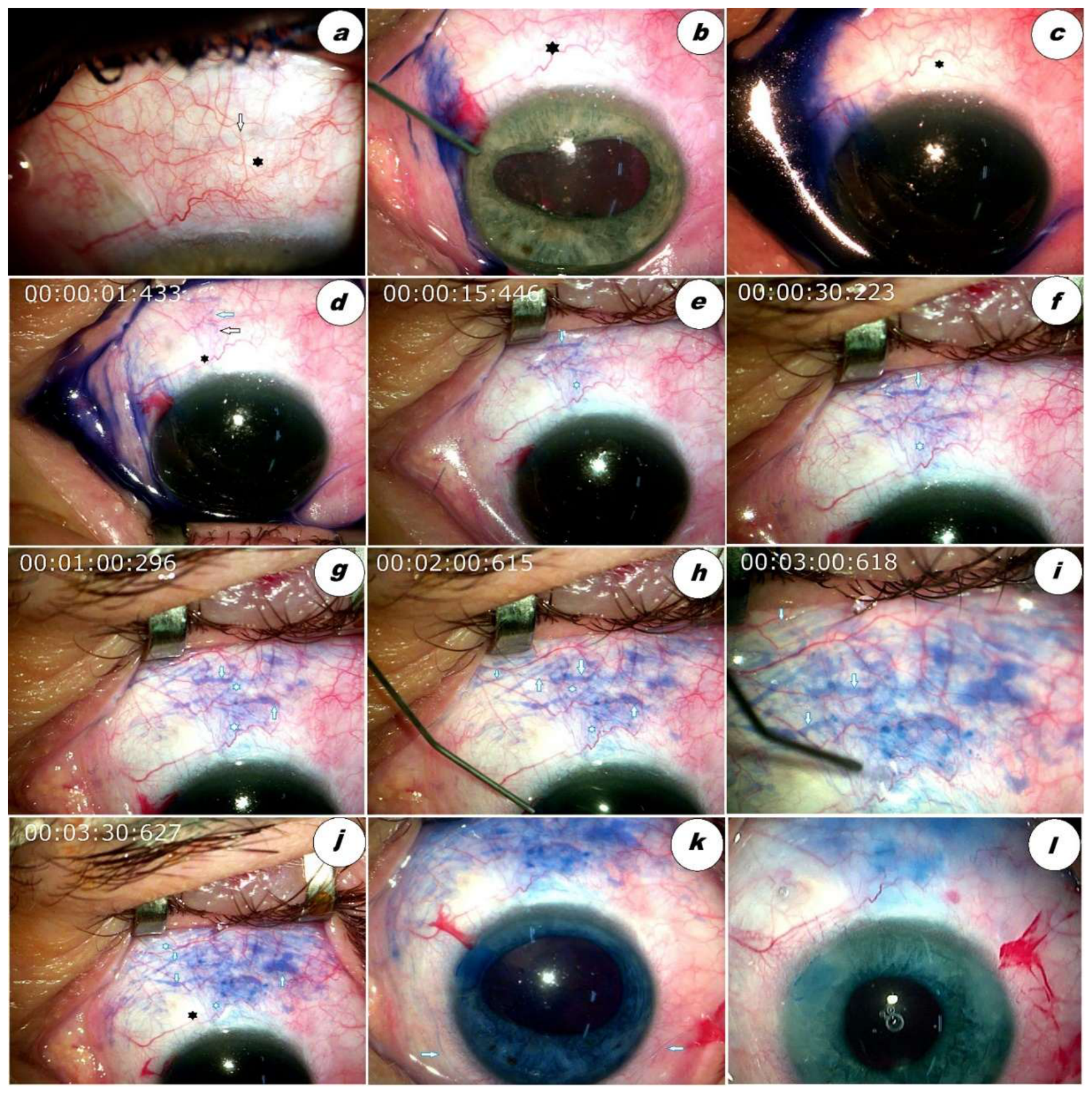
Figure 16.
In vivo investigation of conjunctival lymphatic vessels (CLVs) at the time of glaucoma surgery, with direct puncture of the CLV and manifestation of the lymphatic tree. a—Direct injection of trypan blue dye into the lumen of a CLV at 12:00, 4 mm away from the upper limbus; the white arrow with red borders indicates the site of injection and CLVs. b—Dye is seen filling the lumens of other CLVs, which are connected to the original one (white arrow with red borders). c-d—Further filling of the CLVs (white arrow with red borders) with dye. The arrow with black borders indicates a CLV, which will serve as the only connecting vessel with other CLVs running parallel and near to the limbus in the future. e-f—The connecting vessel, ‘the lymphatic bridge’ (pink arrow with black borders), connecting the first CLV from above with the CLV running below, parallel and near to the limbus. g–h—Development of limbal lymphatic capillaries of different calibers (white arrows with lilac borders) from the CLV. i—Different parts of the lymphatic tree of the superior bulbar conjunctiva, with the CLV punctured by an injection needle to inject dye. The connecting ‘lymphatic bridge’ (pink arrow with black borders), different layers of the conjunctival lymphatic system and limbal lymphatic capillaries, and the enlarged view of the area marked by the white rectangle are shown in Figure j. j—Enlarged view of the area captured by the white rectangle in Figure i. Small limbal lymphatic capillaries are indicated by black arrows, which are straight, oriented perpendicular to the edge of the cornea with one end connected with large limbal lymphatic capillaries, and the other end connected with the lymphatic plexuses of sclera and episclera (white 4-pointed stars with black border), light purple arrows with black borders indicate large limbal lymphatic capillaries, one end of which is blind, and the other end is connected with CLV above, these capillaries are oriented parallel to the limbus, a pink arrow with black border indicates the ‘lymphatic bridge’. White 4-pointed asterisks with black borders indicate the episcleral and scleral lymphatic plexuses, lymphatic vessels with poorly defined borders and dull color belong to the deep layer of CLVs and lymphatic vessels with sharp borders and bright dye belong to superficial vessels, which are interconnected superficial and deep lymphatic vessels.
Figure 16.
In vivo investigation of conjunctival lymphatic vessels (CLVs) at the time of glaucoma surgery, with direct puncture of the CLV and manifestation of the lymphatic tree. a—Direct injection of trypan blue dye into the lumen of a CLV at 12:00, 4 mm away from the upper limbus; the white arrow with red borders indicates the site of injection and CLVs. b—Dye is seen filling the lumens of other CLVs, which are connected to the original one (white arrow with red borders). c-d—Further filling of the CLVs (white arrow with red borders) with dye. The arrow with black borders indicates a CLV, which will serve as the only connecting vessel with other CLVs running parallel and near to the limbus in the future. e-f—The connecting vessel, ‘the lymphatic bridge’ (pink arrow with black borders), connecting the first CLV from above with the CLV running below, parallel and near to the limbus. g–h—Development of limbal lymphatic capillaries of different calibers (white arrows with lilac borders) from the CLV. i—Different parts of the lymphatic tree of the superior bulbar conjunctiva, with the CLV punctured by an injection needle to inject dye. The connecting ‘lymphatic bridge’ (pink arrow with black borders), different layers of the conjunctival lymphatic system and limbal lymphatic capillaries, and the enlarged view of the area marked by the white rectangle are shown in Figure j. j—Enlarged view of the area captured by the white rectangle in Figure i. Small limbal lymphatic capillaries are indicated by black arrows, which are straight, oriented perpendicular to the edge of the cornea with one end connected with large limbal lymphatic capillaries, and the other end connected with the lymphatic plexuses of sclera and episclera (white 4-pointed stars with black border), light purple arrows with black borders indicate large limbal lymphatic capillaries, one end of which is blind, and the other end is connected with CLV above, these capillaries are oriented parallel to the limbus, a pink arrow with black border indicates the ‘lymphatic bridge’. White 4-pointed asterisks with black borders indicate the episcleral and scleral lymphatic plexuses, lymphatic vessels with poorly defined borders and dull color belong to the deep layer of CLVs and lymphatic vessels with sharp borders and bright dye belong to superficial vessels, which are interconnected superficial and deep lymphatic vessels.
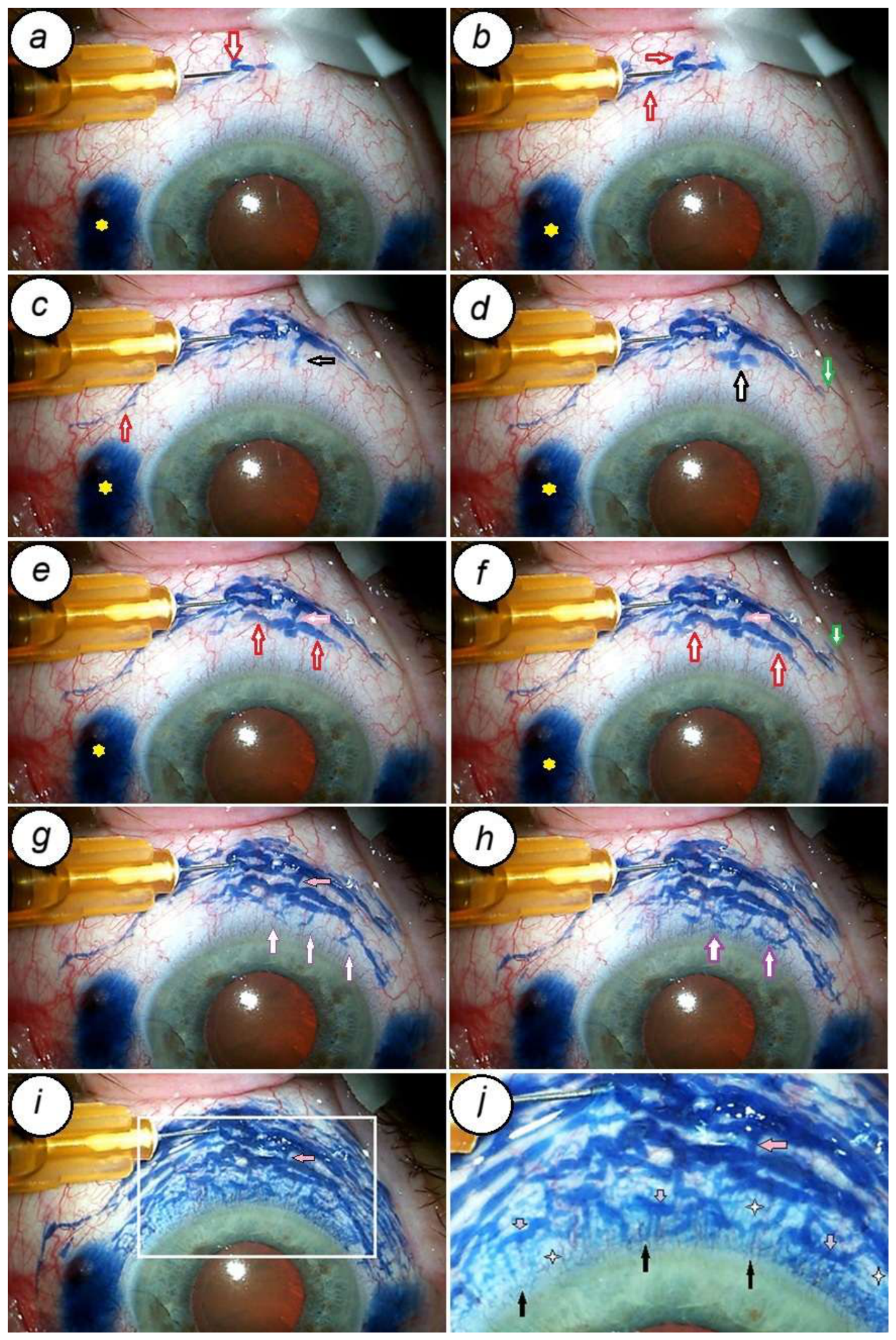

Table 1.
Demographic and baseline characteristics.
| Parameter | Values | P | ||
| All cases | Standalone procedure (22 eyes, 31.4%) | Combined procedure (48 eyes, 68.6%) | ||
| Patients, n | 63 | 20 | 43 | |
| Eyes, n | 70 | 22 | 48 | |
| Gender, female n (%) | 44 (69.8) | 13 (65) | 31 (72.1) | 0.5413 # |
| Age, years, mean (SD) | 75.11 (7.11) | 77.3 (6.4) | 74.1 (7.3) | 0.0837* |
| Laterality, left eye n (%) | 37 (52.8) | 12 (54,5) | 25 (52,1) | 0.8161 # |
| Latest follow-up, weeks, mean (SD) | 104.9 (63.9) | 109.4 (63.1) | 102,8 (64,8) | 0.686 ** |
| Glaucoma type, n (%) | ||||
| Primary open angle | 52 (74.3) | 11 (50) | 41 (85.4) | 0.0023# |
| Previous glaucoma operation | 18 (25.7) | 11 (50) | 7 (14.6) | 7.84-10# |
| Severity, n (%) | ||||
| Moderate | 26 (37.1) | 11 (50) | 15 (31.2) | 0.0369# |
| Severe | 44 (62.9) | 11 (50) | 33 (68.8) | 0.0846# |
| Preoperative IOP, mm Hg, mean (SD) | 28.8 (8.4) | 29 [27.3; 33.8] | 27.7 (7.4) | 0.138** |
| IOP range, mm Hg, n (%) | ||||
| ≤ 18 | 4 (5.7) | 2 (9.1) | 2 (4.2) | 0.1791# |
| 18.1 – 21.0 | 8 (11.4) | 2 (9.1) | 6 (12.5) | 0.4644# |
| 21.1 – 30.0 | 41 (58.6) | 11 (50) | 30 (62.5) | 0.2386# |
| 30.1 – 40.0 | 13 (18.6) | 7 (31.8) | 6 (12.5) | 0.0037# |
| >40.0 | 4 (5.7) | 0 | 4 (8.3) | 0.0039# |
| Previuos glaucoma surgeries, n (%) | 18 (25.7) | 11 (50) | 7 (14.6) | 0.00001# |
| Number of previous glaucoma surgeries, n | ||||
| Surgery performed once | 13 | 9 | 4 | |
| Surgery performed twice | 5 | 2 | 3 | |
| Previous glaucoma surgery, n | ||||
| Trabeculectomy | 7 | 4 | 3 | |
| Deep sclerectomy | 2 | 2 | 0 | |
| Laser Iridotomy | 2 | 2 | 0 | |
| Schlemm’s canal stenting | 2 | 0 | 2 | |
| Modified deep sclerectomy with “Xenoplast” as space maintainer | 6 | 4 | 2 | |
| Other surgeries | 4 | 1 | 3 | |
| Preoperative IOP-lowering medications, median [p25; p75] | 3 [2,3] | 3.00 [2.25; 4.00] | 3 [2.00; 3.00] | 0.074** |
| Classes of hypotensive medications used n (%) | ||||
| Class 1 | 7 (10.0) | 0 | 7 (14.6) | 0.0001 |
| Classes 2 | 19 (27.1) | 6 (27.2) | 13 (27.1) | 0.9891 |
| Classes 3 | 26 (37.2) | 8 (36.4) | 18 (37.5) | 0.9073 |
| Classes 4 | 18 (25.7) | 8 (36.4) | 10 (20.8) | 0.0391 |
| BCVA before surgery, median [p25; p75], logMar | 0.6 [0.2; 1.9] | 0.7 [2.0; 1.8] | 0.5 [0.2; 1.7] | 0.671 ** |
| Lens condition, n (%) | ||||
| No cataract | 1 (1.4) | 1 | 0 | |
| Cataract (intumescent cataract-2 eyes; severe phacodonesis-3 eyes) | 48 (68.6) | 0 | 48 | |
| Pseudophakia (2 cases with IOL dislocation) | 21 (30) | 21 | 0 | |
| Comorbidities | ||||
| Pseudo-exfoliation syndrome | 46 (65.7) | 17 (77.3) | 29 (60.4) | 0.1498 |
| Wet AMD | 2 (2.9) | 1 | 1 | |
| Dry AMD (1 case had 3 intravitreal injections of antiVEGF; epiretinal membrane -1 eye) | 2 (2.9) | 1 | 1 | |
| Mild myopia | 1 (1.4) | 0 | 1 | |
| High myopia | 4 (5.7) | 0 | 4 | |
| Diabetes mellitus | 5 (7.1) | 2 | 3 | |
antiVEGF= anti vascular endothelial growth factor; AMD=age-related macular degeneration; BCVA=best corrected visual acuity; IOP = intraocular pressure; n=number of eyes; logMAR=logarithm of the minimum angle of resolution; n=number of eyes; p25=Q1, first quartile; p75=Q3, 3rd quartile, SD = standard deviation. *=independent samples t-test; **=two-sample t-test with different variables; # = Chi-square test.
Table 2.
IOP reduction at different follow-up intervals.
| IOP reduction,% | Postoperative follow-up intervals, n (%) | ||||||||||
|---|---|---|---|---|---|---|---|---|---|---|---|
| 1 w (n=70) | 1 mo (n=70) | 3 mo (n=69) | 6 mo (n=70) | 9 mo (n=64) | 12 mo (n=60) | 18 mo (n=42) | 24 mo (n=31) | 30 mo (n=26) | 36 mo (n=22) | ||
| <20 | 4 (5.7) | 2 (2.9) | 4 (5.8) | 5 (7.1) | 2 (3.1) | 4 (6.7) | 2 (4.8) | 2 (6.5) | 0 | 0 | |
| 20 and < 30 | 6 (8.6) | 5 (7.1) | 5 (7.2) | 4 (5.7) | 7 (10.9) | 4 (6.7) | 3 (7.1) | 4 (12.9) | 2 (7.7) | 4 (18.2) | |
| 30 and < 40 | 6 (8.6) | 5 (7.1) | 6 (8.7) | 11 (15.9) | 13 (20.3) | 18 (30.0) | 13 (31.0) | 8 (25.8) | 5 (19.2) | 2 (9.1) | |
| 40 and < 50 | 7 (10.0) | 12 9 (17.1) | 12 (17.4) | 12 (17.3) | 14 (21.9) | 9 (15.0) | 6 (14.3) | 6 (19.4) | 4 (15.4) | 6 (27.3) | |
| ≥50 | 44 (62.9) | 45 (64.4) | 42 (60.9) | 36 (52.2) | 27 (42.2) | 22 (36.7) | 16 (38.1) | 11 (35.4) | 14 (53.8) | 10 (45.4) | |
| IOP > baseline IOP | 3 (4.3) | 1 (1.4) | 0 | 1 (1.4) | 1 (1.6) | 3 (5.0) | 2 (4.8) | 0 | 1 (3.8) | 0 | |
| No change in IOP | 0 | 0 | 0 | 1 (1.4) | 0 | 0 | 0 | 0 | 0 | 0 | |
IOP=intraocular pressure; mo=months; n=number of eyes; w=week.
Table 3.
Intraocular pressure changes after surgery.
| Follow-up intervals | n | IOP, Mean (SD) | IOP decrease, mean (SD) |
IOP reduction,%, mean (SD) | Eyes with IOP> baseline IOP, n |
|---|---|---|---|---|---|
| Baseline | 70 | 28.9 (8.4) | |||
| 1w | 70 | 13.4 (6.8) | -15.5 (10.0) | -51.0 (26.7) | 3 |
| 1mo | 70 | 12.3 (4.5) | -16.6 (9.0) | -54.7 (20.2) | 1 |
| 3 mo | 69 | 13.8 (4.6) | -15.1 (8.9) | -49.6 (18.9) | 0 |
| 6 mo | 70 | 14.5 (4.4) | -14.5 (8.9) | -47.0 (19.5) | 1 |
| 9 mo | 64 | 15.0 (5.5) | -14.2 (8.3) | -46.6 (17.2) | 1 |
| 12 mo | 60 | 15.6 (5.3) | -12.0 (9.3) | -42.5 (21.3) | 3 |
| 18 mo | 42 | 15.9 (6.1) | -13.3 (8.2) | -43.6 (19.3) | 2 |
| 24 mo | 31 | 15.7 (3.7) | - 11.0 (8.9) | - 44.3 (17.2) | 0 |
| 30 mo | 26 | 14.6 (3.6) | -15.2 (9.0) | -47.7 (19.8) | 1 |
| 36 mo | 22 | 14.5 (2.5) | -13.7 (7.7) | - 46.2 (13.8) | 0 |
IOP=intraocular pressure; mo=months; n=number of eyes; SD = standard deviation; w=week.
Table 4.
Changes in use of hypotensive medications after surgery.
| Hypotensive medications, median [p25;p75], mean (SD) | |||||||||
|---|---|---|---|---|---|---|---|---|---|
| Baseline | 1 mo | 3 mo | 6 mo | 9 mo | 12 mo | 18 mo | 24 mo | 30 mo | 36 mo |
| 3 [2;3] | 0 [0;0] | 0 [0;0] | 0 [0;1] | 0 [0;1.5] | 0 [0;1.5] | 0 [0;1] | 1 [0;1] | 0 [0;1] | 1.4 (1.1) |
mo=months; p25=Q1; first quartile; p75=Q3; 3rd quartile; SD = standard deviation.
Table 5.
Medication burden after surgery.
| Follow-up intervals | Number of eyes not requiring medication, n (%) | Number of eyes in need of additional hypotensive medications, n (%) | Total number of eyes under observation, n | ||
| 1 class, n (%) | 2 classes, n (%) | 3 classes, n (%) | |||
| 6 mo | 48 (68.6) | 8 (11.4) | 6 (8.6) | 8 (11.4) | 70 |
| 12 mo | 34 (56.7) | 10 (16.7) | 6 (10.0) | 10 (16.6) | 60 |
| 24 mo | 15 (48.4) | 10 (32.2) | 3 (9.7) | 3 (9.6) | 31 |
| 36 mo | 6 (27.3) | 6 (27.3) | 6 (27.3) | 4 (18.1) | 22 |
mo=months; n=number of eyes.
Table 6.
Changes in visual acuity after surgery.
| Visual acuity, logMar, median [p25;p75] | ||||||||||
|---|---|---|---|---|---|---|---|---|---|---|
| Preoperative | 1 mo | 3 mo | 6 mo | 9 mo | 12 mo | 18 mo | 24 mo | 30 mo | 36 mo | |
| 0.6[0.2;1.9] | 0.2[0;0.8] | 0.2[0;0.7] | 0.2[0;0.7] | 0.2[0;0.7] | 0.2[0; 0.8] | 0.2[0; 0.4] | 0.2[0; 0.6] | 0.1[0; 0.4] | 0[0; 0.4] | |
logMar = logarithm of the minimum angle of resolution; mo=months, p25=Q1, first quartile; p75=Q3, third quartile.
Table 7.
Visualization of conjunctival lymphatic vessels on slit lamp examination.
| CLV | Postoperative follow-up intervals, n | ||||||||
|---|---|---|---|---|---|---|---|---|---|
| 1 d | 1 w | 1 mo | 3 mo | 6 mo | 9 mo | 12 mo | 18 mo | 30 mo | |
| follow-up CLV was observed at | 10 | 8 | 13 | 2 | 3 | 1 | |||
| period of visibility | 11 | 4 | 12 | 4 | 1 | 0 | 1 | 1 | 3 |
CLV=conjucntival lymphatic vessel; d=day; w=week; mo=month; months; n=number of eyes.
Table 8.
Distribution of conjunctival lymphatic vessels in the superior bulbar conjunctiva, as observed on slit lamp microscope.
Table 8.
Distribution of conjunctival lymphatic vessels in the superior bulbar conjunctiva, as observed on slit lamp microscope.
| Site of CLV visualization on slit lamp examination, number of times | |||||
|---|---|---|---|---|---|
| SS | SN | ST | SS+SN | SS+ST | SN+ST |
| 5 | 46 | 17 | 6 | 6 | 7 |
CLV=conjunctival lymphatic vessels; SS=surgical site; SN= supranasally; ST= supratemporally.
Table 9.
OCT evaluation of superior bulbar conjunctiva after surgery at different follow-up intervals and distribution of CLVs as per areas of their identification.
Table 9.
OCT evaluation of superior bulbar conjunctiva after surgery at different follow-up intervals and distribution of CLVs as per areas of their identification.
| Follow-up intervals | CLVs identified at, n | CLVs not identified, n (%) | № of times OCT performed, n (%) | № of times OCT not performed (%) | ||||||
| SS | SN | ST | SS+ SN | SS+ ST | SN+ ST | SS+ SN+ ST | ||||
| 1d | 2 | 2 | 0 | 1 | 1 | 0 | 2 | 0 | 8 | 62 |
| 1w | 3 | 5 | 0 | 7 | 2 | 2 | 5 | 1 | 25 | 45 |
| 1m | 3 | 7 | 3 | 4 | 4 | 3 | 5 | 0 | 29 | 41 |
| 3 m | 5 | 5 | 7 | 7 | 2 | 1 | 7 | 0 | 34 | 36 |
| 6 m | 3 | 4 | 6 | 2 | 0 | 1 | 5 | 4 | 25 | 45 |
| 9 m | 1 | 2 | 2 | 4 | 0 | 0 | 4 | 5 | 18 | 46 |
| 12 m | 2 | 2 | 3 | 4 | 0 | 0 | 7 | 5 | 23 | 37 |
| 18 m | 0 | 3 | 1 | 1 | 1 | 1 | 3 | 5 | 15 | 27 |
| 24 m | 0 | 2 | 0 | 3 | 1 | 0 | 3 | 3 | 12 | 19 |
| 30 m | 0 | 2 | 0 | 3 | 1 | 0 | 3 | 5 | 14 | 12 |
| 36 m | 1 | 0 | 0 | 0 | 0 | 0 | 3 | 2 | 6 | 16 |
| Total | 20 | 34 | 22 | 36 | 12 | 8 | 47 | 30 | 209 | 386 |
| % | 9.6 | 16.3 | 10.5 | 17.2 | 5.7 | 3.8 | 22.5 | 14.4 | 35.1 | 64.9 |
d=day; w=week; LV=lymphatic vessels; mo=months; n= number of eyes; OCT=optical coherence tomography; SS=surgical site; SN =superior nasal quadrant; ST= superior temporal quadrant; LV=lymphatic vessel.
Disclaimer/Publisher’s Note: The statements, opinions and data contained in all publications are solely those of the individual author(s) and contributor(s) and not of MDPI and/or the editor(s). MDPI and/or the editor(s) disclaim responsibility for any injury to people or property resulting from any ideas, methods, instructions or products referred to in the content. |
© 2026 by the authors. Licensee MDPI, Basel, Switzerland. This article is an open access article distributed under the terms and conditions of the Creative Commons Attribution (CC BY) license (http://creativecommons.org/licenses/by/4.0/).
Copyright: This open access article is published under a Creative Commons CC BY 4.0 license, which permit the free download, distribution, and reuse, provided that the author and preprint are cited in any reuse.