

Submitted:
12 August 2026
Posted:
13 August 2026
You are already at the latest version
Abstract
The genetic architecture of neurodevelopmental disorders (NDDs) is increasingly well described, yet the biological meaning of many variants remains uncertain. A molecular diagnosis may identify the affected gene without explaining how the alteration perturbs brain development or why the same or related variants produce different outcomes. Variant effects are conditioned by dosage, developmental timing, cell identity and the wider genomic, epigenetic and environmental context, while compensatory responses may modify or obscure the phenotype. Functional interpretation therefore depends on linking the molecular defect to the relevant developmental and physiological consequences. This review brings together current knowledge of NDD mechanisms with the experimental and computational strategies used to test that link. No model reproduces the complete disorder, and greater complexity does not necessarily confer greater validity. The most informative system is the one that captures the biological process and developmental window relevant to the question. Concordance across models can strengthen a proposed mechanism, whereas discordant findings may reveal cellular, developmental or species-specific effects that would otherwise be missed. The same standard applies to therapeutic development: correction of a molecular abnormality is meaningful only when it produces durable functional benefit and can be achieved with appropriate central nervous system distribution, dosage and safety. Integrating functional evidence with detailed phenotyping and longitudinal clinical data may improve variant interpretation, distinguish biologically meaningful patient groups and support therapeutic strategies directed at mechanism rather than diagnostic category.
Keywords:
neurodevelopmental disorders
; variant interpretation
; functional genomics
; disease modeling
; translational genetics
; targeted therapies
1. Introduction
Neurodevelopmental disorders (NDDs) comprise a heterogeneous group of conditions arising from disruptions in brain development and function, leading to impairments in cognition, communication, behavior, social interaction, and motor abilities. This broad category includes autism spectrum disorder (ASD), attention-deficit/hyperactivity disorder (ADHD), intellectual disability (ID), developmental and epileptic encephalopathies, and a wide range of neurogenetic syndromes (Silverman & Ellegood, 2018). Although these conditions differ considerably in clinical presentation and severity, they frequently share overlapping developmental trajectories, biological mechanisms, and functional consequences, making their diagnosis and management particularly challenging.
NDDs represent a substantial clinical and public health burden and are commonly encountered in pediatric neurology and child psychiatry. Although they are typically diagnosed during childhood, certain clinical features may evolve or become more evident during adolescence or adulthood. Affected individuals frequently present with communication difficulties, repetitive behaviors, executive dysfunction, learning disabilities, and additional neurological or psychiatric comorbidities, further complicating differential diagnosis and therapeutic management (Hanly et al., 2021). Their frequent association with anxiety, depression, and other neuropsychiatric manifestations also suggests partially shared genetic and neurobiological mechanisms across diagnostic categories (Doherty et al., 2018).
Over the past decade, advances in next-generation sequencing (NGS), whole-exome sequencing (WES), and functional genomics have substantially expanded our understanding of the molecular architecture of NDDs. Targeted sequencing panels have also significantly improved the early molecular diagnosis of childhood epilepsy and related neurodevelopmental disorders, complementing broader exome and genome sequencing approaches (Amadori et al., 2020). These approaches have enabled the identification of pathogenic variants and dysregulated pathways involved in neuronal development, synaptic function, and neural connectivity. In parallel, emerging experimental platforms, including patient-derived cells, induced pluripotent stem cells (iPSCs), brain organoids, animal models, and computational approaches, have provided new opportunities to investigate disease mechanisms in biologically relevant systems. Nevertheless, translating genomic and molecular discoveries into effective clinical interventions remains challenging because of marked genetic heterogeneity, variable penetrance, phenotypic diversity, and the complexity of human brain development (Pietropaolo et al., 2017).
This review integrates current knowledge of the molecular mechanisms and translational approaches relevant to NDDs. It discusses genetic, epigenetic, and environmental contributions, together with cellular, organoid, animal, and computational models used for functional and mechanistic investigation. Emerging RNA-based and gene-targeted therapies and the main challenges limiting clinical translation are also addressed. Rather than identifying a universally optimal experimental platform, the central challenge is to select the model that best captures the biological process, developmental stage, and mechanistic question under investigation.
2. Molecular and Developmental Basis of NDDs
2.1. Disrupted Neurodevelopmental Processes
The clinical heterogeneity of NDDs reflects not only the molecular pathway affected but also the developmental stage, brain region, and cell population in which disruption occurs. Altered neural progenitor proliferation and fate specification can modify cortical growth and cellular composition, whereas defects in neuronal migration, axon guidance, and synaptogenesis disturb circuit organization and connectivity. For example, gain-of-function variants in RAC3 disrupt neuronal differentiation, migration, and axonogenesis during cortical development, illustrating how perturbation of cytoskeletal signaling pathways can lead to severe neurodevelopmental disorders (Nishikawa et al., 2023; Scala et al., 2022). Consistent with this, Rho-family GTPases have emerged as key regulators of neurite extension, axon guidance, synapse formation, and other developmental processes that represent convergent biological mechanisms across multiple neurodevelopmental disorders (Scala et al., 2021). Likewise, loss of WWOX impairs neuronal migration and early cortical development (Iacomino et al., 2020), whereas disruption of extracellular signaling pathways such as Reelin further emphasizes that defects affecting distinct molecular systems ultimately converge on shared abnormalities of cortical organization, synaptic maturation, and autism-related neurodevelopmental phenotypes (Scala et al., 2022). Later abnormalities in excitation–inhibition balance, synaptic pruning, myelination, and activity-dependent plasticity may impair circuit maturation even in the absence of major structural abnormalities (Figure 1).
Genes with different molecular functions can therefore converge on shared developmental processes, although their downstream effects remain cell-type and stage dependent. Perturbation of NDD-associated genes produces distinct transcriptional consequences in neural progenitors, glutamatergic neurons, and GABAergic neurons, with prominent convergence on synaptic and regulatory pathways after neuronal maturation (Fernandez Garcia et al., 2026). Of note, convergence is not restricted to cytoskeletal or signaling pathways. In fact, perturbation of fundamental cellular processes can similarly impair brain development, as illustrated by CTU2 deficiency, in which defective tRNA thiolation disrupts translational homeostasis and results in a severe neurodevelopmental syndrome (Shaheen et al., 2019). Neuroimaging abnormalities in cortical organization and white-matter connectivity should consequently be interpreted as downstream manifestations of altered development rather than evidence of a specific molecular mechanism (Kiser et al., 2015; Hanly et al., 2021).
2.2. Genetic and Genomic Contributions
The genetic architecture of NDDs encompasses de novo and inherited sequence variants, copy-number and structural variants, noncoding alterations, postzygotic mosaicism, and common polygenic variation. Large sequencing studies have demonstrated enrichment of damaging de novo variants in constrained genes involved in early development, while inherited rare variants and common genetic background contribute to disease liability and phenotypic variability (Deciphering Developmental Disorders Study, 2017; Coe et al., 2019; Fu et al., 2022; Huang et al., 2024).
The consequences of a variant depend on its molecular effect, dosage sensitivity, developmental expression, and interaction with the wider genomic background. Recurrent CNVs such as 16p11.2 and 22q11.2 affect multiple dosage-sensitive genes and show incomplete penetrance and variable expressivity, contributing to overlapping combinations of ID, ASD, epilepsy, and psychiatric manifestations. Postzygotic variants introduce further heterogeneity because their effects depend on the timing and extent of the mutated cell population and may be missed by conventional analysis when present at low allelic fractions (Wright et al., 2019). Genome sequencing has also expanded disease discovery beyond protein-coding regions, as illustrated by pathogenic variants in the spliceosomal RNA gene RNU4-2 (Chen et al., 2024; Di Letto et al., 2025). Despite this diversity, implicated genes frequently converge on chromatin regulation, transcription, RNA processing, ion-channel activity, cytoskeletal organization, and synaptic signaling. Examples include biallelic variants in ADARB1, encoding an RNA-editing enzyme essential for post-transcriptional regulation, which cause a severe developmental and epileptic encephalopathy (Maroofian et al., 2021).
2.3. Epigenetic Mechanisms
Epigenetic dysregulation provides an important link between genetic variation and altered neurodevelopmental gene expression. Pathogenic variants affecting regulators such as MECP2, CHD8, and EHMT1 can disturb the temporal control of transcriptional programs required for progenitor proliferation, neuronal identity, maturation, and synaptic plasticity (Reichard & Zimmer-Bensch, 2021; Byres et al., 2021).
These effects are strongly context dependent because chromatin accessibility and regulatory-element activity change across developmental stages, brain regions, and cell types (Ziffra et al., 2021). Imprinting and X-chromosome regulation introduce additional dosage effects, as illustrated by parent-of-origin-dependent UBE3A expression and the dosage sensitivity of MECP2. Epigenetic alterations may therefore contribute both to shared disease mechanisms and to differences in severity among individuals with similar genetic variants. Disease-associated methylation or chromatin signatures may support molecular classification, but they must be interpreted cautiously because they may represent primary mechanisms, compensatory responses, or secondary consequences of altered cellular composition.
2.4. Gene–Environment Interactions
Environmental exposures are better understood as modifiers of developmentally vulnerable systems than as uniform and independent causes of NDDs. Their effects depend on developmental timing, exposure intensity, placental and immune responses, fetal sex, and underlying genetic susceptibility.
Maternal immune activation illustrates this context dependence. In experimental models, maternal inflammation alters fetal-brain gene expression, protein translation, proteostasis, and stress-response pathways, with some effects differing between male and female offspring (Kalish et al., 2021). Human studies have also associated maternal interleukin-6 levels with neonatal brain connectivity and later cognitive outcomes, although such observations establish association rather than direct causation (Rudolph et al., 2018).
Prenatal valproate exposure provides stronger evidence linking a defined exposure with adverse neurodevelopmental outcomes. Beyond its effects on neurotransmission and histone deacetylase activity, valproate alters transcriptional programs involved in neurogenesis, neuronal fate, synaptic development, excitation–inhibition balance, and inflammatory signaling (Cattane et al., 2020; Ibi et al., 2019; Dorsey et al., 2024). Valproate-based models are therefore useful for investigating exposure-induced developmental disruption but should not be considered complete representations of genetically heterogeneous NDDs. Collectively, these findings indicate that disease penetrance and clinical trajectory emerge from interactions among genetic liability, epigenetic regulation, developmental timing, and environmental exposure.
3. Experimental Models in NDD Research
3.1. Patient-Derived Cellular Models
Neurodevelopmental disorders arise from pathological disruptions during early embryogenesis, driven by a complex interplay of genetic susceptibility and environmental influences (Sabitha et al., 2021). Alongside well-defined NDDs, many rare genetic variants present with highly heterogeneous clinical manifestations and varying degrees of severity, largely shaped by patient-specific genetic and epigenetic backgrounds. To systematically dissect the molecular and cellular mechanisms underlying these disorders, researchers have increasingly relied on patient-derived cell models. Integrating patient-derived iPSCs with CRISPR/Cas9 genome editing and directed differentiation protocols allows for the generation of functional human neuronal and glial populations (Lewis et al., 2018; Nam et al., 2020). These cellular platforms preserve the patient’s exact genomic context, providing a highly physiologically relevant framework for disease modeling, high-throughput drug screening, and therapeutic discovery that conventional animal models often fail to replicate due to interspecies differences (Lee et al., 2019; Telias & Ben-Yosef, 2014). Patient-derived fibroblasts and iPSC-derived neuronal models have successfully clarified the pathogenic mechanisms of several recently identified NDD genes, including DENND5B, revealing defects in intracellular membrane trafficking and neuronal development that were not evident from genomic data alone (Scala et al., 2024). Consequently, patient-derived models facilitate the detailed investigation of human-specific neuronal signaling, synaptic connectivity, and electrophysiological properties, driving the development of personalized gene therapies and small-molecule interventions (Maussion et al., 2019; Muotri, 2016). These approaches are particularly valuable for X-linked neurodevelopmental disorders, where functional studies may reveal disease mechanisms that are not readily inferred from genetic findings alone, as recently demonstrated for DDX53-associated autism spectrum disorder (Scala et al., 2025). The workflow from patient-derived somatic cells to neural differentiation, organoid generation, phenotypic characterization, and gene correction is summarized in Figure 2.
3.1.1. Heterologous Expression Systems
The primary objective of heterologous expression systems is not to model disease complexity, but to establish the direct molecular consequences of disease-associated variants under highly controlled experimental conditions. Before transitioning to complex stem cell models, heterologous expression systems, such as Human Embryonic Kidney 293 (HEK293) and Chinese Hamster Ovary (CHO) cells, remain workhorses for the rapid functional characterization of genetic variants in highly controlled environments (EpiPM Consortium, 2015). HEK293 cells are widely favored for their high transfection efficiency and robust protein expression, making them ideal for high-throughput electrophysiological and biochemical assays (Abaandou et al., 2021). Meanwhile, CHO cells, characterized by stable genetic backgrounds and human-like post-translational modifications, are frequently utilized to study receptor pharmacology and protein-protein interactions (Li et al., 2022). These simplified platforms are invaluable for studying ion channelopathies, neurotransmitter receptor kinetics, and synaptic protein alterations, as illustrated by functional studies demonstrating altered sodium-channel activity caused by biallelic SCN1B variants (Osorio-Querejeta et al., 2018; Scala et al., 2021). Combined electrophysiological and pharmacological studies of disease-associated GRID1 and GRID2 variants also illustrate how heterologous systems can connect altered receptor function with variant-specific rescue strategies (Allen et al., 2024). However, their lack of complex neural morphology, cellular diversity, and three-dimensional synaptic network connectivity limits their capacity to model cell-type-specific pathology or complex neuronal behavior (Pramotton et al., 2024; Dolmetsch & Geschwind, 2011).
To bridge this gap, primary somatic cells, such as patient-derived skin fibroblasts and peripheral blood mononuclear cells (PBMCs), are utilized (Marchetto et al., 2010). Dermal fibroblasts allow for the direct investigation of systemic metabolic abnormalities, mitochondrial dysfunction, and gene expression dysregulation associated with NDDs (Sabitha et al., 2021; Ma et al., 2021). On the other hand, PBMCs provide a window into the systemic immune dysregulation often observed in ASD and ADHD, which is characterized by altered inflammatory cytokines (e.g., IL-6, TNF-α, and IL-1β) and oxidative stress markers like superoxide dismutase (SOD) and malondialdehyde (MDA) (Harper et al., 2023). Furthermore, transcriptomic analyses of patient-derived PBMCs have successfully identified differentially expressed genes associated with cognitive deficits in schizophrenia and bipolar disorder, serving as accessible reservoirs for biomarker discovery and multi-omics profiling (Lee et al., 2019; O’Hara et al., 2023).
3.1.2. Induced pluripotent stem cells (iPSCs)
iPSC technology has revolutionized NDD research by facilitating the creation of patient-derived neuronal models that accurately reflect physiologically pertinent disease pathways. iPSCs developed into particular neuronal subtypes, astrocytes, and oligodendrocytes, replicate essential features of human neurodevelopment and disease pathophysiology (Chailangkarn et al., 2012). iPSC-derived neurons have proven crucial in investigating ASD, uncovering irregularities in synaptic density, excitatory/inhibitory equilibrium, and neural connection (Marchetto et al., 2017). In RTT, these models have revealed abnormalities in dendritic spine structure and synaptic plasticity, which correlate with clinical symptoms (Marchetto et al., 2010). Research on schizophrenia has revealed deficiencies in calcium signaling, neuronal migration, and synaptic vesicle release (Brennand et al., 2011), whereas models of bipolar disorder demonstrate hyperexcitability and mitochondrial dysfunction, corroborating hypotheses regarding energy metabolism dysregulation (Mertens et al., 2015). In fragile X syndrome (FXS), neurons produced from iPSCs have compromised synaptic signaling and abnormal dendritic spine development (Telias & Ben-Yosef, 2014). Also, studies on intellectual disabilities utilizing iPSCs have shown genetic anomalies in neuronal development and synaptic plasticity pathways (Muotri et al., 2016).
iPSC-derived neurons function as platforms for high-throughput pharmacological screening and CRISPR-mediated gene editing, hence enhancing precision medicine for NDDs (Takahashi & Yamanaka, 2016; Shi et al., 2012). High-throughput screening has revealed potential medicines for ASD, RTT, schizophrenia, and bipolar disorder (Muotri, 2016). iPSC-derived platforms have been utilized to assess dopaminergic signaling modulators for schizophrenia and lithium-responsive agents for bipolar disorder (Mertens et al., 2015). Single-cell transcriptomics and electrophysiological profiling have enhanced treatment techniques by finding neuronal characteristics sensitive to drugs (Connacher et al., 2018). Advancements in CRISPR-Cas9 technology have allowed accurate genetic alterations in patient-derived neuronal cells, enabling the rectification of pathogenic mutations linked to NDDs such as FXS, ID, and epilepsy, with research indicating partial restoration of synaptic function, neuronal excitability, and gene expression patterns in neuronal cultures derived from patients (Lybrand et al., 2020; Russo et al., 2019). Utilization of gene editing with high-content imaging and automated electrophysiology has improved the characterization of mutation-specific neuronal dysfunction (Michalski & Wen, 2023).
Using fibroblast- and PBMC-derived iPSCs has facilitated the creation of patient-specific in vitro models that accurately reflect the molecular and cellular pathogenesis of NDDs. These models offer significant insights into neural differentiation, synaptic impairment, and gene-environment interactions that contribute to disease phenotypes (Telias & Ben-Yosef, 2014). For instance, iPSC-derived neuronal cultures have shown synaptic abnormalities and modified gene expression profiles in ASD, schizophrenia, and RTT (Brennand et al., 2015). Utilizing iPSC-derived neurons has favored the identification of drugs that reinstate normal neuronal activity, yielding encouraging results for bipolar disorder and FXS (Mertens et al., 2015). The amalgamation of CRISPR gene editing with iPSC-derived neurons has facilitated the rectification of harmful mutations, hence advancing gene therapy for ID and epilepsy (Lybrand et al., 2020). Furthermore, iPSC-derived neurons have been employed in drug screening platforms to evaluate potential drugs that restore synaptic equilibrium and improve neuronal function (Chailangkarn et al., 2012).
3.2. Brain Organoids
Brain organoids are three-dimensional in vitro models derived from human pluripotent stem cells that reproduce selected features of early brain architecture, cellular diversity, and network development. Compared with conventional two-dimensional cultures, they provide a more physiologically relevant system for investigating neurogenesis, neuronal migration, synaptic connectivity, and disease-associated molecular changes (Cheffer et al., 2020; Trujillo et al., 2019).
Patient-derived organoids have been used to model several NDDs. Forebrain organoids from individuals with autism spectrum disorder (ASD) have revealed altered neuronal differentiation, increased excitability, abnormal migration, and disrupted synaptic organization (Mariani et al., 2015). Organoids modeling fragile X syndrome (FXS) show impaired cortical development and synaptic transmission, whereas Rett syndrome (RTT) models demonstrate abnormalities in neuronal maturation, dendritic spine density, MeCP2 expression, and synaptic plasticity (Telias & Ben-Yosef, 2014; Acharya et al., 2023).
Brain organoids have also provided important insights into Zika virus-associated microcephaly. Zika virus preferentially infects neural progenitor cells and radial glia, reducing their proliferation and promoting premature differentiation, apoptosis, and depletion of progenitor populations. These effects impair cortical expansion and involve disruption of cell-cycle regulation, Notch signaling, and p53-mediated apoptotic pathways (Qian et al., 2016; Cugola et al., 2016; Tang et al., 2016; Garcez et al., 2016). Beyond NDDs, organoids have been applied to study mitochondrial dysfunction, oxidative stress, neuroinflammation, and neuronal vulnerability in Alzheimer disease, Parkinson disease, and Huntington disease. They also support drug screening, gene-editing studies, and the investigation of gene–environment interactions following exposure to hypoxia, neurotoxins, or viral agents (Shou et al., 2020; Hou & Kuo, 2022; Jacob et al., 2021; Yan et al., 2018).
Despite their potential, current brain organoids remain limited by inadequate vascularization, incomplete cellular diversity, variable regional identity, restricted maturation, and batch-to-batch variability. Incorporating microglia, vascular components, advanced biomaterials, and standardized culture protocols may improve their reproducibility and translational relevance. Overall, brain organoids provide complementary platforms for investigating disease mechanisms, evaluating therapeutic strategies, and advancing personalized approaches in neurodevelopmental research (Urrestizala-Arenaza et al., 2024; Faravelli et al., 2019; Muotri, 2016).
4. In Vivo Modeling of Neurodevelopmental Disorders
4.1. Experimental Rationale and Model Generation
In vivo models provide access to developmental, circuit-level, behavioral, and systemic phenotypes that cannot be fully reproduced in cellular cultures or brain organoids. They allow molecular alterations to be examined across defined developmental stages and linked to changes in brain organization, neuronal activity, behavior, and therapeutic response. This is particularly relevant for NDDs, in which the effects of genetic or environmental perturbations may emerge through interactions among multiple cell populations, neural circuits, and peripheral physiological systems. Animal models also support controlled investigation of rare disorders for which patient cohorts and human neural tissues are limited (Prasad et al., 2023; Homberg et al., 2021).
Genetic models can be generated through constitutive or conditional gene disruption, variant-specific knock-in, transgenic expression, or targeted genome editing. Knockout models are commonly used to investigate loss-of-function mechanisms, whereas knock-in models introduce defined disease-associated variants and may reproduce loss-of-function, gain-of-function, or altered-function effects. CRISPR/Cas9 is not a separate category of model but a genome-editing method that can be used to generate knockout, knock-in, conditional, and regulatory models more rapidly than conventional embryonic stem-cell approaches. Its application nevertheless requires consideration of mosaicism, off-target editing, genetic background, and developmental timing (Powell et al., 2017; Kalebic et al., 2016; Redman et al., 2016).
Environmental paradigms provide a complementary means of investigating how prenatal inflammation, teratogenic exposure, hypoxic–ischemic injury, and other developmental insults modify neurodevelopmental trajectories. Genetic and environmental models should not be expected to reproduce the complete clinical spectrum of an NDD; rather, they are used to isolate defined mechanisms and connect molecular perturbations with organism-level phenotypes. The comparative strengths, limitations, and translational implications of these systems are considered separately in Section 8. The principal in vivo systems used to investigate NDD mechanisms, ranging from genetically tractable invertebrates to mammalian models with greater physiological complexity, are summarized in Figure 3.
4.2. Model Organisms and Applications
4.2.1. Drosophila melanogaster
Drosophila models have been established to investigate NDD-relevant genes and phenotypes associated with Angelman syndrome (UBE3A), neurofibromatosis type 1 (NF1), and fragile X syndrome (FMR1), while transgenic expression of human MECP2 has been used to examine selected Rett syndrome-related phenotypes. These models reproduce selected features such as motor deficits, synaptic dysfunction, and learning impairments (Gatto et al., 2011; Mariano et al., 2020). UBE3A-deficient flies exhibit motor and circadian defects; human MECP2 transgenic flies have been used to investigate dendritic and locomotor abnormalities; NF1 mutants show GABAergic dysregulation and memory impairments; and FMR1-deficient flies reveal defective synaptic pruning and sensory hypersensitivity. Drosophila models are also useful for investigating genes associated with ID and ASD, including CHD8, ARID1B, SHANK3, and SYNGAP1, supported by efficient genome-editing approaches (Mariano et al., 2020). These models enable the investigation of genetic interactions, conserved disease mechanisms, and drug-discovery studies. Beyond developmental signaling pathways, Drosophila has also proven valuable for investigating neurometabolic disorders. Modeling ASAH2 deficiency demonstrated that disruption of sphingolipid homeostasis results in progressive neuromotor dysfunction, highlighting the value of this model for investigating conserved metabolic mechanisms underlying neurological disease (Scala et al., 2026).
Drosophila is limited in its ability to model higher-order cognitive skills and complex social behaviors because it lacks a neocortex. Variations in gene expression regulation, splicing, and post-translational modifications between Drosophila and Homo sapiens require careful consideration when generalizing results (Gatto et al., 2011; Mariano et al., 2020).
4.2.2. Caenorhabditis elegans
Caenorhabditis elegans is a tractable system for investigating conserved mechanisms of neuronal development, synaptic transmission, and network excitability because of its 302-neuron nervous system, mapped synaptic connectivity, short generation time, and compatibility with genetic, behavioral, and pharmacological screening (Bessa et al., 2013; Schmeisser & Parker, 2018). Its stereotyped neural anatomy and quantifiable phenotypes, including locomotion, chemotaxis, sensory adaptation, and associative learning, enable molecular perturbations to be linked to defined neuronal and behavioral outcomes.
NDD-related studies in C. elegans generally examine conserved ion-channel and synaptic mechanisms rather than direct equivalents of complex human clinical phenotypes. The nca-1 and nca-2 genes encode homologues of the mammalian NALCN leak channel and regulate neuronal excitability, propagation of depolarizing signals, synaptic transmission, and locomotion; they should not be described as SCN1A orthologues. Loss of NCA channel activity reduces synaptic transmission and produces intermittent locomotor arrest, whereas gain-of-function mutations increase synaptic activity, illustrating how the system can distinguish opposing effects on neuronal excitability (Yeh et al., 2008).
The model has also become particularly valuable for functional interpretation of patient-specific variants. In humanized STXBP1 models, expression of wild-type human STXBP1 rescued the severe locomotor phenotype caused by disruption of its worm orthologue, unc-18, whereas several epilepsy-associated missense variants produced abnormal locomotor, electrophysiological, or biochemical phenotypes (Zhu et al., 2020). A subsequent whole-gene humanization strategy combined CRISPR-based variant introduction with automated phenotyping and machine-learning classification, allowing pathogenic and benign STXBP1 variants to be distinguished and identifying functionally abnormal variants of uncertain significance (Hopkins et al., 2023). The clinical relevance of such functional stratification is reinforced by the relatively characteristic epilepsy course and developmental trajectories observed in STXBP1-related disorders (Balagura et al., 2022).
Comparable approaches have supported the interpretation of other NDD-associated genes. Modeling the de novo NBEA p.(Gly1967Arg) variant in the worm orthologue sel-2 disrupted neuronal potassium-channel trafficking and cell-fate regulation, providing functional evidence for a strong loss-of-function effect (Boulin et al., 2021). More recently, conserved UNC13A variants introduced into unc-13 produced variant-specific alterations in neurotransmitter release, contributing to the distinction between loss-of-function, gain-of-function, and regulatory mechanisms underlying an UNC13A-associated neurodevelopmental syndrome (Asadollahi et al., 2025).
C. elegans is particularly suited to mechanistic studies of conserved neuronal and synaptic pathways and to the functional assessment of disease-associated variants in vivo. Its interpretive value, however, depends on the degree of conservation of the affected gene, protein function, and cellular pathway. The absence of myelination, cortical organization, and complex social and cognitive circuitry limits its capacity to model higher-order human phenotypes. Findings from worm models should therefore be integrated with evidence from human cellular systems or vertebrate models before broader conclusions are drawn about NDD pathogenesis or therapeutic relevance.
4.2.3. Xenopus laevis
Xenopus laevis is a useful vertebrate model for investigating neurodevelopmental mechanisms relevant to NDDs because of its externally developing embryos, conserved developmental pathways, and experimental tractability (McCluskey et al., 2025; Sater et al., 2017; Nenni et al., 2019). Embryonic accessibility permits live imaging, electrophysiological recording, and behavioral assessment, allowing molecular perturbations to be linked with changes in neural development and circuit function.
Genetic and knockdown models have been used to investigate selected consequences of NDD-associated gene dysfunction. Reduction of MECP2 expression produces abnormalities in neural migration, dendritic morphology, and synaptic development relevant to Rett syndrome (Stancheva et al., 2003). Similarly, FMR1 knockdown alters dendritic-spine development and synaptic transmission, providing a model for selected mechanisms implicated in fragile X syndrome (Truszkowski et al., 2016). Perturbation of CACNA1C has also been used to examine altered calcium signaling and neurobehavioral phenotypes associated with neuropsychiatric disease (Wemhöner et al., 2015).
The model is particularly valuable for studying early neurodevelopment and circuit formation. Calcium imaging and electrophysiological approaches can monitor neuronal activity, synaptic connectivity, and network development in vivo (Exner et al., 2021; Offner et al., 2020). Behavioral assays in tadpoles, including visual tracking and sensorimotor paradigms, can detect alterations in sensory processing, locomotor activity, and behavioral flexibility relevant to NDD-associated functional domains (Huot et al., 2005; Bolkhovitinov et al., 2022).
Important limitations remain. Xenopus lacks a layered neocortex and has a restricted behavioral repertoire, limiting its ability to model higher-order cognitive and social phenotypes associated with ASD and schizophrenia (Willsey et al., 2021; Kloc et al., 2014). Species-specific differences in calcium regulation, synaptic maturation, and neurotransmitter systems must also be considered when interpreting pharmacological findings (Willsey et al., 2021). Xenopus is therefore most informative for defined questions concerning early development, neuronal differentiation, circuit formation, and gene-environment interactions rather than for reproducing complete human diagnostic phenotypes.
4.2.4. Zebrafish
Zebrafish (Danio rerio) serve as an important model for the investigation of NDDs owing to their rapid development, optical transparency, and genetic manipulability (Howe et al., 2013; Rea et al., 2021). About 70% of human genes and 84% of disease-related genes possess orthologs in zebrafish, underscoring their significance in modeling disorders such as ASD, ADHD, epilepsy, and FXS (Howe et al., 2013; Rea et al., 2021; de Abreu et al., 2020; Vaz et al., 2019). More recently, zebrafish models have also been successfully used to validate novel NDD genes such as CACHD1, demonstrating abnormal neuronal development and supporting pathogenic variant interpretation through complementary in vivo functional assays (Scala et al., 2024).
ASD zebrafish models incorporate mutations in genes including ARID1B, CHD8, FMR1, MECP2, SHANK3, and SYNGAP1 (Rea et al., 2021; de Abreu et al., 2020), which influence synaptic development and neuronal migration. CHD8 mutants demonstrate macrocephaly, hyperactivity, and modified social behaviors (Rea et al., 2021), whereas SHANK3 and FMR1 mutants show deficits in social interaction and atypical dendritic spine morphology, respectively (de Abreu et al., 2020; den Broeder et al., 2009). On the other hand, Zebrafish models with mutations in LPHN3, PER1B, DEPDC5, and CNTNAP2 exhibit hyperactivity and modified reactions to psychostimulants, as demonstrated by de Abreu et al. (de Abreu et al., 2020). Models of epilepsy featuring mutations in SCN1A, SCN2A, and DEPDC5 demonstrate spontaneous seizures and altered sleep-wake cycles (Vaz et al., 2019).
Species-specific differences are evident despite their utility, zebrafish do not possess a layered neocortex, which restricts the modeling of complex cognition (Howe et al., 2013). Additionally, variations in social behavior and neurotransmitter systems must be taken into account when extrapolating findings to humans (Rea et al., 2021; Vaz et al., 2019). Also, zebrafish models enable screening for neuroactive compounds (Rea et al., 2021; de Abreu et al., 2020), but species-specific differences (Vaz et al., 2019) require validation in mammalian models to fully characterize NDD pathophysiology and therapies (Rea et al., 2021).
4.2.5. Rodent Models
Rodents, particularly mice and rats, are essential models for studying NDDs due to their genetic, physiological, and neurological similarities to humans. About 95% of mouse genes have human counterparts, and their short lifespan facilitates the study of disease progression. Depending on the study objectives, genetic, toxin-induced, and spontaneous models are employed (Gonzalez-Sulser, A, 2019; Till S et al., 2022).
Rodent models have been widely utilized to investigate various NDDs owing to their genetic manipulability and translational significance. SHANK3 knockout mice have been extensively utilized for ASD, exhibiting disrupted synapse architecture and behavioral anomalies that mirror fundamental ASD characteristics (Peça et al., 2011; Yoo et al., 2022). Additional ASD-related models encompass rats with mutations in CNTNAP2, Fmr1, and NLGN3, which are also employed for investigating FXS and impairments in social communication (Bernardet & Crusio, 2006; Peñagarikano et al., 2011; Hamilton et al., 2014; Truong et al., 2015). Models with Tsc1 or Tsc2 mutations exhibit characteristics of tuberous sclerosis complex, such as neuronal hyperexcitability and modified cortical development (Waltereit et al., 2011; Kashii et al., 2023). Mutations in MECP2 in rats replicate the symptoms of RTT, encompassing motor and cognitive deficits (Na et al., 2012; Wu et al., 2016). Animal models for ADHD that focus on the dopamine transporter (DAT), Snap-25, and guanylyl cyclase-c genes demonstrate hyperactivity and attention abnormalities (Savchenko et al., 2023; Regan et al., 2022). Rodents with DISC1 mutations have been utilized to simulate schizophrenia, demonstrating impaired dopaminergic and glutamatergic transmission (Seshadri et al., 2015; Dahoun et al., 2017). Mutations in SCN1A, STXBP1, and KCNQ2 have generated dependable rodent models exhibiting seizure symptoms and modified neuronal excitability in the setting of epilepsy (Chen et al., 2020; Das et al., 2021; Brun et al., 2022). ARX mutant mice exhibit GABAergic abnormalities pertinent to both ID and ASD (Friocourt, 2010; Jackson et al., 2017), whereas UBE3A knockout animals are employed to investigate the genetic and behavioral characteristics of Angelman syndrome (Avagliano Trezza et al., 2019). Of note, rodent models have also provided mechanistic insights into developmental channelopathies beyond classical epilepsy genes, including ATP-sensitive potassium channel disorders caused by ABCC9 variants (Efthymiou et al., 2024).
4.2.6. Non-Human Primates (NHPs)
Non-human primates (NHPs) serve as essential models for investigating NDDs because of their close phylogenetic ties to humans and their analogous brain structure, function, and behavior (Izpisua Belmonte et al., 2015). Their developed prefrontal cortex and intricate social behaviors render them significant for the study of ASD, RTT, FXS, and Phelan–McDermid syndrome (Dettmer et al., 2014; Sliwa & Freiwald, 2017). NH Ps more accurately reflect human higher-order cognition and emotional processing than rodents; however, it is essential to account for species-specific variations in cognition, synaptic architecture, and immune responses (Beran et al., 2017).
Recent advancements in gene editing have facilitated the accurate development of NHP models with mutations in MECP2, SHANK3, and FMR1, which replicate significant molecular, cellular, and behavioral phenotypes observed in humans (Liu et al., 2016; Tu et al., 2019). MECP2-deficient macaques exhibit motor deficits and repetitive behaviors similar to those observed in RTT; however, the model does not fully replicate progressive language regression (Chen et al., 2020; Luo et al., 2021). Monkeys deficient in SHANK3 exhibit social deficits and abnormal vocalizations akin to those observed in ASD, whereas FMR1 mutant monkeys demonstrate dendritic abnormalities and social anxiety, characteristic features of FXS (Tu et al., 2019; Luo et al., 2021). However, variations in social reward processing and neurodevelopmental compensation restrict direct translational extrapolation (Beran et al., 2017).
Environmental models such as MIA involve exposing pregnant NHPs to inflammatory stimuli to investigate prenatal risk factors for NDDs. Offspring exhibit cytokine dysregulation, microglial activation, synaptic abnormalities, and modified social behaviors, reflecting characteristics of ASD (Bauman et al., 2014; Estes & McAllister, 2016). Interspecies variations in placental and immune responses complicate direct comparisons to human ASD pathogenesis (Beran et al., 2017).
CRISPR-Cas9 technology has enhanced NHPs disease modeling; however, challenges persist, such as off-target effects, mosaicism, and the impact of developmental timing on phenotype severity (Tu et al., 2019). Although NHPs hold translational significance, ethical and logistical challenges restrict their extensive application (Prescott, 2020). High maintenance costs, extended reproductive cycles, and genetic heterogeneity require meticulous study designs (Dettmer et al., 2014; Beran et al., 2017).
4.3. Circuit-Level and Synaptic Analyses in Animal Models
Many NDD-associated variants disrupt neuronal excitability, synaptic transmission, circuit assembly, or activity-dependent plasticity. Recent studies have further shown that disruption of proteins regulating neurite development and axon initial segment organization, such as KCTD3, results in circuit-level dysfunction that can be quantified using complementary anatomical and electrophysiological approaches (Oh et al., 2026). Animal models allow these effects to be examined within intact neural networks and across defined developmental stages, thereby linking molecular alterations to electrophysiological, anatomical, and behavioral outcomes. This level of investigation is important because similar behavioral phenotypes may arise from disturbances in different neuronal populations, brain regions, or periods of circuit maturation. Circuit dysfunction should therefore be localized to specific cellular and developmental contexts rather than inferred from behavioral abnormalities alone (Stampanoni Bassi et al., 2019; Luo, 2021; Del Pino et al., 2018).
Electrophysiological approaches, including patch-clamp recording, field-potential analysis, and multielectrode recordings, can identify alterations in intrinsic excitability, synaptic strength, excitation–inhibition balance, and network synchronization. Calcium imaging, two-photon microscopy, functional magnetic resonance imaging, and anatomical tracing provide complementary information about neuronal activity, structural connectivity, and circuit organization (Glasgow et al., 2019; Mandino et al., 2020). Optogenetic and chemogenetic approaches further permit defined neuronal populations or pathways to be manipulated with spatial and temporal precision, allowing causal relationships between circuit dysfunction and behavior to be tested (Chen et al., 2022). Experimental elevation of the excitation–inhibition ratio in the mouse prefrontal cortex impaired information processing and social behavior, whereas modulation of prefrontal circuits in Cntnap2-deficient mice improved social interaction (Yizhar et al., 2011; Selimbeyoglu et al., 2017).
Synaptic plasticity can be investigated through measurements of long-term potentiation, long-term depression, dendritic-spine dynamics, and activity-dependent changes in synaptic transmission (Appelbaum et al., 2023). These approaches have been applied to models of ASD, intellectual disability, developmental and epileptic encephalopathies, and related neurogenetic syndromes to determine how pathogenic variants alter experience-dependent circuit adaptation. In Fmr1-deficient mice, enhanced metabotropic glutamate receptor-dependent long-term depression provided early evidence that altered synaptic plasticity contributes to fragile X syndrome pathophysiology (Huber et al., 2002). Viral vectors, conditional genetic systems, and targeted genome editing can additionally restrict gene manipulation to selected neuronal populations or developmental windows, helping to distinguish cell-specific effects from broader developmental consequences.
Circuit-level abnormalities nevertheless require cautious interpretation. Changes in electrophysiological or imaging signals may reflect developmental delay, compensatory adaptation, seizure activity, anesthesia, or differences in behavioral state rather than a primary disease mechanism. An altered excitation-inhibition ratio should not automatically be interpreted as network hyperexcitability because coordinated changes in excitatory and inhibitory inputs may preserve neuronal firing through homeostatic compensation (Antoine et al., 2019). Circuit and synaptic findings are therefore most informative when linked to defined molecular changes and validated across complementary experimental and behavioral approaches. Such integrative analyses have highlighted recurrent convergence of genetically diverse neurodevelopmental disorders on conserved signaling pathways, including Rho-family GTPase networks regulating neuronal morphology, synaptic development, and circuit assembly (Scala et al., 2021).
5. Environmental Perturbation Models and Behavioral Phenotyping
Environmental paradigms complement genetic models by perturbing neurodevelopment during defined prenatal or early postnatal windows. Their value lies in isolating specific exposure-related mechanisms and linking them to molecular, circuit-level, and functional outcomes, rather than in reproducing the complete clinical spectrum of a single neurodevelopmental disorder (NDD). Behavioral phenotyping provides the principal bridge between these developmental perturbations and organism-level function, but individual assays should be interpreted as measures of specific domains rather than direct equivalents of human diagnostic features.
5.1. Prenatal and Perinatal Environmental Perturbations
Maternal immune activation (MIA) models are used to examine how prenatal inflammatory signaling alters fetal brain development. In rodents, MIA is commonly induced by administering the viral mimic polyinosinic-polycytidylic acid [poly(I:C)] or bacterial lipopolysaccharide during gestation. Offspring may show changes in cytokine signaling, microglial activity, synaptic organization, sensory processing, social interaction, ultrasonic vocalization, and repetitive behavior. For example, poly(I:C)-induced MIA has been associated with reduced pup and adult ultrasonic vocalizations, decreased sociability, and increased self-grooming or marble-burying behavior (Malkova et al., 2012; Oskvig et al., 2012; Lammert et al., 2019). These outcomes are strongly influenced by gestational timing, immune agonist, dose, maternal response, offspring sex, and genetic background. MIA should therefore be regarded as a model of prenatal inflammatory perturbation rather than as a complete model of autism spectrum disorder (ASD).
Prenatal valproic acid (VPA) exposure provides a defined teratogenic model in which developmental timing and dose can be experimentally controlled. Rodent studies have reported alterations in cortical development, excitation-inhibition balance, sensory processing, social interaction, repetitive behavior, locomotor activity, and learning (Nicolini and Fahnestock, 2018; Ibi et al., 2019; Sakade et al., 2019). Related paradigms have also been developed in zebrafish and pigs, extending the model across systems with different anatomical complexity and experimental throughput (Rea and Van Raay, 2021; Qiu et al., 2024). Nevertheless, VPA exposure represents one defined developmental insult and should not be interpreted as reproducing the etiological or phenotypic heterogeneity of idiopathic ASD or intellectual disability.
Perinatal hypoxic-ischemic injury models address a distinct mechanism of developmental brain disruption. The Vannucci paradigm typically combines unilateral carotid artery ligation with systemic hypoxia in neonatal rodents and can produce cortical, hippocampal, striatal, and white-matter injury. Depending on postnatal age, injury severity, and sex, affected animals may develop motor impairment, altered sensorimotor integration, learning and memory deficits, and changes in spontaneous activity. This model is particularly relevant to cerebral palsy and other NDDs associated with perinatal brain injury, although the extent and distribution of damage vary across laboratories and experimental conditions (Rumajogee et al., 2016; Vannucci et al., 2022).
Pharmacological lesion models can also be used to investigate selected neurobehavioral domains. Neonatal administration of 6-hydroxydopamine (6-OHDA), which reduces dopaminergic innervation, has historically been used to study hyperactivity, impaired attention, and inhibitory-control deficits relevant to attention-deficit/hyperactivity disorder (ADHD). In mice, the model has been evaluated using open-field activity, social interaction, novel-object recognition, and the five-choice serial reaction-time task (5-CSRTT), with some studies also demonstrating responsiveness to methylphenidate (Bouchatta et al., 2018; Stanford, 2022). Because a neonatal neurotoxic lesion does not reproduce the heterogeneous genetic and developmental origins of ADHD, its strongest use is the analysis of dopamine-dependent behavioral mechanisms rather than disorder-level modeling.
5.2. Behavioral Phenotyping Across Functional Domains
No single behavioral assay captures the multidimensional phenotype of an NDD model. Testing should therefore be organized around functional domains and supported by complementary assays that distinguish the phenotype of interest from motor, sensory, motivational, and anxiety-related confounders (Sukoff Rizzo and Crawley, 2017).
5.2.1. Early Development and Sensorimotor Maturation
Developmental phenotyping is particularly important because abnormalities may emerge before adult behavioral testing. Surface righting, negative geotaxis, pivoting, locomotor development, acoustic startle, limb clasping, and other sensorimotor milestones can identify delayed or atypical maturation during the neonatal and preweaning periods. Separation-induced ultrasonic vocalizations provide an additional measure of early pup-mother communication and affective reactivity, although call number and acoustic structure are also influenced by temperature, maternal cues, respiratory function, and general health (Scattoni et al., 2009; Harper et al., 2023). Longitudinal assessment is preferable to single-age testing when the model is expected to show developmental progression, regression, or age-dependent compensation.
5.2.2. Social Interaction and Communication
Social behavior can be assessed using the three-chamber sociability test, preference for social novelty, reciprocal social interaction, juvenile play, social-recognition paradigms, and home-cage observation. Ultrasonic vocalizations and scent-marking assays provide complementary information about communication and social motivation. MIA offspring, for example, have shown changes in sociability, pup isolation calls, and adult courtship-related vocalizations (Malkova et al., 2012). These measurements should not be described as direct tests of autism; they quantify selected social and communicative domains that may be altered across several NDDs.
5.2.3. Repetitive Behavior and Cognitive Flexibility
Self-grooming, repetitive digging, and marble burying are often used to detect stereotyped or perseverative behavior, but each assay is sensitive to locomotor activity, anxiety, and species-typical motivation. Reversal-learning paradigms provide a more direct measure of cognitive flexibility because the animal must suppress a previously learned response and acquire a new rule. In Fmr1-knockout mice, acquisition in the Morris water maze may remain relatively preserved while reversal learning reveals a more selective deficit, illustrating the importance of matching the assay to the expected cognitive process (Nolan and Lugo, 2018). Touchscreen discrimination and reversal tasks can provide additional measures of rule learning, perseveration, and response inhibition.
5.2.4. Learning, Memory, Attention, and Executive Function
Different tasks interrogate distinct components of cognition. Spontaneous alternation in Y- or T-mazes is commonly used to examine working memory, novel-object recognition assesses recognition memory, and the Barnes and Morris water mazes evaluate spatial learning and memory. Contextual and cued fear conditioning measure associative learning, whereas attentional set-shifting and reversal paradigms examine executive control. A broad behavioral battery applied to adolescent Scn1a-haploinsufficient mice identified impaired spatial and social-recognition memory, atypical fear expression, and altered acoustic startle responses despite preserved general locomotion and sociability, demonstrating that multidomain assessment can reveal a selective profile that would be missed by a single test (Bahceci et al., 2020).
For ADHD-related phenotypes, open-field hyperactivity alone is insufficient because increased locomotion does not establish impaired attention or impulsivity. The 5-CSRTT provides more specific measures of sustained attention and inhibitory control through response accuracy, omissions, premature responses, and reaction times. Spontaneously hypertensive rats and neonatal 6-OHDA models have shown abnormalities in these parameters, although sex, strain, motivation, and food-restriction protocols can substantially affect performance (Bayless et al., 2015; Bouchatta et al., 2018). Operant tasks should therefore be interpreted together with locomotor, motivational, and sensory controls.
5.2.5. Motor Function and Sensorimotor Gating
Motor phenotyping is essential in NDD models with movement abnormalities and is also required to interpret performance in cognitive and social assays. Open-field activity measures spontaneous locomotion and habituation, while rotarod, balance-beam, gait, grip-strength, and footprint analyses assess coordination, balance, strength, and motor learning. In Mecp2-deficient mice, open-field and rotarod testing has demonstrated age-dependent changes in exploratory behavior and motor performance, emphasizing the need to consider disease stage when defining endpoints (Smith et al., 2019). Acoustic startle and prepulse inhibition assess startle reactivity and sensorimotor gating, but hearing deficits must be excluded before abnormal responses are attributed to altered information processing.
5.3. Methodological Considerations in Behavioral Assessment
Behavioral outcomes are sensitive to age, sex, genetic background, litter effects, housing, social isolation, circadian timing, experimenter handling, and prior exposure to other tests. Test order is particularly important because stressful or highly trained procedures can alter performance in later assays; batteries are therefore commonly arranged from less invasive measures to more stressful or cognitively demanding tasks, and separate cohorts may be required for procedures with substantial carryover effects (McIlwain et al., 2001).
Interpretation also requires explicit control of alternative explanations. Reduced social interaction may reflect impaired olfaction, hearing, mobility, or anxiety; poor maze performance may result from visual or motor deficits; altered ultrasonic vocalization may reflect respiratory dysfunction or reduced social motivation; and changes in open-field activity may reflect anxiety, sedation, seizures, or motor impairment rather than hyperactivity. Behavioral endpoints should therefore be predefined, assessed by blinded investigators, and supported by appropriate sensory and motor controls, independent cohorts, and complementary assays.
Animals should be described as modeling a defined genetic or environmental perturbation and selected functional domains, rather than as reproducing an entire human diagnosis. This distinction is particularly important for ASD and other clinically heterogeneous NDDs, for which no single rodent phenotype can reproduce the complete disorder. Greater emphasis on construct validity, developmental trajectory, reproducibility, and transparent reporting will improve the mechanistic and translational value of behavioral studies (Silverman et al., 2022).
6. Computational Structural Modeling in NDDs
Computational structural approaches can help connect sequence variation to altered protein function and can support the prioritization of molecular mechanisms and therapeutic hypotheses in neurodevelopmental disorders (NDDs). These methods are particularly valuable when experimental structures are unavailable or when a variant is predicted to affect folding, stability, conformational dynamics, molecular interactions, or ligand binding. Their outputs remain model-dependent, however, and should be interpreted as mechanistic hypotheses rather than direct evidence of pathogenicity or therapeutic efficacy. A coherent workflow therefore combines structure prediction, molecular dynamics, stability analysis, and interaction modeling with biochemical, cellular, or in vivo validation (Figure 4).
6.1. Protein Structure Prediction and Variant Interpretation
Template-based modeling predicts the three-dimensional structure of a target protein from experimentally determined structures of homologous proteins. Its reliability depends primarily on template quality, sequence identity, alignment accuracy, and the structural conservation of the region under investigation. Representative platforms such as MODELLER, SWISS-MODEL, I-TASSER, Rosetta, and Phyre2 can generate structural models for subsequent analysis of domain organization, residue environment, molecular interfaces, and candidate ligand-binding sites (Webb and Sali, 2016; Waterhouse et al., 2018; Yang and Zhang, 2015; Leaver-Fay et al., 2011; Kelley et al., 2015). Template-based models are generally most informative for well-conserved domains, whereas low-homology regions, intrinsically disordered segments, and flexible loops remain difficult to model reliably.
AI-based structure prediction has substantially expanded the structural coverage of disease-associated proteins. AlphaFold2, RoseTTAFold, and ESMFold infer protein structures from sequence and evolutionary information, while AlphaFold3 extends prediction to complexes involving proteins, nucleic acids, ions, and small molecules (Jumper et al., 2021; Baek et al., 2021; Lin et al., 2023; Abramson et al., 2024). These methods can identify folded domains and plausible interaction interfaces even when close experimental templates are unavailable. Confidence scores and local structural uncertainty must nevertheless be examined carefully, particularly for disordered regions, alternative conformations, multimeric assemblies, and proteins whose function depends on membrane context or post-translational modification.
Structural interpretation of missense variants can identify whether an altered residue lies within a catalytic site, ligand-binding pocket, protein-protein interface, transmembrane segment, or structurally constrained core. Changes in side-chain size, charge, polarity, hydrogen bonding, or steric compatibility may then suggest plausible mechanisms such as destabilization, impaired catalysis, or disruption of molecular interactions (Nosrati et al., 2025; Pettersen et al., 2020). Variant-effect models such as AlphaMissense can provide complementary proteome-wide prioritization by integrating evolutionary and structural information (Cheng et al., 2023). However, neither a predicted structural difference between wild-type and variant models nor a high computational pathogenicity score establishes pathogenicity independently. Structural predictions should be integrated with population data, segregation, phenotype consistency, functional evidence, and established clinical-interpretation frameworks.
Wild-type and variant proteins should therefore not be compared solely by visual inspection of independently generated static structures. Predicted differences may arise from model uncertainty rather than genuine mutation-induced rearrangement, and structure-prediction systems do not directly reproduce protein folding pathways, thermodynamic stability, or conformational ensembles in solution. Where a structural mechanism is proposed, it should be tested using complementary stability calculations, molecular dynamics simulations, biochemical assays, or experimentally determined structures.
6.2. Molecular Dynamics and Protein Stability Analysis
Molecular dynamics (MD) simulations provide an atomistic, time-resolved representation of molecular motion and can examine conformational flexibility, local structural rearrangement, solvent exposure, interface stability, and the propagation of variant-associated effects through a protein. Widely used simulation environments include GROMACS, AMBER, CHARMM, and NAMD (Abraham et al., 2015; Case et al., 2025; Brooks et al., 2009; Phillips et al., 2005). Rather than demonstrating a mechanism directly, MD trajectories allow comparison of predefined structural hypotheses under specified force fields, solvent conditions, simulation lengths, and boundary assumptions.
The interpretive value of MD depends on the quality of the starting model, system preparation, force-field selection, equilibration, sampling, and replication. Short or single simulations may fail to capture rare conformational transitions and can overemphasize stochastic fluctuations. Analyses should therefore focus on reproducible differences across independent trajectories and use multiple descriptors, such as structural deviation, residue flexibility, hydrogen-bond persistence, solvent accessibility, interaction networks, and free-energy landscapes, rather than relying on a single metric.
Protein-stability predictors estimate the effect of amino-acid substitutions on thermodynamic stability, commonly expressed as a change in folding free energy (ΔΔG). Sequence-based methods can be applied when no reliable structure is available, whereas structure-based approaches incorporate local packing, residue environment, solvent exposure, and molecular interactions. Destabilizing variants may promote misfolding, aggregation, or degradation, while excessive stabilization can also impair function by restricting conformational flexibility or disrupting regulated transitions (Marabotti et al., 2021).
Predicted stability changes require cautious interpretation because performance varies across proteins, structural environments, and mutation classes. Benchmarking has shown that many tools perform better for destabilizing than stabilizing substitutions and that no individual predictor achieves uniformly high accuracy on unseen data (Zheng et al., 2024). Consensus predictions may improve prioritization but do not eliminate shared training-set biases or dependence on structural quality. Stability estimates are therefore most useful when they support a defined molecular hypothesis and are corroborated by protein-expression, thermal-shift, folding, aggregation, localization, or functional assays.
6.3. Molecular Docking and Interaction Modeling
Molecular docking predicts plausible binding orientations and interaction geometries between proteins and small molecules or between biomolecular partners. In NDD research, docking can be used to examine whether a variant alters a known interaction interface, prioritize candidate ligands for disease-relevant proteins, or propose compounds for subsequent experimental screening. Protein–ligand platforms include AutoDock4, AutoDock Vina, and SwissDock, whereas HADDOCK, ClusPro, and RosettaDock support protein–protein or integrative interaction modeling (Morris et al., 2009; Trott and Olson, 2010; Bugnon et al., 2024; Giulini et al., 2025; Kozakov et al., 2017; Lyskov and Gray, 2008). AutoDock4 incorporates limited receptor flexibility, while AutoDock Vina introduced a different scoring and optimization framework designed to improve computational speed and binding-pose prediction.
Docking results are sensitive to receptor conformation, protonation and tautomeric states, ligand preparation, solvent treatment, search strategy, and scoring function. Although docking algorithms can generate experimentally plausible binding poses, scoring functions are often less reliable for identifying the correct pose or estimating binding affinity. A highly ranked pose therefore does not establish target engagement or biological activity, and static docking may inadequately represent protein flexibility and allosteric conformational changes (Warren et al., 2006; ten Brink and Exner, 2009). Protein–protein docking is similarly constrained by uncertainty in interface definition, conformational rearrangement, and the limited ability of scoring functions to predict binding affinity. Experimental restraints, known interaction residues, mutational evidence, or distance information can improve model selection when available (Kastritis and Bonvin, 2010; Kozakov et al., 2017).
Virtual screening can prioritize compounds predicted to interact with a disease-relevant protein or altered binding site, thereby reducing the number of candidates advanced to experimental testing. More robust workflows may combine docking with consensus analysis, structural filtering, molecular-dynamics refinement, and post-docking rescoring. These approaches can improve compound prioritization in some datasets, but their performance remains target dependent and does not replace experimental measurement of binding or activity (Houston and Walkinshaw, 2013; Sahakyan, 2021).
For CNS-directed therapies, computationally prioritized compounds must also be evaluated for target engagement, cellular rescue, toxicity, metabolic stability, and brain exposure. Physicochemical properties such as lipophilicity, polarity, ionization state, molecular size, and hydrogen-bonding capacity influence CNS penetration and unbound drug exposure, but computational prioritization must be followed by cellular and in vivo validation (Wager et al., 2010; Loryan et al., 2015). Docking should therefore be regarded as a hypothesis-generating and prioritization strategy rather than evidence that a compound restores the function of a defective protein.
Taken together, these analyses can link a variant to a plausible molecular mechanism and help prioritize candidates for experimental testing. Their interpretation should nevertheless rely on agreement across complementary methods and confirmation in biochemical or cellular models.
7. Interplay of Modeling Platforms in Translational and Therapeutic Development for NDDs
Moving therapeutic discoveries from experimental studies to clinical application remains difficult in NDDs because findings from individual models often capture only part of the disease process. Evidence from complementary platforms can improve the evaluation of candidate therapies, while biomarkers may help identify patients most likely to benefit from specific interventions (Silverman & Ellegood, 2018). Functional studies therefore provide not only evidence for variant pathogenicity but also the biological rationale for selecting the most appropriate therapeutic strategy. This process is placed within a translational framework that begins with the patient and returns to clinical assessment (Figure 5).
7.1. Gene Editing, RNA Therapeutics, and Pharmacological Strategies
Gene-targeted and RNA-based therapies have expanded treatment possibilities for monogenic NDDs. CRISPR-Cas systems, antisense oligonucleotides (ASOs), and RNA interference can correct pathogenic variants or modulate abnormal gene expression in disorders such as Rett syndrome, Angelman syndrome, fragile X syndrome, and Dravet syndrome (Turner et al., 2021; Coradin et al., 2024). Their translation remains constrained by off-target effects, immune responses, dosage sensitivity, developmental timing, and delivery to the CNS. Base and prime editing may improve editing precision, but therapeutic benefit still requires validation of molecular rescue, cell-type specificity, and long-term safety across complementary models (Goodspeed et al., 2022).
ASO treatments represent a promising gene-targeting strategy that influences gene expression at the RNA level. These treatments have been developed for various NDDs, including Angelman syndrome, where the silencing of UBE3A antisense transcripts can reactivate the usually inactive paternal allele. Initial clinical trials suggest that ASO-based therapies may lead to notable improvements in cognitive and behavioral functions, although their long-term efficacy and safety are still being evaluated (Zardetto et al., 2024). ASO treatments targeting SCN1A expression for Dravet syndrome have advanced to late-stage clinical trials, demonstrating promising preliminary results that highlight their potential for practical application (Han et al., 2020). In addition to gene therapies, targeted pharmaceutical interventions have shown promise in mitigating core symptoms associated with ASD and other NDDs. Pharmacological investigations of GABAergic modulators, glutamatergic agents, and neurosteroids have demonstrated favorable results in clinical trials, indicating their potential impact on NDDs (Doherty et al., 2018). These therapies offer new alternatives for symptom management, particularly for individuals for whom behavioral and traditional pharmaceutical treatments have been insufficient.
7.2. Preclinical Model Selection and Cross-Platform Validation
Preclinical model selection should begin with the mechanism of the proposed therapy. The appropriate platform depends on whether treatment is intended to restore a deficient protein, suppress a toxic transcript, correct a sequence variant, modulate an ion channel, or compensate for a downstream pathway. The affected cell population, developmental expression of the target, expected reversibility of the phenotype, and availability of a measurable pharmacodynamic biomarker should also be defined before efficacy studies begin. Heterologous systems may establish direct biochemical or electrophysiological effects, whereas patient-derived neurons and isogenic controls can determine whether target modulation corrects the relevant molecular and cellular phenotype within a human genetic background. Organoids are particularly informative when treatment effects depend on progenitor behavior, neuronal migration, regional identity, or interactions among multiple neural cell types. Whole-organism models are then required to evaluate whether molecular rescue is maintained in intact circuits and can be achieved at exposures compatible with systemic and neurological safety (Chailangkarn et al., 2012; Trujillo et al., 2019; Rea et al., 2021; Till et al., 2022).
Cross-platform validation should not simply demonstrate improvement in unrelated endpoints in several models. The same mechanistic sequence should be followed across systems: exposure to the therapeutic agent, engagement of the intended target, correction of the proximal molecular abnormality, normalization of downstream cellular or circuit function, and improvement in a clinically relevant functional domain. Advancement criteria should therefore be defined prospectively and include the magnitude and persistence of target correction, concentration-response relationships, rescue in independent patient-derived lines, comparison with genetically corrected isogenic controls, and confirmation using an orthogonal assay. Failure to rescue a downstream phenotype despite adequate target engagement may indicate that treatment was delivered outside the relevant developmental window, that the selected phenotype is not mechanistically linked to the target, or that irreversible secondary changes have already developed.
Recent NDD studies illustrate the value of this endpoint-linked approach. In an HNRNPH2-related disorder, a human-specific ASO produced the expected molecular response in iPSC-derived neurons, whereas administration of the corresponding murine ASO in mutant mice enabled assessment of brain target engagement, seizure susceptibility, motor function, cognition, treatment timing, and phenotypes that remained uncorrected. The incomplete rescue of survival, body weight, and hydrocephalus despite improvement in several neurological outcomes demonstrates why molecular correction should not be treated as equivalent to comprehensive therapeutic efficacy (Korff et al., 2026). Similarly, an AAV-based MECP2 therapy was evaluated across two Rett syndrome mouse models in independent laboratories and subsequently assessed for expression control and safety in non-human primates. This design addressed variant context, reproducibility, dose-response, cross-species delivery, and the risk of MeCP2 overexpression within a coordinated preclinical programme (Powers et al., 2023).
7.3. High-Throughput Therapeutic Screening
High-throughput screening should be built around a phenotype that is reproducible, quantitatively robust, and mechanistically connected to the disease process, with sufficient dynamic range to distinguish disease and control conditions. Scalability alone does not ensure that an assay is biologically informative. The primary readout should capture a validated disease-associated process, such as restoration of protein expression, correction of ion-channel activity, normalization of neurite development, or recovery of neuronal-network function (Moffat et al., 2017; Coussens et al., 2018). Parallel measurements of cell number, viability, and cytotoxicity should be incorporated as counter-screens to exclude apparent rescue caused by cell loss, altered cellular metabolism, or assay interference (Riss et al., 2016; Awada et al., 2025).
The screening cascade should separate initial hit identification from confirmation of biological relevance. Primary hits should undergo concentration-response testing, replicate confirmation, chemical-interference assessment, and evaluation in an independent assay measuring a different level of the proposed mechanism. Genetically corrected isogenic controls are especially valuable because they can establish whether a compound preferentially rescues the disease state rather than producing a general shift in neuronal activity. Multiparametric functional readouts are preferable to single endpoints when the disease phenotype is heterogeneous. In fragile X syndrome, high-throughput all-optical electrophysiology applied across patient-derived and CRISPR-generated isogenic neuronal lines identified a reproducible combination of excitability and network features and quantified how much FMRP re-expression was required for functional rescue. This illustrates how assay development can define a therapeutic threshold rather than merely classify cultures as affected or unaffected (Fink et al., 2024).
Three-dimensional neural systems and zebrafish can be incorporated at different stages according to assay maturity rather than being assigned automatically to either primary or secondary screening. Standardized neural spheroids can support multiparametric calcium-based screening, whereas organoids become suitable for larger campaigns only when size, cellular composition, maturation, imaging, and batch variability are sufficiently controlled. Zebrafish provide an early in vivo assessment of absorption, developmental toxicity, seizure liability, sensorimotor function, and behavioral effects, but drug-induced suppression of a behavioral phenotype should subsequently be linked to target engagement and validated in a human neural system. A recent pharmaco-behavioral study profiled 520 approved drugs across nine zebrafish models of high-effect ASD genes and identified compounds whose behavioral signatures opposed selected mutant phenotypes; candidate effects were then examined across genetic and experimental contexts rather than being inferred from a single locomotor measurement (Jamadagni et al., 2026). Candidate progression should therefore depend on convergence among molecular, cellular, and functional readouts, not normalization of one surrogate endpoint (Mertens et al., 2015; Rea et al., 2021; de Abreu et al., 2020).
7.4. Safety, Efficacy, and CNS Delivery Assessment
Assessment of CNS-directed therapies should establish a quantitative exposure-response relationship connecting the administered dose with drug concentration, regional distribution, cell-type exposure, target engagement, and functional benefit. Total plasma, cerebrospinal-fluid, or whole-brain concentration cannot alone establish therapeutic exposure in the affected cells. For small molecules, unbound brain concentration and target occupancy are more informative than total tissue levels. For ASOs, relevant measurements include tissue concentration, intracellular uptake, transcript reduction or splice correction, duration of action, and regional and cellular variation in target engagement. For viral vectors, vector-genome distribution should be distinguished from transgene RNA and protein expression because the presence of vector DNA does not demonstrate adequate or appropriately regulated expression (Wager et al., 2010; Loryan et al., 2015).
Central administration of ASOs can produce broad CNS activity, but distribution is not uniform. Studies in rodents and non-human primates have shown that centrally administered RNase H-active ASOs can reach multiple brain regions and neural cell types, while also revealing exposure gradients across the neuraxis and differences between tissue accumulation and pharmacological activity (Jafar-Nejad et al., 2021). More recent single-cell analysis of non-human-primate thalamus, caudate, and putamen demonstrated broad neuronal target reduction but variation among non-neuronal populations and weaker exposure in some deep-brain regions. These findings indicate that bulk-tissue measurements may conceal therapeutically relevant differences among neuronal, glial, vascular, and anatomical compartments (Frei et al., 2025). Route, injection volume, dosing interval, molecular chemistry, regional disease involvement, and the need for repeated administration should therefore be evaluated together rather than treating intrathecal or intracerebroventricular delivery as evidence of uniform CNS exposure.
Gene-replacement, RNA-interference, and genome-editing strategies require additional assessment of vector tropism, promoter activity, transgene copy number, expression variability, immune responses, off-target activity, peripheral-organ exposure, and long-term persistence. Safety testing should be performed in species that meaningfully reproduce the relevant target sequence, vector tropism, route of administration, and CNS anatomy. Non-human-primate studies may reveal toxicities not predicted by rodent efficacy experiments. For example, AAV-mediated RNA-interference therapy that had produced sustained benefit in mouse models caused unexpected cerebellar toxicity after delivery to non-human-primate brain, despite earlier short-term studies suggesting acceptable safety (Keiser et al., 2021). Conversely, controlled promoter design and dose selection allowed a Rett syndrome vector to achieve widespread MeCP2 expression and efficacy in mouse models without evident overexpression toxicity during extended assessment in non-human primates (Powers et al., 2023). These contrasting results show that safety depends on the complete vector–cargo–dose–route combination rather than on the delivery platform alone.
The therapeutic window should ultimately be defined using separate molecular, functional, and toxicological thresholds. The minimum effective dose should produce sufficient target engagement and sustained functional rescue, whereas the upper boundary should account for neurological toxicity, systemic toxicity, immune activation, off-target modification, and overcorrection of dosage-sensitive genes. This distinction is particularly important for MECP2, UBE3A, ion-channel genes, and other targets for which both insufficient and excessive expression can be harmful. A candidate should not progress solely because the primary molecular defect has been corrected; the achieved exposure must also be anatomically appropriate, durable, functionally beneficial, and separated from toxicity by an acceptable safety margin (Turner et al., 2021; Goodspeed et al., 2022; Han et al., 2020; Zardetto et al., 2024).
7.5. Challenges in Clinical Translation
Clinical translation remains limited by genetic and phenotypic heterogeneity, incomplete model validity, and differences between experimental and human neurodevelopment. A response observed in one model may depend on genetic background, developmental stage, cell type, or species and may not predict benefit across the broader patient population.
Therapeutic timing is particularly important because some developmental abnormalities may become difficult to reverse after circuit formation. Preclinical studies should therefore distinguish molecular correction from meaningful recovery of neuronal, behavioral, and clinical function and examine whether treatment remains effective across relevant developmental windows.
Reproducibility also remains a major concern. Variation in cell lines, differentiation protocols, animal strains, housing conditions, outcome measures, and analytical pipelines can produce inconsistent results and obscure genuine therapeutic effects. Standardized protocols, adequately powered studies, blinded assessment, and replication across independent models are necessary before clinical progression (Löscher et al., 2020; Percie du Sert et al., 2020).
Human-derived systems can reduce some species-specific uncertainty and support the 3Rs, but they do not replace whole-organism evaluation of delivery, pharmacokinetics, toxicity, and long-term effects. Clinical translation will therefore depend on convergent evidence across complementary platforms, together with transparent reporting of limitations, safety risks, and uncertainty.
8. NDD Modeling Platforms: Strengths, Limitations, and Methodological Pitfalls
The complementary nature of NDD modeling platforms is accompanied by important methodological limitations that can influence data interpretation and translational relevance. Model selection should therefore be guided not only by experimental feasibility but also by the biological level being investigated, the developmental context of the phenotype, and the specific sources of bias associated with each platform. Particular caution is required when findings obtained in one system are extrapolated directly to human disease without validation in complementary models (Table 1).
In Vitro Systems: Experimental Control Versus Biological Complexity
Heterologous expression systems, patient-derived iPSCs, and brain organoids provide increasing levels of biological complexity for investigating NDD-associated variants. Heterologous systems enable rapid and reproducible assessment of protein expression, localization, biochemical activity, and electrophysiological function, but their non-neuronal identity and reliance on artificial overexpression limit interpretation of neuronal and developmental effects (Abaandou et al., 2021; Li et al., 2022; Dolmetsch & Geschwind, 2011). Patient-derived iPSCs address some of these limitations by preserving the patient’s genetic background and enabling analysis in relevant neuronal and glial populations; however, variability in reprogramming, clonal selection, differentiation, maturation, and epigenetic state can complicate phenotype attribution, requiring multiple clones, independent differentiations, and isogenic controls (Chailangkarn et al., 2012; Lee et al., 2019; Russo et al., 2019; Michalski & Wen, 2023). Brain organoids further improve developmental and spatial relevance by reproducing selected features of tissue organization, neuronal migration, regional patterning, and cell–cell interactions, yet their utility is constrained by batch variability, incomplete vascularization and cellular diversity, cellular stress, and restricted maturation. Consequently, these platforms are most informative when used sequentially and cross-validated, with simpler systems establishing direct molecular effects and more complex models determining their relevance to human neurodevelopment (Trujillo et al., 2019; Faravelli et al., 2019; Urrestizala-Arenaza et al., 2024).
In Vivo Systems: Experimental Tractability Versus Translational Relevance
Animal models extend molecular and cellular findings to development, neural circuits, behavior, and whole-organism therapeutic responses, but increasing similarity to humans is generally accompanied by reduced experimental flexibility and greater ethical burden. Invertebrate and zebrafish models enable rapid genetic manipulation and large-scale screening, although their limited cortical and behavioral complexity restricts interpretation of higher-order phenotypes. Rodents offer greater mammalian relevance, but species-specific differences in brain organization, behavior, and drug metabolism, together with limitations of knockout and CRISPR-generated models, can reduce direct clinical extrapolation. Non-human primates provide the closest approximation to human neuroanatomy and social behavior, yet their scientific advantages must be weighed against small sample sizes, limited reproducibility, high costs, prolonged experimental procedures, and substantial welfare concerns. Thus, as translational relevance increases, stronger evidence of scientific necessity and model validity is required. Applying the 3Rs therefore involves not only replacing animals when suitable alternatives exist, but also selecting adequately powered and reproducible designs and refining procedures to minimize burden without compromising scientific value (Powell et al., 2017; Tu et al., 2019; Prescott, 2020; Percie du Sert et al., 2020). Model selection should ultimately balance experimental tractability, translational relevance, reproducibility, and ethical justification, with conclusions validated across complementary systems.
In Silico Platforms: Predictive Power Versus Experimental Dependency
In silico platforms combine speed, scalability, and relatively low cost with the ability to investigate proteins lacking experimentally resolved structures, but this strength is accompanied by substantial uncertainty. When crystallographic, cryo-EM, or NMR data are unavailable, predicted models can support structural interpretation; however, errors in poorly conserved regions, intrinsically disordered segments, partner-dependent folds, alternative conformations, and ligand- or cofactor-binding sites may propagate into all subsequent analyses. Global prediction confidence can also conceal local inaccuracies, while independently generated wild-type and mutant models may fail to reproduce genuine mutation-induced structural disruption (Buel & Walters, 2022; Akdel et al., 2022; Hekkelman et al., 2023). Downstream methods introduce additional assumptions: molecular dynamics depends on the starting model, force field, simulation duration, and conformational sampling; stability predictors may disagree or preferentially detect destabilizing variants; and docking results are influenced by receptor flexibility, protonation, solvation, binding-site definition, and scoring functions. Agreement across multiple algorithms may reduce method-specific bias but does not establish pathogenicity, molecular binding, or therapeutic efficacy. Computational findings should therefore guide experimental prioritization rather than serve as standalone evidence and require validation in appropriate biochemical, cellular, and in vivo models. Taken together, these limitations highlight that confidence in disease mechanisms arises not from reliance on any single platform, but from the convergence of independent evidence across complementary experimental systems. Accordingly, robust mechanistic inference depends less on any individual model than on integrating complementary platforms that collectively resolve molecular, cellular, tissue, circuit, and organismal aspects of disease biology.
9. Conclusions
The rapid expansion of genes and variants associated with neurodevelopmental disorders has shifted the central challenge of the field from gene discovery to defining disease mechanisms. No single experimental platform can fully recapitulate the biological complexity of these disorders. Instead, each model interrogates a distinct level of disease biology, from molecular function and cell-autonomous mechanisms to tissue architecture, neuronal circuits, and organismal phenotypes. Biological integration, rather than model replacement, should therefore guide future neurodevelopmental research.
This integrative framework is equally critical for therapeutic development. Functional studies not only establish variant pathogenicity but also identify the underlying disease mechanism, providing the biological rationale for mechanism-based therapeutic strategies. Integrating complementary experimental systems with detailed clinical phenotyping, longitudinal follow-up, and emerging biomarkers will improve variant interpretation, refine biologically meaningful patient stratification, and accelerate the translation of genetic discoveries into precision therapies for neurodevelopmental disorders.
Acknowledgment
This work was supported by the Italian Ministero dell’Istruzione, dell’Università e della Ricerca (MUR) through the PNRR Extended Partnership-MNESYS (PNRR-MUR-M4C2 PE0000006, “A multiscale integrated approach to the study of the nervous system in health and disease”), the Italian Ministry of Health (PNRR-MR1-2022-12376528), and the German Research Foundation (DFG - Grant number 510672168). Additionally, IRCCS ‘G. Gaslini’ is a member of ERN-Epicare. We also acknowledge the European Reference Network for Rare Intellectual Disability, Autism and Other Neurodevelopmental Disorders (ERN-ITHACA) for its contribution to improving clinical practice across the EU.
Author Contributions
Conceptualization, M.S.S.N. and M.S.; Investigation and literature review, M.S.S.N., A.D., Z.H.T., M.D., A.H.N., M.T., S.B., F.R., F.M., P.D.M., M.D.O., V.C. and A.B.; writing original draft preparation, M.S.S.N., A.D., Z.H.T., M.D., A.H.N., M.T., S.B., F.R., F.M., P.D.M., M.D.O., V.C. and A.B.; visualization, M.S.S.N.; writing review and editing, M.S.S.N., A.D., Z.H.T., M.D., A.H.N., M.T., S.B., F.R., F.M., P.D.M., M.D.O., V.C., A.B., F.Z. and M.S.; supervision, F.Z. and M.S.; project administration, M.S. All authors have read and agreed to the published version of the manuscript.
Funding
No specific funding was received for the preparation of this review.
Conflicts of Interest
The authors declare no conflicts of interest.
Data Availability Statement
No new data were created or analyzed in this study. Data sharing is not applicable to this article.
Declaration of Generative AI and AI-Assisted Technologies in the Manuscript Preparation Process
During the preparation of this work, the authors used a generative AI-assisted tool solely to improve grammar and language. The authors reviewed and edited the final manuscript and take full responsibility for its content.
References
- Silverman JL, Ellegood J. Behavioral and neuroanatomical approaches in models of neurodevelopmental disorders: opportunities for translation. Curr Opin Neurol. 2018;31(2):126-133. [CrossRef]
- Hanly C, Shah H, Au PYB, Murias K. Description of neurodevelopmental phenotypes associated with 10 genetic neurodevelopmental disorders: a scoping review. Clin Genet. 2021;99(3):335-346. [CrossRef]
- Doherty JL, Cooper M, Thapar A. Advances in our understanding of the genetics of childhood neurodevelopmental disorders. Evid Based Ment Health. 2018;21(4):171-172. [CrossRef]
- Amadori E, Scala M, Cereda GS, Vari MS, Marchese F, Di Pisa V, et al. Targeted re-sequencing for early diagnosis of genetic causes of childhood epilepsy: the Italian experience from the ‘beyond epilepsy’ project. Ital J Pediatr. 2020;46(1):92. [CrossRef]
- Pietropaolo S, Crusio WE, Feldon J. Gene-environment interactions in neurodevelopmental disorders. Neural Plast. 2017;2017:9272804. [CrossRef]
- Nishikawa M, Scala M, Umair M, Ito H, Waqas A, Striano P, et al. Gain-of-function p.F28S variant in RAC3 disrupts neuronal differentiation, migration and axonogenesis during cortical development, leading to neurodevelopmental disorder. J Med Genet. 2023;60(3):223-232. [CrossRef]
- Scala M, Nishikawa M, Ito H, Tabata H, Khan T, Accogli A, et al. Variant-specific changes in RAC3 function disrupt corticogenesis in neurodevelopmental phenotypes. Brain. 2022;145(9):3308-3327. [CrossRef]
- Scala M, Nishikawa M, Nagata KI, Striano P. Pathophysiological mechanisms in neurodevelopmental disorders caused by Rac GTPases dysregulation: What’s behind Neuro-RACopathies. Cells. 2021;10(12):3395. [CrossRef]
- Iacomino M, Baldassari S, Tochigi Y, Kośla K, Buffelli F, Torella A, et al. Loss of Wwox perturbs neuronal migration and impairs early cortical development. Front Neurosci. 2020;14:644. [CrossRef]
- Scala M, Grasso EA, Di Cara G, Riva A, Striano P, Verrotti A. The pathophysiological link between Reelin and autism: overview and new insights. Front Genet. 2022;13:869002. [CrossRef]
- Fernandez Garcia M, Retallick-Townsley K, Pruitt A, Davidson EA, Balafkan N, Warrell J, et al. Transcriptomic and phenotypic convergence of neurodevelopmental disorder risk genes in vitro and in vivo. Nat Neurosci. 2026;29(5):1079-1094. [CrossRef]
- Shaheen R, Mark P, Prevost CT, AlKindi A, Alhag A, Estwani F, et al. Biallelic variants in CTU2 cause DREAM-PL syndrome and impair thiolation of tRNA wobble U34. Hum Mutat. 2019;40(11):2108-2120. [CrossRef]
- Kiser DP, Rivero O, Lesch KP. Annual research review: the (epi)genetics of neurodevelopmental disorders in the era of whole-genome sequencing-unveiling the dark matter. J Child Psychol Psychiatry. 2015;56(3):278-295. [CrossRef]
- Deciphering Developmental Disorders Study. Prevalence and architecture of de novo mutations in developmental disorders. Nature. 2017;542(7642):433-438. [CrossRef]
- Coe BP, Stessman HAF, Sulovari A, Geisheker MR, Bakken TE, Lake AM, et al. Neurodevelopmental disease genes implicated by de novo mutation and copy number variation morbidity. Nat Genet. 2019;51(1):106-116. [CrossRef]
- Fu JM, Satterstrom FK, Peng M, Brand H, Collins RL, Dong S, et al. Rare coding variation provides insight into the genetic architecture and phenotypic context of autism. Nat Genet. 2022;54(9):1320-1331. [CrossRef]
- Huang QQ, Wigdor EM, Malawsky DS, Campbell P, Samocha KE, Chundru VK, et al. Examining the role of common variants in rare neurodevelopmental conditions. Nature. 2024;636(8042):404-411. [CrossRef]
- Wright CF, Prigmore E, Rajan D, Handsaker J, McRae J, Kaplanis J, et al. Clinically-relevant postzygotic mosaicism in parents and children with developmental disorders in trio exome sequencing data. Nat Commun. 2019;10(1):2985. [CrossRef]
- Chen Y, Dawes R, Kim HC, Ljungdahl A, Stenton SL, Walker S, et al. De novo variants in the RNU4-2 snRNA cause a frequent neurodevelopmental syndrome. Nature. 2024;632(8026):832-840. [CrossRef]
- Di Letto P, De Leonibus C, Palmieri FP, Zanobio M, Scarpato M, Cetrangolo V, et al. Reanalysis of undiagnosed neurodevelopmental disorder cases: from RNU4-2 variants to clinical phenotypes. Neurol Genet. 2025;11(6):e200312. [CrossRef]
- Maroofian R, Sedmík J, Mazaheri N, Scala M, Zaki MS, Keegan LP, et al. Biallelic variants in ADARB1, encoding a dsRNA-specific adenosine deaminase, cause a severe developmental and epileptic encephalopathy. J Med Genet. 2021;58(7):495-504. [CrossRef]
- Reichard J, Zimmer-Bensch G. The epigenome in neurodevelopmental disorders. Front Neurosci. 2021;15:776809. [CrossRef]
- Byres LP, et al. Identification of TIA1 mRNA targets during human neuronal development. Mol Biol Rep. 2021;48(9):6349-6361. [CrossRef]
- Ziffra RS, Kim CN, Ross JM, Wilfert A, Turner TN, Haeussler M, et al. Single-cell epigenomics reveals mechanisms of human cortical development. Nature. 2021;598(7879):205-213. [CrossRef]
- Kalish BT, Kim E, Finander B, Duffy EE, Kim H, Gilman CK, et al. Maternal immune activation in mice disrupts proteostasis in the fetal brain. Nat Neurosci. 2021;24(2):204-213. [CrossRef]
- Rudolph MD, Graham AM, Feczko E, Miranda-Dominguez O, Rasmussen JM, Nardos R, et al. Maternal IL-6 during pregnancy can be estimated from newborn brain connectivity and predicts future working memory in offspring. Nat Neurosci. 2018;21(5):765-772. [CrossRef]
- Cattane N, Richetto J, Cattaneo A. Prenatal exposure to environmental insults and enhanced risk of developing schizophrenia and autism spectrum disorder: focus on biological pathways and epigenetic mechanisms. Neurosci Biobehav Rev. 2020;117:253-278. [CrossRef]
- Ibi D, et al. Paternal valproic acid exposure in mice triggers behavioral alterations in offspring. Neurotoxicol Teratol. 2019;76:106837. [CrossRef]
- Dorsey SG, Mocci E, Lane MV, et al. Rapid effects of valproic acid on the fetal brain transcriptome: implications for brain development and autism. Transl Psychiatry. 2024;14(1):482. [CrossRef]
- Sabitha KR, Shetty AK, Upadhya D. Patient-derived iPSC modeling of rare neurodevelopmental disorders: Molecular pathophysiology and prospective therapies. Neurosci Biobehav Rev. 2021;121:201-219. [CrossRef]
- Lewis EM, Kroll KL. Development and disease in a dish: the epigenetics of neurodevelopmental disorders. Epigenomics. 2018;10(2):219-231. [CrossRef]
- Nam KH, Yi SA, Jang HJ, Han JW, Lee J. In vitro modeling for inherited neurological diseases using induced pluripotent stem cells: from 2D to organoid. Arch Pharm Res. 2020;43(9):877-889. [CrossRef]
- Lee KM, Hawi Z, Parkington HC, Parish CL, Kumar PV, Polo JM, et al. The application of human pluripotent stem cells to model the neuronal and glial components of neurodevelopmental disorders. Mol Psychiatry. 2020;25(2):368-378. [CrossRef]
- Telias M, Ben-Yosef D. Modeling Neurodevelopmental Disorders Using Human Pluripotent Stem Cells. Stem Cell Rev Rep. 2014;10(4):494-511. [CrossRef]
- Scala M, Tomati V, Ferla M, Lena M, Cohen JS, Fatemi A, et al. De novo variants in DENND5B cause a neurodevelopmental disorder. Am J Hum Genet. 2024;111(3):529-543. [CrossRef]
- Maussion G, Rocha C, Bernard G, Beitel LK, Durcan TM. Patient-derived stem cells, another in vitro model, or the missing link toward novel therapies for autism spectrum disorders? Front Pediatr. 2019;7:225. [CrossRef]
- Muotri AR. The iPSC technology to study neurodevelopmental disorders. In: Sala C, Verpelli C, editors. Neuronal and Synaptic Dysfunction in Autism Spectrum Disorder and Intellectual Disability. Amsterdam: Academic Press; 2016. p. 295-300. [CrossRef]
- Scala M, Bradley CA, Howe JL, Trost B, Salazar NB, Shum C, et al. Genetic variants in DDX53 contribute to autism spectrum disorder associated with the Xp22.11 locus. Am J Hum Genet. 2025;112(1):154-167. [CrossRef]
- EpiPM Consortium. A roadmap for precision medicine in the epilepsies. Lancet Neurol. 2015;14(12):1219-1228. [CrossRef]
- Abaandou L, Quan D, Shiloach J. Affecting HEK293 cell growth and production performance by modifying the expression of specific genes. Cells. 2021;10(7):1667. [CrossRef]
- Li ZM, Fan ZL, Wang XY, Wang TY. Factors affecting the expression of recombinant protein and improvement strategies in Chinese hamster ovary cells. Front Bioeng Biotechnol. 2022;10:880155. [CrossRef]
- Osorio-Querejeta I, Alberro A, Muñoz-Culla M, Mäger I, Otaegui D. Therapeutic potential of extracellular vesicles for demyelinating diseases: challenges and opportunities. Front Mol Neurosci. 2018;11:434. [CrossRef]
- Scala M, Efthymiou S, Sultan T, De Waele J, Panciroli M, Salpietro V, et al. Homozygous SCN1B variants causing early infantile epileptic encephalopathy 52 affect voltage-gated sodium channel function. Epilepsia. 2021;62(6):e82-e87. [CrossRef]
- Allen JP, Garber KB, Perszyk R, Khayat CT, Kell SA, Kaneko M, et al. Clinical features, functional consequences, and rescue pharmacology of missense GRID1 and GRID2 human variants. Hum Mol Genet. 2024;33(4):355-373. [CrossRef]
- Pramotton FM, et al. Challenges and Future Perspectives in Modeling Neurodegenerative Diseases Using Organ-on-a-Chip Technology. Adv Sci (Weinh). 2024;11(32):e2403892. [CrossRef]
- Dolmetsch R, Geschwind DH. The human brain in a dish: the promise of iPSC-derived neurons. Cell. 2011;145(6):831-4. [CrossRef]
- Marchetto MCN, et al. A model for neural development and treatment of Rett syndrome using human induced pluripotent stem cells. Cell. 2010;143(4):527-39. [CrossRef]
- Ma L, et al. Human iPSC lines from a Christianson syndrome patient with NHE6 W523X mutation, a biologically-related control, and CRISPR/Cas9 gene-corrected isogenic controls. Stem Cell Res. 2021;54:102435. [CrossRef]
- Harper KM, Harp S, Moy SS. Prenatal stress unmasks behavioral phenotypes in genetic mouse models of neurodevelopmental disorders. Front Behav Neurosci. 2023;17:1271225. [CrossRef]
- O’Hara P, Talero P, Earland T. Early Detection of Neurodevelopmental Disorders in Pediatric Primary Care: A Scoping Review. Am J Occup Ther. 2023;77(Suppl 2):7711510205p1. [CrossRef]
- Chailangkarn T, Acab A, Muotri A. Modeling neurodevelopmental disorders using human neurons. Curr Opin Neurobiol. 2012;22(5):785-790. [CrossRef]
- Marchetto MC, et al. Altered proliferation and networks in neural cells derived from idiopathic autistic individuals. Mol Psychiatry. 2017;22(6):820-835. [CrossRef]
- Brennand KJ, Simone A, Jou J, Gelboin-Burkhart C, Tran N, Sangar S, et al. Modelling schizophrenia using human induced pluripotent stem cells. Nature. 2011;473(7346):221-225. [CrossRef]
- Mertens J, et al. Differential responses to lithium in hyperexcitable neurons from patients with bipolar disorder. Nature. 2015;527(7576):95-9. [CrossRef]
- Takahashi K, Yamanaka S. A decade of transcription factor-mediated reprogramming to pluripotency. Nat Rev Mol Cell Biol. 2016;17(3):183-93. [CrossRef]
- Shi Y, et al. Directed differentiation of human pluripotent stem cells to cerebral cortex neurons and neural networks. Nat Protoc. 2012;7(10):1836-1846. [CrossRef]
- Connacher RJ, DiCicco-Bloom E, Millonig JH. Using human induced neural precursor cells to define early neurodevelopmental defects in syndromic and idiopathic autism. Curr Pharmacol Rep. 2018;4:422-435. [CrossRef]
- Lybrand Z, Goswami S, Hsieh J. Stem cells: A path towards improved epilepsy therapies. Neuropharmacology. 2020;168:107781. [CrossRef]
- Russo F, Brito A, De Freitas A, Castanha A, De Freitas B, Beltrão-Braga P. The use of iPSC technology for modeling Autism Spectrum Disorders. Neurobiol Dis. 2019;130:104483. [CrossRef]
- Michalski C, Wen Z. Leveraging iPSC technology to assess neuro-immune interactions in neurological and psychiatric disorders. Front Psychiatry. 2023;14:1291115. [CrossRef]
- Brennand KJ, Savas JN, Kim Y, Tran N, Simone A, Hashimoto-Torii K, et al. Phenotypic differences in hiPSC NPCs derived from patients with schizophrenia. Mol Psychiatry. 2015;20(3):361-368. [CrossRef]
- Cheffer A, Flitsch LJ, Krutenko T, Röderer P, Sokhranyaeva L, Iefremova V, et al. Human stem cell-based models for studying autism spectrum disorder-related neuronal dysfunction. Mol Autism. 2020;11(1):99. [CrossRef]
- Trujillo CA, et al. Complex Oscillatory Waves Emerging from Cortical Organoids Model Early Human Brain Network Development. Cell Stem Cell. 2019;25(4):558-569.e7. [CrossRef]
- Mariani J, et al. FOXG1-Dependent Dysregulation of GABA/Glutamate Neuron Differentiation in Autism Spectrum Disorders. Cell. 2015;162(2):375-390. [CrossRef]
- Acharya P, Choi NY, Shrestha S, Jeong S, Lee MY. Brain organoids: a revolutionary tool for modeling neurological disorders and development of therapeutics. Biotechnol Bioeng. 2024;121(2):489-506. [CrossRef]
- Qian X, et al. Brain-Region-Specific Organoids Using Mini-bioreactors for Modeling ZIKV Exposure. Cell. 2016;165(5):1238-1254. [CrossRef]
- Cugola FR, et al. The Brazilian Zika virus strain causes birth defects in experimental models. Nature. 2016;534(7606):267-71. [CrossRef]
- Tang H, et al. Zika Virus Infects Human Cortical Neural Progenitors and Attenuates Their Growth. Cell Stem Cell. 2016;18(5):587-90. [CrossRef]
- Garcez PP, et al. Zika virus impairs growth in human neurospheres and brain organoids. Science. 2016;352(6287):816-8. [CrossRef]
- Shou Y, Liang F, Xu S, Li X. The Application of Brain Organoids: From Neuronal Development to Neurological Diseases. Front Cell Dev Biol. 2020;8:579659. [CrossRef]
- Hou P, Kuo H. Central nervous system organoids for modeling neurodegenerative diseases. IUBMB Life. 2022;74(8):812-825. [CrossRef]
- Jacob F, et al. Building the brain from scratch: Engineering region-specific brain organoids from human stem cells to study neural development and disease. Curr Top Dev Biol. 2021;142:477-530. [CrossRef]
- Yan Y, Song L, Bejoy J, Zhao J, Kanekiyo T, Bu G, et al. Modeling Neurodegenerative Microenvironment Using Cortical Organoids Derived from Human Stem Cells. Tissue Eng Part A. 2018;24(13-14):1125-1137. [CrossRef]
- Urrestizala-Arenaza N, Cerchio S, Cavaliere F, Magliaro C. Limitations of human brain organoids to study neurodegenerative diseases: a manual to survive. Front Cell Neurosci. 2024;18:1419526. [CrossRef]
- Faravelli I, Costamagna G, Tamanini S, Corti S. Back to the origins: Human brain organoids to investigate neurodegeneration. Brain Res. 2019;1727:146561. [CrossRef]
- Prasad T, et al. In vivo models to study neurogenesis and associated neurodevelopmental disorders-Microcephaly and autism spectrum disorder. Wiley Interdiscip Rev Mech Dis. 2023;15(4):e1603. [CrossRef]
- Homberg JR, et al. The continued need for animals to advance brain research. Neuron. 2021;109(15):2374-2379. [CrossRef]
- Powell SK, et al. Application of CRISPR/Cas9 to the study of brain development and neuropsychiatric disease. Mol Cell Neurosci. 2017;82:157-166. [CrossRef]
- Kalebic N, et al. CRISPR/Cas9-induced disruption of gene expression in mouse embryonic brain and single neural stem cells in vivo. EMBO Rep. 2016;17(3):338-48. [CrossRef]
- Redman M, et al. What is CRISPR/Cas9? Arch Dis Child Educ Pract Ed. 2016;101(4):213-5. [CrossRef]
- Gatto CL, Broadie K. Drosophila modeling of heritable neurodevelopmental disorders. Curr Opin Neurobiol. 2011;21(6):834-41. [CrossRef]
- Mariano V, et al. Modelling Learning and Memory in Drosophila to Understand Intellectual Disabilities. Neuroscience. 2020;445:12-30. [CrossRef]
- Scala M, Sahu RK, Severino M, Traverso M, Iacomino M, Pedemonte M, et al. ASAH2 deficiency affects sphingolipid homeostasis and neuromotor control, causing a progressive neurological disorder. HGG Adv. 2026;7(2):100587. [CrossRef]
- Bessa C, Maciel P, Rodrigues AJ. Using C. elegans to decipher the cellular and molecular mechanisms underlying neurodevelopmental disorders. Mol Neurobiol. 2013;48(3):465-489. [CrossRef]
- Schmeisser K, Parker JA. Worms on the spectrum-C. elegans models in autism research. Exp Neurol. 2018;299(Pt A):199-206. [CrossRef]
- Yeh E, et al. A Putative Cation Channel, NCA-1, and a Novel Protein, UNC-80, Transmit Neuronal Activity in C. elegans. PLoS Biol. 2008;6(3):e55. [CrossRef]
- Zhu B, Mak JCH, Morris AP, et al. Functional analysis of epilepsy-associated variants in STXBP1/Munc18-1 using humanized Caenorhabditis elegans. Epilepsia. 2020;61(4):810-821. [CrossRef]
- Hopkins CE, McCormick K, Brock T, et al. Clinical variants in Caenorhabditis elegans expressing human STXBP1 reveal a novel class of pathogenic variants and classify variants of uncertain significance. Genet Med Open. 2023;1(1):100823. [CrossRef]
- Balagura G, Xian J, Riva A, Marchese F, Ben Zeev B, Rios L, et al. Epilepsy course and developmental trajectories in STXBP1-DEE. Neurol Genet. 2022;8(3):e676. [CrossRef]
- Boulin T, Itani O, El Mouridi S, et al. Functional analysis of a de novo variant in the neurodevelopment and generalized epilepsy disease gene NBEA. Mol Genet Metab. 2021;134(1-2):195-202. [CrossRef]
- Asadollahi R, Ahmad A, Boonsawat P, et al. Pathogenic UNC13A variants cause a neurodevelopmental syndrome by impairing synaptic function. Nat Genet. 2025;57(11):2691-2704. [CrossRef]
- McCluskey KE, Stovell KM, Law K, Kostyanovskaya E, Schmidt JD, Exner CRT, et al. Autism gene variants disrupt enteric neuron migration and cause gastrointestinal dysmotility. Nat Commun. 2025;16(1):2238. [CrossRef]
- Sater AK, Moody SA. Using Xenopus to understand human disease and developmental disorders. Genesis. 2017;55(1-2):e22997. [CrossRef]
- Nenni MJ, Fisher ME, James-Zorn C, Pells TJ, Ponferrada V, Chu S, et al. Xenbase: facilitating the use of Xenopus to model human disease. Front Physiol. 2019;10:154. [CrossRef]
- Stancheva I, et al. A mutant form of MeCP2 protein associated with human Rett syndrome cannot be displaced from methylated DNA by notch in Xenopus embryos. Mol Cell. 2003;12(2):425-35. [CrossRef]
- Truszkowski TLS, et al. Fragile X mental retardation protein knockdown in the developing Xenopus tadpole optic tectum results in enhanced feedforward inhibition and behavioral deficits. Neural Dev. 2016;11(1):14. [CrossRef]
- Wemhöner K, et al. Gain-of-function mutations in the calcium channel CACNA1C (Cav1.2) cause non-syndromic long-QT but not Timothy syndrome. J Mol Cell Cardiol. 2015;80:186-95. [CrossRef]
- Exner CRT, Willsey HR. Xenopus leads the way: Frogs as a pioneering model to understand the human brain. Genesis. 2021;59(1-2):e23405. [CrossRef]
- Offner T, Daume D, Weiss L, Hassenklöver T, Manzini I. Whole-brain calcium imaging in larval Xenopus. Cold Spring Harb Protoc. 2020;2020(12):pdb.prot106815. [CrossRef]
- Huot ME, et al. The RNA-binding protein fragile X-related 1 regulates somite formation in Xenopus laevis. Mol Biol Cell. 2005;16(9):4350-4361. [CrossRef]
- Bolkhovitinov L, Weselman BT, Shaw GA, Dong C, Giribhattanavar J, Saha MS. Tissue Rotation of the Xenopus Anterior-Posterior Neural Axis Reveals Profound but Transient Plasticity at the Mid-Gastrula Stage. J Dev Biol. 2022;10(3):38. [CrossRef]
- Willsey HR, et al. Parallel in vivo analysis of large-effect autism genes implicates cortical neurogenesis and estrogen in risk and resilience. Neuron. 2021;109(5):788-804.e8. [CrossRef]
- Kloc M, Kubiak JZ, editors. Xenopus development. Hoboken (NJ): Wiley-Blackwell; 2014.
- Howe K, Clark MD, Torroja CF, Torrance J, Berthelot C, Muffato M, et al. The zebrafish reference genome sequence and its relationship to the human genome. Nature. 2013;496(7446):498-503. [CrossRef]
- Rea V, Van Raay TJ. Corrigendum: using zebrafish to model autism spectrum disorder: a comparison of ASD risk genes between zebrafish and their mammalian counterparts. Front Mol Neurosci. 2021;14:695317. [CrossRef]
- de Abreu MS, et al. Zebrafish as a Model of Neurodevelopmental Disorders. Neuroscience. 2020;445:3-11. [CrossRef]
- Vaz R, Hofmeister W, Lindstrand A. Zebrafish Models of Neurodevelopmental Disorders: Limitations and Benefits of Current Tools and Techniques. Int J Mol Sci. 2019;20(6):1296. [CrossRef]
- Scala M, Khan K, Beneteau C, Fox RG, von Hardenberg S, Khan A, et al. Biallelic loss-of-function variants in CACHD1 cause a novel neurodevelopmental syndrome with facial dysmorphism and multisystem congenital abnormalities. Genet Med. 2024;26(4):101057. [CrossRef]
- den Broeder MJ, et al. Generation and characterization of FMR1 knockout zebrafish. PLoS One. 2009;4(11):e7910. [CrossRef]
- Gonzalez-Sulser A. Rodent genetic models of neurodevelopmental disorders and epilepsy. Eur J Paediatr Neurol. 2020;24:66-69. [CrossRef]
- Till SM, et al. Cross-species considerations in models of neurodevelopmental disorders. Trends Neurosci. 2022;45(3):171-172. [CrossRef]
- Peça J, et al. Shank3 mutant mice display autistic-like behaviours and striatal dysfunction. Nature. 2011;472(7344):437-42. [CrossRef]
- Yoo T, et al. Age, brain region, and gene dosage-differential transcriptomic changes in Shank3-mutant mice. Front Mol Neurosci. 2022;15:1017512. [CrossRef]
- Bernardet M, Crusio WE. Fmr1 KO mice as a possible model of autistic features. ScientificWorldJournal. 2006;6:1164-1176. [CrossRef]
- Peñagarikano O, et al. Absence of CNTNAP2 leads to epilepsy, neuronal migration abnormalities, and core autism-related deficits. Cell. 2011;147(1):235-46. [CrossRef]
- Hamilton SM, et al. Fmr1 and Nlgn3 knockout rats: novel tools for investigating autism spectrum disorders. Behav Neurosci. 2014;128(2):103-109. [CrossRef]
- Truong DT, et al. Auditory processing and morphological anomalies in medial geniculate nucleus of Cntnap2 mutant mice. Behav Neurosci. 2015;129(6):731-743. [CrossRef]
- Waltereit R, et al. Epilepsy and Tsc2 haploinsufficiency lead to autistic-like social deficit behaviors in rats. Behav Genet. 2011;41(3):364-372. [CrossRef]
- Kashii H, et al. Tsc2 mutation rather than Tsc1 mutation dominantly causes a social deficit in a mouse model of tuberous sclerosis complex. Hum Genomics. 2023;17(1):4. [CrossRef]
- Na ES, et al. A mouse model for MeCP2 duplication syndrome: MeCP2 overexpression impairs learning and memory and synaptic transmission. J Neurosci. 2012;32(9):3109-3117. [CrossRef]
- Wu Y, et al. Characterization of Rett syndrome-like phenotypes in Mecp2-knockout rats. J Neurodev Disord. 2016;8:23. [CrossRef]
- Savchenko A, et al. Dopamine transporter deficient rodents: perspectives and limitations for neuroscience. Biomolecules. 2023;13(5):806. [CrossRef]
- Regan SL, et al. Review of rodent models of attention deficit hyperactivity disorder. Neurosci Biobehav Rev. 2022;132:621-637. [CrossRef]
- Seshadri S, et al. Interneuronal DISC1 regulates NRG1-ErbB4 signalling and excitatory-inhibitory synapse formation in the mature cortex. Nat Commun. 2015;6:10118. [CrossRef]
- Dahoun T, Trossbach SV, Brandon NJ, Korth C, Howes OD. The impact of Disrupted-in-Schizophrenia 1 (DISC1) on the dopaminergic system: a systematic review. Transl Psychiatry. 2017;7(1):e1015. [CrossRef]
- Chen W, et al. Stxbp1/Munc18-1 haploinsufficiency impairs inhibition and mediates key neurological features of STXBP1 encephalopathy. eLife. 2020;9:e48705. [CrossRef]
- Das A, et al. Interneuron dysfunction in a new mouse model of SCN1A GEFS+. eNeuro. 2021;8(2):ENEURO.0394-20.2021. [CrossRef]
- Brun L, et al. Mouse models of Kcnq2 dysfunction. Epilepsia. 2022;63(11):2813-2826. [CrossRef]
- Friocourt G, Parnavelas JG. Mutations in ARX result in several defects involving GABAergic neurons. Front Cell Neurosci. 2010;4:4. [CrossRef]
- Jackson MR, et al. Extensive phenotyping of two ARX polyalanine expansion mutation mouse models that span clinical spectrum of intellectual disability and epilepsy. Neurobiology of disease. 2017;105:245-256. [CrossRef]
- Avagliano Trezza R, et al. Loss of nuclear UBE3A causes electrophysiological and behavioral deficits in mice and is associated with Angelman syndrome. Nat Neurosci. 2019;22(8):1235-1247. [CrossRef]
- Efthymiou S, Scala M, Nagaraj V, Ochenkowska K, Komdeur FL, Liang RA, et al. Novel loss-of-function variants expand ABCC9-related intellectual disability and myopathy syndrome. Brain. 2024;147(5):1822-1836. [CrossRef]
- Izpisua Belmonte JC, et al. Brains, genes, and primates. Neuron. 2015;86(3):617-31. [CrossRef]
- Dettmer AM, Suomi SJ. Nonhuman primate models of neuropsychiatric disorders: influences of early rearing, genetics, and epigenetics. ILAR J. 2014;55(2):361-370. [CrossRef]
- Sliwa J, Freiwald WA. A dedicated network for social interaction processing in the primate brain. Science. 2017;356(6339):745-749. [CrossRef]
- Beran MJ, Menzel CR, Parrish AE, Perdue BM, Sayers K, Smith JD, et al. Primate cognition: attention, episodic memory, prospective memory, self-control, and metacognition as examples of cognitive control in nonhuman primates. Wiley Interdiscip Rev Cogn Sci. 2016;7(5):294-316. [CrossRef]
- Liu Z, et al. Autism-like behaviours and germline transmission in transgenic monkeys overexpressing MeCP2. Nature. 2016;530(7588):98-102. [CrossRef]
- Tu Z, et al. CRISPR/Cas9-mediated disruption of SHANK3 in monkey leads to drug-treatable autism-like symptoms. Hum Mol Genet. 2019;28(4):561-571. [CrossRef]
- Luo L. Architectures of neuronal circuits. Science. 2021;373(6559):eabg7285. [CrossRef]
- Bauman MD, et al. Activation of the maternal immune system during pregnancy alters behavioral development of rhesus monkey offspring. Biol Psychiatry. 2014;75(4):332-341. [CrossRef]
- Estes ML, McAllister AK. Maternal immune activation: Implications for neuropsychiatric disorders. Science. 2016;353(6301):772-777. [CrossRef]
- Prescott MJ. Ethical and Welfare Implications of Genetically Altered Non-Human Primates for Biomedical Research. J Appl Anim Ethics Res. 2020;2(2):151-176. [CrossRef]
- Oh Y, Lee Y, Jin H, Scala M, Capra V, Torella A, et al. KCTD3 deficiency disrupts axon initial segment organization and neurite outgrowth in a neurodevelopmental disorder mouse model. Commun Biol. 2026. [CrossRef]
- Stampanoni Bassi M, Iezzi E, Gilio L, Centonze D, Buttari F. Synaptic plasticity shapes brain connectivity: implications for network topology. Int J Mol Sci. 2019;20(24):6193. [CrossRef]
- Del Pino I, et al. Neural circuit dysfunction in mouse models of neurodevelopmental disorders. Curr Opin Neurobiol. 2018;48:174-182. [CrossRef]
- Glasgow SD, et al. Approaches and limitations in the investigation of synaptic transmission and plasticity. Front Synaptic Neurosci. 2019;11:20. [CrossRef]
- Mandino F, et al. Animal functional magnetic resonance imaging: trends and path toward standardization. Front Neuroinform. 2020;13:78. [CrossRef]
- Chen W, et al. The roles of optogenetics and technology in neurobiology: a review. Front Aging Neurosci. 2022;14:867863. [CrossRef]
- Yizhar O, Fenno LE, Prigge M, et al. Neocortical excitation/inhibition balance in information processing and social dysfunction. Nature. 2011;477(7363):171-178. [CrossRef]
- Selimbeyoglu A, Kim CK, Inoue M, et al. Modulation of prefrontal cortex excitation/inhibition balance rescues social behavior in CNTNAP2-deficient mice. Sci Transl Med. 2017;9(401):eaah6733. [CrossRef]
- Appelbaum LG, Shenasa MA, Stolz L, et al. Synaptic plasticity and mental health: methods, challenges and opportunities. Neuropsychopharmacology. 2023;48(1):113-120. [CrossRef]
- Huber KM, Gallagher SM, Warren ST, Bear MF. Altered synaptic plasticity in a mouse model of fragile X mental retardation. Proc Natl Acad Sci U S A. 2002;99(11):7746-7750. [CrossRef]
- Antoine MW, Langberg T, Schnepel P, Feldman DE. Increased excitation-inhibition ratio stabilizes synapse and circuit excitability in four autism mouse models. Neuron. 2019;101(4):648-661.e4. [CrossRef]
- Malkova NV, Yu CZ, Hsiao EY, Moore MJ, Patterson PH. Maternal immune activation yields offspring displaying mouse versions of the three core symptoms of autism. Brain Behav Immun. 2012;26(4):607-616. [CrossRef]
- Oskvig DB, et al. Maternal immune activation by LPS selectively alters specific gene expression profiles of interneuron migration and oxidative stress in the fetus without triggering a fetal immune response. Brain Behav Immun. 2012;26(4):623-634. [CrossRef]
- Lammert CR, Lukens JR. Modeling autism-related disorders in mice with maternal immune activation (MIA). Methods Mol Biol. 2019;1960:227-236. [CrossRef]
- Nicolini C, Fahnestock M. The valproic acid-induced rodent model of autism. Exp Neurol. 2018;299(Pt A):217-227. [CrossRef]
- Sakade Y, Yamanaka K, Soumiya H, Furukawa S, Fukumitsu H. Exposure to valproic acid during middle to late-stage corticogenesis induces learning and social behavioral abnormalities with attention deficit/hyperactivity in adult mice. Biomed Res. 2019;40(5):179-188. [CrossRef]
- Qiu S, et al. Development and evaluation of an autism pig model. Lab Anim (NY). 2024;53(12):376-386. [CrossRef]
- Rumajogee P, et al. Rodent hypoxia-ischemia models for cerebral palsy research: a systematic review. Front Neurol. 2016;7:57. [CrossRef]
- Vannucci SJ, Back SA. The Vannucci Model of Hypoxic-Ischemic Injury in the Neonatal Rodent: 40 years Later. Dev Neurosci. 2022;44(4-5):186-193. [CrossRef]
- Bouchatta O, Manouze H, Bouali-Benazzouz R, et al. Neonatal 6-OHDA lesion model in mouse induces attention-deficit/hyperactivity disorder-like behaviour. Sci Rep. 2018;8(1):15349. [CrossRef]
- Stanford SC. Animal models of ADHD? In: Stanford SC, Sciberras E, editors. New Discoveries in the Behavioral Neuroscience of Attention-Deficit Hyperactivity Disorder. Current Topics in Behavioral Neurosciences, vol. 57. Cham: Springer; 2022. p. 363-393. [CrossRef]
- Sukoff Rizzo SJ, Crawley JN. Behavioral phenotyping assays for genetic mouse models of neurodevelopmental, neurodegenerative, and psychiatric disorders. Annu Rev Anim Biosci. 2017;5:371-389. [CrossRef]
- Scattoni ML, Crawley J, Ricceri L. Ultrasonic vocalizations: a tool for behavioural phenotyping of mouse models of neurodevelopmental disorders. Neurosci Biobehav Rev. 2009;33(4):508-515. [CrossRef]
- Harper KM, Nikolova VD, Conrad ME, Moy SS. Neonatal behavioral screen for mouse models of neurodevelopmental disorders. Methods Mol Biol. 2023;2583:159-173. [CrossRef]
- Nolan SO, Lugo JN. Reversal learning paradigm reveals deficits in cognitive flexibility in the Fmr1 knockout male mouse. F1000Res. 2018;7:711. [CrossRef]
- Bahceci D, Anderson LL, Brown CVOH, Zhou C, Arnold JC. Adolescent behavioral abnormalities in a Scn1a+/− mouse model of Dravet syndrome. Epilepsy Behav. 2020;103(Pt A):106842. [CrossRef]
- Bayless DW, Perez MC, Daniel JM. Comparison of the validity of the use of the spontaneously hypertensive rat as a model of attention deficit hyperactivity disorder in males and females. Behav Brain Res. 2015;286:85-92. [CrossRef]
- Smith ES, Smith DR, Eyring C, et al. Altered trajectories of neurodevelopment and behavior in mouse models of Rett syndrome. Neurobiol Learn Mem. 2019;165:106962. [CrossRef]
- McIlwain KL, Merriweather MY, Yuva-Paylor LA, Paylor R. The use of behavioral test batteries: effects of training history. Physiol Behav. 2001;73(5):705-717. [CrossRef]
- Silverman JL, Thurm A, Ethridge SB, et al. Reconsidering animal models used to study autism spectrum disorder: current state and optimizing future. Genes Brain Behav. 2022;21(5):e12803. [CrossRef]
- Webb B, Sali A. Comparative protein structure modeling using MODELLER. Curr Protoc Bioinformatics. 2016;54:5.6.1-5.6.37. [CrossRef]
- Waterhouse A, Bertoni M, Bienert S, et al. SWISS-MODEL: homology modelling of protein structures and complexes. Nucleic Acids Res. 2018;46(W1):W296-W303. [CrossRef]
- Yang J, Zhang Y. I-TASSER server: new development for protein structure and function predictions. Nucleic Acids Res. 2015;43(W1):W174-W181. [CrossRef]
- Leaver-Fay A, Tyka M, Lewis SM, et al. ROSETTA3: an object-oriented software suite for the simulation and design of macromolecules. Methods Enzymol. 2011;487:545-574. [CrossRef]
- Kelley LA, Mezulis S, Yates CM, Wass MN, Sternberg MJE. The Phyre2 web portal for protein modeling, prediction and analysis. Nat Protoc. 2015;10(6):845-858. [CrossRef]
- Jumper J, Evans R, Pritzel A, et al. Highly accurate protein structure prediction with AlphaFold. Nature. 2021;596(7873):583-589. [CrossRef]
- Baek M, DiMaio F, Anishchenko I, et al. Accurate prediction of protein structures and interactions using a three-track neural network. Science. 2021;373(6557):871-876. [CrossRef]
- Lin Z, Akin H, Rao R, et al. Evolutionary-scale prediction of atomic-level protein structure with a language model. Science. 2023;379(6637):1123-1130. [CrossRef]
- Abramson J, Adler J, Dunger J, et al. Accurate structure prediction of biomolecular interactions with AlphaFold 3. Nature. 2024;630(8016):493-500. [CrossRef]
- Nosrati MSS, Doustmohammadi A, Severino M, et al. Novel KIF26A variants associated with pediatric intestinal pseudo-obstruction and brain developmental defects. Clin Genet. 2025;107(1):83-90. [CrossRef]
- Pettersen EF, Goddard TD, Huang CC, Meng EC, Couch GS, Croll TI, et al. UCSF ChimeraX: structure visualization for researchers, educators, and developers. Protein Sci. 2021;30(1):70-82. [CrossRef]
- Cheng J, Novati G, Pan J, et al. Accurate proteome-wide missense variant effect prediction with AlphaMissense. Science. 2023;381(6664):eadg7492. [CrossRef]
- Abraham MJ, Murtola T, Schulz R, et al. GROMACS: high performance molecular simulations through multi-level parallelism from laptops to supercomputers. SoftwareX. 2015;1-2:19-25. [CrossRef]
- Case DA, Cerutti DS, Cruzeiro VWD, et al. Recent developments in Amber biomolecular simulations. J Chem Inf Model. 2025;65(15):7835-7843. [CrossRef]
- Brooks BR, Brooks CL III, MacKerell AD Jr, Nilsson L, Petrella RJ, Roux B, et al. CHARMM: the biomolecular simulation program. J Comput Chem. 2009;30(10):1545-1614. [CrossRef]
- Phillips JC, Braun R, Wang W, et al. Scalable molecular dynamics with NAMD. J Comput Chem. 2005;26(16):1781-1802. [CrossRef]
- Marabotti A, Del Prete E, Scafuri B, Facchiano A. Performance of web tools for predicting changes in protein stability caused by mutations. BMC Bioinformatics. 2021;22(Suppl 7):345. [CrossRef]
- Zheng F, Liu Y, Yang Y, Wen Y, Li M. Assessing computational tools for predicting protein stability changes upon missense mutations using a new dataset. Protein Sci. 2024;33(1):e4861. [CrossRef]
- Morris GM, Huey R, Lindstrom W, et al. AutoDock4 and AutoDockTools4: automated docking with selective receptor flexibility. J Comput Chem. 2009;30(16):2785-2791. [CrossRef]
- Trott O, Olson AJ. AutoDock Vina: improving the speed and accuracy of docking with a new scoring function, efficient optimization, and multithreading. J Comput Chem. 2010;31(2):455-461. [CrossRef]
- Bugnon M, Röhrig UF, Goullieux M, et al. SwissDock 2024: major enhancements for small-molecule docking with Attracting Cavities and AutoDock Vina. Nucleic Acids Res. 2024;52(W1):W324-W332. [CrossRef]
- Giulini M, Reys V, Teixeira JMC, et al. HADDOCK3: a modular and versatile platform for integrative modeling of biomolecular complexes. J Chem Inf Model. 2025;65(13):7315-7324. [CrossRef]
- Kozakov D, Hall DR, Xia B, et al. The ClusPro web server for protein-protein docking. Nat Protoc. 2017;12(2):255-278. [CrossRef]
- Lyskov S, Gray JJ. The RosettaDock server for local protein-protein docking. Nucleic Acids Res. 2008;36(Web Server issue):W233-W238. [CrossRef]
- Warren GL, Andrews CW, Capelli AM, et al. A critical assessment of docking programs and scoring functions. J Med Chem. 2006;49(20):5912-5931. [CrossRef]
- ten Brink T, Exner TE. Influence of protonation, tautomeric, and stereoisomeric states on protein-ligand docking results. J Chem Inf Model. 2009;49(6):1535-1546. [CrossRef]
- Kastritis PL, Bonvin AMJJ. Are scoring functions in protein-protein docking ready to predict interactomes? Clues from a novel binding-affinity benchmark. J Proteome Res. 2010;9(5):2216-2225. [CrossRef]
- Houston DR, Walkinshaw MD. Consensus docking: improving the reliability of docking in a virtual-screening context. J Chem Inf Model. 2013;53(2):384-390. [CrossRef]
- Sahakyan H. Improving virtual-screening results with MM/GBSA and MM/PBSA rescoring. J Comput Aided Mol Des. 2021;35(6):731-736. [CrossRef]
- Wager TT, Hou X, Verhoest PR, Villalobos A. Moving beyond rules: the development of a central nervous system multiparameter optimization approach to enable alignment of druglike properties. ACS Chem Neurosci. 2010;1(6):435-449. [CrossRef]
- Loryan I, Sinha V, Mackie C, et al. Molecular properties determining unbound intracellular and extracellular brain exposure of CNS drug candidates. Mol Pharm. 2015;12(2):520-532. [CrossRef]
- Turner TJ, Zourray C, Schorge S, Lignani G. Recent advances in gene therapy for neurodevelopmental disorders with epilepsy. J Neurochem. 2021;157(2):229-262. [CrossRef]
- Coradin J, De Castro Y, Caiado F, Destefani A, Destefani V. Unraveling the complex genetic architecture of autism spectrum disorder: insights into pathophysiology and therapeutic implications. Rev Ibero-Am Humanid Cienc Educ. 2024;10(8). [CrossRef]
- Goodspeed K, et al. Gene therapy: novel approaches to targeting monogenic epilepsies. Front Neurol. 2022;13:805007. [CrossRef]
- Zardetto B, van Roon-Mom W, Aartsma-Rus A, Lauffer MC. Treatability of the KMT2-associated neurodevelopmental disorders using antisense oligonucleotide-based treatments. Hum Mutat. 2024;2024:9933129. [CrossRef]
- Han Z, et al. Antisense oligonucleotides increase Scn1a expression and reduce seizures and SUDEP incidence in a mouse model of Dravet syndrome. Sci Transl Med. 2020;12(558):eaaz6100. [CrossRef]
- Korff A, Yang X, Ozdemir O, Samanta A, Wang YD, Patni T, et al. Preclinical evaluation of antisense oligonucleotide therapy in a mouse model of HNRNPH2-related neurodevelopmental disorder. Sci Transl Med. 2026;18(846):eadx3491. [CrossRef]
- Powers S, Likhite S, Gadalla KK, Miranda CJ, Huffenberger AJ, Dennys C, et al. Novel MECP2 gene therapy is effective in a multicenter study using two mouse models of Rett syndrome and is safe in non-human primates. Mol Ther. 2023;31(9):2767-2782. [CrossRef]
- Moffat JG, Vincent F, Lee JA, Eder J, Prunotto M. Opportunities and challenges in phenotypic drug discovery: an industry perspective. Nat Rev Drug Discov. 2017;16(8):531-543. [CrossRef]
- Coussens NP, Sittampalam GS, Guha R, Brimacombe K, Grossman A, Chung TDY, et al. Assay Guidance Manual: quantitative biology and pharmacology in preclinical drug discovery. Clin Transl Sci. 2018;11(5):461-470. [CrossRef]
- Riss TL, Moravec RA, Niles AL, Duellman S, Benink HA, Worzella TJ, et al. Cell viability assays. 2013 May 1 [updated 2016 Jul 1]. In: Markossian S, Grossman A, Baskir H, et al., editors. Assay Guidance Manual [Internet]. Bethesda (MD): Eli Lilly & Company and the National Center for Advancing Translational Sciences; 2004-.
- Awada B, Williams I, Moreau T, et al. Phenotypic screening with primary and human iPSC-derived neurons. 2013 Oct 15 [updated 2025 Dec 1]. In: Markossian S, Grossman A, Baskir H, et al., editors. Assay Guidance Manual [Internet]. Bethesda (MD): Eli Lilly & Company and the National Center for Advancing Translational Sciences; 2004-. PMID: 41359787.
- Fink JJ, Delaney-Busch N, Dawes R, Nanou E, Folts C, Harikrishnan K, et al. Deep functional measurements of Fragile X syndrome human neurons reveal multiparametric electrophysiological disease phenotype. Commun Biol. 2024;7(1):1447. [CrossRef]
- Jamadagni P, Dai Y, Liu Y, Mendes HW, Pruitt A, Khan S, et al. Pharmaco-behavioral profiling identifies suppressors of autism gene-associated phenotypes in zebrafish. Proc Natl Acad Sci U S A. 2026;123(12):e2518846123. [CrossRef]
- Jafar-Nejad P, Powers B, Soriano A, Zhao H, Norris DA, Matson J, et al. The atlas of RNase H antisense oligonucleotide distribution and activity in the CNS of rodents and non-human primates following central administration. Nucleic Acids Res. 2021;49(2):657-673. [CrossRef]
- Frei JA, Gentile JE, Lian Y, Mortberg MA, Capitanio J, Jafar-Nejad P, et al. Cell type distribution of intrathecal antisense oligonucleotide activity in deep brain regions of non-human primates. Nucleic Acid Ther. 2025;35(5):234-240. [CrossRef]
- Keiser MS, Ranum PT, Yrigollen CM, Carrell EM, Smith GR, Muehlmatt AL, et al. Toxicity after AAV delivery of RNAi expression constructs into nonhuman primate brain. Nat Med. 2021;27(11):1982-1989. [CrossRef]
- Löscher W, Potschka H, Sisodiya SM, Vezzani A. Drug Resistance in Epilepsy: Clinical Impact, Potential Mechanisms, and New Innovative Treatment Options. Pharmacol Rev. 2020;72(3):606-638. [CrossRef]
- Percie du Sert N, Hurst V, Ahluwalia A, Alam S, Avey MT, Baker M, et al. The ARRIVE guidelines 2.0: Updated guidelines for reporting animal research. Br J Pharmacol. 2020;177(16):3617-3624. [CrossRef]
- Buel GR, Walters KJ. Can AlphaFold2 predict the impact of missense mutations on structure? Nat Struct Mol Biol. 2022;29(1):1-2. [CrossRef]
- Akdel M, Pires DEV, Pardo EP, et al. A structural biology community assessment of AlphaFold2 applications. Nat Struct Mol Biol. 2022;29(11):1056-1067. [CrossRef]
- Hekkelman ML, de Vries I, Joosten RP, Perrakis A. AlphaFill: enriching AlphaFold models with ligands and cofactors. Nat Methods. 2023;20(2):205-213. [CrossRef]
Figure 1.
Synaptic mechanisms implicated in neurodevelopmental disorders. Presynaptic voltage-gated calcium channels and the SNARE complex regulate glutamate release, which activates postsynaptic NMDA and AMPA receptors. PSD-95, SHANK3 and Homer organize receptor-associated signalling within the postsynaptic density, supporting synaptic strength and plasticity. FMRP regulates local protein translation, whereas GABAergic input through GABAA receptors counterbalances glutamatergic excitation. Disruption of these mechanisms can alter excitation–inhibition balance, synaptic plasticity and circuit function, contributing to cognitive, sensory and behavioral phenotypes across NDDs. SHANK3- and FMR1-related disorders are shown as representative examples. Abbreviations: AMPA, α-amino-3-hydroxy-5-methyl-4-isoxazolepropionic acid; FMRP, fragile X messenger ribonucleoprotein; GABA, γ-aminobutyric acid; NDD, neurodevelopmental disorder; NMDA, N-methyl-D-aspartate; PSD-95, postsynaptic density protein 95; SNARE, soluble N-ethylmaleimide-sensitive factor attachment protein receptor.
Figure 1.
Synaptic mechanisms implicated in neurodevelopmental disorders. Presynaptic voltage-gated calcium channels and the SNARE complex regulate glutamate release, which activates postsynaptic NMDA and AMPA receptors. PSD-95, SHANK3 and Homer organize receptor-associated signalling within the postsynaptic density, supporting synaptic strength and plasticity. FMRP regulates local protein translation, whereas GABAergic input through GABAA receptors counterbalances glutamatergic excitation. Disruption of these mechanisms can alter excitation–inhibition balance, synaptic plasticity and circuit function, contributing to cognitive, sensory and behavioral phenotypes across NDDs. SHANK3- and FMR1-related disorders are shown as representative examples. Abbreviations: AMPA, α-amino-3-hydroxy-5-methyl-4-isoxazolepropionic acid; FMRP, fragile X messenger ribonucleoprotein; GABA, γ-aminobutyric acid; NDD, neurodevelopmental disorder; NMDA, N-methyl-D-aspartate; PSD-95, postsynaptic density protein 95; SNARE, soluble N-ethylmaleimide-sensitive factor attachment protein receptor.
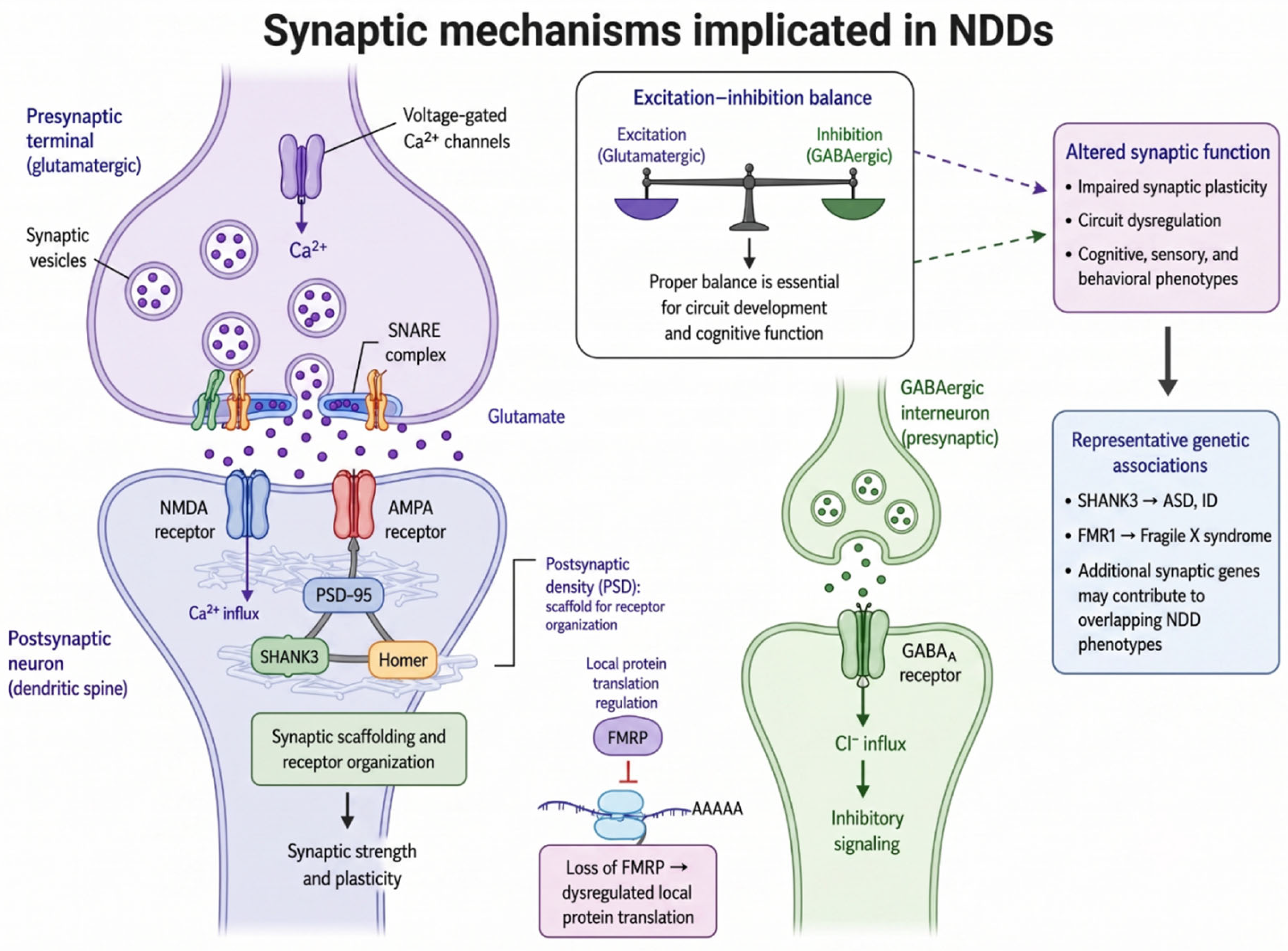
Figure 2.
Patient-derived iPSC and brain organoid models in neurodevelopmental disorders. Patient fibroblasts or peripheral blood mononuclear cells are reprogrammed into iPSCs and differentiated into two-dimensional neural cultures or three-dimensional brain organoids. CRISPR/Cas9 editing enables correction of patient variants or introduction of variants into control lines to generate isogenic pairs. Molecular, cellular and electrophysiological characterization can define disease-associated phenotypes and support therapeutic screening, gene- or RNA-based intervention, and assessment of functional rescue. Abbreviations: CRISPR/Cas9, clustered regularly interspaced short palindromic repeats/CRISPR-associated protein 9; iPSC, induced pluripotent stem cell; NDD, neurodevelopmental disorder; PBMC, peripheral blood mononuclear cell.
Figure 2.
Patient-derived iPSC and brain organoid models in neurodevelopmental disorders. Patient fibroblasts or peripheral blood mononuclear cells are reprogrammed into iPSCs and differentiated into two-dimensional neural cultures or three-dimensional brain organoids. CRISPR/Cas9 editing enables correction of patient variants or introduction of variants into control lines to generate isogenic pairs. Molecular, cellular and electrophysiological characterization can define disease-associated phenotypes and support therapeutic screening, gene- or RNA-based intervention, and assessment of functional rescue. Abbreviations: CRISPR/Cas9, clustered regularly interspaced short palindromic repeats/CRISPR-associated protein 9; iPSC, induced pluripotent stem cell; NDD, neurodevelopmental disorder; PBMC, peripheral blood mononuclear cell.
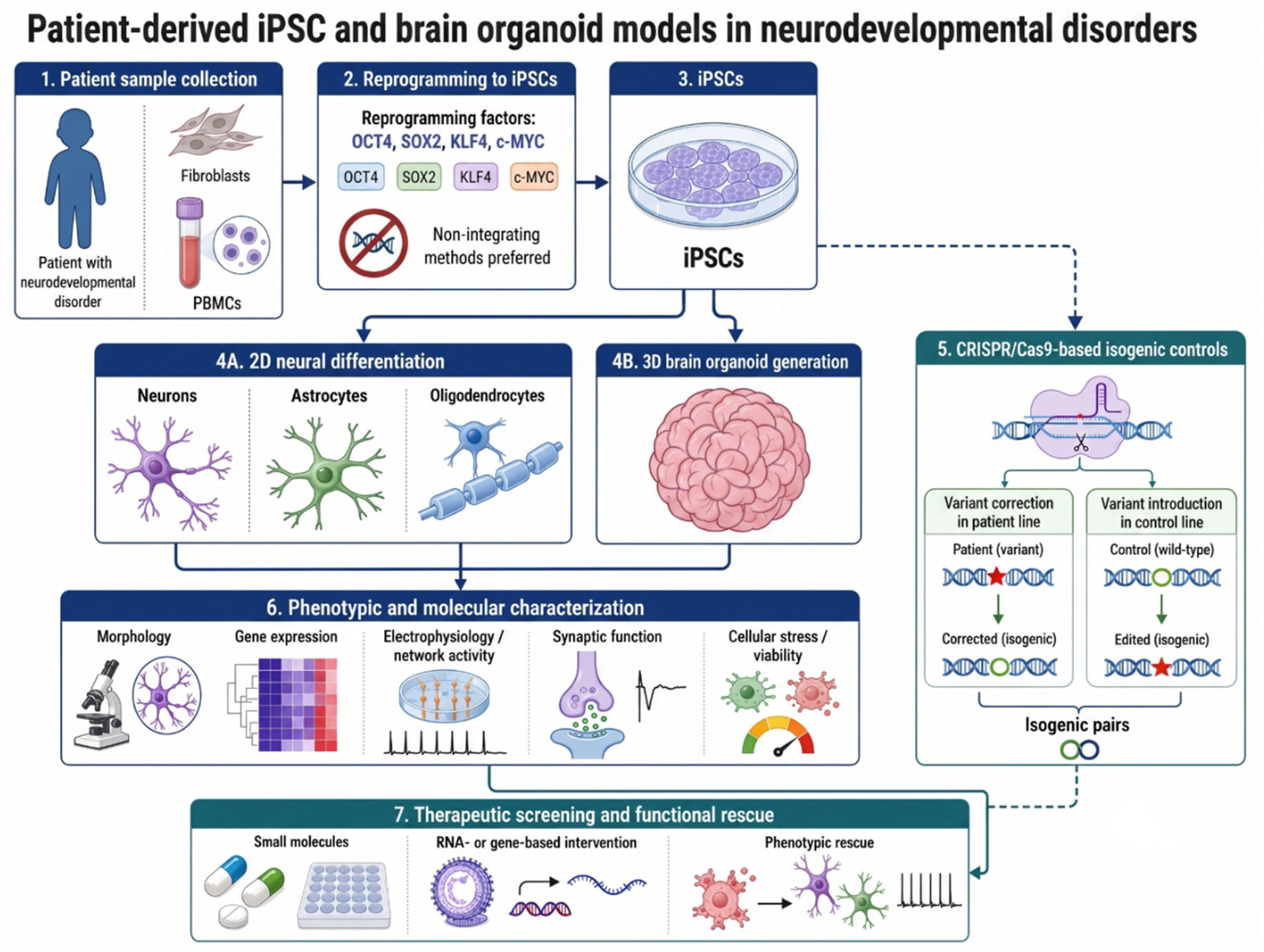
Figure 3.
Comparative overview of animal models used in neurodevelopmental disorder research. Model organisms differ in genetic tractability, throughput, anatomical complexity and relevance to human neurobiology. Caenorhabditis elegans, Drosophila melanogaster, Xenopus laevis and zebrafish support rapid investigation of conserved molecular, synaptic and developmental mechanisms, whereas rodents permit broader analysis of brain development, neural circuits, behavior, pharmacokinetics and environmental perturbations. Non-human primates provide closer similarity to human brain anatomy and selected cognitive and social functions but are constrained by cost, limited sample sizes and ethical considerations. Representative NDD-associated genes, principal applications and key advantages are shown for each model. The model organism selection should reflect the biological question rather than organismal complexity alone. Abbreviations: MIA, maternal immune activation; NDD, neurodevelopmental disorder; VPA, valproic acid.
Figure 3.
Comparative overview of animal models used in neurodevelopmental disorder research. Model organisms differ in genetic tractability, throughput, anatomical complexity and relevance to human neurobiology. Caenorhabditis elegans, Drosophila melanogaster, Xenopus laevis and zebrafish support rapid investigation of conserved molecular, synaptic and developmental mechanisms, whereas rodents permit broader analysis of brain development, neural circuits, behavior, pharmacokinetics and environmental perturbations. Non-human primates provide closer similarity to human brain anatomy and selected cognitive and social functions but are constrained by cost, limited sample sizes and ethical considerations. Representative NDD-associated genes, principal applications and key advantages are shown for each model. The model organism selection should reflect the biological question rather than organismal complexity alone. Abbreviations: MIA, maternal immune activation; NDD, neurodevelopmental disorder; VPA, valproic acid.
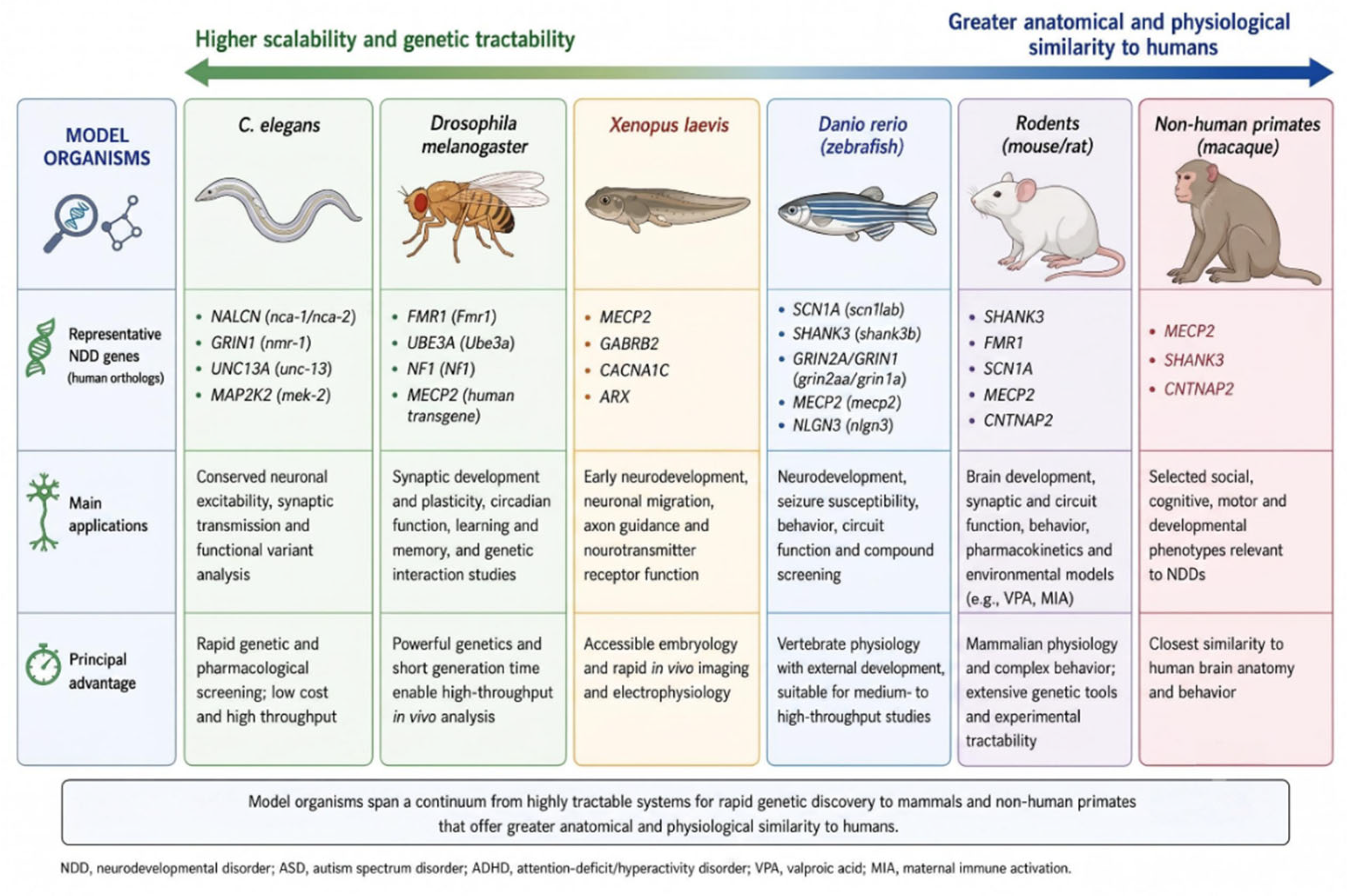
Figure 4.
Experimental and computational workflow for functional interpretation of NDD-associated variants. Candidate variants are prioritized using clinical phenotype, inheritance, population frequency and predicted molecular consequence. Structural mapping and confidence assessment define the residue context, while stability prediction, molecular dynamics and interaction modeling generate testable hypotheses regarding altered protein function. These predictions are evaluated through biochemical and cellular assays assessing expression, localization, activity, molecular interactions, electrophysiological function and disease-relevant phenotypes. Experimental findings can refine the computational model and contribute to variant interpretation under ACMG/AMP criteria. Interaction modeling may also support compound prioritization, followed by assessment of target engagement, functional rescue and toxicity. Computational predictions do not independently establish pathogenicity, molecular binding or therapeutic efficacy. Abbreviations: ACMG/AMP, American College of Medical Genetics and Genomics/Association for Molecular Pathology; MD, molecular dynamics; NDD, neurodevelopmental disorder; WT, wild type.
Figure 4.
Experimental and computational workflow for functional interpretation of NDD-associated variants. Candidate variants are prioritized using clinical phenotype, inheritance, population frequency and predicted molecular consequence. Structural mapping and confidence assessment define the residue context, while stability prediction, molecular dynamics and interaction modeling generate testable hypotheses regarding altered protein function. These predictions are evaluated through biochemical and cellular assays assessing expression, localization, activity, molecular interactions, electrophysiological function and disease-relevant phenotypes. Experimental findings can refine the computational model and contribute to variant interpretation under ACMG/AMP criteria. Interaction modeling may also support compound prioritization, followed by assessment of target engagement, functional rescue and toxicity. Computational predictions do not independently establish pathogenicity, molecular binding or therapeutic efficacy. Abbreviations: ACMG/AMP, American College of Medical Genetics and Genomics/Association for Molecular Pathology; MD, molecular dynamics; NDD, neurodevelopmental disorder; WT, wild type.
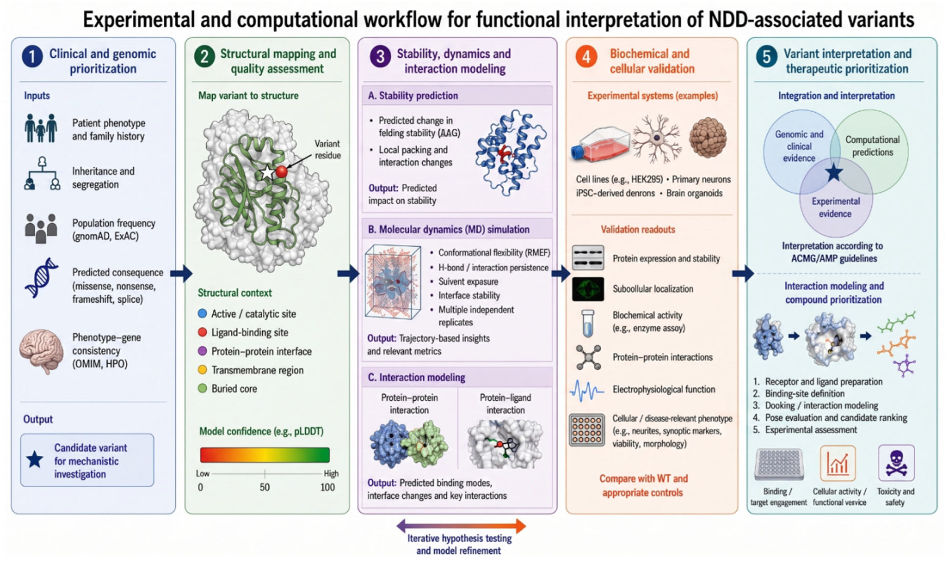
Figure 5.
Patient-centred translational framework for neurodevelopmental disorders. Clinical characterization, family history, longitudinal data, and genomic or selected multi-omic profiling support the identification and prioritization of candidate variants. Mechanistic hypotheses are evaluated through complementary structural, cellular, organoid, animal, and systems-level approaches. The resulting evidence can inform biomarker development, therapeutic development, drug screening and validation, and mechanistic interpretation. These outputs may support molecular diagnosis, prognostic assessment, mechanism-informed patient stratification, treatment prioritization, and longitudinal outcome monitoring. Clinical follow-up provides feedback for variant reassessment and refinement of disease models. Abbreviations: HPO, Human Phenotype Ontology; NDD, neurodevelopmental disorder; RNA, ribonucleic acid; WES, whole-exome sequencing; WGS, whole-genome sequencing.
Figure 5.
Patient-centred translational framework for neurodevelopmental disorders. Clinical characterization, family history, longitudinal data, and genomic or selected multi-omic profiling support the identification and prioritization of candidate variants. Mechanistic hypotheses are evaluated through complementary structural, cellular, organoid, animal, and systems-level approaches. The resulting evidence can inform biomarker development, therapeutic development, drug screening and validation, and mechanistic interpretation. These outputs may support molecular diagnosis, prognostic assessment, mechanism-informed patient stratification, treatment prioritization, and longitudinal outcome monitoring. Clinical follow-up provides feedback for variant reassessment and refinement of disease models. Abbreviations: HPO, Human Phenotype Ontology; NDD, neurodevelopmental disorder; RNA, ribonucleic acid; WES, whole-exome sequencing; WGS, whole-genome sequencing.
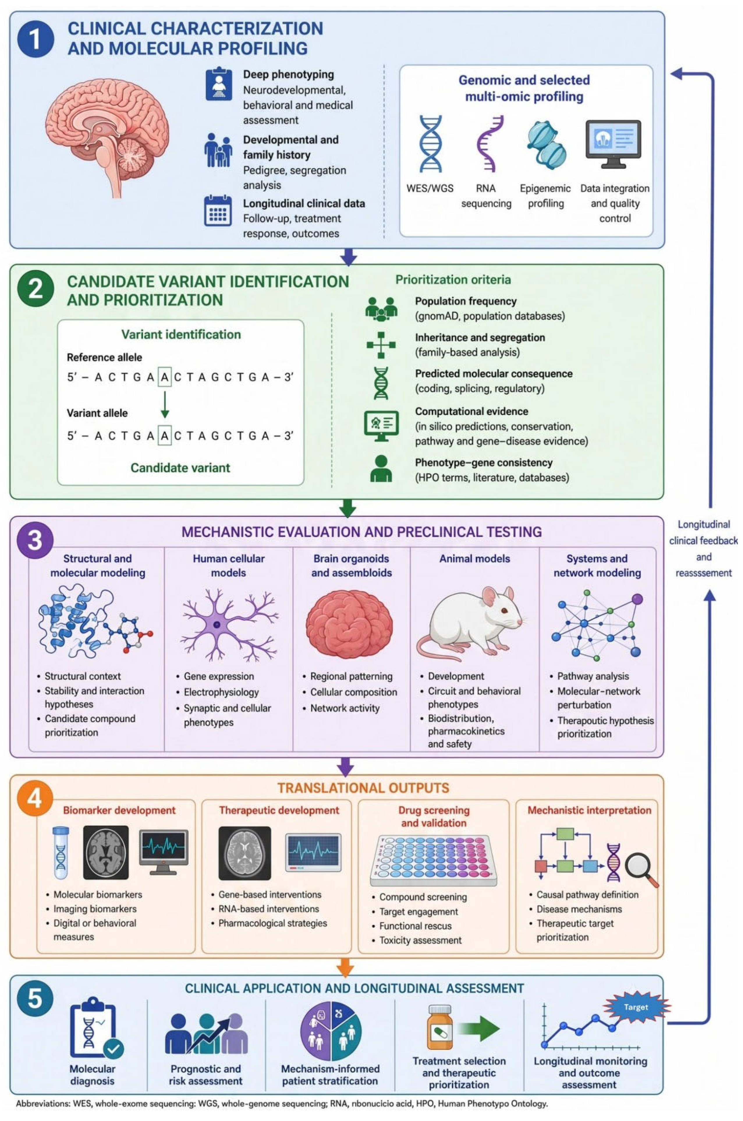

Table 1.
Comparative strengths, limitations, and methodological pitfalls of major platforms used in neurodevelopmental disorder modeling.
Table 1.
Comparative strengths, limitations, and methodological pitfalls of major platforms used in neurodevelopmental disorder modeling.
| Modeling platform | Key strengths | Main limitations and methodological pitfalls | Representative references |
| Heterologous systems (HEK293 and CHO cells) | High experimental control, rapid manipulation, and reproducible biochemical and electrophysiological assessment | Non-neuronal identity, artificial expression levels, and absence of developmental, cellular, and synaptic context | Abaandou et al., 2021; Li et al., 2022; Damianidou et al., 2022 |
| Two-dimensional iPSC-derived neural models | Preserve patient-specific genetic backgrounds; enable neural differentiation, functional analysis, and drug screening | Variable reprogramming and differentiation, limited maturation, and incomplete spatial and multicellular organization | Chailangkarn et al., 2012; Telias & Ben-Yosef, 2014; Lee et al., 2019 |
| Three-dimensional brain organoids | Reproduce selected features of human neurodevelopment, tissue organization, cellular diversity, and network formation | Batch variability, incomplete vascularization and cellular composition, cellular stress, and restricted maturation | Cheffer et al., 2020; Trujillo et al., 2019; Urrestizala-Arenaza et al., 2024 |
| Lower organisms (Drosophila and C. elegans) | Short generation times, low costs, strong genetic tractability, and suitability for large-scale functional and pharmacological screening | Limited conservation of brain architecture, gene regulation, and higher-order cognitive and social phenotypes | Gatto & Broadie, 2011; Bessa et al., 2013; Schmeisser & Parker, 2018; Mariano et al., 2020 |
| Rodent models | Mammalian physiology, extensive genetic manipulability, and robust developmental, behavioral, and neural-circuit analyses | Species-specific differences in cortical development, behavioral interpretation, genetic compensation, and drug metabolism | Gonzalez-Sulser, 2020; Till et al., 2022 |
| Non-human primates | Close similarity to humans in cortical organization, developmental timing, cognition, and social behavior | High financial and ethical burden, small sample sizes, long reproductive cycles, genetic heterogeneity, and limited replication | Izpisua Belmonte et al., 2015; Dettmer & Suomi, 2014; Sliwa & Freiwald, 2017; Prescott, 2020 |
| Computational modeling | Rapid and scalable variant prioritization, structural analysis, molecular simulation, and virtual screening | Dependence on input quality and model assumptions, limited reliability for unresolved or dynamic structures, algorithm-specific bias, and requirement for experimental validation | Abraham et al., 2015; Jumper et al., 2021; Marabotti et al., 2021; Zheng et al., 2024; Morris et al., 2009 |
Abbreviations: CHO, Chinese hamster ovary; HEK293, human embryonic kidney 293; iPSC, induced pluripotent stem cell.
Disclaimer/Publisher’s Note: The statements, opinions and data contained in all publications are solely those of the individual author(s) and contributor(s) and not of MDPI and/or the editor(s). MDPI and/or the editor(s) disclaim responsibility for any injury to people or property resulting from any ideas, methods, instructions or products referred to in the content. |
© 2026 by the authors. Licensee MDPI, Basel, Switzerland. This article is an open access article distributed under the terms and conditions of the Creative Commons Attribution (CC BY) license (http://creativecommons.org/licenses/by/4.0/).
Copyright: This open access article is published under a Creative Commons CC BY 4.0 license, which permit the free download, distribution, and reuse, provided that the author and preprint are cited in any reuse.