

Submitted:
11 August 2026
Posted:
13 August 2026
You are already at the latest version
Abstract
Background: The zoonotic epidemic of sporotrichosis caused by Sporothrix brasiliensis has expanded throughout Brazil and other countries, with a rise in deep forms of disease. Detailed comparative data on presentation, treatment and outcome of cutaneous and deep disease are needed to follow how Brazilian cohorts are evolving. Methods: Retrospective observational cohort of patients with sporotrichosis treated at a quaternary university hospital in Belo Horizonte between 2011 and 2026, restricted to culture-positive cases. Localized cutaneous disease was compared with deep infection using logistic regression adjusted for age and sex. Results: Of 313 patients, 213 had a positive culture. Median age was 41 years and 54.0% were women. Lymphocutaneous disease predominated (54.0%) and contact with cats was reported by 53.1%. Median treatment duration was 150 days. Among the 101 patients with a defined outcome, 97 were cured (96.0%), with no documented treatment failure. All four sporotrichosis-related deaths occurred in the deep infection group, which was independently associated with comorbidity (OR 9.69; 95% CI 2.52 to 37.23) and with an immunosuppressive condition (OR 6.61; 95% CI 1.89 to 23.09). Conclusions: Localized cutaneous sporotrichosis was cured in nearly all patients with documented follow-up, whereas mortality was confined to deep infection associated with comorbidity and immunosuppression.
Keywords:
sporotrichosis
; Sporothrix brasiliensis
; itraconazole
; implantation mycosis
; cohort
; Brazil
1. Introduction
Sporotrichosis is one of the most frequent implantation mycoses worldwide, caused by thermodimorphic fungi of the genus Sporothrix, and is most prevalent in tropical and subtropical areas [1]. Infection results from traumatic inoculation of the fungus into the skin, classically from soil, plants or decaying organic matter, which made the disease an occupational hazard for almost a century [2]. Outside South America this remains the predominant presentation, caused mainly by S. schenckii and S. globosa [3].
This pattern changed at the end of the 1990s. Zoonotic transmission from cats infected with Sporothrix brasiliensis, an emerging species first described in Brazil and later in other South American countries, became the main mechanism in the country, moved the disease into the urban environment and began to affect mainly adult women who care for the animals at home, with disease also occurring in children [4,5,6]. Brazil is currently the epicentre of the ongoing epidemic [7]. Cat-transmitted disease has also been described in Asian countries, caused by S. schenckii [8,9].
The usual presentation is cutaneous, in the fixed cutaneous and lymphocutaneous forms, with good prognosis and response to itraconazole [10]. The expansion of the epidemic caused by S. brasiliensis, a more virulent species, has been accompanied by an increase in deep forms, with osteoarticular, pulmonary and central nervous system involvement and disseminated disease, which require prolonged treatment and are associated with functional sequelae and mortality [11,12,13,14,15,16,17]. These forms occur mainly in patients with comorbidities, alcohol use or immunosuppression, and are often recognized late [18,19,20].
Sporotrichosis is described by the Brazilian Ministry of Health as the most prevalent implantation mycosis in the country, occurring in all regions. The true magnitude of the problem, however, remains unknown: the disease was only added to the National List of Notifiable Diseases in March 2025, and surveillance of endemic mycoses is expected to be implemented in all federal units by 2027 [21]. State and municipal series show consistent growth: in the state of São Paulo, 7,834 cases were notified between 2011 and 2025, of which 6,784 in the last five years [22]; in Curitiba the incidence of S. brasiliensis infection increased steadily between 2011 and 2022 [23]; and in Pernambuco sporotrichosis became the second leading cause of hospital admission for fungal disease between 2016 and 2024 [24].
In Belo Horizonte, human sporotrichosis has been a notifiable disease at municipal level since 2018. Between 2016 and 2026, 1,262 cases were confirmed among residents of the municipality, with sustained growth over the period: 15 cases in 2016, 94 in 2020, 203 in 2023, 227 in 2024 and 380 in 2025. Women account for 64% of cases and 67% occur between 20 and 69 years of age. Among identified exposures, cats were involved in 74.8% and dogs in 15.4%, and the most frequent mechanism was scratching, followed by biting and contact with skin lesions of the animal [25]. The expansion of animal and human disease in the state since 2016 has been documented in molecular and epidemiological studies [26,27].
Treatment is prolonged, with a recommended duration of about four months for cutaneous forms and more than six months for deep forms [21], which raises issues of adherence, tolerability and laboratory monitoring. Clinical knowledge about the disease in Brazil derives largely from a single referral institution in Rio de Janeiro, the source of the largest series on severe forms, including disease in people living with HIV, bone sporotrichosis and pulmonary disease [12,13,18]. Descriptions of culture-confirmed cohorts from other regions, covering the full spectrum of disease and reporting actual treatment duration, tolerability of prolonged antifungal therapy and outcome by clinical form, are lacking. This study describes the clinical and epidemiological characteristics, treatment, tolerability and outcomes of a cohort of patients with culture-confirmed sporotrichosis treated at a quaternary university hospital in Belo Horizonte, and compares localized cutaneous disease with deep infection.
2. Materials and Methods
2.1. Study Design and Setting
This was a retrospective observational cohort of patients with sporotrichosis treated at the hospital complex of Hospital das Clínicas, Universidade Federal de Minas Gerais (HC-UFMG), in Belo Horizonte, Minas Gerais, Brazil.
HC-UFMG is a quaternary university hospital with more than 400 beds, comprising a complex of dedicated units and outpatient clinics that includes a hospital devoted to ophthalmology and otorhinolaryngology, with a 24-hour ophthalmology emergency service, as well as dermatology, oncology and haematology services and the Orestes Diniz Training and Referral Centre for Infectious and Parasitic Diseases. As a state referral centre for high-complexity care, it treats patients exclusively through the Brazilian Unified Health System, including patients immunosuppressed by onco-haematological diseases and those in solid organ and haematopoietic stem cell transplant programmes. The presence of a dedicated ophthalmology hospital explains the high proportion of ocular cases, and the presence of transplant and oncology services broadens the spectrum of hosts compared with services dedicated exclusively to infectious diseases.
Patients were treated predominantly on an outpatient basis, in the infectious diseases, dermatology and ophthalmology clinics of the complex, with hospital admission reserved for severe forms.
Patients were identified from the mycology laboratory database and from hospital diagnostic codes, and their clinical data were extracted from HC-UFMG medical records. A subgroup of patients followed at the infectious diseases outpatient clinic has been followed prospectively since 2016, with a structured protocol for clinical and laboratory data collection.
2.2. Study Population and Inclusion Criteria
All patients with a diagnosis of sporotrichosis recorded in the database were considered eligible. The analysis population was restricted to patients with a positive culture for Sporothrix spp. Patients with a clinical and epidemiological diagnosis without culture confirmation were not included in this analysis. This criterion corresponds to the definition of a laboratory-confirmed case adopted by the Brazilian Ministry of Health, which requires a positive culture or polymerase chain reaction for Sporothrix spp. [21]. Data were recorded in an electronic form in REDCap [28], hosted by the telehealth service of HC-UFMG. No patient without a positive culture had a positive polymerase chain reaction, so that the inclusion criterion adopted coincided, in this case series, with the definition of a laboratory-confirmed case. The study period extended from 2011 to 8 August 2026, when the database was closed.
2.3. Definitions
Clinical form was classified as fixed cutaneous, lymphocutaneous, disseminated cutaneous, ocular, osteoarticular, pulmonary or disseminated [29]. In the description of baseline characteristics the forms are presented in a non-exclusive manner, and patients with more than one recorded form appear in each corresponding category. For the comparison between groups, localized cutaneous disease comprised the fixed cutaneous and lymphocutaneous forms, and deep infection comprised the disseminated, osteoarticular, pulmonary and disseminated cutaneous forms. Patients with more than one recorded form were allocated according to the most severe form, following the hierarchy deep infection, localized cutaneous disease, ocular disease; accordingly, patients with ocular disease combined with a deep form were allocated to the deep infection group, and patients with ocular disease combined with a localized cutaneous form were allocated to the localized cutaneous group. Patients with isolated ocular disease were not included in either group, as they constitute a distinct clinical entity with its own diagnosis, management and outcome, and will be described in a separate study. Species identification was performed by polymerase chain reaction in a subgroup of isolates, according to a previously described protocol [30]. The primary outcome was clinical cure, defined as complete epithelialization of all lesions recorded by the attending physician or, in the absence of such a record, as completion of antifungal treatment with documented start and end dates. In the latter situation, patients who died, who had the antifungal discontinued for any reason, who remained on chronic suppressive therapy or on treatment at database closure, and those with recorded loss to follow-up were excluded. The service protocol includes follow-up for two months after treatment discontinuation to check for relapse, but this return visit was not documented for all patients. Outcomes were classified into mutually exclusive categories: cure, spontaneous cure without treatment, treatment failure, sporotrichosis-related death, unrelated death, chronic suppressive therapy, ongoing treatment at database closure, loss to follow-up and no recorded outcome. The relationship between death and sporotrichosis was defined by medical record review. Patients who never received antifungal treatment were not classified as treatment failures. The cure proportion was calculated only among patients with a defined outcome; patients without a defined outcome were excluded from the denominator. Laboratory tests requested during treatment were considered, including aminotransferases, alkaline phosphatase, gamma-glutamyl transferase, bilirubin, amylase, lipase, creatinine and complete blood count. The highest value of each analyte during treatment was recorded. Aminotransferase elevation was graded retrospectively according to the Common Terminology Criteria for Adverse Events, version 5.0 [31], using the upper limit of normal of the local laboratory (aspartate aminotransferase 40 U/L and alanine aminotransferase 41 U/L) and the higher of the two ratios for each patient. The database did not record laboratory tests performed before the start of treatment, so that grading assumes a normal baseline value and the abnormalities observed cannot be attributed with certainty to the antifungal agent.
2.4. Statistical Analysis
Categorical variables were described as absolute frequencies and proportions, and continuous variables as median and interquartile range. Proportions were calculated over the whole study population, with missing data presented as a separate category. Continuous variables were compared using the Mann-Whitney U test and categorical variables using the chi-square or Fisher’s exact test. Binary logistic regression models adjusted for age and sex were built for two outcomes: deep infection and laboratory abnormality during treatment. Comorbidity and immunosuppressive condition were analysed in separate models because of conceptual overlap. Results are presented as odds ratios with 95% confidence intervals. No model was built for cure or death, given the insufficient number of unfavourable outcomes. Analyses were performed in Python 3.12.3, using the pandas 3.0.2, numpy 2.4.4, scipy 1.17.1 and statsmodels 0.14.6 libraries.
2.5. Ethics
The study was approved by the Research Ethics Committee of Universidade Federal de Minas Gerais (CAAE 76717223.4.0000.5149). Informed consent was waived for the retrospective data collection. Patients followed prospectively at the infectious diseases outpatient clinic since 2016 signed an informed consent form.
3. Results
A total of 313 patients with sporotrichosis confirmed by clinical and epidemiological criteria were identified, of whom 213 also had laboratory confirmation by a positive culture for Sporothrix spp. and constitute the study population. Among the 100 patients without culture confirmation, 69 had no culture performed or no recorded result and 31 had a negative culture (Figure 1).
3.1. Patient Characteristics
Of the 213 patients, 115 were female (54.0%). Median age was 41 years (IQR 24 to 55), with 29 patients younger than 18 years (13.6%) and 42 aged 60 years or older (19.7%). Mixed race skin colour was reported by 133 patients (62.4%). One hundred and seven patients lived in Belo Horizonte (50.2%), 54 in the metropolitan region (25.4%) and 16 in the interior of Minas Gerais (7.5%) (Table 1). At least one comorbidity was recorded in 86 of the 213 patients (40.4%), with arterial hypertension in 42 (19.7%) and diabetes mellitus in 16 (7.5%). An immunosuppressive condition was identified in 15 patients (7.0%), comprising rheumatological disease in 9 (4.2%), HIV infection in 2 (0.9%), and inflammatory bowel disease, glomerulopathy, solid organ transplantation and intracranial tumour in 1 patient each (0.5%). The lymphocutaneous form was the most frequent, in 115 patients (54.0%), followed by ocular disease in 47 (22.1%) and fixed cutaneous disease in 44 (20.7%). Disseminated disease was recorded in 13 patients (6.1%), osteoarticular disease in 12 (5.6%), disseminated cutaneous disease in 3 (1.4%) and pulmonary disease in 2 (0.9%). More than one clinical form was recorded in 19 patients (8.9%). Hospital admission was recorded in 16 patients (7.5%). The most frequently affected site was the upper limbs, in 102 patients (47.9%). Contact with cats was reported by 113 patients (53.1%). Among those exposed to cats, the animal was ill in 84 cases (74.3%). Contact with soil, earth or gardening was reported by 21 patients (9.9%). Species identification was performed by polymerase chain reaction in 11 isolates (5.2%), all Sporothrix brasiliensis. In the remaining 202 patients (94.8%) there was no species identification.
3.2. Treatment, Tolerability and Outcome
Itraconazole 100 mg twice daily was the most frequently recorded regimen, in 53 patients (24.9%), followed by 200 mg daily in 44 (20.7%) and 400 mg daily in 21 (9.9%). Potassium iodide was used in 13 patients (6.1%) and amphotericin B, in any formulation, in 6 (2.8%). More than one regimen was recorded in 22 patients (10.3%), 3 patients received no antifungal agent (1.4%) and treatment was not recorded in 75 (35.2%). Median treatment duration was 150 days (IQR 93 to 217), calculable in 106 patients (49.8%) (Table 2). Laboratory abnormality during treatment was recorded in 52 patients (24.4%) and antifungal discontinuation for any reason in 11 (5.2%). Cure was documented in 95 patients (44.6%) and two patients were cured without having received antifungal treatment (0.9%). No case was classified as treatment failure. Four patients died from a sporotrichosis-related cause (1.9%) and one from an unrelated cause (0.5%). Three patients remained on chronic suppressive therapy (1.4%) and 19 were lost to follow-up (8.9%). Outcome was not recorded in 87 patients (40.8%). Among the 101 patients with a defined outcome, 97 were cured (96.0%). The four sporotrichosis-related deaths occurred in patients with deep infection. One patient died from complications of central nervous system involvement. Another had disseminated disease, never received treatment and was diagnosed after death, by blood culture and autopsy. A third, a 36-year-old man with alcohol use disorder, was admitted with disseminated skin lesions of six months’ duration and neurological manifestations, and received empirical amphotericin B deoxycholate for eight days, with a cumulative dose of 400 mg, before diagnostic confirmation; autopsy demonstrated meningeal, cerebral, pulmonary, renal, testicular, prostatic and adrenal involvement [32]. The fourth patient, a 52-year-old woman with lymphocutaneous disease that progressed to presumed disseminated infection, with fever, weight loss and concomitant Staphylococcus aureus infection, received amphotericin B lipid complex followed by itraconazole and died three weeks after the start of antifungal treatment.
3.3. Laboratory Monitoring and Toxicity
The presence or absence of laboratory abnormality during treatment was recorded in 92 patients (43.2%), of whom 52 (56.5%) had some abnormality. The most frequent abnormalities were in the complete blood count, in 27 patients, gamma-glutamyl transferase, in 25, and alkaline phosphatase, in 18. Alanine aminotransferase was altered in 11 patients and aspartate aminotransferase in 8 (Table S1). Among the 101 patients with an available peak aminotransferase value, a group that does not coincide with the 101 patients with a defined outcome, 83 had no elevation (82.2%), 13 had grade 1 elevation (12.9%), 4 grade 2 (4.0%) and 1 grade 3 (1.0%), according to the Common Terminology Criteria for Adverse Events, version 5.0 [31]. Total bilirubin above 1.5 mg/dL was observed in 2 of 76 patients (2.6%) and creatinine above 1.3 mg/dL in 9 of 83 (10.8%). Patients with laboratory abnormality were older, with a median age of 50 years (IQR 38 to 56) versus 36 years (IQR 20 to 47) in the remainder (p = 0.003), and had more comorbidity, 73.1% versus 50.0% (p = 0.023). Disseminated disease was recorded in 8 patients in the group with abnormality and in none of the group without abnormality (p = 0.009), and osteoarticular disease in 7 and none (p = 0.017). The median number of laboratory panels requested was 4 (IQR 2 to 6) in the group with abnormality and 2 (IQR 1 to 2) in the group without abnormality (p < 0.001). There was no difference in the proportion of patients with documented cure (71.2% versus 72.5%; p = 0.887) or in antifungal discontinuation (7.7% versus 5.0%; p = 0.694) (Table 3).
3.4. Localized Cutaneous Disease and Deep Infection
A total of 146 patients with localized cutaneous disease and 22 with deep infection were compared. The 45 patients with isolated ocular disease were not included in this comparison, as they constitute a distinct clinical entity, and will be described in a separate study. Patients with deep infection were older, with a median age of 50 years (IQR 43 to 62) versus 40 years (IQR 25 to 56) (p = 0.024), and had more comorbidity, 81.8% versus 40.4% (p < 0.001). An immunosuppressive condition was identified in 6 of the 22 patients with deep infection (27.3%) and in 8 of the 146 with localized cutaneous disease (5.5%) (p = 0.004). The proportion of women was 31.8% in the deep infection group versus 54.1% in the localized cutaneous disease group (p = 0.051). Alcohol use was reported by 6 of 13 patients in the deep infection group and by 13 of 66 in the localized cutaneous disease group (p = 0.071). Itraconazole 400 mg daily was used in 27.3% versus 8.2% (p = 0.016) and amphotericin B only in deep infection, in 6 patients (p < 0.001). Median treatment duration was 98 days in the deep infection group versus 150 days in the localized cutaneous disease group (p = 0.330). Hospital admission was recorded in 9 of the 22 patients with deep infection (40.9%) and in 6 of the 146 with localized cutaneous disease (4.1%; p < 0.001) (Table 4). Among monitored patients, laboratory abnormality occurred in 11 of 12 with deep infection (91.7%) and in 36 of 67 with localized cutaneous disease (53.7%) (p = 0.022). Cure was documented in 76 of 76 patients with localized cutaneous disease and in 5 of 9 with deep infection (p < 0.001). The four sporotrichosis-related deaths and the three patients on chronic suppressive therapy occurred in the deep infection group (p < 0.001 and p = 0.002). Sporotrichosis-related case fatality was 2 of the 15 patients with an immunosuppressive condition (13.3%) and 2 of the remaining 198 (1.0%; p = 0.026).
In the multivariable logistic regression analysis, adjusted for age and sex, both the presence of at least one comorbidity (OR 9.69; 95% CI 2.52 to 37.23; p < 0.001) and an immunosuppressive condition (OR 6.61; 95% CI 1.89 to 23.09; p = 0.003) were independently associated with deep infection, in separate models, since the two variables overlap conceptually. Female sex was associated with lower odds of deep infection (OR 0.28; 95% CI 0.10 to 0.78; p = 0.015 in the model with comorbidity), and age was not associated. For the outcome laboratory abnormality, after adjustment for age, sex, comorbidity and deep infection, only the number of laboratory panels requested remained associated (OR 1.43 per additional panel; 95% CI 1.09 to 1.88; p = 0.009) (Table 5).
4. Discussion
In this cohort of 213 patients with culture-confirmed sporotrichosis, outcome depended on clinical form: cure was documented in all 76 patients with localized cutaneous disease and in 5 of 9 with deep infection (p < 0.001), and the four sporotrichosis-related deaths occurred in the deep infection group. In models adjusted for age and sex, deep infection was independently associated with the presence of comorbidity (OR 9.69; 95% CI 2.52 to 37.23) and with an immunosuppressive condition (OR 6.61; 95% CI 1.89 to 23.09), whereas female sex was associated with lower odds of deep infection. Among patients with deep infection only 31.8% were women, versus 54.1% among those with localized cutaneous disease (p = 0.051). Prolonged antifungal treatment was well tolerated, with a single case of grade 3 aminotransferase elevation and discontinuation for any reason in 5.2%.
The pattern observed reproduces that described in Brazilian cohorts of the zoonotic epidemic. In Rio de Janeiro, the historical epicentre, cases in humans and animals have been reported since the end of the 1990s, with cats established as the main source and itraconazole as first-line treatment [29,33]. In Duque de Caxias, a geo-epidemiological cohort of 827 cases showed a predominance of women aged 25 to 59 years and an association with low income and poor sanitation [34]. In Curitiba, 216 cases followed a rising incidence between 2011 and 2022, with lymphocutaneous disease in 63%, fixed cutaneous disease (termed localized cutaneous in that series) in 24% and ocular disease in 10% [23]. In the state of Amazonas, 950 human cases were notified between 2020 and 2023, with animal contact in 77.7% and S. brasiliensis in 65.4% of positive cultures [35]. The historical systematic review of Brazilian cases between 1907 and 2020 gathered 10,400 patients, with 55.98% women, lymphocutaneous disease in 56.14%, systemic involvement in 14.34% and a cure rate of 85.83% [36].
Female predominance is a consistent finding in these series, and municipal surveillance in Belo Horizonte follows the same direction, with 64% of cases in women and 67% between 20 and 69 years of age [25]. In this cohort the proportion of women was 54.0%, at the lower limit of what is described in the literature. The difference follows from the way the case series was assembled: a predominantly outpatient cohort from a referral service, restricted to culture-confirmed cases, that receives referrals of atypical, ocular and deep forms from across the region. Typical cutaneous cases, in which female predominance is more marked, are largely diagnosed and treated outside tertiary services, often without culture, and are not part of this case series.
The median age of 41 years and the concentration between 18 and 59 years, which accounted for 65.7% of the cohort, are consistent with other Brazilian series, in which the disease affects predominantly working-age adults, with a median of 46 years in São Paulo [37] and concentration between 25 and 59 years in Duque de Caxias [34]. Twenty-nine patients (13.6%) were younger than 18 years, aged 8 to 17 years, and none had deep infection. Contact with cats was reported by 18 of these 29 patients (62.1%), a higher proportion than in the cohort as a whole, consistent with household exposure to infected animals and with the recognition of children as a risk group in the zoonotic epidemic [6].
Contact with cats was reported by 113 patients (53.1%), with an ill animal in 84 of the 113 exposures (74.3%). This proportion probably underestimates true exposure, since the information was not recorded in 39.0% of cases, and is lower than that described in Brazilian series: 87.6% in the classic Rio de Janeiro series [33], 96.5% in São Paulo [37] and 77.7% in the state of Amazonas [35]. In that Rio de Janeiro series, a scratch or a bite from a cat was reported by 97 of the 178 patients, so that direct traumatic inoculation accounted for only part of the reported exposures. In municipal surveillance in Belo Horizonte, cats account for 74.8% of identified exposures and scratching is the most frequent mechanism [25]. Transmission is not restricted to traumatic injury. Cats with sporotrichosis may shed S. brasiliensis from the front paws even without apparent lesions, from the nasal cavity and through respiratory droplets, which broadens the routes of exposure and includes contamination of the household environment [38]. A recent household outbreak documented clonal transmission from an index cat to three people and a dog, with distinct clinical presentations among contacts [39]. These findings support the interpretation that female predominance and the concentration in young adults reflect the role of caregivers of household cats, and reinforce that control of the feline source is the most effective preventive strategy [40].
Species identification was performed by polymerase chain reaction in 11 isolates, all Sporothrix brasiliensis. In the remaining 202 patients there was no species identification. This matches the national picture, in which S. brasiliensis accounts for about 85% of Brazilian isolates, originating in Rio de Janeiro and spreading to at least six states through independent emergence events followed by animal-to-animal and animal-to-human transmission [7], and with the expansion documented in Minas Gerais [20,27]. S. schenckii accounts for about 14% of isolates in the country, with a regional distribution pattern and greater genetic diversity, consistent with sporadic environmental transmission; in Espírito Santo, S. brasiliensis predominated in the metropolitan area whereas S. schenckii was more frequent in the mountainous region [5,41]. Since species is related to severity and to atypical presentations [11,16], the absence of routine molecular identification is a limitation of clinical care, and not only a methodological one. In this cohort it was not possible to assess whether clinical form or outcome varied according to species, and isolates were not submitted to antifungal susceptibility testing.
This analysis contrasts localized cutaneous disease with deep infection, rather than the usual division between cutaneous and extracutaneous disease. Ocular sporotrichosis comprises, in the great majority of cases, adnexal and surface involvement, with granulomatous conjunctivitis, Parinaud oculoglandular syndrome and dacryocystitis, an entity that the literature already explicitly distinguishes from true intraocular infection [17]. A recent series of 25 cases of ocular sporotrichosis in a hyperendemic region of southern Brazil recorded no case of intraocular involvement, with granulomatous conjunctivitis in 56%, Parinaud syndrome in 40% and dacryocystitis with eyelid lesion in 4% [42]. In this cohort there was no case of endophthalmitis with culture confirmation, so that the group with ocular disease comprised exclusively surface involvement. True intraocular involvement is rare and would constitute deep infection.
Resolution of ocular surface disease is assessed by specific parameters, such as resolution of conjunctivitis and patency of the lacrimal system, distinct from the cure criteria applicable to skin lesions, and is described in this way in dedicated ophthalmological series [43]. For these reasons ocular cases were analysed separately and will be described in a separate study. The proportion observed in this case series, 22.1%, is higher than that described in Curitiba, where the ocular form represented 10% of 216 cases [23], and reflects the referral profile of the service, which has a dedicated ophthalmology hospital with a 24-hour emergency service, as well as greater ophthalmological diagnostic capacity, a factor already identified as a determinant of how often this form is recognized [42].
At least one comorbidity was recorded in 81.8% of patients with deep infection, versus 40.4% of those with localized cutaneous disease, and an immunosuppressive condition in 27.3% versus 5.5%. Both remained independently associated with deep infection after adjustment for age and sex, with odds ratios of 9.69 (95% CI 2.52 to 37.23) and 6.61 (95% CI 1.89 to 23.09), respectively. Female sex was associated with lower odds of deep infection, a finding consistent with the pattern of the zoonotic epidemic, in which women predominate among cases but severe forms concentrate in men with comorbidities. Sporotrichosis-related case fatality was 13.3% among patients with an immunosuppressive condition and 1.0% among the remainder (p = 0.026). Unlike what is described in series from infectious diseases services, in which immunosuppression results mainly from HIV infection, in this cohort it was predominantly of rheumatological cause, present in 9 of the 15 immunosuppressed patients, which reflects the referral profile of a university hospital with all specialties.
This agrees with the Brazilian literature, in which immunosuppression, alcohol use and diabetes mellitus are the main determinants of dissemination. In a cohort from the same state, all patients living with HIV developed the systemic form, with increased mortality, and alcohol use was specifically associated with pulmonary involvement [19]; in the individual participant data review, alcohol use disorder remained independently associated with systemic disease, with an odds ratio of 4.76 (95% CI 1.45 to 18.05) [44]; and in an osteoarticular series from Belo Horizonte diabetes was an independent factor, with an odds ratio of 4.11 [20]. In this cohort none of these isolated factors reached significance: diabetes was not independently associated with deep infection and alcohol use was borderline (p = 0.071), with information missing for more than half of the patients. HIV infection was recorded in only 2 patients (0.9%), a proportion far lower than in series of systemic disease, which reflects both underreporting and the referral profile of the service. What remained associated was the broader category, comorbidity or immunosuppression, which suggests that, in services with a wide spectrum of hosts, the determinant of severity is the presence of immunocompromise itself rather than a specific condition.
From a clinical care perspective, these findings support that the initial assessment of every patient with sporotrichosis should include active search for predisposing conditions, with HIV testing, assessment of blood glucose and history of alcohol use, regardless of clinical presentation. Immunosuppression of non-infectious cause, predominant in this case series, tends to be less often considered a risk factor for implantation mycosis than HIV infection. The risk of iatrogenic immunosuppression deserves particular attention: cutaneous sporotrichosis may be mistaken for inflammatory dermatoses, and disseminated disease has been described after corticosteroid therapy started for suspected pyoderma gangrenosum [45]. In this cohort, a patient on methotrexate and prednisone for rheumatoid arthritis had a biopsy initially interpreted as pyoderma gangrenosum, with culture revealing Sporothrix spp., which reinforces the need for mycological investigation before intensifying immunosuppression in chronic ulcerated skin lesions.
Cure was documented in all 76 patients with localized cutaneous disease and in 5 of 9 with deep infection, and the four sporotrichosis-related deaths and the three patients maintained on chronic suppressive therapy belong to the deep infection group. The difference in cure according to clinical form is similar to that described in the individual participant data review [44] and to that observed in a global cohort, in which disseminated disease, malignancy and older age were associated with higher one-year mortality [46]. Treatment also reproduced the described pattern: localized cutaneous disease responds to itraconazole at conventional doses for three to six months, whereas deep forms require higher doses or amphotericin B, reserved for systemic cases [17,47]. The number of patients with deep infection and a defined outcome is small, which precludes risk estimation, but the direction of the finding is consistent with series of systemic, osteoarticular and pulmonary disease from different Brazilian states [12,13,16,18,20].
All four sporotrichosis-related deaths occurred in patients with disseminated disease, although dissemination was presumed rather than microbiologically confirmed in one patient, and in two the diagnosis was established only at the time of death or afterwards. One patient never received antifungal treatment, and the diagnosis was made after death, by blood culture and autopsy. Another, a 36-year-old man with six months of disseminated skin lesions, received empirical amphotericin B deoxycholate and had the biopsy result released one day before death, with the definitive culture report available only after death [32]. A third died from complications of central nervous system involvement. Two of the four patients who died had central nervous system involvement, an uncommon complication with high lethality, described predominantly in immunocompromised patients and associated with unfavourable outcome even under prolonged treatment [15].
These patients reached the service already with disseminated disease and in severe condition, after a prolonged course. This database contains no information on symptom onset or on the previous care pathway, but the presentation suggests that dissemination resulted from the chronic evolution of untreated disease, and not from the interval between admission and the start of treatment. The increase in hospital admissions for sporotrichosis, a disease typically managed on an outpatient basis, has been described in other regions of the country: in Pernambuco the mycosis became the second leading cause of hospital admission for fungal disease between 2016 and 2024, with an increase from 5 to 28 annual admissions and disseminated disease in 17.3% of specified cases [24]. The critical point lies before access to the referral service, in the recognition of the disease in its early forms.
With a median treatment duration of 150 days, aminotransferase elevation was restricted to grades 1 and 2 in almost all patients assessed, with a single case of grade 3, and antifungal discontinuation for any reason occurred in 5.2%. The most frequent abnormalities were in the complete blood count and gamma-glutamyl transferase, non-specific findings, and were more frequent than aminotransferase elevation. The profile is similar to that described in the largest series of treatment of cutaneous sporotrichosis with itraconazole, with 645 patients, in which clinical adverse events occurred in 18.1% of those treated with 100 mg daily and in 21.9% of those receiving 200 to 400 mg daily, predominantly nausea and epigastric pain [48]. Hepatotoxicity is the most relevant adverse effect of prolonged azole use, although rare at conventional doses [49].
These data support outpatient management of itraconazole with targeted rather than systematic laboratory monitoring, in patients without previous liver or kidney disease and on conventional doses. Regimens recommended for deep infection may involve higher doses and courses exceeding twelve months, which increase cumulative exposure and justify periodic monitoring of liver function tests, particularly in patients with liver comorbidity, concomitant use of other hepatotoxic agents or alcohol consumption [50].
The comparison between patients with and without laboratory abnormality should be interpreted with caution. Patients with abnormality were older, had more comorbidity and more deep infection, but also had twice as many laboratory panels requested, and in the multivariable analysis only the number of panels remained associated with the outcome. Part of the difference reflects intensity of investigation rather than toxicity. The absence of a difference in cure and in antifungal discontinuation between the two groups suggests that the abnormalities found had little clinical consequence.
Limitations
This is a retrospective cohort, and data were extracted from records that were not generated for research purposes, so that completeness varies across variables. Outcome was not recorded in 40.8% of patients, which restricts the cure proportion to a subgroup with documented follow-up and may overestimate sustained cure.
The database did not record laboratory tests performed before the start of treatment, so that the abnormalities observed cannot be attributed with certainty to the antifungal agent and grading assumes a normal baseline value. Laboratory monitoring was not protocol-driven and its intensity varied according to case severity, which introduces detection bias.
The proportion of patients with HIV infection was 0.9%, far lower than the 15% described among osteoarticular cases from an infectious diseases referral service in the same city [20]. Part of this reflects the referral profile: HC-UFMG receives patients from all specialties and immunosuppression in the cohort is more heterogeneous, including solid organ transplant recipients and patients on immunosuppressive drugs for autoimmune disease, whereas series from hospitals dedicated to infectious diseases concentrate patients living with HIV.
Species identification was performed in only 11 isolates, which precludes any inference about species distribution; Brazilian cohort studies, however, confirm that the great majority of cases in the south and southeast regions are caused by Sporothrix brasiliensis. The temporal distribution of cases reflects the case-finding process and should not be read as an epidemic curve. Finally, with 21 events and three variables, the models for deep infection had seven events per variable, below the usual rule of ten, and the confidence intervals are wide.
5. Conclusions
In a cohort of 213 patients with culture-confirmed sporotrichosis at a referral centre in Belo Horizonte, the disease affected mainly adult women, with lymphocutaneous disease and exposure to ill cats, and cure was documented in 96.0% of patients with a defined outcome. Prolonged antifungal treatment was well tolerated, with grade 3 aminotransferase elevation in a single patient and antifungal discontinuation in 5.2%. Deep infection concentrated older patients with comorbidity, required more intensive regimens and accounted for all sporotrichosis-related mortality, including two cases diagnosed only at the time of death or afterwards. Early recognition of deep presentations and routine species identification are the two needs identified by this study.
Use of Artificial Intelligence: During the preparation of this manuscript, the authors used Claude (Anthropic, model Opus 5) for the following purposes: checking numerical consistency within the text, verifying bibliographic references, translation and English-language editing, and improving the coherence and fluency of the English text. The tool was not used to generate original scientific content, to design the study, or to analyze or interpret the data. The authors have reviewed and edited all output and take full responsibility for the content of this publication.
Supplementary Materials
The following supporting information can be downloaded at the website of this paper posted on Preprints.org, [URL to be inserted by the Editorial Office]. Table S1: Laboratory monitoring, toxicity and antifungal discontinuation (n = 213).
Author Contributions
Conceptualization, H.D., F.F.A. and A.C.C.R.; methodology, H.D., F.F.A., A.C.C.R. and M.O.F.; software, H.D. and A.S.B.; validation, H.D., F.F.A., A.S.B. and M.O.F.; formal analysis, H.D., F.F.A. and M.O.F.; investigation, H.D., F.F.A., A.S.B., V.B.M., K.V.S., B.V.M., H.F.P., R.L.S., M.V.C.A., F.T.C.T., G.B.G., A.C.C.R., L.C.S., S.M.L. and M.O.F.; resources, H.D. and S.M.L.; data curation, H.D., F.F.A., A.S.B., V.B.M., K.V.S., B.V.M., H.F.P., R.L.S., M.V.C.A., F.T.C.T., G.B.G. and L.C.S.; writing—original draft preparation, H.D.; writing—review and editing, H.D., F.F.A., A.S.B., V.B.M., K.V.S., B.V.M., H.F.P., R.L.S., M.V.C.A., F.T.C.T., G.B.G., A.C.C.R., L.C.S., S.M.L. and M.O.F.; visualization, H.D.; supervision, H.D. and F.F.A.; project administration, H.D.; funding acquisition, H.D. All authors have read and agreed to the published version of the manuscript.
Funding
This research was supported by undergraduate research scholarships from the Brazilian National Council for Scientific and Technological Development (CNPq) and the Minas Gerais State Research Foundation (FAPEMIG).
Institutional Review Board Statement
The study was conducted in accordance with the Declaration of Helsinki and approved by the Research Ethics Committee of Universidade Federal de Minas Gerais (CAAE 76717223.4.0000.5149).
Informed Consent Statement
Informed consent was obtained from all patients followed prospectively at the infectious diseases outpatient clinic since 2016. The requirement for informed consent was waived for the retrospective data collection by the Research Ethics Committee.
Data Availability Statement
The data presented in this study are available on request from the corresponding author. The data are not publicly available because they contain information that could compromise the privacy of research participants.
Conflicts of Interest
The authors declare no conflicts of interest. The funders had no role in the design of the study; in the collection, analyses, or interpretation of data; in the writing of the manuscript; or in the decision to publish the results.
References
- Chakrabarti, A.; Bonifaz, A.; Gutierrez-Galhardo, M.C.; Mochizuki, T.; Li, S. Global epidemiology of sporotrichosis. Med. Mycol. 2015, 53, 3–14. [Google Scholar] [CrossRef] [PubMed]
- Rodrigues, A.M.; de Hoog, G.S.; de Camargo, Z.P. Sporothrix species causing outbreaks in animals and humans driven by animal–animal transmission. PLoS Pathog. 2016, 12, e1005638. [Google Scholar] [CrossRef] [PubMed]
- Zhang, Y.; Hagen, F.; Stielow, B.; Rodrigues, A.M.; Samerpitak, K.; Zhou, X.; Feng, P.; Yang, L.; Chen, M.; Deng, S.; et al. Phylogeography and evolutionary patterns in Sporothrix spanning more than 14,000 human and animal case reports. Persoonia 2015, 35, 1–20. [Google Scholar] [CrossRef] [PubMed]
- Gremião, I.D.F.; Miranda, L.H.M.; Reis, E.G.; Rodrigues, A.M.; Pereira, S.A. Zoonotic epidemic of sporotrichosis: cat to human transmission. PLoS Pathog. 2017, 13, e1006077. [Google Scholar] [CrossRef] [PubMed]
- de Carvalho, J.A.; Beale, M.A.; Hagen, F.; Fisher, M.C.; Kano, R.; Bonifaz, A.; Toriello, C.; Negroni, R.; Rego, R.S.M.; Gremião, I.D.F.; et al. Trends in the molecular epidemiology and population genetics of emerging Sporothrix species. Stud. Mycol. 2021, 100, 100129. [Google Scholar] [CrossRef] [PubMed]
- Queiroz-Telles, F.; Bonifaz, A.; Cognialli, R.; Lustosa, B.P.R.; Vicente, V.A.; Ramírez-Marín, H.A. Sporotrichosis in children: case series and narrative review. Curr. Fungal Infect. Rep. 2022, 16, 33–46. [Google Scholar] [CrossRef] [PubMed]
- Ribeiro dos Santos, A.; Misas, E.; Min, B.; Le, N.; Bagal, U.R.; Parnell, L.A.; Sexton, D.J.; Lockhart, S.R.; de Souza Carvalho Melhem, M.; Takahashi, J.P.F.; et al. Emergence of zoonotic sporotrichosis in Brazil: a genomic epidemiology study. Lancet Microbe 2024, 5, e282–e290. [Google Scholar] [CrossRef] [PubMed]
- Han, H.S.; Kano, R. Feline sporotrichosis in Asia. Braz. J. Microbiol. 2021, 52, 125–134. [Google Scholar] [CrossRef] [PubMed]
- Jirawattanadon, P.; Bunyaratavej, S.; Leeyaphan, C.; Chongtrakool, P.; Sitthinamsuwan, P.; Panjapakkul, W.; Prasertsook, S.; Saengthong-Aram, P.; Wareesawetsuwan, N.; Posri, J.; et al. Clinical manifestations, antifungal drug susceptibility, and treatment outcomes for emerging zoonotic cutaneous sporotrichosis, Thailand. Emerg. Infect. Dis. 2024, 30, 2583–2592. [Google Scholar] [CrossRef] [PubMed]
- Orofino-Costa, R.; de Macedo, P.M.; Rodrigues, A.M.; Bernardes-Engemann, A.R. Sporotrichosis: an update on epidemiology, etiopathogenesis, laboratory and clinical therapeutics. An. Bras. Dermatol. 2017, 92, 606–620. [Google Scholar] [CrossRef] [PubMed]
- Almeida-Paes, R.; de Oliveira, M.M.E.; Freitas, D.F.S.; do Valle, A.C.F.; Zancopé-Oliveira, R.M.; Gutierrez-Galhardo, M.C. Sporotrichosis in Rio de Janeiro, Brazil: Sporothrix brasiliensis is associated with atypical clinical presentations. PLoS Negl. Trop. Dis. 2014, 8, e3094. [Google Scholar] [CrossRef] [PubMed]
- Ramos, V.; Astacio, G.S.; do Valle, A.C.F.; de Macedo, P.M.; Lyra, M.R.; Almeida-Paes, R.; Zancopé-Oliveira, R.M.; Gutierrez-Galhardo, M.C.; Freitas, D.F.S. Bone sporotrichosis: 41 cases from a reference hospital in Rio de Janeiro, Brazil. PLoS Negl. Trop. Dis. 2021, 15, e0009250. [Google Scholar] [CrossRef] [PubMed]
- Fichman, V.; Mota-Damasceno, C.G.; Procópio-Azevedo, A.C.; Almeida-Paes, R.; de Macedo, P.M.; Medeiros, D.M.; Zancopé-Oliveira, R.M.; Gutierrez-Galhardo, M.C.; Freitas, D.F.S. Pulmonary sporotrichosis caused by Sporothrix brasiliensis: a 22-year, single-center, retrospective cohort study. J. Fungi 2022, 8, 536. [Google Scholar] [CrossRef] [PubMed]
- Xavier, M.O.; Poester, V.R.; Trápaga, M.R.; Stevens, D.A. Sporothrix brasiliensis: epidemiology, therapy, and recent developments. J. Fungi 2023, 9, 921. [Google Scholar] [CrossRef] [PubMed]
- de Oliveira, V.F.; Petrucci, J.F.; Taborda, M.; Brener, P.Z.; Kremer, P.G.D.B.B.; Randi, B.A.; Magri, A.S.G.K.; Magri, M.M.C.; Levin, A.S.; Silva, G.D. Clinical characteristics, diagnosis, and treatment of central nervous system sporotrichosis: systematic review and meta-analysis. Mycoses 2024, 67, e13697. [Google Scholar] [CrossRef] [PubMed]
- Poester, V.R.; Xavier, M.O.; Munhoz, L.S.; Basso, R.P.; Zancopé-Oliveira, R.M.; Almeida-Paes, R.; Stevens, D.A. Sporothrix brasiliensis causing atypical sporotrichosis in Brazil: a systematic review. J. Fungi 2024, 10, 287. [Google Scholar] [CrossRef] [PubMed]
- Ramírez-Soto, M.C. Extracutaneous sporotrichosis. Clin. Microbiol. Rev. 2025, 38, e0014024. [Google Scholar] [CrossRef] [PubMed]
- Freitas, D.F.S.; do Valle, A.C.F.; da Silva, M.B.T.; Campos, D.P.; Lyra, M.R.; de Souza, R.V.; Veloso, V.G.; Zancopé-Oliveira, R.M.; Bastos, F.I.; Galhardo, M.C.G. Sporotrichosis: an emerging neglected opportunistic infection in HIV-infected patients in Rio de Janeiro, Brazil. PLoS Negl. Trop. Dis. 2014, 8, e3110. [Google Scholar] [CrossRef] [PubMed]
- Magalhães, V.C.R.; Colombo, S.A.; Peres, N.T.A.; Moura, A.S.; de Ávila, R.E.; Azevedo, M.I.; Santos, D.A. Clinical factors associated with systemic sporotrichosis in Brazil. Mycoses 2024, 67, e13656. [Google Scholar] [CrossRef] [PubMed]
- Magalhães, V.C.R.; Moura, A.S.; do Nascimento Sales, F.; Colombo, S.A.; de Ávila, R.E.; César, J.M.S.; Viegas, D.M.; Pizarro, A.C.S.T.; Lyon, A.C.; Carvalho, S.H.; et al. Osteoarticular sporotrichosis in a Brazilian endemic setting: associated factors and outcomes. Mycoses 2026, 69, e70211. [Google Scholar] [CrossRef] [PubMed]
- Brazil; Ministry of Health. Technical Note No. 20/2025-CGTM/DATHI/SVSA/MS: Updates for the Surveillance and Notification of Human Sporotrichosis at National Level; (In Portuguese). Ministério da Saúde: Brasília, Brazil, 2025. [Google Scholar]
- São Paulo State Health Department; Alexandre Vranjac Epidemiological Surveillance Centre. Epidemiological Bulletin on Sporotrichosis; (In Portuguese). CVE/SES: São Paulo, Brazil, 2026. [Google Scholar]
- Cognialli, R.C.R.; Cáceres, D.H.; Bastos, F.A.G.D.; Cavassin, F.B.; Lustosa, B.P.R.; Vicente, V.A.; Breda, G.L.; Santos-Weiss, I.; Queiroz-Telles, F. Rising incidence of Sporothrix brasiliensis infections, Curitiba, Brazil, 2011–2022. Emerg. Infect. Dis. 2023, 29, 1330–1339. [Google Scholar] [CrossRef] [PubMed]
- Prohaska-Batista, F.; Bridi Cavassin, F.; de Queiroz-Telles, F. From neglected to notable: a growing public health challenge driven by hospitalization for sporotrichosis in Pernambuco, Northeast Brazil (2016–2024). PLoS Negl. Trop. Dis. 2025, 19, e0013795. [Google Scholar] [CrossRef] [PubMed]
- Belo Horizonte Municipal Health Department. Epidemiological Report: Human Sporotrichosis; (In Portuguese). SMSA: Belo Horizonte, Brazil, 2026. [Google Scholar]
- Lecca, L.O.; Paiva, M.T.; de Oliveira, C.S.F.; Morais, M.H.F.; de Azevedo, M.I.; Bastos, C.V.; Keller, K.M.; Ecco, R.; Santos, D.A.; Silva, T.M.; et al. Associated factors and spatial patterns of the epidemic sporotrichosis in a high density human populated area: a cross-sectional study from 2016 to 2018. Prev. Vet. Med. 2020, 176, 104939. [Google Scholar] [CrossRef] [PubMed]
- Colombo, S.A.; Bicalho, G.C.; de Oliveira, C.S.F.; Keller, K.M.; Silva, M.X.; Santos, D.A.; Azevedo, M.I. Emergence of zoonotic sporotrichosis due to Sporothrix brasiliensis in Minas Gerais, Brazil: a molecular approach to the current animal disease. Mycoses 2023, 66, 911–922. [Google Scholar] [CrossRef] [PubMed]
- Harris, P.A.; Taylor, R.; Thielke, R.; Payne, J.; Gonzalez, N.; Conde, J.G. Research electronic data capture (REDCap): a metadata-driven methodology and workflow process for providing translational research informatics support. J. Biomed. Inform. 2009, 42, 377–381. [Google Scholar] [CrossRef] [PubMed]
- Barros, M.B.L.; de Almeida Paes, R.; Schubach, A.O. Sporothrix schenckii and sporotrichosis. Clin. Microbiol. Rev. 2011, 24, 633–654. [Google Scholar] [CrossRef] [PubMed]
- Spruijtenburg, B.; Bombassaro, A.; Meijer, E.F.J.; Rodrigues, A.M.; Grisolia, M.E.; Vicente, V.A.; de Queiroz-Telles, F.; Meis, J.F.; de Groot, T. Sporothrix brasiliensis genotyping reveals numerous independent zoonotic introductions in Brazil. J. Infect. 2023, 86, 610–613. [Google Scholar] [CrossRef] [PubMed]
- National Cancer Institute; Division of Cancer Treatment and Diagnosis. Common Terminology Criteria for Adverse Events (CTCAE) Version 5.0. Available online: https://ctep.cancer.gov/protocolDevelopment/electronic_applications/ctc.htm (accessed on 8 August 2026).
- Duani, H.; Palmerston, M.F.; Rosa Júnior, J.F.; Ribeiro, V.T.; Alcântara Neves, P.L. Meningeal and multiorgan disseminated sporotrichosis: a case report and autopsy study. Med. Mycol. Case Rep. 2019, 26, 47–51. [Google Scholar] [CrossRef] [PubMed]
- Barros, M.B.; Schubach, A.O.; do Valle, A.C.; Gutierrez Galhardo, M.C.; Conceição-Silva, F.; Schubach, T.M.; Reis, R.S.; Wanke, B.; Marzochi, K.B.; Conceição, M.J. Cat-transmitted sporotrichosis epidemic in Rio de Janeiro, Brazil: description of a series of cases. Clin. Infect. Dis. 2004, 38, 529–535. [Google Scholar] [CrossRef] [PubMed]
- Alzuguir, C.L.C.; Pereira, S.A.; Magalhães, M.A.F.M.; Almeida-Paes, R.; Freitas, D.F.S.; Oliveira, L.F.A.; Pimentel, M.I.F. Geo-epidemiology and socioeconomic aspects of human sporotrichosis in the municipality of Duque de Caxias, Rio de Janeiro, Brazil, between 2007 and 2016. Trans. R. Soc. Trop. Med. Hyg. 2020, 114, 99–106. [Google Scholar] [CrossRef] [PubMed]
- Mesquita, V.A.; Talhari, S.; Leturiondo, A.L.; de Souza, G.C.; de Brito, E.M.; de Andrade, S.L.; de Lima Fernandes, D.C.; Frota, M.Z.M.; da Silva Cruz, R.C.; Guimarães, J.A.R.; et al. Zoonotic sporotrichosis outbreak: emerging public health threat in the Amazon State, Brazil. PLoS Negl. Trop. Dis. 2024, 18, e0012328. [Google Scholar] [CrossRef] [PubMed]
- Rabello, V.B.S.; Almeida, M.A.; Bernardes-Engemann, A.R.; Almeida-Paes, R.; de Macedo, P.M.; Zancopé-Oliveira, R.M. The historical burden of sporotrichosis in Brazil: a systematic review of cases reported from 1907 to 2020. Braz. J. Microbiol. 2022, 53, 231–244. [Google Scholar] [CrossRef] [PubMed]
- Bittencourt, A.A.; Oyafuso, L.K.M.; Cavalin, R.F.; Palhares, R.B.; Benard, G.; Gimenes, V.M.F.; Del Negro, G.M.B.; Siqueira, L.P.M.; de Freitas Xavier, R.S.; Lopes-Bezerra, L.M.; et al. A neglected disease. Human sporotrichosis in a densely populated urban area in São Paulo, Brazil: clinical-epidemiological and therapeutic aspects. Braz. J. Microbiol. 2022, 53, 739–748. [Google Scholar] [CrossRef] [PubMed]
- de Andrade Galliano Daros Bastos, F.; de Farias, M.R.; Gremião, I.D.F.; Bridi Cavassin, F.; dos Santos Monti, F.; Vilas Boas, R.R.; Lustosa, B.P.R.; Razzolini, E.; Vicente, V.A.; Queiroz-Telles, F. Cat-transmitted sporotrichosis by Sporothrix brasiliensis: focus on its potential transmission routes and epidemiological profile. Med. Mycol. 2025, 63, myaf051. [Google Scholar] [CrossRef] [PubMed]
- Berber-Freitas, B.B.; Batista, L.R.; Prudente, B.S.; Silva, L.E.A.E.; Argenton-Perrella, S.R.; Coimbra, É.R.; de Souza, A.B.O.; Araújo, L.S.; Fuga, M.P.; Rocha, I.H.; et al. Cat-transmitted sporotrichosis: a clonal household outbreak with atypical human and canine disease due to Sporothrix brasiliensis. Mycopathologia 2026, 191, 53. [Google Scholar] [CrossRef] [PubMed]
- Araújo, A.A.; Codeço, C.; Freitas, D.F.S.; de Macedo, P.M.; Pereira, S.A.; Gremião, I.D.F.; Coelho, F.C. Mathematical model of the dynamics of transmission and control of sporotrichosis in domestic cats. PLoS ONE 2023, 18, e0272672. [Google Scholar] [CrossRef] [PubMed]
- da Cruz Bahiense Rocha, I.; Terra, P.P.D.; Cardoso de Oliveira, R.; Lubianca Zanotti, R.; Falqueto, A.; de Camargo, Z.P.; Rodrigues, A.M.; Gonçalves, S.S. Molecular-based assessment of diversity and population structure of Sporothrix spp. clinical isolates from Espírito Santo, Brazil. Mycoses 2021, 64, 420–427. [Google Scholar] [CrossRef] [PubMed]
- Poester, V.R.; Basso, R.P.; Blan, B.D.S.; Trápaga, M.R.; Munhoz, L.S.; de Lima, D.C.; Neuberger, A.; Sanchotene, K.O.; dos Santos, M.C.; Goedel, E.R.; et al. Ocular adnexal sporotrichosis: case series from a hyperendemic region in southern Brazil. Med. Mycol. 2026, 64, myag054. [Google Scholar] [CrossRef] [PubMed]
- Silva Neto, F.; Gadelha, C.; Silva, L.; Pazos, N.; Silva, A.; Silva, M.; Sousa, B.; Bernardes Dulgheroff, A.C.; Guerra, F.; Magalhães da Silveira, V.; et al. Case report: ocular sporotrichosis—a seven-case series. Am. J. Trop. Med. Hyg. 2023, 108, 1109–1114. [Google Scholar] [CrossRef] [PubMed]
- Duani, H.; Bonifaz, A.; Tirado-Sánchez, A.; Magalhães, V.C.R.; Ramírez-Soto, M.C. Sporotrichosis in patients with diabetes mellitus: a systematic review of individual participant data. Med. Mycol. 2026, 64, myag060. [Google Scholar] [CrossRef] [PubMed]
- White, M.; Adams, L.; Phan, C.; Erdag, G.; Totten, M.; Lee, R.; Lu, X.; Mehta, S.; Miller, L.S.; Zhang, S.X. Disseminated sporotrichosis following iatrogenic immunosuppression for suspected pyoderma gangrenosum. Lancet Infect. Dis. 2019, 19, e385–e391. [Google Scholar] [CrossRef] [PubMed]
- Schapiro, S.; Pulciano, N.; Galindo-Ramirez, J.; Rau, N.; Tuells, J.; Agudelo Higuita, N.I.; Thompson, G.R., 3rd; Chastain, D.B.; Henao-Martínez, A.F. Epidemiology and outcomes of sporotrichosis: a descriptive real-world analysis from a global cohort. Mycoses 2026, 69, e70152. [Google Scholar] [CrossRef] [PubMed]
- Queiroz-Telles, F.; Fahal, A.H.; Falci, D.R.; Caceres, D.H.; Chiller, T.; Pasqualotto, A.C. Neglected endemic mycoses. Lancet Infect. Dis. 2017, 17, e367–e377. [Google Scholar] [CrossRef] [PubMed]
- de Lima Barros, M.B.; Schubach, A.O.; de Vasconcellos Carvalhaes de Oliveira, R.; Martins, E.B.; Teixeira, J.L.; Wanke, B. Treatment of cutaneous sporotrichosis with itraconazole: study of 645 patients. Clin. Infect. Dis. 2011, 52, e200–e206. [Google Scholar] [CrossRef] [PubMed]
- Benitez, L.L.; Carver, P.L. Adverse effects associated with long-term administration of azole antifungal agents. Drugs 2019, 79, 833–853. [Google Scholar] [CrossRef] [PubMed]
- Thompson, G.R., 3rd; Le, T.; Chindamporn, A.; Kauffman, C.A.; Alastruey-Izquierdo, A.; Ampel, N.M.; Andes, D.R.; Armstrong-James, D.; Ayanlowo, O.; Baddley, J.W.; et al. Global guideline for the diagnosis and management of the endemic mycoses: an initiative of the European Confederation of Medical Mycology in cooperation with the International Society for Human and Animal Mycology. Lancet Infect. Dis. 2021, 21, e364–e374. [Google Scholar] [CrossRef] [PubMed]
Figure 1.
Study flow chart. Patients with sporotrichosis assessed during the study period, showing the culture-confirmed population and the subgroup with a defined outcome. Patients without culture confirmation had a clinical and epidemiological diagnosis of sporotrichosis and are not included in this analysis.
Figure 1.
Study flow chart. Patients with sporotrichosis assessed during the study period, showing the culture-confirmed population and the subgroup with a defined outcome. Patients without culture confirmation had a clinical and epidemiological diagnosis of sporotrichosis and are not included in this analysis.
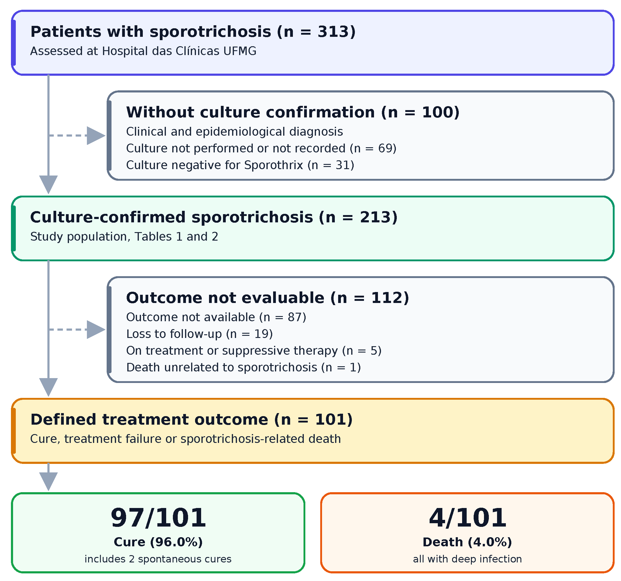

Table 1.
Baseline characteristics of patients with culture-confirmed sporotrichosis (n = 213).
| Characteristic | Patients (n = 213) |
| Sex | |
| Female | 115 (54.0) |
| Male | 98 (46.0) |
| Age, years | |
| Median (IQR) | 41 (24–55) [n=211] |
| 0–17 | 29 (13.6) |
| 18–39 | 72 (33.8) |
| 40–59 | 68 (31.9) |
| ≥60 | 42 (19.7) |
| Not reported | 2 (0.9) |
| Self-reported skin colour | |
| Mixed race (parda) | 133 (62.4) |
| White | 22 (10.3) |
| Black | 14 (6.6) |
| Asian | 3 (1.4) |
| Not reported | 41 (19.2) |
| Place of residence | |
| Belo Horizonte | 107 (50.2) |
| Metropolitan Region of Belo Horizonte | 54 (25.4) |
| Interior of Minas Gerais | 16 (7.5) |
| Other Brazilian states | 1 (0.5) |
| Not reported | 35 (16.4) |
| Comorbidities | |
| At least one comorbidity | 86 (40.4) |
| Hypertension | 42 (19.7) |
| Diabetes mellitus | 16 (7.5) |
| Smoking | 15 (7.0) |
| Depression or anxiety | 12 (5.6) |
| Obesity (BMI ≥30) | 7 (3.3) |
| Chronic kidney disease | 8 (3.8) |
| Heart disease | 4 (1.9) |
| Hypothyroidism | 5 (2.3) |
| Asthma or COPD | 6 (2.8) |
| Liver disease | 4 (1.9) |
| Other psychiatric disorder | 5 (2.3) |
| Other comorbidity | 27 (12.7) |
| Immunosuppressive condition | |
| Any immunosuppressive condition | 15 (7.0) |
| Rheumatological disease | 9 (4.2) |
| HIV infection | 2 (0.9) |
| Inflammatory bowel disease | 1 (0.5) |
| Glomerulopathy | 1 (0.5) |
| Solid organ transplant | 1 (0.5) |
| Intracranial tumour | 1 (0.5) |
| Clinical form | |
| Lymphocutaneous | 115 (54.0) |
| Fixed cutaneous | 44 (20.7) |
| Ocular | 47 (22.1) |
| Disseminated | 13 (6.1) |
| Osteoarticular | 12 (5.6) |
| Disseminated cutaneous | 3 (1.4) |
| Pulmonary | 2 (0.9) |
| More than one form recorded | 19 (8.9) |
| Hospital admission | |
| Hospital admission recorded | 16 (7.5) |
| Site of involvement | |
| Upper limbs | 102 (47.9) |
| Lower limbs | 41 (19.2) |
| Face | 11 (5.2) |
| Trunk | 11 (5.2) |
| Ocular | 36 (16.9) |
| Neck | 7 (3.3) |
| Nose | 4 (1.9) |
| Mouth | 2 (0.9) |
| Not reported | 47 (22.1) |
| Exposure | |
| Contact with cats | 113 (53.1) |
| Not reported (cat contact) | 83 (39.0) |
| Sick cat, among those exposed to cats | 84/113 (74.3) |
| Contact with animals other than cats | 23 (10.8) |
| Contact with soil, gardening | 21 (9.9) |
| Not reported (soil contact) | 130 (61.0) |
| Any animal exposure | 119 (55.9) |
| Mycological diagnosis | |
| Sporothrix spp., not identified to species | 202 (94.8) |
| Sporothrix brasiliensis (molecular identification) | 11 (5.2) |
Data are n (%) unless otherwise stated. IQR, interquartile range. HIV infection is reported once, under immunosuppressive condition. Categories of comorbidity are not mutually exclusive, and patients may appear in more than one row. “Other comorbidity” comprises conditions entered as free text, most frequently dyslipidaemia (n = 5), rhinitis or allergy (n = 5), gastrointestinal disease (n = 4), malignancy (n = 3), thrombosis or coagulation disorder (n = 3) and epilepsy (n = 2); rheumatological disease is shown in its own row, under immunosuppressive condition.
Table 2.
Treatment, tolerability and outcome of patients with culture-confirmed sporotrichosis (n = 213).
Table 2.
Treatment, tolerability and outcome of patients with culture-confirmed sporotrichosis (n = 213).
| Variable | Patients (n = 213) |
|---|---|
| Treatment received | |
| Itraconazole 100 mg twice daily | 53 (24.9) |
| Itraconazole 200 mg once daily | 44 (20.7) |
| Itraconazole 400 mg daily | 21 (9.9) |
| Potassium iodide | 13 (6.1) |
| Terbinafine | 5 (2.3) |
| Amphotericin B deoxycholate | 3 (1.4) |
| Liposomal amphotericin B | 1 (0.5) |
| Amphotericin B lipid complex | 4 (1.9) |
| Amphotericin B, any formulation | 6 (2.8) |
| Local heat | 1 (0.5) |
| Other | 16 (7.5) |
| More than one regimen recorded | 22 (10.3) |
| No antifungal treatment | 3 (1.4) |
| Treatment not recorded | 75 (35.2) |
| Treatment duration | |
| Median (IQR), days | 150 (93–217) |
| Available, n (%) | 106 (49.8) |
| Tolerability | |
| Laboratory abnormality during treatment | 52 (24.4) |
| Antifungal discontinuation for any reason | 11 (5.2) |
| Outcome | |
| Cure after antifungal treatment | 95 (44.6) |
| Spontaneous cure without treatment | 2 (0.9) |
| Treatment failure | 0 (0.0) |
| Death, sporotrichosis-related | 4 (1.9) |
| Death, unrelated to sporotrichosis | 1 (0.5) |
| Chronic suppressive therapy | 3 (1.4) |
| On treatment at database closure | 2 (0.9) |
| Loss to follow-up | 19 (8.9) |
| No outcome recorded | 87 (40.8) |
| Overall cure among patients with a defined outcome | 97/101 (96.0) |
Data are n (%) unless otherwise stated. IQR, interquartile range. Treatment categories are not mutually exclusive: 22 patients received more than one regimen, and the six patients treated with amphotericin B are also counted under the individual formulations received.
Table 3.
Comparison of patients with and without laboratory abnormalities during antifungal treatment (n = 92).
Table 3.
Comparison of patients with and without laboratory abnormalities during antifungal treatment (n = 92).
| Variable | Laboratory abnormality (n = 52) | No abnormality (n = 40) | p value |
|---|---|---|---|
| Demographic characteristics | |||
| Age, years, median (IQR) | 50 (38–56) [n=52] | 36 (20–47) [n=40] | 0.003 |
| Age ≥60 years | 11 (21.2) | 6 (15.0) | 0.451 |
| Female sex | 29 (55.8) | 26 (65.0) | 0.371 |
| Comorbidities | |||
| At least one comorbidity | 38 (73.1) | 20 (50.0) | 0.023 |
| Hypertension | 19 (36.5) | 9 (22.5) | 0.147 |
| Diabetes mellitus | 7 (13.5) | 3 (7.5) | 0.505 |
| Liver disease | 1 (1.9) | 1 (2.5) | 1.000 |
| Chronic kidney disease | 4 (7.7) | 1 (2.5) | 0.383 |
| Obesity (BMI ≥30) | 4 (7.7) | 1 (2.5) | 0.383 |
| Clinical form | |||
| Lymphocutaneous | 27 (51.9) | 23 (57.5) | 0.594 |
| Fixed cutaneous | 13 (25.0) | 11 (27.5) | 0.787 |
| Ocular | 7 (13.5) | 8 (20.0) | 0.400 |
| Disseminated | 8 (15.4) | 0 (0.0) | 0.009 |
| Osteoarticular | 7 (13.5) | 0 (0.0) | 0.017 |
| Antifungal regimen | |||
| Itraconazole 100 mg twice daily | 24 (46.2) | 16 (40.0) | 0.555 |
| Itraconazole 200 mg once daily | 14 (26.9) | 19 (47.5) | 0.041 |
| Itraconazole 400 mg daily | 11 (21.2) | 3 (7.5) | 0.085 |
| Amphotericin B, any formulation | 1 (1.9) | 0 (0.0) | 1.000 |
| Potassium iodide | 1 (1.9) | 0 (0.0) | 1.000 |
| More than one regimen recorded | 9 (17.3) | 3 (7.5) | 0.219 |
| Treatment and monitoring | |||
| Treatment duration, days, median (IQR) | 162 (94–304) [n=46] | 122 (90–176) [n=30] | 0.068 |
| Test panels requested, median (IQR) | 4 (2–6) [n=49] | 2 (1–2) [n=39] | <0.001 |
| Outcome | |||
| Documented cure | 37 (71.2) | 29 (72.5) | 0.887 |
| Loss to follow-up | 4 (7.7) | 5 (12.5) | 0.495 |
| Death, any cause | 2 (3.8) | 0 (0.0) | 0.503 |
| Antifungal discontinuation | 4 (7.7) | 2 (5.0) | 0.694 |
IQR, interquartile range. The analysis is restricted to patients in whom the presence or absence of laboratory abnormality during treatment was recorded. Documented cure is calculated over the whole group in each column, and not over the patients with a defined outcome. Numbers in square brackets indicate the patients with available data for the variable.
Table 4.
Comparison of localized cutaneous disease and deep infection (n = 168).
| Variable | Localized cutaneous (n = 146) | Deep infection (n = 22) | p value |
|---|---|---|---|
| Demographic characteristics | |||
| Age, years, median (IQR) | 40 (25–56) [n=141] | 50 (43–62) [n=21] | 0.024 |
| Age ≥60 years | 30 (20.5) | 6 (27.3) | 0.577 |
| Female sex | 79 (54.1) | 7 (31.8) | 0.051 |
| Comorbidities | |||
| At least one comorbidity | 59 (40.4) | 18 (81.8) | <0.001 |
| Hypertension | 35 (24.0) | 5 (22.7) | 0.898 |
| Diabetes mellitus | 12 (8.2) | 4 (18.2) | 0.232 |
| HIV infection | 1 (0.7) | 1 (4.5) | 0.245 |
| Chronic kidney disease | 4 (2.7) | 2 (9.1) | 0.177 |
| Immunosuppressive condition | 8 (5.5) | 6 (27.3) | 0.004 |
| Alcohol use, among those with information | 13/66 (19.7) | 6/13 (46.2) | 0.071 |
| Hospital admission | |||
| Hospital admission recorded | 6 (4.1) | 9 (40.9) | <0.001 |
| Exposure | |||
| Contact with cats | 86 (58.9) | 10 (45.5) | 0.235 |
| Sick cat, among those exposed to cats | 67/86 (77.9) | 6/10 (60.0) | 0.245 |
| Contact with soil or gardening | 15 (10.3) | 3 (13.6) | 0.710 |
| Antifungal treatment | |||
| Itraconazole 100 mg twice daily | 42 (28.8) | 3 (13.6) | 0.135 |
| Itraconazole 200 mg once daily | 33 (22.6) | 4 (18.2) | 0.787 |
| Itraconazole 400 mg daily | 12 (8.2) | 6 (27.3) | 0.016 |
| Potassium iodide | 11 (7.5) | 1 (4.5) | 1.000 |
| Terbinafine | 5 (3.4) | 0 (0.0) | 1.000 |
| Amphotericin B, any formulation | 0 (0.0) | 6 (27.3) | <0.001 |
| More than one regimen recorded | 15 (10.3) | 5 (22.7) | 0.147 |
| Treatment duration, days, median (IQR) | 150 (96–216) [n=78] | 98 (44–331) [n=12] | 0.330 |
| Tolerability | |||
| Laboratory abnormality, if monitored | 36/67 (53.7) | 11/12 (91.7) | 0.022 |
| Antifungal discontinuation | 9 (6.2) | 2 (9.1) | 0.639 |
| Outcome | |||
| Cure, among defined outcomes | 76/76 (100.0) | 5/9 (55.6) | <0.001 |
| Death, sporotrichosis-related | 0 (0.0) | 4 (18.2) | <0.001 |
| Chronic suppressive therapy | 0 (0.0) | 3 (13.6) | 0.002 |
| Loss to follow-up | 15 (10.3) | 1 (4.5) | 0.698 |
| No outcome recorded | 52 (35.6) | 9 (40.9) | 0.630 |
IQR, interquartile range. Localized cutaneous disease includes the fixed cutaneous and lymphocutaneous forms. Deep infection includes the disseminated, osteoarticular, pulmonary and disseminated cutaneous forms. Patients with more than one recorded clinical form were allocated to a single group, according to the most severe form, so that the group totals do not correspond to the sum of the non-exclusive clinical form counts shown in Table 1. The 45 patients with isolated ocular disease are not shown. Numbers in square brackets indicate the patients with available data for the variable.
Table 5.
Multivariable logistic regression for deep infection and for laboratory abnormality during treatment.
Table 5.
Multivariable logistic regression for deep infection and for laboratory abnormality during treatment.
| Variable | Odds ratio | 95% CI | p value |
|---|---|---|---|
| Deep infection: any comorbidity as exposure (n = 162, 21 events) | |||
| Age, per 10 years | 1.10 | 0.84–1.45 | 0.475 |
| Female sex | 0.28 | 0.10–0.78 | 0.015 |
| At least one comorbidity | 9.69 | 2.52–37.23 | <0.001 |
| Deep infection: immunosuppressive condition as exposure (n = 162, 21 events) | |||
| Age, per 10 years | 1.24 | 0.96–1.61 | 0.100 |
| Female sex | 0.36 | 0.13–1.01 | 0.051 |
| Immunosuppressive condition | 6.61 | 1.89–23.09 | 0.003 |
| Laboratory abnormality during treatment (n = 88, 49 events) | |||
| Age, per 10 years | 1.23 | 0.92–1.62 | 0.157 |
| Female sex | 0.71 | 0.26–1.91 | 0.493 |
| At least one comorbidity | 0.92 | 0.30–2.83 | 0.890 |
| Deep infection | 2.60 | 0.24–27.61 | 0.428 |
| Number of test panels requested | 1.43 | 1.09–1.88 | 0.009 |
CI, confidence interval. Binary logistic regression models estimated by maximum likelihood, with Wald confidence intervals. Sample sizes are smaller than the corresponding group totals because patients with missing covariate data were excluded from each model (complete-case analysis): 162 of the 168 patients compared in Table 4 had age recorded, and 88 of the 92 patients in Table 3 had the number of laboratory panels recorded.
Disclaimer/Publisher’s Note: The statements, opinions and data contained in all publications are solely those of the individual author(s) and contributor(s) and not of MDPI and/or the editor(s). MDPI and/or the editor(s) disclaim responsibility for any injury to people or property resulting from any ideas, methods, instructions or products referred to in the content. |
© 2026 by the authors. Licensee MDPI, Basel, Switzerland. This article is an open access article distributed under the terms and conditions of the Creative Commons Attribution (CC BY) license (http://creativecommons.org/licenses/by/4.0/).
Copyright: This open access article is published under a Creative Commons CC BY 4.0 license, which permit the free download, distribution, and reuse, provided that the author and preprint are cited in any reuse.