

Submitted:
12 August 2026
Posted:
13 August 2026
You are already at the latest version
Abstract
Annona amazonica R.E. Fries (Annonaceae) is a tropical species native to Central and South America whose phytochemistry remains poorly explored despite its promising chemical diversity and biological potential. As part of our ongoing investigation of bioactive metabolites from Amazonian Annonaceae, the bark of A. amazonica was subjected to phytochemical investigation using classical chromatographic methods. This study led to the isolation and structural elucidation of a new N-formyl aporphine alkaloid, N-formylactinodaphnine, which occurs as an E/Z rotamer because of restricted rotation around the amide bond, together with 17 known metabolites, including diterpenes, sesquiterpenes, steroids, triterpenoids, and aporphine alkaloids. Most of these compounds are reported for the first time from A. amazonica. The cytotoxic activities of the isolated compounds were evaluated against HepG2, HCT116, MDA-MB-231, MCF-7, and U-87 MG human cancer cell lines, as well as MRC-5 noncancerous fibroblasts, using the Alamar blue assay after 72 h of incubation. Among the tested compounds, actinodaphnine exhibited the strongest cytotoxic activity, with IC50 values ranging from 5.59 to 11.73 μg/mL1 against all the tumor cell lines evaluated, whereas N-methylactinodaphnine showed the greatest activity against HCT116 cells (IC50 = 4.27 μg/mL1). These findings expand the phytochemical knowledge of A. amazonica, describe a new naturally occurring N-formyl aporphine alkaloid, and demonstrate that this Amazonian species represents a promising source of cytotoxic natural products for further pharmacological investigation.
Keywords:
aporphinoids and terpenoids
; rotamers
; cytotoxic activity
1. Introduction
The genus Annona L. belongs to the family Annonaceae and comprises approximately 166–170 currently accepted species of trees, shrubs, and, more rarely, lianas [1,2]. It is among the best-known genera of the family because several of its species produce edible fruits that are widely appreciated throughout the world, such as “graviola” (Annona muricata L.), “ata, “pinha” or “fruta do conde” (Annona squamosa L.), “cherimola or cherimoia” (Annona cherimola Mill.), and “atemoya” (A. cherimola × A. squamosa), which are the best-known cultivated species. Many other Annona species are commonly known in Brazil as “pinha” or “araticum”. In addition to their edible fruits, several Annona species have been traditionally used for medicinal purposes. Different parts of plants are prepared as decoctions, infusions, macerates, ointments, or medicinal baths for the treatment of diseases caused by bacteria, fungi, protozoa, tumors, and insect bites, among other traditional applications [3,4]. Although Annona is among the best-known genera of Annonaceae because of its edible fruits, it remains relatively underexplored from phytochemical and biological perspectives. Most studies have focused on only a few species, especially A. muricata, A. squamosa, A. reticulata L., A. cherimola, A. senegalensis Pers., and A. coriacea Mart., mainly because of their economic importance. Consequently, many other species have received little attention and remain chemically and biologically unexplored [5,6,7,8,9,10].
As in the family Annonaceae in general, the major classes of secondary metabolites reported for Annona species include alkaloids, annonaceous acetogenins, flavonoids, terpenoids, phenolic compounds, and cyclopeptides. These metabolites are associated with several biological activities, including cytotoxic, antitumor, antimicrobial, antifungal, antiprotozoal, antimalarial, insecticidal, larvicidal, molluscicidal, anti-inflammatory, analgesic, antioxidant, neuropharmacological, immunosuppressive, and antidiabetic activities [5,6,7,8,9,10].
Considering the approximately 166 currently accepted species of Annona, it is estimated that only approximately 28–30% have been investigated phytochemically, whereas approximately 11–13% have been evaluated in more comprehensive biological or pharmacological studies. These estimates were obtained by integrating the reviews of Al-Kazman et al. [6], Kusmardiyani et al. [7], Dey et al. [9], Corrêa et al. [10], and other recent reviews, indicating that most species of the genus remain poorly investigated from both chemical and pharmacological perspectives.
Among these understudied species are Annona amazonica R. E. Fr. (syn. Annona amazonica var. lancifolia R.E. Fr.), popularly known in the Brazilian Amazon as “envira”, “araticum”, “araticum árvore gigante”, and “ata”. It is a tropical tree that reaches 20–30 m in height and can be distinguished from other Annona species by its very small flowers, congenitally fused outer and inner petals, and glaucous fruits. The species is distributed from Costa Rica to northern Brazil, where its fruits are traditionally consumed by local populations [11,12,13]. Previous phytochemical investigations of A. amazonica have reported the presence of cyanogenic compounds in the leaves, seeds, and fruits [14]. Pinheiro et al. [15] isolated high yields of acanthoic acid from the stem and demonstrated its trypanocidal activity. More recently, Alcantara et al. [16] identified (E)-caryophyllene and linalool as the major constituents of essential oils obtained from leaves and branches, respectively. More recently, Castro et al. [17] investigated the chemical composition of the essential leaf oil of A. amazonica and evaluated its antitumor activity both in vitro and in vivo. The major constituents identified were (E)-caryophyllene (32.01%), 1,8-cineole (13.93%), α-copaene (7.77%), α-humulene (7.15%), and α-pinene (5.13%). The essential oil induced apoptosis and significantly reduced the viability of HepG2 cells. DNA fragmentation, cell shrinkage, and a significant reduction in the mitochondrial membrane potential (ΔΨm) confirmed that the essential oil induced apoptosis. Furthermore, treatment with essential oil inhibited tumor growth by 39.2% in an in vivo model, indicating its potential against liver cancer [17].
As part of our continuing phytochemical studies on Amazonian Annonaceae species and aiming to expand the phytochemical knowledge of this family, the present work describes the chemical constituents isolated from the bark of A. amazonica and evaluates their cytotoxic activity. To the best of our knowledge, no phytochemical study has specifically investigated the bark of this species. Therefore, the results of the present study contribute to expanding the phytochemical knowledge of A. amazonica through the isolation and structural characterization of a new N-formyl aporphine alkaloid, rotamer, together with known aporphine alkaloids and terpenoids, as well as the evaluation of their cytotoxic properties.
2. Results and Discussion
2.1. Structural elucidation and identification of the isolated compounds
A phytochemical investigation of the bark of A. amazonica led to the isolation of eighteen compounds (1–18), including diterpenes (1), sesquiterpenes (2 and 9), steroids (3, 4 and 5), ketosteroids (6, 7 and 8), aporphinoid alkaloids (9–16), and tetrahydroprotoberberine alkaloids (17 and 18). Among them, compound 16 was identified as a previously undescribed N-formyl aporphine alkaloid occurring as a mixture of two slowly interconverting rotamers (E/Z), whereas compounds 1–15 and 17–18 were identified by spectroscopic analyses and comparison with the literature data. The structures of all the isolated compounds are shown in Figure 1.
Compound 16 was obtained as a brown, amorphous solid. The HRESIMS spectrum revealed a protonated molecular ion [M + H]⁺ at m/z 340.1177 (calcd. for C₁₉H₁₈NO₅⁺, m/z 340.1185; Δ = −2.3 ppm), establishing its molecular formula as C₁₉H₁₇NO₅. The IR spectrum showed a broad absorption at 3271 cm⁻¹, which is indicative of a phenolic hydroxyl group, together with a strong absorption at 1651 cm⁻¹, characteristic of an amide carbonyl, suggesting the presence of an N-formyl substituent [18]. Additional absorptions at 1460, 1393, 1269, 1194, 1102, 1074, 1013, 873, and 830 cm⁻¹ were assigned to aromatic ring vibrations and methylenedioxy group stretching and deformation modes, corroborating the presence of an oxygenated aporphine alkaloid skeleton.
The 1H NMR spectrum of 16 (CDCl3, 500 MHz) exhibited duplicated resonances for several signals, indicating the presence of two slowly interconverting species in solution. This behavior was particularly evident for the formyl proton, which appeared as two singlets at δH 8.25 and 8.38, as well as for several aliphatic resonances adjacent to the nitrogen atom, suggesting restricted rotation around the N-formyl bond. Similarly, the 13C NMR spectrum displayed duplicated signals for the amide carbonyl (δC 162.35 and δC 162.30) and for several carbons in the tetrahydroisoquinoline moiety, confirming the occurrence of two conformational rotamers rather than two distinct compounds (Table 1).
Apart from these duplicated resonances, the NMR data were fully consistent with an oxygenated aporphine skeleton. Three isolated aromatic singlets at δH 6.53, δH 6.84, and δH 7.66 (major rotamer) together with a methylenedioxy group [δH 5.97 (d, J = 1.4 Hz) and δH 6.10 (d, J = 1.4 Hz)] and a methoxy singlet at δH 3.93 indicated a highly substituted aromatic system (Table 1). The corresponding 13C NMR spectrum revealed nineteen carbon signals, including one amide carbonyl carbon, twelve sp2 carbons assigned to two aromatic rings, one methylenedioxy carbon, one methoxy carbon, and four aliphatic carbons characteristic of a tetrahydroaporphine framework.
The HSQC spectrum allowed the direct assignment of all the protonated carbons, whereas the 1H–1H COSY spectrum established the spin systems corresponding to H-4/H-5 and H-6a/H-7. The location of the methylenedioxy bridge between C-1 and C-2 was confirmed by the HMBC correlations of -OCH2O- (δH 5.97 and 6.10) with C-1 (δC 142.5) and C-2 (δC 147.1). Similarly, the methoxy group was assigned to C-10 on the basis of the three-bond correlation between the methoxy protons (δH 3.93) and C-10 (δC 145.6). Long-range HMBC correlations from H-3 to C-1, C-2, C-3a, and C-4 (Table 1), together with those observed from H-11 to C-9, C-10, and C-11a, established the substitution pattern of both aromatic rings, which was identical to that found in actinodaphnine.
The presence of an N-formyl substituent was unequivocally established by the characteristic HMBC correlation of the formyl proton with the amide carbonyl carbon (δC 162.35 for the major rotamer and δC 162.30 for the minor rotamer), together with the significant downfield displacement of the nitrogen-bearing carbon resonances compared with those of actinodaphnine (Table 1). These observations, combined with the molecular formula obtained by HRESIMS, demonstrated that compound 16 corresponded to the N-formyl derivative of actinodaphnine.
Detailed analysis of the duplicated resonances indicated that the two species correspond to rotamers generated by restricted rotation about the amide C−N bond. The major and minor rotamers were identified on the basis of the relative integrals of their respective formyl proton signals. The major component was assigned as the Z rotamer, whereas the minor component corresponded to the E rotamer. This assignment was supported by the NOESY experiment. With respect to the major rotamer, the formyl proton (δH 8.25) clearly enhanced the nuclear Overhauser signal with H-5 (δH 3.80), indicating spatial proximity between these nuclei and supporting the Z arrangement of the N-formyl group (Figure 2). In contrast, the NOESY correlation between the formyl proton (δH 8.38), H-6a (δH 4.58), and H-7 (δH 2.74) of the minor rotamer was consistent with the alternative E orientation (Figure 2). Additional NOESY cross-peaks observed within each rotamer were fully compatible with the proposed conformations and excluded the possibility that the duplicated resonances originated from a mixture of different alkaloids (Figure 2).
The occurrence of observable E/Z rotamers at room temperature results from the partial double-bond character of the amide C−N bond, which substantially increases the rotational barrier and slows conformational exchange on the NMR timescale. This phenomenon has been reported for several N-formyl alkaloids [18,19] and amides but remains relatively uncommon among naturally occurring aporphine alkaloids. The small but consistent differences in the chemical shifts of the formyl proton, carbonyl carbon, and aliphatic carbons adjacent to nitrogen are entirely consistent with the different magnetic environments experienced by the two rotamers.
Taken together, the HRESIMS, IR, and comprehensive 1D and 2D NMR spectroscopic data (HSQC, COSY, HMBC, and NOESY) unequivocally established compound 16 as a previously undescribed aporphine alkaloid, named N-formylactinodaphnine, occurring naturally as a mixture of slowly interconverting Z (major) and E (minor) rotamers. The stereochemical assignment at C-6a was based on a comparison of the specific optical rotation with the values reported in the literature for aporphine alkaloids possessing established absolute configurations [20].
In addition to compound 16, seventeen known metabolites were isolated and identified as acanthoic acid (1) [15] (15- Pinheiro et al., 2009), caryophyllene oxide (2) [21] (21- Ramírez-Santos), β-sitosterol (3) [22,23], stigmasterol (4) [22], campesterol (5) [23], β-sitostenone (6) [24], stigmasta-4,22-dien-3-one (7) [24], campest-4-en-3-one (8) [24], cadinane-4β,5α,10β-triol (9) [25], liriodenine (10) [26], lysicamine (11) [27], anonaine (12) [28], nornuciferine (13) [29], actinodaphnine (14) [30], N-methylactinodaphnine or cassythicine (15) [15], discretamine (17) [28], and 10-demethyldiscretine (18) [31]. Their structures were elucidated by comprehensive spectroscopic analyses, including 1D and 2D NMR experiments and mass spectrometry, and by comparison with published data. In addition, the NMR data of compounds 10, 14, 15, 17, and 18 were critically re-evaluated using highly purified samples. Complete 1H and 13C NMR assignments (Table 1 and Table 2), unequivocally supported by COSY, HSQC, and HMBC correlations, allowed the refinement of previously published assignments, the resolution of ambiguous signal multiplicities, and the confirmation of proton and carbon resonances. Consequently, the revised NMR data presented herein provide a more complete and reliable spectroscopic characterization of these aporphine alkaloids, establishing a robust reference for their future identification and structural studies.
The occurrence of aporphine alkaloids as the predominant alkaloidal constituents of A. amazonica is consistent with the phytochemical profiles reported for several Annona species. Nevertheless, the isolation of compound 16 represents the first report of an N-formyl derivative of actinodaphnine from the genus Annona, expanding the structural diversity of aporphine alkaloids in Annonaceae. Remarkably, acanthoic acid (1) was recovered in an exceptionally high amount (17.89 g), corresponding to at least 59.63% of the crude hexane extract. Because substantial amounts of compound 1 remained in the mother liquor and in enriched chromatographic fractions that were not exhaustively purified, its actual abundance in the hexane extract is expected to be even higher. These findings indicate that A. amazonica is an unusually rich natural source of acanthoic acid.
2.2. Chemophenetic significance
The phytochemical profile of A. amazonica is characterized by the predominance of aporphine alkaloids, a chemophenetic feature widely recognized for the genus Annona and the family Annonaceae. Aporphine alkaloids, together with annonaceous acetogenins, constitute the principal diagnostic classes of secondary metabolites in Annona species and are regarded as valuable chemophenetic markers reflecting conserved biosynthetic pathways within the family [5,32,33].
Prior to the present investigation, the phytochemical knowledge of A. amazonica was limited to a single study reporting the isolation of acanthoic acid (1), caryophyllene oxide (2), β-sitosterol (3), stigmasterol (4), liriodenine (10), and N-methylactinodaphnine, also known as cassythicine (15), from the stems of this species [15]. In the present study, we report the isolation of additional metabolites, including campesterol (5), β-sitostenone (6), stigmas-ta-4,22-dien-3-one (7), campest-4-en-3-one (8), cadinane-4β,5α,10β-triol (9), lysicamine (11), anonaine (12), nornuciferine (13), actinodaphnine (14), the new alkaloid N-formylactinodaphnine (16), discretamine (17), and 10-demethyldiscretine (18), which were described for the first time from A. amazonica. Additionally, liriodenine (10) and N-methylactinodaphnine (15) were confirmed on the basis of spectroscopic data and comparisons with literature values. These findings substantially expand the phytochemical profile of A. amazonica, revealing a rich diversity of aporphine alkaloids and providing new insights into the chemophenetic significance of this species within the genus.
Among the isolated alkaloids, actinodaphnine (14), N-methylactinodaphnine (15), and N-formylactinodaphnine (16) constitute a biologically coherent series. Their simultaneous occurrence strongly suggests that N-methylation and subsequent N-formylation represent late-stage modifications in the biosynthesis of aporphine alkaloids in A. amazonica. To the best of our knowledge, compound 16 is the first N-formyl derivative of actinodaphnine reported from the genus Annona, further expanding the structural diversity of aporphine alkaloids known for Annonaceae [5,32].
Another remarkable feature of A. amazonica is the exceptional accumulation of acanthoic acid (1). A total of 17.89 g of this diterpene was isolated, corresponding to 59.63% of the crude hexane extract. Because appreciable amounts of compound 1 remained in the mother liquor and in chromatographic fractions that were not exhaustively purified, its actual abundance in the extract is expected to be even higher. Diterpenoids are well documented in Annonaceae and constitute one of the major classes of nonalkaloidal metabolites in the family. In particular, species of Annona and Xylopia have yielded numerous diterpenes, predominantly belonging to the ent-kaurane class, which are considered characteristic constituents of these genera [5,32,34]. In contrast, acanthoic acid is an ent-pimarane (ent-pimaradiene) diterpene, a structural type that has been reported much less frequently in Annonaceae. Therefore, the occurrence of acanthoic acid as the overwhelmingly predominant constituent of the hexane extract distinguishes A. amazonica from other chemically investigated species of Annona. The remarkable accumulation of an ent-pimarane diterpene also suggests a diterpenoid biosynthetic profile that differs from that commonly observed in the genus, where ent-kaurane derivatives generally predominate. In addition to representing an unusually rich natural source of acanthoic acid, A. amazonica broadens the known diterpenoid diversity of Annona and contributes new chemophenetic evidence for the evolutionary diversification of diterpene biosynthesis within Annonaceae [5,32,33].
The isolation of cadinane-4β,5α,10β-triol (9) also deserves particular attention. Although cadinane-type sesquiterpenes have been reported from several plant families, this highly oxygenated derivative has not been recognized as a characteristic metabolite of Annona or Annonaceae in the available phytochemical literature and comprehensive reviews of the family. Instead, Annonaceae species are characterized predominantly by the presence of volatile cadinane derivatives such as cadinol, cadinene, and related sesquiterpenes in essential oils. Thus, the occurrence of cadinane-4β,5α,10β-triol in A. amazonica considerably broadens the known sesquiterpenoid diversity of the species and represents an uncommon chemophenetic finding that should be further investigated in other representatives of the genus.
Finally, the combined occurrence of abundant ent-kaurane diterpenes together with a structurally diverse assemblage of aporphine alkaloids, including the new N-formylactinodaphnine (16), reinforces the distinctive chemophenetic position of A. amazonica within the genus Annona. These findings substantially expand the phytochemical knowledge of the species and provide new insights into the evolution and diversification of secondary metabolism in Annonaceae.
2.3. Cytotoxicity Assay
The hexane and methanol extracts, together with the neutral and alkaloid-rich phases obtained from the acid–base extraction of the methanol extract, were subjected to initial cytotoxic screening at a single concentration of 50 μg/mL against human hepatocellular carcinoma (HepG2) and human colorectal carcinoma (HCT116) cell lines to identify samples capable of inhibiting tumor cell growth (Table 3). Among the samples evaluated, only the alkaloid-rich phase displayed relevant cytotoxic activity, inhibiting cell growth by 55.81% and 63.32% against HepG2 and HCT116 cells, respectively. These findings indicated that the cytotoxic activity of the methanol extract was associated mainly with the alkaloid-rich phase. Therefore, this phase was selected for detailed phytochemical investigation and bioactivity-guided isolation of its secondary metabolites.
On the basis of the results of the initial cytotoxic screening, the alkaloid-rich phase was selected for phytochemical investigation. Chromatographic separation of this phase afforded compounds 10–18. Among these compounds, compounds 14–18 were subsequently evaluated for their cytotoxic activity against the human tumor cell lines HepG2, HCT116, MDA-MB-231 (human breast carcinoma), MCF-7 (human breast adenocarcinoma), and U-87 MG (human glioblastoma), as well as the nontumor human lung fibroblast line MRC-5, to identify the metabolites responsible for the cytotoxic activity observed in the alkaloid-rich phase (Table 4). Additionally, acanthoic acid (compound 10), the major diterpenoid isolated from the hexane extract, was included in the cytotoxic evaluation because of its high abundance and previously reported biological activities, despite the lack of significant cytotoxic activity exhibited by the crude hexane extract.
Among the isolated compounds, actinodaphnine (14) exhibited the most pronounced cytotoxic activity, with IC₅₀ values ranging from 5.59 (HCT116) to 14.63 (MCF-7) μg/mL against all the tumor cell lines evaluated (Table 4). N-methylactinodaphnine (15) also demonstrated relevant cytotoxic activity, displaying an IC₅₀ value of 4.27 μg/mL against HCT116 cells. This broad-spectrum activity indicates that actinodaphnine and N-methylactinodaphnine are among of the major contributors to the cytotoxic effect observed for the alkaloid-rich phase. However, actinodaphnine also displayed comparable cytotoxicity toward the non-tumor MRC-5 cell line (IC₅₀ = 6.84 μg/mL), indicating limited tumor selectivity under the experimental conditions. In contrast, N-methylactinodaphnine exhibited lower cytotoxicity against MRC-5 cells (IC₅₀ = 12.16 μg/mL), suggesting a more favorable selectivity profile. These findings are consistent with those of previous reports demonstrating the cytotoxic potential of this aporphine alkaloid. Stévigny et al.[30] first reported the cytotoxic activity of actinodaphnine against human melanoma (Mel-5) and promyelocytic leukemia (HL-60) cells, with IC₅₀ values of 25.7 and 15.4 μM, respectively. Hsieh et al. [35] subsequently demonstrated that actinodaphnine induces apoptosis in Mahlavu human hepatoma cells through increased reactive oxygen species (ROS) and nitric oxide (NO) production, mitochondrial membrane depolarization, caspase-3/7 activation, and down-regulation of NF-κB signaling. Therefore, although the mechanism of action was not investigated in the present study, the potent cytotoxic activity observed for compound 14 is consistent with previous evidence supporting apoptosis as one of the principal mechanisms underlying its antitumor activity.
In contrast, N-formylactinodaphnine (16), discretamine (17), and 10-demethyldiscretine (18) displayed weak or no cytotoxic activity (IC₅₀ > 25 μg/mL for most cell lines). Likewise, acanthoic acid (1), despite being the major diterpenoid isolated from the hexane extract, exhibited only moderate activity against HepG2 and HCT116 cells, corroborating the low cytotoxicity observed for the crude hexane extract (Table 4).
Comparisons of the cytotoxic profiles of actinodaphnine (14), N-methylactinodaphnine (15), and N-formylactinodaphnine (16) provide valuable insight into the structure–activity relationships of these closely related aporphine alkaloids (Table 4). The three compounds differ only in the substitution pattern of the nitrogen atom. Actinodaphnine possesses a free secondary amine, N-methylactinodaphnine, which contains a tertiary amine, whereas N-formylactinodaphnine has an N-formyl amide. Among them, actinodaphnine exhibited the broadest and most potent cytotoxic profile and was active against all the tumor cell lines evaluated. N-methylactinodaphnine also exhibited potent cytotoxic activity against HCT116 cells. However, due to the limited amount of compound available, its cytotoxic activity was evaluated only in this cell line, precluding comparisons of its cytotoxic spectrum with that of actinodaphnine. In contrast, N-formylactinodaphnine displayed weak or no cytotoxic activity against most tumor cell lines, showing only moderate activity against HCT116 cells (IC₅₀ = 21.7 μg/mL). These findings suggest that the nature of the nitrogen substituent plays a crucial role in modulating the cytotoxic properties of aporphine alkaloids. In particular, the presence of a free secondary amine appears to be favorable for broad-spectrum cytotoxic activity, whereas N-methylation confers greater selectivity, and N-formylation markedly reduces antitumor potency. Interestingly, N-formylactinodaphnine was also non-cytotoxic toward the nontumor MRC-5 cell line (IC₅₀ > 25 μg/mL), suggesting that N-formylation decreases both tumor and normal cell cytotoxicity.
Although liriodenine (11) and lysicamine (12) were not included in the present cytotoxic evaluation because of the limited amounts isolated, both oxoaporphine alkaloids have been consistently reported to exhibit significant cytotoxic activity against several human cancer cell lines through multiple mechanisms, including DNA intercalation, inhibition of topoisomerase II, induction of apoptosis, and cell cycle arrest. Recent studies have also demonstrated that both compounds possess antiproliferative activity against breast, liver, colon, gastric, and lung cancer cell lines, highlighting their relevance as bioactive constituents of Annonaceae species [36,37,38,39,40].
Therefore, the cytotoxic activity observed for the alkaloid-rich phase is likely not attributable to a single constituent but rather to the combined action of several bioactive aporphine and oxoaporphine alkaloids, including actinodaphnine, N-methylactinodaphnine, liriodenine, and lysicamine. This hypothesis is further supported by the fact that the alkaloid-rich phase exhibited higher overall activity than several of the isolated compounds did, suggesting additive or synergistic interactions among its constituents.
These results indicate that the cytotoxic activity of A. amazonica is associated mainly with its aporphine alkaloid content rather than with its terpenoid constituents. Although the newly identified N-formylactinodaphnine displayed only weak cytotoxic activity, its characterization as a naturally occurring N-formyl aporphine alkaloid existing as E/Z rotamers expands the structural diversity currently known for this class of natural products. In contrast, the pronounced cytotoxic activities of actinodaphnine and N-methylactinodaphnine identify aporphine alkaloids as the principal bioactive constituents isolated from this species and support further studies aimed at understanding their mechanisms of action and structure–activity relationships.
3. Materials and Methods
3.1. General Experimental Procedures
Fourier transform infrared (FTIR) spectra were recorded on an IRSpirit FTIR spectrophotometer (Shimadzu, Kyoto, Japan). Optical rotations were measured in CHCl₃ using a Jasco P-2000 polarimeter (Jasco, Tokyo, Japan) at the sodium D line (589 nm).
One- and two-dimensional NMR data were acquired in CDCl3 (chloroform-d), CD3OD (methanol-d4), or a mixture of these solvents, at 298 K on AVANCE III HD and AVANCE NEO NMR spectrometers (Bruker, Billerica, MA, USA) operating at 11.75 and 14.1 T (1H at 500 and 600 MHz and 13C at 125 and 150 MHz, respectively). All the 1H-NMR and 13C-NMR chemical shifts (δ) are presented in ppm relative to the tetramethylsilane signal at 0.00 ppm as an internal reference, and the coupling constants (J) are given in Hz. The NMR spectrometer was equipped with a 5-mm multinuclear inverse detection probe (for 1D and 2D NMR experiments) with a z gradient. One-bond (HSQC) and two- and three-bond (HMBC) 1H-13C-NMR correlation experiments were optimized for average coupling constants of 1J(C,H) and LRJ(C,H) of 140 and 8 Hz, respectively. For low-resolution mass spectrometry (LR‒MS) analysis, the samples of the isolated compounds were resuspended in methanol (HPLC grade), creating stock solutions (1 mg/mL). Aliquots (5 µL) of the stock solutions were further diluted to 5 µg/mL and analyzed by direct infusion into a triple quadrupole mass spectrometer, model TSQ Quantum Access (Thermo Scientific, San Jose, CA, USA), equipped with electrospray ionization (ESI) or atmospheric pressure chemical ionization (APCI) sources in negative or positive mode. An HPLC Shimadzu (Kyoto, Japan) coupled with a MicroTOF II (Bruker Daltonics, Billerica, MA, USA) with an electrospray ionization (ESI) source was used to obtain high-resolution mass spectra (HRESIMS) in positive mode. The parameters were as follows: capillary voltage of 4.5 kV, ESI in positive mode, final plate offset of 500 V, nebulizer pressure of 40 psi, dry gas (N2) flow rate of 8 mL/min and temperature of 200 °C. The mass spectra (m/z 50–1000) were recorded every 2 s. Silica gel 60 (Sigma‒Aldrich, San Luis, MO, USA, 70–230 mesh) was used for column chromatography (CC), whereas silica gel 60 F254 (Macherey-Nagel, Düren, Germany, 0.25 mm, aluminum) was used for analysis and preparation with thin layer chromatography (PTLC) (Macherey-Nagel, 1.00 mm, glass). The compounds were visualized by exposure to UV254/365 light, spraying with p-anisaldehyde reagent, heating on a hot plate, and spraying with Dragendorff’s reagent.
Gas chromatography–mass spectrometry (GC–MS) analyses were performed on a Trace Ultra gas chromatograph coupled to an ISQ single quadrupole mass spectrometer (Thermo Scientific, Waltham, MA, USA). The instrument was equipped with a TriPlus autosampler and a TR-5MS fused-silica capillary column (30 m × 0.25 mm i.d., 0.25 μm film thickness). Helium (99.999% purity) was used as the carrier gas at a constant flow rate of 1.2 mL/min. The GC oven temperature was programmed from 120 °C (held for 2 min) to 320 °C at a rate of 20 °C/min, followed by a final hold at 320 °C for 10 min, resulting in a total analysis time of 22 min. Samples were dissolved in dichloromethane (CH₂Cl₂), and 0.5 μL was injected in split mode (split ratio of 1:50). The injector temperature was maintained at 250 °C, and the GC–MS transfer line temperature was set at 280 °C. Mass spectra were acquired in full-scan mode using electron ionization (EI) at 70 eV over a m/z range of 40–600 with a scan time of 0.5 s.
3.2. Plant material
In the present investigation, the botanical material (bark) of A. amazonica was collected on December 3, 2021, on the Adolpho Ducke Reserve (geographic coordinates: 02o 54’ 49.9” S and 59o 58’ 43.6” W), Manaus, Amazonas State, Brazil, and identified by Prof. Dr. Antonio Carlos Webber, a plant taxonomist of the Department of Biology of the Universidade Federal do Amazonas (DB/UFAM). A voucher specimen (number 12098) was deposited at the Herbarium of DB/UFAM. The access (specimen) was registered in the Sistema Nacional de Gestão do Patrimônio Genético e do Conhecimento Tradicional Associado (SISGEN) with the record A70EDCD.
3.3. Preparation of NaHCO₃-treated silica gel
Silica gel for column chromatography (0.063–0.200 mm) was treated with a 10% aqueous solution of sodium bicarbonate (NaHCO₃). Briefly, a 10% NaHCO₃ solution (1 L) was prepared by dissolving sodium bicarbonate (100 g) in distilled water and adjusting the final volume to 1000 mL. Silica gel (300 g) was suspended in the NaHCO₃ solution until completely immersed and maintained under periodic stirring for 1 h, followed by standing for 24 h at room temperature.
The treated silica gel was collected by filtration under reduced pressure, transferred to a porcelain dish, and dried at 170–200 °C with occasional stirring until a free-flowing powder was obtained. The resulting NaHCO₃-treated silica gel was used as the stationary phase for column chromatography.
3.4. Extraction and isolation
The bark of A. amazonica was initially air-dried at room temperature for 24 h and then dried in a forced-air circulation oven at 40 °C for 48 h. The dried material was pulverized using a four-knife mill grinder (Marconi) to yield 1,621.5 g of powdered bark. The powdered material was exhaustively extracted by maceration with hexane (6 × 3.5 L), followed by MeOH (6 × 3.5 L). The resulting extracts were concentrated under reduced pressure at 40–50 °C using a rotary evaporator to obtain the hexane (37.02 g) and MeOH (177.94 g) extracts.
After solvent removal, the hexane extract resulted in the abundant formation of yellow crystals. Because of the large amount of crystals formed, a portion of the hexane extract (30.0 g) was subjected to successive washings with hexane, followed by recrystallization from a hexane–CH2Cl2 (4:1, v/v) mixture, yielding compound 1 (12,717.8 mg) as a white crystalline solid together with a mother-liquor enriched fraction (17,301.6 mg). Since this enriched fraction still contained a high concentration of compound 1, an aliquot (12.0 g) was subjected to silica gel column chromatography (6.0 × 60.0 cm, i.d. × height). Elution was performed successively with hexane (100%); hexane–CH₂Cl₂ (80:20, 70:30, 50:50, 30:70, and 20:80, v/v); CH₂Cl₂ (100%); CH₂Cl₂–EtOAc (80:20, 70:30, 50:50, 30:70, and 20:80, v/v); EtOAc (100%); and EtOAc–MeOH (90:10, v/v). Fractions of 500 mL were collected throughout the chromatographic separation, affording fourteen pooled fractions (F1–F14).
Fractions F3 (1,741.5 mg), eluted with hexane–CH2Cl2 (70:30, v/v), and F4 (3,991.1 mg), eluted with hexane–CH2Cl2 (50:50, v/v), consisted of a white crystalline solid containing a small amount of colorless oil. These fractions were combined and subjected once again to washing with hexane, followed by recrystallization from hexane–CH2Cl2 (4:1, v/v), affording additional compound 1 (5.055 g) together with an enriched fraction (620.0 mg). Approximately 150.0 mg of this enriched fraction was purified by preparative TLC and developed twice with hexane–EtOAc (90:10, v/v), yielding compounds 1 (117.5 mg) and 2 (12.7 mg).
Fraction F5 (2,699.7 mg), eluted with hexane–CH2Cl2 (30:70, v/v), was obtained as a white crystalline solid similar to compound 1. Fraction F7 (479.9 mg), eluted with CH2Cl2 (100%), was purified by preparative TLC (60.0 mg), and developed twice with hexane–EtOAc (80:20, v/v), affording a mixture of compounds 3–5 (14.0 mg) and a mixture of compounds 6–8 (12.7 mg). Fraction F11 (209.6 mg), eluted with CH2Cl2–EtOAc (30:70, v/v), was further purified by preparative TLC (60.0 mg), and developed twice with CHCl3–MeOH (90:10, v/v), to afford compound 9 (5.3 mg).
TLC analysis after spraying with Dragendorff’s reagent indicated a high abundance of nitrogen-containing compounds, suggesting the presence of alkaloids in the MeOH extract. Therefore, an aliquot of the MeOH extract (167.26 g) was subjected to acid–base extraction according to the literature [18], affording an alkaloid-rich phase (2.37 g) and a neutral phase (11.38 g).
A portion of the alkaloid-rich phase (2.30 g) was subsequently chromatographed over a silica gel column (3.0 × 60 cm, i.d. × height) previously treated with a 10% aqueous NaHCO₃ solution [41]. Elution was performed successively with hexane (100%); hexane–CH₂Cl₂ (90:10, 80:20, 70:30, 60:40, 50:50, 40:60, 30:70, 20:80, and 10:90, v/v); CH₂Cl₂ (100%); CH₂Cl₂–EtOAc (90:10, 80:20, 70:30, 60:40, 50:50, 40:60, 30:70, 20:80, and 10:90, v/v); EtOAc (100%); EtOAc–MeOH (95:5, 90:10, 85:15, 80:20, 75:25, 70:30, 60:40, and 50:50, v/v); and finally MeOH (100%). For each solvent system, approximately 200 mL of mobile phase was used, and fractions were collected in 30 mL vials, yielding a total of 157 fractions. For some solvent gradients, additional volumes of 100 mL or more were required to ensure complete elution of the compounds from the chromatographic column. The fractions were monitored by TLC using CH₂Cl₂–MeOH (95:5, 90:10, 85:15, and 80:20, v/v) as the development system. On the basis of their chromatographic profiles, similar fractions were combined to afford sixteen pooled fractions (FA1–FA16).
Fraction FA3 (53.0 mg), eluted with hexane–CH₂Cl₂ (40:60 and 30:70, v/v), was purified by preparative TLC using CH₂Cl₂–MeOH (97:3, v/v; three consecutive developments), yielding compounds 10 (4.7 mg), 11 (4.4 mg), and 12 (1.8 mg).
Fraction FA4 (28.1 mg), eluted with hexane–CH₂Cl₂ (10:90, v/v) and CH₂Cl₂ (100%), and fraction FA5 (23.9 mg), eluted with CH₂Cl₂ (100%) and CH₂Cl₂–EtOAc (90:10, v/v), exhibited similar TLC profiles and were therefore combined (52.0 mg). The resulting fraction was purified by preparative TLC using CH₂Cl₂–MeOH (97:3, v/v; three consecutive developments), affording compounds 10 (4.4 mg) and 13 (2.1 mg).
Fraction FA6 (48.6 mg), eluted with CH₂Cl₂–EtOAc (90:10, v/v), was purified by preparative TLC using CH₂Cl₂–MeOH (97:3, v/v; three consecutive developments) to afford compounds 10 (3.6 mg) and 14 (6.5 mg).
Fractions FA7 (44.2 mg), eluted with CH₂Cl₂–EtOAc (80:20, v/v); FA8 (49.4 mg), eluted with CH₂Cl₂–EtOAc (80:20, v/v); FA9 (62.5 mg), eluted with CH₂Cl₂–EtOAc (80:20, v/v); FA10 (54.4 mg), eluted with CH₂Cl₂–EtOAc (70:30, v/v); and FA11 (329.0 mg), eluted with CH₂Cl₂–EtOAc (70:30, 60:40, 50:50, 40:60, and 30:70, v/v), exhibited similar chromatographic profiles and were therefore combined to afford a pooled fraction (539.5 mg). The combined material was rechromatographed over a silica gel column (3.0 × 30.0 cm, i.d. × height) previously treated with a 10% aqueous NaHCO₃ solution [12] under the same chromatographic conditions employed for the initial fractionation of the alkaloid-rich fraction. Approximately 100 mL of mobile phase was used for each solvent system, and the eluates were collected in 20 mL vials, yielding a total of 95 fractions. Additional volumes of 50 mL or more were used for selected solvent gradients whenever necessary to ensure complete elution of the constituents from the column. The collected fractions were analyzed by TLC using CH₂Cl₂–MeOH (95:5, 90:10, 85:15, and 80:20, v/v) as the development system. Fractions displaying similar chromatographic profiles were combined to afford sixteen pooled fractions, designated FA7–11.1 to FA7–11.16.
Fractions FA7–11.8 (42.5 mg), eluted with CH₂Cl₂–EtOAc (70:30, v/v), and FA7–11.9 (55.4 mg), eluted with CH₂Cl₂–EtOAc (60:40, v/v), exhibited similar chromatographic profiles and were therefore combined (97.9 mg). The resulting fraction was purified by preparative TLC using CH₂Cl₂–MeOH (97:3, v/v; three consecutive developments) to afford compounds 14 (5.1 mg), 15 (4.4 mg), 17 (6.1 mg), and 18 (13.4 mg).
Fraction FA7–11.10 was purified by recrystallization from CH₂Cl₂–ethanol (1:1, v/v), affording additional compound 18 (22.5 mg).
Fraction FA12 (63.2 mg), eluted with CH₂Cl₂–EtOAc (30:70 and 20:80, v/v), was purified by preparative TLC using CH₂Cl₂–MeOH (97:3, v/v; three consecutive developments) to afford compounds 14 (7.6 mg), 17 (10.0 mg), 18 (18.9 mg), and 16 (3.8 mg).
Acanthoic acid (1): White crystalline solid (recrystallized from hexane–CH₂Cl₂, 4:1, v/v). [α]D 25 −42 (c 1.4, CHCl3). The structure was established by analysis of the 1H and 13C NMR spectroscopic data, which were in agreement with those previously reported in the literature [15]. GC–MS: m/z 302 [M]+∙.
Caryophyllene oxide (2): Colorless oil. The structure was established by analysis of the 1H and 13C NMR spectroscopic data, which were in agreement with those previously reported in the literature [21].
Mixture of β-sitosterol (3), stigmasterol (4), and campesterol (5): White needle-shaped crystals (recrystallized from hexane–CH₂Cl₂, 3:1, v/v). The structures were established by analysis of the 1H and 13C NMR spectroscopic data, which were in agreement with those previously reported in the literature [22,23]. GC–MS: m/z 414, 412, and 400 [M]+∙.
Mixture of β-sitostenone (6), stigmasta-4,22-dien-3-one (7), and campest-4-en-3-one (8): White, amorphous solid. The structures were established by analysis of the 1H and 13C NMR spectroscopic data, which were in agreement with those previously reported in the literature [24]. GC–MS: m/z 412, 410, and 398 [M]+∙.
Cadinane-4β,5α,10β-triol (9): White needle-shaped crystals (recrystallized from CH₂Cl₂–MeOH, 2:1, v/v). The structure was established by analysis of the 1H and 13C NMR spectroscopic data, which were in agreement with those previously reported in the literature [25].
Liriodenine (10): Yellow needle-shaped crystals (recrystallized from CH₂Cl₂–ethanol, 3:1, v/v). The structure was established by analysis of the 1H and 13C NMR spectroscopic data (Table 2), which were in agreement with those previously reported in the literature [26]. LR-APCI(+)-MS: m/z 276 [M + H]+.
Lysicamine (11): Yellow needle-shaped crystals (recrystallized from CH₂Cl₂–ethanol, 3:1, v/v). The structure was established by analysis of the 1H and 13C NMR spectroscopic data, which were in agreement with those previously reported in the literature [27]. LR-APCI(+)-MS: m/z 292 [M + H]+.
Anonaine (12): Brown amorphous solid. The structure was established by analysis of the 1H and 13C NMR spectroscopic data, which were in agreement with those previously reported in the literature [28]. LR-APCI(+)-MS: m/z 266 [M + H]+.
Nornuciferine (13): Brown amorphous solid. The structure was established by analysis of the 1H and 13C NMR spectroscopic data, which were in agreement with those previously reported in the literature [29]. LR-APCI(+)-MS: m/z 282 [M + H]+.
Actinodaphnine (14): Brown amorphous solid. The structure was established by analysis of the 1H and 13C NMR spectroscopic data (Table 2), which were in agreement with those previously reported in the literature [30]. LR-APCI(+)-MS: m/z 312 [M + H]+.
N-Methylactinodaphnine (15): Brown amorphous solid. The structure was established by analysis of the 1H and 13C NMR (Table 2) spectroscopic data, which were in agreement with those previously reported in the literature [15]. LR-APCI(+)-MS: m/z 326 [M + H]+.
N-Formylactinodaphnine(16E/16Z): Brown amorphous solid. [α]D 25 +76 (c 0.06, CHCl3). IR (KBr) cm-1: 3271, 2923; 2852, 1651, 1608, 1591, 1461,1444, 1410, 1393, 1364, 1326, 1292, 1269, 1243, 1194, 1102, 1074, 1048, 1013, 976, 942, 873, 830, 750, 640, 600. The structure was established by analysis of the 1H and 13C NMR (Table 1) spectroscopic data, including COSY, HSQC, and HMBC correlations. HRESIMS: m/z 340.1177 (calcd. for C₁₉H₁₈NO₅⁺, m/z 340.1185; Δ = −2.3 ppm). LR-APCI(−)-MS: m/z 338 [M − H]−.
3.5. In vitro cytotoxic assay
3.5.1. Cells
The HepG2 (human hepatocellular carcinoma), HCT116 (human colorectal carcinoma), MDA-MB-231 (human breast carcinoma), MCF-7 (human breast adenocarcinoma), U-87 MG (human glioblastoma), and MRC-5 (human lung fibroblast) cell lines were obtained from the American Type Culture Collection (ATCC; Manassas, VA, USA) and were cultured as recommended by the American Type Culture Collection (ATCC) animal culture guide [42]. All the cell lines were tested for mycoplasma via a mycoplasma stain kit (Sigma‒Aldrich) to validate the use of cells that were free from contamination.
3.5.2. Cytotoxicity assay
For the cytotoxicity assay, cell viability was quantified via the Alamar blue method, as previously described [43]. For all the experiments, the cells were plated in 96-well plates. The chemical constituents evaluated varied in purity between 90% and 99.8%, which was determined on the basis of the relative integrals of the signals of the constituents in relation to the signals of the impurities contained. Most of the constituents had purities greater than 95%, except for constituents 4 and 5, whose purities were approximately 90%. The chemical components were dissolved in dimethyl sulfoxide (DMSO; Vetec Química Fina Ltd.a., Duque de Caxias, RJ, Brazil), added to each well and incubated for 72 h. Doxorubicin (doxorubicin hydrochloride, purity ≥ 95%, Laboratory IMA S.A.I.C., Buenos Aires, Argentina) was used as a positive control. At the end of treatment, 20 µL of a stock solution (0.312 mg/mL) of resazurin (Sigma‒Aldrich Co.) was added to each well. The absorbances at 570 nm and 600 nm were measured via a SpectraMax 190 Microplate Reader (Molecular Devices, Sunnyvale, CA, USA). The half-inhibitory concentration (IC50) was obtained via nonlinear regression with 95% confidence intervals (CIs 95%) via the software GraphPad Prism (Intuitive Software for Science; San Diego, CA, USA).
5. Conclusions
This study provides the first comprehensive phytochemical and biological investigation of the bark of A. amazonica. Bioassay-guided fractionation of the methanol extract, together with a phytochemical investigation of the hexane extract, resulted in the isolation and identification of eighteen secondary metabolites belonging to four major classes of natural products, namely, diterpenes, sesquiterpenes, phytosterols, and aporphine alkaloids. Most of these compounds are reported for the first time from A. amazonica, considerably expanding the phytochemical knowledge of this Amazonian species.
Among the isolated metabolites, N-formylactinodaphnine was characterized as a new naturally occurring N-formyl aporphine alkaloid. Its occurrence as an E/Z rotamer, arising from restricted rotation around the amide C–N bond, represents an uncommon structural feature among naturally occurring aporphine alkaloids and further expands the structural diversity of this metabolite class. Moreover, the combined occurrence of aporphine alkaloids, pimarane diterpenes, sesquiterpenes, and phytosterols provides new chemotaxonomic information for A. amazonica and contributes to a broader understanding of the chemical diversity within the genus Annona and the family Annonaceae.
The results of the cytotoxic evaluation demonstrated that the alkaloid-rich phase was the source of the metabolites responsible for the biological activity observed for A. amazonica. Among the isolated compounds, actinodaphnine exhibited the broadest cytotoxic activity against all the human cancer cell lines evaluated, whereas N-methylactinodaphnine was the most potent compound against HCT116 cells. Although N-formylactinodaphnine displayed only weak cytotoxic activity, its unique structural characteristics, together with the pronounced activity of closely related aporphine alkaloids, reinforce the relevance of this metabolite class for future mechanistic investigations and structure–activity relationship studies.
Overall, the present work substantially expands the phytochemical, chemophenetic and biological knowledge of A. amazonica. The discovery of a new naturally occurring N-formyl aporphine alkaloid, together with the identification of cytotoxic aporphine alkaloids and the chemophenetic significance generated herein, demonstrates that A. amazonica represents a valuable source of structurally diverse secondary metabolites and supports further investigations on Amazonian Annonaceae as a source of new anticancer lead compounds.
Supplementary Materials
The following supporting information can be downloaded at the website of this paper posted on Preprints.org. Figures S1–S273: 1H and 13C 1D and 2D NMR, IR and MS spectra for all the compounds.
Author Contributions
Conceptualization, E.V.C. and D.P.B.; methodology, J.F.A., T.R.B.M., M.V.L.d.C., M.B.P.S, D.P.B, and E.V.C.; formal analysis, J.F.A.; M.B.P.S., D.P.B., and E.V.C.; investigation, J.F.A., T.R.B.M., M.L.B.P.; M.V.L.d.C., M.B.P.S, R.S.G., J.F.T., D.P.B, and E.V.C.; data curation, J.F.A., T.R.B.M., M.L.B.P.; M.V.L.d.C., M.B.P.S, R.S.G., J.F.T., D.P.B., and E.V.C.; resources, E.V.C. and D.P.B.; writing-original draft preparation, E.V.C. and D.P.B.; writing-review and editing, R.S.G., J.F.T., D.P.B., and E.V.C.; visualization, M.B.P.S., R.S.G., J.F.T., D.P.B., and E.V.C.; supervision, D.P.B and E.V.C.; funding acquisition, M.B.P.S., D.P.B. and E.V.C.
Funding
This work was financially supported by the Brazilian agencies Coordenação de Aperfeiçoamento de Pessoal de Nível Superior (CAPES, code 001), Conselho Nacional de Desenvolvimento Científico e Tecnológico (CNPq Grant: 306335/2023-9), FitoAmazônia Research Network (Pró-Amazônia/CNPq Grant: 445615/2024-9), and Fundação Oswaldo Cruz (FIOCRUZ).
Institutional Review Board Statement
Not applicable.
Informed Consent Statement
Not applicable.
Data Availability Statement
The data presented in this study are available in the Supplementary Materials.
Acknowledgments
The authors gratefully acknowledge the Central Analítica, Centro de Apoio Multidisciplinar, Federal University of Amazonas (CA/CAM/UFAM), for the low-resolution MS, GC–MS, and NMR analyses; the Laboratório Temático de Química de Produtos Naturais (LTQPN), Instituto Nacional de Pesquisas da Amazônia (INPA), for the 600 MHz NMR analyses; Prof. Josean Fechine Tavares (Federal University of Paraíba, UFPB) for the HRMS and IR analyses; Prof. Antonio Carlos Webber (Department of Biology, Federal University of Amazonas, UFAM) for the botanical identification of the plant material; and the Federal University of Amazonas (UFAM) for providing the infrastructure necessary to carry out this research.
Conflicts of Interest
The authors declare that they have no conflicts of interest.
Sample Availability
Samples of the isolated compounds are available from the authors.
References
- Chatrou, L.W. The Annonaceae and the Annonaceae project: a brief overview of the state of affairs. Acta Hortic. 1999, 497, 43–49. [Google Scholar] [CrossRef]
- WFO. World Flora Online. Version [2026]. Available online: http://www. worldfloraonline.org (accessed on 16 July 2026).
- Corrêa, M.P. Dicionário das Plantas Úteis do Brasil e das Exóticas Cultivadas; Ministério da Agricultura/IBDF: Rio de Janeiro, Brazil, 1984. [Google Scholar]
- Gemtchújnicov, I.D. Manual de Taxonomia Vegetal: Plantas de Interesse Econômico; Editora Agronômica Ceres: São Paulo, Brazil, 1976; 368 p. [Google Scholar]
- Leite, D.O.D.; Nonato, C.F.A.; Camilo, C.J.; Carvalho, N.K.G.; Nobrega, M.G.L.A.; Pereira, R.C.P.; Costa, J.G.M. Annona Genus: Traditional Uses, Phytochemistry and Biological Activities. Curr. Pharm. Des. 2020, 26(33), 4056–4091. [Google Scholar] [CrossRef] [PubMed]
- Al-Kazman, B.S.M.; Hamett, J.E.; Hanrahan, J.R. Traditional uses, phytochemistry and pharmacological activities of Annonacae. Molecules 2022, 27, 3462. [Google Scholar] [CrossRef] [PubMed]
- Nugraha, A.S.; Damayanti, Y.D.; Wangchuk, P.; Keller, P.A. Anti-infective and anti-cancer properties of the Annona species: their ethnomedicinal uses, alkaloid diversity, and pharmacological activities. Molecules 2019, 24, 4419. [Google Scholar] [CrossRef]
- Kusmardiyani, S.; Suharli, Y.A.; Insanu, M.; Fidrianny, I. Phytochemistry and pharmacological activities of Annona genus: A review. Curr. Res. Biosci. Biotechnol. 2020, 2, 77–88. [Google Scholar] [CrossRef]
- Dey, P.; Gogoia, H.; Goyary, D.; Mandal, S. Unveiling the bioactive phytocompounds of various Annona species with their potential role in pharmacological progression. S. Afr. J. Bot. 2024, 173, 85–98. [Google Scholar] [CrossRef]
- Corrêa, P.L.C.; Martínez-Váquez, M.; Dela-Cruz-Chacón, I.; Mimi, C.O.; Ferreira, G. Anticancer activity of Annona species: a systematic review. Phytochem. Rev. 2026, 25, 2545–2582. [Google Scholar] [CrossRef]
- Maas, P.J.M.; Maas, H.; Miralha, J.M.S.; Junikka, L. Flora da reserva Ducke, Amazonas, Brasil: Annonaceae. Rodriguésia 2007, 58, 617. [Google Scholar] [CrossRef]
- The Herbarium Catalogue, Royal Botanic Gardens, Kew. Available online: http://www.kew.org/herbcat (accessed on 16 July 2026).
- Mendes-Silva, I.; Lopes, J.C.; Silva, L.V.; Bazante, M.L. Annona in flora e funga do Brasil. Jardim Botânico do Rio de Janeiro. Disponível em: . Accessed on 16 July 2026.
- Thomsen, K.; Brimer, L. Cyanogenic constituents in woody plants in natural lowland rain forest in Costa Rica. Bot. J. Linn. Soc. 1997, 124, 273–294. [Google Scholar] [CrossRef]
- Pinheiro, M.L.B.; Xavier, C.M.; De Souza, A.D.; Rabelo, D.D.M.; Batista, C.L.; Batista, R.L.; Costa, E.V.; Campos, F.R.; Barison, A.; Valdez, R.H.; Ueda-Nakamura, T.; Nakamura, C.V. Acanthoic acid and other constituents from the stem of Annona amazonica (Annonaceae). J. Braz. Chem. Soc. 2009, 20, 1095–1102. [Google Scholar] [CrossRef]
- Alcântara, J. M.; Lucena, J. M. V. M.; Marques, M. O. M.; Lima, J. A. O.; Lima, M. P. Chemical composition and antibacterial activity of the essential oils of the Amazon Annona species. Orbital Electron. J. Chem. 2024, 16, 263–266. [Google Scholar] [CrossRef]
- Castro, M.V.L.; de Lima, M.C.F.; da C. Barbosa, G.A.; Carvalho, S.G.; Coelho, A.M.R.M.; de S. Santos, L.; Silva, V.R.; Dias, R.B.; Soares, M.B.P.; Costa, E.V.; et al. In vitro and in vivo efficacy of the essential oil from the leaves of Annona amazonica R.E. Fries (Annonaceae) against liver cancer. Molecules 2025, 30, 3248. [Google Scholar] [CrossRef] [PubMed]
- Pachaly, P.; Adnan, A.Z.; Will, G. NMR-Assignments of N-Acylaporphine Alkaloids from Tinospora crispa. Planta Med. 1992, 58(2), 184–187. [Google Scholar] [CrossRef] [PubMed]
- Achenbach, H.; Renner, C.; Addae-Mensah, I. Inhaltsstoffe Westafrikanischer Arzneipflanzen, X. Untersuchung der Inhaltsstoffe von Hexalobus crispiflorus. Liebigs Ann. Chem. 1982, 1982(9), 1623–1633. [Google Scholar] [CrossRef]
- Ringdahl, B.; Chan, R.P.K.; Craig, J.C.; Cava, M.P.; Shamma, M. Circular Dichroism of Aporphines. J. Nat. Prod. 1981, 44, 80–85. [Google Scholar] [CrossRef]
- Ramírez-Santos, J.; Calzada, F.; García-Hernández, N.; Barbosa, E.; Velázquez, C.; Valdes, M. Caryophyllene oxide, a bicyclic terpenoid isolated from Annona macroprophyllata with Antitumor Activity: In vivo, In vitro, and in silico studies. Int. J. Mol. Sci. 2024, 25, 13355. [Google Scholar] [CrossRef] [PubMed]
- Duguma, T.; Melaku, Y.; Garg, A.; Ensermu, U. In vitro antibacterial, DPPH radical scavenging activities, and in silico molecular modeling of isolated compounds from the roots of Clematis hirsuta. Adv. Pharmacol. Pharm. Sci. 2024, 2024, 3152929. [Google Scholar] [CrossRef] [PubMed]
- Uttu, A.J.; Sallau, M.S.; Ibrahim, H.; Iyun, O.R.A. Isolation, characterization, and docking studies of campesterol and β-sitosterol from Strychnos innocua (Delile) root bark. J. Taibah Univ. Med. Sci. 2023, 18, 566–578. [Google Scholar] [CrossRef] [PubMed]
- do Nascimento, I.A. Estudo Fitoquímico de Mimosa artemisiana Heringer e Paula. Ph.D. Thesis, Universidade Federal Rural do Rio de Janeiro, Seropédica, RJ, Brazil, 2013. [Google Scholar]
- Kuo, Y.H.; Chyu, C.F.; Lin, H.C. Cadinane-type sesquiterpenes from the roots of Taiwania cryptomerioides Hayata. Chem. Pharm. Bull. 2003, 51, 986–989. [Google Scholar] [CrossRef] [PubMed]
- Costa, E.V.; Pinheiro, M.L.B.; Souza, A.D.L.d.; Barison, A.; Campos, F.R.; Valdez, R.H.; Ueda-Nakamura, T.; Filho, B.P.D.; Nakamura, C.V. Trypanocidal activity of oxoaporphine and pyrimidine-β-carboline alkaloids from the branches of Annona foetida Mart. (Annonaceae). Molecules 2011, 16, 9714–9720. [Google Scholar] [CrossRef] [PubMed]
- de Oliveira Teles, M.N.; Dutra, L.M.; Barison, A.; Costa, E.V. Alkaloids from leaves of Annona salzmannii and Annona vepretorum (Annonaceae). Biochem. Syst. Ecol. 2015, 61, 465–469. [Google Scholar] [CrossRef]
- Costa, E.V.; Sampaio, M.F.C.; Salvador, M.S.; Nepel, A.; Barison, A. Chemical constituents from the stem of Annona pickelii (Annonaceae). Quim. Nova 2015, 38, 769–776. [Google Scholar] [CrossRef]
- Dutra, L.M.; Costa, E.V.; Moraes, V.R.S.; Nogueira, P.C.L.; Vendramin, M.E.; Barison, A.; Prata, A.P.N. Chemical constituents from the leaves of Annona pickelii (Annonaceae). Biochem. Syst. Ecol. 2012, 41, 115–118. [Google Scholar] [CrossRef]
- Stévigny, C.; Block, S.; De Pauw-Gillet, M.C.; de Hoffmann, E.; Llabrès, G.; Adjakidjé, V.; Quetin-Leclercq, J. Cytotoxic aporphine alkaloids from Cassytha filiformis. Planta Med. 2002, 68, 1042–1044. [Google Scholar] [CrossRef] [PubMed]
- Hocquemiller, R.; Debitus, C.; Roblot, F.; Cavé, A.; Jacquemin, H. Alcaloïdes des Annonacées, XLVIII. Alcaloïdes des Écorces de Guatteria discolor. J. Nat. Prod. 1984, 47, 353–362. [Google Scholar] [CrossRef]
- Leboeuf, M.; Cave, A.; Bhaumik, P.K.; Mukherjee, B.; Mukherjee, R. The phytochemistry of the Annonaceae. Phytochemistry 1982, 21, 2783–2813. [Google Scholar] [CrossRef]
- Guinaudeau, H.; Leboeuf, M.; Cavé, A. Aporphinoid Alkaloids. V. J. Nat. Prod. 1994, 57, 1033–1135. [Google Scholar] [CrossRef]
- Moreira, I.C.; Roque, N.F.; Vilegas, W.; Zalewski, C.A.; Lago, J.H.G.; Funasaki, M. Genus Xylopia (Annonaceae): chemical and biological aspects. Chem. Biodivers. 2013, 10, 1921–1943. [Google Scholar] [CrossRef] [PubMed]
- Hsieh, T.-J.; Liu, T.-Z.; Lu, F.-J.; Hsieh, P.-Y.; Chen, C.-H. Actinodaphnine induces apoptosis through increased nitric oxide, reactive oxygen species and down-regulation of NF-κB signaling in human hepatoma Mahlavu cells. Food Chem. Toxicol. 2006, 44, 344–354. [Google Scholar] [CrossRef] [PubMed]
- Kong, L.-T.; Zhao, C.-Y.; Xin, H.-Y.; Gu, W.-Y.; Su, Y.-X.; Jia, X.-H.; Tang, W.-Z. Confocal image of three oxoaporphine alkaloids in cancer cell lines and their interaction with DNA by multispectroscopic and molecular docking techniques. Int. J. Biol. Macromol. 2024, 280, 135870. [Google Scholar] [CrossRef] [PubMed]
- Tran, L.T.T.; Pham, L.-H.D.; Dang, N.Y.T.; Le, N.T.N.; Nguyen, H.B.; Nguyen, T.K. Phytochemicals derived from Goniothalamus elegans Ast exhibit anticancer activity by inhibiting epidermal growth factor receptor. Nat. Prod. Commun. 2022, 17, 1934578X221138435. [Google Scholar] [CrossRef]
- Tran, L.T.T.; Dang, N.Y.T.; Le, N.T.N.; Nguyen, H.T.; Ho, D.V.; Do, T.T. In silico and in vitro evaluation of alkaloids from Goniothalamus elegans Ast. for breast cancer treatment. Nat. Prod. Commun. 2022, 17, 1934578X221088110. [Google Scholar] [CrossRef]
- Costa, E.V.; Freitas, J.G.C.; Manickchand, S.P.; Araújo, M.S.; Silva, V.R.; Santos, L.S.; Koolen, H.H.F.; Silva, F.M.A.; Soares, M.B.P.; Bezerra, D.P. Gouregine, an α-gem-dimethyltetradehydrocularine alkaloid, and other aporphinoid alkaloids from the bark of Guatteria olivacea (Annonaceae) and their in vitro cytotoxic activities. Molecules 2024, 29, 3834. [Google Scholar] [CrossRef] [PubMed]
- De Souza, C.A.S.; Nardelli, V.B.; Paz, W.H.P.; Pinheiro, M.L.B.; Rodrigues, A.C.B.C.; Bomfim, L.M.; Soares, M.B.P.; Bezerra, D.P.; Chaar, J.S.; Koolen, H.H.F.; Da Silva, F.M.A.; Costa, E.V. Asarone-derived phenylpropanoids and isoquinoline-derived alkaloids from the bark of Duguetia pycnastera (Annonaceae) and their cytotoxicities. Quim. Nova 2020, 43, 1397–1403. [Google Scholar] [CrossRef]
- Costa, E.V.; Pinheiro, M.L.B.; Xavier, C.M.; Silva, J.R.A.; Amaral, A.C.F.; Souza, A.D.L.; Barison, A.; Campos, F.R.; Ferreira, A.G.; Machado, G.M.C.; et al. A pyrimidine-β-carboline and other alkaloids from Annona foetida with antileishmanial activity. J. Nat. Prod. 2006, 69, 292–294. [Google Scholar] [CrossRef] [PubMed]
- ATCC Animal Cell Culture Guide. Get Time-Tested Tips for Culturing ATCC Animal Cells. 2024. Available online: https://www.atcc.org/resources/culture-guides/animal-cell-culture-guide (accessed on 3 June 2024).
- Ahmed, S.A.; Gogal, R.M., Jr.; Walsh, J.E. A new rapid and simple non-radioactive assay to monitor and determine the proliferation of lymphocytes: An alternative to [^3H]-thymidine incorporation assay. J. Immunol. Methods 1994, 170, 211–224. [Google Scholar] [CrossRef] [PubMed]
Figure 1.
Chemical structures of the isolated compounds from the bark of A. amazonica.
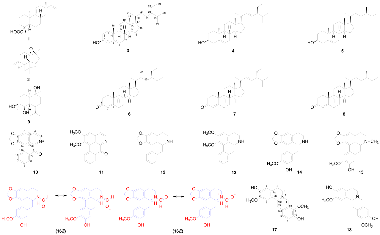
Figure 2.
Key HMBC (→) and NOESY (↔) correlations were observed for the previously undescribed aporphine alkaloid N-formylactinodaphnine (16), supporting the structural assignment of the Z (major) and E (minor) rotamers.
Figure 2.
Key HMBC (→) and NOESY (↔) correlations were observed for the previously undescribed aporphine alkaloid N-formylactinodaphnine (16), supporting the structural assignment of the Z (major) and E (minor) rotamers.
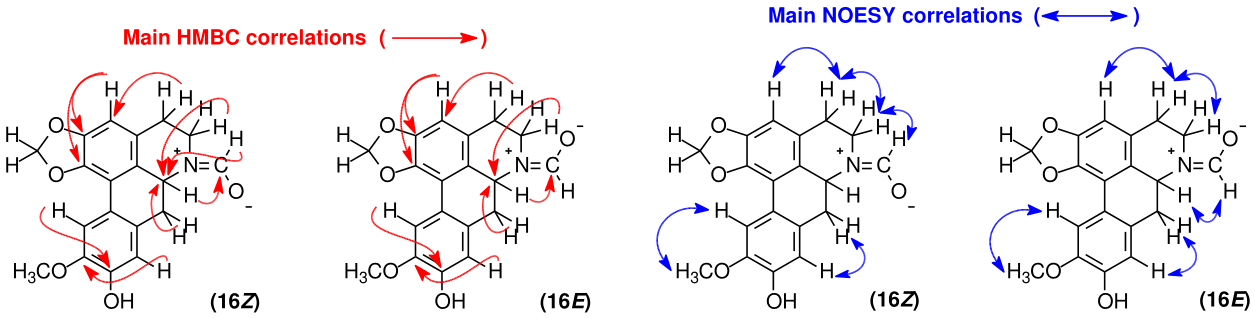

Table 1.
NMR spectroscopic data (500 MHz for ¹H and 125 MHz for ¹³C) for alkaloids 16 (N-formylactinodaphnine) and 15 (N-methylactinodaphnine) recorded in CDCl₃, and alkaloid 14 (actinodaphnine) recorded in CDCl₃ containing a drop of CD₃OD.
Table 1.
NMR spectroscopic data (500 MHz for ¹H and 125 MHz for ¹³C) for alkaloids 16 (N-formylactinodaphnine) and 15 (N-methylactinodaphnine) recorded in CDCl₃, and alkaloid 14 (actinodaphnine) recorded in CDCl₃ containing a drop of CD₃OD.
| Position | 16Z rotamer | 16E rotamer | 14 | 15 | ||||
|---|---|---|---|---|---|---|---|---|
| δC |
δH mult. (J in Hz) |
δC | δH mult. (J in Hz) | δC | δH mult. (J in Hz) | δC | δH mult. (J in Hz) | |
| 1 | 142.5 | - | 142.3 | - | 141.7 | - | 141.7 | - |
| 1a | 118.0 | - | 117.6 | - | 116.7 | - | 116.8 | - |
| 2 | 147.1 | - | 147.4 | - | 146.9 | - | 146.7 | - |
| 3 | 106.9 | 6.53 s | 107.3 | 6.56 s | 107.0 | 6.49 s | 106.6 | 6.51 s |
| 3a | 126.9 | - | 127.9 | - | 126.3 | - | 125.7 | - |
| 3b | 124.3 | - | 123.7 | - | 126.5 | - | 128.8 | - |
| 4 | 31.3 | 2.71 m 2.86 m |
30.0 | 2.68 m 2.77 m |
28.8 | 2.63 m 2.98 m |
28.8 | 2.63 m 3.15 m |
| 5 | 42.4 | 3.39 td (12.5; 2.7) 3.80 ddd (12.7; 4.5; 1.8) |
36.4 | 3.07 m 4.46 ddd (12.6; 4.5; 3.2) |
42.9 | 2.98 m 3.37 m |
53.3 | 2.56 m 3.09 m |
| 6a | 49.9 | 5.02 dd (13.9; 4.5) | 53.7 | 4.58 dd (14.6; 4.3) | 53.5 | 3.91 dd (14.0; 5.0) | 62.2 | 3.20 m |
| 7 | 33.2 | 2.73 t (13.9) 3.12 dd (13.9; 4.5) |
37.4 | 2.74 m 3.08 m |
35.8 | 2.69 dd (14.2; 14.0) 2.81 dd (14.2; 5.0) |
33.6 | 2.63 br d (14.2) 3.04 dd (14.2; 4.3) |
| 7a | 129.2 | - | 128.4 | - | 128.4 | - | 128.6 | - |
| 8 | 115.1 | 6.84 s | 114.6 | 6.82 s | 114.8 | 6.74 s | 114.3 | 6.81 s |
| 9 | 145.7 | - | 145.9 | - | 145.6 | - | 145.1 | - |
| 10 | 145.6 | - | 145.6 | - | 146.0 | - | 145.4 | - |
| 11 | 110.3 | 7.66 s | 110.37 | 7.66 s | 110.5 | 7.61 s | 119.0 | 7.63 s |
| 11a | 122.7 | - | 122.9 | - | 122.8 | - | 122.9 | - |
| 1-OCH2O-2 | 101.10 | 5.97 d (1.4) 6.10 d (1.4) |
101.19 | 5.98 d (1.4) 6.11 d (1.4) |
100.7 | 5.90 d (1.4) 6.05 d (1.4) |
100.7 | 5.92 d (1.5) 6.07 d (1.5) |
| 9-OH | - | -* | - | -* | - | -* | - | -* |
| 10-OCH3 | 56.40 | 3.93 s | 56.43 | 3.95 s | 56.2 | 3.88 s | 56.1 | 3.92 s |
| N-CHO | 162.30 | 8.25 br s | 162.35 | 8.38 br s | - | - | - | - |
| N-CH3 | - | - | - | - | - | - | 43.5 | 2.56 s |
Note: The data were obtained at 298 K with TMS as an internal reference (0.00 ppm) in CDCl3. (*) Phenolic OH was not observed because of exchange.
Table 2.
NMR spectroscopic data in CDCl3 plus CD3OD (600 MHz for 1H and 150 MHz for 13C) for alkaloid 10 (liriodenine) and CDCl3 plus CD3OD (500 MHz for 1H and 125 MHz for 13C) for alkaloids 17 (discretamine) and 18 (10-demethyldiscretine).
Table 2.
NMR spectroscopic data in CDCl3 plus CD3OD (600 MHz for 1H and 150 MHz for 13C) for alkaloid 10 (liriodenine) and CDCl3 plus CD3OD (500 MHz for 1H and 125 MHz for 13C) for alkaloids 17 (discretamine) and 18 (10-demethyldiscretine).
| Position | 10 | Position | 17 | 18 | |||
|---|---|---|---|---|---|---|---|
| δC |
δH mult. (J in Hz) |
δC |
δH mult. (J in Hz) |
δC | δH mult. (J in Hz) | ||
| 1 | 148.4 | 1 | 108.2 | 6.71 s | 107.7 | 6.71 s | |
| 1a | 107.9 | 2 | 145.8 | - | 145.2 | - | |
| 2 | 152.1 | 3 | 144.5 | - | 144.0 | - | |
| 3 | 103.4 | 7.15 s | 4 | 114.7 | - | 114.3 | 6.67 s |
| 3a | 136.1 | 4a | 125.8 | - | 127.4 | - | |
| 3b | 123.3 | 5 | 28.2 | 2.67 m 3.10 m |
28.7 | 2.63 m 3.10 m |
|
| 4 | 124.7 | 7.76 d (5.1) | 6 | 51.4 | 2.68 m 3.22 m |
51.3 | 2.58 m 3.11 m |
| 5 | 144.4 | 8.77 d (5.1) | 7 | - | - | - | - |
| 6a | 144.9 | - | 8 | 53.7 | 3.60 d (15.6) 4.23 d (15.6) |
58.0 | 3.63 d (14.7) 3.90 d (14.7) |
| 7 | 182.6 | - | 8a | 127.3 | - | 126.9 | - |
| 7a | 131.1 | - | 9 | 143.6 | - | 111.8 | 6.633 s |
| 8 | 128.6 | 8.49 dd (7.8; 1.4) | 10 | 147.1 | - | 143.8 | - |
| 9 | 128.7 | 7.55 ddd (7.8; 7.2; 1.0) | 11 | 115.2 | 6.77 d (8.2) | 145.3 | - |
| 10 | 134.2 | 7.72 ddd (8.2; 7.2; 1.4) | 12 | 124.6 | 6.80 d (8.2) | 110.7 | 6.638 s |
| 11 | 127.5 | 8.57 d (8.2; 0.5) | 12a | 126.2 | - | 125.5 | - |
| 11a | 133.0 | - | 13 | 35.6 | 2.84 dd (15.8; 11.6) 3.28 dd (15.8; 3.8) |
36.4 | 2.83 dd (15.7; 11.3) 3.22 dd (15.7; 3.7) |
| 1-OCH2O-2 | 102.8 | 6.37 s | 13a | 59.5 | 3.64 dd (11.6; 3.8) | 59.7 | 3.57 dd (11.3; 3.7) |
| 13b | 128.3 | - | 129.1 | - | |||
| 2-OCH3 | 56.1 | 3.88 s | 56.0 | 3.89 s | |||
| 3-OH | - | -* | -* | ||||
| 9-OCH3 | 60.3 | 3.82 s | - | - | |||
| 10-OH | - | -* | - | -* | |||
| 11-OCH3 | - | - | 55.9 | 3.86 s | |||
Note: The data were obtained at 298 K with TMS as an internal reference (0.00 ppm) in CDCl3 plus CD3OD. (*) Phenolic OH was not observed because of exchange.
Table 3.
Cytotoxic screening of extracts and phases from A. amazonica bark against human tumor cell lines.
Table 3.
Cytotoxic screening of extracts and phases from A. amazonica bark against human tumor cell lines.
| Sample | Growth inhibition % | |
|---|---|---|
| HepG2 | HCT116 | |
| Hexane extract | 4.84±0.79 | 16.00±1.83 |
| Methanol extract | 1.11±0.61 | 0.78±0.18 |
| Neutral chloroform phase | 5.43±0.24 | 0.28±0.03 |
| Alkaloid-rich chloroform phase | 55.81±1.53 | 63.32±2.79 |
| Doxorubicin* | 81.17±0.85 | 83.74±4.08 |
Note: Data are expressed as the percentage of growth inhibition (%GI) ± standard error of the mean (SEM) from two independent experiments performed in triplicate, as determined by the Alamar blue assay after 72 h of exposure to HepG2 (human hepatocellular carcinoma) and HCT116 (human colorectal carcinoma) cells. Doxorubicin was used as the positive control. * Doxorubicin was tested at a single concentration of 25 μg/mL.
Table 4.
Cytotoxic activity of the isolated compounds from the bark of A. amazonica.
| Compounds | IC50, in µg mL−1 (µmol/L), and 95% CIa | |||||
|---|---|---|---|---|---|---|
| HepG2 | HCT116 | MDA MB 231 | MCF-7 | U-87 MG | MRC-5 | |
| Acanthoic acid (1) | 14.63 (48.37) 10.67—20.07 |
21.25 (70.26) 13.25—34.10 |
>25 (82.66) | >25 (82.66) | >25 (82.66) | >25 (82.66) |
| Actinodaphnine (14) | 7.21 (23.16) 6.10—8.53 |
5.59 (17.96) 4.34—7.21 |
8.12 (26.08) 5.93—11.10 |
11.73 (37.68) 9.01—15.26 |
10.92 (35.07) 8.99—13.25 |
6.84 (21.07) 6.23—8.39 |
| N-Methylactinodaphnine (15) | NT | 4.27 (13.12) 3.27—5.59 |
NT | NT | NT | 12.16 (37.37) 8.62—17.17 |
| N-Formylactinodaphnine (16) | >25 (73.67) | 21.07 (62.09) 12.75—34.84 |
22.69 (66.86) 17.79—26.04 |
>25 (73.67) | >25 (73.67) | >25 (73.67) |
| Discretamine (17) | >25 (76.36) | >25 (76.36) | >25 (76.36) | >25 (76.36) | >25 (76.36) | >25 (76.36) |
| 10-Demethyldiscretine (18) | >25 (76.36) | 22.08 (67.45) 11.16—33.67 |
>25 (76.36) | >25 (76.36) | >25 (76.36) | >25 (76.36) |
| Doxorubicinb | 0.43 (0.79) 0.37—0.50 |
0.57 (1.05) 0.39—0.85 |
0.62 (1.14) 0.37—1.02 |
0.36 (0.66) 0.20—0.63 |
0.66 (1,21) 0.32—1.34 |
1.96 (3.61) 1.37—2.80 |
Note: a Data are presented as IC50 values in µg mL−1 (μmol L−1) and their 95% confidence intervals (CIs) obtained by nonlinear regression from three independent experiments performed in duplicate, measured via the Alamar blue assay after 72 h of incubation. The cancer cell lines used were HepG2 (human hepatocellular carcinoma), HCT116 (human colorectal carcinoma), MDA MB 231 (human breast carcinoma), MCF-7 (human breast adenocarcinoma), U-87 MG (human glioblastoma) cells. Noncancerous MRC-5 cells (human lung fibroblasts) were used. b Doxorubicin was used as a positive control. N.T: not tested because of insufficient sample amount.
Disclaimer/Publisher’s Note: The statements, opinions and data contained in all publications are solely those of the individual author(s) and contributor(s) and not of MDPI and/or the editor(s). MDPI and/or the editor(s) disclaim responsibility for any injury to people or property resulting from any ideas, methods, instructions or products referred to in the content. |
© 2026 by the authors. Licensee MDPI, Basel, Switzerland. This article is an open access article distributed under the terms and conditions of the Creative Commons Attribution (CC BY) license (http://creativecommons.org/licenses/by/4.0/).
Copyright: This open access article is published under a Creative Commons CC BY 4.0 license, which permit the free download, distribution, and reuse, provided that the author and preprint are cited in any reuse.