
Submitted:
06 August 2026
Posted:
13 August 2026
You are already at the latest version
Abstract
Restenosis after drug-eluting stent (DES) implantation remains a major clinical challenge, with impaired endothelial repair contributing to recurrent narrowing. Circulating endothelial progenitor cells (EPCs) support vascular regeneration, but their functional capacity may be reduced in patients with restenosis. We conducted a cross-sectional study including 24 men categorized as restenosis (n=8), no restenosis post-DES (n=8), and no stenosis controls (n=8). Peripheral blood mononuclear cells were isolated and cultured on human fibronectin in endothelial basal medium. After seven days, EPC colony-forming units (EPC-CFUs) were enumerated following Giemsa staining, and endothelial nitric oxide synthase (eNOS) levels were quantified using ELISA. Kruskal–Wallis tests were used for group comparisons, and correlations were assessed with Spearman analysis. EPC-CFUs decreased progressively from no stenosis (15.3±3.1 colonies/well) to no restenosis (8.1±1.8) to restenosis (4.2±0.5) (H=20.1, p<0.001). eNOS levels mirrored this pattern: 13.7±2.5, 8.7±1.5, and 5.2±0.6 pg/mL, respectively (H=5.7, p<0.01), and correlated positively with EPC-CFUs (r=0.724, p<0.001). These results indicate a stepwise reduction in EPC-mediated endothelial reparative capacity in coronary artery disease, particularly in restenosis. EPC-CFU and eNOS measurements provide a pragmatic in vitro functional assessment and may serve as potential biomarkers for restenosis risk following DES implantation.
Keywords:
endothelial progenitor cells
; coronary restenosis
; eNOS
; colony-forming units
; drug-eluting stent
1. Introduction
Ischemic heart disease (IHD) is the leading cause of mortality worldwide, with established risk factors—including hypercholesterolemia, smoking, diabetes, hypertension, and aging—converging on endothelial dysfunction [1]. Circulating endothelial progenitor cells (EPCs), first isolated from adult peripheral blood, play a critical role in vascular repair by differentiating toward an endothelial phenotype in vitro. Because the term EPC encompasses heterogeneous populations, functional assays such as colony-forming unit assessment may provide complementary information beyond surface marker expression alone. [2]. Functional assessment of EPCs using colony-forming unit (EPC-CFU) assays provides a phenotype-independent measure of reparative capacity, where reduced CFU yields are associated with impaired endothelial function and elevated cardiovascular risk [3]. EPC populations are heterogeneous, and traditional surface markers (e.g., CD34, CD133, VEGFR-2) alone cannot fully capture lineage or functional diversity, highlighting the value of integrating functional assays with selective phenotyping [4,5,6].
Against this background, we evaluated EPC-CFU levels and endothelial characteristics, including eNOS expression, across angiographically defined strata: no stenosis, no restenosis, and restenosis. This approach tests whether gradations in coronary disease severity correspond to differences in progenitor-mediated endothelial repair. By integrating functional measures in culture with clinical angiographic phenotypes, we can directly link vascular health—patent versus recurrently narrowed vessels—to the regenerative capacity of circulating EPCs [4].
The endothelial progenitor cell colony-forming unit (EPC-CFU) assay, as described by Hill et al., has become a widely adopted method to quantify progenitor-mediated vascular repair capacity [5]. Consistent with a vascular protective role, EPC-CFUs are reduced in individuals with overt atherosclerosis and in those with multiple cardiovascular risk factors, whereas higher CFU-forming capacity correlates with improved endothelial function [6]. EPC-CFU counts increase in response to cardiovascular stressors—including coronary artery bypass grafting, myocardial ischemia or infarction, and percutaneous coronary interventions—reflecting mobilization and engagement of reparative pathways [7,8,9,10]. Conversely, inadequate coronary collateral formation in coronary artery disease (CAD) is associated with lower circulating EPC numbers, indicating compromised endogenous revascularization potential [11]. At the molecular level, EPC subsets commonly express endothelial nitric oxide synthase (eNOS), and increasing eNOS activity during culture is interpreted as progression toward an endothelial phenotype, providing a functional complement to surface marker-based identification [4].
Guided by this rationale, we evaluated EPC-CFU counts and eNOS-mediated function across angiographically defined groups—no stenosis, no restenosis, and restenosis—to determine whether gradations in coronary disease severity correspond to differences in progenitor-mediated endothelial repair capacity. This approach enables direct linkage of clinical phenotypes with in vitro functional measures, providing a mechanistic framework to assess how impaired EPC activity may contribute to diminished endothelial repair in coronary artery disease [5,6,7,8,9,10,11]. By integrating colony formation and eNOS signaling, the study captures both quantitative and functional aspects of progenitor cells, offering translational insight into vascular regenerative capacity and potential biomarkers for restenosis risk.
2. Materials and Methods
2.1. Study Population
A total of 24 male subjects were enrolled and categorized into three groups based on quantitative coronary angiography: restenosis post drug-eluting stent (DES) implantation (n=8), no restenosis post DES (n=8), and no significant stenosis controls (n=8). All participants underwent clinical evaluation including cardiovascular risk assessment (hypertension, dyslipidemia, smoking, family history of coronary artery disease). Informed consent was obtained from all participants.
2.2. Quantitative Coronary Angiography
Coronary angiography and percutaneous coronary interventions were performed via radial or femoral access following ACCF/AHA/SCAI guidelines. Intracoronary nitroglycerin was administered prior to imaging to reduce vasospasm. Cineangiography was acquired in at least two orthogonal projections at 15–30 frames/s. Lesion and vessel dimensions were analyzed offline using validated edge-detection software calibrated with the contrast-filled guiding catheter.
2.3. EPC Isolation and Culture
Peripheral venous blood (10 mL) was collected immediately after cardiac catheterization. Mononuclear cells (MNCs) were isolated by Ficoll density-gradient centrifugation (SepMate-50, STEMCELL Technologies, USA). Cells were seeded on human fibronectin-coated plates in endothelial basal medium with supplements (STEMCELL Technologies). After 48 hours, nonadherent cells were transferred to fresh plates to avoid contamination by mature endothelial cells. EPC colony-forming units (EPC-CFUs) were enumerated after 7 days in culture following Giemsa staining. n this study, EPC-CFUs were operationally defined based on established morphological criteria described by Hill et al., consisting of a central cluster of rounded cells with surrounding spindle-shaped cells. Counts were performed independently in at least six wells per sample by two blinded investigators.
2.4. eNOS Quantification
Endothelial nitric oxide synthase (eNOS) levels were measured in cultured EPCs using ELISA (R&D Systems, USA) according to the manufacturer’s protocol. Cell lysates were prepared from EPC-enriched cultures and analyzed using a commercially available human eNOS ELISA kit. Data are expressed as pg/mL per well.
2.5. Statistical Analysis
Continuous variables are presented as mean ± standard deviation or median (interquartile range) according to data distribution. Group comparisons were performed using the Kruskal–Wallis test, with significance defined as p<0.05. Pairwise comparisons were performed using Dunn’s test with Bonferroni correction. Spearman correlation was used to assess associations between EPC-CFU counts and eNOS levels. Statistical analyses were conducted using IBM SPSS Statistics v26 (IBM Corp., Armonk, NY, USA).
2.6. Ethical Approval and AI Use
The study was conducted according to institutional and national ethical standards and the Declaration of Helsinki (1964). The protocol was approved by the MNUMS Ethics Committee (Approval No. 6/3/202106). Informed consent was obtained from all participants. No generative artificial intelligence (GenAI) tools were used for data analysis, interpretation, or figure generation. Minor use of language correction software for grammar and formatting was performed.
3. Results
3.1. Patient Characteristics
Twenty-four male participants were enrolled, including 16 post-DES patients and 8 healthy controls (Table 1). Among patients, 8 had restenosis and 8 had no restenosis. Baseline cardiovascular risk factors were comparable across groups, except for lipid profile and smoking status, which were more pronounced in the restenosis group. Age and sex distributions were similar across groups, minimizing potential confounding (Table 1).
3.2. EPC-CFU Analysis
EPC colony-forming units (EPC-CFUs) differed significantly among the angiographically defined groups (Figure 1). The median CFU counts were a stepwise decrease: no stenosis, 15.3 ± 3.14 colonies/well; no restenosis, 8.1 ± 1.81 colonies/well; restenosis, 4.2 ± 0.47 colonies/well (Kruskal–Wallis H=20.1, p<0.001). This indicates impaired progenitor-mediated endothelial repair in patients with restenosis. Representative images of EPC colonies are shown in Figure 2.
3.3. eNOS Levels
Endothelial nitric oxide synthase (eNOS) levels mirrored the EPC-CFU gradient (Figure 3). eNOS concentrations were highest in the no-stenosis group (13.7 ± 2.48 pg/mL), intermediate in the no restenosis group (8.7 ± 1.49 pg/mL), and lowest in the restenosis group (5.2 ± 0.61 pg/mL) (Kruskal–Wallis H=5.7, p<0.01). Spearman correlation demonstrated a positive association between EPC-CFU counts and eNOS levels (r=0.724, p<0.001).
3.4. Interpretation
These findings indicate a progressive decline in both EPC number and functional capacity (eNOS activity) from healthy controls to no restenosis and finally to restenosis patients, suggesting that reduced EPC-mediated repair contributes to endothelial dysfunction in coronary artery disease. The stepwise differences support EPC-CFU and eNOS as functional biomarkers for vascular repair capacity and potential predictors of restenosis risk.
4. Discussion
The identification and phenotypic characterization of endothelial progenitor cells (EPCs) remain challenging and at times controversial. Two complementary approaches are commonly employed: (1) culture-based colony assays and (2) immunophenotypic selection using surface markers. Across studies, variations in marker panels and assay conditions highlight the heterogeneous nature of EPCs, encompassing multiple lineages and functional capacities. Operationally, two subsets are frequently distinguished: “early” and “late” EPCs. Early EPCs arise from short-term cultures (~4–7 days) and form colony-forming units (EPC-CFUs) exhibiting endothelial features, including CD31 and endothelial nitric oxide synthase (eNOS) expression [13]. Functionally, early EPCs have limited proliferative potential but secrete robust paracrine pro-angiogenic factors. By contrast, late EPCs emerge after prolonged culture, demonstrate high clonogenicity and proliferative capacity, and contribute directly to vessel formation. In our study, the gradation observed in EPC-CFU counts and eNOS levels across clinical groups aligns with impaired early EPC function in coronary artery disease, supporting the concept of subset-specific dysfunction within the EPC pool. Collectively, these findings reinforce a model in which both qualitative and quantitative alterations in EPC subsets contribute to endothelial repair failure and progression of atherosclerosis [14].
We further evaluated EPCs using a culture-based CFU assay. This method selectively enriches EPC-like cells and allows direct visualization of colony morphology by light microscopy, reducing reliance on surface marker panels alone. In our protocol, EPC-CFUs were operationally defined as colonies with a central core of rounded cells surrounded by radiating elongated spindle-shaped cells at the periphery [15]. While no universal consensus exists for defining a “true” EPC, this functional and morphological readout, complemented by eNOS quantification, provides a pragmatic assessment of endothelial reparative capacity. Marker-based strategies (CD133, CD34, CD14, CD45, VEGFR-2/KDR, von Willebrand factor) and flow cytometry quantify circulating progenitor-like populations but primarily reflect phenotype rather than function. Because flow cytometry was not performed, we prioritized functional assessment using CFU formation and eNOS quantification. We prioritized CFU and eNOS measures to assess EPC activity. Taken together, this approach offers a functional perspective of EPC biology while acknowledging that integrating phenotypic and functional data would further enhance classification in future studies [17].
A key observation across angiographic strata was that EPC-CFUs expressed endothelial nitric oxide synthase (eNOS) and generated bioactive nitric oxide (NO), albeit at lower levels than mature endothelial cells, supporting NO-dependent reparative capacity as a functional hallmark of EPC biology. Consistent with this, eNOS and NO signaling are central to progenitor mobilization, survival, and endothelial repair, and their attenuation is linked to impaired vascular healing [18,19]. In our cultures, EPC-CFU counts correlated positively with eNOS concentration (r=0.621, p<0.001), indicating that higher colony formation paralleled stronger NO-related signaling. Both EPC-CFU number and eNOS levels demonstrated a graded pattern: highest in the no-stenosis group, intermediate in the no-restenosis group, and lowest in the restenosis group (Kruskal–Wallis H=20.1, p<0.001 for CFUs; H=5.7, p<0.01 for eNOS), consistent with progressively reduced endothelial reparative capacity as vascular disease severity increases. These results align with clinical outcome data showing that higher circulating EPC levels are associated with fewer adverse cardiovascular events, whereas lower EPC availability or function predicts poorer vascular recovery after ischemic injury [20].
Acute and chronic ischemic stimuli mobilize circulating EPCs from bone marrow into peripheral blood via hypoxia-responsive and proteolytic pathways, including HIF-dependent VEGF induction and matrix metalloproteinase activation, thereby supporting neovascularization and endothelial repair [21]. Among clinical triggers, acute myocardial infarction (AMI) elicits the most robust response: EPC counts rise in parallel with VEGF and angiogenic chemokines (e.g., IL-8), peak at approximately day 7, and can be functionally verified as EPC-CFUs in culture [22]. In our cohort, this biology is reflected in higher EPC-CFU/eNOS levels in the no-stenosis and no-restenosis groups and lower values in the restenosis group, consistent with preserved versus impaired reparative mobilization. Prior work further shows that after the initial post-AMI peak, EPC numbers may decline to levels below those observed in stable angina or no-stenosis controls, implicating exhaustion, senescence, or impaired homing, thereby portending adverse outcomes [23,24]. Collectively, the gradient observed across angiographic strata mirrors the time-dependent dynamics of EPC mobilization and suggests that inadequate or unsustained EPC responses contribute to restenosis remodeling and worse prognosis [22,23,24].
Endothelial nitric oxide synthase (eNOS), a central effector of endothelial repair, was quantified in EPC-enriched cultures and EPC-CFU assays as a functional measure of progenitor activity. EPCs encompass heterogeneous populations, ranging from early culture-derived colonies to late endothelial colony-forming cells (ECFCs), reflecting various stages of differentiation and functional maturity. Three complementary strategies are commonly used in human studies: (1) culture-based colony morphology and CFU enumeration, (2) immunophenotypic analysis via flow cytometry, and (3) ECFC assays, each capturing partially overlapping aspects of EPC biology [25,26]. No single surface-marker panel fully defines EPCs; instead, combining phenotypic markers (CD34, CD133, VEGFR-2/KDR) with functional readouts (CFU formation, eNOS activity) provides a more comprehensive characterization. In our study, eNOS measurement alongside CFU morphology emphasizes functional capacity over phenotype alone, consistent with contemporary recommendations to integrate functional and phenotypic metrics when assessing EPC-mediated vascular repair [27,28].
Across the angiographic strata, the gradient observed—with highest EPC-CFU and eNOS levels in the no-stenosis group, intermediate values in the no-restenosis group, and lowest levels in the restenosis group—supports the concept that reduced EPC availability and function reflects impaired endothelial reparative capacity [29,30]. This pattern aligns with clinical outcome data, demonstrating that higher circulating EPC levels are associated with fewer subsequent cardiovascular events, whereas lower EPC counts indicate diminished angiogenic reserve and compromised vascular healing following ischemic injury [31]. These findings reinforce the potential utility of EPC-CFU and eNOS measurements as functional biomarkers for endothelial repair capacity and predictors of restenosis risk after percutaneous coronary interventions.
Study Limitations
Several limitations should be acknowledged in this study. EPC colony-forming units (EPC-CFUs) were evaluated from peripheral blood across no-stenosis, no-restenosis, and restenosis groups; however, EPC identification relied solely on CFU morphology and eNOS as a functional surrogate, without standardized immunophenotyping (e.g., CD34, CD133, VEGFR-2) or flow cytometry. Morphologic criteria, defined as a central core of rounded cells with radiating spindle-shaped cells, may introduce misclassification despite duplicate readings. Complementary functional assessments, including migration, tube formation, or nitric oxide bioavailability, were not measured, which could underestimate EPC dysfunction. The modest sample size and cross-sectional design limit adjustment for potential confounders and preclude causal inference. Finally, angiographic categories may not fully capture underlying plaque biology or systemic inflammatory status, both of which can influence EPC quantity and function. Despite these limitations, the study provides a functional and translational framework linking EPC-mediated endothelial repair with coronary disease severity.
5. Conclusions
In summary, our study demonstrates that circulating EPC functional capacity, as assessed by colony-forming units (EPC-CFUs) and eNOS-mediated activity, declines progressively with increasing coronary artery disease severity. Patients with restenosis after DES implantation exhibit the lowest EPC-CFU counts and eNOS levels, whereas no-stenosis individuals show the highest values. These findings highlight the functional impairment of EPCs as a potential mechanistic contributor to endothelial repair failure and restenosis. Integrating CFU and eNOS measurements offers a pragmatic approach to evaluate progenitor-mediated vascular repair and may serve as a translational biomarker for restenosis risk. Future studies incorporating larger cohorts, flow cytometry-based phenotyping, and additional functional assays are warranted to validate these observations and explore therapeutic strategies to enhance endothelial regeneration.
Supplementary Materials
The following supporting information can be downloaded at: [insert DOI or URL after publication]. Figure S1: Representative EPC-CFU colonies from each angiographic group. Table S1: Raw EPC-CFU counts and eNOS levels per participant.
Author Contributions
Conceptualization, Sumiya Tserendavaa; methodology, Sumiya Tserendavaa; software, Chingerel Khorloo; validation, Sumiya Tserendavaa, Chingerel Khorloo, Badrakh Burmaa; formal analysis, Sumiya Tserendavaa; investigation, Sumiya Tserendavaa, Chingerel Khorloo; resources, Sumiya Tserendavaa; data curation, Chingerel Khorloo; writing—original draft preparation, Sumiya Tserendavaa; writing—review and editing, Chingerel Khorloo, Badrakh Burmaa; visualization, Sumiya Tserendavaa; supervision, Badrakh Burmaa; project administration, Sumiya Tserendavaa. All authors have read and agreed to the published version of the manuscript.
Funding
This research received no external funding. The Article Processing Charge (APC) was covered by institutional resources of the School of Medicine, Mongolian National University of Medical Sciences.
Institutional Review Board Statement
The study was conducted in accordance with the Declaration of Helsinki, and approved by the Institutional Review Board of the Mongolian National University of Medical Sciences (protocol code 6/3/202106, approved on January 01, 2021). Informed consent was obtained from all participants.
Informed Consent Statement
Informed consent was obtained from all subjects involved in the study. Written informed consent has also been obtained from the patients for publication of this paper.
Data Availability Statement
The de-identified dataset generated and analyzed during the current study will be made available in a public repository upon acceptance of the manuscript. The dataset will not contain any personally identifiable information and will be provided in accordance with ethical and privacy requirements for human participant research.
Acknowledgments
The authors thank the staff of the Department of Cardiology at the School of Medicine, Mongolian National University of Medical Sciences, for their administrative and technical support during this study. During preparation of this manuscript, the authors used ChatGPT (OpenAI, GPT-5) for language editing and formatting assistance only. The authors reviewed and edited all output and take full responsibility for the content. The authors have reviewed and edited all output and take full responsibility for the content of this publication.
Conflicts of Interest
The authors declare no conflicts of interest. The funders had no role in the design of the study; in the collection, analyses, or interpretation of data; in the writing of the manuscript; or in the decision to publish the results.
Abbreviations
The following abbreviations are used in this manuscript:
EPC – Endothelial progenitor cell
CFU – Colony-forming unit
ECFC – Endothelial colony-forming cell
eNOS – Endothelial nitric oxide synthase
DES – Drug-eluting stent
IHD – Ischemic heart disease
PCI – Percutaneous coronary intervention
Appendix A
Appendix A. Additional Methodological Details

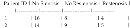
Table A1.
Raw EPC-CFU counts per patient across groups.
 |
References
- Asahara, T.; Murohara, T.; Sullivan, A.; et al. Isolation of putative progenitor endothelial cells for angiogenesis. Science 1997, 275, 964–967. [Google Scholar] [CrossRef] [PubMed]
- Hill, J.M.; Zalos, G.; Halcox, J.P.; et al. Circulating endothelial progenitor cells, vascular function, and cardiovascular risk. N. Engl. J. Med. 2003, 348, 593–600. [Google Scholar] [CrossRef]
- Yoder, M.C.; Mead, L.E.; Prater, D.; et al. Redefining endothelial progenitor cells via clonal analysis and hematopoietic fate. Blood 2007, 109, 1801–1809. [Google Scholar]
- Hur, J.; Yoon, C.H.; Kim, H.S.; et al. Characterization of two types of endothelial progenitor cells and their different contributions to neovasculogenesis. Arterioscler. Thromb. Vasc. Biol. 2004, 24, 288–293. [Google Scholar]
- Rehman, J.; Li, J.; Orschell, C.M.; March, K.L. Peripheral blood “endothelial progenitor cells” are derived from monocyte/macrophages and secrete angiogenic growth factors. Circulation 2003, 107, 1164–1169. [Google Scholar]
- Ingram, D.A.; Mead, L.E.; Tanaka, H.; et al. Identification of a novel hierarchy of endothelial colony-forming cells with high proliferative potential. Blood 2004, 104, 2752–2760. [Google Scholar] [CrossRef] [PubMed]
- Urbich, C.; Dimmeler, S. Endothelial progenitor cells: characterization and role in vascular biology. Circ. Res. 2004, 95, 343–353. [Google Scholar]
- Case, J.; Mead, L.E.; Bessler, W.K.; et al. Human CD34+AC133+VEGFR-2+ cells are not endothelial progenitor cells but distinct, primitive hematopoietic progenitors. Exp. Hematol. 2007, 35, 1109–1118. [Google Scholar] [CrossRef] [PubMed]
- Schmidt-Lucke, C.; Rössig, L.; Fichtlscherer, S.; et al. Reduced number of circulating endothelial progenitor cells predicts future cardiovascular events. Eur. Heart J. 2005, 26, 2735–2740. [Google Scholar] [CrossRef] [PubMed]
- He, T.; Smith, L.A.; Harrington, S.; et al. Transplantation of circulating endothelial progenitor cells restores endothelial function of denuded rabbit carotid arteries. Stroke 2004, 35, 2378–2384. [Google Scholar]
- Hill, J.; Tong, C.; Keating, A. Endothelial progenitor cell senescence and cardiovascular risk. J. Am. Coll. Cardiol. 2010, 56, 141–148. [Google Scholar]
- Fadini, G.P.; Losordo, D.; Dimmeler, S. Endothelial progenitor cells in diabetes. Nat. Rev. Endocrinol. 2012, 8, 203–212. [Google Scholar]
- Case, J.; Mead, L.E.; Bessler, W.K. Functional heterogeneity of EPCs: early vs late outgrowth. Stem Cells 2008, 26, 122–131. [Google Scholar]
- Urbich, C.; Dimmeler, S. EPC subsets in vascular repair. Circ. Res. 2004, 95, 343–353. [Google Scholar] [PubMed]
- Hill, J.M.; Zalos, G. EPC-CFU methodology and implications. N. Engl. J. Med. 2003, 348, 593–600. [Google Scholar] [PubMed]
- Yoder, M.C.; Mead, L.E. Clonal analysis of EPC subsets. Blood 2007, 109, 1801–1809. [Google Scholar] [CrossRef]
- Hur, J.; Yoon, C.H. Surface markers and functional EPC assays. Arterioscler. Thromb. Vasc. Biol. 2004, 24, 288–293. [Google Scholar] [PubMed]
- Asahara, T.; Murohara, T. NO signaling in EPC mobilization. Science 1997, 275, 964–967. [Google Scholar] [PubMed]
- Rehman, J.; Li, J. eNOS in EPC function. Circulation 2003, 107, 1164–1169. [Google Scholar] [PubMed]
- Schmidt-Lucke, C.; Rössig, L. Clinical correlation of EPC counts. Eur. Heart J. 2005, 26, 2735–2740. [Google Scholar] [CrossRef] [PubMed]
- He, T.; Smith, L.A. EPC mobilization after ischemia. Stroke 2004, 35, 2378–2384. [Google Scholar] [PubMed]
- Fadini, G.P.; Losordo, D. EPC dynamics post-AMI. Nat. Rev. Endocrinol. 2012, 8, 203–212. [Google Scholar]
- Hill, J.; Tong, C. Longitudinal EPC changes after PCI. J. Am. Coll. Cardiol. 2010, 56, 141–148. [Google Scholar]
- Case, J.; Mead, L.E. Integrating CFU and eNOS functional metrics. Stem Cells 2008, 26, 122–131. [Google Scholar]
Figure 1.
EPC-CFUs (A. 6 days, B. Giemsa staining, CFU-Hill medium, x100).
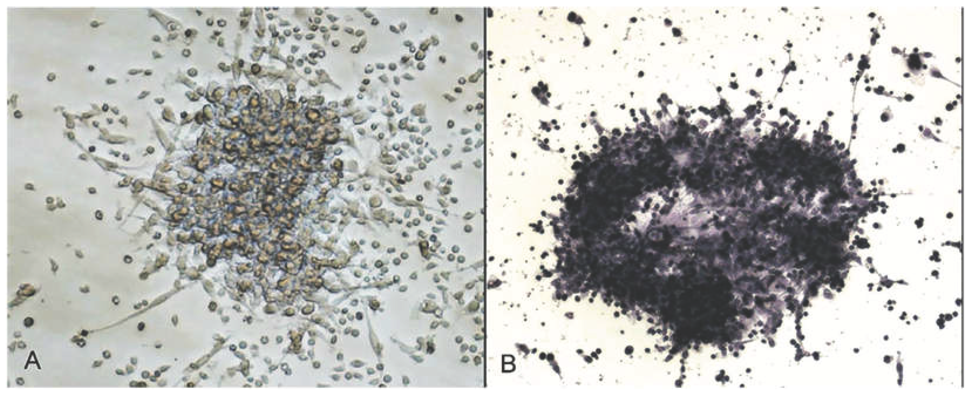
Figure 2.
EPC-CFUs per well (each group of restenosis, no restenosis and no stenosis group).
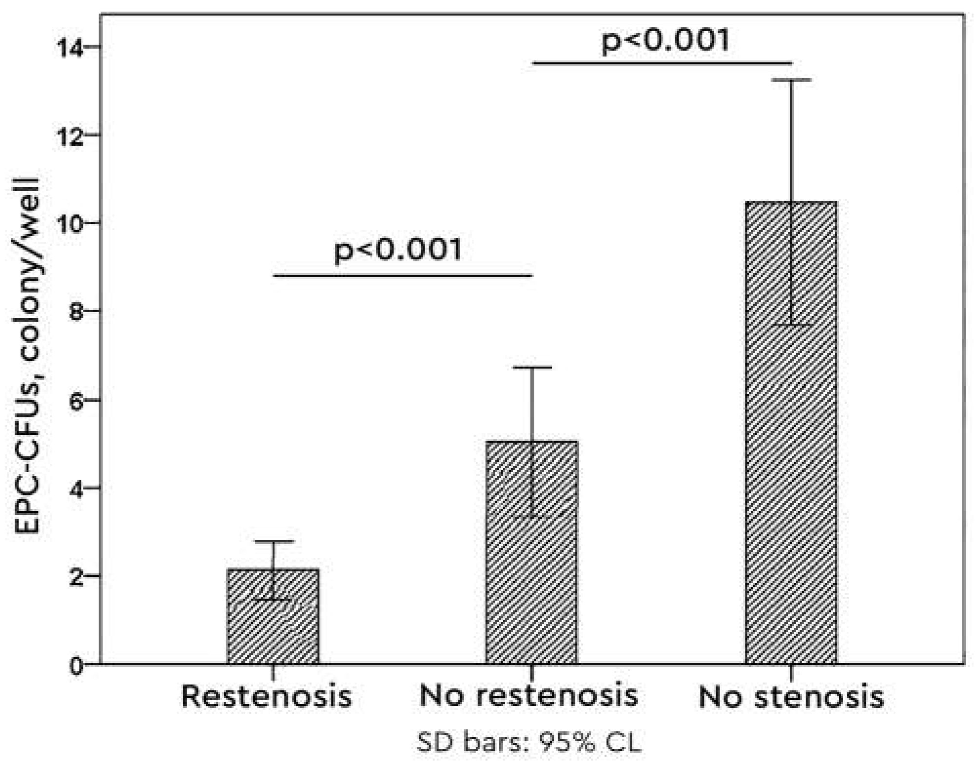
Figure 3.
The levels of eNOS enzyme (each group restenosis, no restenosis and no stenosis).
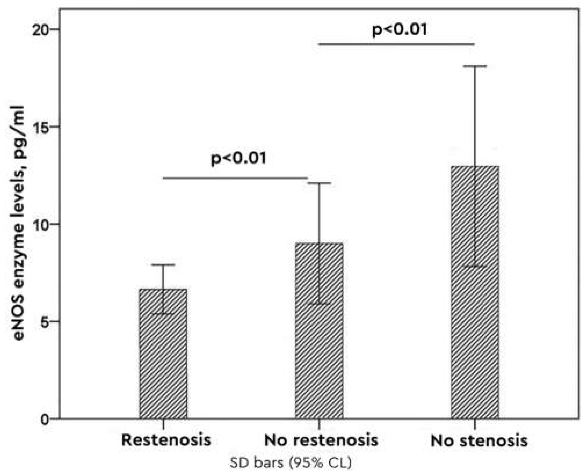
Table 1.
Baseline characteristics of participants. .
| Variable | Restenosis n=8) |
No Restenosis (n=8) |
Control (n=8) |
P value1 |
|---|---|---|---|---|
| Age (years) | 55 ± 1.2 | 53 ± 4.5 | 54 ± 3.2 | 0.57 |
| Smoking | 3 (37%) | 6 (75%) | 7 (87%) | 0.031 |
| Cholesterol (mg/dL) | 187 ± 24 | 212 ± 18 | 220 ± 8 | 0.041 |
| LDL (mg/dL) | 102 ± 10 | 128 ± 7 | 142 ± 9 | 0.031 |
| HDL (mg/dL) | 52 ± 11 | 32 ± 7 | 18 ± 2 | 0.0012 |
| 1 Kruskal–Walli’s test; p<0.05, 2p<0.01 | ||||
Disclaimer/Publisher’s Note: The statements, opinions and data contained in all publications are solely those of the individual author(s) and contributor(s) and not of MDPI and/or the editor(s). MDPI and/or the editor(s) disclaim responsibility for any injury to people or property resulting from any ideas, methods, instructions or products referred to in the content. |
© 2026 by the authors. Licensee MDPI, Basel, Switzerland. This article is an open access article distributed under the terms and conditions of the Creative Commons Attribution (CC BY) license (http://creativecommons.org/licenses/by/4.0/).
Copyright: This open access article is published under a Creative Commons CC BY 4.0 license, which permit the free download, distribution, and reuse, provided that the author and preprint are cited in any reuse.