

Submitted:
12 August 2026
Posted:
13 August 2026
You are already at the latest version
Abstract
Background: Sequential cytokine conditioning of human peripheral blood mononuclear cell (PBMC)-derived cultures can generate heterogeneous extracellular outputs whose biological identity is easily oversimplified. Methods: We retrospectively integrated archived records from an IL-4/GM-CSF-supported differentiation sequence, subsequent IL-10/TGF-β1 conditioning, and recovery of a multicomponent extracellular preparation. Each analytical domain retained its documented experimental unit and was linked within a source-to-output framework. Results: One represented Annexin V/7-AAD output comprised 26,071 events with 99.66% viability. Archived cell-associated regions were integrated descriptively. The recovered preparation showed a heterogeneous particle-associated signal predominantly at approximately 170–240 nm together with reported detection of CD9, CD63, CD81, and TSG101. An aggregate summary of ten conditioned cultures reported mean extracellular values of 25,320.16 pg/mL IL-10, 659.35 pg/mL IL-4, 99.61 pg/mL IL-6, and 3.01 pg/mL TNF-α; IL-10 and IL-4 were conditioning inputs and were therefore interpreted as final-environment attributes. Conclusions: The study establishes a traceable source-to-output framework for a human PBMC-derived multicomponent extracellular preparation, preserves a biogenesis-neutral identity for its vesicular and non-vesicular components, and provides a transferable evidence-to-claim pathway for tolerance-oriented biologic development.
Keywords:
regulatory conditioning
; human PBMC-derived culture
; multicomponent extracellular preparation
; tolerance-oriented development
; interleukin-10
; extracellular-vesicle-associated components
; membrane microdomains
; source-to-output characterization
1. Introduction
Dendritic cells and related antigen-presenting-cell populations integrate antigenic, co-stimulatory, inhibitory, cytokine-derived, and metabolic inputs to determine whether immune encounters favor activation, restraint, or peripheral tolerance [1]. Human peripheral-blood-mononuclear-cell (PBMC)-derived culture systems have consequently become useful experimental platforms for studying how sequential environmental cues shape cellular state and extracellular output [2,3].
IL-4 and granulocyte–macrophage colony-stimulating factor (GM-CSF) are widely used to support the differentiation of monocytes into antigen-presenting cells [4]. Contemporary PBMC- and monocyte-derived culture protocols further demonstrate the utility of controlled cytokine combinations for generating experimentally defined dendritic-cell populations [5]. Subsequent IL-10 and transforming growth factor beta 1 (TGF-β1) exposure can restrain inflammatory maturation and favor regulatory-associated programs [6]. However, acquisition of a regulatory phenotype does not by itself establish stable or antigen-specific tolerogenic function, which requires dedicated phenotypic and functional validation [7]. This source-state logic provides the biological basis for testing whether sequential conditioning is preserved in the composition and future activity of the recovered extracellular output.
A second challenge is analytical: material recovered from conditioned cultures may include extracellular vesicles (EVs), non-vesicular extracellular particles, membrane-derived fragments, lipoprotein-associated structures, soluble proteins, cytokines, and other lipid–protein assemblies [8]. MISEV guidance therefore favors method-explicit, biogenesis-neutral terminology when endosomal origin and product purity have not been established [9]. Orthogonal integration is particularly important because particle size alone does not determine biogenesis, and co-isolated non-vesicular material may contribute substantially to the biological system [10].
Regulatory dendritic-cell-derived vesicles provide relevant biological precedent. Vesicular and vesicle-mimetic preparations derived from tolerogenic dendritic-cell programs have been associated with suppression of T-lymphocyte activation and other tolerance-oriented immunomodulatory effects [11]. Regulatory immune-cell-derived extracellular vesicles have subsequently been investigated across transplantation, autoimmunity, inflammation, and therapeutic immune modulation [12]. Related evidence from regulatory T-cell-derived extracellular vesicles further supports the principle that the phenotype and functional state of the source immune cell can influence the immunological properties of its extracellular output [13]. These reports establish source-state-dependent extracellular immunobiology and create a useful benchmark against which a human PBMC-derived multicomponent preparation can be positioned.
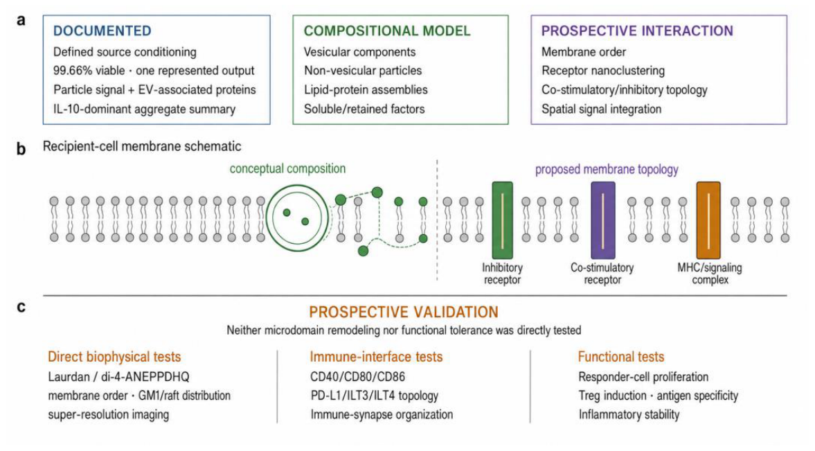
Membrane microdomains provide a complementary mechanistic framework. Lipid order, raft-associated segregation, receptor nanoclustering, and the spatial organization of signaling complexes affect antigen presentation and immune-synapse behavior [14]. In dendritic cells, the nanoscale organization of antigen-presentation machinery demonstrates that molecular positioning and membrane topology can regulate immune signaling even when the relevant molecular constituents are present [15]. More broadly, interactions between membrane topography and lateral membrane domains can influence the localization and activation of signaling proteins [16]. For a regulatory-oriented preparation, the relevant hypothesis is therefore not universal “raft repair” or activation; rather, it is the context-dependent modulation of membrane order and the relative spatial organization of activating and inhibitory interfaces.
In this study, multicomponent extracellular preparation is used as a biogenesis-neutral operational descriptor for the complete recovered output containing vesicle-associated and non-vesicular material. The descriptor identifies the analytical unit under study without collapsing it into a homogeneous extracellular-vesicle subtype. This distinction is consistent with process-defined extracellular formulations in which phospholipid-, protein-, vesicle-, and non-vesicular-associated components are preserved as an integrated analytical object rather than being designated automatically as purified exosomes [17]. Sequential IL-4/GM-CSF-supported differentiation followed by IL-10/TGF-β1 conditioning defines the source-programming axis evaluated in the retrospective archive.
The primary objective was to integrate the scientifically defensible information recoverable from a documented source-to-output archive while preserving method-specific analytical units. The analysis combines source-culture viability and cell-associated regions, an approximate particle-associated size region, reported EV-associated proteins, and a multiculture aggregate cytokine summary. A secondary objective was to translate those compositional observations into a staged membrane-microdomain and immune-function validation program. The central contribution of this study is threefold: (i) it establishes a traceable source-to-output framework for a sequentially cytokine-conditioned human PBMC-derived extracellular preparation; (ii) it prevents reduction of a heterogeneous vesicular and non-vesicular output to an unverified exosome identity; and (iii) it converts compositional observations into an explicit evidence-to-claim and prospective validation framework for tolerance-oriented biologic development. This separation between documented preparation attributes and prospective functional claims is consistent with previously proposed ex vivo traceability approaches for multicomponent phospholipoproteomic formulations [18]. The complete architecture is summarized in Figure 1.
2. Materials and Methods
2.1. Study Design and Data Provenance
This retrospective, descriptive study integrated analytical outputs from one documented human PBMC-derived source-programming workflow. Each analytical domain retained its source-specific experimental unit and was linked through a single provenance rule: only values explicitly preserved in the archive were entered into the integrated dataset [19]. No unavailable instrument-level, replicate-level, or protocol-level values were reconstructed, imputed, or inferred. This approach also preserves transparent reporting boundaries by distinguishing documented flow-cytometric events, percentages, and gated regions from raw instrument outputs that were not available for retrospective review [20]. Figure 1, Figure 2, Figure 4, and Figure 6 are conceptual or evidence-summary schematics; Figure 3 and Figure 5 visualize archived numerical summaries.
Figure 2.
Stage-level source programming and analytical workflow. The biological sequence connects established human PBMC-derived cultures to differentiation support, regulatory conditioning, extracellular recovery, and multilevel analytical integration. The icons are illustrative and do not depict measured cellular morphology, absolute cell number, culture scale, or operational processing parameters.
Figure 2.
Stage-level source programming and analytical workflow. The biological sequence connects established human PBMC-derived cultures to differentiation support, regulatory conditioning, extracellular recovery, and multilevel analytical integration. The icons are illustrative and do not depict measured cellular morphology, absolute cell number, culture scale, or operational processing parameters.
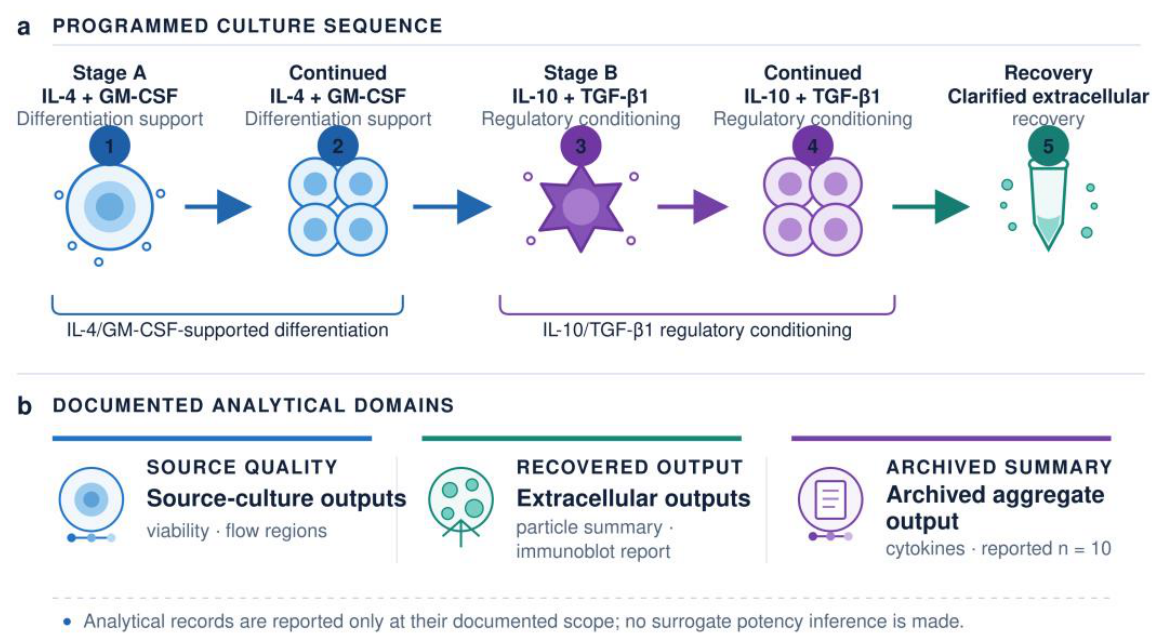
Figure 3.
Source-culture viability and cell-associated analytical distributions visualized from archived numerical summaries. (a) Annexin V/7-AAD viability composition. (b) Parent-gate and secondary CD34/CD38-defined regions.
Figure 3.
Source-culture viability and cell-associated analytical distributions visualized from archived numerical summaries. (a) Annexin V/7-AAD viability composition. (b) Parent-gate and secondary CD34/CD38-defined regions.
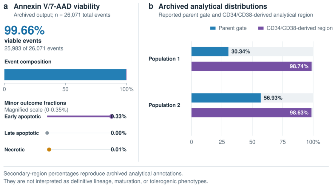
Figure 4.
Integrated profile of the recovered extracellular preparation. Particle-associated and EV-associated protein layers converge on a biogenesis-neutral multicomponent operational identity. The particle and protein panels summarize archived qualitative records and are not raw NTA traces or uncropped immunoblot images.
Figure 4.
Integrated profile of the recovered extracellular preparation. Particle-associated and EV-associated protein layers converge on a biogenesis-neutral multicomponent operational identity. The particle and protein panels summarize archived qualitative records and are not raw NTA traces or uncropped immunoblot images.
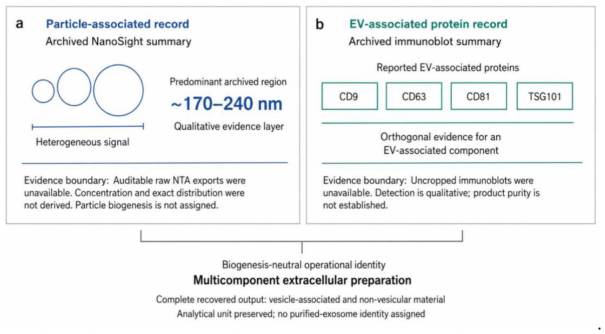
Figure 5.
Archived aggregate cytokine-associated profile. (a) Reported aggregate means on a logarithmic scale. (b) Interpretation boundary integrating the documented pattern, process context, and evidence boundary.
Figure 5.
Archived aggregate cytokine-associated profile. (a) Reported aggregate means on a logarithmic scale. (b) Interpretation boundary integrating the documented pattern, process context, and evidence boundary.
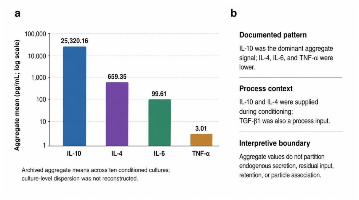
Figure 6.
Evidence-to-claim pathway for tolerance-oriented development. Measured attributes support a multicomponent operational identity, which is linked prospectively to mechanistic and functional validation modules. The membrane and receptor representations are prospective conceptual schematics and do not constitute direct mechanistic or functional evidence.
Figure 6.
Evidence-to-claim pathway for tolerance-oriented development. Measured attributes support a multicomponent operational identity, which is linked prospectively to mechanistic and functional validation modules. The membrane and receptor representations are prospective conceptual schematics and do not constitute direct mechanistic or functional evidence.
2.2. Cellular Material and Ethical Provenance
Established, de-identified, research-grade human PBMC-derived precursor cultures were maintained within the archived research program under internal study culture code FIC-BX-hPBMC-IRC-2026-01 for research use only and under controlled in vitro laboratory conditions. The cultures and associated analytical records were pre-existing and were used exclusively for retrospective analytical integration and extracellular-preparation characterization. The extracellular preparation evaluated in this study corresponds to the recovered output of the documented source-programming and extracellular-recovery workflow described below. No new human biological samples were collected for the purposes of this study, no intervention involving human participants was performed, no identifiable donor information or associated clinical data were accessed, and no novel human cell line was generated or genetically modified by the authors for the purposes of this work [21].
2.3. Sequential Cytokine Conditioning
The source cultures underwent an established sequential research procedure comprising IL-4/GM-CSF-supported differentiation, subsequent IL-10/TGF-β1 regula-tory conditioning, recovery of conditioned medium, serial clarification, and size-based extracellular-fraction recovery [22,23]. The study reports the biological sequence and pro-cessing classes required for source-to-output interpretation; implementation-specific parameters were outside the analytical scope of this retrospective integration (Table 1).
2.4. Viability and Cell-Associated Flow Cytometry
Viability was assessed with Annexin V-PE and 7-aminoactinomycin D on a FACSAria III Cell Sorter, following the standard interpretive logic of phosphatidylserine exposure and membrane permeability [24]. The represented output contained 26,071 events summarized across viable, early-apoptotic, late-apoptotic, and necrotic quadrants. Separate archived analyses reported two parent gates and corresponding CD34/CD38-defined secondary regions, retained under the neutral labels Population 1 and Population 2.
2.5. Extracellular-Fraction Recovery
Conditioned medium was recovered after the sequential source-programming stage. The archive documented serial clarification followed by a size-based extracellular-fraction recovery step [25]. The resulting complete preparation was carried forward to particle, immunoblot, and cytokine-context integration. This stage-level description defines the analytical source-to-output linkage evaluated in the present study.
2.6. Nanoparticle Tracking Analysis
Particle-associated characteristics were evaluated on a NanoSight platform [26]. The archived analytical summary described a heterogeneous signal with a predominant region at approximately 170–240 nm. This particle-associated region was integrated as a qualitative compositional layer of the recovered preparation.
2.7. Immunoblotting
The archived immunoblot summary reported evaluation and detection of CD9, CD63, CD81, and TSG101 in the recovered extracellular preparation [27]. These proteins were interpreted jointly with the particle-associated record as orthogonal evidence for an EV-associated component within the complete multicomponent output
2.8. Multiculture Cytokine Analysis
The archived analytical summary described a multiplex cytometric bead immunoassay with FCAP Array processing and labelled the cytokine output as representing ten conditioned cultures [28]. IL-10, IL-4, IL-6, TNF-α, IFN-γ, IL-2, and IL-17A were listed as assessed analytes; aggregate mean values were available for IL-10, IL-4, IL-6, and TNF-α. Because IL-10 and IL-4 were supplied during conditioning, their concentrations were interpreted as attributes of the final extracellular environment.
2.9. Statistical Analysis
The analysis reports the aggregate arithmetic means preserved in the archived cytokine summary and the directly documented event and region percentages from flow cytometry. Because the integration was descriptive and culture-level replicate values were not available across all analytical domains, no variance estimates, confidence intervals, or inferential hypothesis tests were reconstructed or performed across domains [29].
2.10. Use of Generative AI and AI-Assisted Technologies
Generative AI and AI-assisted technologies were used during manuscript preparation only to support language editing, document structuring, and the code-assisted preparation of figures explicitly identified as conceptual or evidence-summary schematics. These tools were not used to generate, modify, infer, or analyze experimental data, raw plots, replicate-level values, statistical outputs, or numerical results. Final scientific figures were reconstructed and verified using deterministic vector- and code-based workflows. All AI-assisted outputs were critically reviewed, verified, and edited by the authors, who take full responsibility for the integrity and content of the manuscript [30].
3. Results
3.1. Analytical Architecture and Experimental Units
The integrated dataset comprised source-culture, recovered-preparation, and ex-tracellular final-environment outputs. Experimental units were preserved separately for each analytical domain [31,32] (Table 2), creating a traceable bridge from source quality [33,34] to recovered-output composition [35,36].
3.2. Source-Culture Viability and Cell-Associated Analytical Regions
Among 26,071 analyzed events, 25,983 (99.66%) were viable, 86 (0.33%) were early apoptotic, no late-apoptotic events were recorded, and 2 (0.01%) were necrotic. The represented output therefore provides a high-viability source-quality anchor for the downstream extracellular observations.
Two archived cell-associated parent-gate distributions were reported as Population 1 (30.34%) and Population 2 (56.93%). Their corresponding CD34/CD38-defined secondary regions contained 98.74% and 98.63% of events, respectively. These values preserve the archived cell-associated analytical structure within the source-to-output framework.
3.3. Extracellular Particle and EV-Associated Protein Profile
The recovered extracellular preparation displayed a heterogeneous parti-cle-associated signal with a predominant archived region at approximately 170–240 nm. This observation establishes the particle-associated layer of the integrated output.
The archived immunoblot summary reported detection of CD9, CD63, CD81, and TSG101. Convergence of the particle-associated and EV-associated protein layers sup-ports a multicomponent operational identity containing a vesicle-associated compo-nent while preserving the complete recovered preparation as the analytical unit (Table 3).
3.4. Multiculture Cytokine-Associated Profile
The archived aggregate summary, labelled as representing ten conditioned cultures, reported mean cytokine-associated values of 25,320.16 pg/mL IL-10, 659.35 pg/mL IL-4, 99.61 pg/mL IL-6, and 3.01 pg/mL TNF-α. The resulting profile was strongly IL-10-dominant and adds a multiculture final-environment layer to the source-to-output framework.
IL-10 and IL-4 were inputs to the conditioning program. Their measured concentrations are therefore used as process-aware final-environment descriptors, whereas IL-6 and TNF-α are retained as aggregate extracellular signals.

Table 4.
Reported aggregate cytokine-associated outputs.
| Analyte | Reported mean (pg/mL) | Process context | Interpretive use |
| IL-10 | 25,320.16 | Conditioning input | Final-environment descriptor |
| IL-4 | 659.35 | Differentiation/conditioning input | Final-environment descriptor |
| IL-6 | 99.61 | No recorded exogenous input | Aggregate extracellular signal |
| TNF-α | 3.01 | No recorded exogenous input | Aggregate extracellular signal |
The archived summary designated ten conditioned cultures as the multiculture analytical unit.
3.5. Integrated Evidence Boundary and Microdomain-Oriented Framework
The convergent signature integrates sequential source programming, a high-viability represented source output, a heterogeneous particle-associated region, reported EV-associated proteins, and an IL-10-dominant multiculture final-environment summary [37,38]. This cross-domain convergence establishes a process-defined multicomponent extracellular preparation and converts its characterization into an explicit evidence-to-claim framework [39,40] (Table 5).
4. Discussion
The central contribution of this study is threefold: (i) it establishes a traceable source-to-output framework for a sequentially cytokine-conditioned human PBMC-derived extracellular preparation; (ii) it prevents reduction of a heterogeneous vesicular and non-vesicular output to an unverified exosome identity [41]; and (iii) it converts compositional observations into an explicit evidence-to-claim and prospective validation framework for tolerance-oriented biologic development [42]. This contribution is methodologically substantive because the archive spans different analytical units that are often either pooled inappropriately or reported as disconnected observations [43].
4.1. Analytical Significance of the Multicomponent Source-to-Output Framework
The represented source-culture output showed 99.66% viability, providing a source-quality anchor for the extracellular observations. The recovered preparation then displayed convergent particle- and protein-associated signals: a heterogeneous predominant region at approximately 170–240 nm and reported detection of CD9, CD63, CD81, and TSG101 [44]. Together with the aggregate extracellular cytokine profile, these findings define a coherent analytical object across source, recovery, and final-environment levels.
The multicomponent designation is scientifically useful rather than merely cautious. A preparation containing interacting vesicular, non-vesicular, protein, lipid-associated, and soluble components cannot be represented faithfully by particle size or canonical EV markers alone [45]. The biogenesis-neutral operational identity adopted here therefore preserves the complete analytical object and creates a direct route to fraction-resolved testing of complete, EV-enriched, EV-depleted, and soluble components under a common normalization scheme [46].
The IL-10-dominant aggregate final environment adds a distinct process layer. Because IL-10 and IL-4 participated in conditioning, their measured values are treated as final-environment attributes [47]. This process-aware interpretation is transferable to other conditioned secretome studies in which retained inputs, extracellular association, and source-cell output must be separated analytically [48].
4.2. Positioning Relative to Regulatory Dendritic-Cell Vesicle Studies
Regulatory dendritic-cell vesicle studies establish biological precedent for source-state-dependent extracellular immunobiology, but they examine different analytical products and validation depths. Table 6 compares the principal studies directly relevant to the present design.
The comparison identifies a complementary contribution. Earlier studies linked purified murine regulatory-DC exosomes to direct functional outcomes. The present study instead supplies the human, pre-functional analytical bridge required before a heterogeneous preparation can be assigned a reproducible identity, fractionated rationally, and advanced to functional testing. Its novelty therefore lies in the integration logic and claim architecture as well as in the specific conditioned source-to-output sequence.
4.3. Study Boundaries and Prospective Validation
The study boundaries are concentrated at the design level. The retrospective ar-chive did not contain a contemporaneous IL-4/GM-CSF-only comparator, an immuno-genic-conditioning comparator, a cell-free cytokine-processing control, a complete source-cell regulatory phenotype, auditable raw NTA and FCS exports, uncropped quantitative immunoblots, culture-level cytokine dispersion, or a responder-cell func-tional assay. Accordingly, the present evidence supports descriptive integration and an operational product identity, while conditioning causality, quantitative reproducibility, antigen specificity, membrane-microdomain remodeling, and functional tolerogenicity remain assigned to their dedicated validation modules.
The next stage follows directly from the evidence-to-claim framework. Analytical traceability should first be closed with original instrument files, quantitative im-munoblots, and fraction-level recovery records [52]. Source-cell phenotype and cell-free cy-tokine controls should then define the origin of the final-environment signature [53]. Finally, normalized comparisons of the complete preparation with EV-enriched, EV-depleted, and soluble fractions should be linked to responder proliferation, CD69/CD25 activation, Treg-associated phenotypes, cytokine shifts, antigen specificity, inflammatory rechal-lenge, membrane order, and receptor-nanoclustering endpoints [54]. This hierarchy turns the current descriptive study into an executable development program.
5. Conclusions
This study establishes a traceable source-to-output framework for a sequentially conditioned human PBMC-derived multicomponent extracellular preparation. High viability in one represented source output, a heterogeneous particle-associated region, reported EV-associated proteins, and an IL-10-dominant multiculture final-environment summary converge on a coherent, biogenesis-neutral operational identity. The integration distinguishes the complete preparation from an exosome-only product while preserving a direct route to fraction-resolved, mechanistic, and functional testing.More broadly, the paper provides a transferable methodology for characterizing multicomponent extracellular preparations without prematurely assigning exosome identity or biological function. Its evidence-to-claim structure can be applied to other conditioned secretome programs to connect source quality, compositional layers, process context, and claim-specific validation. Functional tolerogenicity, antigen specificity, and recipient-cell membrane-microdomain remodeling are therefore positioned as defined next-stage endpoints rather than diffuse interpretive caveats.
Supplementary Materials
The following supporting information can be downloaded at the website of this paper posted on Preprints.org. The accompanying Supplementary Information contains the analytical-unit and evidence-provenance matrix, the stage-level source-programming workflow, directly documented source-culture outputs, the integrated extracellular-preparation profile, reported aggregate cytokine-associated means, the claim matrix, the prospective validation framework, and a schematic of source-programming and analytical integration.
Author Contributions
Conceptualization, R.G.-S.; methodology, R.G.-S., F.G.-C. and N.M.-G.; validation, R.G.-S., F.G.-C. and N.M.-G.; formal analysis, R.G.-S.; investigation, F.G.-C., N.M.-G., A.L., I.M., J.I. and F.K.; resources, R.G.-S.; data curation, F.G.-C., N.M.-G., A.L., I.M., J.I. and F.K.; writing—original draft preparation, R.G.-S.; writing—review and editing, R.G.-S., I.R. and C.P.-V.; visualization, R.G.-S. and A.T.; supervision, R.G.-S.; project administration, R.G.-S.; funding acquisition, R.G.-S. All authors have read and agreed to the published version of the manuscript.
Funding
This research was supported by Fundación Biotech under grant number FB-20222-1091. The Article Processing Charge (APC) was funded by Biogenica Foundation. The funders had no role in the design of the study; in the collection, analysis, or interpretation of data; in the writing of the manuscript; or in the decision to submit the results for publication.
Institutional Review Board Statement
Not applicable. The present study was limited to retrospective analytical integration of established, de-identified, research-grade human PBMC-derived cultures, recovered extracellular material, and archived laboratory outputs under in vitro conditions. No participants were recruited, no new human biological samples or tissue biopsies were collected, no identifiable donor information or associated clinical data were accessed, and the evaluated preparation was not administered to human subjects. No novel human cell line was created, and no genetic modification of human cell cultures was performed by the authors for this study.
Informed Consent Statement
Not applicable. No human participants were recruited, and no new human samples, identifiable human tissue, identifiable donor information, or associated clinical data were collected or accessed for this study.
Data Availability Statement
The data supporting the findings of this retrospective integration are available within the article and its Supplementary Information. Additional de-identified archived analytical summaries and supporting records that remain available to the authors may be obtained from the corresponding author upon reasonable request, subject to confidentiality, record-retention, intellectual-property, and material-transfer requirements.
Acknowledgments
The authors thank the technical teams who contributed to the experimental and analytical work. The use of generative AI and AI-assisted technologies during manuscript preparation is disclosed in Section 2.10.
Conflicts of Interest
Some authors are affiliated with OGRD Alliance LLC or collaborating entities engaged in scientific research and development related to extracellular preparations. Biogenica Foundation funded the Article Processing Charge. The funding entities had no role in study design, data analysis, interpretation, manuscript preparation, peer-review response, or the decision to submit the work for publication. The authors declare no other conflicts of interest.
Abbreviations
Abbreviations used in this manuscript.
| Abbrev. | Definition |
| CBA | Cytometric bead array |
| EV | Extracellular vesicle |
| GM-CSF | Granulocyte–macrophage colony-stimulating factor |
| NTA | Nanoparticle tracking analysis |
| PBMC | Peripheral blood mononuclear cell |
| TEM | Transmission electron microscopy |
| TGF-β1 | Transforming growth factor beta 1 |
| 7-AAD | 7-aminoactinomycin D |
References
- Lutz, M.B.; Ali, S.; Audiger, C.; Autenrieth, S.E.; Berod, L.; Bigley, V.; Cyran, L.; Dalod, M.; Dörrie, J.; Dudziak, D.; et al. Guidelines for Mouse and Human DC Generation. Eur. J. Immunol. 2023, 53, e2249816. [Google Scholar] [CrossRef] [PubMed]
- Avancini, D.; Testori, A.; Fresolone, L.; Cazzaniga, V.; Doni, A.; Riva, F.; Sommariva, M.; Colombo, F.S.; Russo, V.; Manfredi, A.A.; et al. Aryl Hydrocarbon Receptor Activity Downstream of IL-10 Signaling Is Required to Promote Regulatory Functions in Human Dendritic Cells. Cell Rep. 2023, 42, 112193. [Google Scholar] [CrossRef] [PubMed]
- Koller, N.; Höllthaler, P.; Barends, M.; Döring, M.; Spahn, C.; Durán, V.; Costa, B.; Becker, J.; Heilemann, M.; Kalinke, U.; et al. Nanoscale Organization of the MHC I Peptide-Loading Complex in Human Dendritic Cells. Cell. Mol. Life Sci. 2022, 79, 477. [Google Scholar] [CrossRef] [PubMed]
- Chometon, T.Q.; Siqueira, M.D.S.; Sant’Anna, J.C.; Almeida, M.R.; Gandini, M.; Martins de Almeida Nogueira, A.C.; Antas, P.R.Z. A protocol for rapid monocyte isolation and generation of singular human monocyte-derived dendritic cells. PLoS ONE 2020, 15, e0231132. [Google Scholar] [CrossRef] [PubMed]
- Meskini, M.; Amanzadeh, A.; Salehi, F.; Bouzari, S.; Karimipoor, M.; Fuso, A.; Fateh, A.; Siadat, S.D. A Protocol to Isolate and Characterize Pure Monocytes and Generate Monocyte-Derived Dendritic Cells through FBS-Coated Flasks. Sci. Rep. 2024, 14, 23956. [Google Scholar] [CrossRef] [PubMed]
- Passeri, L.; Andolfi, G.; Bassi, V.; et al. Tolerogenic IL-10-Engineered Dendritic Cell-Based Therapy to Restore Antigen-Specific Tolerance in T Cell-Mediated Diseases. J. Autoimmun. 2023, 138, 103051. [Google Scholar] [CrossRef] [PubMed]
- Dao Nyesiga, G.; Pool, L.; Englezou, P.C.; Hylander, T.; Ohlsson, L.; Appelgren, D.; Sundstedt, A.; Tillerkvist, K.; Romedahl, H.R.; Wigren, M. Tolerogenic Dendritic Cells Generated In Vitro Using a Novel Protocol Mimicking Mucosal Tolerance Mechanisms Represent a Potential Therapeutic Cell Platform for Induction of Immune Tolerance. Front. Immunol. 2023, 14, 1045183. [Google Scholar] [CrossRef] [PubMed]
- Jeppesen, D.K.; Zhang, Q.; Coffey, R.J. Extracellular Vesicles and Nanoparticles at a Glance. J. Cell Sci. 2024, 137, jcs260201. [Google Scholar] [CrossRef] [PubMed]
- Welsh, J.A.; Goberdhan, D.C.I.; O’Driscoll, L.; et al. Minimal Information for Studies of Extracellular Vesicles (MISEV2023): From Basic to Advanced Approaches. J. Extracell. Vesicles 2024, 13, e12404. [Google Scholar] [CrossRef] [PubMed]
- Jeppesen, D.K.; Zhang, Q.; Franklin, J.L.; Coffey, R.J. Extracellular Vesicles and Nanoparticles: Emerging Complexities. Trends Cell Biol. 2023, 33, 667–681. [Google Scholar] [CrossRef] [PubMed]
- Lin, M.; Lei, S.; Chai, Y.; et al. Immunosuppressive Microvesicles-Mimetic Derived from Tolerant Dendritic Cells to Target T-Lymphocytes for Inflammation Diseases Therapy. J. Nanobiotechnol. 2024, 22, 201. [Google Scholar] [CrossRef] [PubMed]
- Avalos-de Leon, C.G.; Thomson, A.W. Regulatory Immune Cell-Derived Exosomes: Modes of Action and Therapeutic Potential in Transplantation. Transplantation 2025, 109, 1124–1137. [Google Scholar] [CrossRef] [PubMed]
- Wu, T.; Wang, L.; Gao, C.; Jian, C.; Liu, Y.; Fu, Z.; Shi, C. Treg-Derived Extracellular Vesicles: Roles in Diseases and Theranostics. Mol. Pharm. 2024, 21, 2659–2672. [Google Scholar] [CrossRef] [PubMed]
- Gutiérrez-Sandoval, R.; et al. Structural Determinants and Repair of Membrane Microdomains in Dendritic Cell-Mediated Antitumor Immunity: An Integrative Mechanistic Synthesis. Int. J. Mol. Sci. 2026, 27, 2305. [Google Scholar] [CrossRef] [PubMed]
- Barends, M.; Koller, N.; Schölz, C.; Durán, V.; Bošnjak, B.; Becker, J.; Döring, M.; Blees, H.; Förster, R.; Kalinke, U.; et al. Dynamic interactome of the MHC I peptide loading complex in human dendritic cells. Proc. Natl. Acad. Sci. USA 2023, 120, e2219790120. [Google Scholar] [CrossRef] [PubMed]
- Ghosh, S.; Wagenknecht-Wiesner, A.; Desai, S.; et al. Synergy between Membrane Topography and Domains to Control Signaling Protein Localization in Mast Cells Facilitates Their Activation. Proc. Natl. Acad. Sci. USA 2025, 122, e2424427122. [Google Scholar] [CrossRef] [PubMed]
- Gutiérrez-Sandoval, R.; Gutiérrez-Castro, F.; Muñoz-Godoy, N.; et al. Beyond Exosomes: An Ultrapurified Phospholipoproteic Complex (PLPC) as a Scalable Immunomodulatory Platform for Reprogramming Immune Suppression in Metastatic Cancer. Cancers 2025, 17, 1658. [Google Scholar] [CrossRef] [PubMed]
- Gutiérrez-Sandoval, R.; et al. Ex Vivo Traceability Platform for Phospholipoproteomic Formulations: Functional Evidence without Clinical Exposure. Biomedicines 2025, 13, 2101. [Google Scholar] [CrossRef] [PubMed]
- Wittner, R.; Holub, P.; Mascia, C.; Frexia, F.; Müller, H.; Plass, M.; Geiger, J.; et al. Toward a Common Standard for Data and Specimen Provenance in Life Sciences. Learn. Health Syst. 2024, 8, e10365. [Google Scholar] [CrossRef] [PubMed]
- Sorigue, M.; Salvia, R.; Petriz, J. Transparency and Reporting in the Evaluation of PD1 by Flow Cytometry. Cytom. A 2025, 107, 745–749. [Google Scholar] [CrossRef] [PubMed]
- Canadian Institutes of Health Research; Natural Sciences and Engineering Research Council of Canada; Social Sciences and Humanities Research Council. Tri-Council Policy Statement: Ethical Conduct for Research Involving Humans—TCPS 2 (2022); Secretariat on Responsible Conduct of Research: Ottawa, ON, Canada, 2022; Volume Chapter 12, p. Article 12.21. [Google Scholar]
- Robertson, H.; Li, J.; Kim, H.J.; Rhodes, J.W.; Harman, A.N.; Patrick, E.; Rogers, N.M. Transcriptomic Analysis Identifies a Tolerogenic Dendritic Cell Signature. Front. Immunol. 2021, 12, 733231. [Google Scholar] [CrossRef] [PubMed]
- Suuring, M.; Amodio, G.; Rouel, M.; Raux, A.; Delhaye, T.; Royer, A.L.; et al. Protocol of Generation of Tolerogenic Dendritic Cells Affects Their Transcriptional and Metabolic Profiles Leading to Specific Tolerogenic Functions. Mol. Ther. Methods Clin. Dev. 2025, 33, 101605. [Google Scholar] [CrossRef] [PubMed]
- Hähnel, V.; Dormann, F.; Kronenberg, K.; Hutchinson, J.A.; Burkhardt, R.; Ahrens, N. Validation of an apoptosis assay for extracorporeal photopheresis. Transfus. Med. 2021, 31, 113–120. [Google Scholar] [CrossRef] [PubMed]
- Soukup, R.; Gerner, I.; Gültekin, S.; Baik, H.; Oesterreicher, J.; Grillari, J.; Jenner, F. Characterisation of Extracellular Vesicles from Equine Mesenchymal Stem Cells. Int. J. Mol. Sci. 2022, 23, 5858. [Google Scholar] [CrossRef] [PubMed]
- Auger, C.; Brunel, A.; Darbas, T.; Akil, H.; Perraud, A.; Bégaud, G.; Bessette, B.; Christou, N.; Verdier, M. Extracellular Vesicle Measurements with Nanoparticle Tracking Analysis: A Different Appreciation of Up and Down Secretion. Int. J. Mol. Sci. 2022, 23, 2310. [Google Scholar] [CrossRef] [PubMed]
- Fordjour, F.K.; Guo, C.; Ai, Y.; Daaboul, G.G.; Gould, S.J. A Shared, Stochastic Pathway Mediates Exosome Protein Budding along Plasma and Endosome Membranes. J. Biol. Chem. 2022, 298, 102394. [Google Scholar] [CrossRef] [PubMed]
- Liu, J.; Allo, B.; Winnik, M.A. Development of Multiplexed Bead-Based Immunoassays for Profiling Soluble Cytokines and CD163 Using Mass Cytometry. ACS Meas. Sci. Au 2022, 2, 629–640. [Google Scholar] [CrossRef] [PubMed]
- Nusser, S.M. The Role of Statistics in Promoting Data Reusability and Research Transparency. Annu. Rev. Stat. Appl. 2023, 10, 1–22. [Google Scholar] [CrossRef]
- Nature Editorials. Tools Such as ChatGPT Threaten Transparent Science; Here Are Our Ground Rules for Their Use. Nature 2023, 613, 612. [Google Scholar] [CrossRef] [PubMed]
- Holzmeister, F.; Johannesson, M.; Böhm, R.; et al. Heterogeneity in Effect Size Estimates. Proc. Natl. Acad. Sci. USA 2024, 121, e2403490121. [Google Scholar] [CrossRef] [PubMed]
- Moresis, A.; Restivo, L.; Bromilow, S.; et al. A Minimal Metadata Set (MNMS) to Repurpose Nonclinical In Vivo Data for Biomedical Research. Lab Anim. 2024, 53, 67–79. [Google Scholar] [CrossRef] [PubMed]
- Rimac, V.; Bojanić, I.; Škifić, M.; Dabelić, S.; Golubić Ćepulić, B. Quality Assessment of Cryopreserved Peripheral Blood Stem Cell Products: Evaluation of Two Methods for Flow Cytometric Viability Testing. Int. J. Lab. Hematol. 2025, 47, 93–100. [Google Scholar] [CrossRef] [PubMed]
- Cai, Y.; Prochazkova, M.; Kim, Y.S.; Jiang, C.; Ma, J.; Moses, L.; Martin, K.; Pham, V.; Zhang, N.; Highfill, S.L.; et al. Assessment and Comparison of Viability Assays for Cellular Products. Cytotherapy 2024, 26, 201–209. [Google Scholar] [CrossRef] [PubMed]
- Zhang, J.; Brown, A.; Johnson, B.; Diebold, D.; Asano, K.; Marriott, G.; Lu, B. Genetically Engineered Extracellular Vesicles Harboring Transmembrane Scaffolds Exhibit Differences in Their Size, Expression Levels of Specific Surface Markers and Cell-Uptake. Pharmaceutics 2022, 14, 2564. [Google Scholar] [CrossRef] [PubMed]
- Suwakulsiri, W.; Xu, R.; Rai, A.; Shafiq, A.; Chen, M.; Greening, D.W.; Simpson, R.J. Comparative Proteomic Analysis of Three Major Extracellular Vesicle Classes Secreted from Human Primary and Metastatic Colorectal Cancer Cells: Exosomes, Microparticles, and Shed Midbody Remnants. Proteomics 2024, 24, 2300057. [Google Scholar] [CrossRef] [PubMed]
- Morales-Sanfrutos, J.; Etxeberria-Ugartemendia, J.; Barroso-Gomila, O.; González, E.; Sendino, M.; Ximénez-Embún, P.; García, F.; Zarzuela, E.; Falcón-Pérez, J.M.; Peinado, H.; et al. Defining the Reference Proteomes for Small Extracellular Vesicles and Non-Vesicular Components. Nat. Cell Biol. 2026, 28, 622–639. [Google Scholar] [CrossRef] [PubMed]
- Peshkova, M.; Korneev, A.; Suleimanov, S.; Vlasova, I.I.; Svistunov, A.; Kosheleva, N.; Timashev, P. MSCs’ Conditioned Media Cytokine and Growth Factor Profiles and Their Impact on Macrophage Polarization. Stem Cell Res. Ther. 2023, 14, 142. [Google Scholar] [CrossRef] [PubMed]
- Lai, J.J.; Chau, Z.L.; Chen, S.-Y.; Hill, J.J.; Korpany, K.V.; Liang, N.-W.; Lin, L.-H.; Lin, Y.-H.; Liu, J.K.; Liu, Y.-C.; et al. Exosome Processing and Characterization Approaches for Research and Technology Development. Adv. Sci. 2022, 9, 2103222. [Google Scholar] [CrossRef] [PubMed]
- Dang, L.E.; Gruber, S.; Lee, H.; Dahabreh, I.J.; Stuart, E.A.; Williamson, B.D.; Wyss, R.; Díaz, I.; Ghosh, D.; Kıcıman, E.; et al. A Causal Roadmap for Generating High-Quality Real-World Evidence. J. Clin. Transl. Sci. 2023, 7, e212. [Google Scholar] [CrossRef] [PubMed]
- Morales-Sanfrutos, J.; Muñoz, J. Unraveling the Complexity of the Extracellular Vesicle Landscape with Advanced Proteomics. Expert Rev. Proteom. 2022, 19, 89–101. [Google Scholar] [CrossRef] [PubMed]
- Barran, G.; Naamane, N.; Baru, A.M.; Anderson, A.E.; Falconer, J.; Hilkens, C.M.U. Human Tolerogenic Dendritic Cell Subtypes Exert Divergent Effects on Induction of Cytotoxic CD4+ T Cells. Front. Immunol. 2025, 16, 1698413. [Google Scholar] [CrossRef] [PubMed]
- Zimmerman, K.D.; Espeland, M.A.; Langefeld, C.D. A practical solution to pseudoreplication bias in single-cell studies. Nat. Commun. 2021, 12, 738. [Google Scholar] [CrossRef] [PubMed]
- Menck, K.; Preußer, C. Heterogeneity in Extracellular Vesicles: Same Same—But Different? Trillium Extracell. Vesicles 2024, 6, 18–21. [Google Scholar] [CrossRef]
- Wolf, M.; Poupardin, R.W.; Ebner-Peking, P.; Andrade, A.C.; Blöchl, C.; Obermayer, A.; Gomes, F.G.; Vari, B.; Maeding, N.; Eminger, E.; et al. A Functional Corona around Extracellular Vesicles Enhances Angiogenesis, Skin Regeneration and Immunomodulation. J. Extracell. Vesicles 2022, 11, e12207. [Google Scholar] [CrossRef] [PubMed]
- Cheng, L.; Hill, A.F. Therapeutically Harnessing Extracellular Vesicles. Nat. Rev. Drug Discov. 2022, 21, 379–399. [Google Scholar] [CrossRef] [PubMed]
- Lapuente, J.P.; Blázquez-Martínez, A.; Marco-Brualla, J.; Gómez, G.; Desportes, P.; Sanz, J.; Fernández, P.; García-Gil, M.; Bermejo, F.; San Martín, J.V.; et al. Cytokine profile and anti-inflammatory activity of a standardized conditioned medium obtained by coculture of monocytes and mesenchymal stromal cells (PRS CK STORM). Biomolecules 2022, 12, 534. [Google Scholar] [CrossRef] [PubMed]
- Holthaus, M.; Santhakumar, N.; Wahlers, T.; Paunel-Görgülü, A. The Secretome of Preconditioned Mesenchymal Stem Cells Drives Polarization and Reprogramming of M2a Macrophages toward an IL-10-Producing Phenotype. Int. J. Mol. Sci. 2022, 23, 4104. [Google Scholar] [CrossRef] [PubMed]
- Liu, Y.Y.; Fan, H.H.; Ren, Y.N.; Yang, J.; Nie, X.X.; Zhao, L.H.; Lin, J.J. Immune Tolerance Induced by Exosomes Derived from Regulatory Dendritic Cells of Mice. Zhongguo Shi Yan Xue Ye Xue Za Zhi 2008, 16, 406–410. [Google Scholar] [PubMed]
- Elashiry, M.; Elashiry, M.M.; Elsayed, R.; Rajendran, M.; Auersvald, C.; Zeitoun, R.; Rashid, M.H.; Ara, R.; Meghil, M.M.; Liu, Y.; et al. Dendritic Cell-Derived Exosomes Loaded with Immunoregulatory Cargo Reprogram Local Immune Responses and Inhibit Degenerative Bone Disease In Vivo. J. Extracell. Vesicles 2020, 9, 1795362. [Google Scholar] [CrossRef] [PubMed]
- Elashiry, M.; Elsayed, R.; Elashiry, M.M.; et al. Proteomic Characterization, Biodistribution, and Functional Studies of Immune-Therapeutic Exosomes: Implications for Inflammatory Lung Diseases. Front. Immunol. 2021, 12, 636222. [Google Scholar] [CrossRef] [PubMed]
- Knecht, S.; Eberl, H.C.; Kreisz, N.; Ugwu, U.J.; Starikova, T.; Kuster, B.; Wilhelm, S. An Introduction to Analytical Challenges, Approaches, and Applications in Mass Spectrometry-Based Secretomics. Mol. Cell. Proteom. 2023, 22, 100636. [Google Scholar] [CrossRef] [PubMed]
- Papait, A.; Silini, A.R.; Gazouli, M.; Malvicini, R.; Muraca, M.; O’Driscoll, L.; Pacienza, N.; Toh, W.S.; Yannarelli, G.; Ponsaerts, P.; et al. Perinatal Derivatives: How to Best Validate Their Immunomodulatory Functions. Front. Bioeng. Biotechnol. 2022, 10, 981061. [Google Scholar] [CrossRef] [PubMed]
- Papait, A.; Ragni, E.; Cargnoni, A.; Vertua, E.; Romele, P.; Masserdotti, A.; Perucca Orfei, C.; Signoroni, P.B.; Magatti, M.; Silini, A.R.; et al. Comparison of EV-Free Fraction, EVs, and Total Secretome of Amniotic Mesenchymal Stromal Cells for Their Immunomodulatory Potential: A Translational Perspective. Front. Immunol. 2022, 13, 960909. [Google Scholar] [CrossRef] [PubMed]
Figure 1.
Source-to-output framework and transferable analytical contribution. Sequential human PBMC-derived source programming is linked to the complete recovered extracellular preparation, its documented analytical layers, and an evidence-to-claim validation pathway. The schematic is conceptual and does not represent direct imaging, absolute particle size, particle concentration, or measured membrane topology.
Figure 1.
Source-to-output framework and transferable analytical contribution. Sequential human PBMC-derived source programming is linked to the complete recovered extracellular preparation, its documented analytical layers, and an evidence-to-claim validation pathway. The schematic is conceptual and does not represent direct imaging, absolute particle size, particle concentration, or measured membrane topology.
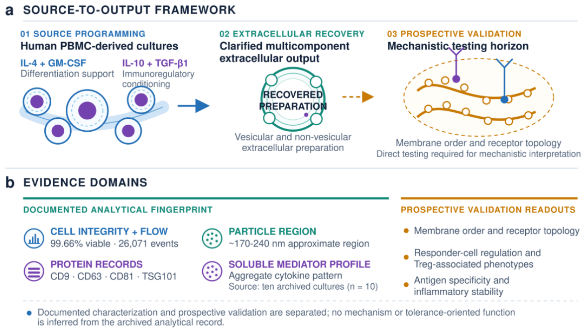
Table 1.
Stage-level source programming and analytical workflow.
| Stage | Source-culture context | Primary operation | Material carried forward | Analytical connection | Reporting level |
| 1 | Human PBMC-derived precursor cultures | Controlled in vitro maintenance | Established source cultures | Ethical and source provenance | Stage level |
| 2 | Differentiation support | IL-4/GM-CSF-supported programming | Differentiating cultures | Source-state traceability | Cytokine classes and order |
| 3 | Regulatory conditioning | Sequential IL-10/TGF-β1 support | Regulatory-oriented cultures | Final-environment profile | Cytokine classes and order |
| 4 | Extracellular recovery | Serial clarification and size-based recovery | Complete extracellular preparation | Particle, protein, and cytokine layers | Processing classes |
| 5 | Prospective validation | Claim-specific functional modules | Normalized test articles | Membrane and immune endpoints | Study-design level |
The workflow is reported by biological stage and method class.
Table 2.
Analytical domains and method-specific experimental units.
| Domain | Material | Method | Experimental unit | Integrated output |
| Source viability | Conditioned source culture | Annexin V/7-AAD flow cytometry | One represented analytical output | Viable, apoptotic, and necrotic events |
| Cell-associated regions | Conditioned source culture | Archived flow-cytometric gates | Reported parent and secondary regions | Population 1/2 and CD34/CD38 regions |
| Particle profile | Recovered extracellular preparation | NTA/NanoSight | One archived qualitative summary | Approximate predominant size region |
| Protein profile | Recovered extracellular preparation | Immunoblot | One archived band-level summary | Reported EV-associated proteins |
| Cytokine profile | Extracellular final environment | Multiplex bead immunoassay | Aggregate summary; reported n = 10 | Reported analyte means |
EV, extracellular vesicle; NTA, nanoparticle tracking analysis. Reported n = 10 denotes the experimental unit stated in the archived cytokine summary.
Table 3.
Integrated extracellular-preparation observations.
| Analytical domain | Documented observation | Integrated interpretation | Role in source-to-output framework |
| Particle analysis | Heterogeneous signal; predominant reported region ~170–240 nm | Particle-associated material is present | Particle-distribution layer |
| Immunoblot | CD9, CD63, CD81, and TSG101 reported as detected | EV-associated protein component | Orthogonal protein layer |
| Integrated identity | Particle and protein observations converge | Multicomponent extracellular preparation | Biogenesis-neutral operational identity |
EV-associated denotes one compositional layer of the complete multicomponent preparation.
Table 5.
Evidence-to-claim framework.
| Evidence level | Current contribution | Transferable analytical use | Next validation module |
| Measured attributes | Source programming, viability, particle region, EV-associated proteins, and cytokine context | Source-to-output traceability | Independent and fraction-resolved replication |
| Operational identity | Multicomponent vesicular/non-vesicular extracellular preparation | Biogenesis-neutral product definition | Complete vs EV-enriched, EV-depleted, and soluble fractions |
| Mechanistic framework | Membrane-organization hypothesis linked to defined readouts | Claim-specific assay mapping | Membrane order, GM1 mapping, and receptor nanoclustering |
| Development orientation | Tolerance-oriented validation pathway | Staged functional progression | Responder-cell, Treg, antigen-specificity, and stability assays |
Table 6.
Comparison with principal regulatory dendritic-cell-derived vesicle studies.
| Study/source | Source programming and analyzed product | Characterization and functional scope | Relevance to the present study |
| Liu et al. [49]; murine BMDCs | Immature DCs exposed to TGF-β1 and IL-10; regulatory-DC exosomes with an immature-DC exosome comparator. | Flow phenotype; ultrafiltration/ultracentrifugation; mixed-lymphocyte response, donor hyporeactivity, and skin-allograft survival. | Functional precedent for purified murine exosomes; differs from the complete human PBMC-derived multicomponent preparation analyzed here. |
| Elashiry et al. [50]; murine BMDCs | Immunoregulatory DC program with TGF-β1/IL-10 cargo enrichment; purified regulatory-DC exosomes with immature and stimulatory comparators. | Particle/vesicle profile, cargo protection, and recipient-cell uptake; recipient-DC and Th17 suppression, Treg recruitment, and reduced inflammatory bone eg in vivo. | Demonstrates regulatory-DC exosome function; the present study instead defines a process-traceable, pre-functional human multicomponent baseline. |
| Elashiry et al. [51]; murine BMDCs | GM-CSF/IL-4 DC generation followed by TGF-β1/IL-10 programming; purified cytokine-loaded regulatory-DC exosomes. | NTA, TEM, immunoblot, LC–MS/MS, and biodistribution; recipient-cell TGF-β1 signaling, organ distribution, and ACE2 modulation in vitro. | Provides egi characterization of purified murine exosomes; the present study prioritizes transferability of the source-to-output method. |
| Present study; human PBMC cultures | Sequential IL-4/GM-CSF differentiation and IL-10/TGF-β1 conditioning; complete recovered multicomponent egiónlular preparation. | Source viability, cell-associated regions, particle egión, EV-associated proteins, and aggregate cytokines; no direct functional assay. | Establishes traceability across heterogeneous domains without reducing the complete output to an exosome-only identity. |
Disclaimer/Publisher’s Note: The statements, opinions and data contained in all publications are solely those of the individual author(s) and contributor(s) and not of MDPI and/or the editor(s). MDPI and/or the editor(s) disclaim responsibility for any injury to people or property resulting from any ideas, methods, instructions or products referred to in the content. |
© 2026 by the authors. Licensee MDPI, Basel, Switzerland. This article is an open access article distributed under the terms and conditions of the Creative Commons Attribution (CC BY) license (http://creativecommons.org/licenses/by/4.0/).
Copyright: This open access article is published under a Creative Commons CC BY 4.0 license, which permit the free download, distribution, and reuse, provided that the author and preprint are cited in any reuse.