

Submitted:
11 August 2026
Posted:
12 August 2026
You are already at the latest version
Abstract
Background/Objectives: Minimal invasive temporary spanning plate (SP) fixation has been established as a valuable treatment for complex distal radius fractures; while short-term outcomes are promising, long-term data remains limited. This study conducts a long-term outcome analysis of patients treated with SP fixation to address this gap within the current literature. Methods: We conducted a retrospective single-center study of patients treated with a spanning plate for distal radius fractures at a level I trauma center between 2018 and 2025. Indications for spanning plate fixation included distal radius fractures with severe metaphyseal comminution, radiocarpal fracture dislocations and very distal intra-articular fractures where standard osteosynthesis was not feasible. The inclusion criteria required a minimum follow-up of one year. Hospital records were screened for healing data and adverse events. Included patients were contacted for assessment of functional scores using the Patient-Rated Wrist Evaluation (PRWE) and a Visual Analogue Scale (VAS) for pain, as well as a 5-point Likert scale for satisfaction. Results: A total of 35 patients (mean age 57; 89% AO type C) were included. The SP was removed on average 3.8 months postoperatively. Radiological union was achieved at a mean of 8.6 weeks, with one non-union requiring reoperation. Complications included one implant failure and two Extensor Pollicis Longus (EPL) ruptures. Three other reoperations occurred: two for DRUJ instability and one wrist denervation due to osteoarthritis. Twenty-six patients responded to functional questionnaires at a mean follow-up of 58.3 months. They reported a mean satisfaction score of 4.1, mean resting VAS pain of 1.7, and a mean PRWE score of 16.7. Conclusions: Radiological, functional, and patient-rated outcomes demonstrate that excellent short-term results are successfully maintained over a 4.5-year period. The spanning plate technique proves to be a reliable and durable treatment option for complex distal radius fractures with a low long-term complication rate.
Keywords:
distal radius fracture
; wrist spanning plate
; dorsal bridge plate
; long-term outcome
; internal fixator
1. Introduction
Distal radius fractures (DRFs) are among the most common adult injuries, accounting for up to 18% of all adult fractures [1]. The data demonstrate a bimodal distribution, with high-energy trauma frequently observed in younger patients and fragility fractures more commonly encountered in older adults, particularly women over the age of 65 [1,2]. Complex patterns involving metaphyseal comminution and intra-articular extension present significant management challenges. These injuries are highly unstable, making it difficult to achieve and maintain anatomical reduction, which increases the risk of long-term functional impairment [2,3,4].
Historically, these complex, unstable fractures were often managed with external fixation utilizing ligamentotaxis for stabilization. However, external fixators are associated with several disadvantages, including high rates of pin-tract infections, hand stiffness, malreduction, and significant patient discomfort [5,6].
As an alternative, the dorsal spanning plate (SP) has emerged for the treatment of these highly comminuted and unstable DRFs. This technique involves spanning the fracture using ligamentotaxis in the same manner as an external fixator does, albeit with an internal fixator (spanning plate) placed from the radial shaft to the second or third metacarpal, effectively bypassing the wrist joint [3,4,7]. This internal distraction method avoids pin-site complications and provides a more stable construct that allows for earlier weight-bearing and functional use of the extremity compared to external fixation [3,5].
While current literature indicates that spanning plates provide reliable stabilization, most existing data is limited to short-term outcomes. Systematic reviews show a median follow-up of only 24 months, leaving a gap regarding long-term clinical effectiveness beyond the first years [1,2,8]. What remains unknown is the durability of these results and the comparative effectiveness over time [2,3,8].
This study presents a retrospective, single-center analysis of patients treated with a spanning plate for complex distal radius fractures with a mean follow-up of 4.5 years. Our objective was to evaluate whether the functional and patient-rated outcomes observed in the short term are successfully maintained over the long term, addressing a critical gap in long-term data within the current literature.
2. Materials and Methods
2.1. Design and Study Population
This is a single-center, retrospective study conducted at a level one trauma center in Switzerland. The electronic hospital records were screened to identify all patients who were treated surgically for a distal radius fracture between 2018 and 2025. All patients who were treated with a spanning plate, older than 18 years, had a minimum follow-up period of one year and signed a general informed consent form for use of their data for research were included in the study. Patients who did not have German or English language skills or were unable to complete the questionnaire were excluded.
2.2. Surgical Technique
The procedure was performed as previously described by Beeres et al. [3]. Under anesthesia and upper arm tourniquet hemostasis, a precontoured 2.4/3.5 mm wrist spanning plate (Arthrex Inc., Naples, FL, USA) was utilized. A dorsal incision over the third extensor compartment allowed for EPL release and PIN excision. Following provisional reduction, fluoroscopy guided plate sizing and the choice of distal anchorage (2nd or 3rd metacarpal). Incisions were made over the metacarpal and radial shaft. The plate was inserted retrograde under the EPL and fixed distally with three locking screws. Traction was applied for final alignment before proximal fixation with one cortical and two locking screws, with an optional middle screw added for stability if required.
2.3. Postoperative Management and Follow-Up
Immediately post-surgery, patients were given a removable wrist brace for comfort. Patients were allowed to weight bear as tolerated. Patients were seen at the outpatient clinic at 6 weeks, 3 months and one year after surgery. The spanning plate was typically removed between three and four months postoperatively, once X-ray confirmed fracture healing.
2.4. Data Collection
2.4.1. Baseline Characteristics
Electronic patient charts were reviewed to extract baseline characteristics, which included age, gender, comorbidities (diabetes, smoking), and the American Society of Anaesthesiologists (ASA) score defined by the treating anaesthesiologist. The initial radiographs and CT scans were used to determine the fracture type. The AO/OTA classification was used for fracture categorization [9]. Surgical data was extracted from the surgical reports and included type of implant, metacarpal bone used for distal anchorage and presence of additional osteosynthesis.
2.4.2. Outcome Parameters
The outpatient clinical records and X-rays were screened for the occurrence of complications such as surgical site infections (SSI), non-union, extensor tendon lesion, reoperation rates, complex regional pain syndrome (CRPS), time of SP removal, time to radiological fracture healing and range of motion were collected. SSI was classified as superficial or deep according to the definition of the Centers for Disease Control and Prevention [10]. Non-union was defined as absence of fracture consolidation 6 months after treatment, with the lack of radiological bridging callus at three out of four cortices [11]. CRPS Type I was diagnosed based on the Budapest criteria [12]. The duration of fracture healing was defined as the time span between the date of surgery and the date of first radiological evidence of fracture union. Radiographs were analyzed for radial inclination, volar tilt and ulnar variance after radiological evidence of fracture healing. Range of motion was extracted from the most recent outpatient clinical record.
All patients were additionally contacted by phone to obtain functional scores. After providing consent, the study documents were sent to them by mail and included a 5-point Likert scale for satisfaction (0–5), VAS for pain during rest (0–10) and the Patient-Rated Wrist Evaluation (PRWE). The PRWE is a validated questionnaire used to assess pain and disability in patients with wrist injuries. It has a score range from 0 to 100, where a lower score is better, indicating less pain and improved function [13]. Additionally, patients were asked whether any complications occurred since the last known clinical follow-up and if they had been treated for CRPS Type I.
Patients who did not return the documents within two weeks were contacted again. This procedure was repeated up to three times. Those who did not respond after three attempts were classified as lost to follow-up (non-responders).
The study was approved by the Ethics Committee of Central Switzerland (EKNZ; protocol number 2024-02472) and was conducted in accordance with the principles of the Declaration of Helsinki. All participating patients provided written informed consent prior to data collection.
3. Results
3.1. Study Population and Baseline Characteristics
A total of 50 patients treated with a Spanning Plate (SP) were initially identified within the given time period (Figure 1). Fifteen patients were excluded. Seven had a follow-up period of less than one year and eight were deceased due to unrelated causes. This resulted in a study population of 35 patients.
Table 1 details the demographic, fracture and procedure characteristics of the 35 patients included in the analysis. The mean age of the patients was 57 years (22–92). The gender distribution was equal male (18, 51%), female (17, 49%). Thirty-one (89%) of the fractures were classified as Type C according to the AO classification. An external fixator was used for temporary fixation in 21 (60%) cases. The mean time from injury to surgery (SP fixation) was 7.1 days (range, 4–11 days). Additional fracture fixation was performed in 30 (86%) patients. The mean duration of the SP fixation was 3.8 months (range, 1.8–12.9 months).
3.2. Clinical Outcomes
Table 2 summarises the clinical and radiological outcomes, complications and reoperations collected from hospital records. The mean active range of motion arc was 86 (35–135) in extension/flexion and 150 (70–180) in pronation/supination.
3.3. Radiological Outcomes
The mean time to fracture union was 8.6 weeks (5–29). The mean radial inclination was 23.2° (16°–32°), the mean volar tilt was 2.0° (−12° to 14°), and the mean ulnar variance was −0.3 mm (−9 to 5).
3.4. Complications and Reoperations
A total of seven adverse events (20%) were documented in the long-term follow-up group. No cases of surgical site infection were observed. Complex Regional Pain Syndrome (CRPS) Type I was diagnosed and treated in three patients (9%).
Complications directly associated with the implant or surgical technique included one implant failure (3%) following renewed trauma, which required hardware removal and secondary volar osteosynthesis. Furthermore, two patients (6%) experienced a rupture of the extensor pollicis longus (EPL) tendon. In these cases, the EPL tendon had not been positioned superficial to the plate during the initial operation. Treatment consisted of spanning plate removal and tendon repair via extensor indicis transposition onto the distal EPL remnant.
Delayed union occurred in two patients (6%), both of whom achieved sufficient consolidation after six months following postponed plate removal. One patient (3%) developed a non-union accompanied by impaired wrist and finger mobility. Proper bone healing was achieved after revision surgery five months postoperatively, which included cancellous bone grafting, re-osteosynthesis, metacarpophalangeal joint arthrolysis, and tenolysis. Although postoperative mobility improved, the patient did not regain full extension and fist closure. Another patient (3%) presented with persistent distal radioulnar joint (DRUJ) instability and pain nine months postoperatively. A satisfactory clinical outcome was achieved following a corrective osteotomy and triangular fibrocartilage complex (TFCC) refixation. Finally, symptomatic post-traumatic osteoarthritis necessitated surgical intervention in two cases (6%) after exhausting conservative treatment options. One patient underwent a denervation of the wrist. A second, working-age patient suffered from persistent pain unresponsive to hardware removal and arthroscopic arthrolysis, leading to a total wrist arthroplasty two years after the initial injury. However, due to ongoing symptoms, conversion to wrist arthrodesis and DRUJ resection is currently under discussion for this patient.
3.5. Functional Outcomes
Out of the 35 included patients, 26 responded to the long-term follow-up questionnaires or telephonic interviews (responders). There were 9 non-responders. One refused to participate, four were unreachable, and four participants did not complete the questionnaire after three attempts and were classified as lost to follow-up. A missing case analysis of the non-responders (Table 3) showed no significant differences in baseline characteristics or fracture treatment and reoperations between the 26 responders and the 9 non-responders, except for smoker status (p = 0.01).
The functional results are shown in Table 4. The mean follow-up period was 58.3 months (13–90 months). The mean satisfaction score (5-point scale) was 4.1 (range 2–5). The mean pain score on the Visual Analog Scale (VAS) was 1.7 (range 0–4). The mean Patient-Rated Wrist Evaluation (PRWE) score Total was 16.7 (range 0–62.5), comprising a mean Pain score of 8.2 (range 0–24) and a mean Function score of 8.4 (range 0–47.5).
4. Discussion
4.1. Summary of Results
This study aimed to evaluate the long-term functional and radiological outcomes of temporary spanning plate (SP) fixation for complex distal radius fractures (DRFs). Our cohort of 35 patients, characterized by a high proportion of AO Type C fractures (89%), demonstrated excellent maintenance of reduction. The functional outcomes of the responder group indicate a high satisfaction (4.1 on a 5-point Likert scale) and good functional results (average PRWE 16.7) after a follow-up of 58.3 months (over 4.5 years). Except one non-union, all fractures healed with an average time to union of 8.6 weeks. In all patients the spanning plate was removed after an average time interval of 3.8 months. Documented complications and reoperations were directly attributable to the spanning plate in only two cases, both involving EPL tendon ruptures. All other reoperations were more likely a consequence of the initial injury's severity.
4.2. Comparison to Literature
This long-term follow-up study, with an observation period of over four years, provides the longest documented follow-up to date for the treatment of complex distal radius fractures using a dorsal spanning plate. In the current literature, as summarized in the systematic review by Fares et al. [1], the longest median follow-up reported is 24 months. Other relevant studies, such as Lauder et al. [2], report a mean follow-up of approximately 32 months. Our findings thus address a gap in the literature by demonstrating that clinical effectiveness and patient satisfaction remain stable beyond the previously documented two-year mark. It confirms the effectiveness and lasting benefits of temporary spanning plate (SP) fixation for complex distal radius fractures. Comparing our current long-term data with the 12-month outcomes reported by Liechti et al. [14] in a similar patient group from the same institution in their prospective cohort study demonstrates the durability of excellent results. Functional and patient-rated outcomes were successfully maintained in the long term. The mean Patient-Rated Wrist Evaluation (PRWE) score of 16.7 in the current study is slightly higher than the mean score of 17.9 reported at 12 months. Similarly, the long-term mean satisfaction score of 4.1 which approximately equals 8.2 is comparable to the mean score of 8.3 out of 10 reported at one year. The long-term mean resting pain score (VAS) remains low at 1.7, though this is slightly higher than 0.8 at 12 months. These results indicate stable and satisfactory long-term functional status.
4.3. Limitations
Several limitations of this study should be acknowledged. First, the retrospective design may introduce selection and reporting bias, although the missing case analysis showed no significant demographic differences between responders and non-responders. Second, the sample size is relatively small, with 26 responders from an initial cohort of 35 patients, which may limit the ability to detect rare long-term complications. Finally, while our findings confirm the durability of this technique over time, supporting the conclusion that the spanning plate is a safe procedure for complex distal radius fractures with no observed long-term disadvantages, prospective, randomized controlled trials remain the gold standard for comparing long-term outcomes against other advanced fixation methods.
4.4. Interpretation of Results
The spanning plate (SP) is primarily indicated for fractures where standard osteosynthesis is not feasible due to severe comminution, poor bone quality or limited fixation anchorage [1,3]. In this capacity, it serves effectively as an "internal fixator", offering a robust alternative to traditional external fixation. Our study, characterized by a high proportion of AO Type C fractures (89%), demonstrates that the SP technique provides reliable maintenance of reduction and alignment in these severely comminuted injuries.
There are distinct advantages to utilizing a spanning plate over an external fixator [5]. Compared to external fixation, the spanning plate avoids external hardware complications and permits immediate weight-bearing, a functional benefit that facilitates daily activities during the healing process. The long-term outcomes of this study confirm the durability of these benefits, showing excellent functional results and high patient satisfaction over a mean follow-up of 58.3 months.
Additionally, the SP technique allows for the combination of ligamentotaxis with internal fixation. In our cohort, additional screws or volar osteosynthesis were used when deemed necessary by the surgeon. This approach aligns with the concept that partial reconstruction of the articular surface yields better results than distraction alone [8].
Despite these advantages, specific technical pitfalls must be managed in order to minimize complications. The most critical soft tissue complication encountered was a rupture of the extensor pollicis longus (EPL) tendon. Within our long-term cohort, the two observed EPL ruptures occurred in patients whose EPL tendon was not positioned superficial to the plate during the initial surgery, which, early in our series, was performed using a minimal invasive technique without tendon exposure. This finding underlines the potential importance of the surgical technique [3] involving an incision of the extensor tendon compartment to identify and free the EPL from its compartment, superficializing it over the plate to prevent irritation and subsequent rupture. After the EPL was routinely superficialized, ruptures were no longer seen.
A secondary concern with spanning plates is the potential for wrist stiffness if the implant is retained too long. Literature regarding plate duration and range of motion suggests a threshold of 100–120 days, beyond which stiffness becomes associated with prolonged fixation [15]. The mean duration of SP fixation in our study was 3.8 months (approximately 114 days). This finding suggests that the practice of removing the plate once X-ray-confirmed fracture healing is evident is effective in balancing the requirement for stability against the risk of long-term stiffness.
We also observed a CRPS Type I incidence of 9% (3 patients). While this is higher than some literature reviews suggest [16], it is difficult to determine if this is a result of the distraction provided by the plate or the nature of the injury itself. Literature does not unequivocally establish that temporary fracture distraction increases the risk of CRPS [17]. Therefore, this incidence should be interpreted in the context of the highly complex, high-energy nature of the fractures in our patient group.
While the long-term functional results were generally excellent, the mean PRWE score of 16.7 and a VAS pain score of 1.7 indicate that some residual disability and pain persist. However, these slightly elevated scores are most likely a reflection of the initial severity of the complex fracture patterns treated, rather than a shortcoming of the spanning plate technique itself. The majority of reoperations, such as revisions due to non-union or DRUJ instability, occurred in the short term and underscore the complexity of the initial injury. With regard to the risk of post-traumatic arthritis of the wrist joint, it has been demonstrated that even with good anatomical results, the probability is approximately 10–30% following Type C distal radius fractures [18]. In the event of symptomatic osteoarthritis, such cases continue to present a considerable challenge, particularly when patients are of working age and have a high demand for wrist function. Ultimately, the sustained maintenance of reduction and low complication rate confirm that the spanning plate is a highly reliable treatment option for preserving function in these complex cases over the long term.
Author Contributions
Conceptualization, L.S.; methodology, L.S.; formal analysis, L.S.; investigation, L.S.; data curation, L.S.; writing—original draft preparation, L.S.; writing—review and editing, B.W.; supervision, F.B.; project administration, F.B.; funding acquisition, B.W. All authors have read and agreed to the published version of the manuscript.
Funding
This research was funded by a clinical research grant from Arthrex GmbH.
Institutional Review Board Statement
The study was conducted in accordance with the Declaration of Helsinki, and approved by the Ethics Committee of Central Switzerland (EKNZ) (protocol code 2024-02472).
Informed Consent Statement
Informed consent was obtained from all subjects involved in the study.
Data Availability Statement
The data presented in this study are available on request from the corresponding author due to privacy and ethical restrictions.
Acknowledgments
During the preparation of this manuscript, the authors used OpenEvidence and Gemini for the purposes of literature searches and proofreading. The authors have reviewed and edited the output and take full responsibility for the content of this publication.
Conflicts of Interest
The authors declare no conflicts of interest. The funders had no role in the design of the study; in the collection, analyses, or interpretation of data; in the writing of the manuscript; or in the decision to publish the results.
Abbreviations
The following abbreviations are used in this manuscript:
| AO/OTA | Arbeitsgemeinschaft für Osteosynthesefragen/Orthopaedic Trauma Association |
| ASA | American Society of Anesthesiologists |
| CRPS | Complex regional pain syndrome |
| CT | Computed tomography |
| DRF | Distal radius fracture |
| DRUJ | Distal radioulnar joint |
| EPL | Extensor pollicis longus |
| Fix Ex | External fixator |
| MC | Metacarpal |
| PIN | Posterior interosseous nerve |
| PRWE | Patient-Rated Wrist Evaluation |
| ROM | Range of motion |
| SP | Spanning plate |
| SSI | Surgical site infection |
| TFCC | Triangular fibrocartilage complex |
| VAS | Visual Analogue Scale |
References
- Fares, A.B.; Childs, B.R.; Polmear, M.M.; Clark, D.R.M.; Nesti, L.J.; Dunn, J.C. Dorsal Bridge Plate for Distal Radius Fractures: A Systematic Review. J. Hand Surg. Am. 2021, 46, 627.e1–627.e8. [Google Scholar] [CrossRef] [PubMed]
- Lauder, A.; Agnew, S.; Bakri, K.; Allan, C.H.; Hanel, D.P.; Huang, J.I. Functional Outcomes Following Bridge Plate Fixation for Distal Radius Fractures. J. Hand Surg. Am. 2015, 40, 1554–1562. [Google Scholar] [CrossRef] [PubMed]
- Beeres, F.J.P.; Liechti, R.; Link, B.C.; Babst, R. Role of a Spanning Plate as an Internal Fixator in Complex Distal Radius Fractures. Oper. Orthop. Traumatol. 2021, 33, 77–88. [Google Scholar] [CrossRef] [PubMed]
- Gausepohl, T.; Pennig, D.; Mader, K. Principles of External Fixation and Supplementary Techniques in Distal Radius Fractures. Injury 2000, 31 (Suppl. 1), 56–70. [Google Scholar] [CrossRef] [PubMed]
- Mohamed, M.A.; Abdel-Wanis, M.E.; Said, E.; Abdel-Aziz, I.A.; Ahmed, A.M.; Addosooki, A. Dorsal Bridge Plating versus Bridging External Fixation for Management of Complex Distal Radius Fractures. Injury 2022, 53, 3344–3351. [Google Scholar] [CrossRef] [PubMed]
- Handoll, H.H.G.; Huntley, J.S.; Madhok, R. External Fixation versus Conservative Treatment for Distal Radial Fractures in Adults. Cochrane Database Syst. Rev. 2007, CD006194. [Google Scholar] [CrossRef] [PubMed]
- Wolf, J.C.; Weil, W.M.; Hanel, D.P.; Trumble, T.E. A Biomechanic Comparison of an Internal Radiocarpal-Spanning 2.4-mm Locking Plate and External Fixation in a Model of Distal Radius Fractures. J. Hand Surg. Am. 2006, 31, 1578–1586. [Google Scholar] [CrossRef] [PubMed]
- Modest, J.M.; Raducha, J.E.; Schilkowsky, R.M.; et al. Supplementary Fixation Improves Stability of Intra-Articular Distal Radius Fractures Managed with a Spanning Plate. J. Hand Surg. Am. 2024, 49, 1039.e1–1039.e9. [Google Scholar] [CrossRef] [PubMed]
- Meinberg, E.G.; Agel, J.; Roberts, C.S.; Karam, M.D.; Kellam, J.F. Fracture and Dislocation Classification Compendium—2018. J. Orthop. Trauma 2018, 32, S1–S170. [Google Scholar] [CrossRef] [PubMed]
- Horan, T.C.; Gaynes, R.P.; Martone, W.J.; Jarvis, W.R.; Emori, T.G. CDC Definitions of Nosocomial Surgical Site Infections, 1992: A Modification of CDC Definitions of Surgical Wound Infections. Am. J. Infect. Control 1992, 20, 271–274. [Google Scholar] [CrossRef] [PubMed]
- Schemitsch, E.H.; Bhandari, M.; Guyatt, G.H.; et al. Interobserver and Intraobserver Variation in the Assessment of the Healing of Tibial Fractures after Intramedullary Fixation. J. Bone Jt. Surg. Br. 2002, 84. [Google Scholar] [CrossRef] [PubMed]
- Harden, R.N.; Bruehl, S.; Perez, R.S.G.M.; et al. Validation of Proposed Diagnostic Criteria (the “Budapest Criteria”) for Complex Regional Pain Syndrome. Pain 2010, 150, 268–274. [Google Scholar] [CrossRef] [PubMed]
- Shafiee, E.; MacDermid, J.; Farzad, M.; Karbalaei, M. A Systematic Review and Meta-Analysis of Patient-Rated Wrist (and Hand) Evaluation (PRWE/PRWHE) Measurement Properties, Translation, and/or Cross-Cultural Adaptation. Disabil. Rehabil. 2022, 44, 6551–6565. [Google Scholar] [CrossRef] [PubMed]
- Liechti, R.; Babst, R.; Hug, U.; et al. The Spanning Plate as an Internal Fixator in Complex Distal Radius Fractures: A Prospective Cohort Study. Eur. J. Trauma Emerg. Surg. 2022, 48, 2369–2377. [Google Scholar] [CrossRef] [PubMed]
- Welch, J.M.; Lauck, B.J.; Pidgeon, T.S.; et al. The Effect of Bridge Plate Duration on Range of Motion: A Breakpoint Modelling Approach for Distal Radius Fractures Treated with Dorsal Wrist Spanning Bridge Plate. J. Am. Acad. Orthop. Surg. online ahead of print. 2025. [Google Scholar] [CrossRef] [PubMed]
- Lorente, A.; Mariscal, G.; Lorente, R. Incidence and Risk Factors for Complex Regional Pain Syndrome in Radius Fractures: Meta-Analysis. Arch. Orthop. Trauma Surg. 2023, 143, 5687–5699. [Google Scholar] [CrossRef] [PubMed]
- Roh, Y.H.; Lee, B.K.; Noh, J.H.; et al. Factors Associated with Complex Regional Pain Syndrome Type I in Patients with Surgically Treated Distal Radius Fracture. Arch. Orthop. Trauma Surg. 2014, 134, 1775–1781. [Google Scholar] [CrossRef] [PubMed]
- Bolmers, A.; Luiten, W.E.; Doornberg, J.N.; et al. A Comparison of the Long-Term Outcome of Partial Articular (AO Type B) and Complete Articular (AO Type C) Distal Radius Fractures. J. Hand Surg. Am. 2013, 38, 753–759. [Google Scholar] [CrossRef] [PubMed]
Figure 1.
Flowchart detailing the patient selection process, including the initial number of patients treated with a spanning plate, reasons for exclusion, and the final number of responders and non-responders for the long-term functional outcome assessment.
Figure 1.
Flowchart detailing the patient selection process, including the initial number of patients treated with a spanning plate, reasons for exclusion, and the final number of responders and non-responders for the long-term functional outcome assessment.
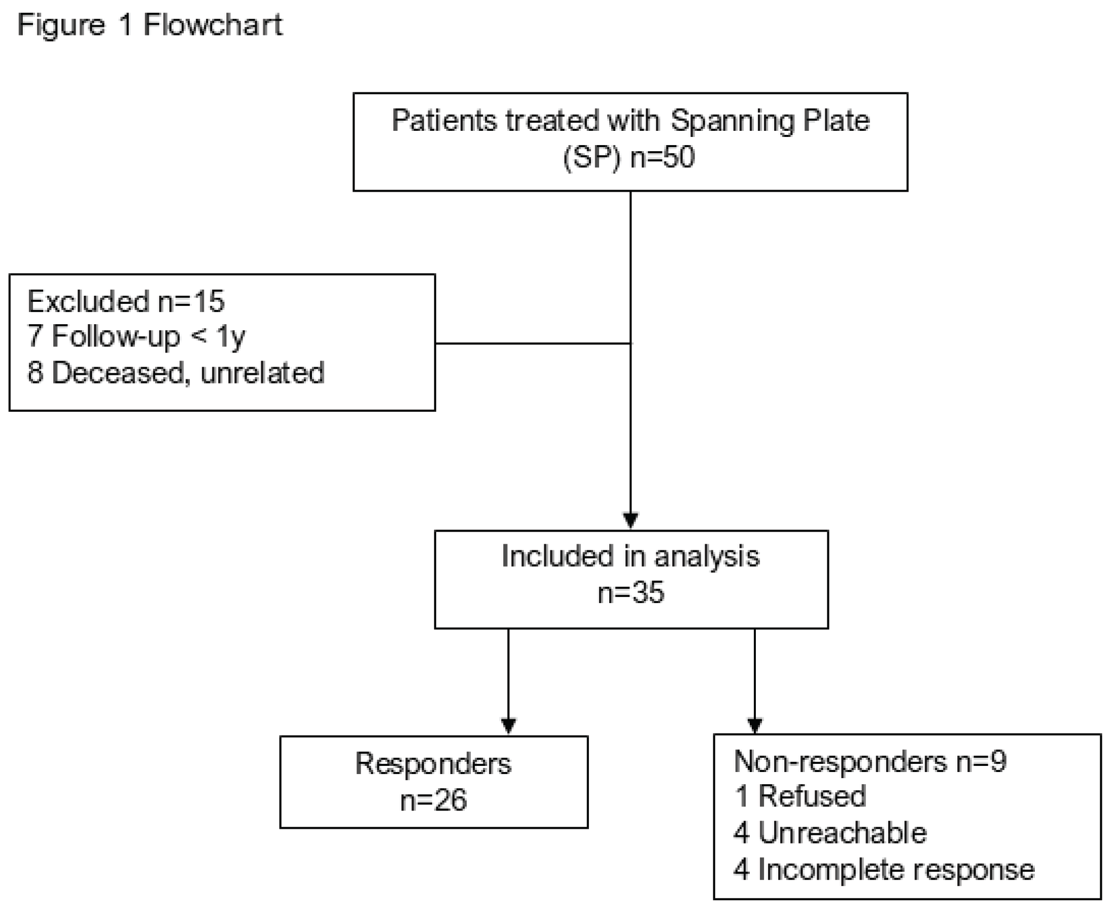

Table 1.
Patient characteristics (n = 35).
| Characteristic | Value |
| Mean age, yr (range) | 57 (22–92) |
| Gender | |
| Male, n (%) | 18 (51) |
| Female, n (%) | 17 (49) |
| Smoker, n (%) | 6 (17) |
| Diabetes, n (%) | 4 (11) |
| ASA score, mean (range) | 2 (1–4) |
| AO classification | |
| Type A fracture, n (%) | 0 (0) |
| Type B fracture, n (%) | 4 (11) |
| Type C fracture, n (%) | 31 (89) |
| Previous Fix Ex, n (%) | 21 (60) |
| Time to surgery (SP fixation), mean d (range) | 7.1 (4–11) |
| Implant used | |
| 2.4/3.5 mm SP, n (%) | 35 (100) |
| Metacarpal SP fixation | |
| MC 3, n (%) | 27 (77) |
| MC 2, n (%) | 8 (23) |
| Additional fracture fixation, n (%) | 30 (86) |
| Mean duration of SP fixation, months (range) | 3.8 (1.8–12.9) |
yr, years; ASA, American Society of Anesthesiologists; AO, Arbeitsgemeinschaft für Osteosynthesefragen; Fix Ex, external fixator; SP, spanning plate; d, days; MC, metacarpal.
Table 2.
Long-term follow-up radiological outcomes, range of motion, complications and reoperations (n = 35).
Table 2.
Long-term follow-up radiological outcomes, range of motion, complications and reoperations (n = 35).
| Outcome | Value |
| Follow-up duration in months, mean (range) | 54.8 (13–79) |
| Weeks until fracture union, mean (range) | 8.6 (5–29) |
| Delayed union, n (%) | 2 (6) |
| Non-union, n (%) | 1 (3) |
| Mean radial inclination, ° (range) | 23.2 (16–32) |
| Mean volar tilt, ° (range) | 2.0 (−12 to 14) |
| Mean ulnar variance, mm (range) | −0.3 (−9 to 5) |
| Active ROM | |
| Extension/flexion arc, mean (range) | 86 (35–135) |
| Pronation/supination arc, mean (range) | 150 (70–180) |
| CRPS I, n (%) | 3 (9) |
| Complications and reoperations, n (%) | 7 (20) |
| Implant failure, n (%) | 1 (3) |
| Surgical site infection, n (%) | 0 (0) |
| Tendon rupture, n (%) | 2 (6) |
| DRUJ stabilization, n (%) | 1 (3) |
| Non-union repair, n (%) | 1 (3) |
| Denervation of the wrist, n (%) | 1 (3) |
| Secondary total wrist arthroplasty, n (%) | 1 (3) |
ROM, range of motion; CRPS I, complex regional pain syndrome type I; DRUJ, distal radioulnar joint.
Table 3.
Missing case analysis.
| Responders (n = 26) | Non-Responders (n = 9) | p-Value | |
| Mean age, yr (range) | 57 (22–92) | 57 (24–89) | 0.95 |
| Gender | 0.78 | ||
| Male, n (%) | 13 (50) | 5 (56) | |
| Female, n (%) | 13 (50) | 4 (44) | |
| Smoker, n (%) | 2 (8) | 4 (44) | 0.01 |
| Diabetes, n (%) | 4 (15) | 0 (0) | 0.22 |
| ASA score, mean (range) | 2 (1–4) | 2 (1–3) | 0.5 |
| AO classification | 0.97 | ||
| Type A fracture, n (%) | 0 (0) | 0 (0) | |
| Type B fracture, n (%) | 3 (12) | 1 (11) | |
| Type C fracture, n (%) | 23 (88) | 8 (89) | |
| Previous Fix Ex, n (%) | 15 (58) | 6 (67) | 0.65 |
| Time to surgery (SP fixation), mean d (range) | 6.8 (4–11) | 7.8 (4–11) | 0.25 |
| Implant used | 0.41 | ||
| 2.4/3.5 mm SP, n (%) | 26 (100) | 9 (100) | |
| Metacarpal SP fixation | 0.96 | ||
| MC 3, n (%) | 20 (77) | 7 (78) | |
| MC 2, n (%) | 6 (23) | 2 (22) | |
| Additional fracture fixation, n (%) | 21 (81) | 9 (100) | 0.16 |
| Mean duration of SP fixation, months (range) | 3.8 (1.8–12.9) | 3.6 (2.9–4.5) | 0.73 |
| Reoperations, n (%) | 5 (19) | 2 (22) | 1.0 |
yr, years; ASA, American Society of Anesthesiologists; AO, Arbeitsgemeinschaft für Osteosynthesefragen; Fix Ex, external fixator; SP, spanning plate; d, days; MC, metacarpal. Bold indicates statistical significance (p < 0.05).
Table 4.
Long-term follow-up functional outcomes (n = 26).
| Outcome | Value |
| Follow-up duration in months, mean (range) | 58.3 (13–90) |
| Satisfaction score (5-point scale), mean (range) | 4.1 (2–5) |
| VAS score, mean (range) | 1.7 (0–4) |
| PRWE score Pain, mean (range) | 8.2 (0–24) |
| PRWE score Function, mean (range) | 8.4 (0–47.5) |
| PRWE score Total, mean (range) | 16.7 (0–62.5) |
VAS, Visual Analog Scale; PRWE, Patient-Rated Wrist Evaluation.
Disclaimer/Publisher’s Note: The statements, opinions and data contained in all publications are solely those of the individual author(s) and contributor(s) and not of MDPI and/or the editor(s). MDPI and/or the editor(s) disclaim responsibility for any injury to people or property resulting from any ideas, methods, instructions or products referred to in the content. |
© 2026 by the authors. Licensee MDPI, Basel, Switzerland. This article is an open access article distributed under the terms and conditions of the Creative Commons Attribution (CC BY) license (http://creativecommons.org/licenses/by/4.0/).
Copyright: This open access article is published under a Creative Commons CC BY 4.0 license, which permit the free download, distribution, and reuse, provided that the author and preprint are cited in any reuse.