

Submitted:
11 August 2026
Posted:
12 August 2026
You are already at the latest version
Abstract
Acute myeloid leukemia (AML) is cytogenetically and phenotypically heterogeneous, and this diversity contributes to differences in how patients respond to therapies that target apoptosis. Venetoclax, a selective BCL-2 inhibitor, has been shown to improve outcomes when used in combination with hypomethylating agents (HMAs) such as azacitidine or decitabine; however, clinical experience has shown resistance and relapse are common. This review examines the current evidence linking chromosomal abnormalities and cellular differentiation state to mitochondrial apoptotic pathways, with an emphasis on how these factors influence dependence on certain anti-apoptotic BCL-2 family proteins such as BCL-2, MCL-1, and BCL-XL. We summarize how specific cytogenetic subtypes and high-risk groups (including monosomy 7/del(7q) and complex karyotype/TP53-altered AML) frequently show stress-adaptive signaling and reliance on multiple anti-apoptotic pathways, which can limit the durability of response to BCL-2 inhibition. Lineage-associated dependencies are also examined, such as monocytic differentiation (which leads to increased MCL-1 reliance) and erythroid/megakaryocytic differentiation, which has been associated with increased BCL-XL dependence and venetoclax resistance. Resistance mechanisms can be grouped into three categories: primary resistance (low BCL-2 dependence), adaptive resistance (therapy-induced MCL-1/BCL-XL upregulation), and execution failure (impaired BAX/BAK function). Finally, we discuss the therapeutic implications of dependence mapping, including venetoclax combinations and direct MCL-1/BCL-XL targeting, and propose promising biomarker strategies that can detect dependence shifts early and guide appropriate treatment selection.
Keywords:
acute myeloid leukemia
; cytogenetics
; venetoclax
; BCL-2 family proteins
; BH3 mimetics
; mitochondrial apoptosis
; therapeutic resistance
; targeted therapy
1. Introduction
Acute myeloid leukemia (AML) is an aggressive hematologic cancer that is typically fatal within weeks or months if left untreated. It has a cytogenetically heterogenous profile, in which a number of chromosomal aberrations can shape its’ clinical behavior and therapeutic response. Diagnostic frameworks developed by European LeukemiaNet (ELN) have cytogenetics placed at the center of AML risk classification and treatment planning since specific chromosomal changes are linked to distinct AML subtypes and clinical outcomes [1]. Simultaneously, there has also been a shift towards lower-intensity therapies for older, medically unfit patients since previous risk classifications were developed based on data from younger, more medically fit patients receiving higher-intensity treatments. This has led to the cytogenetic risk categories to be reinterpreted using newer treatment regimens, not just intensive chemotherapy [2]. This creates an opportunity to study the different cytogenetic groups and how they interact with today’s newer therapies.
These newer, lower-intensive therapies have changed the paradigm for patients who were previously ineligible for intense chemotherapy regimens. Venetoclax, a selective BCL-2 inhibitor, received full FDA approval in 2020 when used in combination with hypomethylating agents such as azacitidine or decitabine [3]. During phase 3 of the VIALE-A trial, patients ineligible for intensive chemotherapy had higher overall survival and rates of remission with azacitidine and venetoclax compared to azacitidine alone [4]. Biological studies have suggested that part of the reason why azacitidine-venetoclax is effective is due to their disruption of leukemic stem cell metabolism, thus linking to apoptotic pathways which vary across the AML risk categories [5]. These developments are clinically important, but patient response can vary and eventually lead to a relapse, indicating that the main challenge here is understanding why some patients respond well while others relapse with resistance after initially responding.
Mechanistically, venetoclax targets the anti-apoptotic (pro-survival) role of BCL-2 mainly at the mitochondria. When AML cells emerge that rely on other anti-apoptotic proteins such as MCL-1 or BCL-XL, the BCL-2 inhibition can be ineffective and lead to the survival of cell populations with different maturity or survival pathways. One study has already shown that monocytic-differentiated AML is more resistant to azacitidine-venetoclax due to transcriptional changes that reduce BCL-2 and upregulate MCL-1 survival pathways, leaving to selective growth of a monocytic differentiation at relapse [6]. Similarly, erythroid or megakaryocytic leukemic cell lines have also shown resistance to venetoclax by switching survival dependence to BCL-XL [7]. Because survival dependency is linked to a cell’s differentiation state, resistance to venetoclax can be seen as a predictable, lineage-associated phenomenon. Conversely, relapse can also be seen outside these lineage patterns, as seen in populations with acquired mutations to BAX and resistance to BH3 proteins during venatoclax therapy [8]. These relapses highlight that resistance can potentially arise from any part of the survival pathway.
These findings suggest a link between cytogenetics and apoptotic dependence, since chromosomal aberrations can change the transcriptional and signaling pathways that directly affect cell differentiation and reliance on BCL-2 protein family. The question is whether cytogenetic features can predict which survival pathway a patient’s leukemic cells rely on, and whether it will be sensitive to venetoclax or prone to resistance through MCL-1 or BCL-XL resistance. Recent studies have suggested that this approach is feasible, showing that BCL-2 protein expression in leukemic stem cells can predict clinical response to azacitidine-venetoclax therapy [9]. Therefore, the literature supports the idea that cytogenetic features shape cell phenotypes and signaling, which in turn determine apoptotic dependence and explain why venetoclax has variable clinical responses.
This review will therefore focus on how cytogenetic aberrations and their differentiation states impact BCL-2 protein family dependence in AML, emphasizing how venetoclax effectiveness is reduced and escape pathways develop around MCL-1 or BCL-XL. The current AML classifications and their main cytogenetic categories will first be summarized and linked to the mitochondrial apoptosis pathway targeted by BH3 mimetics [1,2]. Then, the cytogenetic and phenotypic AML subsets will be examined to explain why some respond to venetoclax and others do not, with a focus on BCL-XL-dependent and monocytic cells that are less dependent on BCL-2 [6,7]. Finally, the review will summarize the key resistance pathways, including shifts in differentiation state and impaired apoptosis, and will explore potential treatment strategies such as combination regimens to limit MCL-1 or BCL-XL escape and biomarkers to monitor therapeutic efficacy [8,9].
2. AML Cytogenetics
Cytogenetic characterization is critical to AML classification because each abnormality can be classified into a disease subtype that can guide treatment selection, prognosis, and even the risk of relapse. Current ELN guidelines match chromosomal aberrations with certain gene mutations to stratify AML subtypes into favorable, intermediate, and adverse-risk groups, based on outcome data collected from large cohorts [1,10]. One study created an ELN genetic risk classification was proposed to reflect patients receiving less-intensive therapies such as venetoclax, recognizing that a shift to less-intensive treatment has changed the prognostics of subtypes [2]. More recently, the WHO 2022 and ICC 2022 have released guidelines that reinforce the idea that cytogenetic lesions found in leukemic cells are not just prognostic, but disease-defining features [11,12].
In these classification systems, chromosomal aberrations are matched into distinct AML subtypes that have unique biological characteristics. For example, the abnormalities t(8;21)(q22;22.1) and inv(16)(p13.1q22), these are categorized as favorable risk. Other abnormalities, such as MLLT3::KMT2A, are generally considered intermediate risk with their own subgroup defined by a unique transcriptional profile and clinical outcome. At the adverse end of classification, the ELN and other organizations classify complex karyotypes, unbalanced abnormalities such as -5/5q- or -7/7q-, and high-risk rearrangements such as inv(3)(q21.3q26.2) as cytogenetic features with poor clinical behavior [1,10]. In their 2022 framework, the WHO formally recognized a wider range of genetically defined AML subtypes and abolished the mandatory 20% blast requirement. This underscores that cytogenetic aberrations can define AML biology even when morphology was previously borderline [12,13].
In terms of the pathogenesis, chromosomal aberrations drive leukemic growth through three main mechanisms: (i) the formation of fused genes that disrupt normal transcription and block differentiation, (ii) changes in genetic material that directly impact tumor suppressor and apoptotic pathways, and (iii) genetic instability, as pointed out by the complex karyotypes and those with TP53 dysfunction and thus resistance to therapy [1,14]. These kinds of cytogenetic lesions impact how leukemic cells behave by locking them into a specific differentiation, promoting uncontrolled cell growth, and dysregulating their regulation mechanisms. Clinically, this matters when it comes to choosing therapies because characterizing the cytogenetic features reflects a broader cell pathway dysfunction rather than a single gene change [1,2].
The cytogenetic changes seen in different AML subtypes provide clues about which survival pathway leukemic cells depend on and their implications on treatment choice. These chromosomal abnormalities can impact the transcriptional and signaling machinery cells rely on to regulate apoptotic pathways, determining whether leukemic cells depend on BCL-2, MCL-1, or BCL-XL for survival [1,12]. These connections help explain why venetoclax works well in some AML subtypes but loses effectiveness or becomes resistant in others, since the cytogenetics shape which anti-apoptotic pathway leukemic cells depend on to develop resistance [1,2].
3. Apoptosis and BH3 Mimetics in AML
The BCL-2 protein family regulates what is called the intrinsic mitochondrial pathway of apoptosis, which permeabilizes the mitochondrial membrane and leads to cell death. The anti-apoptotic BCL-2 protein family, including MCL-1 and BCL-XL, keep the mitochondria intact by neutralizing pro-apoptotic proteins such as BAX and BAK. When these pro-apoptotic proteins are no longer inhibited, this leads to membrane permeabilization. Regulators of these pro-apoptotic proteins include BH3-only proteins, which can sense cellular stress to either directly activate BAX/BAK and promote apoptosis or neutralize them [15,16]. These regulatory pathways are frequently disrupted in AML, both because of altered BCL-2 expression and changes to cell differentiation and signaling that make cells more resistant to apoptosis [17].
One way to characterize this resistance to apoptosis is mitochondrial priming, which refers to a cell’s “readiness” for triggering apoptosis via membrane outer membrane permeabilization (MOMP). BH3 profiling is one functional assay that can be used to measure this apoptotic threshold by exposing the mitochondrial membrane to BH3 peptides and measuring the MOMP. Studies have shown that the use of BH3 profiling can identify apoptotic deficits and cells reliant on BCL-2 for survival, including cell susceptibility to BCL-2 antagonists [18]. This technique is important in AML since what matters is not just which protein is more apoptotic, but rather which protein is actually neutralizing pro-apoptotic signals.
Building on this concept, AML cells often rely on a dominant anti-apoptotic protein to inhibit pro-apoptotic signals and maintain survival. BH3 mimetics take advantage of this vulnerability by mimicking natural BH3-only proteins and binding to anti-apoptotic proteins, freeing them to activate a cell’s apoptotic pathway via BAX and BAK and cause cell death [16,19]. However, the BCL-2 protein family is functionally redundant since AML cells can shift their survival dependence from one anti-apoptotic protein to another, switching from BCL-2 to either MCL-1 or BCL-XL. This concept, known as buffering or dependency switching, can explain why venetoclax often demonstrates a strong initial clinical response that then wanes. It also highlights the importance of measuring levels of multiple anti-apoptotic proteins instead of just BCL-2. One notable study used flow cytometry to measure expression of multiple anti-apoptotic proteins in leukemic stem cells to create a combinational score that predicted azacitidine-venetoclax response, emphasizing that measuring multi-protein expression has prognostic implications [9].
Venetoclax is the most clinically mature and widely approved BH3 mimetic in AML that selectively targets BCL-2. Early preclinical studies demonstrated this when venetoclax targeting of BCL-2 led to cell death with BH3 profiling predicting its’ efficacy [20]. Later studies showed that venetoclax monotherapy had modest activity in AML patients with relapsed or unfit AML, consistent with the fact that many AML cases are not solely dependent on BCL-2 [21]. This has led to the current consensus that venetoclax efficacy can be potentiated when in combination with hypomethylating agents. Research illustrates that combination therapy is biologically sound since azacitidine leads to disruption in leukemic stem cell metabolism and oxidative phosphorylation, in addition to improved clinical efficacy [5]. Other studies have suggested that these hypomethylating agents improve efficacy by “priming” AML cells for venetoclax treatment by upregulating pro-apoptosis factors such as NOXA, weakening the leukemic cell’s ability to develop escape pathways and promoting apoptosis [22,23].
A consistent finding across studies is that whatever survival protein AML cells are dependent on is not fixed but can change in the course of treatment. This means that to fully understand cell survival requires characterization before and during treatment. The use of dynamic BH3 profiling has been shown to better detect apoptotic dependence before and during treatment when compared to standard BH3 profiling that captures a snapshot of the apoptotic state [24,25]. Another approach measures protein expression ratios, such as BCL-2 versus MCL-1 or BCL-XL, in leukemic stem cells to determine survival dependence and is easier to apply to clinical studies [9]. These assays establish the basis for linking the cytogenic and phenotypic AML features to venetoclax response and their escape mechanisms.
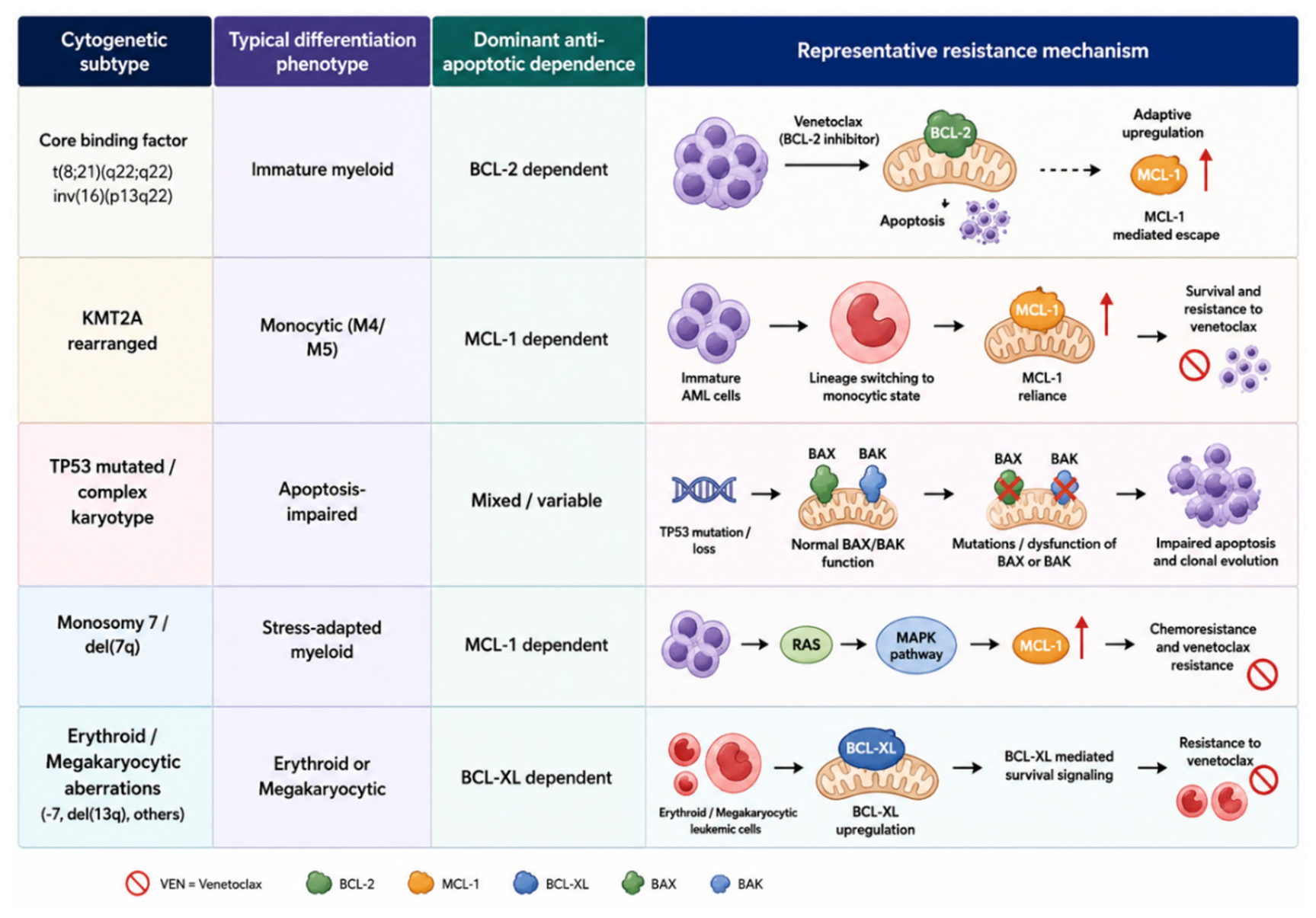
Figure 1.
Cytogenetic subtype-specific mechanisms of venetoclax resistance in acute myeloid leukemia. AML subtypes exhibit differentiation phenotypes and anti-apoptotic dependencies, enabling resistance through MCL-1 or BCL-XL upregulation, monocytic lineage switching, impaired BAX/BAK-mediated apoptosis, and activation of RAS-MAPK signaling.
Figure 1.
Cytogenetic subtype-specific mechanisms of venetoclax resistance in acute myeloid leukemia. AML subtypes exhibit differentiation phenotypes and anti-apoptotic dependencies, enabling resistance through MCL-1 or BCL-XL upregulation, monocytic lineage switching, impaired BAX/BAK-mediated apoptosis, and activation of RAS-MAPK signaling.
4. Cytogenetic and Phenotypic Drivers of BCL-2 Dependence
4.1. Core-Binding Factor (CBF)-AML
CBF-AML is a subtype defined by the chromosomal rearrangements t(8;21) and inv(16). These cytogenetic lesions are classified by the ELN as a favorable risk group, although co-mutations and other genetic factors can influence prognosis too [1,26]. Clinical data proves that CBF-AML is very responsive to chemotherapy, but it is important to delineate between t(8;21) and inv(16) because data between them is not identical. Indeed, studies have shown differences among these aberrations when it comes to relapse risk and long-term outcomes [10,26].
Most evidence for venetoclax with hypomethylating agents in CBF-AML comes from studies that also offer insight into BCL-2 dependence in this subgroup. In the large CBF-AML cohort study by Zhang et al., responses to venetoclax plus hypomethylating agents were better in the inv(16) group, but clinically suboptimal in the t(8;21) group [27]. Additional studies also support that while venatoclax combination therapy has shown clinical efficacy in select CBF-AML patients treated with low-intensity regimens, BCL-2 dependence cannot be generalized across the CBF subtype [27,28]. Thus, it is reasonable to conclude that CBF-AML is vulnerable to BCL-2 inhibition, but this vulnerability can be variable and influenced by other biological factors rather than just BCL-2 dependence [27,28].
4.2. KMT2A-Rearranged AML
This subtype of AML is defined by oncogenic KMT2A gene fusions that disrupt normal gene regulation, leading to inappropriate activation of MEIS1 and HOXA cluster genes. Clinical data has shown aggressive clinical behavior and increased risk of relapses in this subtype [29]. The KMT2A fusions often recruit helper proteins, such as menin, that keep leukemic cells in a stable epigenetic and transcriptional state to support leukemic cell survival and growth. This underscores that KMT2A-rearranged AML is driven by an altered epigenetic and transcriptional state rather than a single downstream signaling pathway [29,30].
This altered state has been reported to alter mitochondrial survival pathways too. In primary patient samples, KMT2A-rearranged AM has shown partial sensitivity to MCL-1 inhibition, consistent with a reduced reliance on BCL-2 mediated survival pathways [31]. This helps explain why venetoclax therapy alone is not sufficient, since pro-apoptotic proteins are sequestered by MCL-1 or BCL-XL instead of BCL-2, thus allowing cells to evade BCL-2 inhibition. Conversely, one preclinical trial has demonstrated that co-targeting of BCL-2 and MCL-1 can restore apoptotic wiring and overcome venetoclax resistance, indicating that MCL-1 represents a rational co-target when BCL-2 inhibition alone is insufficient [32].
KMT2A-rearranged AML commonly exhibits monocytic differentiation, a lineage state associated with reduced sensitivity to venetoclax-based therapies. Clinical studies with azacitidine-venetoclax have demonstrated that treatment efficacy is correlated to a cell’s differentiation state, with monocytic lineages at highest risk for resistance and relapses [6]. This cell lineage distinction is relevant because this subtype may have reduced dependence on BCL-2 at baseline, and starting venetoclax-based therapy may cause these leukemic cells to adapt even further by shifting survival dependence to other anti-apoptotic proteins such as MCL-1 or BCL-XL [6].
Basically, there are two treatment strategies that can be explored for this subtype: (i) disrupting KMT2A-driven transcriptional alterations or (ii) inhibiting secondary anti-apoptotic pathways. An example of the first strategy has been explored in one preclinical study, where menin-KMT2A inhibition synergizes with azacitidine-venetoclax therapy in KMT2A-rearranged AML models, supporting the idea of potential triple therapy [33,34]. This has been translated into a clinical study, where the menin inhibitor revumenib has been combined with azacitidine-venetoclax in adults with KMT2A-rearranged AML, demonstrating early feasibility and providing a clinical link between HOXA/MEIS1 transcriptional alterations and a rational triplet therapy approach [35].
4.3. Monosomy 7/Deletion 7q and Other High-Risk Groups
The monosomy 7/del(7q) AML subtype is classified as an adverse risk group that arises from prior disease or treatment and survives by activating a broad range of stress and survival pathways, rather than just one oncogenic driver [36]. In a study by Shah et al., chromosome 7 abnormalities in venetoclax-treated cohorts were associated with shorter survival and overall inferior outcomes, suggesting that BCL-2 dependence is weakened in high-risk cytogenetic groups [37]. These findings have been emphasized in other literature regarding venatoclax-resistant AML, where adverse risk groups use secondary anti-apoptotic channels including RAS/MAPK and MCL-1 to survive [38].
RAS/MAPK have been noted to promote MCL-1 stability via ERK-mediated phosphorylation, allowing leukemic cells to bypass BCL-2 inhibition under stress [39]. In a study by Zhang et al., data had shown that the RAS/MAPK pathway promoted resistance to venetoclax through upregulation of MCL-1, linking this pathway to an escape pathway [38]. All of these findings are clinically relevant because they support the idea of combination therapy in high-risk cytogenetic groups such as monosomy 7/del(7q). Combining venetoclax with MAPK inhibition to prevent MCL-1 stabilization, or targeting MCL-1 directly, may help overcome BCL-2 resistance [38].
4.4. Complex Karyotype and TP53-Altered AML
Complex karyotype AML is a cytogenetically unstable subtype that is frequently associated with TP53 alterations and is classified as adverse risk according to ELN criteria [1]. Much like the monosomy 7/del(7q) AML subtype, this category demonstrates a broad range of apoptosis-resistant pathways rather than survival on one anti-apoptotic protein. The disruption of TP53 impairs normal stress responses, leading to reduced mitochondrial priming and increased stress tolerance. As a result, activation of the mitochondrial apoptosis pathway is impaired due to disruption of BAX/BAK, reducing the efficacy of therapies that rely on the intrinsic apoptotic pathway for cell death [8,40].
With venetoclax-based therapy, these cytogenetic lesions are associated with responses that may occur but are typically not sustained compared to TP53-wild type AML. Among the VIALE-A patients with cytogenetically-confirmed poor risk subtype, azacitidine-venetoclax therapy saw improved remission rates in TP53-altered AML but did not substantially improve the remission duration or overall survival compared to azacitidine alone, indicating a limited durability of response [41]. Relapses after venetoclax therapy can occur when leukemic cells acquire genetic lesions that make apoptosis harder to trigger; specifically, inactivating mutations in BAX can disable mitochondrial apoptosis and can confer a survival advantage under treatment pressure [8].
Collectively, these findings allow two possible conclusions to be drawn for this subtype. First, venetoclax may be effective initially in complex karyotype/TP53-altered AML, but therapeutic response is short-lived. Cells adapt by switching to survival pathways such as MCL-1 or BCL-XL or by damaging the BAX pathway and preventing apoptosis. When this occurs, cells will stop responding to venatoclax and treatment efficacy falls [8]. Secondly, there [40,41]is a need for new treatment strategies and combinations that move beyond reliance for BCL-2 or apoptosis, targeting parallel survival pathways and non-apoptotic vulnerabilities to limit therapeutic resistance and escape [40,41].
4.5. Trisomy 8 and Normal Karyotype
Trisomy 8 and normal-karyotype AML illustrate a limitation of cytogenetics, as both are cytogenetically broad, heterogeneous categories in which apoptotic dependence, particularly BCL-2 reliance, is driven more by mutation status and cell state than karyotype [1,10]. In real-world trials, cohorts with trisomy 8 were shown to have poorer survival with azacitidine-venetoclax therapy, suggesting that adverse co-features play a bigger role than trisomy 8 itself [42]. To better delineate the features that influence apoptotic dependency, functional and qualitive assays add value. BH3 profiling, including dynamic BH3 profiling, measures mitochondrial priming and identifies which anti-apoptotic proteins a cell depends on, enabling the prediction for response to BH3 mimetics. [24,43]. One study has shown that measuring multiple BCL-2 family proteins at the same time could predict which patients could respond to azacitidine-venetoclax therapy, providing a practical bridge from broad cytogenetic categories treatments that target apoptosis dependence and improve venetoclax sensitivity [9].
4.6. Phenotypic Correlates with Cytogenetic Groups
Among AML subtypes, cytogenetic lesions are often associated with specific lineages. In contrast, resistance to BH3 mimetics often follows the differentiation state rather than the cytogenetics of the cell. While the cytogenetics predict which differentiation state the cells will adopt, venetoclax sensitivity largely depends on the apoptotic dependencies and other anti-apoptotic pathways maintaining cell survival [9,44].
Monocytic-differentiated AML is one example of differentiation state being correlated to venetoclax sensitivity. In a study by Pei et al., paired patient and experimental samples treated with azacitidine-venetoclax therapy revealed selection of monocytic-differentiated cells at relapse, with resistance being linked to the intrinsic features of monocytic differentiation [6]. Previous studies have characterized monocytic AML as less dependent on BCL-2 and more reliant on alternative anti-apoptotic pathways, such as MCL-1, therefore reducing the effectiveness of BCL-2 inhibition [6,44]. These findings are consistent with other mechanistic studies showing that venetoclax resistance arises from reduced mitochondrial apoptotic priming and enhanced alternative anti-apoptotic pathways, rather than the emergence of new recurrent gene mutations [44,45].
Erythroid/megakaryocytic-differentiated AML also represents a distinct lineage state that follows a different anti-apoptotic dependance. A study by Kuusanmäki et al. showed that AML cells with erythrocytic and megakaryocytic differentiation had increased dependence on BCL-XL instead of BCL-2 when profiled [7]. Thus, increased resistance to venetoclax therapy is expected, since survival depends less on BCL-2 and more on BCL-XL, opening the door to approaches that target BCL-XL in erythroid/megakaryocytic-differentiated cells [7].
Phenotype-associated dependencies also explain why targeting a single marker is inconsistent across these cohorts. Multiple studies have supported transitioning away from phenotype-based treatment, and instead to therapies that target multi-protein dependance states, such as azacitidine-venetoclax therapy, which better capture broad apoptotic vulnerabilities across leukemic cell groups [9,44].
Overall, the studies have shown support for the idea that resistance is tied to differentiation state. Monocytic-differentiated AML shifts anti-apoptotic dependence away to MCL-1, and erythroid/megakaryocytic differentiation shift dependence towards BCL-XL. Both phenotypes lead to reduced efficacy of targeted BCL-2 inhibition [6,7]. By integrating phenotypic findings with cytogenetics, this approach links cytogenetic categories to venetoclax outcomes and supports the idea for dependence-guided therapies discussed in subsequent sections [9,44].
5. Mechanisms of Resistance for BCL-2 Inhibition
5.1. Primary Resistance
Primary venetoclax resistance reflects a state in which BCL-2 inhibition is insufficient to induce mitochondrial apoptosis and ultimate cell death. Clinically, this is reflected in data by Konopleva et al. that shows a modest benefit in venetoclax monotherapy in refractory/relapsed AML. There was increased activity in leukemic cells with greater BCL-2 dependence, yet most patients showed minimal responses. This indicates that BCL-2 was not the main anti-apoptotic protein in these cells [21]. This can be explained mechanistically, as resistance is often linked to cells using a broader array of anti-apoptotic pathways such as MCL-1 or BCL-XL, combined with lower mitochondrial priming. As a result, inhibiting BCL-2 does not create a sufficient amount of cell stress to trigger apoptosis. This is supported by the work done in one study, which integrated multiple biological and clinical data types and showed assessing multiple BCL-2 dependency states, rather than a single one, more accurately captures venetoclax responsiveness [9].
Differentiation state is another key determinant of therapy effectiveness, as the key mechanism of resistance lies in a cell’s preexisting pathways rather than new recurrent gene mutations. As discussed earlier, monocytic-differentiated AML is less sensitive to azacitidine-venetoclax therapy, and biochemical analyses showed reduced BCL-2 expression and greater dependence on MCL-1, creating a baseline state where BCL-2 inhibition is not effective [9]. Meanwhile, erythroid and megakaryocytic differentiated AML cells offer a parallel case, instead relying on BCL-XL and resistant to venetoclax, further illustrating that primary resistance arises from a shift in anti-apoptotic dependence away from BCL-2 [7]. Nonetheless, clinical prediction based on phenotype alone is not sufficient. Studies integrating cell lineage with multi-protein dependency measures show that primary resistance is best understood as a measurable baseline dependency state that can guide rational treatment combinations [9].
5.2. Adaptive Resistance Under Treatment
Leukemic cells may initially be responsive to venetoclax therapy but later adapt to venetoclax therapy to develop resistance and survive. This phenomenon occurs due to loss of BCL-2 dependence, driven by increased expression and reliance on other apoptotic proteins, such as MCL-1 and BCL-XL, which capture BH3-only activators and prevent BAX/BAK activation. In AML cell lines made venetoclax-resistant by prolonged drug exposure, resistant cells were observed to have upregulated MCL-1 and sometimes BCL-XL, resulting in a state in which BCL-2 was no longer effective to induce apoptosis [32,46]. Other mechanistic studies support this idea, showing that venetoclax resistance is often associated with signaling-related changes that upregulate dependance on MCL-1 and maintain mitochondrial function, highlighting the compensatory roles for MCL-1 and BCL-XL in cell survival [38,47].
Figure 2.
FTL3-signalling and MCL-1-mediated drug resistance in acute myeloid leukemia. Constitutive signaling by mutant FLT3 activates PI3K/AKT, STAT5, and RAS/MEK/ERK pathways, increasing MCL-dependent survival and resistance to apoptosis. FLT3 inhibitors target ITD and TKD-mutant receptors.
Figure 2.
FTL3-signalling and MCL-1-mediated drug resistance in acute myeloid leukemia. Constitutive signaling by mutant FLT3 activates PI3K/AKT, STAT5, and RAS/MEK/ERK pathways, increasing MCL-dependent survival and resistance to apoptosis. FLT3 inhibitors target ITD and TKD-mutant receptors.
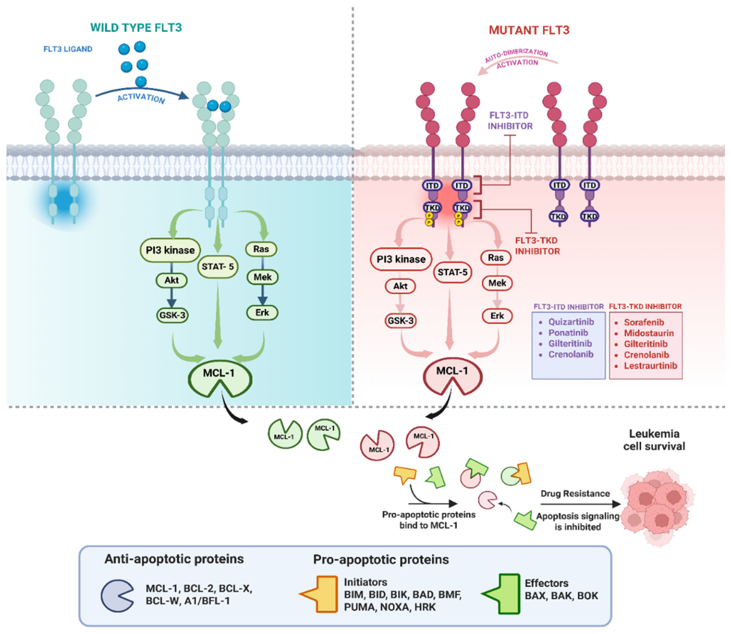
This compensation by MCL-1 upregulation is often maintained by oncogenic signaling that stabilizes MCL-1 and preserves mitochondrial stability, despite BCL-2 blockade. In AML samples, the activation of RAS/MAPK signaling has been shown to drive venetoclax resistance via MCL-1, establishing a direct mechanistic link between pathway activation and escape via BCL-2 inhibition [38]. MCL-1 has an intrinsically short half-life, but contemporary literature supports the idea that ERK-medicated phosphorylation, such as at Thr163, can stabilize the protein and extend its half-life, preserving anti-apoptotic buffering under therapy [48,49]. Alongside ERK signaling, PI3K/AKT signaling can also stabilize MCL-1 by inhibiting GSK3β-dependent degradation pathways. This AKT-GSK3β axis has been shown to modulate MCL-1 levels and apoptotic sensitivity in leukemic cell models [50]. Finally, in FLT3-dependent AML, downstream STAT5 signaling regulates anti-apoptotic pathways, including the BCL-2 protein family, explaining why kinase pathway inhibition can help restore venetoclax sensitivity [51].
The bone marrow can further promote adaptive resistance by providing cytokines and survival signals that upregulate MCL-1 and activate the pathways discussed above. Signals from the bone marrow, both stromal cell interactions and cytokine pathways such as IL-6 and JAK/STAT, can increase anti-apoptotic protein expression and reduce sensitivity to BCL-2 inhibitors. Thus, venetoclax sensitivity can be driven by the bone marrow microenvironment and not cytogenetics alone [52,53]. Functionally, this implies that cytokines such as GM-CSF and G-CSF can trigger bone marrow-like signaling that helps cancer cells avoid death. Blockade of the JAK pathway has been previously explored in studies as a way to counteract these cytokine-mediated resistance programs that protect leukemic cells from apoptosis [54]. Overall, these results show support for the rationale behind venetoclax combination therapy that lowers MCL-1 or BCL-XL activity or blocks bone marrow signaling, particularly in patients at increased susceptibility of switching of apoptotic dependence [38,46].
5.3. Execution Failure
Even if venetoclax is successful in disrupting anti-apoptotic proteins, complete cell death still depends on a functional mitochondrial death machinery. The loss or impairment of key effectors BAX and BAK prevents MOMP, enabling leukemic cells to survive despite BCL-2 inhibition. One
CRISPR screening study in AML models showed that the loss of TP53, BAX, and PMAIP1 (NOXA) genes confers venetoclax resistance, linking impaired p53-BH3 signaling and reduced BAX-mediated execution to reduced venetoclax efficacy [55]. Clinically, patients who relapse after venetoclax therapy have been found to have acquired BAX mutations that cause resistance to BH3 mimetics, showing that failure to execute apoptosis can also be a relapse mechanism [8].
6. Therapeutic Strategies Based on Dependence
6.1. Venetoclax with Hypomethylating Agents
The standard low-intensity combination regimen pairs venetoclax with a hypomethylating agent, most commonly azacitidine or decitabine. Clinically, results from VIALE-A have shown this combination regimen improves remission rates and survival time compared to azacitidine alone in older or unfit patients, with this also seen in earlier phase 1B studies [4,56]. Hypomethylating agents appear to promote apoptotic priming in a way that heightens cell sensitivity to BCL-2 inhibition. In patients, this combination targets oxidative phosphorylation and suppresses leukemic stem cells, supporting a synergistic mechanism that goes beyond reducing cell count [5]. Preclinical studies have already demonstrated that azacitidine can induce NOXA (PMAIP1) through a non-epigenetic mechanism, counteracting MCL-mediated resistance and enhancing venetoclax activity [23].

Table 1.
Therapeutic Strategies Targeting Anti-Apoptotic Dependence and Venetoclax Resistance in Acute Myeloid Leukemia.
Table 1.
Therapeutic Strategies Targeting Anti-Apoptotic Dependence and Venetoclax Resistance in Acute Myeloid Leukemia.
| Drug/Class | Target | Mechanism Relevant to Venetoclax | AML Subgroup | Key Findings |
|---|---|---|---|---|
| Venetoclax | BCL-2 | Releases BAX/BAK-mediated apoptosis | Broad AML | Backbone low-intensity therapy |
| Azacitidine | DNA methylation / OXPHOS | Primes apoptosis, induces NOXA | Older/unfit AML | Synergistic with venetoclax |
| Gilteritinib | FLT3/AXL | Suppresses MCL-1 signaling | FLT3-mut AML | Restores venetoclax sensitivity |
| Quizartinib | FLT3 | Reduces FLT3-driven survival | FLT3-ITD AML | Rational combo partner |
| AMG-176 | MCL-1 | Blocks compensatory MCL-1 escape | Venetoclax-resistant AML | Strong preclinical synergy |
| VU661013 | MCL-1 | Dual BH3 targeting | Resistant AML | Overcomes adaptive resistance |
| Revumenib | Menin-KMT2A | Disrupts HOXA/MEIS1 programs | KMT2A-r AML | Rational triplet strategy |
| DT2216 | BCL-XL PROTAC | Targets BCL-XL with platelet sparing | Erythroid/megakaryocytic AML | Reduced thrombocytopenia |
| Navitoclax | BCL-2/BCL-XL | Dual inhibition | BCL-XL-dependent AML | Limited by thrombocytopenia |
| JAK inhibitors | JAK/STAT | Blocks cytokine-mediated resistance | Microenvironment-driven resistance | Sensitizes to venetoclax |
| MAPK inhibitors | RAS/MAPK | Prevents MCL-1 stabilization | RAS-driven AML | Reduces adaptive resistance |
However, resistance ultimately develops as leukemic cells can adapt to therapeutic pressure by shifting away from BCL-2 dependence. One common resistance pathway involves increased MCL-1 expression or stabilization, allowing it to sequester pro-apoptotic factors released after BCL-2 inhibition and prevent BAX/BAK activation. Evidence from acquired resistance models and patient-linked analysis shows that RAS/MAPK signaling drives this resistance through MCL-1 upregulation and mitochondrial fitness, directly supporting combinational approaches that inhibit this pathway [38]. A second route of resistance is lineage-associated buffering, in which differentiation state can drives a shift away from BCL-2 toward MCL-1 or BCL-XL dependence, leading in venetoclax dependence, as discussed in detail earlier [6,7]. Together, these findings indicate that venetoclax with hypomethylating agents is effective when leukemic cells remain dependent on BCL-2 and apoptotic priming pathways are undisturbed, but responses are often short-lived when resistance emerges through MCL-1 or BCL-XL escape, providing a rationale for other combination strategies discussed later [7,38].
6.2. Venetoclax with FLT3 Inhibitors
Combining venetoclax with an FLT3 inhibitor is appealing because FLT3 inhibition weakens MCL-1-mediated mitochondrial survival and shifts leukemic cells towards BCL-2 dependence. In a study using FLT3-ITD AML models, inhibition of the FLT3 suppressed MCL-1-dependent survival, sensitizing cells to venetoclax-induced apoptosis [51]. Likewise, preclinical studies with the FLT3 inhibitor gilteritinib reveal synergy with venetoclax through suppression of MCL-1, indicating that inhibition of FLT3/AXL signaling disrupts an MCL-1-mediated escape pathway [57]. Clinically, this biology has been translated into meaningful clinical activity, with venetoclax combined with gilteritinib achieving high clinical and molecular response rates in relapsed/refractory cases of FLT3-mutated AML, consistent with restored sensitivity to BCL-2 inhibition [58].
6.3. Venetoclax with MCL-1 Inhibitors
The rationale of combining venetoclax with an MCL-1 inhibitor is that MCL-1 often serves as a common escape mechanism, preserving mitochondrial integrity when BCL-2 has been inhibited. By directly inhibiting MCL-1, a key escape mechanism for venetoclax resistance is disrupted, preventing the re-sequestration of pro-apoptotic factors, thereby allowing BAX/BAK activation and MOMP. In AML models, selective MCL-1 inhibitors, such as VU661013 and AMG 176, triggered apoptosis in MCL-1-dependent cells and acted synergistically with venetoclax, including in venetoclax-resistant models and patient-derived xenografts, demonstrating that dual BH3-mimetic therapy can overcome a cell’s compensatory anti-apoptosis pathway [32,36].
Safety remains a key limitation, as MCL-1 plays a critical role in normal tissues, making bone marrow suppression and heart toxicity likely on-target risks. Early clinical evaluation of the direct MCL-1 inhibitor AZD5991 revealed a high frequency of troponin elevation and severe cytopenic events, including febrile neutropenia and anemia, underscoring both cardiac monitoring concerns and on-target hematologic toxicity [59]. Cardiac toxicity has also been reported in other MCL-1 inhibitor development programs, including an FDA clinical hold for AMG 397, reinforcing the need for optimized dosing schedules, strict eligibility criteria, and frequent cardiac monitoring in future venetoclax-based combination regimens [59].
6.4. BCL-XL Targeting
A subset of AML cases is naturally resistant to venatoclax due to BCL-XL survival dependence instead of BCL-2. This is most evident in AML cells with erythroid/megakaryocytic differentiation, a relationship between differentiation state and BCL-XL-dependent survival that was discussed earlier [7]. In these lineage-skewed states, BCL-2 inhibition alone is often ineffective, as it does not target the dominant anti-apoptotic protein and instead requires overcoming BCL-XL buffering [7,60]. These findings provide a strong biological rationale for targeting BCL-XL in erythroid and megakaryocytic AML, as well as in other contexts where BCL-XL is dominant [7].
The main challenge is that by directly targeting BCL-XL in leukemic cells, it also causes on-target platelet toxicity due to the essential role that BCL-XL has in platelet survival. Clinical studies with the dual BCL-2/BCL-XL inhibitor navitoclax showed dose-limiting thrombocytopenia, which mechanistic studies have confirmed as on-target consequence of platelet dependence on BCL-XL [61,62]. Newer therapies have therefore focused on targeting tumor-selective BCL-XL and sparing platelets. One promising strategy is the use of platelet-sparing BCL-XL-targeting PROTACs such as DT2216, which leverage low VHL expression in platelets to mitigate thrombocytopenia and have advanced into the first-in-human studies [63]. Another approach involves using delivery modalities such as antibody-drug conjugates to enrich BCL-XL-directed payloads in tumor cells while reducing systemic platelet exposure [64]. Together, these approaches reflect the emerging strategy of matching BCL-XL-dependent AML phenotypes with BCL-XL-directed therapy while circumventing on-target platelet toxicity and targeting the tumor microenvironment.
6.5. Treatment Sequencing and Timing
In clinical practice, the primary constraint for venetoclax-based combinations is treatment-limiting cytopenia, underscoring the importance of timing and duration when designing an appropriate regimen. In VIALE-A, management of cytopenias included protocol-guided treatment interruptions and reduction of venetoclax from 28 to 21 days per cycle, with long-term follow-up confirming that many patients with sustained remission are maintained on 21 days or fewer per cycle [4,65]. This supports the practice of intermittent dosing when combining venetoclax with agents that target MCL-1 or BCL-XL escape, enabling sufficient on-target leukemic cell eradication while allowing hematopoietic recovery and adjusting treatment intensity based on apoptotic dependence. Functional monitoring tools can support this approach, as BH3 profiling directly measures mitochondrial priming and anti-apoptotic dependence, thus complementing protein-expression ratios in identifying emerging dependence switching in leukemic cells [24,66]. Lastly, adverse effects specific to each target should guide how venetoclax combinations are decided. BCL-XL inhibition is limited by platelet-dependent thrombocytopenia, whereas early clinical experience with direct MCL-1 inhibitors highlights the need to monitor cardiac biomarkers such as troponin and overlapping myelosuppression [59,62,63].
7. Biomarkers and Future Therapeutic Directions
7.1. Biomarkers to Guide Dependence-Based Therapy
The use of BH3 profiling, also known as functional priming, offers a direct functional measurement of mitochondrial priming and anti-apoptotic dependence that complements cytogenetic data when predicting response to BH3 mimetics. In AML, reduced mitochondrial priming correlates with venetoclax resistance, supporting the use of functional priming as a treatment-relevant biomarker instead of a description of phenotype [44]. More recently, dynamic BH3 profiling has been adapted as a rapid (48-hour) ex vivo assay for patient samples. It predicts clinical outcomes by measuring how drugs altar apoptotic priming, helping detect early changes in cellular dependence during therapy [67]. Early clinical studies in frontline azacitidine-venetoclax treatment show that BH3 profiling during therapy can identify treatment responders vs non-responders, emphasizing its’ applicability in clinical settings [68].
Given that dependence is commonly distributed across multiple BCL-2 family proteins, clinically useful biomarkers are increasingly focused on multi-protein patterns rather than single markers. One notable example is a Cancer Discovery study that demonstrated that combining transcriptional, proteomic, functional, and clinical data can predict response to azacitidine-venetoclax treatment through coordinated BCL-2 family expression in AML stem cells [9]. Using this approach, clinically practical readouts rely on relative measures of BCL-2 versus MCL-1 or BCL-XL to identify the dominant anti-apoptotic protein, providing an alternative to full functional priming while still capturing underlying dependence [9].
As the risk of relapse is often driven by leukemic stem cells and the mitochondrial pathways they rely on, biomarkers that target stem and progenitor cells may better identify treatment vulnerabilities than profiling leukemic cells alone. Studies have shown that venetoclax combined with azacitidine can disrupt energy metabolism and oxidative phosphorylation and selectively targets leukemic stem cells. These effects provide a mechanistic basis for using leukemic stem cell dependency and metabolic profiling as tools to predict treatment response [5,69]. In line with these findings, when dependency signatures such as the BCL-2 family proteins were measured in AML cells, they were found to correlate with clinical response to azacitidine and venetoclax [9]. This supports the idea of a future approach in which baseline and treatment leukemic stem cell dependence states are measured to guide both initial therapy selection and adaptation as resistance emerges [9].
7.2. Future Directions
An important next step is the development of prospective trials that move beyond single-factor risk grouping to jointly integrate cytogenetic risk with functional anti-apoptotic dependence. Existing frameworks do already classify genetic risk across treatment intensities, including hypomethylating agent and venetoclax regimens, creating a consistent structure for cytogenetic grouping [1,2]. Advances now make it possible to incorporate functional measures of anti-apoptotic dependence, including stem and progenitor BCL-2 family signatures or priming assays, to distinguish treatment states within each genetic risk category. A stem cell-focused combinatorial BCL-2 family expression biomarker that is predictive of azacitidine-venetoclax treatment response illustrates how dependence states can be measured and used to guide clinical trial design [9]. Early clinical experience shows that targeted therapies can be layered onto azacitidine-venetoclax to target specific molecular subgroups, enabling stratified approaches that can be applied without moving away from the established standard treatments [70].
Another promising approach is early identification of dependency switching during therapy, with treatment intensity adjusted according to predefined rules. Dynamic BH3 profiling provides a rapid functional test that measures treatment-induced changes in apoptotic sensitivity and predicts response to chemotherapy and targeted therapies, enabling early detection of shifts toward MCL-1 or BCL-XL dependence before relapse becomes clinically apparent [67]. Clinical trials incorporating these early functional readouts must also address treatment tolerance. Venetoclax-based regimens commonly require adjustments in dose scheduling due to cytopenia risk, with evidence from real-world studies and VIALE-A suggesting that shorter or intermittent venetoclax exposure can preserve efficacy while reducing toxicity [71,72]. This supports a treatment paradigm that integrates dependency-aligned drug combinations with sequenced or time-limited dosing and targeted monitoring to maximize leukemic cell death while limiting toxicity and other on-target effects [67,71]
8. Conclusions
Cytogenetic abnormalities, together with their phenotypic maturation, offer a useful framework for interpreting the regulatory mechanisms of mitochondrial apoptosis in AML. The presence of certain chromosomal arrangements and other high-risk cytogenetic patterns cause unique transcriptional and signaling pathways that impact mitochondrial priming, altering the balance of translation and apoptosis within the BCL-2 family. Modern classification and risk-stratification guidelines reflect this biology by recognizing many chromosomal abnormalities as both disease-defining and prognostically significant. These same features also help explain the variability in Venetoclax response across the different AML subtypes.
Resistance to BCL-2 inhibition most commonly arises through a shift in anti-apoptotic dependence, rather than a single uniform mechanism. When leukemic cells are treated with Venetoclax, they can shift their survival dependence to alternative anti-apoptotic proteins, particularly MCL-1 or BCL-XL. These adaptations allow them to re-sequester pro-apoptotic activators and block BAX/BAK-mediated mitochondrial outer membrane permeabilization. Previous studies have clarified that RAS/MAPK pathway activation promotes MCL-1-mediated resistance. Concurrently, phenotypic studies have shown that monocytic differentiation is associated with reduced BCL-2 dependence, while erythroid and megakaryocytic differentiation are more reliant on BCL-XL. Overall, both patterns correlate with decreased efficacy of Venetoclax.
The impact of this anti-apoptotic dependence switching from BCL-2 reliance has led to important therapeutic implications. In this context, Venetoclax is best utilized as a backbone and combined with other agents that target the dominant resistance pathway, while preserving dose tolerance through optimized dosing and scheduling. Clinical studies with the azacitidine/venetoclax combination therapy have demonstrated meaningful clinical benefit, but also treatment-related cytopenias that necessitate careful dose and duration adjustments as key to feasibility for combination regimens. Looking ahead, the integration of predictive biomarkers such as BH3 profiling, multi-protein dependence signatures, and stem and progenitor focused profiling offers a strategy to detect anti-apoptotic dependencies early during therapy and to adapt treatment regimens before overt relapse. This will ultimately enable a more precise and proactive approach to achieving clinically meaningful, durable, and sustainable responses to AML.
Acknowledgments
This work is dedicated to the memory of Dr. William “Bill” Kocher, whose mentorship and devotion to teaching inspired countless students, including the first author, to pursue a deeper interest in hematology-oncology. This article reflects the enduring passion for leukemia research that he instilled through his guidance, encouragement, and example.
References
- Döhner, H.; Wei, A.H.; Appelbaum, F.R.; Craddock, C.; DiNardo, C.D.; Dombret, H.; et al. Diagnosis and management of AML in adults: 2022 recommendations from an international expert panel on behalf of the ELN. Blood 2022, 140(12), 1345–77. [Google Scholar] [CrossRef] [PubMed]
- Döhner, H.; DiNardo, C.D.; Appelbaum, F.R.; Craddock, C.; Dombret, H.; Ebert, B.L.; et al. Genetic risk classification for adults with AML receiving less-intensive therapies: the 2024 ELN recommendations. Blood 2024, 144(21), 2169–73. [Google Scholar] [CrossRef] [PubMed]
- Vachhani, P.; Flahavan, E.M.; Xu, T.; Ma, E.; Montez, M.; Gershon, A.; et al. Venetoclax and Hypomethylating Agents as First-line Treatment in Newly Diagnosed Patients with AML in a Predominately Community Setting in the US. The Oncologist 2022, 27(11), 907–18. [Google Scholar] [CrossRef] [PubMed]
- DiNardo, C.D.; Jonas, B.A.; Pullarkat, V.; Thirman, M.J.; Garcia, J.S.; Wei, A.H.; et al. Azacitidine and Venetoclax in Previously Untreated Acute Myeloid Leukemia. N. Engl. J. Med. 2020, 383(7), 617–29. [Google Scholar] [CrossRef] [PubMed]
- Pollyea, D.A.; Stevens, B.M.; Jones, C.L.; Winters, A.; Pei, S.; Minhajuddin, M.; et al. Venetoclax with azacitidine disrupts energy metabolism and targets leukemia stem cells in patients with acute myeloid leukemia. Nat. Med. 2018, 24(12), 1859–66. [Google Scholar] [CrossRef] [PubMed]
- Pei, S.; Pollyea, D.A.; Gustafson, A.; Stevens, B.M.; Minhajuddin, M.; Fu, R.; et al. Monocytic Subclones Confer Resistance to Venetoclax-Based Therapy in Patients with Acute Myeloid Leukemia. Cancer Discov. 2020, 10(4), 536–51. [Google Scholar] [CrossRef] [PubMed]
- Kuusanmäki, H.; Dufva, O.; Vähä-Koskela, M.; Leppä, A.-M.; Huuhtanen, J.; Vänttinen, I.; et al. Erythroid/megakaryocytic differentiation confers BCL-XL dependency and venetoclax resistance in acute myeloid leukemia. Blood 2023, 141(13), 1610–25. [Google Scholar] [CrossRef] [PubMed]
- Moujalled, D.M.; Brown, F.C.; Chua, C.C.; Dengler, M.A.; Pomilio, G.; Anstee, N.S.; et al. Acquired mutations in BAX confer resistance to BH3-mimetic therapy in acute myeloid leukemia. Blood 2023, 141(6), 634–44. [Google Scholar] [CrossRef] [PubMed]
- Waclawiczek, A.; Leppä, A.-M.; Renders, S.; Stumpf, K.; Reyneri, C.; Betz, B.; et al. Combinatorial BCL2 Family Expression in Acute Myeloid Leukemia Stem Cells Predicts Clinical Response to Azacitidine/Venetoclax. Cancer Discov. 2023, 13(6), 1408–27. [Google Scholar] [CrossRef] [PubMed]
- Mrózek, K.; Kohlschmidt, J.; Blachly, J.S.; Nicolet, D.; Carroll, A.J.; Archer, K.J.; et al. Outcome Prediction By the New 2022 European Leukemia Net (ELN) Genetic-Risk Classification for Adult Patients (Pts) with Acute Myeloid Leukemia (AML): An Alliance Study. Blood 2022, 140 (Supplement 1), 6318–20. [Google Scholar] [CrossRef]
- Arber, D.A.; Orazi, A.; Hasserjian, R.P.; Borowitz, M.J.; Calvo, K.R.; Kvasnicka, H.-M.; et al. International Consensus Classification of Myeloid Neoplasms and Acute Leukemias: integrating morphologic, clinical, and genomic data. Blood 2022, 140(11), 1200–28. [Google Scholar] [CrossRef] [PubMed]
- Khoury, J.D.; Solary, E.; Abla, O.; Akkari, Y.; Alaggio, R.; Apperley, J.F.; et al. The 5th edition of the World Health Organization Classification of Haematolymphoid Tumours: Myeloid and Histiocytic/Dendritic Neoplasms. Leukemia 2022, 36(7), 1703–19. [Google Scholar] [CrossRef] [PubMed]
- CC C, A. K. What’s new in AML Classification (WHO 2022 vs International Consensus Classification): College of American Pathologists. 2023. Available online: https://www.cap.org/member-resources/articles/whats-new-in-aml-classification-who-2022-vs-international-consensus-classification.
- Mrózek, K. Molecular cytogenetics in acute myeloid leukemia in adult patients: practical implications. Pol. Arch. Intern Med. 2022, 132, 7–8. [Google Scholar] [CrossRef] [PubMed]
- Qian, S.; Wei, Z.; Yang, W.; Huang, J.; Yang, Y.; Wang, J. The role of BCL-2 family proteins in regulating apoptosis and cancer therapy. Front Oncol. 2022, 12, 985363. [Google Scholar] [CrossRef] [PubMed]
- Vogler, M.; Braun, Y.; Smith, V.M.; Westhoff, M.A.; Pereira, R.S.; Pieper, N.M.; et al. The BCL2 family: from apoptosis mechanisms to new advances in targeted therapy. Signal Transduct. Target Ther. 2025, 10(1), 91. [Google Scholar] [CrossRef] [PubMed]
- Wei, Y.; Cao, Y.; Sun, R.; Cheng, L.; Xiong, X.; Jin, X.; et al. Targeting Bcl-2 Proteins in Acute Myeloid Leukemia. Frontiers in Oncology 2020, 10. [Google Scholar] [CrossRef] [PubMed]
- Deng, J.; Carlson, N.; Takeyama, K.; Dal Cin, P.; Shipp, M.; Letai, A. BH3 profiling identifies three distinct classes of apoptotic blocks to predict response to ABT-737 and conventional chemotherapeutic agents. Cancer Cell 2007, 12(2), 171–85. [Google Scholar] [CrossRef] [PubMed]
- Roberts, A.W.; Wei, A.H.; Huang, D.C.S. BCL2 and MCL1 inhibitors for hematologic malignancies. Blood 2021, 138(13), 1120–36. [Google Scholar] [CrossRef] [PubMed]
- Pan, R.; Hogdal, L.J.; Benito, J.M.; Bucci, D.; Han, L.; Borthakur, G.; et al. Selective BCL-2 Inhibition by ABT-199 Causes On-Target Cell Death in Acute Myeloid Leukemia. Cancer Discov. 2014, 4(3), 362–75. [Google Scholar] [CrossRef] [PubMed]
- Konopleva, M.; Pollyea, D.A.; Potluri, J.; Chyla, B.; Hogdal, L.; Busman, T.; et al. Efficacy and Biological Correlates of Response in a Phase II Study of Venetoclax Monotherapy in Patients with Acute Myelogenous Leukemia. Cancer Discov. 2016, 6(10), 1106–17. [Google Scholar] [CrossRef] [PubMed]
- Garciaz, S.; Saillard, C.; Hicheri, Y.; Hospital, M.A.; Vey, N. Venetoclax in Acute Myeloid Leukemia: Molecular Basis, Evidences for Preclinical and Clinical Efficacy and Strategies to Target Resistance. Cancers 2021, 13(22). [Google Scholar] [CrossRef] [PubMed]
- Jin, S.; Cojocari, D.; Purkal, J.J.; Popovic, R.; Talaty, N.N.; Xiao, Y.; et al. 5-Azacitidine Induces NOXA to Prime AML Cells for Venetoclax-Mediated Apoptosis. Clin. Cancer Res. 2020, 26(13), 3371–83. [Google Scholar] [CrossRef] [PubMed]
- Certo, M.; Del Gaizo Moore, V.; Nishino, M.; Wei, G.; Korsmeyer, S.; Armstrong, S.A.; et al. Mitochondria primed by death signals determine cellular addiction to antiapoptotic BCL-2 family members. Cancer Cell 2006, 9(5), 351–65. [Google Scholar] [CrossRef] [PubMed]
- Montero, J.; Letai, A. Dynamic BH3 profiling-poking cancer cells with a stick. Mol. Cell. Oncol. 2016, 3(3), e1040144. [Google Scholar] [CrossRef] [PubMed]
- Darwish, C.; Farina, K.; Tremblay, D. The core concepts of core binding factor acute myeloid leukemia: Current considerations for prognosis and treatment. Blood Rev. 2023, 62, 101117. [Google Scholar] [CrossRef] [PubMed]
- Zhang, K.; Zhang, X.; Xu, Y.; Xue, S.; Qiu, H.; Tang, X.; et al. Efficacy of venetoclax combined with hypomethylating agents in young, and unfit patients with newly diagnosed core binding factor acute myeloid leukemia. Blood Cancer J. 2023, 13(1), 155. [Google Scholar] [CrossRef] [PubMed]
- Arslan, S.; Zhang, J.; Dhakal, P.; Moran, J.A.; Naidoo, N.; Lombardi Story, J.; et al. Outcomes of Therapy with Venetoclax Combined with Hypomethylating Agents in Favorable-Risk Acute Myeloid Leukemia (AML). Blood 2020, 136 (Supplement 1), 41–2. [Google Scholar] [CrossRef]
- Zehtabcheh, S.; Soleimani Samarkhazan, H.; Asadi, M.; Zabihi, M.; Parkhideh, S.; Mohammadi, M.H. Insights into KMT2A rearrangements in acute myeloid leukemia: from molecular characteristics to targeted therapies. Biomark. Res. 2025, 13(1), 73. [Google Scholar] [CrossRef] [PubMed]
- Thakur, R.K.; Wang, E.S. The promise of menin inhibitors: from approval to triplet regimens. Hematology 2025, 2025(1), 599–606. [Google Scholar] [CrossRef] [PubMed]
- Safa-Tahar-Henni, S.; Páez Martinez, K.; Gress, V.; Esparza, N.; Roques, É.; Bonnet-Magnaval, F.; et al. Comparative small molecule screening of primary human acute leukemias, engineered human leukemia and leukemia cell lines. Leukemia 2025, 39(1), 29–41. [Google Scholar] [CrossRef] [PubMed]
- Ramsey, H.E.; Fischer, M.A.; Lee, T.; Gorska, A.E.; Arrate, M.P.; Fuller, L.; et al. A Novel MCL1 Inhibitor Combined with Venetoclax Rescues Venetoclax-Resistant Acute Myelogenous Leukemia. Cancer Discov. 2018, 8(12), 1566–81. [Google Scholar] [CrossRef] [PubMed]
- Issa, G.C.; Cai, S.F.; Bataller, A.; Kantarjian, H.M.; Stein, E.M. Combination Strategies with Menin Inhibitors for Acute Leukemia. Blood Cancer Discov. 2025, 6(6), 547–60. [Google Scholar] [CrossRef] [PubMed]
- Kwon, M.C.; Thuring, J.W.; Querolle, O.; Dai, X.; Verhulst, T.; Pande, V.; et al. Preclinical efficacy of the potent, selective menin-KMT2A inhibitor JNJ-75276617 (bleximenib) in KMT2A- and NPM1-altered leukemias. Blood 2024, 144(11), 1206–20. [Google Scholar] [CrossRef] [PubMed]
- Zeidner, J.F.; Lin, T.L.; Welkie, R.L.; Curran, E.; Koenig, K.; Stock, W.; et al. Azacitidine, Venetoclax, and Revumenib for Newly Diagnosed NPM1-Mutated or KMT2A-Rearranged AML. J. Clin. Oncol. 2025, 43(23), 2606–15. [Google Scholar] [CrossRef] [PubMed]
- Abbas, H.A.; Ayoub, E.; Sun, H.; Kanagal-Shamanna, R.; Short, N.J.; Issa, G.; et al. Clinical and molecular profiling of AML patients with chromosome 7 or 7q deletions in the context of TP53 alterations and venetoclax treatment. Leuk. Lymphoma 2022, 63(13), 3105–16. [Google Scholar] [CrossRef] [PubMed]
- Shah, M.V.; Chhetri, R.; Dholakia, R.; Kok, C.H.; Gangat, N.; Alkhateeb, H.B.; et al. Outcomes following venetoclax-based treatment in therapy-related myeloid neoplasms. Am. J. Hematol. 2022, 97(8), 1013–22. [Google Scholar] [CrossRef] [PubMed]
- Zhang, Q.; Riley-Gillis, B.; Han, L.; Jia, Y.; Lodi, A.; Zhang, H.; et al. Activation of RAS/MAPK pathway confers MCL-1 mediated acquired resistance to BCL-2 inhibitor venetoclax in acute myeloid leukemia. Signal Transduct. Target Ther. 2022, 7(1), 51. [Google Scholar] [CrossRef] [PubMed]
- Cook, S.J.; Stuart, K.; Gilley, R.; Sale, M.J. Control of cell death and mitochondrial fission by ERK1/2 MAP kinase signalling. Febs j. 2017, 284(24), 4177–95. [Google Scholar] [CrossRef] [PubMed]
- Shahzad, M.; Amin, M.K.; Daver, N.G.; Shah, M.V.; Hiwase, D.; Arber, D.A.; et al. What have we learned about TP53-mutated acute myeloid leukemia? Blood Cancer J. 2024, 14(1), 202. [Google Scholar] [CrossRef] [PubMed]
- Pollyea, D.A.; Pratz, K.W.; Wei, A.H.; Pullarkat, V.; Jonas, B.A.; Recher, C.; et al. Outcomes in Patients with Poor-Risk Cytogenetics with or without TP53 Mutations Treated with Venetoclax and Azacitidine. Clin. Cancer Res. 2022, 28(24), 5272–9. [Google Scholar] [CrossRef] [PubMed]
- Othman, J.; Lam, H.P.J.; Leong, S.; Basheer, F.; Abdallah, I.; Fleming, K.; et al. Real-world outcomes of newly diagnosed AML treated with venetoclax and azacitidine or low-dose cytarabine in the UK NHS. Blood Neoplasia 2024, 1(3), 100017. [Google Scholar] [CrossRef] [PubMed]
- Montero, J.; Sarosiek, K.A.; DeAngelo, J.D.; Maertens, O.; Ryan, J.; Ercan, D.; et al. Drug-induced death signaling strategy rapidly predicts cancer response to chemotherapy. Cell 2015, 160(5), 977–89. [Google Scholar] [CrossRef] [PubMed]
- Bhatt, S.; Pioso, M.S.; Olesinski, E.A.; Yilma, B.; Ryan, J.A.; Mashaka, T.; et al. Reduced Mitochondrial Apoptotic Priming Drives Resistance to BH3 Mimetics in Acute Myeloid Leukemia. Cancer Cell 2020, 38(6), 872–90.e6. [Google Scholar] [CrossRef] [PubMed]
- Liu, J.; Chen, Y.; Yu, L.; Yang, L. Mechanisms of venetoclax resistance and solutions. Front Oncol. 2022, 12, 1005659. [Google Scholar] [CrossRef] [PubMed]
- Ong, F.; Kim, K.; Konopleva, M.Y. Venetoclax resistance: mechanistic insights and future strategies. Cancer Drug Resist. 2022, 5(2), 380–400. [Google Scholar] [CrossRef] [PubMed]
- Condoluci, A.; Rossi, D. Mechanisms of resistance to venetoclax. Blood 2022, 140(20), 2094–6. [Google Scholar] [CrossRef] [PubMed]
- Senichkin, V.V.; Streletskaia, A.Y.; Gorbunova, A.S.; Zhivotovsky, B.; Kopeina, G.S. Saga of Mcl-1: regulation from transcription to degradation. Cell Death Differ. 2020, 27(2), 405–19. [Google Scholar] [CrossRef] [PubMed]
- Nifoussi, S.K.; Vrana, J.A.; Domina, A.M.; De Biasio, A.; Gui, J.; Gregory, M.A.; et al. Thr 163 Phosphorylation Causes Mcl-1 Stabilization when Degradation Is Independent of the Adjacent GSK3-Targeted Phosphodegron, Promoting Drug Resistance in Cancer. PLoS ONE 2012, 7(10), e47060. [Google Scholar] [CrossRef] [PubMed]
- Wang, R.; Xia, L.; Gabrilove, J.; Waxman, S.; Jing, Y. Downregulation of Mcl-1 through GSK-3β activation contributes to arsenic trioxide-induced apoptosis in acute myeloid leukemia cells. Leukemia 2013, 27(2), 315–24. [Google Scholar] [CrossRef] [PubMed]
- Singh Mali, R.; Zhang, Q.; DeFilippis, R.A.; Cavazos, A.; Kuruvilla, V.M.; Raman, J.; et al. Venetoclax combines synergistically with FLT3 inhibition to effectively target leukemic cells in FLT3-ITD+ acute myeloid leukemia models. Haematologica 2021, 106(4), 1034–46. [Google Scholar] [CrossRef] [PubMed]
- Gupta, V.A.; Matulis, S.M.; Conage-Pough, J.E.; Nooka, A.K.; Kaufman, J.L.; Lonial, S.; et al. Bone marrow microenvironment–derived signals induce Mcl-1 dependence in multiple myeloma. Blood 2017, 129(14), 1969–79. [Google Scholar] [CrossRef] [PubMed]
- Leverson, J.D.; Cojocari, D. Hematologic Tumor Cell Resistance to the BCL-2 Inhibitor Venetoclax: A Product of Its Microenvironment? Frontiers in Oncology 2018, 8. [Google Scholar] [CrossRef] [PubMed]
- Karjalainen, R.; Pemovska, T.; Popa, M.; Liu, M.; Javarappa, K.K.; Majumder, M.M.; et al. JAK1/2 and BCL2 inhibitors synergize to counteract bone marrow stromal cell–induced protection of AML. Blood 2017, 130(6), 789–802. [Google Scholar] [CrossRef] [PubMed]
- Nechiporuk, T.; Kurtz, S.E.; Nikolova, O.; Liu, T.; Jones, C.L.; D'Alessandro, A.; et al. The TP53 Apoptotic Network Is a Primary Mediator of Resistance to BCL2 Inhibition in AML Cells. Cancer Discov. 2019, 9(7), 910–25. [Google Scholar] [CrossRef] [PubMed]
- DiNardo, C.D.; Pratz, K.; Pullarkat, V.; Jonas, B.A.; Arellano, M.; Becker, P.S.; et al. Venetoclax combined with decitabine or azacitidine in treatment-naive, elderly patients with acute myeloid leukemia. Blood 2019, 133(1), 7–17. [Google Scholar] [CrossRef] [PubMed]
- Janssen, M.; Schmidt, C.; Bruch, P.-M.; Blank, M.F.; Rohde, C.; Waclawiczek, A.; et al. Venetoclax synergizes with gilteritinib in FLT3 wild-type high-risk acute myeloid leukemia by suppressing MCL-1. Blood 2022, 140(24), 2594–610. [Google Scholar] [CrossRef] [PubMed]
- Daver, N.; Perl, A.E.; Maly, J.; Levis, M.; Ritchie, E.; Litzow, M.; et al. Venetoclax Plus Gilteritinib for FLT3-Mutated Relapsed/Refractory Acute Myeloid Leukemia. J. Clin. Oncol. 2022, 40(35), 4048–59. [Google Scholar] [CrossRef] [PubMed]
- Desai, P.; Lonial, S.; Cashen, A.; Kamdar, M.; Flinn, I.; O’Brien, S.; et al. A Phase 1 First-in-Human Study of the MCL-1 Inhibitor AZD5991 in Patients with Relapsed/Refractory Hematologic Malignancies. Clin. Cancer Res. 2024, 30(21), 4844–55. [Google Scholar] [CrossRef] [PubMed]
- Nachmias, B.; Aumann, S.; Haran, A.; Schimmer, A.D. Venetoclax resistance in acute myeloid leukaemia-Clinical and biological insights. Br. J. Haematol. 2024, 204(4), 1146–58. [Google Scholar] [CrossRef] [PubMed]
- Debrincat, M.A.; Pleines, I.; Lebois, M.; Lane, R.M.; Holmes, M.L.; Corbin, J.; et al. BCL-2 is dispensable for thrombopoiesis and platelet survival. Cell Death Dis. 2015, 6(4), e1721–e. [Google Scholar] [CrossRef] [PubMed]
- Gandhi, L.; Camidge, D.R.; Ribeiro de Oliveira, M.; Bonomi, P.; Gandara, D.; Khaira, D.; et al. Phase I study of Navitoclax (ABT-263), a novel Bcl-2 family inhibitor, in patients with small-cell lung cancer and other solid tumors. J. Clin. Oncol. 2011, 29(7), 909–16. [Google Scholar] [CrossRef] [PubMed]
- Mahadevan, D.; Barve, M.; Mahalingam, D.; Parekh, J.; Kurman, M.; Strauss, J.; et al. First in human phase 1 study of DT2216, a selective BCL-xL degrader, in patients with relapsed/refractory solid malignancies. J. Hematol. Oncol. 2025, 18(1), 98. [Google Scholar] [CrossRef] [PubMed]
- Judd, A.S.; Bawa, B.; Buck, W.R.; Tao, Z.-F.; Li, Y.; Mitten, M.J.; et al. BCL-XL-targeting antibody-drug conjugates are active in preclinical models and mitigate on-mechanism toxicity of small-molecule inhibitors. Sci. Adv. 2024, 10(40), eado7120. [Google Scholar] [CrossRef] [PubMed]
- Pratz, K.W.; Jonas, B.A.; Pullarkat, V.; Thirman, M.J.; Garcia, J.S.; Döhner, H.; et al. Long-term follow-up of VIALE-A: Venetoclax and azacitidine in chemotherapy-ineligible untreated acute myeloid leukemia. Am. J. Hematol. 2024, 99(4), 615–24. [Google Scholar] [CrossRef] [PubMed]
- Del Gaizo Moore, V.; Letai, A. BH3 profiling--measuring integrated function of the mitochondrial apoptotic pathway to predict cell fate decisions. Cancer Lett. 2013, 332(2), 202–5. [Google Scholar] [CrossRef] [PubMed]
- Minihane, E.-J.; Krotee, L.; Wu, J.; Thornton, L.; Fell, G.; Herter, S.; et al. Dynamic BH3 profiling predicts clinical outcomes in acute myeloid leukemia. bioRxiv 2025. 2025.10.14.682414. [Google Scholar]
- Cluzeau, T.; Favreau, C.; Bourgoin, M.; Venton, G.; Fanciullino, R.; Boucher, M.; et al. Venetoclax dosages, BH3 profiling, and bcl-2 expression predict response to azacitidine + venetoclax regimen in first line AML not eligible to intensive chemotherapy: First results of venetacible study. Blood 2025, 146, 5250. [Google Scholar] [CrossRef]
- Staff, N. Drug Combination May Target the Unique Metabolism of Leukemia Stem Cells: National Cancer Institute. 2018. Available online: https://www.cancer.gov/news-events/cancer-currents-blog/2018/aml-venetoclax-azacitidine-target-leukemia-stem-cells?cid=eb_govdel.
- Short, N.J.; Daver, N.; Dinardo, C.D.; Kadia, T.; Nasr, L.F.; Macaron, W.; et al. Azacitidine, Venetoclax, and Gilteritinib in Newly Diagnosed and Relapsed or Refractory FLT3-Mutated AML. J. Clin. Oncol. 2024, 42(13), 1499–508. [Google Scholar] [CrossRef] [PubMed]
- Vachhani, P.; Ma, E.; Xu, T.; Montez, M.; Worth, S.; Yellow-Duke, A.; et al. Post-remission cytopenia management in patients with AML treated with venetoclax in combination with hypomethylating agents: Pre- versus post-VIALE-A real-world experience from a predominantly US community setting. Cancer Med. 2023, 12(17), 17914–23. [Google Scholar] [CrossRef] [PubMed]
- Gangat, N.; Tefferi, A. Venetoclax schedule in AML: 7 vs 14 vs 21 vs 28 days. Blood Cancer J. 2025, 15(1), 56. [Google Scholar] [CrossRef] [PubMed]
Disclaimer/Publisher’s Note: The statements, opinions and data contained in all publications are solely those of the individual author(s) and contributor(s) and not of MDPI and/or the editor(s). MDPI and/or the editor(s) disclaim responsibility for any injury to people or property resulting from any ideas, methods, instructions or products referred to in the content. |
© 2026 by the authors. Licensee MDPI, Basel, Switzerland. This article is an open access article distributed under the terms and conditions of the Creative Commons Attribution (CC BY) license (http://creativecommons.org/licenses/by/4.0/).
Copyright: This open access article is published under a Creative Commons CC BY 4.0 license, which permit the free download, distribution, and reuse, provided that the author and preprint are cited in any reuse.