

Submitted:
10 August 2026
Posted:
12 August 2026
You are already at the latest version
Abstract
Surgical mitral valve repair is the gold standard for severe primary mitral regurgitation (MR), yet when repairs fail, open reintervention carries substantial perioperative risk. Following band annuloplasty, minimally invasive options, such as transcatheter mitral valve replacement (TMVR), remain largely unexplored due to a hypothetical concern around the lack of circumferential radial support. We report the first transcatheter mitral valve-in-band (ViB) replacement in a large-animal model of failed mitral repair. A 68 kg, 20-month-old female Yucatan mini pig underwent placement of a 28 mm mitral annuloplasty band, oversized to create MR, via clamshell sternotomy under cardiopulmonary bypass; a simulated dilated left atrium enabled transcatheter access. A 23 mm Edwards Sapien Ultra valve was deployed within the band using the Commander delivery system under fluoroscopic guidance. The valve achieved stable fixation without embolization, paravalvular leak (PVL), or left ventricular outflow tract (LVOT) obstruction with resolution of MR on echocardiography. The native anterior leaflet and annulus contributed circumferential radial support, and the animal tolerated the procedure without complications. This first-in-animal study demonstrates the technical feasibility and acute safety of TMVR within a mitral annuloplasty band and supports further investigation of percutaneous band modification strategies to optimize ViB outcomes.
Keywords:
transcatheter mitral valve replacement
; valve-in-band
; mitral annuloplasty
; failed mitral valve repair
; mitral regurgitation
; swine model
; structural heart intervention
1. Introduction
Mitral regurgitation (MR) is among the most common valvular lesions, with the prevalence of clinically significant valve disease rising with age, reaching 13.3% in those aged ≥75 years [1]. Surgical mitral valve repair is the gold standard for severe primary MR, offering superior long-term outcomes to valve replacement [2], including lower operative mortality (3.9% versus 8.9%) and superior 1-, 5-, and 10-year survival [3,4]. At high volume centers, roughly three quarters of degenerative valves are repaired rather than replaced [5]. Repair is completed with an annuloplasty device, either a complete ring or a partial band, and device choice varies by surgeon and center. Partial posterior bands, favored to preserve anterior annular and leaflet mobility and the physiologic saddle-shaped dynamics of the annulus, predominate at some centers (roughly two-thirds of repairs in one series [6]), whereas complete rings remain the majority for degenerative mitral repair in the United States [7]. Repairs are durable but not always permanent. They can fail through a variety of mechanisms, including incomplete correction of MR at the baseline operation, continued disease progression, or leaflet calcification [8]. Significant regurgitation recurs progressively in the years after surgery [9], affecting approximately 16.5% of patients with reported reoperation rates ranging from 4.5-10% [10]. Because repair volumes are large and many affected patients are elderly or otherwise poor candidates for redo sternotomy, a substantial and growing population lives with a failing repair, yet minimally invasive options to avoid redo sternotomy remain limited.
For patients who are poor surgical candidates, transcatheter mitral valve replacement (TMVR) has emerged as a reintervention strategy, with established valve-in-valve (ViV) and valve-in-ring (ViR) options for failed bioprostheses and complete rings. Yet, for failed bands, valve-in-band (ViB) intervention remains largely unexplored, limited to an isolated case report [11], with no US Food and Drug Administration (FDA)-approved option. The principal concern for valve-in-band is incomplete annular support; partial bands reinforce only the posterior and lateral annuli, leaving the anterior annulus unsupported and theoretically predisposing to insufficient fixation, embolization, left ventricular outflow tract (LVOT) obstruction, and paravalvular leak (PVL) [12]. Although the expanding valve-in-ring experience offers transferable lessons in device selection, sizing, and pre-procedural LVOT-obstruction screening, no comparable framework yet exists for valve-in-band. The aim of our work is to begin building such a framework. Our model is part of a broader research program (IACUC Protocol #00002378) evaluating percutaneous ring completion and transcatheter valve deployment following atrioventricular-valve band placement in both the mitral and tricuspid positions. Building on a swine model that recreates failed repair by generating MR within an annuloplasty band, we deployed a TMVR device within the band in a large animal.
2. Materials and Methods
Study design and animal. This was a single-animal, non-survival, first-in-animal feasibility study conducted under a protocol approved by the Dartmouth College Institutional Animal Care and Use Committee (IACUC Protocol #00002378; approved 4 November 2025; Animal Welfare Assurance D16-00166). The study protocol, including its objectives and design, was defined a priori and approved by the IACUC; it was not entered in a public registry. Reporting follows the ARRIVE 2.0 guidelines. One 20-month-old, 68 kg female Yucatan mini pig (Sinclair Bio Resources, Auxvasse, MO, USA) was studied and was housed and acclimated in the Dartmouth College Center for Comparative Medicine and Research. A single animal was used because the objective was proof of technical feasibility rather than a statistically powered comparison. The experimental unit was the individual animal; no control group was included, and no a priori sample-size calculation was performed, consistent with a single-arm feasibility design. Randomization, blinding, and formal inclusion/exclusion criteria were not applicable to a single-animal study, and there were no exclusions. The animal was healthy at enrollment, with no prior experimental procedures.
Anesthesia and monitoring. Following a 12 h fast, the animal was premedicated with intramuscular tiletamine-zolazepam (Telazol, 4.4 mL) and xylazine (0.44 mL), intubated (7.5 mm endotracheal tube), and maintained on isoflurane (2-3%) in oxygen (2 L/min). Neuromuscular blockade was provided with rocuronium, and heparin, calcium chloride, magnesium sulfate, lidocaine, and sodium bicarbonate were administered intravenously as indicated. Monitoring included pulse oximetry, end-tidal CO2, invasive arterial pressure, temperature, and serial arterial blood gases. Anesthesia and analgesia were titrated to minimize pain and distress; as a terminal procedure performed entirely under general anesthesia, the study had no humane endpoints or recovery-monitoring schedule, and no unexpected adverse events occurred.
Surgical preparation and cardiopulmonary bypass. The animal was aseptically prepped and draped in the hybrid operating suite (Center for Surgical Innovation). The chest was opened via clamshell incision with division of the internal mammary arteries; to maximize exposure, the sternum and ribs were divided to approximately the midaxillary line, a retractor was placed, and the pericardium was opened. A hypoplastic aorta (~1.5 cm) was noted without other structural abnormalities. After heparinization to an activated clotting time > 400 s, central cannulation was performed (16 Fr arterial, 36 Fr venous) and cardiopulmonary bypass was initiated using a Medtronic oxygenator with a Plasma-Lyte prime (~1400 mL). The aorta was cross-clamped and the heart was arrested with antegrade cold del Nido cardioplegia (4:1; ~1.9 L), with mild systemic hypothermia (~34-35 °C) during the arrest.
Model creation. Through a left atriotomy (accessed by following the right pulmonary vein), the mitral valve was exposed and circumferential 2-0 Ethibond sutures were placed around the posterior annulus from trigone to trigone. A 28 mm mitral annuloplasty band, oversized by 2 mm to generate mitral regurgitation, was seated and secured with core knots (Figure 1). To enable subsequent transcatheter access, a Gelweave mesh conduit with an 8 mm side-arm was anastomosed to the left atrium to simulate a dilated atrium. The cross-clamp was removed after 75 min of ischemia and the heart resumed sinus rhythm.
Device selection and transcatheter deployment. A 23 mm Edwards Sapien Ultra was selected on the basis of ex vivo measurement of the implanted band. With the animal on partial cardiopulmonary bypass for hemodynamic support, guidewire access to the left ventricle was established through the left atrial graft under fluoroscopic guidance (Figure 2), and the valve was deployed within the band using the Commander delivery system (Figure 3). Fluoroscopy and echocardiography guided and documented positioning.
Assessments and euthanasia. The primary outcome was technical success, defined as deployment of the transcatheter valve within the band with stable fixation and without device embolization. Secondary outcomes were resolution of mitral regurgitation, paravalvular leak, LVOT obstruction, valve thrombus, and periprocedural survival. Valve position, apposition, mitral regurgitation, paravalvular leak, thrombus, and left ventricular outflow tract patency were assessed by intraoperative echocardiography with color Doppler and corroborated by direct inspection and postmortem dissection. Given the single-animal design, outcomes are reported descriptively, without formal statistical analysis. At completion of this non-survival protocol, euthanasia was performed by the attending veterinarian with intravenous pentobarbital-phenytoin (Euthasol), and death was confirmed by the absence of cardiac function.
3. Results
The animal tolerated the procedure without perioperative complications. After band placement, severe MR was confirmed by intraoperative echocardiography, and fluoroscopy confirmed correct TMVR positioning within the band. Following deployment, the valve frame was stable with appropriate contact, without redundant leaflet motion, frame prolapse, or embolization. The native anterior leaflet and annulus contributed circumferential radial support, complementing the posterior and lateral fixation provided by the band. Echocardiography demonstrated a well-functioning valve with resolution of MR (Figure 4) without thrombus or paravalvular leak on color Doppler. No LVOT obstruction was identified on echocardiography. The pig remained hemodynamically stable, and no obstruction was evident on postmortem dissection. Direct visualization confirmed apposition of the valve frame against the band without focal gaps or LVOT obstruction (Figure 5).
4. Discussion
Failed partial-band mitral repair leaves a large and growing population without a minimally invasive option. A partial posterior band is often chosen because it stabilizes the dilated posterior annulus while preserving anterior leaflet and annular mobility [13], with durability comparable to a complete ring [14]. The trade-off is that structural support is confined to the posterior and lateral annulus. Valve-in-valve and valve-in-ring succeed because the bioprosthetic valve or complete ring gives the transcatheter valve a circumferential landing zone [15,16]. A partial band does not; thus, valve-in-band has remained mainly theoretical until this point [12]. This study asked whether a band can nonetheless anchor a transcatheter valve. The valve seated stably within the band without embolization or paravalvular leak, and the preserved native anterior leaflet and annulus supplied the circumferential radial support the band lacks (Figure 4 and Figure 5). The finding is consistent with the preserved native anterior annulus and leaflet supplying the anterior radial support a partial band omits, completing a circumferential anchor for the valve in the acute setting.
The valve-in-valve and valve-in-ring experience, though built on a different anchoring substrate, offers a template for preoperative planning and screening for valve-in-band candidates. Recent literature has found that valve-in-ring and valve-in-mitral annular calcification are feasible, but not without risk, citing 22.4% 1-year mortality, a 9.1% reintervention rate, and left ventricular outflow tract obstruction as a leading cause of death [15,16]. Preprocedural strategies for ViR can be adapted to the ViB setting, such as preprocedural computed tomography sizing of the annulus and modeling the potential LVOT obstruction. This strategy can also assess individual anterior leaflet anatomy and flag patients at risk of obstruction [17,18] and inform whether adjunctive leaflet modification is warranted [19]. Valve-in-band can adopt this planning and screening playbook.
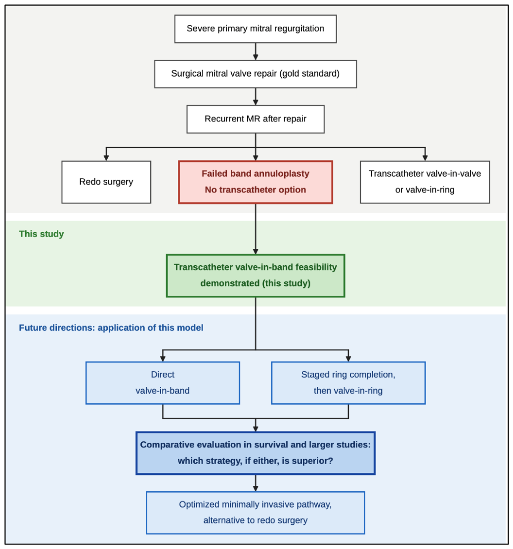
Building on the success of transcatheter deployment of the valve in the swine model, two strategies now warrant further evaluation (Figure 6). The first is direct valve-in-band, relying on the native anterior leaflet for anterior support. The second is staged percutaneous ring completion, bridging the unsupported anterior annulus between the fibrous trigones to convert the band into a complete ring before deployment; this strategy restores circumferential support and reframes the procedure as the better characterized valve-in-ring [16,17]. Comparing the two is the explicit aim of the program under which this study was performed (IACUC Protocol #00002378, approved for up to 30 animals). Definitive translation will require a fully percutaneous, transseptal approach, survival data, and adequately powered comparison against redo surgery [10] using standardized Mitral Valve Academic Research Consortium endpoints [20].
These conclusions must be tempered by the limitations inherent to a single, acute, non-survival experiment. With one animal, we cannot assess reproducibility, chronic valve function, durability, or the delayed risk of LVOT obstruction, and outcomes are necessarily descriptive rather than statistical. The preparation also carries model-specific features, including a surgically simulated dilated left atrium for access and an incidentally hypoplastic aorta, that may not reflect human anatomy or a fully percutaneous procedure. These constraints define the immediate next steps: survival studies in additional animals to characterize durability and the safety profile before any clinical translation.
5. Conclusions
In a large-animal model of failed band annuloplasty, transcatheter valve-in-band was technically feasible and acutely safe, achieving stable fixation with resolution of mitral regurgitation and without embolization or paravalvular leak. Critically, the preserved native anterior leaflet supplied the circumferential support that a partial band cannot, providing the first direct evidence that a failed band, long regarded as an unsuitable landing zone, can anchor a transcatheter valve. These findings establish a platform for survival and comparative studies of direct valve-in-band versus staged percutaneous ring completion, using standardized endpoints, to determine whether the approach can mature into a durable, minimally invasive alternative to redo mitral surgery.
Author Contributions
Conceptualization, H.J.T. and J.E.K.; Investigation, H.J.T., J.M.R., D.A., E.K.L. and A.M.T.; Formal analysis, J.E.K., J.M.R. and M.A.M.; Writing—original draft, J.E.K. and J.M.R.; Writing—review and editing, H.J.T., M.A.M., D.A., E.K.L. and A.M.T.; Supervision, H.J.T. All authors have read and agreed to the published version of the manuscript.
Funding
This work was supported by the Avery Endowment for Cardiothoracic Surgery (Internal Department Funding). The funder had no role in the design of the study; in the collection, analysis, or interpretation of data; or in the writing of the manuscript.
Institutional Review Board Statement
The animal study protocol was approved by the Institutional Animal Care and Use Committee (IACUC) of Dartmouth College (protocol code #00002378, approved 4 November 2025). All procedures were performed in accordance with institutional and national ARRIVE 2.0 guidelines for the care and use of laboratory animals.
Informed Consent Statement
Not applicable.
Data Availability Statement
The data supporting the findings of this study are available from the corresponding author upon request.
Acknowledgments
Not applicable.
Conflicts of Interest
The authors declare no conflicts of interest.
Abbreviations
The following abbreviations are used in this manuscript:
| MR | Mitral regurgitation |
| TMVR | Transcatheter mitral valve replacement |
| ViB | Valve-in-band |
| PVL | Paravalvular leak |
| ViV | Valve-in-valve |
| ViR | Valve-in-ring |
| LVOT | Left ventricular outflow tract |
| IACUC | Institutional Animal Care and Use Committee |
| FDA | US Food and Drug Administration |
References
- Nkomo, V.T.; Gardin, J.M.; Skelton, T.N.; Gottdiener, J.S.; Scott, C.G.; Enriquez-Sarano, M. Burden of valvular heart diseases: A population-based study. Lancet 2006, 368, 1005–1011. [Google Scholar] [CrossRef] [PubMed]
- Alfieri, O.; De Bonis, M. Repair options for mitral regurgitation. Eur. Cardiol. 2008, 4, 88. [Google Scholar] [CrossRef]
- Vassileva, C.M.; Mishkel, G.; McNeely, C.; Boley, T.; Markwell, S.; Scaife, S.; Hazelrigg, S. Long-term survival of patients undergoing mitral valve repair and replacement: A longitudinal analysis of Medicare fee-for-service beneficiaries. Circulation 2013, 127, 1870–1876. [Google Scholar] [CrossRef] [PubMed]
- Otto, C.M.; Nishimura, R.A.; Bonow, R.O.; Carabello, B.A.; Erwin, J.P., III; Gentile, F.; Jneid, H.; Krieger, E.V.; Mack, M.; McLeod, C.; et al. 2020 ACC/AHA Guideline for the Management of Patients With Valvular Heart Disease: Executive Summary. Circulation 2021, 143, e35–e71. [Google Scholar] [CrossRef] [PubMed]
- Gammie, J.S.; O'Brien, S.M.; Griffith, B.P.; Ferguson, T.B.; Peterson, E.D. Influence of hospital procedural volume on care process and mortality for patients undergoing elective surgery for mitral regurgitation. Circulation 2007, 115, 881–887. [Google Scholar] [CrossRef] [PubMed]
- Yaffee, D.W.; Loulmet, D.F.; Zias, E.A.; Ursomanno, P.A.; Rabinovich, A.E.; Galloway, A.C.; Grossi, E.A. Long-term results of mitral valve repair with semi-rigid posterior band annuloplasty. J. Heart Valve Dis. 2014, 23, 66–71. [Google Scholar] [PubMed]
- Schulte, L.J.; Melby, S.J. Mitral annuloplasty ring design and selection: Flexible bands are (mostly) all you need. J. Thorac. Cardiovasc. Surg. 2023, 166, 88–90. [Google Scholar] [CrossRef] [PubMed]
- Dreyfus, G.; Dulguerov, F. Why do surgical mitral valve repairs fail? JTCVS Tech 2024, 25, 70–73. [Google Scholar] [CrossRef] [PubMed]
- Flameng, W.; Herijgers, P.; Bogaerts, K. Recurrence of mitral valve regurgitation after mitral valve repair in degenerative valve disease. Circulation 2003, 107, 1609–1613. [Google Scholar] [CrossRef] [PubMed]
- Yu, C.; Arjomandi Rad, A.; He, H.; Yang, Y.; Maessen, J.G.; Sardari Nia, P. The impact of recurrent mitral regurgitation after surgical or transcatheter mitral valve repair: A comprehensive review and a meta-analysis. Interdiscip. Cardiovasc. Thorac. Surg. 2025, 40, ivaf109. [Google Scholar] [CrossRef] [PubMed]
- Feyrer, R.; Joeckel, J.; Endlich, M. Valve-in-band treatment of failed mitral valve repair: The future method of choice? Struct. Heart 2020, 4, 38. [Google Scholar] [CrossRef]
- Ueyama, H.A.; Gleason, P.T.; Babaliaros, V.C.; Greenbaum, A.B. Transcatheter mitral valve replacement in failed bioprosthetic valve, ring, and mitral annular calcification associated mitral valve disease using balloon expandable transcatheter heart valve. Methodist DeBakey Cardiovasc. J. 2023, 19, 37–49. [Google Scholar] [CrossRef] [PubMed]
- Bothe, W.; Kuhl, E.; Kvitting, J.-P.E.; Rausch, M.K.; Göktepe, S.; Swanson, J.C.; Farahmandnia, S.; Ingels, N.B., Jr.; Miller, D.C. Rigid, complete annuloplasty rings increase anterior mitral leaflet strains in the normal beating ovine heart. Circulation 2011, 124((11) Suppl 1, S81–S96. [Google Scholar] [CrossRef] [PubMed]
- Bogachev-Prokophiev, A.V.; Afanasyev, A.V.; Zheleznev, S.I.; Nazarov, V.M.; Sharifulin, R.M.; Karaskov, A.M. Mid-term results of mitral valve repair using flexible bands versus complete rings in patients with degenerative mitral valve disease: a prospective, randomized study. J. Cardiothorac. Surg. 2017, 12, 113. [Google Scholar] [CrossRef] [PubMed]
- Yoon, S.H.; Whisenant, B.K.; Bleiziffer, S.; Delgado, V.; Dhoble, A.; Schofer, N.; Eschenbach, L.; Bansal, E.; Murdoch, D.J.; Ancona, M.; et al. Outcomes of transcatheter mitral valve replacement for degenerated bioprostheses, failed annuloplasty rings, and mitral annular calcification. Eur. Heart J. 2019, 40, 441–451. [Google Scholar] [CrossRef] [PubMed]
- Guerrero, M.E.; Bapat, V.N.; Mahoney, P.; Krishnaswamy, A.; Eleid, M.F.; Eng, M.H.; Yadav, P.; Coylewright, M.; Makkar, R.; Szerlip, M.; et al. Contemporary 1-year outcomes of mitral valve-in-ring with balloon-expandable aortic transcatheter valves in the U.S. JACC Cardiovasc. Interv. 2024, 17, 874–886. [Google Scholar] [CrossRef] [PubMed]
- Sekaran, N.; Horne, B.D.; Doty, J.R.; Reid, B.B.; Miner, E.C.; Harkness, J.R.; Jones, K.W.; Minder, C.M.; Caine, W.T.; Clayson, S.E.; et al. Transcatheter mitral valve in ring, hazards of long anterior mitral leaflet and 3-dimensional rings. Catheter. Cardiovasc. Interv. 2021, 97, 353–358. [Google Scholar] [CrossRef] [PubMed]
- Blanke, P.; Naoum, C.; Dvir, D.; Bapat, V.; Ong, K.; Muller, D.; Cheung, A.; Ye, J.; Min, J.K.; Piazza, N.; et al. Predicting LVOT obstruction in transcatheter mitral valve implantation: Concept of the neo-LVOT. JACC Cardiovasc. Imaging 2017, 10, 482–485. [Google Scholar] [CrossRef] [PubMed]
- Khan, J.M.; Babaliaros, V.C.; Greenbaum, A.B.; Foerst, J.R.; Yazdani, S.; McCabe, J.M.; Paone, G.; Eng, M.H.; Leshnower, B.G.; Gleason, P.T.; et al. Anterior leaflet laceration to prevent ventricular outflow tract obstruction during transcatheter mitral valve replacement. J. Am. Coll. Cardiol. 2019, 73, 2521–2534. [Google Scholar] [CrossRef] [PubMed]
- Stone, G.W.; Adams, D.H.; Abraham, W.T.; Kappetein, A.P.; Généreux, P.; Vranckx, P.; Mehran, R.; Kuck, K.-H.; Leon, M.B.; Piazza, N.; et al. Clinical trial design principles and endpoint definitions for transcatheter mitral valve repair and replacement: Part 2: Endpoint definitions: A consensus document from the Mitral Valve Academic Research Consortium. J. Am. Coll. Cardiol. 2015, 66, 308–321. [Google Scholar] [CrossRef] [PubMed]
Figure 1.
Intraoperative echocardiogram confirming severe mitral regurgitation in the setting of a band annuloplasty, mimicking failure of a mitral repair in humans.
Figure 1.
Intraoperative echocardiogram confirming severe mitral regurgitation in the setting of a band annuloplasty, mimicking failure of a mitral repair in humans.
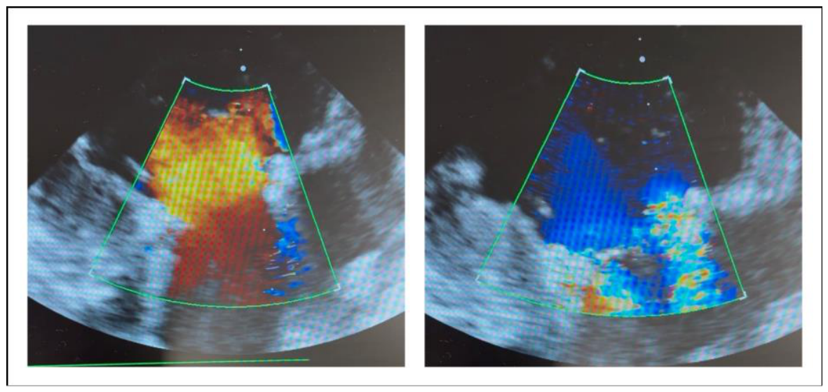
Figure 2.
Fluoroscopic image demonstrating guidewire access to the left ventricle through the left atrial graft.
Figure 2.
Fluoroscopic image demonstrating guidewire access to the left ventricle through the left atrial graft.
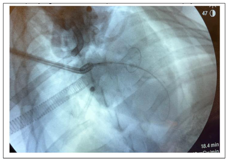
Figure 3.
Fluoroscopic images of transcatheter valve deployment within the mitral annuloplasty band. (A) Pre-deployment positioning of the crimped valve within the band. (B) Post-deployment image confirming successful expansion of the valve within the band.
Figure 3.
Fluoroscopic images of transcatheter valve deployment within the mitral annuloplasty band. (A) Pre-deployment positioning of the crimped valve within the band. (B) Post-deployment image confirming successful expansion of the valve within the band.
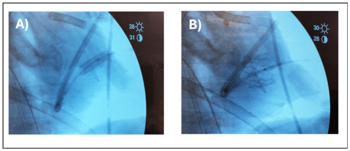
Figure 4.
Intraoperative echocardiography, long-axis view. (A) Valve position and frame-leaflet relationship. (B) Doppler image demonstrating unobstructed transvalvular inflow.
Figure 4.
Intraoperative echocardiography, long-axis view. (A) Valve position and frame-leaflet relationship. (B) Doppler image demonstrating unobstructed transvalvular inflow.
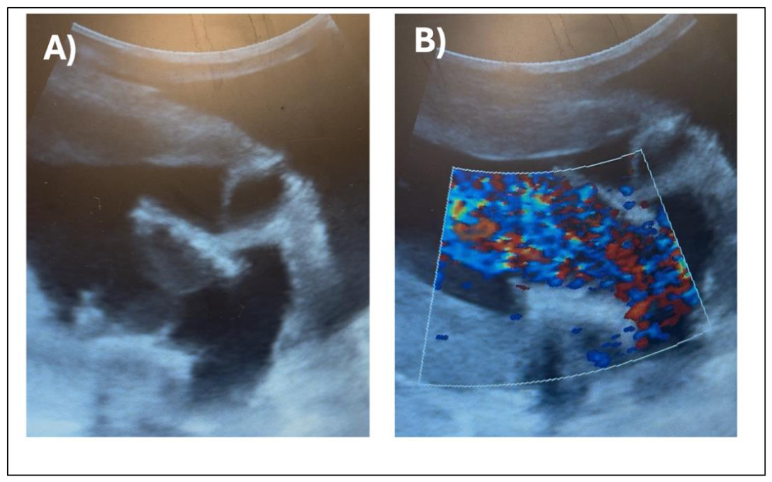
Figure 5.
Direct visualization confirming successful deployment: (A) adequate positioning of the transcatheter valve against the band (white arrow); (B) support of the anterior leaflet and annulus (white arrow) and a patent left ventricular outflow tract without obstruction (blue arrow).
Figure 5.
Direct visualization confirming successful deployment: (A) adequate positioning of the transcatheter valve against the band (white arrow); (B) support of the anterior leaflet and annulus (white arrow) and a patent left ventricular outflow tract without obstruction (blue arrow).
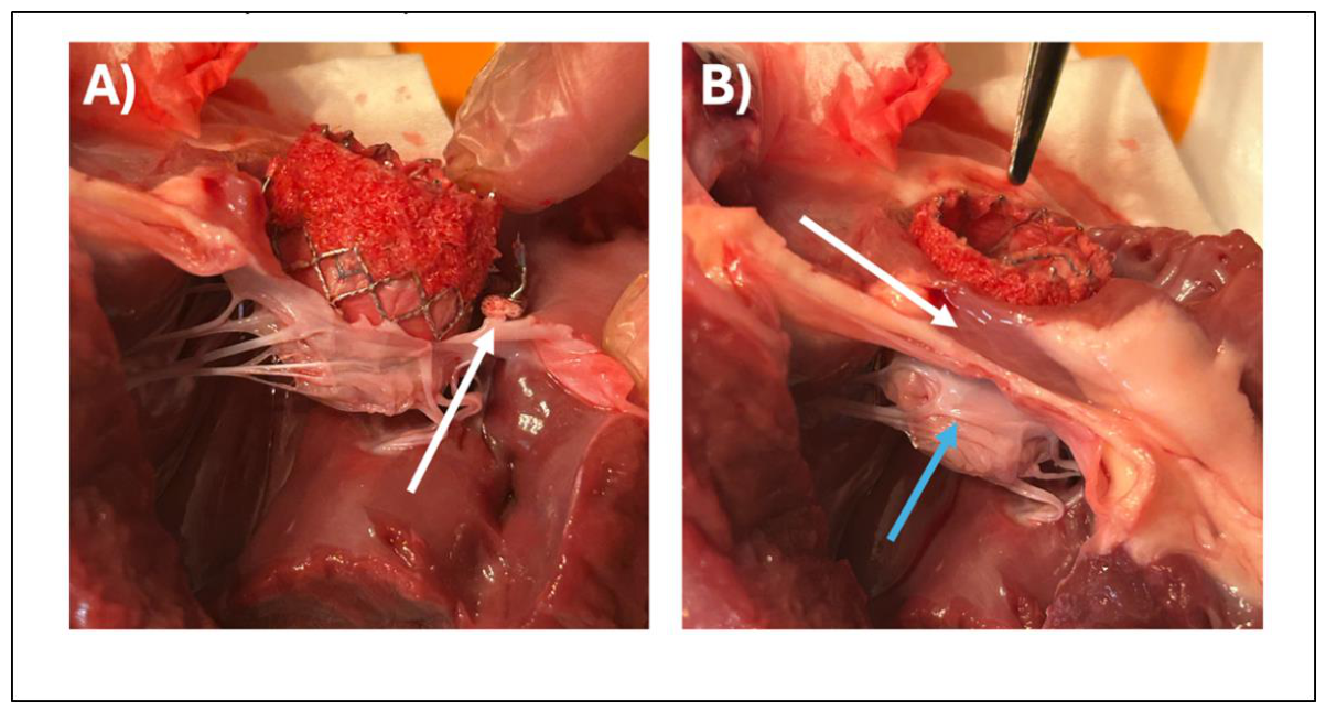
Figure 6.
Current standards and the proposed future application of the valve-in-band model for failed mitral repair.
Figure 6.
Current standards and the proposed future application of the valve-in-band model for failed mitral repair.
Disclaimer/Publisher’s Note: The statements, opinions and data contained in all publications are solely those of the individual author(s) and contributor(s) and not of MDPI and/or the editor(s). MDPI and/or the editor(s) disclaim responsibility for any injury to people or property resulting from any ideas, methods, instructions or products referred to in the content. |
© 2026 by the authors. Licensee MDPI, Basel, Switzerland. This article is an open access article distributed under the terms and conditions of the Creative Commons Attribution (CC BY) license.
Copyright: This open access article is published under a Creative Commons CC BY 4.0 license, which permit the free download, distribution, and reuse, provided that the author and preprint are cited in any reuse.