

Submitted:
10 August 2026
Posted:
11 August 2026
You are already at the latest version
Abstract
Suspended particulate matter (SPM) plays a fundamental role in water bodies by controlling the transport of sediments, nutrients, trace elements, radionuclides, and organic contaminants. The mineral fraction of SPM is particularly important because it governs many of these physicochemical processes and strongly influences contaminant mobility and bioavailability. However, its characterization remains challenging due to the small particle size, complex organo-mineral associations, poor crystallinity of some phases, and the dynamic nature of aquatic particles. This review provides a comprehensive overview of the principal methodologies currently available for investigating the mineral fraction of SPM. First, the advantages and limitations of the main sampling strategies are critically discussed. Then, analytical techniques are reviewed from bulk characterization methods to high-resolution approaches. This review emphasizes that no single analytical technique is sufficient to fully characterize natural SPM. Instead, integrating complementary methods across multiple spatial scales provides the most robust framework for understanding particle composition, reactivity, and contaminant dynamics. Future progress will rely on the harmonization of sampling and analytical protocols, the development of international SPM databases, and the combination of advanced laboratory experiments with field observations to improve our understanding of SPM processes under changing environmental conditions.
Keywords:
SPM
; sampling techniques
; bulk mineral characterization
; particulate mineral characterization
1. Introduction
River suspended particulate matter (SPM) is composed of colloids (1 nm–1 µm) and particles (>1 µm) forming complex and heterogeneous aggregates of mineral, organic, and microbiological components [1] . In addition, SPM is defined and comprises any material in aquatic systems that does not settle and is retained by filtration over 0.45 or 0.22 µm pore size membrane [2] . In natural aquatic systems, these components are frequently formed into floc-like structures whose composition, density, size, and reactivity continuously evolve according to hydrological, physicochemical, and biological conditions [3,4,5] . Consequently, the study of SPM is inherently interdisciplinary and requires combined physical, chemical, and mineralogical approaches to decipher their whole complexity.
Compared with soils and sediments, studies focusing on SPM, moreover freshwater SPM [6] , remain relatively limited due to their high mobility, reactivity, and their difficulty to sample. Their transient nature, rapid aggregation/disaggregation processes, and strong spatial and temporal variability considerably complicate both field investigations and laboratory analyses [7,8] . However, increasing efforts have recently been devoted to the development and adaptation of techniques suitable for the sampling and characterization of natural SPM in aquatic systems.
Rivers and streams constitute the principal vectors of matter fluxes within watersheds and may therefore be considered as the ecological arteries of catchments. SPM enters river systems through multiple mechanisms including soil erosion, runoff, bank erosion, atmospheric deposition, wastewater discharge, mining activities, and sediment resuspension processes before being transported downstream toward estuaries and oceans (Figure 1). The transport of SPM plays a major role in regional and global biogeochemical cycles by facilitating the transfer of carbon, nutrients, trace elements, and contaminants between terrestrial and aquatic environments [9,10] . This is mainly because SPM, due to their fine texture, can hold and trasnport nutrients and contaminants for long distances in water systems [11] . Investigating SPM transport in river catchments is a key component of the environmental monitoring of large and small rivers as they may cause important environmental impacts [12,13] . Suspended particulate matter transport can have impacts on: (i) water turbidity, (ii) fish habitats [14] , (iii) siltation of reservoirs [15] , (iv) transport of contaminants such as polychlorinated biphenyls (PCBs), trace metal elements (TME), radionuclides or nutrients [16,17,18] .
Transport of SPM from land to ocean is estimated from 12.6 to 24 Gt year-1 [12] . The value frequently observed in the literature is around 15 Gt year-1 [19] . However, this value is associated to uncertainties, and does not take into account the recent anthropogenic activities (deforestation, agriculture, mining, urbanization) and/or climate change [20] . The nature and amount of particles in rivers are controlled by the weathering regime. A fast mechanical weathering will engender immature particles and quartz, K-feldspar, illite, calcite and kaolinite [21] . By contrast, a fast chemical weathering will permit the formation of mature particles: poorly ordered kaolinite, goethite and particulate organic matter [22] . The weathering regime is governed by the climate through different processes such as the relief, surface-area of the catchment, climate geology and vegetation as well as human activities [19,23] .
Because of their high specific surface area and strong adsorption capacity, SPM also acts as an important carrier for numerous contaminants in aquatic environments [24,25] . Toxic metals, radionuclides, organic contaminants, and pathogenic microorganisms originating from both natural and anthropogenic sources may adsorb onto particle surfaces, undergo physicochemical transformations, and subsequently be transported, remobilized, or released within aquatic systems [26] . The interactions between mineral particles, natural organic matter, and biological components strongly influence aggregation mechanisms, contaminant binding, and particle stability.
The mineral fraction of SPM is of particular importance because mineral surfaces largely govern adsorption mechanisms, redox reactions, aggregation behavior, and contaminant mobility. Clay minerals are omnipresent in SPM (22 - 72%wt). Feldspars and carbonates are also reported (61%wt) but can be absent due to dissolution during the course of the water [27] . Quartz and feldspars represent 8 – 35%wt and 4 - 41%wt respectively. This proportion can highly increase with water discharge increase [28] . Oxides and (oxy)hydroxides (mainly FeOx) are also reported in the literature but in low mass or number concentrations. FeOx are present as « patchy coatings » on other minerals [29,30] . Co-occurrence of Fe with Al, Si, Ca and discrete FeOx particles are also reported [31,32,33] . Amorphous phases, ferrihydrite, goethite and hematite are frequently reported in the literature [32,34,35,36,37] . Clay minerals, iron and manganese oxides, carbonates, silicates, and authigenic mineral phases may coexist within highly heterogeneous particle assemblages and strongly influence the environmental reactivity of suspended particles [6] . However, the characterization of these mineral phases remains challenging because of their small size, low abundance, poor crystallinity, and intimate association with organic and biological materials.
Recent advances in high-resolution analytical techniques have considerably improved the characterization of the mineral fraction of SPM at multiple spatial scales. In particular, synchrotron-based approaches, high-resolution electron microscopy, and coupled spectroscopic-imaging methods now provide valuable information on particle heterogeneity, mineral speciation, contaminant binding, and organo-mineral associations within complex suspended aggregates. Such approaches have highlighted significant differences between natural SPM and simplified laboratory-controlled particles regarding metal reactivity and association mechanisms, emphasizing the need to investigate natural particulate systems under environmentally relevant conditions [36] .
This review highlights the importance of SPM studies in environmental research with the objective of improving the understanding of biogeochemical processes occurring within the water column. Particular attention is devoted to inland waters, especially rivers and estuaries, where particle concentrations are generally higher than in the open ocean [38] . The first section presents the principal methods used for SPM sampling in aquatic systems. The second section provides a detailed, although non-exhaustive, overview of the analytical techniques currently available for the characterization of the mineral fraction of SPM, from bulk approaches to high-resolution analyses. Finally, future perspectives associated with SPM research are discussed.
2. Sampling Techniques Available to Catch SPM
It is crucial to obtain a representative SPM sample to describe the chemical and physical state of a catchment. SPM sampling is an essential step to ensure the success of their characterization. In particular, contamination can occur due to careless handling or improper protocol preparation. In the literature, different techniques are described for SPM sampling: direct water sampling followed by filtration [39] , in-line filtration [40] , continuous flow centrifugation [41] or the use of particle traps [42] . They differ in terms of duration, yield and sample composition [43] .
2.1. Filtration Technique
The filtration technique permits the total separation of the SPM from the water (except the colloids). It is the most widespread technique for particle removal from water [44] . Usually, 0.45 µm pore size filters are used (Fig. 2). However, this technique is not truly representative because the sample volume is generally small (few liters) and the sampling time is very short. In addition, the SPM composition on the filter is altered, the SPM are aggregated and clogged on the filter compared to the SPM in the water column [45] . Finally, it requires the filtration of huge volumes of water to collect enough materials (> 2g dry weight, d.w.) for exhaustive analyses of SPM [46] . For that purpose, this technique is only used when the other techniques described below are not feasible [43] .
2.2. The Continuous Flow Centrifugation (CFC)
The continuous flow centrifugation is a well-established and reliable technique to sample SPM [47,48] . This technique separates the SPM from the water phase at a defined constant throughput. CFC separates particles according to size and density such as natural sedimentation process [44] . The water is pumped from the river and the SPM are deposited in the inner wall of the rotating cylinder. In order to facilitate the SPM recovery, Teflon plates are installed in the bowl to attach the SPM [36] . The separation efficiency can reach 90-98% [49] . One of the advantages of this technique is that the sample yield is considerably higher than filtration (several grams) and therefore permits a better comprehension of chemical, physical, and mineralogical characterization of the suspended material collected. The CFC can be installed in a monitoring station or on a trailer, or on a boat and can be mobile, however, this technique is voluminous and requires large and stable riverbanks. This technique is grain size dependent and the density between water and SPM also influences the sampling [50] . In addition, different grain size classes may be lost [51] (the average cut off is 1 µm). This information can, in certain cases be crucial, like, for example, the analyses of Polycyclic Aromatic Hydrocarbons (PAH) on SPM [52] . However, [53] showed that CFC can retain nanoparticles (~ 160 nm) with high efficiency (> 90%). This technique is also time consuming and expensive. Thus, it is difficult to investigate the SPM spatial and temporal variability [54] . Finally, there is a possibility of contamination from CFC components (e.g. bearings, lubrication oil) [55,56] and physical effects during the centrifugation such as changes in the cut-off due to the bowl filling up or fragmentation and aggregation of particles (shear stress).
2.3. Hydrocyclone (HC)
The hydrocyclone is a device widely used in petroleum, mineral and environment engineering. Surprisingly, this technique is not widely reported in the literature [57] . SPM is separated from water in the HC using centrifugal force. High flow rate of water is introduced into the upper cylinder. This water is then forced into a downward vortex. Due to the centrifugal forces, the particles are pressed against the wall where they are collected [50] . Several studies showed that HC systems can retain ~ 75% of SPM in the size range 20-30 µm at a very high throughput [50,58] . [53] compared to CFC and HC and they concluded that both of these techniques worked well under real work conditions.
2.4. The Sedimentation Trap (ST)
The sedimentation trap is a technique that uses the natural velocity of the river to collect particles via sedimentation in the inner of the trap. This technique is also called “fresh sediment derived from suspended solids” [59] . It is a low-cost technique that can be easily set up and does not need attendance. This method also integrates a longer period of time (1 to 4 weeks typically) that is well-suited for in situ monitoring over a year [54,60] . Whereas the sampling of SPM by CFC has been investigated and validated since the 1990’s, (see above); the representativeness of the SPM collected with STs is still questionable. Several studies showed that SPM sampled by STs differ from river SPM with a modification of the grain size distribution and organic carbon content [42] . The fine particulate matter can be lost with the overflow [43,59] and the SPM concentration in the water column is not available. This difference could lead to biased concentrations for contaminants [61] . In their study, [46] showed a difference of grain-size distribution between SPM collected by sediment traps (0.1 – 400 µm) and CFC (0.1 – 112 µm). They also observed seasonal differences in POC concentrations due to degradation or phytoplankton blooms. However, they concluded that ST sampling is a valuable technique to assess spatial and temporal trends of particulate contaminants (PCBs and Hg).
2.5. Other Techniques
The use of filtration or centrifugation on field cannot entirely certify the separation of colloids and particles. Field-flow fractionation (FFF) is a relatively new technique that is capable of high-resolution separation of colloids and inorganic particles [62] . The two main principles are sedimentation (SedFFF) and asymmetric flow field-flow fractionation. The separation is achieved by the interaction of sample components with an external generated field that is applied perpendicularly to the direction of the mobile phase flow. It can also be coupled by second cross flow stream resulting in flow field-flow fractionation with a better separation [63,64] . This rapid technique (10-30 min) permits the separation of a continuous large class of particles (from ~1nm to 100 μm) and is based on important properties such as their surface charge, density, size, shape and diffusion coefficient [62,65] . This method has been employed, coupled with different detectors (fluorescence, UV-absorbance) for river colloids [66] . Good introduction of the FFF theory can be found in the literature [67] .
Different studies in the literature try to compare different devices based on the difference of passive sampling versus active sampling. Passive sampling methods are based on lowering the flow speed of the water inside the sample. On the other side, active sampling is based on applying external force to separate SPM from water [68] . For example, [50] compared the efficiency of time-integrated sampling devices (Binnensammler device and Phillips sampler, PS) and discrete sampling devices (CFC and Hydrocyclone, HC). They showed similar physical and chemical parameters for the two time-integrative sampling devices as well as organic pollutants. However, they remarked differences in TOC concentrations between CFC and PS due to degradation processes occurring in the PS device. Figure 2 shows the advantages and limitations of the different sampling techniques. Recently, systematic validation of these approaches has highlighted critical technical biases. Another study [68] demonstrated that passive devices (such as sedimentation boxes or tanks) significantly underestimate fine particles (<75μm) and low-density microplastics (<80μm, with a capture efficiency below 20%), shifting the collected grain-size distribution toward coarser fractions compared to active CFC or filtration methods. Despite these physical limitations, sedimentation boxes remain highly valuable for integrating fluxes over several weeks. Each technique has its benefits and limitations (size range collected; retention capacity of particles). It is then of pivotal importance to carefully consider SPM sampling efficiencies of the device used, especially when dealing with a quantitative approach.
3. Characterization of SPM (Mineral Fraction) at Bulk and Particulate Size
SPM global characterization of the physical and chemical fraction is not be possible via one technique. Different techniques are complementary and permit to better understand the whole complexity of natural SPM and their reactivity towards contaminants (Figure 3).
3.1. Specific Surface Area (SSA)
The Specific surface area (SSA) is a pivotal parameter to better understand the processes that occur at the mineral interface [69]
A routine method for SSA determination of fine grain such as SPM is the gas adsorption method. The analyze of adsorption isotherm of a non-polar gas such as N2, Ar, Kr or CO2 permits to calculate the surface area of SPM (the surface of SPM that is ideally covered by a monolayer of adsorbed gas). The measurement of the specific surface area is determined by the BET method [70] . Microporous surface (pore size ranging from 0.8-2 nm) is obtained by the t-plot method [71] and mesoporous distribution size (pore size ranging from 2 and 80 nm) is determined by the BJH method [72] . The meso- and micropores are evaluated by the hysteresis of the adsorption and desorption isotherm. Prior measurement, this method requires the total removal of adsorbed surface water and interlayer cation [73,74] . However, in the case of natural particles, the temperature of outgasing is crucial as it could alter iron (hydro)oxide [75] . For bulk samples, such as SPM the adsorption of liquid ethylene glycol monomethyl ether (EGME) can be an alternative. Indeed, EGME covers the external surface of particles and penetrate the interlayer space of swelling clay minerals [76] .
The mineral fraction of SPM is mainly composed by clay minerals. They develop two kinds of surfaces: the basal (001) surface that carry negative charges and the edge or lateral surfaces that are pH dependent [77] . Concerning the clays, the mesopores (2-50 nm) are mainly interparticle pores and have minor effect on the gas adsorption surface area [78] . On the other hand, micropores (2 nm) result from the intraparticle space and are N2-accessible [79] . For example, micropores in bentonites contribute significantly to the total specific surface area of gas adsorption measurements [78] .
Through years, the method did not change significantly but increased in precision and accuracy. The SSA is commonly used for synthesized particles and single minerals in the laboratory in order to characterize their physical properties. However, researchers also use this method to characterize the physical properties of natural SPM, even if it is not widely used for natural materials. [80] characterized SPM from a New Zealand watershed and showed that SSA increased with increasing SPM concentration in relation with the increase of flow rate. The result commonly observed for environmental particles is a surface area increase with decreasing particle size such as clays [81] . This larger surface area facilitates the sequestration of contaminants such as trace metals [80,82] . More recently, [6] highlighted that the specific surface area (SSA) of natural SPM is highly dynamic and closely linked to flocculation processes. They demonstrated that the adsorption of natural organic matter (NOM) can either clog mineral pores, reducing the measurable BET surface area, or create new complex organo-mineral micro-architectures that modify the overall particle porosity and reactivity. The interaction between FeOx and phyllosilicates is more studied in the case of natural SPM. This interaction favoring flocculation and enhance surface area of SPM as suggested by several authors [29,32,83] . [29] showed a close relationship between iron oxide content and surface area when interaction with clay minerals. They argue that this relationship could be the result of the abundance of edge sites for electrostatic attraction between iron and clay and addition of the spheres of the iron [82] . [30] also studied the SSA of natural particles from the Moselle River (France). They showed that the SSA of natural particles ranged between 6 and 37 m2 g-1 and showed that the presence of organic matter increased the mean particle size and decreased the SSA through the lower accessibility of external sites. In addition, no significant microporous surface area was measured (≤1 m2 g-1).
3.2. Bulk Chemical Analyses
Bulk chemical analyses or elemental analysis is often conducted to characterize the full spectrum of non metallic, metalloids and metallic species in natural SPM [84] . In environmental laboratories, ICP-MS and ICP-OES are the two most advanced metal analysis techniques routinely used [85] and the selection of method depends on the range of element concentration in the samples (i.e. ICP-MS for ppt range and ICP-OES for ppb as well as ppm [86] . ICP-OES is a method of optical emission spectrometry that uses the emission spectra of sample molecules to identify and quantify the elements present. This technique has two parts: the inductively couple plasma, and the optical emission spectrometer with major components (sampler, pump, nebulizer, spray chamber, ICP torch, monochromator and detector). Conversely, three main units compose the ICP-MS: (I) an argon ICP operating at a temperature of 5000-7400K with a nebulizer, spray chamber, radio-frequency coil and power supplies; (ii) two water-cooled sampling cones (iii) a quadrupole mass spectrometer, detector and associated data collection electronics. Both ICP-MS and ICP-OES rely on similar principles for introduction and plasma generation [87,88] . In ICP, each element emits photon with distinct wave lengths that can be attributed to specific elements [89] . Usually, sample preparation is required for a suitable introduction of the sample into the system. Before any preparation, it is necessary to use trace metal grade acids and solvents, as well as vessels that have been soaked and rinsed with deionized water. This is because deionized water is free of impurities, including metal ions [90] . SPM, as solid samples must encompass different steps including drying, grinding, sieving, acid digestion, filtration and occasionally pre-concentration techniques for homogenization before introduction in the ICP-OES [91,92] . The technique consists in taking an aliquot of several grams of SPM (usually one gram) that is smelted with LiBO3 in Pt–Au crucibles [93] . The resulting sample is then dissolved using an acid. The sample is then introduced in the ICP. The high temperature of the plasma excites electrons above steady-state. For ICP-OES, the electrons return to steady-state, a photon is emitted and can be analyzed by interaction with electromagnetic radiation [94] . Concerning the ICP-MS; the ions formed in the plasma are introduced in a quadrupole [95] . ICP (OES and MS) are suitable for elemental analysis. It is then an excellent technique for the measurement of trace metals. However, this technique is destructive with a complete extraction and digestion of the sample. If the digestion is incomplete, the solution can form co-precipitates [96] . Recent applications on ICP-OES and MS can be highly beneficial for the scientific community. The next paragraph will present non exhaustive results concerning the applications for riverine SPM studies.
Many studies dealing with elemental analysis of natural SPM can be encountered in the literature worldwide. For example [36] investigated the spatial and temporal variability of SPM in the Moselle River (France). They showed an urbanization gradient upstream to downstream the Moselle River and a seasonal variation with more silty particles during high flow regime. [97] studied the suspended load of the Ganga and Bramaputra Rivers using a combination of techniques in order to get the maximum of precision in the identification of the different size fractions. The geochemistry using ICP-MS permitted to show that the surface load was enriched in -metals. They also showed that the chemical variability of suspended load being influenced by hydraulic, weathering, and possibly anthropic effects. [19,98] presented a database of the chemical composition of SPM in World Rivers and gave a snap-shot of elemental fluxes for different continents. [19] found that the anthropogenic fluxes and riverine fluxes are similar revealing that human activities have effects on the cycles of trace elements. [99] analyzed SPM from the Yarlung Tsanpo River (Tibetan Plateau). The selected 10 metal(loid)s (V, Cr, Co, Ni, Cu, Zn, As, Cd, Sb, Cs) and analyzed them by ICP-OES and ICP-MS. They measured highest enrichment values for Cu, As, and Cd. They showed a seasonal enrichment due to hydrodynamic conditions. They also showed that the SPM are mainly anthropogenic and from geothermal activities. [100] measured the impacts of the human-induced flood event on heavy metal transport, spatiotemporal variations in contents and fluxes of metal (Cr, Ni, Cu, Zn, As, Cd) using ICP-MS. They concluded that metals transport was controlled by hydrological processes and dominated by SPM and with strong association with particle size and SPM concentration.
3.3. Bulk Mineral Analyses
3.3.1. X Ray Diffraction (XRD)
After the characterization of the bulk physical properties (SSA) and the elemental analysis, the next step in the SPM characterization process is a better understanding of the mineralogy. Different techniques are available for the mineral investigation in bulk natural SPM. XRD is a non-contact and non-destructive technique for a large panel of materials including minerals [103] . XRD is used to identify and quantify crystalline mineral phases, as well as to evaluate their degree of crystallinity. Indeed, the wavelengths of X-rays are near atomic size. This technique is thus capable to provide information about atomic structures. XRD permits to characterize the position of atoms, to characterize their arrangement and is thus particularly suited for crystallography studies [101,102] . The main use is to identify components in SPM sample by a “fitting” procedure. In addition, the area under the peak is proportional to the amount of each phase present [103] . Therefore, the crytstalline minerals obtained from the diffractograms can be quantified via Rietveld refinement. These complex diffractograms are modeled as a sum of individual mineral phases, and their relative abundances are refined by least-squares fitting of the entire pattern, yielding the mass percentage of each component. Furthermore, the percentage of amorphous materials can also be quantified by using an internal standard [104] . [103] Furthermore, XRD of filtered samples can give additional information as it can measure the degree of orientation of clay particles and can thus, when coupled with electron microscopy study investigate composite particles (individual particles of different mineralogy). XRD is also used to identify and distinguish between various clay minerals using oriented mounts. The main procedures rely on treating the materail with ethylene glycol and to heat the samples; this will lead to various swelling and collapse of the minerals. These information can then be used to clearly distiguish between clay minerals [105,106] . XRD uses X ray waves with a length comprised between 0.1 and 10 Å. The process consists in the interaction between the X-ray beam and the SPM. One part of the beam enters in SPM and one part is diffused in different directions. The diffracted beam is the sum of the different beams diffused. This diffracted beam varies with the different constituents of the SPM [107] . This technique is powerful for the identification of the different crystallized constituents of the SPM such as primary and detrital minerals (quartz, phyllosilicates, and carbonates). It is easy to perform, quick and can be used for other analyses but requires, the least, more than tens or hundreds of milligrams. X-ray diffraction has been used for example to follow the changes of the mineralogy of river particles during a storm event with particles mainly composed of smectites [108] . Other studies, such as [107] showed, in a US stream that during the beginning of the limb of the hydrograph, the quartz is present as discrete particles and at the end of the hydrograph, the SPM is composite with different particles of various sizes. They concluded that XRD can be chosen as a tool to help the sampling procedure. Other studies such as [37] investigated the seasonal variations of SPM mineralogy of the lower Changjiang River using different techniques such as XRD. They found a seasonal variation of the clay mineralogy with distinct behaviors during the rainy season (more illite and less kaolinite proportion) suggesting more intense erosion in the basin during the rainy season. [109] studied the effect of flow rate and lithology on SPM mineralogy in Puerto-Rican Rivers using XRD. They showed that the mineralogy of suspended sediments in the three watersheds studied returned to baseline composition after storm events showing the resilience of the watershed upon strong hydrological conditions.
3.3.2. Fourier Transform Infrared Spectroscopy (FTIR)
Different spectroscopic methods are illustrated in the literature and can be used for particle characterization in bulk mode. This technique is also reported in order to fingerprint (i.e. sources origin) sediments and SPM and is used alternatively to traditional techniques such as geochemistry and the use of radionuclide tracers. Indeed, traditional tracers used for fingerprinting is time-consuming. Visible reflectance [110] in France, visible near infrared (VNIR) in Luxembourg, shortwave infrared (SWIR) in Spain [111,112] diffuse reflectance infrared Fourier transform (DRIFT) spectroscopy [17,113] and near infrared in southern Brazil (NIR) [114] are different techniques documented. Infrared spectroscopy is a technique suitable to investigate the interactions radiation-matter, sensitive to vibration energies of the molecular bonds. Molecular bonds absorb one part of the incident energy and the nucleus vibrates by stretching of bonds. Infrared spectroscopy consists in the measurement of the quantity of radiation absorbed by the sample in function of the incident wave. FTIR technique can be performed in various modes, such as transmission mode, diffuse reflectance mode (DRIFT) and attentuated reflectance (ATR).
This technique is especially suitable to track particulate compounds. [114] used NIR spectroscopy on twenty nine SPM samples in a 1.19 km2 in Brazil. They were able to differentiate the sources of the SPM with this technique. In their study, [115] used FTIR to investigate the variations of the SPM mineral and organic composition. They found that the SPM were mainly composed of quartz (peaks at 696, 780, 800, 1018 and 1868 cm-1) and carbonates (peaks at 713, 729, 874, 1457, 1794, 2517 and 2873 cm-1) with no large variations along the river. [113] used DRIFT spectroscopy to evaluate the efficiency of this technique in tracing sediment sources. They analyzed 50 samples from a small catchment (90 ha) in France and showed that this technique can separate at least four different sources from soil and river channel sediments [114] compared classical geochemical and radionuclides tracers and a method using MIR spectroscopy to fingerprint sediments from rural catchments in Mexico. They found similar results except for one catchment due to the present of high organic matter content. The use of VNIR spectroscopy was also successful for sediment fingerprinting in Ethiopia [116] . In particular cases, the use of spectroscopy method cannot be used. For example, [110] showed that Quaternary deposits in France could not be discriminated because the gypsum was not conservative.
Infrared spectroscopy is widely used to determine the mineral and organic composition of solid matrices, including sediments and SPM. Indeed, well defined set of IR peaks can be used to identify organic matter and minerals, including clay minerals [117,118] ). Furthermore, IR can also be used to identify and distinguish between various carbonates, such as calcite and dolomite [119] . IR is considered a cost-effective and relatively accurate quantification method for carbonates, amorphous silica, and organic matter [120] ). Recent studies are also looking at the quantification of clay minerals [121] . Complex IR spectra from heterogeneous samples, such as SPM and sediments, can be further analyzed by multivariate curve resolution alternating least square (MCR-ALS) [122,123] . This decomposes the IR spectra into multiple components and computes their respective percentage [124,125] . As such, the change or evolution of SPM components can be followed along a river course or as a function of variable river flow. Furthermore, the same approach can be used to detect even minute IR changes that would be not possible without any treatment [126] .
3.4. Particulate to Atomic Mineral Analyses
3.4.1. Transmission Electron Microscopy
Several techniques permit to characterize the mineral composition at micro to nano scale. Transmission electron microscopy (TEM) coupled with energy dispersive spectroscopy (EDS), has been shown to be an extremely powerful tool for examining geochemical questions at the individual particle scale. TEM can assess the size, morphology, crystallinity and elemental composition of single particles as well as to their physical and spatial associations. This technique can provide high-resolution imaging of thin samples, with magnifications reaching up to 106×, with quantitative chemical analysis. TEM consists of an electron gun, a set of electromagnetic lenses, and apertures. The thickness, density, and chemical composition of the sample will constrain the degree of the electrons absorbed. When the samples are very thin, the electron bombarded from high-energy electron beam will be transmitted and can form an image [127] . The imaging mode, will permit the projection of an image of SPM and can be observed on a screen or detector. In diffraction mode, diffraction patterns can be acquired at the microscopic scale in diffraction mode. This mode gives information on the crystalline structure of SPM. The TEM combined with energy-dispersive X-ray spectroscopy (EDS) detector enables the analysis of X-ray photons emitted by the sample, allowing quantitative elemental characterization of the area irradiated by the electron beam. Particularly, the Backscattered Electron Imaging (BEI) mode permits the detection of mineralogy and texture of heavy metal and provide direct information on the presence these particles [36] . Several publications can be encountered in the literature for precise description of the functioning of TEM. Concerning the interaction between electrons and thin specimens; see [128,129] . For the description of TEM (imaging and electron diffraction); see [130] .
The classical preparation requires chemical stabilization, followed by sample dehydration and resin embedding, prior to ultramicrotomy. The multiple steps can lead to morphological artifacts such as aggregation of particles [131] . To avoid these morphological problems, a hydrophilic resin permits to embed the samples [132] . A very easy technique used for SPM consists in re-suspending dried SPM in ethanol and laying down a drop on cover grid [36] . TEM analysis is mainly used in SPM studies to investigate the morphology and nature of particles and the speciation of contaminants. The morphology, crystallinity and elemental composition of colloids at the particle level can be assessed by analytical electron microscopy [133] . Chemical analysis within the microscope can be obtained by energy-dispersive spectrometry (EDS) [134] or by electron energy loss spectrometry (EELS) [135] . EELS is more suitable for thin specimens (30-200 nm), [136] . Several mineral substances can be encountered in the literature [137,138] . Studies concerning aquatic particles using TEM as an analytical tool stated in the late 1980’s. Concerning oxic fresh waters, the inorganic fraction is mainly composed by aluminosilicates, silica and iron oxyhydroxides [36,139] . Iron in SPM have been extensively investigated because of their important role in supplying nutrients to aquatic biota and their role as sorbents. Natural anoxic waters have also been investigated. Especially the colloids composed of elemental sulfur and iron sulfide [140] .
3.4.2. Atomic Force Microscopy (AFM)
Transmission electron microscopy (TEM) and scanning electron microscopy (SEM) are powerful tools to investigate the size and shape of SPM even TEM has higher resolution. However, these techniques sometimes do not permit to distinguish between grains and aggregates [141] . Quite new techniques known as scanning probe microscopy permit to directly imaging particles with very high resolution (up to atomic). Different techniques are available, among them we can cite: atomic force microscopy (AFM), magnetic force microscopy (MFM), electrostatic force microscopy (EFM), lateral force microscopy (LFM), electrochemical atomic force microscopy (ECAFM) [142] . These techniques are very promising in order to explore SPM surfaces, visualize the sorption of organic substances, determine the morphology and measure size and thickness [143] . Several studies concerned the characterization of the morphology of pure minerals forming SPM such as illite, smectite, iron oxides minerals, calcite [144,145,146] .
AFM is by far the most common SPM technique to probe natural SPM.AFM has a high spatial resolution and can image samples in different environments [147] . AFM is constituted by several elements: the tip, the cantilever, the detector, the scanner, the computer controlling all the system. Several limitations and artifacts can come from the different elements [148] . Several articles in the literature deal with the AFM principles. Here is a brief overview of it function. AFM uses a sharp cantilever-mounted tip to scan a sample surface with nanometer resolution. Cantilever deflection is typically measured by a laser reflected onto a photodiode. AFM can operate in constant-force mode, where a feedback loop maintains a constant interaction force by adjusting the sample height, or in constant-height mode, where the tip height remains nearly fixed and changes in cantilever deflection are recorded. The latter is mainly suitable for relatively flat surfaces, as rough topography may damage the tip or the sample.
The studies are generally performed at ambient air conditions. Such preparation may result in artefacts. AFM is a high resolution surface imaging tool, extremely powerful for the determination of colloidal topography/height on the basis of the repulsive or attractive forces between the sample and the TIP [149] . The AFM can be operated in air or in fluid [150] More than 10 different imaging modes can be used in AFM analysis [151] . However, three common imaging modes are encountered in AFM imaging: contact, tapping and non-contact modes. For contact mode, the tip is in contact with the sample all the time. This mode is associated with high shear forces and then can damage samples. In non-contact mode, the tip oscillates at a frequency slightly higher to its resonance frequency, several nanometers above the sample. In tapping mode, the canteliver oscillates near its resonance frequency at an amplitude ranging from 100 and 200 nm and touches the sample [152] . Different sample preparations can be realized such as: particle sorption to a freshly cleaved flat surface, drop deposition and particle ultracentrifugation [153] .
[147] investigated the effects of the analysis mode (contact vs non-contact modes) as well as the analysis environment (liquid vs ambient air) and the sample preparation. They found that the non-contact mode was the most appropriate for the characterization of natural colloids. In addition, the lack of washing after adsorption or sample preparation by drop deposition can result in the formation of aggregation and salt crystallization. Finally, they found that the liquid mode imaging is not necessary for size measurement if minimally perturbing sample preparation are employed. [154] used AFM in tapping mode to study the size distribution, surface coverage and morphology of aquatic colloids. They found three types of natural aquatic colloids based on their shape and size. [155] successfully used AFM to study the surface area and reactivity of several phyllosilicates (kaolinite, hectorite and nontronite). Other studies on phyllosilicates were also enlarged to illite and montmorillonite [156] ; Smectite [144] ; mica and muscovite [157,158] ; oxides [145] ; calcite [146] ; goethite [159] ; hematite [160] .
3.4.3. X-Ray Absorption Fine Structure Spectroscopy
Extended X-ray Absorption Fine Structure (EXAFS) spectroscopy started in the 1920s but the technique gained widespread recognition in the mid-1970s following the advent of synchrotron radiation sources [161] . EXAFS was first applied to mineralogical studies in the 1980s [161,162] , and the subsequent development of second- and third-generation synchrotron facilities greatly expanded its applicability to highly diluted environmental matrices such as SPM.
EXAFS is an element-specific spectroscopic technique that uses X-ray radiation to selectively excite a target atom within a matrix. Depending on the incident photon energy, core electrons are ejected from the 1s shell (K-edge) or higher electronic shells (L- or M-edges) as photoelectrons. The sample absorbance is recorded as a function of the incident X-ray energy, typically extending up to approximately 800 eV beyond the absorption edge of the target element.
Data acquisition can be performed in two modes (i) the Transmission mode determined by measuring the intensity of the X-ray beam passing through the sample (suited for concentrated samples) and (ii) the Fluorescence mode determined by measuring the secondary X-ray fluorescence emitted by the target element (for diluted samples).
Beyond the absorption edge, interference between the outgoing photoelectron wave emitted by the absorbing atom and the waves backscattered by surrounding neighboring atoms generates oscillations in the absorption coefficient. EXAFS provides precise insights into the local chemical and structural environment around the absorber up to a distance of approximately 5–6 Å. This allows for the determination of the coordination number, the chemical identity of neighboring atoms, interatomic distances, and the degree of structural order or disorder within the coordination shells [163,164] .
To extract structural information from EXAFS oscillations, two primary analytical approaches are employed (i) the linear combination fitting and (ii) the numerical simulation and shell fitting.
Linear combination fitting provides an estimation of the local atomic environment without the immediate need for complex numerical simulations. Using a least-squares minimization protocol, the experimental spectrum is modeled as a linear combination of reference spectra obtained from well-characterized standard compounds. The contribution of each reference component is directly proportional to its fractional abundance within the sample. To maintain physical relevance and avoid overfitting, the decomposition of the experimental spectrum is typically restricted to a maximum of three or four reference compounds. While the sum of all components ideally equals 100%, lower sums (e.g., ~70%) can yield acceptable fits in specific diluted or complex systems. This approach serves as an efficient tool for phase identification, despite carrying a structural uncertainty of approximately 10% to 15%. This approach is routinely used for SPM studies. [165] investigated the speciation of Zn in SPM from the Seine River (France). The results showed, Using LCF, significant spatial variations in Zn speciation along the river continuum. Upstream of Paris, Zn was mainly associated with calcite, either adsorbed onto its surface or incorporated into its crystal structure. In contrast, downstream of Paris, amorphous zinc sulfide (ZnS) became the dominant Zn-bearing phase, reflecting the influence of anthropogenic inputs from the Paris metropolitan area. Similar study was conducted for Zn speciation in one affluent of the Sein River (Orge River). Coupled with TEM and SEM analysis, this study showed that Zn speciation depended to urbanization but also from River flow rate [139] . Another study [166] used EXAFS approach coupled with Isotopy (see below paragraph) in order to investigate the influence of land use on trace metal dynamics was investigated in three contrasting sub-basins of the Seine River watershed. Results showed significant anthropogenic impacts on both dissolved and particulate fractions, with distinct sources depending on land use. Zinc speciation, isotopic signatures, and export rates further highlighted the role of forested, agricultural, and urban areas in controlling trace metal transport and cycling. Numerical Simulation and Shell Fitting provides highly precise determination of structural parameters. Numerical modeling is conducted based on the reciprocity of the Fourier Transform (FT). This procedure involves isolating a specific peak from the pseudo-Radial Distribution Function (RDF) using a selective window (filter), followed by an inverse FT to back-transform the signal into k-space. This isolates the specific EXAFS contributions belonging to distinct absorber-backscatterer atomic pairs.
The resulting partial spectrum is subsequently fitted in both frequency and amplitude using a theoretical approach based on the standard EXAFS equation under the single-scattering approximation. The adjustable parameters are categorized into two groups (i) the structural parameters (the coordination number of neighboring atoms (Ni), the Debye-Waller factor (σi, reflecting structural and thermal disorder), the photoelectron mean free path (λi = k/Γi), the interatomic distance (Ri), and the energy threshold shift (ΔE0) between the experimental edge and the theoretical value) (ii) the electronic parameters (the element-specific amplitude (A(k)) and phase shift (Φ(k)) functions for each atomic pair, alongside the amplitude reduction factor (S02, often fixed to 1). These electronic functions are theoretically calculated using ab initio codes such as FEFF and validated against reference compounds with known crystal structures. This numerical optimization yields highly precise structural data, with typical uncertainties of approximately ±0.02 A˚ for interatomic distances (Ri) and ±20% for coordination numbers (Ni). This approach was first used for single minerals. For example, we can cite Zn adsorption on clay minerals [167] ; Montmorillonite [168,169] Smectite [170] calcite [171] or ferrihydrite [34] . Some studies also used this approach for SPM. [30] monitor changes in mineralogy and composition of authigenic material from its source to streams of increasing order. EXAFS analyses revealed that freshly precipitated Fe-rich particles formed from groundwater oxidation consisted mainly of poorly crystalline hydrous ferric oxides with a structure similar to ferrihydrite. The Fe speciation remained largely unchanged during downstream transport, although an increase in Fe–Fe interactions indicated progressive ageing and hydrolysis of the ferrihydrite-like phases. [172] also tried this approach to better understand the Zn speciation in SPM from the Moselle River (North East of France). They realized Zn absorption tests on natural SPM. They observed that at low Zn loadings, the binding mode of Zn with SPM was controlled by its own mineral composition.
3.4.4. Isotopy
Isotopes are atoms of an element that differ for their number of neutrons and thus detains different masses. They can be classified into radioisotopes and stable isotopes [173,174] . Stable isotopes are divided into traditional stable isotopes (C, H, O, N, S) and non-traditional stable isotopes (Pb, Cu, Zn, Cd, Hg, Cr, Mg) [175,176] . They are high atomic weight, small mass difference between isotopes and are in the form of ionic bonds [174] . The international nomenclature is expressed as delta values, δ. It is a relative value compared to reference materials in order to compare the results between laboratories where the common material is the zero baseline [174] . Here, E is an element, and R represents the isotopic relative abundance. The x and y represent the mass numbers of the two elements isotopes. Positive and negative values of δ indicate the relative enrichment of heavy and light isotopes. In the periodic table, only 21 elements are composed of only one stable isotope (e.g., Na, Al, P, Mn) permitting the use of a large panel of elements.
Mass spectrometry is the technique of choice for high-accuracy isotope composition detection. The ionization mechanism permits to classify the different MS (I) isotope ratio mass spectrometry (IRMS), (ii) thermoelectric ionization mass spectrometry (TIMS) and (iii) inductively coupled plasma mass spectrometer (MC-ICP-MS) [177,178,179,180] . IRMS is used for light gaseous elements while TIMS is used for heavy elements [174] . The analysis of many elements cannot be performed by IRMS and TIMS due to their low ionization efficiency. The use of MC-ICP-MS can analyze most of elements with higher accuracy. The laser ablation multi-collector inductively coupled plasma mass spectrometer (LA-MC-ICP-MS) is now a day gradually adopted [181] . The laser ablation permits to sputter the atoms from the surface and then transported to the plasma for ionization. However, the instrument mass bias from laser-induced isotope fractionation is difficult to manage for accurate isotope determination.
This difference of masses causes slight differences in reactivity giving to this technique a strong advantage for better understanding the SPM behavior in the water column. The different signatures of the element isotopes can give information on source tracing. This application is based on the mixing of reservoirs with different isotope signatures. If the isotopic compositions of the involved endmembers are known and the contributions of different source materials are distinct, mixing calculations can help unravel the different sources of SPM. Metal isotopes can be measured using MC-ICP-MS instrument [173] .
The sampling operation is crucial as it is necessary to minimize the interference from metal impurities. Therefore, laboratory procedures are strict. The preparation is performed in cleanroom equipped with Class 100 laminar flow exhaust hood and with positive pressure room to prevent the entry of unpurified air from the exterior [182] . Chemicals used should be double distilled and the instruments used should undergo acid washing and rinsing with ultrapure water [183] . The preparation procedure is not the same for liquids and solids. Concerning SPM, samples are ground and sifted before digestion. HF is mainly used to dissolve silicates in samples and HClO4 and H2O2 are used to remove organic matter from the sample [183,184] . Before the analyses, the targeted element must be separated and purified from the matrix. The common separations are (I) precipitation, (ii) liquid extraction, (iii) chelating resin (iiii) ion exchange chromatography [173,185] . The ion exchange chromatography is based on the ion exchange reactions between the sample and the resin.
Isotopic fractionation is the process that changes the relative abundance of stable isotopes of an element. This change is mostly very small, and the isotopic mass balance of the overall system remains unchanged. In addition, it is necessary to know that an enrichment in a certain reservoir must always be balanced by in corresponding depletion in another reservoir. Isotopic fractionation occur during chemical, physical and biological processes. Although, two main mechanisms can cause isotopic fractionation; (i) the kinetic isotope effect and the thermodynamic isotope effect [173] .
As this paper is dedicated to characterization techniques of SPM, we will present only two stable isotopes that are studied for source contamination investigation. Zinc (Zn) has five stable elements (64Zn, 66Zn, 67Zn, 68Zn, and 70Zn) [186]. It is one of the major elements in aquatic systems. The low temperature bio-geochemical processes (absorption, desorption, dissolution and precipitation) can in fine induce Zn isotopic fractionation [187,188]. Relatively limited research focus on δ66Zn for SPM in rivers. The range of δ66Zn varies from +0.2 – 0.35 ‰ [189]. These values are mainly reported from continental weathering. One of the earliest study concerning Zn isotopes was in the Scheldt River estuary with a δ66Zn values ranged from 0.21 – 1 .13 ‰ [190]. Other examples show that δ66Zn values are ranged from 0.08 – 0.27 ‰ in the Seine River [188]. Additionally, the Zhjiang River Basin exhibited δ66Zn values ranging from -0.11‰ and 0.41‰ [191]. It can be seen that more the river length increase, more the downstream river tend to be enriched in 66Zn, resulting from the migration of Zn from the soil into the river system.
Copper (Cu) has two stable isotopes (63Cu and 65Cu) and the Cu isotopic variations is described as the δ65/63Cu [174]. Cu is a ubiquitous element and is naturally present in various environments and plays a pivotal role in different environmental processes [192]. The factors inducing Cu isotopic fractionation at low temperature are (i) adsorption (ii) conversion of Cu(I) and Cu(II) (iii) mineral dissolution. Copper isotope fractionation was already reported for mine drainage [193] and to trace dissolved metal contaminants in stream affected by mining by-products [194]. Concerning the Yangtze river, we can observe an enrichment of heavier isotopes from upstream to downstream the river. The isotopic evaluation of Cu is rarely reported for SPM. [192] reported the Cu contents of SPM and also the Cu isotopic compositions in the Zhujiang River Basin. They measured a δ65Cu value fluctuating between 0.04 – 0.50‰ with heavier isotopes downstream. Based on isotope ratios, they concluded that rock weathering contributed 76.4 % particulate Cu and the contribution of urban sludge and smelting were 15.4 and 8.2 % respectively. Recent study [192] showed that δ65Cu of SPM is mainly controlled by inorganic isotopic partitioning during weathering and transport. They also observed that the monsoon introduces lighter material redefining the controls on Cu in large river systems.
Suspended particulate matter (SPM) constitutes a fundamental component of riverine ecosystems from headwaters to estuaries and plays a central role in the transport of sediments, nutrients, contaminants, and trace metals. Understanding the spatial and temporal dynamics of SPM is therefore essential for improving our knowledge of biogeochemical processes and for assessing the environmental impacts of natural and anthropogenic pressures.
This review initially presents the main possible sources of SPM and their fate in a watershed, highlighting their heterogeneous and complex nature. Then, the principal sampling strategies for collecting SPM are presented, together with a comprehensive overview of analytical techniques ranging from bulk characterization methods (specific surface area measurements, elemental analyses, X-ray diffraction, infrared spectroscopy) to high-resolution particle-scale and atomic-scale approaches such as electron microscopy, atomic force microscopy, X-ray absorption spectroscopy, and isotopic analyses. Each technique provides complementary information and exhibits specific strengths and limitations in terms of representativeness, spatial resolution, sensitivity, sample preparation, and analytical capabilities. Consequently, no single analytical method is sufficient to fully characterize the mineral fraction of natural SPM. A multi-scale, multi-technique approach is therefore required to obtain a comprehensive understanding of particle composition, structure, reactivity, and contaminant associations.
Future research should focus on improving the standardization of sampling procedures, sample preparation protocols, and analytical methodologies to facilitate comparisons between studies conducted in different river systems worldwide. The development of international databases dedicated to SPM mineralogical, chemical, and physical characteristics, similar to critical zone observatories, would considerably improve our understanding of the effects of climate change and anthropogenic activities on riverine particulate matter.
References
- Stumm, W.; Morgan, J.J. Aquatic Chemistry: Chemical Equilibria and Rates in Natural Waters; John Wiley & Sons, 2013; ISSN ISBN 047167303X. [Google Scholar]
- Eisma, D. Suspended Matter in the Aquatic Environment; Springer Berlin Heidelberg: Berlin, Heidelberg, 1993; ISBN 978-3-642-77724-0. [Google Scholar]
- Droppo, I.G. Rethinking What Constitutes Suspended Sediment. Hydrol. Process. 2001, 15, 1551–1564. [Google Scholar] [CrossRef]
- Henning, K.H.; Damke, H.; Kasbohm, J.; Puff, T.; Breitenbach, E.; Theel, O.; Kießling, A. Schwebstoffbeschaffenheit Im Odersystem; Ernst-Moritz-Arndt-Universität Greifswald: Greifswald, 2001. [Google Scholar]
- Zimmermann-Timm, H. Characteristics, Dynamics and Importance of Aggregates in Rivers - An Invited Review. Int. Rev. Hydrobiol. 2002, 87, 197–240. [Google Scholar] [CrossRef]
- Walch, H.; von der Kammer, F.; Hofmann, T. Freshwater Suspended Particulate Matter—Key Components and Processes in Floc Formation and Dynamics. Water Res. 2022, 220, 118655. [Google Scholar] [CrossRef] [PubMed]
- Ho, Q.N.; Fettweis, M.; Spencer, K.L.; Lee, B.J. Flocculation with Heterogeneous Composition in Water Environments: A Review. Water Res. 2022, 213, 118147. [Google Scholar] [CrossRef] [PubMed]
- Lee, B.J.; Fettweis, M.; Toorman, E.; Molz, F.J. Multimodality of a Particle Size Distribution of Cohesive Suspended Particulate Matters in a Coastal Zone. J. Geophys. Res. Ocean. 2012, 117. [Google Scholar] [CrossRef]
- Collins, A.L.; Walling, D.E.; Leeks, G.J.L. Storage of Fine-Grained Sediment and Associated Contaminants within the Channels of Lowland Permeable Catchments in the UK. 2005. [PubMed]
- Quinton, J.N.; Govers, G.; Van Oost, K.; Bardgett, R.D. The Impact of Agricultural Soil Erosion on Biogeochemical Cycling. Nat. Geosci. 2010, 3, 311–314. [Google Scholar] [CrossRef]
- Walling, D.E.; Collins, A.L. Fine Sediment Transport and Management. In River Science: Research and Management for the 21st Century; Gilvear, D.J., Greenwood, M.T., Thoms, M.C., Wood, P.J., Eds.; John Wiley & Sons, Ltd: Chichester, UK, UK, 2016; pp. 37–60. ISBN 9781118643525. [Google Scholar]
- Walling, D.E. Human Impact on Land–Ocean Sediment Transfer by the World’s Rivers. Geomorphology 2006, 79, 192–216. [Google Scholar] [CrossRef]
- Frings, R.M.; Ten Brinke, W.B.M. Ten Reasons to Set up Sediment Budgets for River Management. Int. J. River Basin Manag. 2018, 16, 35–40. [Google Scholar] [CrossRef]
- Grimardias, D.; Guillard, J.; Cattanéo, F. Drawdown Flushing of a Hydroelectric Reservoir on the Rhône River: Impacts on the Fish Community and Implications for the Sediment Management. J. Environ. Manag. 2017, 197, 239–249. [Google Scholar] [CrossRef] [PubMed]
- Owens, P.N.; Batalla, R.J.; Collins, A.J.; Gomez, B.; Hicks, D.M.; Horowitz, A.J.; Kondolf, G.M.; Marden, M.; Page, M.J.; Peacock, D.H.; et al. Fine-Grained Sediment in River Systems: Environmental Significance and Management Issues. River Res. Appl. 2005, 21, 693–717. [Google Scholar] [CrossRef]
- Dumas, C.; Ludwig, W.; Aubert, D.; Eyrolle, F.; Raimbault, P.; Gueneugues, A.; Sotin, C. Riverine Transfer of Anthropogenic and Natural Trace Metals to the Gulf of Lions (NW Mediterranean Sea). Appl. Geochem. 2015, 58, 14–25. [Google Scholar] [CrossRef]
- Evrard, O.; Laceby, J.P.; Lepage, H.; Onda, Y.; Cerdan, O.; Ayrault, S. Radiocesium Transfer from Hillslopes to the Pacific Ocean after the Fukushima Nuclear Power Plant Accident: A Review. J. Environ. Radioact. 2015, 148, 92–110. [Google Scholar] [CrossRef] [PubMed]
- Horowitz, A.J. Determining Annual Suspended Sediment and Sediment-Associated Trace Element and Nutrient Fluxes. Sci. Total Environ. 2008, 400, 315–343. [Google Scholar] [CrossRef] [PubMed]
- Viers, J.; Dupré, B.; Gaillardet, J. Chemical Composition of Suspended Sediments in World Rivers: New Insights from a New Database. Sci. Total Environ. 2009, 407, 853–868. [Google Scholar] [CrossRef] [PubMed]
- Walling, D.E.; Fang, D. Recent Trends in the Suspended Sediment Loads of the World’s Rivers. Glob. Planet. Change 2003, 39, 111–126. [Google Scholar] [CrossRef]
- Négrel, P.; Grosbois, C. Changes in Chemical and / Signature Distribution Patterns of Suspended Matter and Bed Sediments in the Upper Loire River Basin (France). Chem. Geol. 1999, 156, 231–249. [Google Scholar] [CrossRef]
- Olivié-Lauquet, G.; Allard, T.; Bertaux, J.; Muller, J.-P. Crystal Chemistry of Suspended Matter in a Tropical Hydrosystem, Nyong Basin (Cameroon, Africa). Chem. Geol. 2000, 170, 113–131. [Google Scholar] [CrossRef]
- Gaillardet, J.; Dupré, B.; Allègre, C.J. Geochemistry of Large River Suspended Sediments: Silicate Weathering or Recycling Tracer? Geochim. Cosmochim. Acta 1999, 63, 4037–4051. [Google Scholar] [CrossRef]
- Buffle, J.; Leppard, G.G. Characterization of Aquatic Colloids and Macromolecules. 2. Key Role of Physical Structures on Analytical Results. Environ. Sci. Technol. 1995, 29, 2176–2184. [Google Scholar] [CrossRef] [PubMed]
- Leppard, G.G.; Flannigan, D.T.; Mavrocordatos, D.; Marvin, C.H.; Bryant, D.W.; McCarry, B.E. Binding of Polycyclic Aromatic Hydrocarbons by Size Classes of Particulate in Hamilton Harbor Water. Environ. Sci. Technol. 1998, 32, 3633–3639. [Google Scholar] [CrossRef]
- Montarges-Pelletier, E.; Jeanneau, L.; Faure, P.; Bihannic, I.; Barres, O.; Lartiges, B.S. The Junction of Fensch and Moselle Rivers, France; Mineralogy and Composition of River Materials. Environ. Geol. 2007, 53, 85–102. [Google Scholar] [CrossRef]
- Eidam, J.; Lehmann, J.; Puff, T. Einfluß Des Phasenbestandes von Schwebstoffen Und Sedimenten Des Odermündungsgebietes Auf Die Bindung Und Mobilisierung von Schwermetallen – Beitrag Zur Bilanzierung von Stoffaustauschprozessen. In Institut Für Geologische Wissenschaften; Ernst-Moritz-Arndt-Universität Greifswald: Greifswald, 1997. [Google Scholar]
- Slomberg, D.L.; Ollivier, P.; Radakovitch, O.; Baran, N.; Sani-Kast, N.; Miche, H.; Borschneck, D.; Grauby, O.; Bruchet, A.; Scheringer, M.; et al. Characterisation of Suspended Particulate Matter in the Rhone River: Insights into Analogue Selection. Environ. Chem. 2016, 13, 804–815. [Google Scholar] [CrossRef]
- Poulton, S.W.; Raiswell, R. Chemical and Physical Characteristics of Iron Oxides in Riverine and Glacial Meltwater Sediments. Chem. Geol. 2005, 218, 203–221. [Google Scholar] [CrossRef]
- Le Meur, M.; Montarges-Pelletier, E.; Gley, R.; Briois, V.; Michot, L.; Kanbar, H.; Caillet, C.; Razafitianamaharavo, A.; Villieras, F. Natural Suspended Particulate Matter (SPM) versus Lab-Controlled Particles: Comparison of the Reactivity and Association Mode of Zn. Appl. Geochem. 2022, 140, 105286. [Google Scholar] [CrossRef]
- Atteia, O.; Perret, D.; Adatte, T.; Kozel, R.; Rossi, P. Characterization of Natural Colloids from a River and Spring in a Karstic Basin. Environ. Geol. 1998, 34, 257–269. [Google Scholar] [CrossRef]
- Hirst, C.; Andersson, P.S.; Shaw, S.; Burke, I.T.; Kutscher, L.; Murphy, M.J.; Maximov, T.; Pokrovsky, O.S.; Mörth, C.-M.; Porcelli, D. Characterisation of Fe-Bearing Particles and Colloids in the Lena River Basin, NE Russia. Geochim. Cosmochim. Acta 2017, 213, 553–573. [Google Scholar] [CrossRef]
- Neubauer, E.; Köhler, S.J.; von der Kammer, F.; Laudon, H.; Hofmann, T. Effect of PH and Stream Order on Iron and Arsenic Speciation in Boreal Catchments. Environ. Sci. Technol. 2013, 47, 7120–7128. [Google Scholar] [CrossRef] [PubMed]
- Baken, S.; Sjöstedt, C.; Gustafsson, J.P.; Seuntjens, P.; Desmet, N.; De Schutter, J.; Smolders, E. Characterisation of Hydrous Ferric Oxides Derived from Iron-Rich Groundwaters and Their Contribution to the Suspended Sediment of Streams. Appl. Geochem. 2013, 39, 59–68. [Google Scholar] [CrossRef]
- Kravchishina, M.D.; Dara, O.M. Mineral Composition of the Suspended Particulate Matter in the White Sea. Oceanology 2014, 54, 327–337. [Google Scholar] [CrossRef]
- Le Meur, M.; Montarges-Pelletier, E.; Bauer, A.; Gley, R.; Migot, S.; Barres, O.; Delus, C.; Villiéras, F. Characterization of Suspended Particulate Matter in the Moselle River (Lorraine, France): Evolution along the Course of the River and in Different Hydrologic Regimes. J. Soils Sediments 2016, 16, 1625–1642. [Google Scholar] [CrossRef]
- Mao, C.; Chen, J.; Yuan, X.; Yang, Z.; Balsam, W.; Ji, J. Seasonal Variation in the Mineralogy of the Suspended Particulate Matter of the Lower Changjiang River at Nanjing, China. Clays Clay Miner. 2010, 58, 691–706. [Google Scholar] [CrossRef]
- Röttgers, R.; Dupouy, C.; Taylor, B.B.; Bracher, A.; Woźniak, S.B. Mass-specific Light Absorption Coefficients of Natural Aquatic Particles in the Near-infrared Spectral Region. Limnol. Oceanogr. 2014, 59, 1449–1460. [Google Scholar] [CrossRef]
- Mahler, B.J.; Van Metre, P.C. A Simplified Approach for Monitoring Hydrophobic Organic Contaminants Associated with Suspended Sediment: Methodology and Applications. Arch. Environ. Contam. Toxicol. 2003, 44, 288–297. [Google Scholar] [CrossRef] [PubMed]
- Horowitz, A.J. Comparison of Methods for the Concentration of Suspended Sediment in River Water for Subsequent Chemical Analysis. Environ. Sci. Technol. 1986, 20, 155–160. [Google Scholar] [CrossRef] [PubMed]
- Schäfer, J.; Blanc, G. Relationship between Ore Deposits in River Catchments and Geochemistry of Suspended Particulate Matter from Six Rivers in Southwest France. Sci. Total Environ. 2002, 298, 103–118. [Google Scholar] [CrossRef] [PubMed]
- Pohlert, T.; Hillebrand, G.; Breitung, V. Effects of Sampling Techniques on Physical Parameters and Concentrations of Selected Persistent Organic Pollutants in Suspended Matter. J. Environ. Monit. 2011, 13, 1579–1588. [Google Scholar] [CrossRef] [PubMed]
- Schubert, B.; Heininger, P.; Keller, M.; Claus, E.; Ricking, M. Monitoring of Contaminants in Suspended Particulate Matter as an Alternative to Sediments. TrAC Trends Anal. Chem. 2012, 36, 58–70. [Google Scholar] [CrossRef]
- Rossé, P.; Vignati, D.; Dominik, J. Effects of Continuous Flow Centrifugation on Measurements of Trace Elements in River Water: Intrinsic Contamination and Particle Fragmentation. Hydrol. Process. 2006, 20, 2745–2754. [Google Scholar] [CrossRef]
- Buffle, J.; Perret, D.; Newman, M. The Use of Filtration and Ultrafiltration for Size Fractionation of Aquatic Particles, Colloids, and Macromolecules. In Environmental Particles; CRC Press, 2019; pp. 171–230. [Google Scholar]
- Masson, M.; Angot, H.; Le Bescond, C.; Launay, M.; Dabrin, A.; Miege, C.; Le Coz, J.; Coquery, M. Sampling of Suspended Particulate Matter Using Particle Traps in the Rhône River: Relevance and Representativeness for the Monitoring of Contaminants. Sci. Total Environ. 2018, 637, 538–549. [Google Scholar] [CrossRef] [PubMed]
- Moody, J.A.; Meade, R.H. Evaluation of the Method of Collecting Suspended Sediment from Large Rivers by Discharge-weighted Pumping and Separation by Continuous-flow Centrifugation. Hydrol. Process. 1994, 8, 513–530. [Google Scholar] [CrossRef]
- Breitung, V. Probenahme Mit Einer Durchlaufzentrifuge Zur Gewinnung von Schwebstoffen Für Die Schadstoffanalyse Aus Fließenden Gewässern. Dtsch. Gewässerkundliche Mitteilungen 1997, 41, 113–117. [Google Scholar]
- Burrus, D.; Thomas, R.L.; Dominik, J.; Vernet, J. Recovery and Concentration of Suspended Solids in the Upper Rhone River by Continuous Flow Centrifugation. Hydrol. Process. 1989, 3, 65–74. [Google Scholar] [CrossRef]
- Keßler, S.; Pohlert, T.; Breitung, V.; Wilcsek, K.; Bierl, R. Comparative Evaluation of Four Suspended Particulate Matter (SPM) Sampling Devices and Their Use for Monitoring SPM Quality. Environ. Sci. Pollut. Res. 2020, 27, 5993–6008. [Google Scholar] [CrossRef] [PubMed]
- Baborowski, M.; Claus, E.; Friese, K.; Von der Kammer, F.; Kasimir, P.; Pelzer, J.; Heininger, P. Comparison of Different Monitoring Programs of the 2002 Summer Flood in the River Elbe. Acta Hydrochim. Hydrobiol. 2005, 33, 404–417. [Google Scholar] [CrossRef]
- Abuhelou, F.; Mansuy-Huault, L.; Lorgeoux, C.; Catteloin, D.; Collin, V.; Bauer, A.; Kanbar, H.J.; Gley, R.; Manceau, L.; Thomas, F.; et al. Suspended Particulate Matter Collection Methods Influence the Quantification of Polycyclic Aromatic Compounds in the River System. Environ. Sci. Pollut. Res. 2017, 24, 22717–22729. [Google Scholar] [CrossRef] [PubMed]
- Hildebrandt, L.; Zimmermann, T.; Primpke, S.; Fischer, D.; Gerdts, G.; Pröfrock, D. Comparison and Uncertainty Evaluation of Two Centrifugal Separators for Microplastic Sampling. J. Hazard. Mater. 2021, 414, 125482. [Google Scholar] [CrossRef] [PubMed]
- Schulze, T.; Ricking, M.; Schröter-Kermani, C.; Körner, A.; Denner, H.-D.; Weinfurtner, K.; Winkler, A.; Pekdeger, A. The German Environmental Specimen Bank: Sampling, Processing, and Archiving Sediment and Suspended Particulate Matter. J. Soils Sediments 2007, 7, 361–367. [Google Scholar]
- Horowitz, A.J.; Elrick, K.A.; Hooper, R.C. A Comparison of Instrumental Dewatering Methods for the Separation and Concentration of Suspended Sediment for Subsequent Trace Element Analysis. Hydrol. Process. 1989, 3, 163–184. [Google Scholar] [CrossRef]
- Rees, T.F.; Leenheer, J.A.; Ranville, J.F. Use of a Single-bowl Continuous-flow Centrifuge for Dewatering Suspended Sediments: Effect on Sediment Physical and Chemical Characteristics. Hydrol. Process. 1991, 5, 201–214. [Google Scholar] [CrossRef]
- Liu, Y.; Cheng, Q.; Zhang, B.; Tian, F. Three-Phase Hydrocyclone Separator – A Review. Chem. Eng. Res. Des. 2015, 100, 554–560. [Google Scholar] [CrossRef]
- Pohlert, T. Vergleich Neuartiger Geräte Zur Schwebstoffgewinnung Für Das Chemische Gewässermonitoring – SCHWEBSAM: Ein Forschungs- Und Entwicklungsprojekt Der Bundesanstalt Für Gewässerkunde Und Der Universität Trier; Bundesanstalt für Gewässerkunde: Koblenz, 2015. [Google Scholar]
- Gaumert, T. Entwicklung Der Schadstoffgehalte in Frischen, Schwebstoffbürtigen Sedimenten Der Elbe Bei Schnackenburg: 1984-1991; Arge Elbe, 1992. [Google Scholar]
- Phillips, J.M.; Russell, M.A.; Walling, D.E. Time-integrated Sampling of Fluvial Suspended Sediment: A Simple Methodology for Small Catchments. Hydrol. Process. 2000, 14, 2589–2602. [Google Scholar] [CrossRef]
- Laceby, J.P.; Evrard, O.; Smith, H.G.; Blake, W.H.; Olley, J.M.; Minella, J.P.G.; Owens, P.N. The Challenges and Opportunities of Addressing Particle Size Effects in Sediment Source Fingerprinting: A Review. Earth-Sci. Rev. 2017, 169, 85–103. [Google Scholar] [CrossRef]
- Giddings, J.C. Field-Flow Fractionation: Analysis of Macromolecular, Colloidal, and Particulate Materials. Science 1993, 260, 1456–1465. [Google Scholar] [CrossRef] [PubMed]
- Ulrich, A.; Losert, S.; Bendixen, N.; Al-Kattan, A.; Hagendorfer, H.; Nowack, B.; Adlhart, C.; Ebert, J.; Lattuada, M.; Hungerbühler, K. Critical Aspects of Sample Handling for Direct Nanoparticle Analysis and Analytical Challenges Using Asymmetric Field Flow Fractionation in a Multi-Detector Approach. J. Anal. At. Spectrom. 2012, 27, 1120–1130. [Google Scholar] [CrossRef]
- Hassellöv, M.; von der Kammer, F.; Beckett, R. Characterisation of Aquatic Colloids and Macromolecules by Field-Flow Fractionation. Environ. Colloids Part. Behav. Sep. Characterisation 2006, 10, 223–276. [Google Scholar] [CrossRef]
- Floge, S.A.; Wells, M.L. Variation in Colloidal Chromophoric Dissolved Organic Matter in the Damariscotta Estuary, Maine. Limnol. Oceanogr. 2007, 52, 32–45. [Google Scholar] [CrossRef]
- Stolpe, B.; Guo, L.; Shiller, A.M.; Hassellöv, M. Size and Composition of Colloidal Organic Matter and Trace Elements in the Mississippi River, Pearl River and the Northern Gulf of Mexico, as Characterized by Flow Field-Flow Fractionation. Mar. Chem. 2010, 118, 119–128. [Google Scholar] [CrossRef]
- Chittleborough, D.J.; Tadjiki, S.; Ranville, J.F.; Shanks, F.; Beckett, R. Soil Colloid Analysis by Flow Field-Flow Fractionation. In Proceedings of the 3rd Australian New Zealand Soils Conference, Symposium, 2004; Volume 4. [Google Scholar]
- Harhash, M.; Schroeder, H.; Zavarsky, A.; Kamp, J.; Linkhorst, A.; Lauschke, T.; Dierkes, G.; Ternes, T.A.; Duester, L. Efficiency of Five Samplers to Trap Suspended Particulate Matter and Microplastic Particles of Different Sizes. Chemosphere 2023, 338, 139479. [Google Scholar] [CrossRef] [PubMed]
- Macht, F.; Eusterhues, K.; Pronk, G.J.; Totsche, K.U. Specific Surface Area of Clay Minerals: Comparison between Atomic Force Microscopy Measurements and Bulk-Gas (N2) and-Liquid (EGME) Adsorption Methods. Appl. Clay Sci. 2011, 53, 20–26. [Google Scholar] [CrossRef]
- Brunauer, S.; Emmett, P.H.; Teller, E. Adsorption of Gases in Multimolecular Layers. J. Am. Chem. Soc. 1938, 60, 309–319. [Google Scholar] [CrossRef]
- De Boer, J.H.; Lippens, B.C.; Linsen, B.G.; Broekhoff, J.C.P.; Van den Heuvel, A.; Osinga, T.J. Thet-Curve of Multimolecular N2-Adsorption. J. Colloid Interface Sci. 1966, 21, 405–414. [Google Scholar] [CrossRef]
- Barrett, E.P.; Joyner, L.G.; Halenda, P.P. The Determination of Pore Volume and Area Distributions in Porous Substances. I. Computations from Nitrogen Isotherms. J. Am. Chem. Soc. 1951, 73, 373–380. [Google Scholar] [CrossRef]
- Clausen, L.; Fabricius, I. BET Measurements: Outgassing of Minerals. J. Colloid Interface Sci. 2000, 227, 7–15. [Google Scholar] [CrossRef] [PubMed]
- Thomas, J.; Bohor, B.F. Surface Area of Montmorillonite from the Dynamic Sorption of Nitrogen and Carbon Dioxide. Clays Clay Miner. 1968, 16, 83–91. [Google Scholar] [CrossRef]
- Hofmann, A.; Vantelon, D.; Montarges-Pelletier, E.; Villain, F.; Gardoll, O.; Razafitianamaharavo, A.; Ghanbaja, J. Interaction of Fe(III) and Al(III) during Hydroxylation by Forced Hydrolysis: The Nature of Al-Fe Oxyhydroxy Co-Precipitates. J. Colloid Interface Sci. 2013, 407, 76–88. [Google Scholar] [CrossRef] [PubMed]
- Carter, D.L.; Heilman, M.D.; Gonzales, C.L. Ethylene Glycol Monoethyl Ether for Determining Surface Area of Silicate Minerals. Soil Sci. 1965, 100, 356–360. [Google Scholar] [CrossRef]
- Johnston, C.T.; Tombácz, E. Surface Chemistry of Soil Minerals; 2002; pp. 37–67. [Google Scholar]
- Kaufhold, S.; Dohrmann, R.; Klinkenberg, M.; Siegesmund, S.; Ufer, K. N2-BET Specific Surface Area of Bentonites. J. Colloid Interface Sci. 2010, 349, 275–282. [Google Scholar] [CrossRef] [PubMed]
- Michot, L.J.; Villiéras, F. Chapter 12.9 Surface Area and Porosity. In Handbook of Clay Science; Bergaya, F., Theng, B.K.G., Lagaly, G.B.T.-D., Eds.; Elsevier: Amsterdam, The Netherlands, 2006; Volume 1, pp. 965–978. ISSN ISBN 1572-4352. [Google Scholar]
- Bibby, R.L.; Webster-Brown, J.G. Characterisation of Urban Catchment Suspended Particulate Matter (Auckland Region, New Zealand); a Comparison with Non-Urban SPM. Sci. Total Environ. 2005, 343, 177–197. [Google Scholar] [CrossRef] [PubMed]
- Dzombak, D.A.; Morel, F.M. Surface Complexation Modeling: Hydrous Ferric Oxide; John Wiley & Sons: New York, NY, USA, 1990. [Google Scholar]
- Horowitz, A.J.; Elrick, K.A. The Relation of Stream Sediment Surface Area, Grain Size and Composition to Trace Element Chemistry. Appl. Geochem. 1987, 2, 437–451. [Google Scholar] [CrossRef]
- Jilbert, T.; Asmala, E.; Schröder, C.; Tiihonen, R.; Myllykangas, J.-P.; Virtasalo, J.J.; Kotilainen, A.; Peltola, P.; Ekholm, P.; Hietanen, S. Impacts of Flocculation on the Distribution and Diagenesis of Iron in Boreal Estuarine Sediments. Biogeosciences 2018, 15, 1243–1271. [Google Scholar] [CrossRef]
- Chen, L.C.; Lippmann, M. Effects of Metals within Ambient Air Particulate Matter (PM) on Human Health. Inhal. Toxicol. 2009, 21, 1–31. [Google Scholar] [CrossRef] [PubMed]
- Panwichian, S.; Kantachote, D.; Wittayaweerasak, B.; Mallavarapu, M. Removal of Heavy Metals by Exopolymeric Substances Produced by Resistant Purple Nonsulfur Bacteria Isolated from Contaminated Shrimp Ponds. Electron. J. Biotechnol. 2011, 14, 2. [Google Scholar] [CrossRef]
- Sharma, I. ICP-OES: An Advance Tool in Biological Research. Open J. Env. Biol. 2020, 5, 27–33. [Google Scholar] [CrossRef]
- Thomas, R. Practical Guide to ICP-MS and Other Atomic Spectroscopy Techniques: A Tutorial for Beginners; Taylor & Francis Group / CRC Press, 2023. [Google Scholar]
- Thomas, R. Practical Guide to ICP-MS: A Tutorial for Beginners; CRC press, 2008; ISSN ISBN 0429148720. [Google Scholar]
- Yeung, V.; Miller, D.D.; Rutzke, M.A.B.T.-N.F.A. Atomic Absorption Spectroscopy, Atomic Emission Spectroscopy, and Inductively Coupled Plasma Mass Spectrometry; Springer International Publishing: Cham, 2024; pp. 117–134. [Google Scholar]
- Douvris, C.; Bentil, E.; Ayensu, I.; Osei Akoto, C.; Amponsah, I.K.; Adu, J.; Bussan, D. Trace Metals in Cannabis Seized by Law Enforcement in Ghana and Multivariate Analysis to Distinguish among Different Cannabis Farms. Toxics 2022, 10, 567. [Google Scholar] [CrossRef] [PubMed]
- Mitra, S. Sample Preparation Techniques in Analytical Chemistry; John Wiley & Sons: Hoboken, NJ, USA, 2003. [Google Scholar]
- Kurfürst, U. Solid Sample Analysis Direct and Slurry Sampling Using GF-AAS and ETV-ICP; Springer: Berlin/Heidelberg, Germany, 2011; ISBN 9783642082900. [Google Scholar]
- Carignan, J.; Hild, P.; Mevelle, G.; Morel, J.; Yeghicheyan, D. Routine Analyses of Trace Elements in Geological Samples Using Flow Injection and Low Pressure On-Line Liquid Chromatography Coupled to ICP-MS: A Study of Geochemical Reference Materials BR, DR-N, UB-N, AN-G and GH. Geostand. Geoanalytical Res. 2001, 25, 187–198. [Google Scholar] [CrossRef]
- Brown, R.J.C.; Milton, M.J.T. Analytical Techniques for Trace Element Analysis: An Overview. TrAC Trends Anal. Chem. 2005, 24, 266–274. [Google Scholar] [CrossRef]
- De Hoffman, E.; Stroobant, V. Mass Spectrometry: Principles and Applications, 3rd ed.; John Wiley & Sons: Chichester, UK, 2007. [Google Scholar]
- Walkner, C.; Gratzer, R.; Meisel, T.; Bokhari, S.N.H. Multi-Element Analysis of Crude Oils Using ICP-QQQ-MS. Org. Geochem. 2017, 103, 22–30. [Google Scholar] [CrossRef]
- Garzanti, E.; Andó, S.; France-Lanord, C.; Censi, P.; Vignola, P.; Galy, V.; Lupker, M. Mineralogical and Chemical Variability of Fluvial Sediments 2. Suspended-Load Silt (Ganga–Brahmaputra, Bangladesh). Earth Planet. Sci. Lett. 2011, 302, 107–120. [Google Scholar] [CrossRef]
- Martin, J.M.; Meybeck, M. Elemental Mass-Balance of Material Carried by Major World Rivers. Mar. Chem. 1979, 7, 173–206. [Google Scholar] [CrossRef]
- Xia, Z.; Zhang, J.; Yan, Y.; Zhang, W.; Zhao, Z. Heavy Metals in Suspended Particulate Matter in the Yarlung Tsangpo River, Southwest China. Geosystems Geoenvironment 2024, 3, 100160. [Google Scholar] [CrossRef]
- Chen, J.; Liu, M.; Wang, F.; Ding, Y.; Fan, D.; Wang, H. Accumulation and Migration of Particulate Trace Metals by Artificial Flood Event of the Yellow River: From Xiaolangdi Reservoir to Estuary. Sci. Total Environ. 2024, 912, 168614. [Google Scholar] [CrossRef] [PubMed]
- Ali, A.; Chiang, Y.W.; Santos, R.M. X-Ray Diffraction Techniques for Mineral Characterization: A Review for Engineers of the Fundamentals, Applications, and Research Directions. Minerals 2022, 12, 205. [Google Scholar] [CrossRef]
- Pappas, N. Calculating Retained Austenite in Steel Post Magnetic Processing Using X-Ray Diffraction. 2006. [PubMed]
- Brown, G.; Brindley, G.W. X-Ray Diffraction Procedures for Clay Mineral Identification. In Crystal Structures of Clay Minerals and Their X-ray Identification; Brindley, G.W., Brown, G., Eds.; Mineralogical Society: London, 1980; pp. 305–359. [Google Scholar]
- Rancourt, D.G.; Dang, M.Z. Absolute Quantification by Powder X-Ray Diffraction of Complex Mixtures of Crystalline and Amorphous Phases for Applications in the Earth Sciences. Am. Mineral. 2005, 90, 1571–1586. [Google Scholar] [CrossRef]
- Poppe, L.J.; Paskevich, V.F.; Hathaway, J.C.; Blackwood, D.S. A Laboratory Manual for X-Ray Powder Diffraction. 2001. [CrossRef]
- Holder, C.F.; Schaak, R.E. Tutorial on Powder X-Ray Diffraction for Characterizing Nanoscale Materials. ACS Nano 2019, 13, 7359–7365. [Google Scholar] [CrossRef] [PubMed]
- River, M.; Richardson, C.J. Suspended Sediment Mineralogy and the Nature of Suspended Sediment Particles in Stormflow of the Southern Piedmont of the USA. Water Resour. Res. 2019, 55, 5665–5678. [Google Scholar] [CrossRef]
- Bortoluzzi, E.C.; dos Santos, D.R.; Santanna, M.A.; Caner, L. Mineralogy and Nutrient Desorption of Suspended Sediments during a Storm Event. J. Soils Sediments 2013, 13, 1093–1105. [Google Scholar] [CrossRef]
- Mackowiak, T.J.; Perdrial, N. Monitoring of Suspended Sediment Mineralogy in Puerto-Rican Rivers: Effects of Flowrate and Lithology. Minerals 2023, 13, 208. [Google Scholar] [CrossRef]
- Legout, C.; Poulenard, J.; Nemery, J.; Navratil, O.; Grangeon, T.; Evrard, O.; Esteves, M. Quantifying Suspended Sediment Sources during Runoff Events in Headwater Catchments Using Spectrocolorimetry. J. Soils Sediments 2013, 13, 1478–1492. [Google Scholar] [CrossRef]
- Martínez-Carreras, N.; Krein, A.; Udelhoven, T.; Gallart, F.; Iffly, J.F.; Hoffmann, L.; Pfister, L.; Walling, D.E. A Rapid Spectral-Reflectance-Based Fingerprinting Approach for Documenting Suspended Sediment Sources during Storm Runoff Events. J. Soils Sediments 2010, 10, 400–413. [Google Scholar] [CrossRef]
- Brosinsky, A.; Foerster, S.; Segl, K.; López-Tarazón, J.A.; Piqué, G.; Bronstert, A. Spectral Fingerprinting: Characterizing Suspended Sediment Sources by the Use of VNIR-SWIR Spectral Information. J. Soils Sediments 2014, 14, 1965–1981. [Google Scholar] [CrossRef]
- Poulenard, J.; Perrette, Y.; Fanget, B.; Quetin, P.; Trevisan, D.; Dorioz, J.M. Infrared Spectroscopy Tracing of Sediment Sources in a Small Rural Watershed (French Alps). Sci. Total Environ. 2009, 407, 2808–2819. [Google Scholar] [CrossRef] [PubMed]
- Tiecher, T.; Caner, L.; Minella, J.P.G.; Bender, M.A.; dos Santos, D.R. Tracing Sediment Sources in a Subtropical Rural Catchment of Southern Brazil by Using Geochemical Tracers and Near-Infrared Spectroscopy. Soil Tillage Res. 2016, 155, 478–491. [Google Scholar] [CrossRef]
- Verheyen, D.; Diels, J.; Kissi, E.; Poesen, J. The Use of Visible and Near-Infrared Reflectance Measurements for Identifying the Source of Suspended Sediment in Rivers and Comparison with Geochemical Fingerprinting. J. Soils Sediments 2014, 14, 1869–1885. [Google Scholar] [CrossRef]
- Lartiges, B.S.; Deneux-Mustin, S.; Villemin, G.; Mustin, C.; Barrès, O.; Chamerois, M.; Gerard, B.; Babut, M. Composition, Structure and Size Distribution of Suspended Particulates from the Rhine River. Water Res. 2001, 35, 808–816. [Google Scholar] [CrossRef] [PubMed]
- Madejová, J.; Gates, W.P.; Petit, S. IR Spectra of Clay Minerals. In Developments in Clay Science; Gates, W.P., Kloprogge, J.T., Madejová, J., Bergaya, F., Eds.; Eslevier, 2017; Volume 8, pp. 107–149. ISBN 9780081003558. [Google Scholar]
- Parikh, S.J.; Goyne, K.W.; Margenot, A.J.; Calderón, F.J. Soil Chemical Insights Provided through Vibrational Spectroscopy. Adv. Agron. 2014, 126, 1–148. [Google Scholar] [CrossRef]
- De Lorenzi Pezzolo, A. An Exercise on Calibration: DRIFTS Study of Binary Mixtures of Calcite and Dolomite with Partially Overlapping Spectral Features. J. Chem. Educ. 2013, 90, 118–122. [Google Scholar] [CrossRef]
- Vogel, H.; Rosén, P.; Wagner, B.; Melles, M.; Persson, P. Fourier Transform Infrared Spectroscopy, a New Cost-Effective Tool for Quantitative Analysis of Biogeochemical Properties in Long Sediment Records. J. Paleolimnol. 2008, 40, 689–702. [Google Scholar] [CrossRef]
- Hahn, A.; Vogel, H.; Andó, S.; Garzanti, E.; Kuhn, G.; Lantzsch, H.; Schüürman, J.; Vogt, C.; Zabel, M. Using Fourier Transform Infrared Spectroscopy to Determine Mineral Phases in Sediments. Sediment. Geol. 2018, 375, 27–35. [Google Scholar] [CrossRef]
- Felten, J.; Hall, H.; Jaumot, J.; Tauler, R.; De Juan, A.; Gorzsás, A. Vibrational Spectroscopic Image Analysis of Biological Material Using Multivariate Curve Resolution-Alternating Least Squares (MCR-ALS). Nat. Protoc. 2015, 10, 217–240. [Google Scholar] [CrossRef] [PubMed]
- Felten, J.; Hall, H.; Jaumot, J.; Tauler, R.; de Juan, A.; Gorzsás, A. Addendum: Vibrational Spectroscopic Image Analysis of Biological Material Using Multivariate Curve Resolution–Alternating Least Squares (MCR-ALS). Nat. Protoc. 2019, 14, 3032. [Google Scholar] [CrossRef] [PubMed]
- Mas, S.; Bendoula, R.; Agoda-Tandjawa, G.; de Juan, A.; Roger, J.-M. Study of Time-Dependent Structural Changes of Laponite Colloidal System by Means of near-Infrared Spectroscopy and Hybrid Hard- and Soft-Modelling Multivariate Curve Resolution–Alternating Least Squares. Chemom. Intell. Lab. Syst. 2015, 142, 285–292. [Google Scholar] [CrossRef]
- Kanbar, H.J.; Tran Le, T.; Olajos, F.; Englund, G.; Holmboe, M. Tracking Mineral and Geochemical Characteristics of Holocene Lake Sediments: The Case of Hotagen, West-Central Sweden. J. Soils Sediments 2021, 21, 3150–3168. [Google Scholar] [CrossRef]
- Kanbar, H.J.; Olajos, F.; Englund, G.; Holmboe, M. Geochemical Identification of Potential DNA-Hotspots and DNA-Infrared Fingerprints in Lake Sediments. Appl. Geochem. 2020, 122, 104728. [Google Scholar] [CrossRef]
- Winey, M.; Meehl, J.B.; O’Toole, E.T.; Giddings, T.H., Jr. Conventional Transmission Electron Microscopy. Mol. Biol. Cell 2014, 25, 319–323. [Google Scholar] [CrossRef] [PubMed]
- Loretto, M.H. Electron Beam Analysis of Materials; Springer Science & Business Media, 2012; ISSN ISBN 9400955405. [Google Scholar]
- Williams, D.B.; Carter, C.B. Transmission Electron Microscopy: A Textbook for Materials Science. Micron 1997, 28, 75. [Google Scholar]
- Buseck, P.R. Minerals and Reactions at the Atomic Scale: Transmission Electron Microscopy; Walter de Gruyter GmbH & Co KG, 2018; Volume 27, ISSN ISBN 150150973X. [Google Scholar]
- Leppard, G.G.; Heissenberger, A.; Herndl, G.J. Ultrastructure of Marine Snow. I. Transmission Electron Microscopy Methodology. Oceanogr. Lit. Rev. 1997, 1, 25. [Google Scholar] [CrossRef]
- Liss, S.N.; Droppo, I.G.; Flannigan, D.T.; Leppard, G.G. Floc Architecture in Wastewater and Natural Riverine Systems. Environ. Sci. Technol. 1996, 30, 680–686. [Google Scholar] [CrossRef]
- Leppard, G.G. Size, Morphology and Composition of Particulates in Aquatic Ecosystems: Solving Speciation Problems by Correlative Electron Microscopy. Analyst 1992, 117, 595. [Google Scholar] [CrossRef] [PubMed]
- Glauert, A.M.; Chandler, J.A. X-Ray Microanalysis in the Electron Microscope. 1977. [CrossRef]
- Egerton, R.F. New Techniques in Electron Energy-Loss Spectroscopy and Energy-Filtered Imaging. Micron 2003, 34, 127–139. [Google Scholar] [CrossRef] [PubMed]
- Mavrocordatos; Perret. Quantitative and Qualitative Characterization of Aquatic Iron Oxyhydroxide Particles by EF-TEM. J. Microsc. 1998, 191, 83–90. [Google Scholar] [CrossRef]
- Liss, S.N.; Droppo, I.G.; Leppard, G.G.; Milligan, T.G. Flocculation in Natural and Engineered Environmental Systems; CRC press, 2004; ISSN ISBN 0429204833. [Google Scholar]
- Filella, M. Colloidal Properties of Submicron Particles in Natural Waters. IUPAC Ser. Anal. Phys. Chem. Environ. Syst. 2007, 10, 17. [Google Scholar] [CrossRef]
- Le Pape, P.; Quantin, C.; Morin, G.; Jouvin, D.; Kieffer, I.; Proux, O.; Ghanbaja, J.; Ayrault, S. Zinc Speciation in the Suspended Particulate Matter of an Urban River (Orge, France): Influence of Seasonality and Urbanization Gradient. Environ. Sci. Technol. 2014, 48, 11901–11909. [Google Scholar] [CrossRef] [PubMed]
- Wilkinson, K.; Negre, J.-C.; Buffle, J. Coagulation of Colloidal Material in Surface Waters: The Role of Natural Organic Matter. J. Contam. Hydrol. 1997, 26, 229–243. [Google Scholar] [CrossRef]
- Köllensperger, G.; Friedbacher, G.; Krammer, A.; Grasserbauer, M. Application of Atomic Force Microscopy to Particle Sizing. Fresenius. J. Anal. Chem. 1999, 363, 323–332. [Google Scholar] [CrossRef]
- Binnig, G.; Quate, C.F.; Gerber, C. Atomic Force Microscope. In Scanning Tunneling Microscopy; Springer: Berlin/Heidelberg, Germany, 1993; pp. 55–58. [Google Scholar]
- Scheidegger, A.M.; Sparks, D.L. A Critical Assessment of Sorption-Desorption Mechanisms at the Soil Mineral/Water Interface. Soil Sci. 1996, 161, 813–831. [Google Scholar] [CrossRef]
- Lindgreen, H.; Garnaes, J.; Hansen, P.L.; Besenbacher, F.; Laegsgaard, E.; Stensgaard, I.; Gould, S.A.C.; Hansma, P.K. Ultrafine Particles of North Sea Illite/Smectite Clay Minerals Investigated by STM and AFM. Am. Mineral. 1991, 76, 1218–1222. [Google Scholar]
- Liu, C.; Huang, P.M. Atomic Force Microscopy and Surface Characteristics of Iron Oxides Formed in Citrate Solutions. Soil Sci. Soc. Am. J. 1999, 63, 65–72. [Google Scholar] [CrossRef]
- Rachlin, A.L.; Henderson, G.S.; Goh, M.C. An Atomic Force Microscope (AFM) Study of the Calcite Cleavage Plane: Image Averaging in Fourier Space. Am. Mineral. 1992, 77, 904–910. [Google Scholar]
- Baalousha, M.; Lead, J.R. Characterization of Natural and Manufactured Nanoparticles by Atomic Force Microscopy: Effect of Analysis Mode, Environment and Sample Preparation. Colloids Surf. A Physicochem. Eng. Asp. 2013, 419, 238–247. [Google Scholar] [CrossRef]
- Maurice, P.A. Applications of Atomic-Force Microscopy in Environmental Colloid and Surface Chemistry. Colloids Surf. A Physicochem. Eng. Asp. 1996, 107, 57–75. [Google Scholar] [CrossRef]
- Baalousha, M.; Lead, J.R. Characterization of Natural Aquatic Colloids (<5 Nm) by Flow-Field Flow Fractionation and Atomic Force Microscopy. Environ. Sci. Technol. [CrossRef] [PubMed]
- Amrein, M.; Marti, O. STM and SFM in Biology; Academic Press: Cambridge, MA, USA, 1993; ISSN ISBN 0124745008. [Google Scholar]
- Ubbink, J.; Schär-Zammaretti, P. Probing Bacterial Interactions: Integrated Approaches Combining Atomic Force Microscopy, Electron Microscopy and Biophysical Techniques. Micron 2005, 36, 293–320. [Google Scholar] [CrossRef] [PubMed]
- Martin, M.J.; Wickramasinghe, Y.; Newson, T.P.; Crowe, J.A. Fibre-Optics and Optical Sensors in Medicine. Med. Biol. Eng. Comput. 1987, 25, 597–604. [Google Scholar] [CrossRef] [PubMed]
- Wilkinson, K.J.; Balnois, E.; Leppard, G.G.; Buffle, J. Characteristic Features of the Major Components of Freshwater Colloidal Organic Matter Revealed by Transmission Electron and Atomic Force Microscopy. Colloids Surf. A Physicochem. Eng. Asp. 1999, 155, 287–310. [Google Scholar] [CrossRef]
- Muirhead, D.; Lead, J.R. Measurement of the Size and Structure of Natural Aquatic Colloids in an Urbanised Watershed by Atomic Force Microscopy. Hydrobiologia 2003, 494, 65–69. [Google Scholar] [CrossRef]
- Bickmore, B.R.; Rosso, K.M.; Nagy, K.L.; Cygan, R.T.; Tadanier, C.J. Ab Initio Determination of Edge Surface Structures for Dioctahedral 2: 1 Phyllosilicates: Implications for Acid-Base Reactivity. Clays Clay Miner. 2003, 51, 359–371. [Google Scholar] [CrossRef]
- Macht, F.; Totsche, K.U.; Eusterhues, K.; Pronk, G.; Gilkes, R. Topography and Surface Properties of Clay Minerals Analyzed by Atomic Force Microscopy. In Proceedings of the 19th World Congress of Soil Science, Soil Solutions for a Changing World, 2010; pp. 1–6. [Google Scholar]
- Sharp, T.G.; Oden, P.I.; Buseck, P.R. Lattice-Scale Imaging of Mica and Clay (001) Surfaces by Atomic Force Microscopy Using Net Attractive Forces. Surf. Sci. 1993, 284, L405–L410. [Google Scholar] [CrossRef]
- Hartman, H.; Sposito, G.; Yang, A.; Manne, S.; Gould, S.A.C.; Hansma, P.K. Molecular-Scale Imaging of Clay Mineral Surfaces with the Atomic Force Microscope. Clays Clay Miner. 1990, 38, 337–342. [Google Scholar] [CrossRef]
- Johnsson, P.A.; Eggleston, C.M.; Hochella, M.F. Imaging Molecular-Scale Structure and Microtopography of Hematite with the Atomic Force Microscope. Am. Mineral. 1991, 76, 1442–1445. [Google Scholar]
- Wicks, F.J.; Kjoller, K.; Henderson, G.S. Imaging the Hydroxyl Surface of Lizardite at Atomic Resolution with the Atomic Force Microscope. Can. Miner. 1992, 30, 83–91. [Google Scholar]
- Brown, G.E.; Calas, G.; Waychunas, G.A.; Petiau, J. X-Ray Absorption Spectroscopy; Applications in Mineralogy and Geochemistry. Rev. Mineral. Geochem. 1988, 18, 431–512. [Google Scholar] [CrossRef]
- Manceau, A.; Calas, G. Nickel-Bearing Clay Minerals: II. Intracrystalline Distribution of Nickel: An X-Ray Absorption Study. Clay Miner. 1986, 21, 341–360. [Google Scholar] [CrossRef]
- Teo, B.K. Improvement of EXAFS Theory. In EXAFS: Basic Principles and Data Analysis; Springer: Berlin/Heidelberg, Germany, 1986; pp. 79–113. [Google Scholar]
- Koningsberger, D.C.; Prins, R. X-Ray Absorption: Principles, Applications, Techniques of EXAFS, SEXAFS and XANES; John Wiley & Sons, 1988; ISSN ISBN 0471875473. [Google Scholar]
- Priadi, C.; Le Pape, P.; Morin, G.; Ayrault, S.; Maillot, F.; Juillot, F.; Hochreutener, R.; Llorens, I.; Testemale, D.; Proux, O.; et al. X-Ray Absorption Fine Structure Evidence for Amorphous Zinc Sulfide as a Major Zinc Species in Suspended Matter from the Seine River Downstream of Paris, Ile-de-France, France. Environ. Sci. Technol. 2012, 46, 3712–3720. [Google Scholar] [CrossRef] [PubMed]
- Churakov, S.V.; Dähn, R. Zinc Adsorption on Clays Inferred from Atomistic Simulations and EXAFS Spectroscopy. Environ. Sci. Technol. 2012, 46, 5713–5719. [Google Scholar] [CrossRef] [PubMed]
- Dähn, R.; Scheidegger, A.M.; Manceau, A.; Curti, E.; Baeyens, B.; Bradbury, M.H.; Chateigner, D. Th Uptake on Montmorillonite: A Powder and Polarized Extended X-Ray Absorption Fine Structure (EXAFS) Study. J. Colloid Interface Sci. 2002, 249, 8–21. [Google Scholar] [CrossRef] [PubMed]
- Lee, S.; Anderson, P.R.; Bunker, G.B.; Karanfil, C. EXAFS Study of Zn Sorption Mechanisms on Montmorillonite. Environ. Sci. Technol. 2004, 38, 5426–5432. [Google Scholar] [CrossRef] [PubMed]
- Papelis, C.; Hayes, K.F. Distinguishing between Interlayer and External Sorption Sites of Clay Minerals Using X-Ray Absorption Spectroscopy. Colloids Surf. A Physicochem. Eng. Asp. 1996, 107, 89–96. [Google Scholar] [CrossRef]
- Elzinga, E.J.; Rouff, A.A.; Reeder, R.J. The Long-Term Fate of Cu2+, Zn2+, and Pb2+ Adsorption Complexes at the Calcite Surface: An X-Ray Absorption Spectroscopy Study. Geochim. Cosmochim. Acta 2006, 70, 2715–2725. [Google Scholar] [CrossRef]
- Cismasu, A.C.; Michel, F.M.; Tcaciuc, A.P.; Brown, G.E., Jr. Properties of Impurity-Bearing Ferrihydrite III. Effects of Si on the Structure of 2-Line Ferrihydrite. Geochim. Cosmochim. Acta 2014, 133, 168–185. [Google Scholar] [CrossRef]
- Wiederhold, J.G. Metal Stable Isotope Signatures as Tracers in Environmental Geochemistry. Environ. Sci. Technol. 2015, 49, 2606–2624. [Google Scholar] [CrossRef] [PubMed]
- Wang, P.; Hu, J.; Liu, T.; Liu, J.; Ma, S.; Ma, W.; Li, J.; Zheng, H.; Lu, R. Advances in the Application of Metallic Isotopes to the Identification of Contaminant Sources in Environmental Geochemistry. J. Hazard. Mater. 2023, 458, 131913. [Google Scholar] [CrossRef] [PubMed]
- Aggarwal, J.; Habicht-Mauche, J.; Juarez, C. Application of Heavy Stable Isotopes in Forensic Isotope Geochemistry: A Review. Appl. Geochem. 2008, 23, 2658–2666. [Google Scholar] [CrossRef]
- Baskaran, M. Handbook of Environmental Isotope Geochemistry; Springer Science & Business Media, 2011; ISSN ISBN 3642106374. [Google Scholar]
- Barnes, I.L.; Murphy, T.J.; Gramlich, J.W.; Shields, W.R. Lead Separation by Anodic Deposition and Isotope Ratio Mass Spectrometry of Microgram and Smaller Samples. Anal. Chem. 1973, 45, 1881–1884. [Google Scholar] [CrossRef]
- Wang, L.; Jin, Y.; Weiss, D.J.; Schleicher, N.J.; Wilcke, W.; Wu, L.; Guo, Q.; Chen, J.; O’Connor, D.; Hou, D. Possible Application of Stable Isotope Compositions for the Identification of Metal Sources in Soil. J. Hazard. Mater. 2021, 407, 124812. [Google Scholar] [CrossRef] [PubMed]
- Desaulty, A.-M.; Petelet-Giraud, E. Zinc Isotope Composition as a Tool for Tracing Sources and Fate of Metal Contaminants in Rivers. Sci. Total Environ. 2020, 728, 138599. [Google Scholar] [CrossRef] [PubMed]
- Li, Y.; Huang, Y.; Li, Z.; Tang, X.; Liu, X.; Hughes, S.S. Mechanisms of Chromium Isotope Fractionation and the Applications in the Environment. Ecotoxicol. Environ. Saf. 2022, 242, 113948. [Google Scholar] [CrossRef] [PubMed]
- Zhang, R.; Meija, J.; Huang, Y.; Pei, X.; Mester, Z.; Yang, L. Determination of the Isotopic Composition of Tungsten Using MC-ICP-MS. Anal. Chim. Acta 2019, 1089, 19–24. [Google Scholar] [CrossRef] [PubMed]
- Stürup, S.; Hansen, H.R.; Gammelgaard, B. Application of Enriched Stable Isotopes as Tracers in Biological Systems: A Critical Review. Anal. Bioanal. Chem. 2008, 390, 541–554. [Google Scholar] [CrossRef] [PubMed]
- Croteau, M.-N.; Cain, D.J.; Fuller, C.C. Novel and Nontraditional Use of Stable Isotope Tracers to Study Metal Bioavailability from Natural Particles. Environ. Sci. Technol. 2013, 47, 3424–3431. [Google Scholar] [CrossRef] [PubMed]
- von Blanckenburg, F.; Willenbring, J.K. Cosmogenic Nuclides: Dates and Rates of Earth-Surface Change. Elements 2014, 10, 341–346. [Google Scholar] [CrossRef]
- Lee, C.; Kim, N.B.; Lee, I.C.; Chung, K.S. The Use of a Chelating Resin Column for Preconcentration of Trace Elements from Sea-Water in Their Determination by Neutron-Activation Analysis. Talanta 1977, 24, 241–245. [Google Scholar] [CrossRef] [PubMed]
- Żmijewska, W.; Polkowska-Motrenko, H.; Stokowska, H. Preconcentration of Trace Elements from Water by Coprecipitation and Ion Exchange. J. Radioanal. Nucl. Chem. 1984, 84, 319–328. [Google Scholar] [CrossRef]
- Vance, D.; Matthews, A.; Keech, A.; Archer, C.; Hudson, G.; Pett-Ridge, J.; Chadwick, O.A. The Behaviour of Cu and Zn Isotopes during Soil Development: Controls on the Dissolved Load of Rivers. Chem. Geol. 2016, 445, 36–53. [Google Scholar] [CrossRef]
- Xia, Y.; Gao, T.; Liu, Y.; Wang, Z.; Liu, C.; Wu, Q.; Qi, M.; Lv, Y.; Li, F. Zinc Isotope Revealing Zinc’s Sources and Transport Processes in Karst Region. Sci. Total Environ. 2020, 724, 138191. [Google Scholar] [CrossRef] [PubMed]
- Ding, Z.; Han, G.; Qu, R.; Liu, J.; Wang, P. Review on Zinc Isotopes in River Systems: Distribution and Application. Water 2024, 16, 87. [Google Scholar] [CrossRef]
- Petit, J.C.J.; De Jong, J.; Chou, L.; Mattielli, N. Development of Cu and Zn Isotope MC-ICP-MS Measurements: Application to Suspended Particulate Matter and Sediments from the Scheldt Estuary. Geostand. Geoanalytical Res. 2008, 32, 149–166. [Google Scholar] [CrossRef]
- Zimmermann, T.; Mohamed, A.F.; Reese, A.; Wieser, M.E.; Kleeberg, U.; Pröfrock, D.; Irrgeher, J. Zinc Isotopic Variation of Water and Surface Sediments from the German Elbe River. Sci. Total Environ. 2020, 707, 135219. [Google Scholar] [CrossRef] [PubMed]
- Kimball, B.E. Biogeochemical Cycling of Copper in Acid Mine Drainage. 2009. [PubMed]
- Zeng, J.; Han, G. Preliminary Copper Isotope Study on Particulate Matter in Zhujiang River, Southwest China: Application for Source Identification. Ecotoxicol. Environ. Saf. 2020, 198, 110663. [Google Scholar] [CrossRef] [PubMed]
- CristinaVásquez, A.; Razak, N.S.A.; Hohl, S.; He, Z. Dissolved Trace Metal Sources and Copper Isotopic Fractionation in a Tropical Volcanic River–Insights for Small Mountainous River Systems. J. Geochem. Explor. 2026, 108077. [Google Scholar] [CrossRef]
- Song, S.; Mathur, R.; Ruiz, J.; Chen, D.; Allin, N.; Guo, K.; Kang, W. Fingerprinting Two Metal Contaminants in Streams with Cu Isotopes near the Dexing Mine, China. Sci. Total Environ. 2016, 544, 677–685. [Google Scholar] [CrossRef] [PubMed]
Figure 1.
Sources and fate of SPM in the watershed.
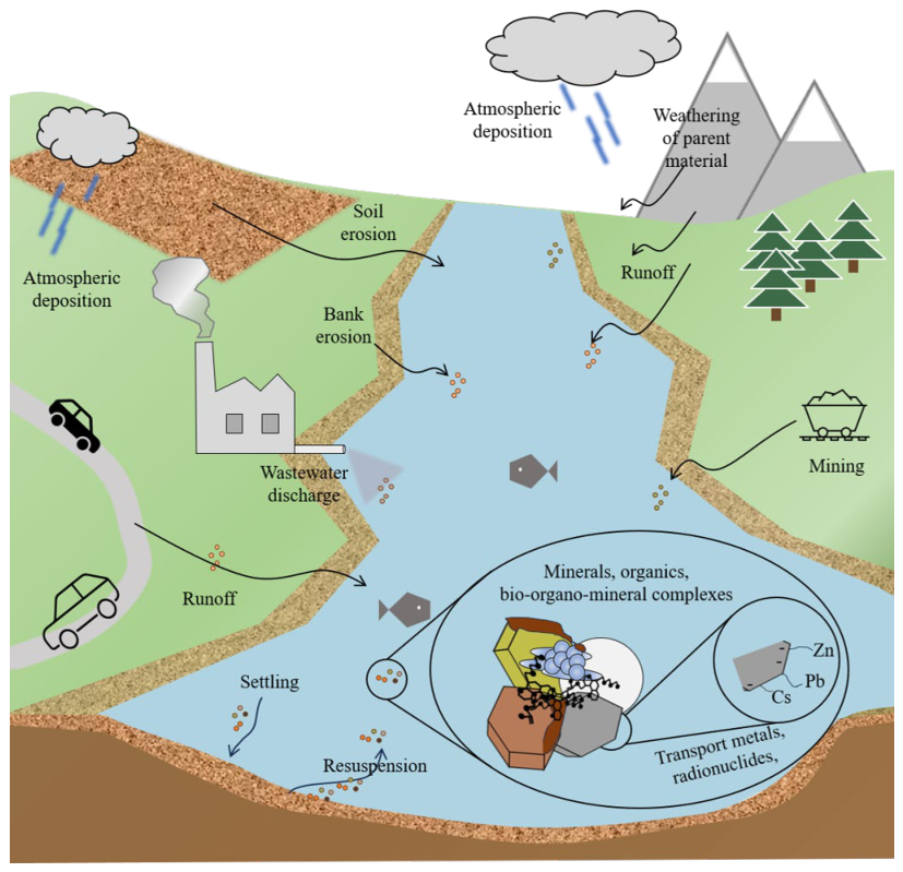
Figure 2.
The various techniques that are used for SPM collection.
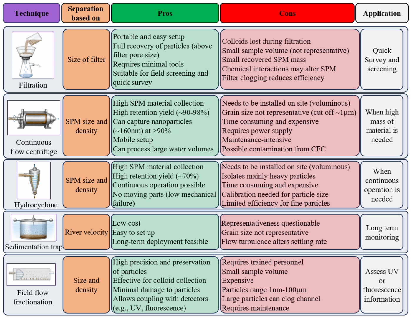
Figure 3.
Main analytical techniques and tools that are used for SPM characterization.
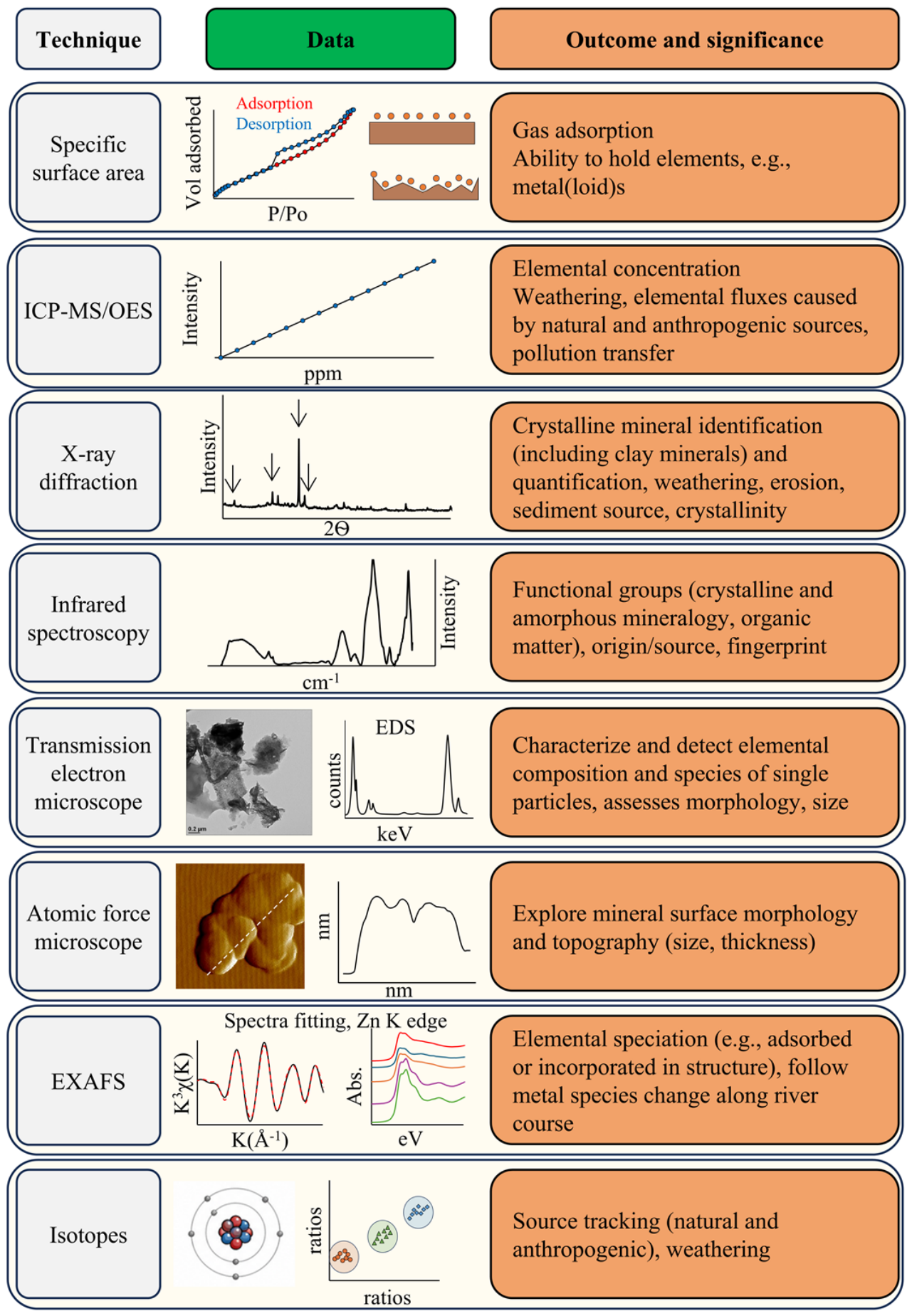
Disclaimer/Publisher’s Note: The statements, opinions and data contained in all publications are solely those of the individual author(s) and contributor(s) and not of MDPI and/or the editor(s). MDPI and/or the editor(s) disclaim responsibility for any injury to people or property resulting from any ideas, methods, instructions or products referred to in the content. |
© 2026 by the authors. Licensee MDPI, Basel, Switzerland. This article is an open access article distributed under the terms and conditions of the Creative Commons Attribution (CC BY) license.
Copyright: This open access article is published under a Creative Commons CC BY 4.0 license, which permit the free download, distribution, and reuse, provided that the author and preprint are cited in any reuse.