

Submitted:
10 August 2026
Posted:
11 August 2026
You are already at the latest version
Abstract
Diagnosing tuberculous pleural effusion (TPE) is challenging, particularly when lung parenchyma appears normal on imaging and sputum smears are negative. This study evaluates the diagnostic utility of medical thoracoscopy in such cases. We conducted a retrospective review of nine patients with exudative pleural effusions and radiographically normal lungs who underwent semi-rigid medical thoracoscopy. Data on demographics, thoracoscopic findings, histopathology, and microbiology (including induced sputum and pleural fluid cultures) were analyzed. The cohort had a mean age of 42 years, with fever (89%) and chest pain (78%) being the most common symptoms. All patients had lymphocytic exudates with elevated adenosine deaminase (ADA). Thoracoscopy revealed characteristic abnormalities, including sago-like nodules and adhesions, in the majority of cases, and provided histological evaluation supporting the diagnosis of tuberculosis in 89% (8/9) of patients. Notably, while all acid-fast bacilli sputum smears were negative, 56% of patients eventually grew M. tuberculosis on induced sputum culture despite the absence of lung parenchymal abnormalities. Medical thoracoscopy is a safe and highly effective diagnostic tool for TPE, providing rapid histological confirmation when non-invasive workup fails. The findings also suggest that patients with normal lung imaging may still harbor active, sputum culture-positive pulmonary tuberculosis, necessitating robust infection-control measures.
Keywords:
tuberculous pleural effusion
; medical thoracoscopy
; pleuroscopy
; adenosine deaminase
; granuloma
; Mycobacterium tuberculosis
; exudative pleural effusion
; induced sputum
; safety-net hospital
1. Introduction
Tuberculosis (TB) remains a global health crisis and a significant diagnostic challenge in clinical practice, particularly in non-endemic regions where the index of suspicion may vary. While pulmonary tuberculosis is the most common presentation, extrapulmonary manifestations account for a significant proportion of cases. Among these, tuberculous pleural effusion (TPE) is the second most common form of extrapulmonary TB in the United States and often presents a distinct set of diagnostic hurdles [1,2].
The pathophysiology of TPE involves a delayed hypersensitivity reaction to mycobacterial antigens entering the pleural space, often resulting in a paucibacillary fluid [3]. Consequently, the traditional initial diagnostic tests often fail; pleural fluid smears for acid-fast bacilli (AFB) are positive in less than 10% of cases, and pleural fluid cultures yield Mycobacterium tuberculosis in only 20% to 50% of patients [4]. This low diagnostic yield frequently forces clinicians to rely on surrogate markers such as adenosine deaminase (ADA) or interferon-gamma release assays (IGRA), or to resort to empiric anti-tuberculous therapy (ATT) [5]. However, empiric treatment without histological or microbiological confirmation carries risks of drug toxicity and delays the diagnosis of alternative etiologies, such as malignancy [6]. Recent literature further emphasizes that while fluid biomarkers are useful, they lack the specificity of tissue diagnosis, particularly in distinguishing TB from malignant pleural effusions in difficult cases [7].
The diagnostic challenge is further compounded when patients present with a pleural effusion but have “normal” lung parenchyma on chest imaging, lacking the characteristic infiltrates, cavities, or nodules associated with active pulmonary TB. In these scenarios, the absence of radiographic parenchymal disease may falsely lower the clinician’s pre-test probability for TB, potentially delaying diagnosis [8].
Medical thoracoscopy (pleuroscopy) has been performed by pulmonary physicians for over 40 years and remains a vital tool in the pulmonologist’s armamentarium. Unlike blind closed pleural biopsy—which has variable sensitivity (ranging from 50–80%) dependent on the uniform distribution of disease—medical thoracoscopy allows for direct visualization of the pleural cavity [9]. It facilitates targeted pleural biopsies of abnormal lesions, such as sago-like nodules or inflamed septations, reportedly increasing diagnostic yield to over 90% [10,11].
Despite its known utility, there is a paucity of literature in the United States specifically examining the role of thoracoscopy in patients with TPE who present with radiographically normal lung parenchyma. This brief retrospective report aims to bridge that gap. We describe the clinical characteristics, thoracoscopic findings, and outcomes of nine patients with undiagnosed pleural effusions and normal lung imaging, highlighting how medical thoracoscopy served as the definitive diagnostic modality when sputum and radiographic workups were unrevealing.
2. Materials and Methods
2.1. Study Design and Patient Selection
The study protocol was approved by the Institutional Review Board (IRB) of the University of Texas Health Science Center at Houston, which waived the requirement for informed consent due to the retrospective nature of the study. This study was conducted at a safety-net hospital. We identified patients who underwent medical thoracoscopy for undiagnosed exudative pleural effusions. Inclusion criteria were strictly defined to isolate the population of interest: (1) presence of an exudative pleural effusion; (2) normal lung parenchyma on chest imaging (defined as the absence of lung opacities, nodules, masses, or cavitations on CT scan); and (3) a final confirmed diagnosis of tuberculous pleuritis.
2.2. Procedures
All patients underwent medical thoracoscopy utilizing a semi-rigid thoracoscope. The procedures were performed in a dedicated endoscopy suite under local anesthesia and moderate sedation, adhering to standard safety protocols. The semi-rigid scope was introduced through a single port and pleural fluid was drained, allowing for thorough inspection of the parietal and visceral pleura.
During the procedure, the pleural space was assessed for characteristic findings of granulomatous disease, including sago-like nodules, adhesions, pleural thickening, and fibrous bands or septations. Multiple targeted parietal pleural biopsies were taken from areas exhibiting these abnormalities using flexible biopsy forceps. A chest tube was placed after the procedure and removed prior to discharge.
2.3. Data Collection
We extracted data from electronic medical records regarding patient demographics (age, gender), comorbidities, and presenting clinical symptoms (fever, cough, chest pain). Pleural fluid analysis was reviewed for cell count, differential, and ADA levels. Microbiological data were analyzed, including sputum AFB smears, induced sputum M. tuberculosis cultures, and pleural fluid cultures. At least two induced sputum samples were obtained for each patient. Histopathological reports from the pleural biopsies were reviewed for the presence of granulomatous inflammation, micro-organisms, and caseating necrosis.
2.4. Outcomes
The primary outcome was the diagnostic yield of medical thoracoscopy (histopathology) compared to sputum and pleural fluid analysis. Secondary outcomes included the description of thoracoscopic visual findings and the clinical resolution of the disease following the initiation of anti-tuberculous therapy (ATT).
3. Results
3.1. Demographics and Clinical Presentation
The study cohort consisted of nine patients. The mean age was 42 years, with a range of 25 to 60 years, reflecting a younger demographic often seen in TPE compared to malignant effusions [12]. The majority of patients were male (N = 6, 67%). Comorbidities were present in 33% of the cohort (N = 3), specifically chronic kidney disease (one patient) and diabetes mellitus (two patients), known risk factors for TB reactivation or post-primary tuberculosis. None of the patients had HIV infection.
The clinical presentation was symptomatic in all cases. Fever was the predominant symptom, reported in 89% (N = 8) of patients, followed closely by pleuritic chest pain in 78% (N = 7) and cough in 67% (N = 6). Seven patients (78%) presented with unilateral pleural effusion (Figure 1E). These findings are summarized in Table 1.
3.2. Pleural Fluid Analysis
Thoracentesis performed prior to or during thoracoscopy revealed lymphocytic exudates in all nine patients (100%). The mean lymphocyte percentage was 85%, consistent with the localized immune response typical of TPE. Adenosine deaminase (ADA) levels were universally elevated (>40 U/L), with a mean of 61.4 U/L (range 42–105 U/L), strongly suggestive of a tuberculous etiology [13].
3.3. Thoracoscopic Findings
Direct visualization of the pleural space provided critical diagnostic clues. The most frequently observed abnormalities were pleural thickening (N = 8, 89%) and adhesions (N = 7, 78%). “Sago-like” nodules—small, white, granulomatous deposits on the parietal pleura—were visualized in 5 patients (56%) (Figure 1C). Fibrous bands and septations were also noted in 56% of cases (Figure 1A,B). Notably, 89% of patients exhibited at least two of these macroscopic findings, reinforcing the value of visual inspection in establishing a high pre-test probability intraoperatively.
3.4. Histopathology and Microbiology
Histopathological examination of the targeted pleural biopsies supported the diagnosis of tuberculosis. It revealed granulomatous inflammation with caseating necrosis in 8 out of 9 patients (89%). In one patient, parietal pleural biopsies were not possible due to severe organized adhesions preventing access, despite the presence of characteristic visual findings; however, this patient was diagnosed via microbiological confirmation.
Microbiological analysis revealed a significant discrepancy between fluid/sputum analysis and biopsy. Pleural fluid cultures yielded M. tuberculosis in only 2 patients (22%). Acid-fast bacilli (AFB) sputum smears were negative in 100% of patients. However, induced M. tuberculosis sputum cultures eventually returned positive in 5 out of 9 patients (56%), including the single patient who had a non-diagnostic pleural biopsy. This occurred despite the complete absence of radiographic evidence of pulmonary tuberculosis. A comparison of diagnostic yields is presented in Table 2.
3.5. Treatment and Follow-Up
All patients tolerated the procedure without any complications. A chest tube was placed in all patients’ post-procedure and removed after 24 hours. Following the procedure, all patients were started on standard anti-tuberculous therapy (ATT) based on pleuroscopic findings and/or histology, prior to culture confirmation, and continued treatment with directly observed therapy (DOT). All nine patients achieved complete clinical and radiological resolution, confirming the efficacy of the expedited diagnosis and treatment.
4. Discussion
This retrospective brief report underscores the indispensable role of medical thoracoscopy in the diagnosis of tuberculous pleural effusion, particularly in the challenging subset of patients with radiographically normal lung parenchyma. In the United States, where TB incidence is lower than in developing nations, TPE can easily be misdiagnosed or delayed, often mistaken for parapneumonic effusions or malignancy [14]. Our study highlights three critical clinical implications: the limitations of non-invasive testing, the risk of delay or unnecessary treatment while waiting for culture results, and the diagnostic power of direct visualization.
4.1. The Limitations of Non-Invasive Testing
Our data reinforces the well-established limitation of smears and fluid cultures in TPE. All nine patients were sputum AFB smear-negative, and pleural fluid cultures were positive in only 22% of cases. Relying solely on these initial tests in patients with normal chest X-rays would have resulted in a significant delay in diagnosis and treatment initiation. While ADA levels were elevated in our cohort (mean 61.4 U/L), supporting the diagnosis, ADA can occasionally be elevated in parapneumonic effusions, empyema, and lymphomas [13,15]. As highlighted by Amipara et al. in 2024, while ADA is a key indicator, relying on it without microbiological confirmation can be risky in non-endemic settings where malignancy is a primary differential [16]. Furthermore, while IGRAs are useful for detecting latent infections, they cannot distinguish active pleural disease from latent TB, limiting their utility in the acute diagnostic setting [17]. Additionally, a major limitation of relying solely on ADA is the inability to perform drug susceptibility testing, leaving clinicians unaware of potential multidrug-resistant tuberculosis.
4.2. The Utility of Medical Thoracoscopy
Medical thoracoscopy provided histological findings supportive of tuberculosis in 89% of our patients. The visualization of “sago-like” nodules is highly suggestive for TB, allowing the pulmonologist to target these areas for biopsy. This targeted approach is superior to blind closed pleural biopsy, which carries a risk of sampling error, particularly if the granulomas are unevenly distributed on the parietal pleura [18]. Recent findings by Rawat et al. (2024) confirm this high diagnostic yield, reporting over 85% success with thoracoscopy in undiagnosed effusions [19]. Furthermore, thoracoscopy allowed for the mechanical breakdown of adhesions and septations in some cases, potentially aiding in fluid drainage, although its primary role here was diagnostic [20]. The safety profile was excellent, with procedures performed under moderate sedation and local anesthesia, avoiding the risks and costs associated with general anesthesia and video-assisted thoracoscopic surgery (VATS) [21].
4.3. Occult Pulmonary Involvement
Perhaps the most intriguing finding of this study is the positivity of induced sputum cultures in 56% of patients, despite normal lung parenchyma on imaging in non-immunocompromised patients. This finding challenges the conventional dogma that TPE is strictly a paucibacillary, sequestered pleural disease. It suggests that a significant number of TPE patients have concomitant active pulmonary TB that is radiographically occult, likely endobronchial or microscopic parenchymal disease [22].
This has profound infection-control implications. Patients with TPE and “normal lungs” are often considered non-infectious and may not be isolated initially. Our findings show that the lungs indeed serve as a source of M. tuberculosis in these patients. Therefore, even in the absence of radiographic infiltrates, clinicians should maintain a high index of suspicion for communicability, and induced sputum cultures should be obtained alongside pleural interventions [23].
4.4. Limitations
This study is limited by its retrospective design and small sample size (N = 9). As a single-center experience, referral biases may exist. However, the strict inclusion criteria (normal parenchyma) make this a unique dataset, as most TPE studies mix patients with and without pulmonary findings.
5. Conclusions
In patients with lymphocytic exudative pleural effusions and radiographically normal lung parenchyma, medical thoracoscopy is a safe and highly effective diagnostic modality. It overcomes the limitations of sputum smears and blind biopsies, facilitating an expedited diagnosis through direct visualization and targeted histopathology. Furthermore, the significant rate of positive sputum cultures in this cohort suggests that “normal” chest imaging does not rule out active pulmonary tuberculosis. Early implementation of thoracoscopy in this patient population prevents delays in anti-tuberculous therapy, improves patient outcomes, and may help mitigate the risk of occult TB transmission.
Author Contributions
Conceptualization, M.A.A. and S.C.; methodology, M.A.A., S.C. and R.M.E.-Y.-M.; validation, S.C. and R.M.E.-Y.-M.; formal analysis, M.A.A. and U.J.; investigation, M.A.A., U.J. and Z.H.; resources, S.C. and R.M.E.-Y.-M.; data curation, M.A.A., U.J. and Z.H.; writing—original draft preparation, M.A.A. and U.J.; writing—review and editing, S.C., R.M.E.-Y.-M. and Z.H.; visualization, M.A.A.; supervision, S.C. and R.M.E.-Y.-M.; project administration, M.A.A. and S.C. All authors have read and agreed to the published version of the manuscript.
Funding
This research received no external funding.
Institutional Review Board Statement
The study was conducted in accordance with the Declaration of Helsinki and approved by the Institutional Review Board of the University of Texas Health Science Center at Houston, which waived the requirement for informed consent due to the retrospective nature of the study.
Informed Consent Statement
Patient consent was waived due to the retrospective design of the study, as approved by the Institutional Review Board. No individually identifiable patient information is included in this manuscript.
Data Availability Statement
The data presented in this study is available on request from the corresponding author. The data is not publicly available due to privacy and ethical restrictions.
Conflicts of Interest
The authors declare no conflicts of interest.
Acknowledgments
None.
Abbreviations
The following abbreviations are used in this manuscript:
| ADA | Adenosine deaminase |
| AFB | Acid-fast bacilli |
| ATT | Anti-tuberculous therapy |
| CT | Computed tomography |
| DOT | Directly observed therapy |
| HIV | Human immunodeficiency virus |
| IGRA | Interferon-gamma release assay |
| IRB | Institutional review board |
| TB | Tuberculosis |
| TPE | Tuberculous pleural effusion |
| VATS | Video-assisted thoracoscopic surgery |
References
- Vorster, M.J.; Allwood, B.W.; Diacon, A.H.; Koegelenberg, C.F. Tuberculous pleural effusions: advances and controversies. J. Thorac. Dis. 2015, 7, 981–991. [Google Scholar] [PubMed]
- Baumann, M.H.; Nolan, R.; Petrini, M.; et al. Pleural tuberculosis in the United States: incidence and drug resistance. Chest 2007, 131, 1125–1132. [Google Scholar] [CrossRef] [PubMed]
- Zhai, K.; Lu, Y.; Shi, H.Z. Tuberculous pleural effusion. J. Thorac. Dis. 2016, 8, E486–E494. [Google Scholar] [CrossRef] [PubMed]
- Porcel, J.M. Tuberculous pleural effusion. Lung 2009, 187, 263–270. [Google Scholar] [CrossRef] [PubMed]
- Gopi, A.; Madhavan, S.M.; Sharma, S.K.; Sahn, S.A. Diagnosis and treatment of tuberculous pleural effusion in 2006. Chest 2007, 131, 880–889. [Google Scholar] [CrossRef] [PubMed]
- Wang, W.; Zhou, Q.; Zhai, K.; Wang, Y.; Shi, H.Z. Diagnostic accuracy of interleukin 27 for tuberculous pleural effusion: two prospective studies and a meta-analysis. Thorax 2018, 73, 240–247. [Google Scholar] [PubMed]
- Zhang, J.; et al. High diagnostic accuracy of flexible thoracoscopy for distinguishing tuberculous from malignant pleural effusions. Am. J. Transl. Res. 2025, 17, 5284–5292. [Google Scholar] [CrossRef] [PubMed]
- Kim, H.J.; Lee, H.J.; Kwon, S.Y.; et al. The prevalence of pulmonary parenchymal tuberculosis in patients with tuberculous pleurisy. Chest 2006, 129, 1253–1258. [Google Scholar] [CrossRef] [PubMed]
- Koegelenberg, C.F.; Diacon, A.H. Pleural controversy: close needle biopsy or thoracoscopy? Respirology 2011, 16, 738–746. [Google Scholar] [CrossRef] [PubMed]
- Diacon, A.H.; Van de Wal, B.W.; Wyser, C.; et al. Diagnostic tools in tuberculous pleurisy: a direct comparative study. Eur. Respir. J. 2003, 22, 589–594. [Google Scholar] [CrossRef] [PubMed]
- Wang, Z.; Xu, L.L.; Wu, Y.B.; et al. Diagnostic value and safety of medical thoracoscopy in tuberculous pleural effusion. Respir. Med. 2015, 109, 1188–1192. [Google Scholar] [CrossRef] [PubMed]
- Light, R.W. Update on tuberculous pleural effusion. Respirology 2010, 15, 451–458. [Google Scholar] [CrossRef] [PubMed]
- Liang, Q.L.; Shi, H.Z.; Wang, K.; Qin, S.M.; Qin, X.J. Diagnostic performance of adenosine deaminase in tuberculous pleural effusion: a meta-analysis. Respir. Med. 2008, 102, 744–754. [Google Scholar] [PubMed]
- Skouras, V.S.; Kalomenidis, I. Tuberculous pleural effusion: likely underdiagnosed. Monaldi Arch. Chest Dis. 2017, 87, 773. [Google Scholar]
- Greco, S.; Girardi, E.; Masciangelo, R.; Capoccetta, G.B.; Saltini, C. Adenosine deaminase and interferon-gamma measurements for the diagnosis of tuberculous pleurisy: a meta-analysis. Int. J. Tuberc. Lung Dis. 2003, 7, 777–786. [Google Scholar] [PubMed]
- Amipara, A.G.; Rangari, A.; Ghewade, B. Diagnosis and management of tuberculous pleural effusion in a patient with chronic obstructive pulmonary disease: a case report. Cureus 2024, 16, e64532. [Google Scholar] [CrossRef] [PubMed]
- Meldau, R.; Peter, J.; Theron, G.; et al. Comparison of same day diagnostic tools including Gene Xpert and unstimulated IFN-γ for the evaluation of pleural tuberculosis: a prospective cohort study. BMC Pulm. Med. 2014, 14, 58. [Google Scholar] [CrossRef] [PubMed]
- Casal, S.R. Medical thoracoscopy. Curr. Opin. Pulm. Med. 2009, 15, 313–320. [Google Scholar] [CrossRef] [PubMed]
- Rawat, J.; Kumar, A.; Mrigpuri, P.; et al. Role of single port rigid thoracoscopy in undiagnosed pleural effusion. Tuberc. Respir. Dis. 2024, 87, 194–199. [Google Scholar] [CrossRef] [PubMed]
- Rahman, N.M.; Ali, N.J.; Brown, G.; et al. Local anaesthetic thoracoscopy: British Thoracic Society pleural disease guideline 2010. Thorax 2010, 65 (Suppl. 2), ii54–ii60. [Google Scholar] [CrossRef] [PubMed]
- Shojaee, S.; Lee, H.J. Thoracoscopy: medical versus surgical—in the management of pleural diseases. J. Thorac. Dis. 2015, 7 (Suppl. 4), S339–S351. [Google Scholar] [CrossRef] [PubMed]
- Ruan, S.Y.; Chuang, Y.C.; Wang, J.Y.; et al. Revisiting tuberculous pleurisy: pleural fluid characteristics and diagnostic yield of mycobacterial culture in an endemic area. Thorax 2012, 67, 822–827. [Google Scholar] [CrossRef] [PubMed]
- Conde, M.B.; Loizos, A.; Rezende, T.; et al. Yield of sputum induction in the diagnosis of pleural tuberculosis. Am. J. Respir. Crit. Care Med. 2003, 167, 723–725. [Google Scholar] [CrossRef] [PubMed]
Figure 1.
Thoracoscopic and radiographic findings. Panels (A,B) show pleuroscopy findings of septations (A) and fibrous bands (B); (C) classic sago-like nodules (arrow); and (D) exudative thick effusion with nodules (arrow), all suggestive of TPE. (E) is a CT scan illustrating the corresponding left pleural effusion (arrow) and thickening.
Figure 1.
Thoracoscopic and radiographic findings. Panels (A,B) show pleuroscopy findings of septations (A) and fibrous bands (B); (C) classic sago-like nodules (arrow); and (D) exudative thick effusion with nodules (arrow), all suggestive of TPE. (E) is a CT scan illustrating the corresponding left pleural effusion (arrow) and thickening.
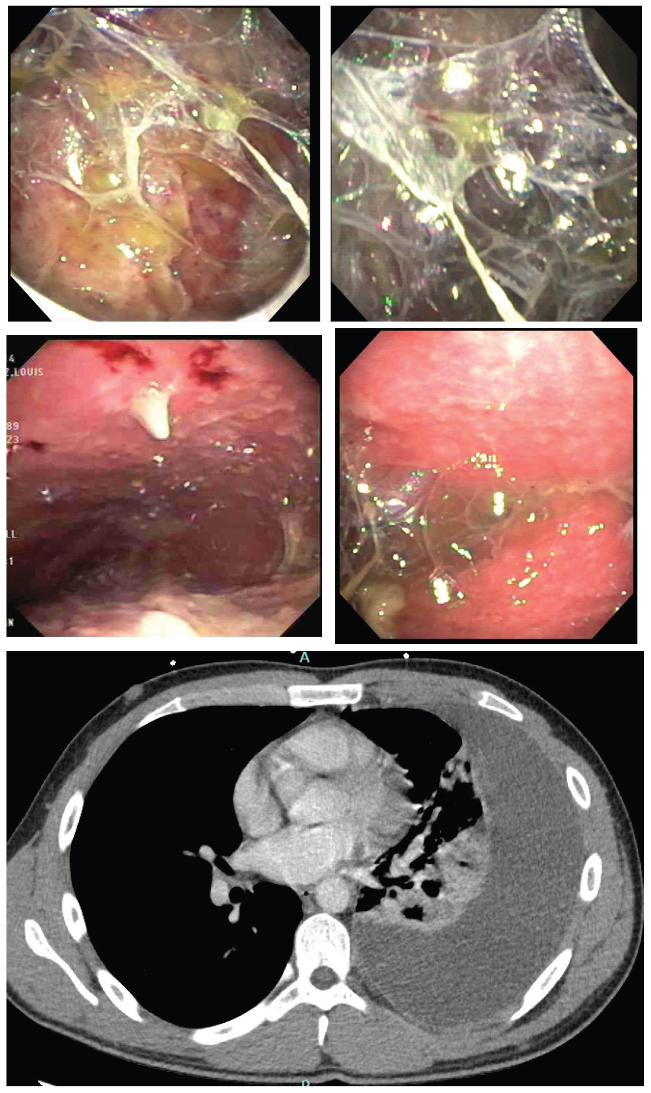

Table 1.
Patient demographics and clinical characteristics (N = 9).
| Characteristic | n (%) or Mean (Range) |
| Age (years) | 42 (25–60) |
| Gender | |
| Male | 6 (67%) |
| Female | 3 (33%) |
| Comorbidities | |
| Diabetes mellitus | 2 (22%) |
| Chronic kidney disease | 1 (11%) |
| None | 6 (67%) |
| Presenting symptoms | |
| Fever | 8 (89%) |
| Chest pain | 7 (78%) |
| Cough | 6 (67%) |
| Effusion laterality | |
| Unilateral | 7 (78%) |
| Bilateral | 2 (22%) |
Table 2.
Comparison of diagnostic modalities.
| Diagnostic Modality | Positive Findings (n/N) | Yield (%) |
| Pleural Fluid Analysis | ||
| Lymphocytic exudate | 9/9 | 100% |
| Elevated ADA (>40 U/L) | 9/9 | 100% |
| Pleural fluid M. tuberculosis culture | 2/9 | 22% |
| Sputum Microbiology | ||
| Sputum AFB smear | 0/9 | 0% |
| Induced sputum M. tuberculosis culture | 5/9 | 56% |
| Medical Thoracoscopy | ||
| Visual abnormalities (≥2 findings) | 8/9 | 89% |
| Histopathology (granulomas) | 8/9 | 89% |
Disclaimer/Publisher’s Note: The statements, opinions and data contained in all publications are solely those of the individual author(s) and contributor(s) and not of MDPI and/or the editor(s). MDPI and/or the editor(s) disclaim responsibility for any injury to people or property resulting from any ideas, methods, instructions or products referred to in the content. |
© 2026 by the authors. Licensee MDPI, Basel, Switzerland. This article is an open access article distributed under the terms and conditions of the Creative Commons Attribution (CC BY) license (http://creativecommons.org/licenses/by/4.0/).
Copyright: This open access article is published under a Creative Commons CC BY 4.0 license, which permit the free download, distribution, and reuse, provided that the author and preprint are cited in any reuse.