
Submitted:
30 July 2026
Posted:
11 August 2026
You are already at the latest version
Abstract
Objective: To challenge the longstanding assumption that the sonographic Shred Sign and the Fractal Sign are synonymous and to redefine their diagnostic relationship in peripheral lung disease. Methods and Study Design: This prospective observational study analysed 700 consecutive comprehensive lung ultrasound examinations obtained from hospitalized patients. All patients underwent chest computed tomography (CT) and lung ultrasound as part of the routine diagnostic evaluation. Final diagnoses were established according to current international disease-specific guidelines by integrating clinical findings, laboratory investigations, CT imaging, microbiological studies, bronchoscopy, histopathology, and clinical follow-up, where appropriate. Morphological analysis was subsequently performed on the lung ultrasound examinations after diagnostic confirmation. Setting: Single tertiary university pulmonary referral centre. Participants: Seven hundred consecutive patients undergoing comprehensive lung ultrasound for peripheral lung disease. Primary outcome measure: Comparative assessment of the morphological characteristics of the Shred Sign across confirmed pulmonary diseases, focusing on boundary geometry, internal echotexture, air bronchogram dynamics, and airway patency. Secondary outcome measure: Identification and characterization of the True Fractal Sign, Non-Fractal Shred Sign, and Fractal Mimicry based on the combined morphological profile. Results: Among 700 examinations, 446 demonstrated an irregular pleural–parenchymal interface (Shred Sign). Of these, 127 exhibited fractal-like morphology; 126 fulfilled the criteria for a True Fractal Sign, whereas one lung cancer represented Fractal Mimicry. The remaining 319 cases were classified as Non-Fractal Shred Signs. True Fractal Signs were identified only in a subset of acute pneumonias, whereas Non-Fractal Shred Signs occurred both in acute pneumonias and in pulmonary infarction, tuberculosis, bronchiectasis, lung cancer, and other structural lung diseases. These findings demonstrate that the Shred Sign and the True Fractal Sign are distinct sonographic entities rather than synonymous findings. Conclusions: The Shred Sign is a broad descriptive sonographic pattern rather than a specific diagnostic entity. The True Fractal Sign represents a distinct geometric subtype associated with preservation of the alveolar branching architecture during acute exudative pneumonia. Based on these findings, we propose a novel four-pillar diagnostic tetrad integrating boundary geometry, internal shade, dynamic air bronchograms, and airway patency to differentiate True Fractal Sign, Non-Fractal Shred Sign, and Fractal Mimicry. This morphology-based framework may improve the diagnostic specificity of bedside lung ultrasound. The observation that many acute pneumonias lack a True Fractal Sign generates the hypothesis that preservation of fractal architecture depends on the integrity of the underlying lung structure and the stage of disease, warranting prospective validation.
Keywords:
lung ultrasound
; shred sign
; fractal sign
; morphological diagnosis
; peripheral lung disease
; pneumonia
Introduction
Lung ultrasound (LUS) has become an essential bedside imaging modality for evaluating peripheral lung diseases, largely owing to the pioneering concepts introduced by point-of-care ultrasound (POCUS) protocols for acute respiratory failure [1]. Within this framework, an irregular pleural–parenchymal interface bordering aerated lung has traditionally been described interchangeably as the Shred Sign or the Fractal Sign [2,3]. This terminology evolved in an emergency setting where rapid recognition of peripheral lung consolidation was prioritized over detailed morphological characterization [4,5].
As a result, the terms Shred Sign and Fractal Sign have remained widely accepted as synonymous in clinical practice and continue to appear interchangeably in major POCUS reviews, educational resources, and respiratory literature [6-13]. While this equivalence is appropriate for rapid identification of peripheral consolidations, it assumes that all irregular pleural–parenchymal boundaries represent the same underlying structural phenomenon.
However, peripheral lung diseases encompass diverse pathological processes. Acute exudative pneumonia, pulmonary infarction, tuberculosis, bronchiectasis, malignant infiltration, and chronic fibrotic remodeling may all produce an irregular pleural–parenchymal interface despite fundamentally different alterations of lung tissue [1,14,15,16,17,18]. Whether these diverse lesions generate the same sonographic morphology has never been systematically investigated.
This distinction may have important clinical implications. If all irregular interfaces are interpreted as a single sonographic entity, acute inflammatory processes may become indistinguishable from chronic structural remodeling, potentially reducing the diagnostic specificity of lung ultrasound beyond the emergency setting [11,19,20].
Therefore, the present study was designed to challenge the longstanding assumption that the Shred Sign and the Fractal Sign are synonymous. By analysing 700 consecutive lung ultrasound examinations across a broad spectrum of peripheral lung diseases, we sought to determine whether these signs represent a single sonographic phenomenon or distinct morphological entities with different diagnostic implications.
Methods
Study Design
This prospective observational study analysed 700 consecutive comprehensive lung ultrasound examinations obtained from hospitalized patients.
All patients underwent chest computed tomography (CT) and lung ultrasound as part of the routine diagnostic evaluation. Final diagnoses were established according to current international disease-specific guidelines by integrating clinical findings, laboratory investigations, CT imaging, microbiological studies, bronchoscopy, histopathology, and clinical follow-up, where appropriate.
Morphological analysis was subsequently performed on the lung ultrasound examinations after diagnostic confirmation.
Lung Ultrasound Examination
Comprehensive lung ultrasound examinations were performed using a Clarius C3 HD3 handheld ultrasound scanner equipped with a convex transducer. A standardized examination protocol was applied in all patients. Ultrasound images were independently evaluated by two experienced lung ultrasonographers.
Morphological Analysis
After the final diagnosis had been established, archived lung ultrasound examinations were systematically re-evaluated.
The objective of the morphological analysis was not to determine the diagnosis by ultrasound, but to characterize the morphological appearance of the pleural–parenchymal interface within each confirmed pulmonary disease. The analysis focused on the presence and morphological characteristics of the Shred Sign, True Fractal Sign, Fractal Mimicry, and Non-Fractal Shred Sign. Particular attention was paid to boundary geometry, internal echotexture, dynamic air bronchograms, and airway patency. Comparative morphological analysis was performed across the spectrum of confirmed pulmonary diseases. The analysis evaluated boundary geometry, internal echotexture, air bronchogram dynamics, and airway patency.
Results
Study Population
A total of 700 consecutive comprehensive lung ultrasound examinations were analysed. The distribution of confirmed pulmonary diagnoses is presented in Table 1.
Distribution of Sonographic Patterns
An irregular parenchymal interface (Shred Sign) was identified in 446 examinations (63.7%). Among these, 127 examinations (28.5%) demonstrated fractal-like morphology, whereas 319 (71.5%) demonstrated non-fractal morphology. Of the 127 examinations with fractal-like morphology, 126 (99.2%) were classified as True Fractal Sign, while one examination (0.8%) represented Fractal Mimicry, observed in a patient with lung cancer.
Distribution Across Confirmed Diseases
The True Fractal Sign was identified in patients with acute pneumonia. The Non-Fractal Shred Sign was observed in acute pneumonia, pulmonary infarction, tuberculosis, bronchiectasis, lung cancer, and other pulmonary diseases associated with irregular parenchymal interfaces.
Morphological Classification
Systematic comparative morphological analysis identified three distinct sonographic patterns: True Fractal Sign, Fractal Mimicry, and Non-Fractal Shred Sign. These observations formed the basis for the proposed four-pillar diagnostic tetrad.
Discussion
The Shred Sign and the True Fractal Sign Are Not Synonymous
The present study demonstrates that the Shred Sign and the True Fractal Sign should not be regarded as synonymous sonographic entities. Although both describe irregular pleural–parenchymal interfaces, they do not represent the same underlying structural organization. Our observations indicate that the Shred Sign is a broad morphological descriptor encompassing geometrically distinct patterns, only one of which fulfills the characteristics of a true biological fractal [11,21,22]. This distinction forms the conceptual basis of the proposed four-pillar diagnostic tetrad.
What Defines a Shred Sign?
Before distinguishing between the Fractal Sign and the Shred Sign, a more fundamental question must first be addressed: what characteristics qualify an ultrasound boundary as a Shred Sign?
Despite its widespread use in lung ultrasound, the Shred Sign has never been systematically defined beyond the generic description of an irregular or shredded deep border [2,5,9]. No objective morphological criteria have been proposed to distinguish which irregular pleural–parenchymal interfaces should be classified as a Shred Sign and which should not [11,23].
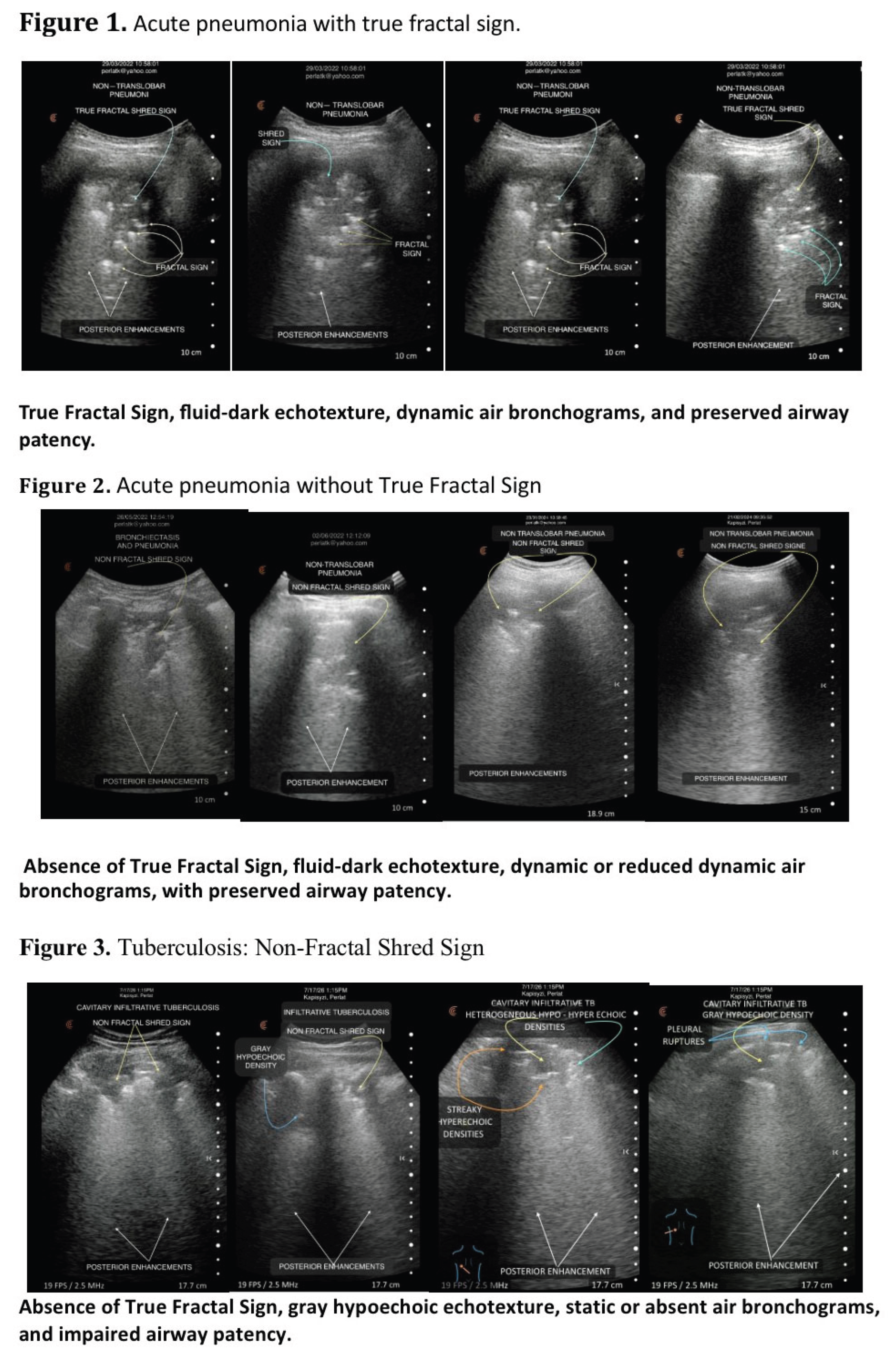
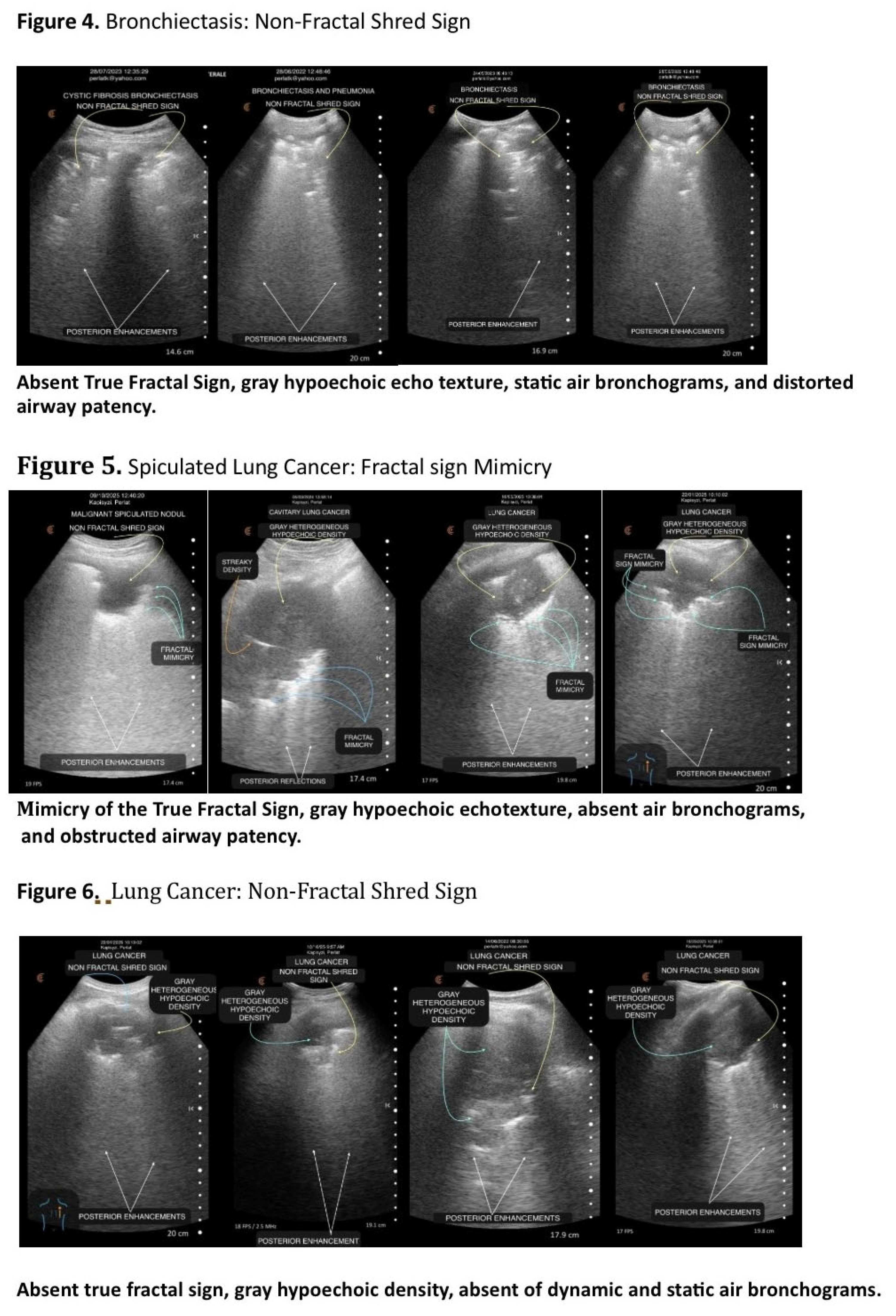
Our findings indicate that the Shred Sign is fundamentally a morphological descriptor rather than an etiological diagnosis. It simply identifies an irregular interface between consolidated and aerated lung, irrespective of the underlying disease. Consequently, restricting this term to pneumonia represents a conceptual simplification rather than a scientific definition [11,23] [Figure 1, Figure 2, Figure 3, Figure 4, Figure 5 and Figure 6]
Revisiting the Historical Equivalence
Once the Shred Sign is recognized as a morphological descriptor, a second question naturally arises: are all Shred Signs morphologically equivalent?
For almost two decades, point-of-care ultrasound (POCUS) algorithms have treated the Shred Sign and the Fractal Sign as interchangeable terms [2,5,6,9,11,12,13]. This simplification has been appropriate for rapid emergency algorithms such as the BLUE Protocol, where the primary objective is the prompt recognition of peripheral consolidation [3,6]. However, in pulmonology and internal medicine, where a much broader spectrum of peripheral lung diseases is encountered, such equivalence may obscure important morphological differences with potential diagnostic implications [2,3,5,6,9,24,25,26,27].
Biological Basis of the True Fractal Sign
The native human respiratory tree is a biological fractal that branches recursively across multiple structural scales [24,25]. During early acute exudative pneumonia, inflammatory fluid fills the pre-existing alveolar–lobular framework while largely preserving its native acinar architecture [1,24,25,26]. Consequently, the pleural–parenchymal interface retains its hierarchical geometric organization and appears sonographically as a True Fractal Sign [18,20].
In contrast, chronic inflammatory, fibrotic, and neoplastic diseases progressively distort or destroy the normal pulmonary architecture [2,28]. Caseous necrosis, fibrosis, cavitation, and tumor infiltration replace the normal alveolar–lobular framework with structurally disorganized tissue [15,17,18,19,20,21,22,23,24,25,26,27,28,29].
A Time- and Severity-Dependent Hypothesis
Our observations also suggest that the True Fractal Sign may represent a time- and severity-dependent phenomenon. It is likely to be most evident during the early exudative phase of acute inflammation, when the native pulmonary architecture remains largely preserved. As inflammation progresses or becomes more severe, increasing tissue distortion may progressively disrupt this geometric organization. Consequently, absence of the True Fractal Sign should not necessarily be interpreted as evidence of chronic structural remodeling alone but may also reflect a more advanced stage or greater severity of acute inflammatory disease [2,18]. This hypothesis requires prospective validation.
Clinical Implications
Recognition of the distinction between the True Fractal Sign and the Non-Fractal Shred Sign has important clinical implications. The presence of an irregular pleural–parenchymal interface should not automatically be interpreted as evidence of acute bacterial pneumonia. Rather, it should prompt a comprehensive morphological assessment integrating boundary geometry, internal echotexture, bronchogram dynamics, and airway patency [19,20,30]. Together, these complementary features constitute the proposed four-pillar diagnostic tetrad. [Table 2]
Discussion
The Shred Sign and the True Fractal Sign Are Not Synonymous
The present study demonstrates that the Shred Sign and the True Fractal Sign should not be regarded as synonymous sonographic entities. Although both describe irregular pleural–parenchymal interfaces, they do not represent the same underlying structural organization. Our observations indicate that the Shred Sign is a broad morphological descriptor encompassing geometrically distinct patterns, only one of which fulfills the characteristics of a true biological fractal. This distinction forms the conceptual basis of the proposed four-pillar diagnostic tetrad.
What Defines a Shred Sign?
Before distinguishing between the Fractal Sign and the Shred Sign, a more fundamental question must first be addressed: what characteristics qualify an ultrasound boundary as a Shred Sign?
Despite its widespread use in lung ultrasound, the Shred Sign has never been systematically defined beyond the generic description of an irregular or shredded deep border [2,5,9]. No objective morphological criteria have been proposed to distinguish which irregular pleural–parenchymal interfaces should be classified as a Shred Sign and which should not.
Our findings indicate that the Shred Sign is fundamentally a morphological descriptor rather than an etiological diagnosis. It simply identifies an irregular interface between consolidated and aerated lung, irrespective of the underlying disease. Consequently, restricting this term to pneumonia represents a conceptual simplification rather than a scientific definition. [Figures1–6]
Revisiting the Historical Equivalence
Once the Shred Sign is recognized as a morphological descriptor, a second question naturally arises: are all Shred Signs morphologically equivalent?
For almost two decades, point-of-care ultrasound (POCUS) algorithms have treated the Shred Sign and the Fractal Sign as interchangeable terms [2,5,6,9]. This simplification has been appropriate for rapid emergency algorithms such as the BLUE Protocol, where the primary objective is the prompt recognition of peripheral consolidation [3,6]. However, in pulmonology and internal medicine, where a much broader spectrum of peripheral lung diseases is encountered, such equivalence may obscure important morphological differences with potential diagnostic implications [2,3,5,6,9,20].
Biological Basis of the True Fractal Sign
The native human respiratory tree is a biological fractal that branches recursively across multiple structural scales [24,25]. During early acute exudative pneumonia, inflammatory fluid fills the pre-existing alveolar–lobular framework while largely preserving its native acinar architecture [1,24,25,26]. Consequently, the pleural–parenchymal interface retains its hierarchical geometric organization and appears sonographically as a True Fractal Sign
In contrast, chronic inflammatory, fibrotic, and neoplastic diseases progressively distort or destroy the normal pulmonary architecture [20,22]. Caseous necrosis, fibrosis, cavitation, and tumor infiltration replace the normal alveolar-lobular framework with structurally disorganized tissue [30,31].
A Time- and Severity-Dependent Hypothesis
Our observations also suggest that the True Fractal Sign may represent a time- and severity-dependent phenomenon. It is likely to be most evident during the early exudative phase of acute inflammation, when the native pulmonary architecture remains largely preserved. As inflammation progresses or becomes more severe, increasing tissue distortion may progressively disrupt this geometric organization. Consequently, absence of the True Fractal Sign should not necessarily be interpreted as evidence of chronic structural remodeling alone but may also reflect a more advanced stage or greater severity of acute inflammatory disease. This hypothesis requires prospective validation.
Clinical Implications
Recognition of the distinction between the True Fractal Sign and the Non-Fractal Shred Sign has important clinical implications. The presence of an irregular pleural–parenchymal interface should not automatically be interpreted as evidence of acute bacterial pneumonia. Rather, it should prompt a comprehensive morphological assessment integrating boundary geometry, internal echotexture, bronchogram dynamics, and airway patency. Together, these complementary features constitute the proposed four-pillar diagnostic tetrad. [Table 2]
Within this framework, the True Fractal Sign represents the geometrically preserved acute subtype of the Shred Sign, whereas the Non-Fractal Shred Sign reflects loss of fractality secondary to architectural remodeling.
Diagnostic Exceptions
Two important diagnostic exceptions deserve consideration.
First, the True Fractal Sign requires preservation of the native pulmonary architecture. Therefore, its absence does not exclude an acute inflammatory process when infection develops within a lung that has already undergone irreversible structural remodeling. In such circumstances, diagnosis should rely on integrated assessment of all four sonographic pillars rather than boundary geometry alone [15,18,20].
Second, peripheral spiculated lung carcinomas may superficially resemble a True Fractal Sign. However, careful geometric analysis demonstrates irregular projections that fail to preserve recursive scaling or statistical self-similarity. Rather than representing preserved biological architecture, this appearance reflects infiltrative tumor growth and desmoplastic remodeling. Accordingly, these lesions should be regarded as Fractal Mimicry rather than a True Fractal Sign [20,24,32,33,34].
Strengths and Limitations
The strengths of this study include the analysis of a large real-world cohort comprising 700 comprehensive lung ultrasound examinations covering a broad spectrum of peripheral pulmonary diseases. Morphological patterns were evaluated across independently established reference diagnoses, allowing comparison of sonographic appearances beyond individual disease entities.
Several limitations should also be acknowledged. This was a single-center observational study, and the proposed morphological classification has not yet undergone external prospective validation or formal assessment of interobserver reproducibility. Future multicenter studies are required to confirm the diagnostic performance and generalizability of the proposed tetrad.
Conclusions
The Shred Sign should be regarded as a broad morphological descriptor rather than a disease-specific sonographic sign. Consequently, the terms Shred Sign and Fractal Sign should no longer be considered synonymous
The True Fractal Sign is not synonymous with the Shred Sign but represents a distinct geometrically preserved subtype within this broader morphological category, reflecting acute exudative infiltration within preserved pulmonary architecture. In contrast, the Non-Fractal Shred Sign denotes shredded interfaces resulting from irreversible architectural remodeling with loss of fractality.
Recognizing a shredded but non-fractal consolidation broadens the bedside differential diagnosis beyond the traditional assumption of acute bacterial pneumonia and emphasizes the need to interpret shred morphology within its underlying pathophysiological context.
Rather than relying on boundary morphology alone, the proposed 4-Pillar Diagnostic Tetrad integrates boundary geometry, internal shade, dynamic air bronchograms, and airway patency to characterize the underlying pathophysiology of subpleural consolidations in real time. By combining these complementary sonographic features, this framework transforms lung ultrasound from a predominantly binary screening modality into a structured pathophysiology-based bedside diagnostic approach, with the potential to improve diagnostic accuracy and clinical decision-making in patients with complex thoracic diseases.
The True Fractal Sign may therefore reflect not only preserved pulmonary architecture, but also the temporal stage and severity of acute inflammatory injury.
Acknowledgements
The authors sincerely thank the physicians and nursing staff for their commitment to patient care and their valuable support throughout this study.
Conflict of interest
The authors declare that they have no competing interests.
Statement of Ethical Approval
This study was based exclusively on routine, non-invasive lung ultrasound examinations performed as part of standard clinical care in patients admitted to the Department of Pulmonology. No additional procedures or interventions were performed for research purposes, and patient management was not influenced by study participation. According to institutional regulations, formal ethics committee approval was not required. Written informed consent for the use of anonymized clinical and imaging data for research and publication was obtained from all participants.
Author contributions
Perlat Kapisyzi conceived the study, developed the study concept and methodolgy, performed lung ultrasound examinations, analysed and interpreted the data, prepared the figures, and drafted the manuscript. Emira Hysa performed lung ultrasound examinations, contributed to data acquisition and manuscript revision. Silvana Bala Head of the Department, contributed to study supervision, critical revision of the manuscript for important intellectual content, and approved the final version for publication. All authors read and approved the final manuscript.
Funding
This research received no specific grant from any funding agency in the public, commercial or not-for-profit sectors
References
- Lichtenstein DA, Lascols N, Mezière G, Gepner A. Ultrasound diagnosis of alveolar consolidation in the critically ill. Intensive Care Med. 2004;30(2):276-281.
- Lichtenstein DA. Lung ultrasound in the critically ill. Ann Intensive Care. 2014; 4:1. [CrossRef]
- Volpicelli G, Elbarbary M, Blaivas M, Lichtenstein DA, Gargani L, et al. international evidence-based recommendations for point-of-care lung ultrasound. Intensive Care Med. 2012;38(4):577-591.
- Lichtenstein DA, Mauriat P. Lung Ultrasound in the Critically Ill Neonate. Curr Pediatr Rev. 2012;8(3):217-223.
- Volpicelli G. Lung sonography. J Ultrasound Med. 2013;32(1):165–171. [CrossRef]
- Nazerian P, Volpicelli G, Vanni S, Gigli C, Betti L, Bartolucci M, Zanobetti M, Ermini FR, Iannello C, Grifoni S. Accuracy of lung ultrasound for the diagnosis of consolidations when compared to chest computed tomography. Am J Emerg Med. 2015;33(5):620–625. [CrossRef]
- Vignon P, Repessé X, Vieillard-Baron A, et al. Critical care ultrasonography in acute respiratory failure. Crit Care. 2016; 20:228. [CrossRef]
- Mongodi S, De Luca D, Colombo A, Stella A, Santangelo E, Corradi F, Gargani L, Rovida S, Volpicelli G, Bouhemad B, Mojoli F. Quantitative Lung Ultrasound: Technical Aspects and Clinical Applications. Anesthesiology. 2021;134(6):949-965. [CrossRef]
- Deng F, Venkatesh M. Shred sign (lungs) in non-translobar consolidation. Radiopaedia Reference Article. 2014; Article ID: 35939.
- Soni NJ, Schnobrich D, Mathews BK, et al. Point-of-Care Ultrasound for Hospitalists: A Position Statement of the Society of Hospital Medicine. J Hosp Med. 2019;14(1): E1-E6. [CrossRef]
- Lichtenstein DA. Current misconceptions in lung ultrasound: a short guide for experts. Chest. 2019;156(1):21-25. [CrossRef]
- Bhoil R, Ahluwalia A, Chopra R, Surya M, Bhoil S. Signs and lines in lung ultrasound. J Ultrason. 2021;21(86): e225-e233. [CrossRef]
- Łyźniak P, Świętoń D, Serafin Z, Szurowska E. Lung ultrasound in a nutshell: lines, signs, some applications, and misconceptions from a radiologist’s point of view. Pol J Radiol. 2023;88: e294-e310. [CrossRef]
- Reissig A, Kroegel C. Transthoracic ultrasound of lung and pleura in the diagnosis of pulmonary embolism: a novel non-invasive bedside approach. Respiration. 2003;70(5):441-452. [CrossRef]
- Di Gennaro F, Pisani L, Veronese N, Pizzol D, Lippolis V, Monno L, et al. Potential diagnostic properties of chest ultrasound in thoracic tuberculosis: a systematic review. Int J Environ Res Public Health. 2018;15(10):2235. [CrossRef]
- Ghany MFA. Transthoracic ultrasound in the diagnosis of bronchiectasis: is it valuable? Egypt J Bronchol. 2019; 13:303-308. [CrossRef]
- Sperandeo M, Varriale A, Sperandeo G, et al. Assessment of ultrasound acoustic features in peripheral lung cancer. Chest. 2014;146(4):1026-1032. [CrossRef]
- Soldati G, Demi L, Inchingolo R, et al. On the physical basis of pulmonary sonographic interstitial syndrome. J Ultrasound Med. 2016; 35:2075-2086. [CrossRef]
- Volpicelli G, Elbarbary M, Blaivas M, Lichtenstein DA, Mathis G, Kirkpatrick AW, et al. international evidence-based recommendations for point-of-care lung ultrasound. Intensive Care Med. 2012;38(4):577-591. [CrossRef]
- Demi M, Prediletto R, Soldati G, Demi L. Physical mechanisms providing clinical information from ultrasound lung images: hypotheses and early confirmations. IEEE Trans Ultrason Ferroelectr Freq Control. 2020;67(3):612-623. [CrossRef]
- Lichtenstein DA. Novel approaches to ultrasonography of the lung and pleural space: where are we now? Breathe (Sheff). 2017;13(2):100-111. [CrossRef]
- Tanabe N, Sato S, Suki B, Hirai T. Fractal analysis of lung structure in chronic obstructive pulmonary disease. Front Physiol. 2020; 11:603197. [CrossRef]
- Lichtenstein DA, Mezière GA. BLUE-protocol and FALLS-protocol: two applications of lung ultrasound in the critically ill. Chest. 2015;147(6):1659-1670. [CrossRef]
- Maina JN. Fractal design of the pulmonary airways and blood vessels: morphologic, morphometric, and functional significance. Respir Physiol Neurobiol.
- Montana Essey, John N Maina. Fractal analysis of concurrently prepared latex rubber casts of the bronchial and vascular systems of the human lung. Open Biology. 2020;10(7):190249. [CrossRef]
- Haefeli-Bleuer B, Weibel ER. Morphometry of the human pulmonary acinus. Anat Rec. 1988;220(4):401–414. [CrossRef]
- Mathis G, Blank W, Reissig A, Lechleitner P, Reuss J, Schuler A, et al. Thoracic ultrasound for diagnosing pulmonary embolism: a prospective multicenter study of 352 patients. Chest. 2005;128(3):1531-1538. [CrossRef]
- Devaraj U, Veluthat C, Venkatnarayan K, Krishnaswamy UM, Ramachandran P. Comparison of Lung Ultrasound Findings in Patients with Pulmonary Tuberculosis and Lobar Pneumonia: A Case–control Study. Journal of Medical Ultrasound. 2025;33(3):216–221. [CrossRef]
- Cozzi D, Bartolucci M, Giannelli F, Cavigli E, Campolmi I, Rinaldi F, et al. Parenchymal cavitations in pulmonary tuberculosis: comparison between lung ultrasound, chest X-ray and computed tomography. Diagnostics (Basel).2024;14(5):522. [CrossRef]
- Demi M, Soldati G. Ultrasound imaging of the lung: why does it work? IEEE Trans Ultrason Ferroelectr Freq Control.2021;68(8):2428-2442. [CrossRef]
- Buonsenso D, Chiaretti A, Curatola A, et al. Chest Imaging in the Diagnosis and Management of Pulmonary Tuberculosis: The Complementary Role of Thoracic Ultrasound. Front Public Health. 2021; 9:753821. [CrossRef]
- Travis WD, Brambilla E, Nicholson AG, Yatabe Y, Austin JHM, Beasley MB, et al.; WHO Panel. The 2015 World Health Organization Classification of Lung Tumors: Impact of Genetic, Clinical and Radiologic Advances Since the 2004 Classification. J Thorac Oncol. 2015;10(9):1243-1260. [CrossRef]
- Mathis G, Beckh S, Görg C. Lung Consolidation. In: Mathis G, editor. Chest Sonography. 4th ed. Cham: Springer International Publishing; 2017. p. 51–97. [CrossRef]
- Yang H, Wang Y, Jiang M, et al. B-mode ultrasound related combined predictive indicators for benign versus malignant subpleural pulmonary lesions. Ultrasound Med Biol. 2024;50(10):1585-1592. [CrossRef]

Table 1.
Distribution of the True Fractal Sign and Non-Fractal Shred Sign across confirmed pulmonary diseases.
Table 1.
Distribution of the True Fractal Sign and Non-Fractal Shred Sign across confirmed pulmonary diseases.
| Pneumonia | Shred +Fractal | Shred sign |
| 359 | 126 | 233 |
| Pulmonary embolism | ||
| 62 | 0 | 12 |
| Lung cancer | ||
| 45 | 1 | 19 |
| Tuberculosis | ||
| 56 | 0 | 37 |
| Bronchiectasis | ||
| 40 | 0 | 18 |
| Diffuse lung disease | ||
| 59 | 0 | 0 |
| COVID | ||
| 44 | 0 | 0 |
| Pulmonary edema | ||
| 35 | 0 | 0 |
Table 2.
Four-Pillar Morphological Framework for Differentiating Acute Exudative and Tissue Remodeling Patterns.
Table 2.
Four-Pillar Morphological Framework for Differentiating Acute Exudative and Tissue Remodeling Patterns.
| Morphological Pillar | Acute Pattern | Remodeling Pattern |
| Boundary Geometry |
True Fractal Sign Preserved self-similar branching interface |
Non-Fractal Shred Sign Irregular, non–self-similar interface |
| Internal Echotexture | Fluid-dark hypoechoic pattern | Gray tissue-like hypoechoic pattern |
| Air Bronchogram Dynamics | Dynamic (hyperkinetic) | Static, sluggish or absent |
| Functional Airway Patency | Preserved | Reduced, obstructed or destroyed |
Disclaimer/Publisher’s Note: The statements, opinions and data contained in all publications are solely those of the individual author(s) and contributor(s) and not of MDPI and/or the editor(s). MDPI and/or the editor(s) disclaim responsibility for any injury to people or property resulting from any ideas, methods, instructions or products referred to in the content. |
© 2026 by the authors. Licensee MDPI, Basel, Switzerland. This article is an open access article distributed under the terms and conditions of the Creative Commons Attribution (CC BY) license (http://creativecommons.org/licenses/by/4.0/).
Copyright: This open access article is published under a Creative Commons CC BY 4.0 license, which permit the free download, distribution, and reuse, provided that the author and preprint are cited in any reuse.