

Submitted:
08 August 2026
Posted:
11 August 2026
You are already at the latest version
Abstract
Background: The odontogenic keratocyst (OKC) is an aggressive intraosseous cyst with a high recurrence rate. Optimal surgical management remains debated, and independent predictors of recurrence require further analysis. The aim of this study is to evaluate surgical treatment outcomes for OKC across a spectrum of approaches and to identify clinical, demographic, and histopathological predictors of recurrence over long-term follow-up. Methods: A multicenter retrospective study was conducted. A total of 113 patients with histologically confirmed OKC treated between 2015 and 2025 were included. univariate and multivariate logistic regression analyses were performed to assess associations between clinical variables and recurrence. Results: First-stage recurrence occurred in 47/113 patients (41.6%). Histotype was the only independent predictor of recurrence, with 85% of parakeratinized and 15% of orthokeratinized cases. No recurrences were observed among 8 patients treated with excision combined with adjuvant therapy (cryotherapy or topical 5-fluorouracil), compared with 44.8% in the remaining cohort. Analyses for second- and third-stage recurrence did not identify significant predictors, likely due to limited event numbers. Conclusions: Parakeratinized histotype is the dominant predictor of OKC recurrence. Adjuvant therapy — particularly topical 5-fluorouracil or cryotherapy — was associated with absence of recurrence and should be considered in standard management. Long-term follow-up is essential.
Keywords:
odontogenic keratocyst
; OKC
; recurrence
; histotype
; surgical treatment
; 5-fluorouracil
; cryotherapy
; jaw cyst
1. Introduction
The odontogenic keratocyst (OKC) is a developmental intraosseous cyst of odontogenic origin, arising from remnants of the dental lamina or, less frequently, from basal cells of the overlying oral epithelium [1,2]. First described by Philipsen in 1956 as a distinct entity defined by its characteristic keratinised epithelial lining, OKC has attracted considerable scientific interest owing to its aggressive biological behaviour, high recurrence rate, and potential association with hereditary syndromes [1,3]. OKC accounts for approximately 10–15% of all odontogenic cysts, representing the third most common jaw cyst after radicular and dentigerous cysts across most populations [4,5]. Large epidemiological series have confirmed this figure across diverse geographic settings; a recent retrospective analysis of 19,352 histologically confirmed jaw cysts spanning nearly five decades found radicular cysts to be most prevalent, followed by dentigerous cysts and OKC, with a regional variation [6].
OKC demonstrates a broad age distribution, with a peak incidence in the second and third decades of life and a secondary peak in older age groups [3,7]. A slight male predominance is consistently reported, with male-to-female ratios typically ranging from 1.3:1 to 1.5:1 in large series [8,9]. The posterior mandible, particularly the angle, ramus, and molar-premolar region is the most frequently involved site, accounting for approximately 60–70% of cases [9,10]. Maxillary involvement is observed in approximately 20–35% of cases and tends to affect the posterior region or premaxilla. Clinically, OKC is often asymptomatic until the lesion reaches a considerable size, as it grows preferentially along the medullary cavity with minimal cortical expansion [3,7]. Radiographically, OKC presents as a well-defined unilocular or multilocular radiolucency with smooth corticated margins, which may be associated with an impacted tooth and may simulate ameloblastoma or dentigerous cyst [3]. Cone-beam computed tomography (CBCT) has substantially improved preoperative delineation of lesion extent, cortical perforation, and proximity to the inferior alveolar canal, enabling more precise surgical planning [11].
Histologically, OKC is characterised by a thin, uniform parakeratinized stratified squamous epithelium, typically five to eight cell layers thick, with a corrugated parakeratin surface layer and a well-defined basal cell layer of columnar or cuboidal cells with hyperchromatic nuclei oriented perpendicularly to the basement membrane [7]. The cyst wall is usually thin and fibrous, with significant lack of inflammatory infiltrate. A clinically critical feature is the frequent presence of satellite (daughter) cysts, epithelial islands, and budding projections within the fibrous wall, which are widely recognised as major contributors to incomplete surgical removal and subsequent recurrence [12]. The orthokeratinized variant of OKC is now classified as a distinct clinicopathological entity, exhibiting a uniform orthokeratinized epithelial lining with a subjacent granular cell layer, absent palisaded basal cells, and markedly lower recurrence rates [13].
The molecular pathogenesis of OKC has been extensively investigated. Mutations of the tumour suppressor gene PTCH1, located on chromosome 9q22.3, represent the most consistently identified molecular alteration [14]. PTCH1 encodes a transmembrane receptor that normally inhibits Smoothened (SMO), a key effector of the Sonic Hedgehog (SHH) signalling pathway; loss-of-function mutations result in constitutive SHH pathway activation, aberrant epithelial proliferation, and impaired apoptosis [14,15]. PTCH1 mutations have been identified in over 80% of syndromic OKCs and in 35–80% of sporadic cases [14]. Immunohistochemical studies have consistently demonstrated elevated Ki-67 labelling indices in OKC compared to non-keratinized odontogenic cysts, alongside overexpression of matrix metalloproteinases (MMP-2, MMP-9), p53, and anti-apoptotic markers such as Bcl-2, supporting the invasive and proliferative phenotype of this lesion [16,17]. Notably, high Ki-67 expression in the basal layer has been proposed as a potential prognostic marker for recurrence following enucleation, suggesting that immunohistochemical profiling may have a role in guiding the intensity of surgical and follow-up strategies [16].
The classification of OKC has undergone substantial revisions across successive editions of the World Health Organization (WHO) Classification of Head and Neck Tumours. Originally classified as a developmental cyst in 1971 and 1992, OKC was reclassified as a cystic neoplasm under the designation ‘keratocystic odontogenic tumour’ (KCOT) in 2005, on the basis of its aggressive behaviour, high proliferative activity, and identified PTCH1 mutations [18]. This designation was reversed in the 2017 edition, which restored the term ‘odontogenic keratocyst’ and returned it to the cyst category [19]. The 2022 WHO Classification maintains this position, due to the continuing scientific debate [20]. The practical implication of this nosological ambiguity extends beyond taxonomy: a neoplastic designation was defined with a more aggressive surgical management, while a cystic classification has been supported with a conservative approaches in selected cases.
A critical clinical association is the relationship between OKC and Gorlin-Goltz syndrome (GGS), also known as nevoid basal cell carcinoma syndrome (NBCCS) . This is an autosomal dominant condition caused by germline mutations of PTCH1 on chromosome 9q22.3 [21]. OKCs are found in 75–80% of patients with GGS and may constitute its first clinical manifestation, frequently presenting as multiple synchronous or metachronous lesions in the first and second decades of life [21]. Syndromic OKCs are associated with a higher recurrence rate compared to sporadic lesions, attributable to a greater number of satellite cysts, epithelial islands, and basal cell budding [22]. The recognition of multiple OKCs, particularly in younger patients, should prompt systematic evaluation for GGS, including clinical screening and PTCH1 genetic analysis. Although rare, malignant transformation of OKC into primary intraosseous squamous cell carcinoma (PIOSCC) has been reported, with an estimated incidence of dysplasia and malignant change of approximately 2.76% in large case series, often associated with chronic inflammation and repeated recurrences [40].
Due to the high recurrence rate and aggressive behaviour of OKC a wide spectrum of surgical approaches has been described, ranging from conservative enucleation to radical resection. Intermediate options include enucleation with peripheral ostectomy, enucleation combined with chemical adjuvants (Carnoy’s solution or modified formulations), enucleation with cryotherapy, enucleation with topical 5-fluorouracil (5-FU), and marsupialization with or without secondary enucleation [23,24]. A systematic review by Blanas et al. reported recurrence rates of 17–56% following simple enucleation [23]. Subsequent large meta-analyses have defined the optimal surgical strategy: Al-Moraissi et al. reported recurrence rates of 23.1% for enucleation alone, 11.5% for enucleation with Carnoy’s solution, 14.5% with cryotherapy, and 8.4% for wide resection, with marsupialization alone with the highest rate at 32% [24]. An overview of systematic reviews by Titinchi et al. (2020), which combined data from five large reviews, proposed a decision-tree algorithm for treatment selection based on lesion size, cortical perforation, locularity, and syndromic status recommending decompression, followed by enucleation with adjuvant therapy as first-line management for most cases [34]. The role of topical 5-FU has gained increasing attention; a 2022 systematic review and meta-analysis by Singh et al. found no recurrences among 56 patients treated with 5-FU [28], corroborated by a 2024 systematic review and meta-analysis which demonstrated that 5-FU was associated with significantly reduced time to recurrence and lower rates of permanent nerve injury compared to modified Carnoy’s solution (MCS) [39]. Marsupialization followed by delayed enucleation has been advocated for large lesions, as preliminary decompression promotes cyst shrinkage and facilitates more complete secondary removal [31,32]. Despite this evidence base, there is no a common agreement about the treatment. Different protocols currently exists, and the management must balance recurrence risk against procedural morbidity, lesion size, histotype, and syndromic status.
The aim of the present multicenter retrospective study is to evaluate the different surgical treatment approaches for OKC, ranging from conservative to more aggressive techniques and to assess their outcomes in terms of recurrence and follow-up in a cohort of patients treated across four surgery departments. In addition, the study investigates clinical, demographic, and histopathological variables associated with recurrence, with the intent of identifying independent prognostic predictors to guide individualised management and surveillance.
2. Materials and Methods
2.1. Study Design and Patient Population
This multicenter retrospective study was conducted in the Departments of Maxillofacial Surgery and Odontostomatology of four institutions in north west of Italy. A total of 113 patients diagnosed with odontogenic keratocyst (OKC) were included. Patients were consecutively retrieved from institutional databases over the study period from 2015 to 2025. Inclusion criteria comprised histologically confirmed OKC and the availability of complete clinical, radiological, and follow-up data. Patients with incomplete records or insufficient follow-up were excluded.
2.2. Data Collection
Demographic and clinical data were collected for all patients, including age at diagnosis, sex, comorbidities, lesion site, histopathological features, and type of surgical treatment. The primary outcome of interest was recurrence, defined as radiological and histological evidence of lesion reappearance during follow-up.
2.3. Statistical Analysis
Univariate analysis (UVA) and multivariate analysis (MVA) were performed to evaluate the association between clinical and demographic variables and recurrence, and to explore patterns within the data.
In the UVA, age was analysed as a continuous variable. Normality was assessed using the Ryan-Joiner test. As age was normally distributed (p > 0.100), comparisons between groups were performed using the two-sample t-test. Categorical variables — including sex, histotype, comorbidities, site, and surgical treatment — were analysed using the chi-square test. A p-value < 0.05 was considered statistically significant.
Multivariate logistic regression analysis was performed including age, histotype, site, and comorbidities to identify independent predictors of recurrence. Variables were selected based on clinical relevance and prior evidence, while ensuring a parsimonious model and minimising the risk of overfitting.
3. Results
Clinical and demographic characteristics of the study population are summarised in Table 1.
In the univariate analysis for recurrence stage I, age was significantly associated with recurrence (p = 0.031), with patients experiencing recurrence being older (mean = 51.4; SD = 19.9) than those without recurrence (mean = 43.5; SD = 18.1). Histotype was significantly associated with recurrence (p < 0.001), with recurrences occurring more frequently in the parakeratinized subtype (85%) than in the orthokeratinized subtype (15%). No significant associations were observed for sex (p = 0.313), comorbidity (p = 0.109), site (p = 0.191), or surgical treatment when dichotomised as excision versus other treatments (p = 0.395). Results of the univariate analysis are reported in Table 2, while the distribution of age and categorical variables is illustrated in Figure 1 and Figure 2.
A more detailed analysis of surgical treatment was subsequently performed by grouping procedures into clinically meaningful categories: excision alone, excision plus adjuvant therapy, excision plus reconstructive procedures, and conservative procedures. No recurrences were observed in patients treated with excision combined with adjuvant therapy (cryotherapy or topical 5-fluorouracil) (0/8), compared with 47/105 (45%) in the remaining patients. This comparison was assessed using Fisher’s exact test due to the small sample size (p = 0.020).
Multivariate logistic regression including age, comorbidity, histotype, and site (maxilla vs mandible) showed that histotype (p < 0.001) was the only variable independently associated with recurrence stage I. An odds ratio of 21.17 indicates that recurrence is substantially more likely in the parakeratinized subtype. Age (p = 0.138), comorbidity (p = 0.586), and site (p = 0.326) were not independently associated with recurrence. The model showed adequate fit as indicated by non-significant goodness-of-fit tests (Deviance p = 0.653; Pearson p = 0.444; Hosmer-Lemeshow p = 0.617).
UVA for recurrence stage II did not reveal any significant associations between recurrence and the investigated variables (all p > 0.05). Similarly, MVA did not identify any significant predictors. The overall model was not statistically significant (p = 0.860), and none of the variables such as age (p = 0.689), comorbidity (p = 0.859), histotype (p = 0.613), or site (p = 0.327) were associated with recurrence.
Due to the limited number of cases, UVA for recurrence stage III was not considered informative. MVA for recurrence stage III did not reveal any significant associations. The overall model was not statistically significant (p = 0.791), and no associations were observed for age (p = 0.205), comorbidity (p = 0.876), histotype (p = 0.978), or site (p = 0.956). However, this analysis was limited by the small number of events (n = 9 and n = 3 at stages II and III, respectively), which may have reduced statistical power and the reliability of the estimates.
4. Discussion
4.1. General Considerations and Comparison with the Literature
The present multicenter retrospective study analysed 113 patients with histologically confirmed OKC, examining clinical, demographic, and histopathological variables in relation to recurrence during follow up.
The overall recurrence rate in our series (47/113 = 41.6% at first recurrence stage) is broadly consistent with published data. Blanas et al. conducted the first systematic review of OKC treatment, reporting recurrence rates of 17–56% following simple enucleation [23]. More recently, Al-Moraissi et al. performed a large meta-analysis encompassing over 2287 lesions, reporting a recurrence rate of approximately 23% for simple enucleation [24]. The somewhat higher first-recurrence rate in our cohort may be partly attributable to the predominance of parakeratinized lesions and the heterogeneity of surgical approaches across the participating centres. A study by Sanchez-Burgos et al. on 55 patients over a decade found comparable recurrence rates and similarly reported a male predominance and mandibular predilection [33], while Bera et al. (2024) confirmed in a recent retrospective review that lesion size, soft tissue extension and multilocularity are among the strongest predictors of recurrence [35].
In terms of demographics, our series showed a male predominance with a peak incidence between the second and fifth decades, consistent with well-established epidemiological data. Myoung et al., in a review of 256 cases, reported a male-to-female ratio of 1.4:1 and a mean age of diagnosis at 30.8 years [8]. The posterior mandible was the most frequently involved anatomical site as reported in large case series [8,9].
4.2. Histotype as a Predictor of Recurrence
One of the most significant findings of our study was the strong dependent association between histotype and first-stage recurrence. In the multivariate logistic regression, the parakeratinized subtype was the only variable significantly associated with recurrence (OR = 21.17; 95% CI: 7.51–59.64; p < 0.001), while age, site, and comorbidity did not retain independent significance. Recurrences occurred in 85% of parakeratinized lesions, compared with only 15% in orthokeratinized cases.
This finding corroborates established histopathological evidence. The landmark study by Crowley et al., the largest comparative analysis of the two subtypes, demonstrated recurrence rates of at least 42.6% for parakeratinized OKC versus only 2.2% for the orthokeratinized variant in a series of 449 cases [25]. The biological basis for this differential behaviour is known: the parakeratinized variant is characterised by higher epithelial mitotic activity, a palisaded basal cell layer with nuclear hyperchromatism, and a greater propensity to form satellite (daughter) cysts within the fibrous wall. This features contribuites to incomplete surgical removal and recurrence [3]. By contrast, the orthokeratinized variant exhibits a thin uniform epithelial lining with a subjacent granular cell layer, lacks palisaded basal cells, and demonstrates minimal clinical aggressiveness [13].
A systematic review by Titinchi F et al. (2022) identified a novel recurrence risk stratification based on analysis of 2,064 OKCs from 23 studies, confirming that parakeratinized histotype, large lesion size (>4 cm), multilocularity, cortical perforation, association with the dentition, presence of daughter cysts, and epithelial budding were all significantly associated with recurrence [37]. The integration of such stratification criteria into clinical decision-making may enable more personalised surgical planning, with adjuvant therapies reserved for high-risk cases. The high odds ratio (OR = 21.17) in our series underscores the paramount importance of histopathological subtyping as a determinant of prognosis and follow-up intensity.
4.3. Impact of Adjuvant Therapies: Cryotherapy and 5-Fluorouracil
Among the 8 patients treated with excision combined with either cryotherapy or topical 5-FU, no recurrences were observed (0/8), compared with a recurrence rate of 44.8% (47/105) in the remaining patients (Fisher’s exact test, p = 0.020). Although the small sample size limits definitive conclusions, this result is clinically meaningful and consistent with the available literature.
The rationale for adjuvant chemical or cryogenic agents applied to the bony cavity following enucleation is well established. Stoelinga advocated Carnoy’s solution in areas of soft tissue adherence combined with excision of the overlying mucosa, reporting markedly reduced recurrence rates [26]. However, concerns about the neurotoxicity of chloroform, a constituent of the original Carnoy’s solution, prompted the development of modified formulations (MCS) and the exploration of alternative adjuvants, including cryotherapy and 5-FU. A systematic review by Winters R. et al. (2023) evaluated the safety and efficacy of Carnoy’s solution, MCS, and 5-FU as adjunctive therapies in patients with non-syndromic OKC treated with enucleation and peripheral ostectomy, concluding that all three agents reduce recurrence compared to enucleation alone, without significant differences in efficacy between MCS and Carnoy’s solution, and that 5-FU showed a favourable morbidity profile [38].
A systematic review by de Castro et al. confirmed that cryotherapy application following enucleation reduces recurrence, though statistical significance was not reached in all analyses [27]. More recently, 5-FU has emerged as a promising alternative. A systematic review and meta-analysis by Singh et al. (2022) found no recurrences among 56 cases treated with topical 5-FU, noting a 20% rate of transient nerve paraesthesia, none of which was permanent [28]. A retrospective cohort study by Caminiti et al. (2020) further demonstrated a significantly decreased recurrence risk with 5-FU compared to MCS [29]. Most recently, a 2024 systematic review and meta-analysis directly comparing 5-FU with MCS demonstrated that 5-FU was associated with significantly reduced time to recurrence and lower rates of permanent peripheral nerve injury [39], while a 2025 meta-analysis confirmed these findings and advocated 5-FU as the preferred adjunct [30]. Our results add further clinical evidence supporting the routine use of adjuvant therapy in OKC management, particularly for parakeratinized lesions (Figure 3 and Figure 4).
4.4. Surgical Approach and the Spectrum from Conservative to Aggressive Management
In the univariate analysis, dichotomised surgical treatment (excision vs other) was not significantly associated with recurrence (p = 0.395), a finding potentially attributable to the heterogeneous composition of the ‘other’ group, which included both conservative procedures with high expected recurrence (marsupialization, drainage) and more aggressive approaches. When stratified into clinically meaningful categories, the protective effect of adjuvant therapy became apparent, as discussed above.
The literature broadly confirms that treatment intensity inversely correlates with recurrence risk. Al-Moraissi et al. reported pooled recurrence rates of 23.1% for enucleation alone, 17.4% for enucleation with curettage, 11.5% for enucleation with Carnoy’s solution, enucleation plus liquid nitrogen cryotherapy (14.5%), marsupialization alone (32.3%), decompression followed by residual cystectomy (14.6%), and resection (8.4%) [24]. Marsupialization alone yielded the highest recurrence rate at approximately 30% [31]. This two-stage approach has been advocated for large lesions, as initial decompression induces cyst shrinkage, new bone formation, and thickening of the cyst wall, facilitating safer and more complete secondary enucleation [32]. A retrospective analysis of 565 Chinese OKC cases by Fidele NB et al. further demonstrated that preservation of involved teeth combined with cortical perforation and the presence of daughter cysts were independent predictors of recurrence, highlighting the importance of wide surgical margins and, when necessary, extraction of associated dentition [36].
The balance between recurrence risk and morbidity should advocate an individualised treatment. Resection, while associated with the lowest recurrence rates, carries significant functional and aesthetic consequences and requires complex reconstructive procedures. For most patients with non-syndromic OKC the current consensus favours enucleation combined with adjuvant therapy and peripheral ostectomy, as this approach achieves acceptable recurrence rates with limited morbidity [7,24].
4.5. Classification, Biological Behaviour, and the Cyst-Neoplasm Debate
The recurring reclassification of OKC in successive WHO editions reflects the unresolved debate between its cystic morphology and neoplastic behaviour. The 2005 WHO designation as KCOT sought to translate its aggressive behaviour and PTCH1 molecular profile into a neoplastic category [18]. The subsequent return to OKC in 2017 and 2022 was based on insufficient evidence for true neoplasia in sporadic cases, particularly given that PTCH1 mutations were found in only 30–35% of sporadic cases in some studies [19,20]. Most OKCs harbour PTCH1 mutations that activate the SHH signalling pathway, leading to aberrant epithelial proliferation [14].
The association between OKC and Gorlin-Goltz syndrome should put a specific attention. OKCs are found in 74–80% of GGS patients, and syndromic cases carry recurrence rates of approximately 63%, likely due to a greater number of satellite cysts and epithelial rests [21,22]. The comorbidity variable in our analysis did not reach statistical significance as a predictor of recurrence.
4.6. Second and Third Recurrence: Limitations and Interpretive Considerations
Analyses of second- and third-stage recurrence did not identify any statistically significant predictors, in either univariate or multivariate models. This should not be interpreted as evidence that clinical or histopathological variables are unrelated to repeated recurrence, but rather as a reflection of the limitations of retrospective studies with small event rates at higher recurrence stages.
This observation highlights the need for extended and structured follow-up protocols. Apaydın et al. recommended a minimum 10-year follow-up period, as recurrences can manifest several years after the initial surgery [9]. Regular annual panoramic radiography, supplemented by CBCT when clinically indicated, represents the current standard of care for long-term monitoring. A study by Sanchez-Burgos et al. found that 75% of recurrences occurred within the first 5 years, while 25% appeared after 5 years, reinforcing the need for extended follow up [33].
4.7. Strengths and Limitations
This study has several strengths, including its multicenter design, histological confirmation of all diagnoses, and the inclusion of a substantial cohort with a stage recurrence analysis. The participation of four distinct centres in north west of Italy introduces real-world heterogeneity while allowing assessment of treatment across diverse clinical settings.
Limitations include the retrospective design, potential information bias and missing data, and variability in surgical techniques across different centres. The absence of standardised postoperative surveillance protocols may have led to differential diagnosis of recurrences. The limited number of cases at stages II and III precluded meaningful multivariate analysis, and the relatively short study period may have underestimated late recurrences.
5. Conclusions
The present multicenter retrospective study confirms that histotype is the predominant independent predictor of first-stage recurrence in OKC, with the parakeratinized variant carrying an approximately 21-fold higher odds of recurrence compared to the orthokeratinized form. The use of adjuvant therapy, particularly cryotherapy or topical 5-fluorouracil, was associated with complete absence of recurrence in the treated subgroup, supporting its routine adoption in OKC management, especially for parakeratinized lesions. These findings reinforce the importance of histopathological subtyping at diagnosis and advocate for individualised treatment strategies that balance recurrence risk with procedural morbidity. Long-term structured follow-up remains essential for all patients, given the well-documented propensity of OKC for late recurrence.
Author Contributions
Conceptualization: FF; Data curation: FF, GC, AN; Formal analysis: FF, AN; Investigation: FF, GC, FR, EZ, CB, MG, LG, EG, FAB, MB, AP; Methodology: FF, GR; Project administration: FF, AN; Resources: FF, GC, FR; Software: AN; Supervision: FF, GR; Validation: GR; Visualization: FF; Writing-original draft: FF, GC, AN, LG; Writing-review and editing: FF, AN.
Funding
This research received no specific grant from any funding agency in the public, commercial, or not-for-profit sectors.
Institutional Review Board Statement
The study was conducted in accordance with the Declaration of Helsinki. No IRB is required for this study.
Informed Consent Statement
Written informed consent was obtained from all participants.
Data Availability Statement
The datasets generated and analyzed during the current study are available from the corresponding author on reasonable request.
Acknowledgments
Not applicable.
Conflicts of Interest
The authors declare that they have no competing financial interests or personal relationships that could have appeared to influence the work reported in this paper.
References
- Philipsen, H.P. Om keratocyster (kolesteatomer) i kaeberne. Tandlaegebladet 1956, 60, 963–971. [Google Scholar]
- Shear, M.; Speight, P. Cysts of the Oral and Maxillofacial Regions, 4th ed.; Blackwell Munksgaard: Oxford, 2007. [Google Scholar]
- Borghesi, A.; Nardi, C.; Giannitto, C.; Tironi, A.; Maroldi, R.; Di Bartolomeo, F.; Preda, L. Odontogenic keratocyst: imaging features of a benign lesion with an aggressive behaviour. Insights Imaging 2018, 9(5), 883–897. [Google Scholar] [CrossRef]
- Franklin, J.R.B.; Vieira, E.L.; Brito, L.N.S.; Castro, J.F.L.; Godoy, G.P. Epidemiological evaluation of jaw cysts according to the new WHO classification: a 30-year retrospective analysis. Braz. Oral Res. 2021, 35, e129. [Google Scholar] [CrossRef]
- Rees, F.; Fonseca, F.P.; Kufeke, M.; Paez, C. Epidemiological features of 4,777 cysts and odontogenic tumors based on the 2022 WHO classification. Oral Dis. 2025. [Google Scholar] [CrossRef] [PubMed]
- Yilmaz, E.; et al. Epidemiologic and clinicopathologic features of 19,352 jaw cysts: a single-center retrospective study. BMC Oral Health 2026. [Google Scholar] [CrossRef] [PubMed]
- Dioguardi, M.; Quarta, C.; Sovereto, D.; Caloro, G.A.; Ballini, A.; Aiuto, R.; et al. Factors and management techniques in odontogenic keratocysts: a systematic review. Eur. J. Med. Res. 2024, 29(1), 287. [Google Scholar] [CrossRef] [PubMed]
- Myoung, H.; Hong, S.P.; Hong, S.D.; Lee, J.I.; Lim, C.Y.; Choung, P.H.; et al. Odontogenic keratocyst: review of 256 cases for recurrence and clinicopathologic parameters. Oral Surg. Oral Med. Oral Pathol. Oral Radiol. Endod. 2001, 91(3), 328–333. [Google Scholar] [CrossRef] [PubMed]
- Apaydın, A.; Erbaş, E.; Veyisoğlu, G. Appropriate follow-up period for odontogenic keratocyst: a retrospective study. Oral Maxillofac. Surg. 2021, 25(3), 349–357. [Google Scholar] [CrossRef]
- Boffano, P.; Cavarra, F.; Agnone, A.M.; Brucoli, M.; Ruslin, M.; Forouzanfar, T.; Ridwan-Pramana, A.; Rodríguez-Santamarta, T.; de Vicente, J.C.; Starch-Jensen, T.; Pechalova, P.; Pavlov, N.; Doykova, I.; Gospodinov, D.; Konstantinovic, V.S.; Jovanovic, M.; Barrabe, A.; Louvrier, A.; Meyer, C.; Tamme, T.; Andrianov, A.; Dovsak, T.; Birk, A.; Masu, L.; Rocchetti, V. The epidemiology and management of odontogenic keratocysts (OKCs): a European multicenter study. J. Craniomaxillofac Surg. Epub. 2022, 50(1), 1–6. [Google Scholar] [CrossRef] [PubMed]
- Gümüşok, M.; Toraman Alkurt, M.; Museyibov, F.; Üçok, O. Evaluation of keratocystic odontogenic tumors using cone-beam computed tomography. J. Istanb. Univ. Fac. Dent. 2016, 50(3), 32–37. [Google Scholar] [CrossRef] [PubMed]
- Woolgar, J.A.; Rippin, J.W.; Browne, R.M. A comparative study of the clinical and histopathological features of recurrent and non-recurrent odontogenic keratocysts. J. Oral Pathol. 1987, 16(3), 124–128. [Google Scholar] [CrossRef] [PubMed]
- Wright, J.M. The odontogenic keratocyst: orthokeratinized variant. Oral Surg. Oral Med. Oral Pathol. 1981, 51(6), 609–618. Available online: https://. [CrossRef] [PubMed]
- Li, T.J. The odontogenic keratocyst: a cyst, or a cystic neoplasm? J. Dent. Res. 2011, 90(2), 133–142. [Google Scholar] [CrossRef] [PubMed]
- Sun, L.S.; Li, X.F.; Li, T.J. PTCH1 and SMO gene alterations in keratocystic odontogenic tumors. J. Dent. Res. 2008, 87(6), 575–579. [Google Scholar] [CrossRef] [PubMed]
- Almeida, L.E.; Loyd, D.; Boettcher, D.; Kraft, O.; Zammuto, S. Immunohistochemical analysis of dentigerous cysts and odontogenic keratocysts associated with impacted third molars: a systematic review. Diagnostics 2024, 14(12), 1246. [Google Scholar] [CrossRef] [PubMed]
- Orikpete, E.V.; Omoregie, O.F.; Ojo, M.A. Proliferative and anti-apoptotic indices of unicystic ameloblastoma, odontogenic keratocyst, dentigerous cyst and radicular cyst. J. Oral Maxillofac. Pathol. 2020, 24(2), 399. [Google Scholar] [CrossRef] [PubMed]
- Thompson, L. World Health Organization classification of tumours: pathology and genetics of head and neck tumours. Ear Nose Throat J. 2006, 85(2), 74. [Google Scholar] [CrossRef]
- Wright, J.M.; Vered, M. Update from the 4th Edition of the World Health Organization Classification of Head and Neck Tumours: Odontogenic and Maxillofacial Bone Tumors. Head. Neck Pathol. 2017, 11, 68–77. Available online: https://. [CrossRef] [PubMed]
- Muller, S.; Tilakaratne, W.M. Update from the 5th edition of the World Health Organization Classification of Head and Neck Tumors: tumours of the oral cavity and mobile tongue. Head. Neck Pathol. 2022, 16(1), 54–62. [Google Scholar] [CrossRef] [PubMed]
- Kimonis, V.E.; Goldstein, A.M.; Pastakia, B.; Yang, M.L.; Kase, R.; DiGiovanna, J.J.; et al. Clinical manifestations in 105 persons with nevoid basal cell carcinoma syndrome. Am. J. Med. Genet. 1997, 69(3), 299–308. [Google Scholar] [CrossRef]
- Woolgar, J.A.; Rippin, J.W.; Browne, R.M. The odontogenic keratocyst and its occurrence in the nevoid basal cell carcinoma syndrome. Oral Surg. Oral Med. Oral Pathol. 1987, 64(6), 727–730. [Google Scholar] [CrossRef] [PubMed]
- Blanas, N.; Freund, B.; Schwartz, M.; Furst, I.M. Systematic review of the treatment and prognosis of the odontogenic keratocyst. Oral Surg. Oral Med. Oral Pathol. Oral Radiol. Endod. 2000, 90(5), 553–558. [Google Scholar] [CrossRef] [PubMed]
- Al-Moraissi, E.A.; Dahan, A.A.; Alwadeai, M.S.; Oginni, F.O.; Al-Jamali, J.M.; Alkhutari, A.S.; et al. What surgical treatment has the lowest recurrence rate following the management of keratocystic odontogenic tumor? A large systematic review and meta-analysis. J. Craniomaxillofac Surg. 2017, 45(1), e131–e144. [Google Scholar] [CrossRef] [PubMed]
- Crowley, T.E.; Kaugars, G.E.; Gunsolley, J.C. Odontogenic keratocysts: a clinical and histologic comparison of the parakeratin and orthokeratin variants. J. Oral Maxillofac. Surg. 1992, 50(1), 22–26. [Google Scholar] [CrossRef] [PubMed]
- Stoelinga, P.J.W. Excision of the overlying, attached mucosa, in conjunction with cyst enucleation and treatment of the bony defect with Carnoy solution. Oral Maxillofac. Surg. Clin. North Am. 2003, 15(3), 407–414. [Google Scholar] [CrossRef] [PubMed]
- de Castro, M.S.; Caixeta, C.A.; de Carli, M.L.; Ribeiro Júnior, N.V.; Miyazawa, M.; Pereira, A.A.C.; et al. Conservative surgical treatments for nonsyndromic odontogenic keratocysts: a systematic review and meta-analysis. Clin. Oral Investig. 2018, 22(5), 2089–2101. [Google Scholar] [CrossRef] [PubMed]
- Singh, A.K.; Khanal, N.; Chaulagain, R.; Bhujel, N.; Singh, R.P. How effective is 5-fluorouracil as an adjuvant in the management of odontogenic keratocyst? A systematic review and meta-analysis. Br. J. Oral Maxillofac. Surg. 2022, 60(6), 746–754. [Google Scholar] [CrossRef] [PubMed]
- Caminiti, M.F.; Dost, P.; Rodriguez-Arias, M. 5-Fluorouracil is associated with a decreased recurrence risk in odontogenic keratocyst management: a retrospective cohort study. J. Oral Maxillofac. Surg. 2021, 79(4), 814–821. [Google Scholar] [CrossRef] [PubMed]
- Mahmoud, R.; Kleinman, S.; Shuster, A.; Arbel, S.; Ianculovici, C.; Peleg, O. Exploring the use and efficacy of 5-fluorouracil in the management of odontogenic keratocysts: a systematic review and meta-analysis. BMC Oral Health 2026, 26, 183. [Google Scholar] [CrossRef] [PubMed]
- Al-Moraissi, E.A.; Pogrel, M.A.; Ellis, E., 3rd. Enucleation with or without adjuvant therapy versus marsupialization with or without secondary enucleation in the treatment of keratocystic odontogenic tumors: a systematic review and meta-analysis. J. Craniomaxillofac Surg. 2016, 44(9), 1395–1403. [Google Scholar] [CrossRef] [PubMed]
- Marker, P.; Brøndum, N.; Clausen, P.P.; Bastian, H.L. Treatment of large odontogenic keratocysts by decompression and later cystectomy: a long-term follow-up and a histologic study of 23 cases. Oral Surg. Oral Med. Oral Pathol. Oral Radiol. Endod. 1996, 82(2), 122–131. [Google Scholar] [CrossRef] [PubMed]
- Sanchez-Burgos, R.; Gonzalez-Martin-Moro, J.; Perez-Fernandez, E.; Burgueno-Garcia, M. Clinical, radiological and therapeutic features of keratocystic odontogenic tumours: a study over a decade. J. Clin. Exp. Dent. 2014, 6(3), e259–e264. [Google Scholar] [CrossRef] [PubMed]
- Titinchi, F. Protocol for management of odontogenic keratocysts considering recurrence according to treatment methods. J. Korean Assoc. Oral Maxillofac. Surg. 2020, 46(5), 358–360. [Google Scholar] [CrossRef] [PubMed]
- Bera, R.N.; Tandon, S.; Tiwari, P.; Mishra, M. Recurrence and prognosticators of recurrence in odontogenic keratocyst of the jaws. J. Maxillofac. Oral Surg. 2024, 23(5), 1304–1315. [Google Scholar] [CrossRef] [PubMed]
- Fidele, N.B.; Yueyu, Z.; Zhao, Y.; Tianfu, W.; Liu, J.; Sun, Y.; Liu, B. Recurrence of odontogenic keratocysts and possible prognostic factors: review of 455 patients. Med. Oral Patol. Oral Cir. Bucal 2019, 24(4), e491-501. [Google Scholar] [CrossRef] [PubMed]
- Titinchi, F. Novel recurrence risk stratification of odontogenic keratocysts: a systematic review. Oral Dis. 2022, 28(7), 1749–1759. [Google Scholar] [CrossRef] [PubMed]
- Winters, R.; Garip, M.; Meeus, J.; Coropciuc, R.; Politis, C. Safety and efficacy of adjunctive therapy in the treatment of odontogenic keratocyst: a systematic review. Br. J. Oral Maxillofac. Surg. 2023, 61, 331–336. [Google Scholar] [CrossRef] [PubMed]
- Jacobs, T.; Patil, D.; Shanti, R.; Ziccardi, V.B. Comparing 5-fluorouracil versus modified Carnoy’s solution for the treatment of odontogenic keratocysts: a systematic review and meta-analysis. J. Oral Maxillofac. Surg. 2024, 82, 1433–1440. [Google Scholar] [CrossRef] [PubMed]
- Lia, J.; Jiang, E.H.; Jiang, S.C.; Liub, B.; Xiong, X.P.; Sun, Y.F.; Deng, W.W. A retrospective study of the malignant change of odontogenic keratocyst. J. Stomatol Oral Maxillofac. Surg. 2023, 124, 101466. [Google Scholar] [CrossRef] [PubMed]
Figure 1.
Distribution of categorical variables according to recurrence stage (I–III) and recurrence status (yes/no).
Figure 1.
Distribution of categorical variables according to recurrence stage (I–III) and recurrence status (yes/no).
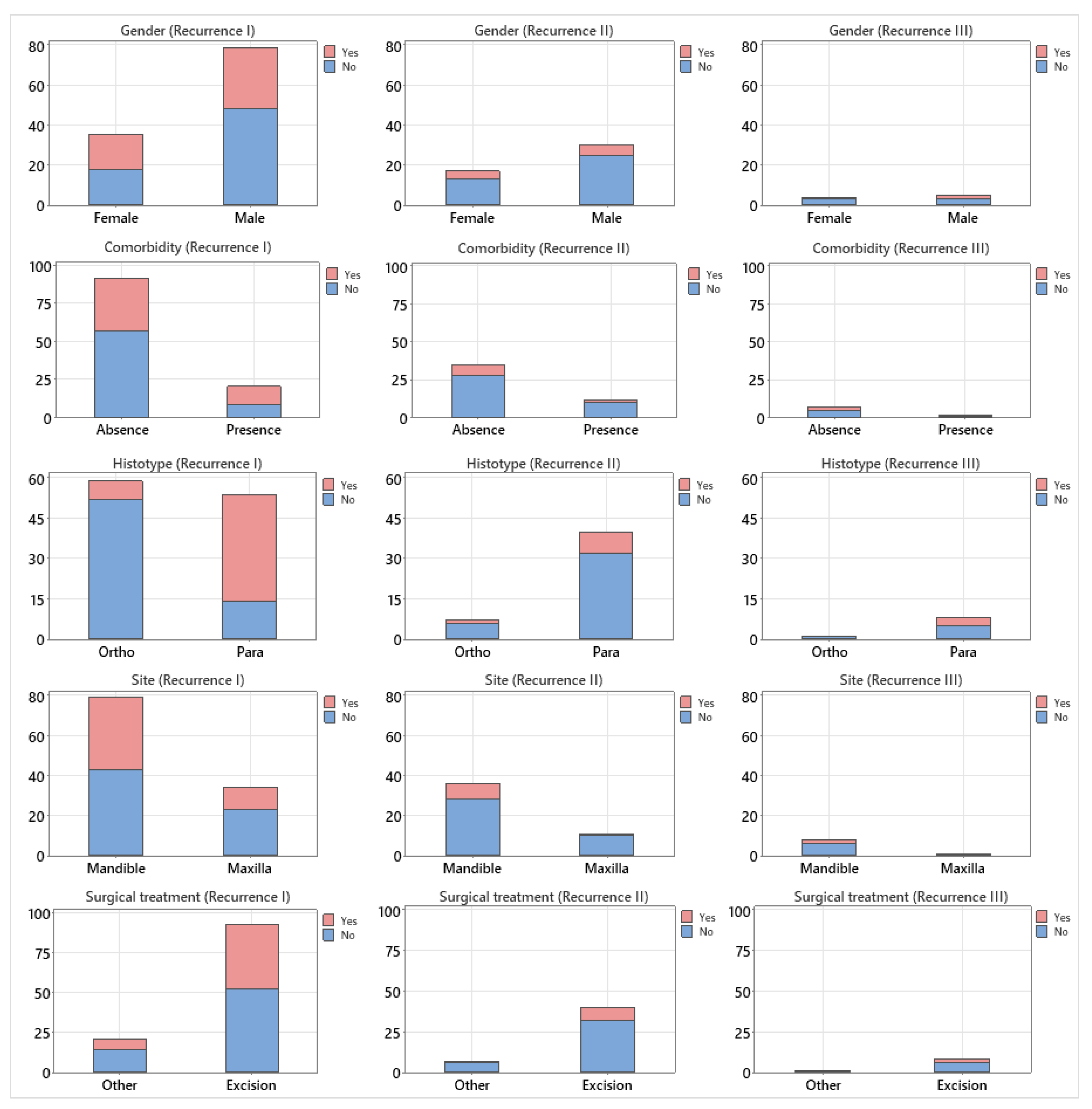
Figure 2.
Distribution of age according to recurrence stage (I–III) and recurrence status (yes/no).
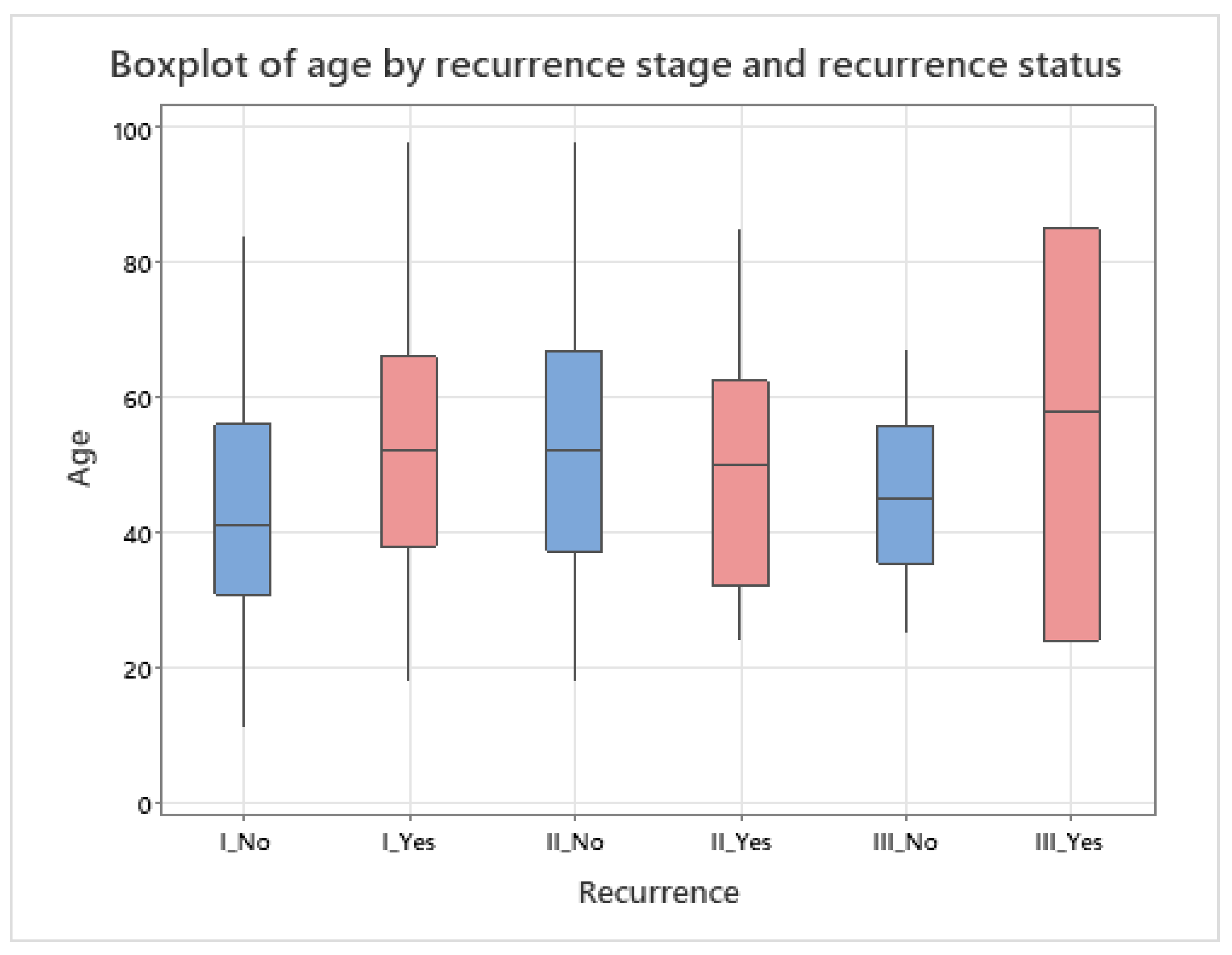
Figure 3.
(a) Surgical incision of keratocyst wall. (b) Application of 5-FU inside of keratocyst. (c) Suture of surgical approach.
Figure 3.
(a) Surgical incision of keratocyst wall. (b) Application of 5-FU inside of keratocyst. (c) Suture of surgical approach.
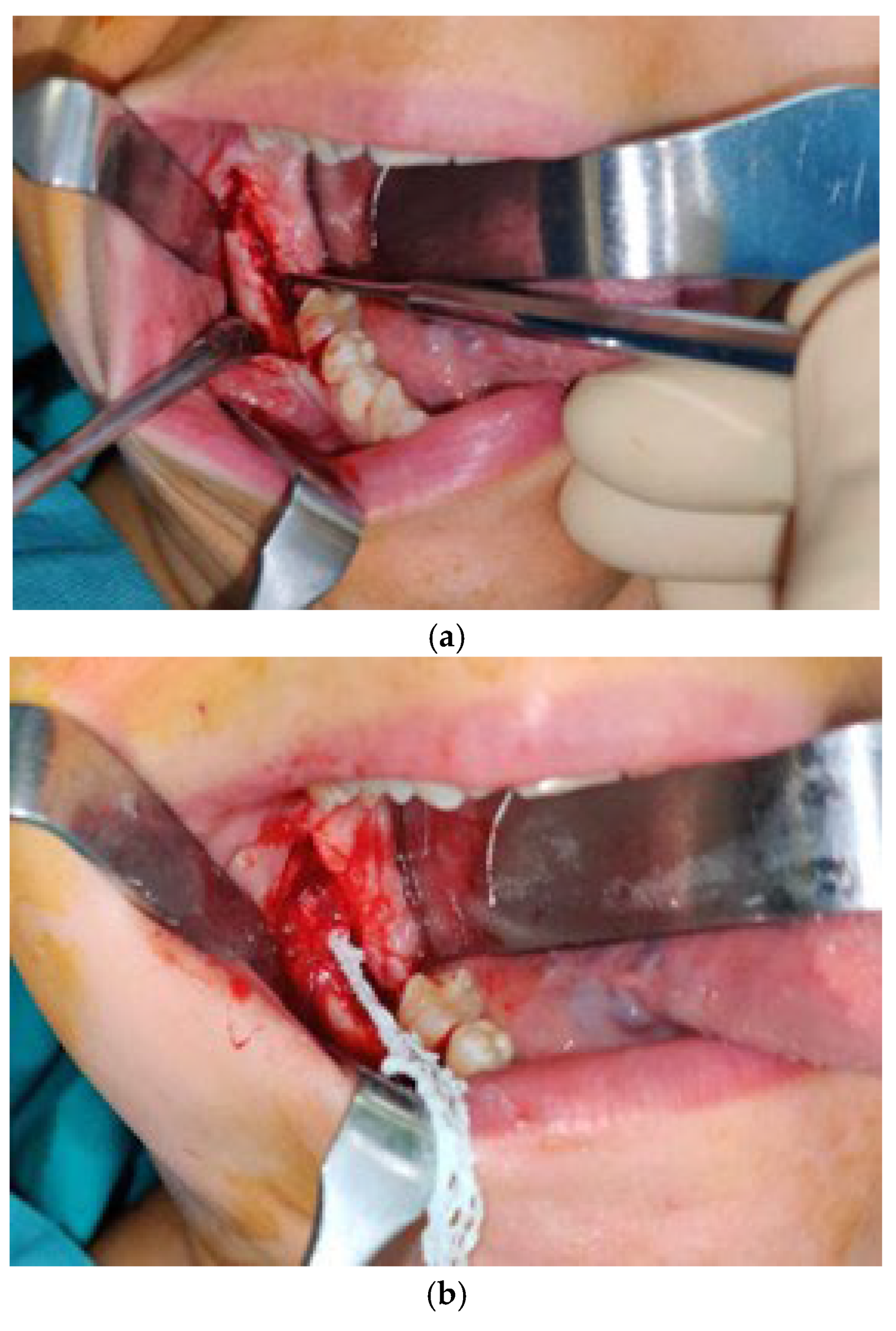
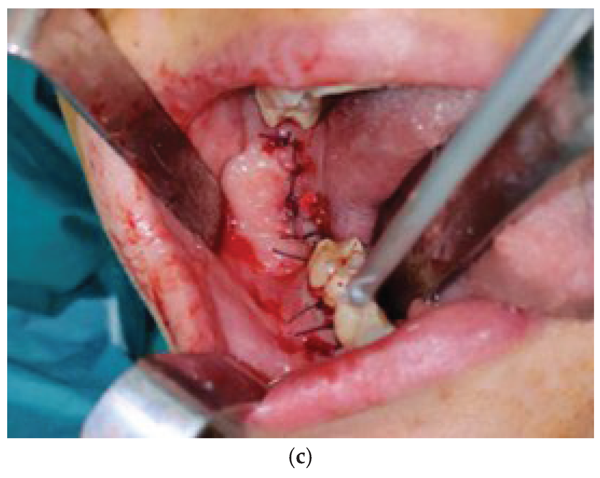
Figure 4.
(a) CT scan of the keratocyst. (b) Virtual surgical planning of bone resection with cutting guides. (c) Virtual surgical planning of recostruction with customized prosthesis.
Figure 4.
(a) CT scan of the keratocyst. (b) Virtual surgical planning of bone resection with cutting guides. (c) Virtual surgical planning of recostruction with customized prosthesis.
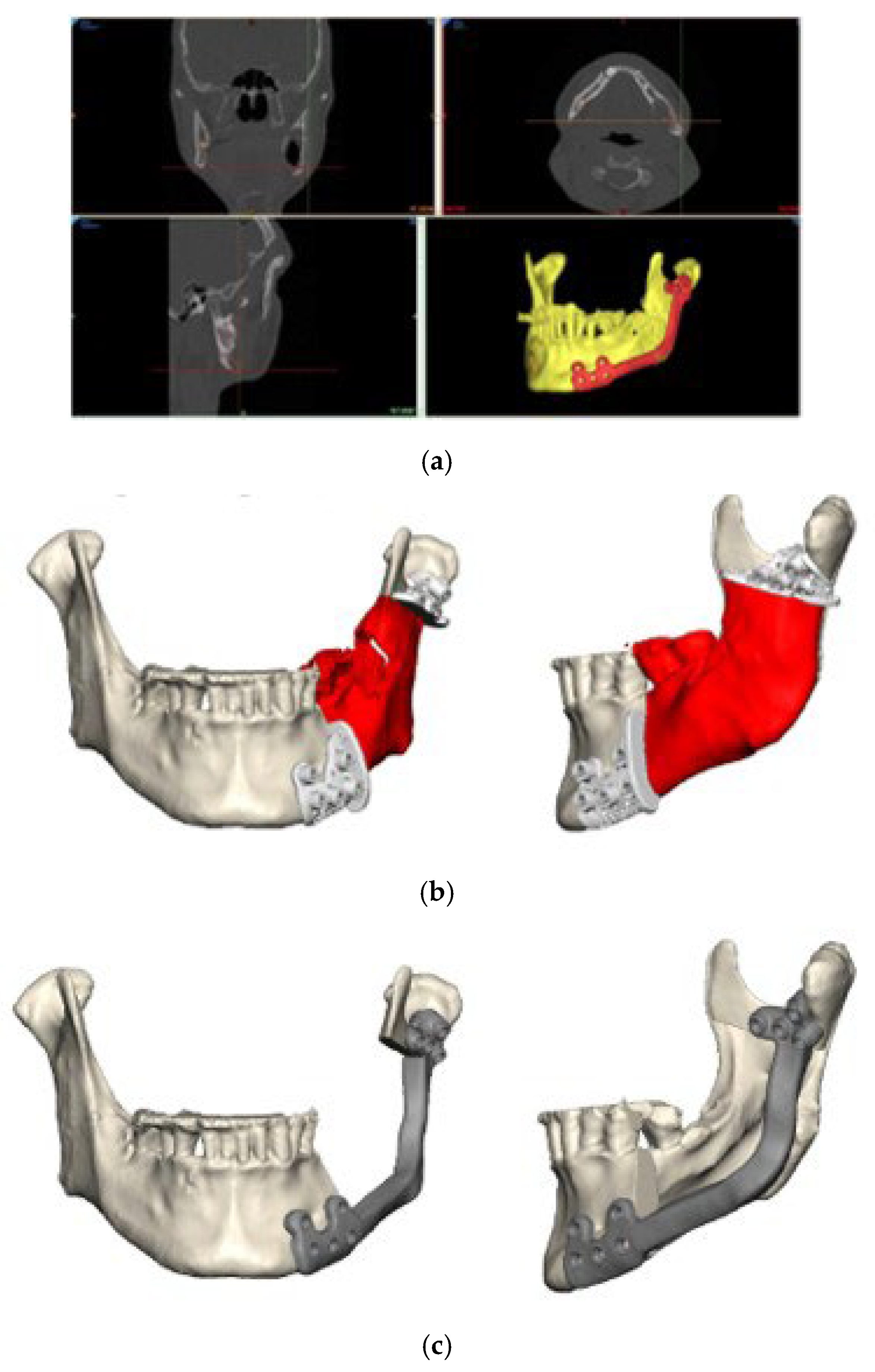

Table 1.
Clinical and demographic characteristics of the study population stratified by recurrence stage (I–III) and recurrence status (yes/no). Age is expressed as mean ± standard deviation (SD); categorical variables are presented as frequencies and column percentages. Dashes indicate treatment categories not applicable at that recurrence stage.
Table 1.
Clinical and demographic characteristics of the study population stratified by recurrence stage (I–III) and recurrence status (yes/no). Age is expressed as mean ± standard deviation (SD); categorical variables are presented as frequencies and column percentages. Dashes indicate treatment categories not applicable at that recurrence stage.
| Recurrence I | Recurrence II | Recurrence III | ||||
|---|---|---|---|---|---|---|
| Variable | No | Yes | No | Yes | No | Yes |
| N | 66 | 47 | 38 | 9 | 6 | 3 |
| Age (mean ± SD) | 43.5 ± 18.1 | 51.4 ± 19.9 | 52.1 ± 20.2 | 48.9 ± 19.7 | 45.5 ± 14.3 | 55.7 ± 30.6 |
| Gender (M/F) | 48/18 | 30/17 | 25/13 | 5/4 | 3/3 | 2/1 |
| Histotype | ||||||
| Orthokeratinized | 52 (79%) | 7 (15%) | 6 (16%) | 1 (11%) | 1 (17%) | 0 (0%) |
| Parakeratinized | 14 (21%) | 40 (85%) | 32 (84%) | 8 (89%) | 5 (83%) | 3 (100%) |
| Comorbidity | ||||||
| Absent | 57 (86%) | 35 (74%) | 28 (74%) | 7 (78%) | 5 (83%) | 2 (67%) |
| Present | 9 (14%) | 12 (26%) | 10 (26%) | 2 (22%) | 1 (17%) | 1 (33%) |
| Site | ||||||
| Maxilla | 23 (35%) | 11 (23%) | 10 (26%) | 1 (11%) | 0 (0%) | 1 (33%) |
| Quadrant I | 11 | 3 | 3 | 0 | 0 | 0 |
| Quadrant II | 6 | 4 | 4 | 0 | 0 | 0 |
| Premaxilla (13–23) | 6 | 4 | 3 | 1 | 0 | 1 |
| Mandible | 43 (65%) | 36 (77%) | 28 (74%) | 8 (89%) | 6 (100%) | 2 (67%) |
| Quadrant III | 14 | 10 | 9 | 1 | 1 | 0 |
| Quadrant IV | 17 | 12 | 10 | 2 | 2 | 0 |
| Right mandibular ramus | 1 | 1 | 1 | 0 | 0 | 0 |
| Left mandibular ramus | 0 | 1 | 0 | 1 | 1 | 0 |
| Right mandib. body & ramus | 7 | 7 | 4 | 3 | 2 | 1 |
| Left mandib. body & ramus | 4 | 5 | 4 | 1 | 0 | 1 |
| Surgical treatment | ||||||
| Excision | 52 (79%) | 40 (85%) | 23 (61%) | 7 (78%) | 3 (50%) | 2 (67%) |
| Other | 14 (21%) | 7 (15%) | 15 (39%) | 2 (22%) | 3 (50%) | 1 (33%) |
| Excision + cryotherapy | 5 | 0 | 8 | 1 | 1 | 0 |
| Excision + fluorouracil | 3 | 0 | 3 | 0 | 1 | 0 |
| Excision + flap reconstruction | 2 | 0 | — | — | — | — |
| Excision + reconstruction plate | 0 | 1 | 1 | 1 | 0 | 1 |
| Marsupialization | 0 | 1 | — | — | — | — |
| Drainage tube | 4 | 5 | — | — | — | — |
| Excision + bone grafting | — | — | 2 | 0 | — | — |
|
Excision +alloplastic prosthesis surgery |
— | — | 1 | 0 | 1 | 0 |
Table 2.
Results of univariate (UVA) and multivariate (MVA) logistic regression analyses for recurrence. UVA was performed using the two-sample t-test for age and the chi-square test for categorical variables. MVA included age, histotype, comorbidity, and site. Odds ratios (OR) with 95% confidence intervals (CI) are reported for variables included in the multivariate model. Dashes indicate variables not included in the MVA.
Table 2.
Results of univariate (UVA) and multivariate (MVA) logistic regression analyses for recurrence. UVA was performed using the two-sample t-test for age and the chi-square test for categorical variables. MVA included age, histotype, comorbidity, and site. Odds ratios (OR) with 95% confidence intervals (CI) are reported for variables included in the multivariate model. Dashes indicate variables not included in the MVA.
| Variable | Statistical test (UVA) | UVA p-value (Stage I) | MVA p-value (Stage I) | OR (95% CI) | MVA p-value (Stage II) | MVA p-value (Stage III) |
|---|---|---|---|---|---|---|
| Age | 2-sample t-test | 0.031 | 0.138 | 1.02 (0.99–1.05) | 0.689 | 0.205 |
| Gender | Chi-square | 0.313 | — | — | — | — |
| Histotype | Chi-square | <0.001 | <0.001 | 21.17 (7.51–59.64) | 0.613 | 0.978 |
| Comorbidity | Chi-square | 0.109 | 0.586 | 0.69 (0.19–2.59) | 0.859 | 0.876 |
| Site | Chi-square | 0.191 | 0.326 | 0.58 (0.20–1.72) | 0.327 | 0.956 |
| Surgical treatment | Chi-square | 0.395 | — | — | — | — |
Disclaimer/Publisher’s Note: The statements, opinions and data contained in all publications are solely those of the individual author(s) and contributor(s) and not of MDPI and/or the editor(s). MDPI and/or the editor(s) disclaim responsibility for any injury to people or property resulting from any ideas, methods, instructions or products referred to in the content. |
© 2026 by the authors. Licensee MDPI, Basel, Switzerland. This article is an open access article distributed under the terms and conditions of the Creative Commons Attribution (CC BY) license (http://creativecommons.org/licenses/by/4.0/).
Copyright: This open access article is published under a Creative Commons CC BY 4.0 license, which permit the free download, distribution, and reuse, provided that the author and preprint are cited in any reuse.