
Submitted:
06 August 2026
Posted:
10 August 2026
You are already at the latest version
Abstract
Enteric fever remains a significant public health burden in sub-Saharan Africa, yet the phylogenetic diversity of circulating Salmonella serovars in Nigeria remains incompletely characterized. While typhoidal strains dominate clinical reports, emerging atypical lineages and non-typhoidal serovars may contribute substantially to febrile illness transmission. A health facility-based cross-sectional study enrolled 425 consecutive febrile patients (aged 1–65 years) with suspected enteric fever in Keffi Local Government Area, Nasarawa State, Nigeria. Blood cultures were processed using the BD Bactec™ automated system, followed by sub-culturing on selective media and presumptive phenotypic screening. Molecular confirmation employed PCR amplification of the 16S rRNA gene (27F/1492R) and Sanger sequencing. Sequence analysis utilized BLASTn against the NCBI GenBank database, and phylogenetic trees were constructed in MEGA 11 using the Maximum Likelihood method with 1,000 bootstrap replicates. Sixty-three (14.8%) blood cultures yielded microbial growth, of which 32 (7.5%) were phenotypically identified as Salmonella. Molecular characterization confirmed 25 isolates (5.9%) as Salmonella species. Phylogenetic analysis resolved four major clades dominated by closely related non-typhoidal Salmonella enterica serovars, particularly S. Typhimurium (n=11), with moderate to strong bootstrap support (68–89%). Emerging atypical lineages, including Salmonella bongori (n=1) and Salmonella enterica subsp. diarizonae (n=1), formed distinct divergent branches separate from the core S. enterica lineages. Additional serovars identified included S. Enteritidis (n=1), S. Typhi (n=1), S. Paratyphi B (n=1), S. Heidelberg (n=1), S. Infantis (n=1), S. Livingstone (n=1), and untyped S. enterica strains (n=6). Substantial phylogenetic diversity exists among Salmonella serovars circulating in suspected enteric fever cases in Keffi, with S. Typhimurium representing the predominant lineage and emerging atypical serovars (S. bongori and S. diarizonae) suggest potential zoonotic and environmental transmission pathways. These findings underscore the urgent need for routine molecular surveillance, enhanced diagnostic capacity, and One Health-based interventions for effective disease control in Nigeria.
Keywords:
Salmonella enterica
; phylogenetic diversity
; emerging serovars
; non-typhoidal Salmonella
; Salmonella bongori
; Salmonella diarizonae
; enteric fever
; Nigeria
; One Health
; molecular epidemiology
1. Introduction
Enteric fever, caused primarily by Salmonella enterica serovars Typhi and Paratyphi, remains a major public health challenge in low- and middle-income countries, particularly in sub-Saharan Africa [1,2]. Globally, an estimated 14.3 million cases of enteric fever occur annually, resulting in over 135,900 deaths [1]. Despite this substantial burden, the true epidemiology of Salmonella infections in Africa remains incompletely understood, largely due to weak surveillance systems and diagnostic approaches that predominantly target typhoidal serovars [3,4].
In recent years, increasing attention has focused on the expanding role of invasive non-typhoidal Salmonella (iNTS) as a cause of bloodstream infections in Africa, particularly among children, the elderly, and immunocompromised individuals [5,6]. These infections are frequently underdiagnosed yet contribute substantially to regional morbidity and mortality. Studies from several African countries have demonstrated that the phylogenetic diversity of circulating Salmonella lineages extends far beyond classical typhoidal strains, encompassing a wide range of non-typhoidal serovars and subspecies with distinct ecological and evolutionary backgrounds [3,7]. The emergence and persistence of these diverse lineages have significant implications for disease transmission, antimicrobial resistance, and public health control strategies [8,9].
The Salmonella genus exhibits remarkable phylogenetic diversity, comprising two main species which include S. enterica and S. bongori, and over 2,600 serovars within S. enterica subsp. enterica [10]. Phylogenetic studies have revealed that S. enterica subspecies demonstrate substantial genetic heterogeneity shaped by ecological pressures, host adaptation, geographic distribution, and evolutionary processes [7,11]. Of particular interest are emerging atypical lineages such as Salmonella bongori and Salmonella enterica subsp. diarizonae, which are evolutionarily distinct from the major S. enterica lineages and have traditionally been associated with environmental reservoirs, reptiles, and other non-human hosts rather than human disease [10,12,13,14]. Although these organisms are infrequently reported in clinical settings, sporadic cases of human infection have been documented and are often linked to zoonotic transmission or environmental exposure [15,16,17,18]. The detection of such emerging atypical lineages in human clinical specimens may indicate the existence of alternative transmission pathways, environmental reservoirs, or ecological factors that facilitate their circulation within endemic communities. Understanding the phylogenetic diversity and emergence patterns of these atypical serovars is critical for comprehensive disease surveillance and control.
In Nigeria, enteric fever remains endemic, driven by rapid urbanization, population growth, inadequate water, sanitation and hygiene (WASH) infrastructure, and close human -animal–environment interactions [19,20,21]. However, routine diagnostic practices in most health facilities rely heavily on phenotypic methods such as blood culture, serology, and biochemical assays, which have limited sensitivity and discriminatory power [22]. These conventional approaches are often unable to reliably differentiate between closely related Salmonella serovars or distinguish Salmonella species from other members of the Enterobacteriaceae family, leading to misclassification and underestimation of the true phylogenetic diversity of circulating pathogens [23,24].
Molecular tools, particularly 16S rRNA gene sequencing and whole-genome sequencing (WGS), have revolutionized the identification and phylogenetic characterization of Salmonella, enabling accurate discrimination of species, subspecies, and serovars while providing valuable insights into evolutionary relationships and transmission dynamics [25,26,27,28]. Recent molecular and genomic studies have documented substantial phylogenetic diversity among both typhoidal and non-typhoidal Salmonella serovars circulating in Africa and other endemic regions [7,29,30]. These studies have demonstrated that genetically distinct lineages may coexist within the same geographical area, reflecting adaptation to different ecological niches and transmission networks. Such diversity often remains undetected by routine diagnostic workflows and underscores the importance of integrating molecular surveillance into public health systems, particularly within a One Health framework that recognizes the interconnected roles of humans, animals, food sources, and the environment in disease transmission [31,32].
Despite these advances, data on the phylogenetic diversity and evolutionary dynamics of Salmonella circulating in North-Central Nigeria remain scarce. Keffi Local Government Area (LGA), Nasarawa State, is characterized by rapid urbanization, small-scale livestock rearing, increasing human population density, environmental exposure to animals, and limited sanitation infrastructure conditions that may facilitate the transmission and persistence of diverse Salmonella lineages, including emerging atypical serovars. Yet, few studies have applied molecular tools to characterize Salmonella isolates from this region.
Therefore, this study aimed to investigate the phylogenetic diversity and evolutionary relationships of Salmonella isolates recovered from febrile patients with suspected enteric fever in Keffi LGA. Using 16S rRNA gene sequencing and phylogenetic analysis, the study sought to: (1) confirm the identity of phenotypically detected Salmonella isolates; (2) characterize the phylogenetic diversity of circulating serovars, including both typhoidal, non-typhoidal, and emerging atypical Salmonella strains; (3) identify the presence of atypical lineages such as S. bongori and S. diarizonae; and (4) determine their evolutionary relationships with globally circulating Salmonella lineages.
2. Materials and Methods
2.1. Study Design and Setting
This facility-based cross-sectional study was conducted over a seven-month period from December 2023 to June 2024 among febrile patients presenting to selected health facilities in Keffi Local Government Area (LGA), Nasarawa State, North-Central Nigeria. The study was conducted following approval of the research proposal.
The study sites comprised the Federal Medical Centre (FMC), Keffi, and six Primary Health Care (PHC) centres within Keffi LGA. Participant recruitment was carried out concurrently across all study sites throughout the study period.
2.2. Study Area
Keffi Local Government Area is located in Nasarawa State and is characterized by tropical Guinea Savannah vegetation, which supports small-scale farming and livestock rearing. Agricultural practices in the area sometimes involve the use of human or animal waste as fertilizer. Additionally, seasonal bush burning during the dry season is commonly practiced for hunting bushmeat, specifically targeting rodents and reptiles, which may serve as reservoirs for food-borne pathogens including Salmonella spp., thereby increasing the risk of zoonotic transmission to humans through handling, processing, and consumption of bushmeat [21]. Rapid urbanization, increasing population density, and limited sanitation infrastructure in Keffi further contribute to the risk of Salmonella infection and other environmental health hazards [19,20].
2.3. Participants’ Recruitment and Ethical Considerations
This study recruited consecutive patients aged 1–65 years who presented to the health facilities with a history of fever (≥38 °C) lasting more than three days and with clinical suspicion of enteric fever. Eligible participants were identified among both in-patients and out-patients during routine clinical evaluation. Demographic and relevant clinical information were collected using structured questionnaires.
Inclusion criteria comprised patients aged 1–65 years presenting with prolonged fever (≥38 °C for >3 days) and other clinical features suggestive of enteric fever or its complications.
Exclusion criteria included patients who had received antibiotic therapy for more than 48 hours prior to presentation, as this could significantly affect culture results and reduce the sensitivity for detecting bacterial pathogens.
Ethical approval for the study was obtained from the Human Research and Ethics Committee of the Federal Medical Centre, Keffi (Approval number: FMC/KF/HREC/02636/24). Written informed consent was obtained from all adult participants prior to enrolment after a clear explanation of the study objectives and procedures. For participants who were minors, informed consent was obtained from parents or legal guardians. Interpreters were used where necessary to ensure adequate understanding. Participants were informed of their right to withdraw from the study at any time without any consequences to their access to medical care or treatment. The study was conducted in accordance with the Declaration of Helsinki.
2.4. Laboratory Analysis
2.4.1. Blood Sample Collection and Culture
Venous blood samples were aseptically collected from 425 study participants for microbiological analysis. Sample volumes of 3–5 mL were obtained from children under five years of age, while 6–10 mL were collected from participants aged five years and above. After disinfection of the venepuncture site with 70% alcohol and 10% povidone iodine, blood samples were collected using a sterile vacutainer system and directly inoculated into aerobic BD Bactec™ blood culture bottles. The inoculated bottles were transported to the microbiology laboratory at Bingham University, Karu, Nasarawa State, within three hours of collection.
Blood culture bottles were incubated in the BD Bactec™ automated blood culture system and monitored for a maximum of seven days. Bottles signalling positive growth were sub-cultured onto chocolate agar and Xylose Lysine Deoxycholate (XLD) agar and incubated at 37 °C for 18–24 hours. Resulting colonies were examined for morphological characteristics consistent with Salmonella species.
2.4.2. Phenotypic Identification
Presumptive isolates were identified using standard phenotypic techniques, including Gram staining and biochemical tests. Gram staining revealed Gram-negative coccobacilli consistent with Salmonella species. Biochemical characterization included indole, methyl red/Voges–Proskauer, citrate utilization, catalase, urease, and Triple Sugar Iron (TSI) tests. Salmonella isolates were identified based on characteristic biochemical reaction patterns, including indole negativity, methyl red positivity, citrate positivity, urease negativity, and hydrogen sulfide production on TSI agar. Confirmed isolates were preserved on nutrient agar slants at 2 °C for further analyses.
2.4.3. DNA Extraction
Genomic DNA was extracted from phenotypic Salmonella isolates using the DNeasy Blood and Tissue Kit (Qiagen, USA), following the manufacturer’s instructions. Briefly, isolates preserved on nutrient agar slants were cultured in nutrient broth and incubated at 37 °C until turbidity equivalent to 0.5 McFarland standards was achieved. Bacterial cells were harvested by centrifugation, washed with phosphate-buffered saline, and lysed to release genomic DNA. DNA purity and concentration were assessed using a NanoDrop spectrophotometer, and DNA samples were standardized to a concentration of 50 ng/µL and stored at −20 °C until PCR amplification.
2.4.4. PCR Amplification of the 16S rRNA Gene
The presence of the 16S rRNA gene was confirmed by PCR using universal primers 27F (5′-AGAGTTTGATCCTGGCTCAG-3′) and 1492R (5′-GGTTACCTTGTTACGACTT-3′), targeting an approximately 1,500 bp fragment. PCR amplification was performed in a 50 µL reaction mixture containing PCR master mix, primers, template DNA, and nuclease-free water. Thermal cycling conditions included an initial denaturation at 94 °C, followed by 30–35 cycles of denaturation, annealing at 55 °C, extension at 72 °C, and a final extension step. PCR products were resolved by agarose gel electrophoresis, and the presence of a single ~1,500 bp band confirmed successful amplification.
2.4.5. Purification and Sequencing
PCR products were purified using the PureLink PCR Purification Kit (Invitrogen, Thermo Fisher Scientific) according to the manufacturer’s protocol. Purified amplicons were subjected to Sanger sequencing to obtain high-quality 16S rRNA gene sequences for downstream analysis.
2.4.6. Sequence Analysis and Phylogenetic Inference
Raw sequence data were assembled, edited, and aligned using BioEdit software. Low-quality and ambiguous bases were trimmed, and consensus sequences were generated from forward and reverse reads. Sequence similarity searches were performed using BLASTn against the NCBI GenBank database to identify closely related reference sequences.
Phylogenetic analysis was conducted using MEGA version 11. Cleaned and aligned sequences were used to construct phylogenetic trees employing the Maximum Likelihood method with the Kimura two-parameter model. Bootstrap analysis with 1,000 replicates was performed to assess tree robustness. The resulting phylogenetic trees were used to infer the evolutionary relationships of the Salmonella isolates and to identify species, serovars, and strains based on sequence similarity and clustering patterns.
2.5. Data Analysis
All questionnaire data were entered into Microsoft Excel and analyzed using SPSS Statistics version 26.0 (IBM Corp., Armonk, NY, USA). Descriptive statistics were used to summarize the demographic characteristics and microbiological findings. Quantitative variables were summarized using means and standard deviations, as appropriate, while categorical variables were summarized using frequencies and percentages. The prevalence of phenotypic Salmonella isolates was expressed as a proportion.
3. Results
3.1. Demographic Characteristics of Study Participants
A total of 425 patients suspected of enteric fever were enrolled. The overall mean age was 22.1 ± 14.4 years (range: 1–65 years). Female participants constituted 54.6% (232/425) of the study population. The mean age of males was 21.66 ± 16.22 years, while females had a mean age of 22.50 ± 12.67 years.
3.2. Blood Culture Yield and Microbial Isolates
All 425 blood samples were cultured, of which 63 (14.8%) yielded bacterial growth, while 362 (85.2%) showed no growth. Phenotypic identification revealed 32 isolates (7.5%) presumptively identified as Salmonella spp. Molecular confirmation using 16S rRNA gene sequencing verified 25 isolates (5.9% of total samples) as Salmonella spp. (Figure 1).
Other bacterial and fungal isolates recovered from blood cultures included Staphylococcus aureus (2.4%), yeast (1.7%), coagulase-negative Staphylococcus spp. (0.9%), Escherichia coli (0.7%), Pseudomonas spp. (0.7%), Klebsiella spp. (0.5%), Streptococcus spp. (0.2%), and Providencia stuartii (0.2%) (Figure 2).
3.3. Genetic Characteristics and Phylogenetic Diversity of Circulating Salmonella Strains
Figure 3 shows the agarose gel electrophoresis of PCR assay products (amplicons) for the 16S rRNA gene of 32 phenotypic Salmonella isolates. The PCR amplicon size for the target 16S rRNA gene is 1500 bp, which is visible for isolates 1–32. This represents an initial presumptive molecular identification of the Salmonella species isolates.
Key:
Lanes 1–32 = PCR amplicons of phenotypically identified Salmonella isolates corresponding to approximately 1500 bp;
Lane M = 250 bp DNA molecular weight marker (Promega);
Lane N = Negative control.
Table1 presents the GenBank accession numbers of the phenotypic Salmonella isolates 16S rRNA sequences (Samples S1–S32) submitted to the NCBI GenBank with the molecularly confirmed bacterial serovars and assigned strains. The number of genetically identified Salmonella was twenty-five (25), giving a molecularly confirmed prevalence of Salmonella among the study population of 5.9% (25/425).

Table 1.
NCBI GenBank database Accession Numbers for the Phenotypic Salmonella Isolates from Suspected Enteric Fever Cases in Keffi LGA.
Table 1.
NCBI GenBank database Accession Numbers for the Phenotypic Salmonella Isolates from Suspected Enteric Fever Cases in Keffi LGA.
| Sample IDs | Ascension Numbers | Bacterial Serovars and Strains |
|---|---|---|
| S1 | PZ413661 | Salmonella enterica subsp. enterica serovar Livingstone strain NSK160426.1 |
| S2 | PZ414891 | Salmonella enterica subsp. enterica serovar Typhimurium strain NSK160426.1 |
| S3 | PZ414892 | Salmonella enterica subsp. enterica strain NSK160426.3 |
| S4 | PZ413669 | Salmonella enterica subsp. enterica serovar Heidelberg strain NSK160426.33 |
| S5 | PZ413662 | Salmonella enterica subsp. enterica serovar Typhimurium strain NSK160426.5 |
| S6 | PZ413663 | Salmonella enterica subsp. enterica serovar Enteritidis strain NSK160426.6 |
| S7 | PZ413664 | Citrobacter freundii strain NSK160426.7 |
| S8 | PZ414895 | Salmonella enterica subsp. enterica serovar Typhimurium strain NSK160426.34 |
| S9 | PZ414896 | Salmonella enterica subsp. enterica strain NSK160426.35 |
| S10 | PZ414897 | Salmonella enterica subsp. enterica strain NSK160426.37 |
| S11 | PZ413633 | Salmonella enterica subsp. enterica serovar Paratyphi B strain NSK160426.11 |
| S12 | PZ413634 | Salmonella enterica subsp. enterica serovar Typhi strain NSK160426.12 |
| S13 | PZ414893 | Salmonella enterica subsp. enterica serovar Typhimurium strain NSK160426.13 |
| S14 | PZ324770 | Salmonella bongori strain NSK230426.14 |
| S15 | PZ324771 | Salmonella enterica subsp. enterica serovar Typhimurium strain NSK230426.15 |
| S16 | PZ414894 | Salmonella enterica subsp. enterica strain NSK160426.16 |
| S17 | PZ324772 | Salmonella enterica subsp. enterica serovar Typhimurium strain NSK230426.17 |
| S18 | PZ324773 | Salmonella enterica subsp. enterica strain NSK230426.18 |
| S19 | PZ324774 | Salmonella enterica subsp. enterica strain NSK230426.19 |
| S20 | PZ324775 | Salmonella enterica subsp. enterica serovar Typhimurium strain NSK230426.20 |
| S21 | PZ324776 | Salmonella enterica subsp. enterica serovar Typhimurium strain NSK230426.21 |
| S22 | PZ413665 | Citrobacter youngae strain NSK160426.22 |
| S23 | PZ414898 | Salmonella enterica subsp. enterica serovar Typhimurium strain NSK160426.38 |
| S24 | PZ324777 | Salmonella enterica subsp. diarizonae strain NSK230426.24 |
| S25 | PZ414899 | Salmonella enterica subsp. enterica serovar Typhimurium strain NSK160426.40 |
| S26 | PZ413671 | Salmonella enterica subsp. enterica serovar Infantis strain NSK160426.41 |
| S27 | PZ413667 | Klebsiella oxytoca strain NSK160426.27 |
| S28 | PZ324778 | Enterobacter oligotrophicus strain NSK230426.28 |
| S29 | PZ324779 | Enterobacter oligotrophicus strain NSK230426.29 |
| S30 | PZ324780 | Enterobacter oligotrophicus strain NSK230426.30 |
| S31 | PZ324781 | Salmonella enterica subsp. enterica serovar Typhimurium strain NSK230426.31 |
| S32 | PZ413668 | Enterobacter hormaechei strain NSK160426.32 |
In descending order of occurrence, the Salmonella serovars were: Salmonella Typhimurium (11), Salmonella Enterica [ Untyped strains] (6), Salmonella Enteritidis (1), Salmonella Heidelberg (1), Salmonella Infantis (1), Salmonella Typhi (1), Salmonella Livingstone (1), Salmonella Paratyphi B (1), Salmonella Diarizonae (1) and Salmonella Bongori (1), as shown in Table 2.
Other molecularly identified organisms in descending order of occurrence include Enterobacter Oligotrophicus (3), Enterobacter Hormaechei (1) Citrobacter Freundii (1), Citrobacter Youngae (1), and Klebsiella Oxytoca (1), as shown in Table 2
3.4. Phylogenetic Analysis of Molecularly Confirmed Salmonella Isolates
Phylogenetic analysis of the 25 molecularly confirmed Salmonella isolates revealed substantial evolutionary diversity, with isolates predominantly clustering within Salmonella enterica subsp. enterica and several distinct serovar-specific lineages observed across the phylogenetic trees (Figure 4). Figure 4 shows the bootstrap-supported phylogenetic tree used to evaluate the reliability of the inferred evolutionary relationships among the isolates.
The phylogenetic tree resolved the isolates into four major clades with several sub-clades:
Major Clade I represented the dominant Salmonella enterica serovar Typhimurium lineage and included isolates S5, S25, S2, S23, S17, S8, S13, S20, and S21. This clade showed moderate to strong bootstrap support values ranging from 68% to 89%, indicating close genetic relatedness and a probable common evolutionary origin among the Typhimurium isolates. Sub-clade I-A included the Enteritidis isolate (S6), which clustered closely with the Typhimurium isolates, while Sub-clade I-B consisted of the Infantis isolate (S26), indicating a related but distinct evolutionary lineage.
Major Clade II comprised mixed Salmonella enterica strains including S3, S18, S19, S16, S1, S4, and S9. This clade demonstrated moderate phylogenetic relatedness among non-Typhimurium Salmonella strains. Within this group, the Heidelberg isolate (S4) clustered closely with untyped Salmonella enterica strains, suggesting evolutionary divergence from the dominant Typhimurium cluster.
Major Clade III consisted of the emerging atypical serovars such as Salmonella bongori (S14) and Salmonella enterica subsp. diarizonae (S24) which formed distinct branches separate from the core Salmonella enterica lineage with bootstrap support values ranging from 31% to 56%. The separation of S. bongori from S. enterica confirms the recognized phylogenetic divergence between these species and highlights the presence of these atypical lineages in clinical specimens from the study area.
Major Clade IV formed the basal lineage and included isolates S10, S15, S11, S31, and S12, representing untyped Salmonella enterica, serovar Typhimurium, Paratyphi B, and Typhi strains. This clade showed the highest bootstrap support value (96–100%), indicating a highly reliable evolutionary relationship among these isolates.
Figure 4.
Phylogenetic Tree of The Molecularly Confirmed Salmonella Isolates with Bootstrap Values (1,000 replicates; values ≥50% shown) from Suspected Enteric Fever Cases in Keffi LGA.
Figure 4.
Phylogenetic Tree of The Molecularly Confirmed Salmonella Isolates with Bootstrap Values (1,000 replicates; values ≥50% shown) from Suspected Enteric Fever Cases in Keffi LGA.
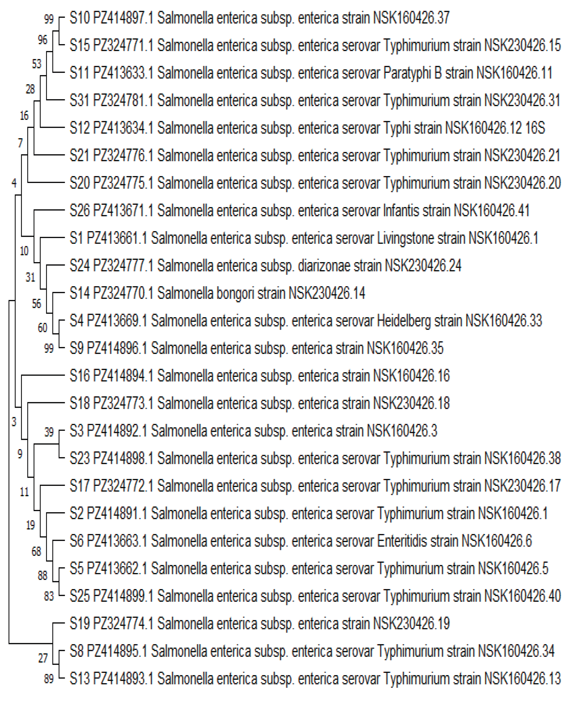
Table 3 provides a textual and tabular summary of the phylogenetic analysis, systematically organizing the major evolutionary groups (Clades I–IV) identified in the study, listing their member isolates, predominant serovars, bootstrap support values, and interpretive significance. It highlights the dominance of S. Typhimurium (Clade I), the presence of moderately related non-Typhimurium strains (Clade II), the distinct evolutionary position of emerging atypical serovars S. bongori and S. diarizonae (Clade III), and a strongly supported basal lineage containing typhoidal serovars (Clade IV). This structured presentation translates the phylogenetic tree (Figure 4) into an accessible format, demonstrating the coexistence of both common and rare Salmonella serovars within the study population.
3.5. Summary of Phylogenetic Diversity Findings
The phylogenetic analysis demonstrated substantial diversity among circulating Salmonella isolates, with the following key observations:
- Predominant Lineage: S. Typhimurium (44% of confirmed Salmonella isolates) represented the most prevalent and phylogenetically conserved lineage.
- Untyped Strains: Six isolates were confidently assigned to S. enterica but could not be resolved to specific serovars using 16S rRNA sequencing alone, suggesting additional hidden phylogenetic diversity.
4. Discussion
This study provides important molecular epidemiological evidence on the phylogenetic diversity of Salmonella serovars circulating among patients with suspected enteric fever in Keffi, North-Central Nigeria, with a particular focus on emerging atypical lineages. Using 16S rRNA gene sequencing, 25 of 32 phenotypically identified isolates were confirmed as Salmonella species, yielding a molecular prevalence of 5.9%. The findings demonstrate substantial phylogenetic diversity comprising both typhoidal and non-typhoidal serovars, with Salmonella Typhimurium emerging as the predominant lineage. Critically, this study identified emerging atypical serovars such as S. bongori and S. diarizonae which formed distinct phylogenetic branches separate from the core S. enterica lineages, suggesting potential zoonotic and environmental transmission pathways previously unrecognized in this setting.
4.1. Phylogenetic Diversity and Predominance of Non-Typhoidal Serovars
The predominance of S. Typhimurium (44% of confirmed Salmonella isolates) observed in this study is consistent with reports from several African countries where invasive non-typhoidal Salmonella (iNTS) disease has emerged as a major public health concern [3,5,6,35]. Historically, typhoidal serovars such as S. Typhi and S. Paratyphi have been considered the principal causes of enteric fever. However, increasing evidence indicates that non-typhoidal serovars, especially S. Typhimurium and S. Enteritidis, are important causes of bloodstream infection and severe febrile illness across sub-Saharan Africa [4,5,6]. The dominance of S. Typhimurium in the present study, therefore, aligns with broader regional trends and may reflect adaptation of this serovar to local ecological and epidemiological conditions.
The phylogenetic analysis revealed that the majority of isolates clustered within Salmonella enterica subsp. enterica and were resolved into four major clades, with the largest and most strongly supported clade consisting predominantly of S. Typhimurium isolates (bootstrap values: 68–89%). This clustering pattern suggests the circulation of genetically related lineages that may share common evolutionary origins, consistent with global genomic studies demonstrating that successful Salmonella lineages frequently undergo clonal expansion while maintaining sufficient genetic diversity to adapt to varying ecological conditions [7,11,29].
The simultaneous detection of typhoidal serovars (S. Typhi and S. Paratyphi B) alongside multiple non-typhoidal serovars suggests the coexistence of diverse transmission networks within the study area. Such phylogenetic coexistence has important epidemiological implications because typhoidal and non-typhoidal Salmonella differ substantially in reservoir ecology, transmission dynamics, and disease manifestations. While typhoidal serovars are largely human-restricted and transmitted through contaminated food and water, many non-typhoidal serovars possess extensive animal reservoirs and are capable of zoonotic transmission [3,6,21,31].
4.2. Detection and Phylogenetic Significance of Emerging Atypical Serovars
One of the most notable findings of this study was the detection of Salmonella bongori and Salmonella enterica subsp. diarizonae in clinical specimens from patients with suspected enteric fever. These organisms are infrequently reported in human clinical studies and are generally associated with environmental habitats, reptiles, and other non-human hosts [10,12,13,14,15]. Their occurrence among patients with suspected enteric fever raises important questions regarding possible zoonotic and environmental transmission pathways.
The phylogenetic placement of these emerging atypical serovars in Major Clade III, distinct from the core S. enterica lineages with bootstrap support values ranging from 31% to 56%, confirms their recognized evolutionary divergence. S. bongori is recognized as a distinct species that diverged early from the S. enterica lineage and occupies a separate evolutionary position within the genus [12,13,14]. Similarly, S. enterica subsp. diarizonae (subspecies IIIb) is one of the six subspecies of S. enterica and is most commonly associated with cold-blooded vertebrates, particularly reptiles [10,17,18].
The detection of these emerging atypical lineages in clinical isolates from Keffi has several important implications. First, the study area is characterized by livestock rearing, wildlife exposure, hunting of reptiles and rodents, and frequent interaction between humans and animals ecological conditions that may facilitate spillover of atypical Salmonella lineages into human populations, with similar observations having been reported from environmental and animal surveillance studies where uncommon Salmonella species and subspecies have been recovered from reptiles, poultry, livestock, and aquatic environments [12,15,21,36]. Second, the detection of S. bongori and S. diarizonae in clinical specimens suggests that the spectrum of Salmonella associated with human disease may be broader than commonly appreciated in endemic settings, as routine diagnostic workflows that focus primarily on typhoidal serovars would miss these emerging atypical lineages, potentially underestimating the true burden of Salmonella-associated febrile illness [22,23]. Third, the presence of these atypical serovars in human clinical specimens strongly supports the application of the One Health framework to understand and control salmonellosis in Nigeria, under which human infections are viewed as the outcome of interconnected ecological processes involving animals, food systems, water sources, and environmental reservoirs [31,32].
4.3. Phylogenetic Resolution and Diagnostic Implications
The identification of six untyped Salmonella enterica strains further emphasizes the genetic complexity of circulating Salmonella populations. These isolates were confidently assigned to S. enterica but could not be resolved into specific serovars using 16S rRNA sequencing alone. This limitation reflects the highly conserved nature of the 16S rRNA gene among closely related Salmonella serovars [23,24]. Consequently, while 16S rRNA sequencing is valuable for species confirmation and broad phylogenetic analysis, it lacks the resolution required for detailed strain-level characterization and serovar identification [25,26,27].
The reduction in isolate numbers from 32 phenotypically identified Salmonella isolates to 25 molecularly confirmed isolates underscores the limitations of conventional diagnostic methods. Phenotypic identification based on culture characteristics and biochemical testing remains the cornerstone of Salmonella diagnosis in many resource-limited settings; however, these methods often lack sufficient discriminatory power to reliably distinguish Salmonella species from closely related members of the Enterobacteriaceae family [8,22,23,24]. The identification of Citrobacter freundii, Citrobacter youngae, Enterobacter oligotrophicus, Enterobacter hormaechei, and Klebsiella oxytoca among the initially presumed Salmonella isolates demonstrates the potential for diagnostic misclassification and highlights the essential role of molecular methods in accurate pathogen identification.
4.4. Ecological and Environmental Drivers of Phylogenetic Diversity
The observed phylogenetic diversity of Salmonella serovars, including the presence of emerging atypical lineages, may be partly explained by environmental and socioeconomic conditions within Keffi, as the study area is characterized by rapid urbanization and increasing human population density, inadequate water and sanitation infrastructure, small-scale livestock rearing and close human–animal contact, seasonal bush burning and hunting of bushmeat (including reptiles and rodents), and the use of human and animal waste as fertilizer in agricultural practices.
These conditions create favourable environments for the maintenance and transmission of multiple Salmonella lineages, including atypical serovars that may originate from environmental or zoonotic reservoirs [19,20,21,37]. These factors are well-recognized drivers of enteric pathogen transmission throughout sub-Saharan Africa and support the growing application of the One Health framework to understand and control salmonellosis [31,32].
4.5. Public Health Implications
The findings of this study have important implications for public health policy and disease surveillance in Nigeria. First, current surveillance systems in many parts of Nigeria remain heavily dependent on clinical diagnosis and conventional microbiological techniques, which may fail to detect the true phylogenetic diversity of circulating Salmonella strains, including emerging atypical serovars; therefore, the incorporation of molecular diagnostics into routine surveillance programmes would improve pathogen identification, facilitate outbreak detection, and strengthen understanding of transmission dynamics [31,32,34]. Second, the detection of non-typhoidal and atypical Salmonella serovars suggests that diagnostic algorithms for febrile illness should not focus exclusively on typhoidal serovars, which is particularly important in settings where invasive non-typhoidal Salmonella (iNTS) disease contributes substantially to the burden of bloodstream infections [5,6,35].
Third, the presence of atypical serovars with known animal and environmental reservoirs underscores the need for integrated One Health interventions that address human, animal, and environmental sources of infection, including improved water and sanitation infrastructure, food safety measures, and regulation of bushmeat hunting and consumption [21,31,32]. Fourth, the diversity of circulating Salmonella serovars, including emerging lineages with potentially different antimicrobial resistance profiles, highlights the importance of antimicrobial stewardship and regular susceptibility testing to guide empirical therapy [8,9].
4.6. Strengths and Limitations
This study has several notable strengths. First, it applied molecular sequencing and phylogenetic analysis to characterize Salmonella isolates from a region where such data are scarce, thereby addressing a critical knowledge gap in North-Central Nigeria. Second, the study identified emerging atypical serovars, including S. bongori and S. diarizonae, in clinical specimens, the findings that challenge the conventional understanding of Salmonella epidemiology in endemic settings and underscore the importance of molecular surveillance [12,13,14,17,18,22,23]. Third, the study provides important baseline phylogenetic data that can serve as a reference for future investigations into the diversity and distribution of Salmonella serovars in the region. Finally, by demonstrating the limitations of phenotypic identification methods which were shown to overestimate Salmonella prevalence compared to molecular confirmation, the study reinforces the need for integrating molecular diagnostics into routine laboratory workflows to improve diagnostic accuracy and patient management [22,23].
However, the study is not without limitations. The use of 16S rRNA sequencing, while valuable for genus-level identification and phylogenetic inference, limits serovar resolution and does not permit detailed analysis of virulence genes, antimicrobial resistance determinants, or transmission pathways [26,28]. Additionally, the study was conducted in a single geographical area, which may not fully represent the diversity of Salmonella serovars circulating across Nigeria. Environmental and animal samples were not investigated, preventing direct assessment of potential reservoirs and transmission routes, which is particularly relevant given the zoonotic potential of several identified serovars [12,15,21,36]. Furthermore, antimicrobial susceptibility testing was not included in this analysis, limiting the ability to assess the clinical relevance of the identified isolates and their resistance profiles [8]. Finally, 16S rRNA sequencing cannot resolve all serovars, particularly closely related ones, which may have resulted in the under-detection of certain strains and an incomplete picture of the true serovar diversity in the study population [26,28].
Future studies incorporating whole-genome sequencing, multilocus sequence typing, or core-genome phylogenetic approaches would provide more precise insight into the evolutionary relationships, antimicrobial resistance determinants, and virulence characteristics of these isolates [27,28]. Additionally, integrated One Health sampling strategies involving human, animal, and environmental specimens would provide deeper insight into the ecology and evolution of Salmonella in the region.
5. Conclusions
This study demonstrates substantial phylogenetic diversity among Salmonella serovars circulating in suspected enteric fever cases in Keffi, North-Central Nigeria. S. Typhimurium represents the predominant lineage, while emerging atypical serovars, S. bongori and S. diarizonae, form distinct phylogenetic branches separate from core S. enterica lineages, suggesting potential zoonotic and environmental transmission pathways previously unrecognized in this setting. The detection of both classical typhoidal serovars and diverse non-typhoidal serovars highlights the complexity of Salmonella epidemiology in the study area and underscores the urgent need for enhanced molecular surveillance, improved diagnostic capacity, and One Health-based control strategies to better understand and mitigate the burden of salmonellosis in Nigeria.
Supplementary Materials
The following supporting information can be downloaded at the website of this paper posted on Preprints.org.
Author Contributions
Conceptualization: C.J.A.; methodology: C.J.A., D.Z.E., and P.M.L.; formal analysis: C.J.A.; investigation: C.J.A.; resources: C.J.A.; data curation: C.J.A.; writing original draft preparation: C.J.A.; writing review and editing: C.J.A., D.Z.E., and P.M.L.; visualization: C.J.A.; supervision: D.Z.E. and P.M.L.; project administration: C.J.A. All authors have read and agreed to the published version of the manuscript.
Funding
This research received no external funding. The study was self-funded by the authors.
Institutional Review Board Statement
The study was conducted in accordance with the Declaration of Helsinki and approved by the Human Research and Ethics Committee of the Federal Medical Centre, Keffi (Approval number: FMC/KF/HREC/02636/24).
Informed Consent Statement
Informed consent was obtained from all subjects involved in the study. Written informed consent was obtained from adult participants, and for minors, consent was obtained from parents or legal guardians.
Data Availability Statement
The sequence data supporting the findings of this study have been deposited in the NCBI GenBank database. The accession numbers include: PZ413661, PZ414891, PZ414892, PZ413669, PZ413662, PZ413663, PZ413664, PZ414895, PZ414896, PZ414897, PZ413633, PZ413634, PZ414893, PZ324770, PZ324771, PZ414894, PZ324772, PZ324773, PZ324774, PZ324775, PZ324776, PZ413665, PZ414898, PZ324777, PZ414899, PZ413671, PZ413667, PZ324778, PZ324779, PZ324780, PZ324781, and PZ413668.
Acknowledgments
The authors thank the staff and management of the Federal Medical Centre, Keffi, and the participating Primary Health Care centres for their support. We also extend our gratitude to the study participants for their cooperation.
Conflicts of Interest
The authors declare no conflicts of interest.
References
- GBD 2017 Typhoid and Paratyphoid Collaborators. The Global burden of typhoid and paratyphoid fever: a systematic analysis for the Global Burden of Disease Study 2017. Lancet Infect Dis 2019, *19*, 369-381. [CrossRef]
- World Health Organization. Typhoid. Geneva: WHO; 2023. https://www.who.int/news-room/fact-sheets/detail/typhoid.
- Crump, J. A.; Heyderman, R. S. A perspective on invasive Salmonella disease in Africa. Clin Infect Dis 2015, *61* (Suppl 4), S235-S240. [CrossRef]
- Uche, I. V.; MacLennan, C. A.; Saul, A. A systematic review of the incidence, risk factors and case fatality rates of invasive nontyphoidal Salmonella (iNTS) disease in Africa (1966 to 2014). PLoS Negl Trop Dis 2017, *11* (1), e0005378. [CrossRef]
- Kariuki, S.; Gordon, M. A.; Feasey, N. A.; Parry, C. M. Antimicrobial resistance and management of invasive Salmonella disease. Vaccine 2015, *33* (Suppl 3), C21-C29. [CrossRef]
- Marchello, C. S.; Birkhold, M.; Crump, J. A.; on behalf of the Vacc-iNTS consortium collaborators. Complications and mortality of non-typhoidal salmonella invasive disease: a global systematic review and meta-analysis. Lancet Infect Dis 2022, *22*, 692-705. doi.org/10.1016/ S1473-3099(21)00615-0.
- Achtman, M.; Zhou, Z.; Alikhan, N. F.; Tyne, W.; Parkhill, J.; Cormican, M.; Chiou, C.; Torpdahl, M.; Litrup, E.; et al. Genomic diversity of Salmonella enterica - The UoWUCC 10K genomes project. Wellcome Open Res 2020, *5*, 223. [CrossRef]
- Iregbu, K. C.; Nwajiobi-Princewill, P. I.; Nwafia, I. N.; Odugbemi, T. O.; Nwokedi, E. O.; Olayinka, B. O.; et al. Antimicrobial resistance patterns of Enterobacteriaceae in a Nigerian tertiary hospital. Afr J Clin Exper Microbiol 2020, *21* (4), 301-308. [CrossRef]
- Wong, V. K.; Baker, S.; Pickard, D. J.; Parkhill, J.; Page, A. J.; Feasey, N. A.; et al. Phylogeographical analysis of the dominant multidrug-resistant H58 clade of Salmonella Typhi identifies inter- and intracontinental transmission events. Nat Genet 2015, *47* (6), 632-639. [CrossRef]
- Brenner, F. W.; Villar, R. G.; Angulo, F. J.; Tauxe, R.; Swaminathan, B. Salmonella nomenclature. J Clin Microbiol 2000, *38* (7), 2465-2467. [CrossRef]
- Alikhan, N. F.; Zhou, Z.; Sergeant, M. J.; Achtman, M. A genomic overview of the population structure of Salmonella. PLoS Genet 2018, *14* (4), e1007261. [CrossRef]
- Fookes, M.; Schroeder, G. N.; Langridge, G. C.; Blondel, C. J.; Mammina, C.; Connor, T. R.; et al. Salmonella bongori provides insights into the evolution of the Salmonellae. PLoS Pathog 2011, *7* (8), e1002191. [CrossRef]
- De Oliveira, A. C. S.; Carvalho, L. F. C.; Da Silva Júnior, J. B.; Nascimento Carneiro, L. C.; Nascimento De Sousa, M.; Dos Santos, T. S.; et al. First reported case of Salmonella bongori causing invasive infection in a child with sickle cell disease in Brazil. J Infect Dev Ctries 2024, *18* (1), 145-149.
- Snapkov, M.; Håkonsholm, F.; Naseer, U.; Lønseth, I.; Aas, C.; Høye, S.; et al. Salmonella bongori bacteremia in a traveler returning from Africa: a case report and literature review. J Med Microbiol 2022, *71* (8), 001568. [CrossRef]
- Teklemariam, Z.; Abayneh, M.; Gumi, B. Non-typhoidal Salmonella epidemiology and control. Epidemiol Infect 2023, *151*, e45.
- Grinevich, E.; Harden, L.; Thakur, S.; Callahan, B. J.; De Jesus, A. J.; Evans, C.; et al. 16S rRNA-based phylogenetic analysis of Salmonella serovars in Africa. Pathogens 2024, *13* (2), 140. [CrossRef]
- Liu, Z.; Zhang, J.; Wang, Y.; et al. Whole-genome sequencing analysis of a rare Salmonella diarizonae clinical strain carrying multiple plasmids and novel gene cassettes. J Glob Antimicrob Resist 2022, *29*, 339-342. . [CrossRef]
- Giner-Lamia, J.; Vinuesa, P.; Betancor, L.; Silva, C.; Bisio, J.; Soleto, L.; Chabalgoity, J. A.; Puente, J. L.; Salmonella CYTED Network; García-Del Portillo, F. Genome analysis of Salmonella enterica subsp. diarizonae isolates from invasive human infections reveals enrichment of virulence-related functions in lineage ST1256. BMC Genomics 2019, *20* (1), 99. doi.org/10.1186/s12864-018-5352-z.
- Ogundele, O. Environmental determinants of enteric infections in Nigeria. Afr J Infect Dis 2017, *11* (2), 49-56.
- Adekanle, O.; Nwachukwu, C.; Ogunniyi, S. Challenges of typhoid fever diagnosis and management in Nigeria. Afr J Clin Microbiol 2020, *21* (2), 45-52.
- Odetoyinbo, J.; Akinyemi, K. O.; Coker, A. O. Non-typhoidal Salmonella infections in Nigeria: reservoirs and transmission dynamics. Pan Afr Med J 2019, *33*, 130.
- Adebayo, A. O.; Olowe, O. A.; Nwachukwu, E. Diagnostic gaps in enteric fever surveillance in Nigeria. J Infect Dev Ctries 2022, *16* (4), 612-620.
- Dekker, J. P.; Frank, K. M. Next-generation sequencing in the clinical microbiology laboratory. Clin Microbiol Rev 2015, *28* (4), 701-736.
- Strommenger, B.; Layer, F.; Werner, G. 16S rRNA sequencing in clinical microbiology. Clin Microbiol Infect 2017, *23* (4), 201-208. [CrossRef]
- Ibrahim, A.; Morin, N. Advances in molecular epidemiology of Salmonella. J Med Microbiol 2018, *67* (7), 859-872.
- Hoffmann, M.; Luo, Y.; Monday, S. R.; Gonzalez-Escalona, N.; Ottesen, A.; Muruvanda, T.; et al. Tracing origins of the Salmonella Bareilly strain causing a food-borne outbreak in the United States. J Clin Microbiol 2016, *54* (3), 685-691. [CrossRef]
- Allard, M. W.; Bell, R.; Ferreira, C. M.; Gonzalez-Escalona, N.; Hoffmann, M.; Muruvanda, T.; et al. Genomics of foodborne pathogens for outbreak detection. Clin Infect Dis 2020, *71* (4), 913-920.
- Kitchens, C.; Chen, J.; Brown, E.; Gonzalez-Escalona, N.; Hoffmann, M.; Allard, M. W.; et al. Phylogenomic approaches for tracing bacterial transmission pathways. Microb Genom 2024, *10* (1), mgen000987.
- Yan, M.; Zhou, Z.; Alikhan, N. F.; Achtman, M.; Brown, D. J.; Petrovska, L.; et al. Local adaptation and evolution of Salmonella lineages. Microb Genom 2021, *7* (5), 000567. [CrossRef]
- Galanis, E.; Lo Fo Wong, D. M.; Patrick, M. E.; Binsztein, N.; Cieslik, A.; Chalermchaikit, T.; et al. Web-based surveillance and global Salmonella diversity. Emerg Infect Dis 2015, *21* (6), 933-941.
- World Health Organization. Integrated surveillance of antimicrobial resistance: One Health approach. Geneva: WHO; 2021.
- Akinyemi, K. O.; Smith, S. I.; Oyefolu, A. O.; Coker, A. O. One Health perspectives on enteric pathogens in Africa. Front Public Health 2023, *11*, 1189042.
- von Kalckreuth, V.; Konings, F.; Aaby, P.; Adu-Sarkodie, Y.; Ali, M.; Aseffa, A.; Baker, S.; Breiman, R. F.; Bjerregaard-Andersen, M.; Clemens, J. D.; et al. The Typhoid Fever Surveillance in Africa Program (TSAP): Clinical, Diagnostic, and Epidemiological Methodologies. Clin Infect Dis 2016, *62* (Suppl 1), S9-S16. [CrossRef]
- Feasey, N. A.; Maasa, C.; Jassi, C.; Faragher, E. B.; Mallewa, J.; Msefula, C. L.; et al. Three decades of invasive Salmonella typhimurium in Malawi: redistribution of bacteremia and pneumonia cases and emergence of a multidrug-resistant clonal lineage. Clin Infect Dis 2015, *61* (Suppl 4), S322-S329. [CrossRef]
- Jibril, A. H.; Okeke, I. N.; Dalsgaard, A.; Olsen, J. E. Prevalence and antimicrobial resistance of Salmonella enterica and S. bongori in poultry in Nigeria. J Health Popul Nutr 2021, *40* (1), 44.
- Lamichhane, R.; Adhikari, B.; Gautam, S.; Mishra, S. R.; Sharma, S.; Pant, N. D.; et al. Environmental drivers of endemic Salmonella transmission in low-resource settings. Int J Infect Dis 2024, *137*, 115-123.
- Antunes, P.; Campos, J.; Mourão, J.; Ribeiro, T. G.; Novais, C.; Peixe, L. High occurrence and unusual serotype diversity of non-typhoidal Salmonella in non-clinical niches, Angola. Epidemiol Infect 2017, *145*, 883-886. [CrossRef]
Figure 1.
Flow Chart of Culture and Molecular Confirmation of Isolates from Blood Culture of Suspected Enteric Fever Cases in Keffi LGA.
Figure 1.
Flow Chart of Culture and Molecular Confirmation of Isolates from Blood Culture of Suspected Enteric Fever Cases in Keffi LGA.
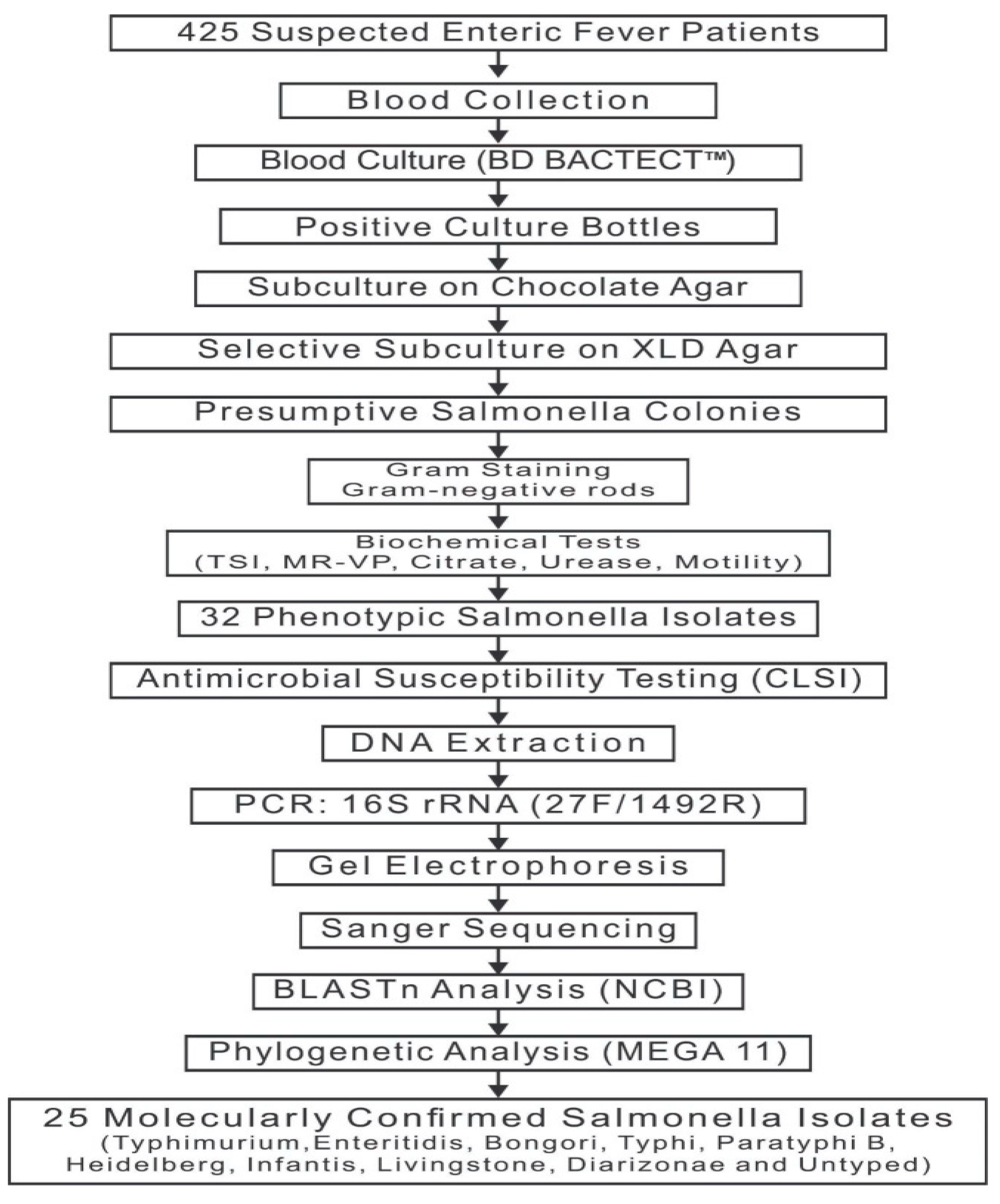
Figure 2.
Distribution of Pathogenic Microorganisms Isolates From Blood Culture of Suspected Enteric Fever Cases in Keffi LGA.
Figure 2.
Distribution of Pathogenic Microorganisms Isolates From Blood Culture of Suspected Enteric Fever Cases in Keffi LGA.
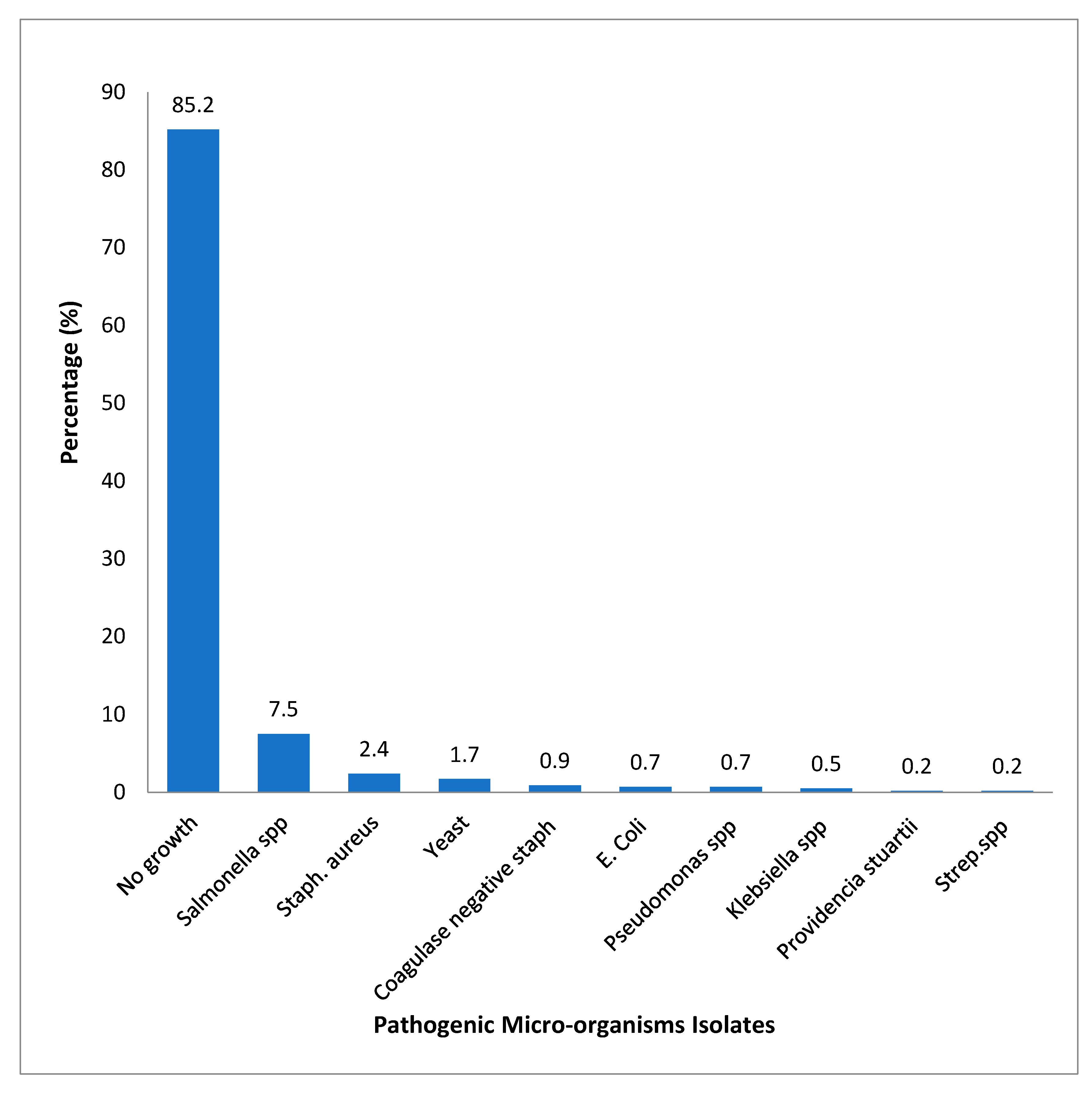
Figure 3.
Agarose Gel Electrophoresis of PCR-Amplified 16S rRNA Gene Products from 32 Phenotypically Identified Salmonella Isolates.
Figure 3.
Agarose Gel Electrophoresis of PCR-Amplified 16S rRNA Gene Products from 32 Phenotypically Identified Salmonella Isolates.
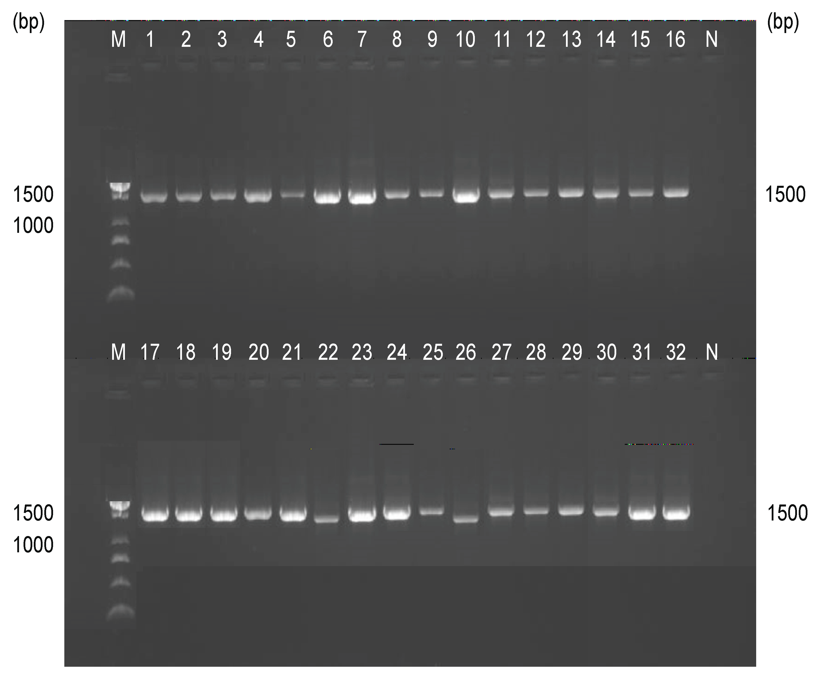
Figure 4.
Phylogenetic Tree of The Molecularly Confirmed Salmonella Isolates with Bootstrap Values (1,000 replicates; values ≥50% shown) from Suspected Enteric Fever Cases in Keffi LGA.
Figure 4.
Phylogenetic Tree of The Molecularly Confirmed Salmonella Isolates with Bootstrap Values (1,000 replicates; values ≥50% shown) from Suspected Enteric Fever Cases in Keffi LGA.
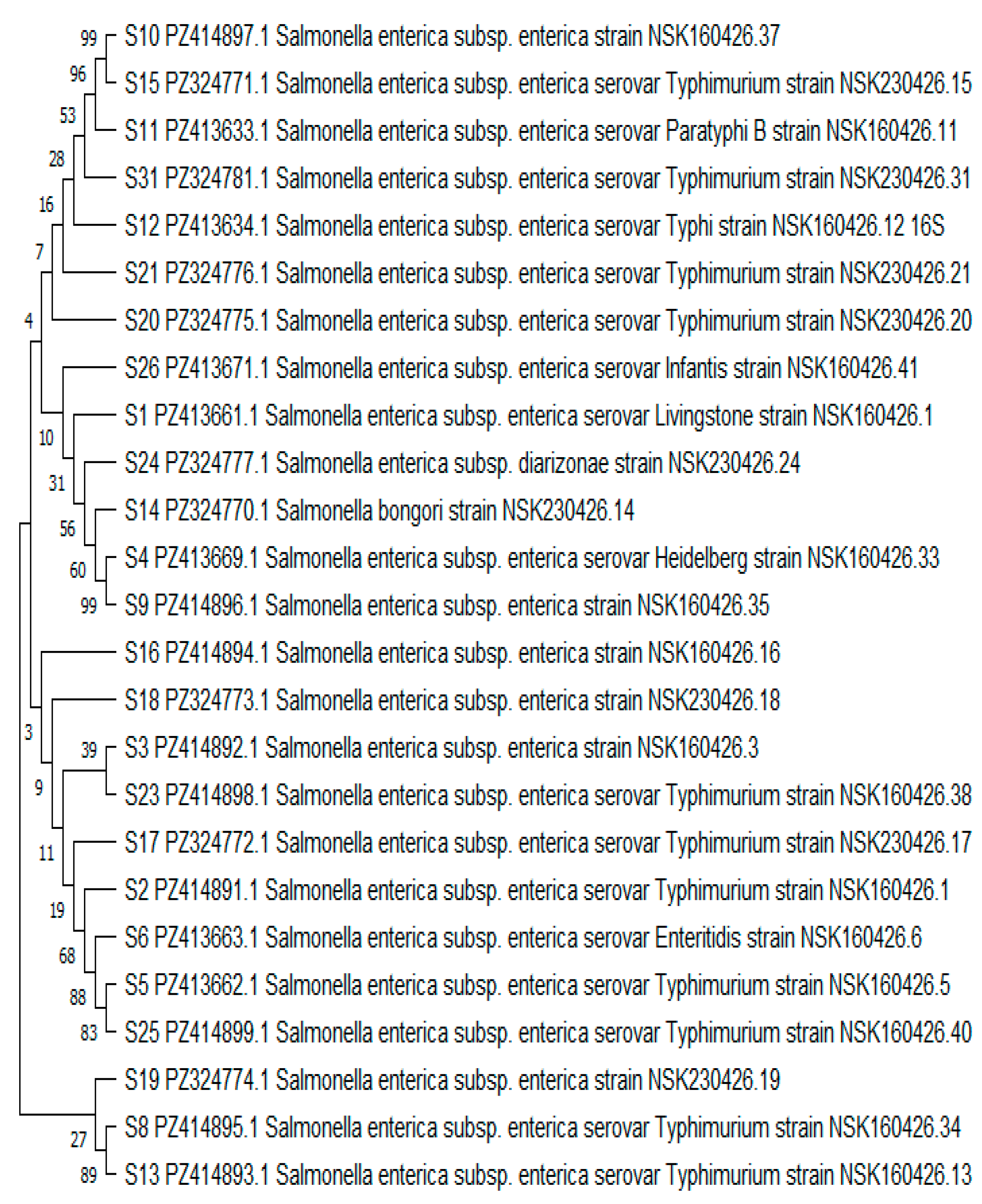
Table 2.
Taxonomic Classification and Distribution of Molecularly Identified Bacterial Isolates from Suspected Enteric Fever Cases in Keffi LGA.
Table 2.
Taxonomic Classification and Distribution of Molecularly Identified Bacterial Isolates from Suspected Enteric Fever Cases in Keffi LGA.
| Taxonomic Group | Isolates Sample(s) ID | Frequency (n=32) |
Percentage (%) |
|---|---|---|---|
|
Salmonella enterica serovar Typhimurium |
S2, S5, S8, S13, S15, S17, S20, S21, S23, S25, S31 |
11 |
34.4 |
| Salmonella enterica serovar Enteritidis | S6 | 1 | 3.1 |
| Salmonella enterica serovar Heidelberg | S4 | 1 | 3.1 |
| Salmonella enterica serovar Infantis | S26 | 1 | 3.1 |
| Salmonella enterica serovar Typhi | S12 | 1 | 3.1 |
| Salmonella enterica serovar Livingstone | S1 | 1 | 3.1 |
| Salmonella enterica serovar Paratyphi B | S11 | 1 | 3.1 |
| Salmonella enterica subsp. enterica (untyped strains) | S3, S9, S10, S16, S18, S19 | 6 | 18.8 |
| Salmonella enterica subsp. Diarizonae | S24 | 1 | 3.1 |
| Salmonella bongori | S14 | 1 | 3.1 |
| Citrobacter freundii | S7 | 1 | 3.1 |
| Citrobacter youngae | S22 | 1 | 3.1 |
| Enterobacter oligotrophicus | S28, S29, S30 | 3 | 9.4 |
| Enterobacter hormaechei | S32 | 1 | 3.1 |
| Klebsiella oxytoca | S27 | 1 | 3.1 |
Table 3.
Phylogenetic Clades of Confirmed Salmonella Isolates with Associated Serovar Groupings from Suspected Enteric Fever Cases in Keffi LGA.
Table 3.
Phylogenetic Clades of Confirmed Salmonella Isolates with Associated Serovar Groupings from Suspected Enteric Fever Cases in Keffi LGA.
| Major Clade | Sub-Clade | Isolate Members | Predominant Species | Bootstrap Support (%) | Interpretation |
|---|---|---|---|---|---|
| Clade I | Typhimurium Core Cluster | S5, S25, S2, S23, S17, S8, S13, S20, S21 | Salmonella enterica serovar Typhimurium | 68–89 | Strongly related Typhimurium lineage |
| Clade I | Typhimurium–Enteritidis Group | S6 | Salmonella enterica serovar Enteritidis | 68 | Closely related serovar |
| Clade I | Typhimurium–Infantis Branch | S26 | Salmonella enterica serovar Infantis | 10 | Related but distinct lineage |
| Clade II | Mixed Salmonella enterica Cluster | S3, S18, S19, S16, S1, S4, S9 | Salmonella enterica | 3–60 | Moderately related enterica strains |
| Clade III | Divergent Salmonella Group | S14, S24 | S. bongori / S. diarizonae | 31–56 | Distinct evolutionary lineage |
| Clade IV | Basal Salmonella Lineage | S10, S15, S11, S31, S12 | Typhimurium, Paratyphi B, Typhi | 96–100 | Strongly supported basal cluster |
Key: S – Salmonella sample.
Disclaimer/Publisher’s Note: The statements, opinions and data contained in all publications are solely those of the individual author(s) and contributor(s) and not of MDPI and/or the editor(s). MDPI and/or the editor(s) disclaim responsibility for any injury to people or property resulting from any ideas, methods, instructions or products referred to in the content. |
© 2026 by the authors. Licensee MDPI, Basel, Switzerland. This article is an open access article distributed under the terms and conditions of the Creative Commons Attribution (CC BY) license.
Copyright: This open access article is published under a Creative Commons CC BY 4.0 license, which permit the free download, distribution, and reuse, provided that the author and preprint are cited in any reuse.


