

Submitted:
07 August 2026
Posted:
10 August 2026
You are already at the latest version
Abstract
Objectives: To evaluate the effect of different healing abutment materials and 3D-printing orientations on microbial adhesion by comparing commercially available titanium abutments with customized composite resin and 3D-printed healing abutments. Materials and Methods: Fifty healing abutments were allocated into five groups (n=10); commercially available titanium (control), customized healing abutment resin composite CHA-Composite, and 3D-printed customized healing abutments printed on 0°, 45°, and 90° orientations; (CHA-3DP-0, CHA-3DP-45, CHA-3DP-90). Following sterilization, specimens were incubated with standardized suspensions of S. aureus and C. albicans for 24 h. Adherent microorganisms were quantified by colony-forming unit (CFU) analysis, and representative specimens were examined using scanning electron microscopy (SEM). Data were analyzed using one-way ANOVA with Tukey’s post hoc test (α = 0.05). Results: Significant differences in microbial adhesion were observed among the experimental groups for both C. albicans and S. aureus (P < 0.001). 3D-printed healing abutments at the 0° orientation demonstrated the lowest CFU counts for both microorganisms, significantly lower than all other groups (P < 0.001). In contrast, the 45° and particularly the 90° printing orientations exhibited significantly greater microbial adhesion. SEM findings corroborated the quantitative results, revealing reduced biofilm accumulation on 0°-3D-printed surfaces and more extensive biofilm formation on the 45° and 90° groups. Conclusions: Healing abutment material and 3D- printed abutments influence early microbial adhesion. The CHA-3DP-0 group showed the lowest S. aureus and C. albicans colonization, suggesting that optimized printing orientation may reduce biofilm formation. Further clinical and in vitro investigations are needed to confirm these findings.
Keywords:
additive manufacturing
; colony forming unit
; peri-implantits
; peri-implant mucositis
; dental composite healing abutment
; microbial adhesion
; stock healing abutment
1. Introduction
Dental implants are widely recognized as a predictable and effective treatment for replacing missing teeth, restoring both masticatory function and esthetics. However, the long-term success of implant therapy extends beyond osseointegration and relies on the preservation and stability of the surrounding peri-implant hard and soft tissues [1]. During the healing period between implant placement and definitive prosthetic restoration, various surgical and prosthetic strategies have been introduced to support soft tissue maturation, preserve peri-implant architecture, and facilitate the development of an optimal emergence profile [2,3,4,5].
After placement of endosseous dental implants, healing abutments are routinely connected during the transmucosal healing phase and maintained until fabrication of the definitive prosthesis. Their primary function is to promote the maturation and maintenance of the peri-implant soft tissue architecture while preserving the stability of the surrounding hard tissues [6,7]. For that, a variety of materials and techniques have been introduced and used [3,4,8,9]. These strategies encompass both prefabricated commercially available healing abutments manufactured from titanium or titanium alloys due to their excellent biocompatibility, corrosion resistance, and mechanical properties and customized healing abutments (CHAs) fabricated from materials such as resin composites, auto-polymerized poly (methyl methacrylate) (PMMA) resin, Polyetheretherketone (PEEK), Zirconia and three-dimensional (3D)-printed biomaterials using CAD-CAM technology, allowing individualized soft tissue contouring and emergence profile development [2,5,9,10].
The diverse materials used during the peri-implant healing phase are continuously exposed to the oral environment, rendering them susceptible to colonization by a variety of oral microorganisms. Differences in the surface characteristics of these materials may affect bacterial adhesion and the accumulation of multispecies biofilms, potentially influencing peri-implant tissue health [11,12,13,14,15].
Unlike periodontitis, which is predominantly associated with a well-characterized group of periodontal pathogens, peri-implant diseases have been shown to involve a more diverse and complex microbial community [16]. Several microorganisms have been implicated in the initiation and progression of peri-implant infections, including classical periodontal pathogens such as Prevotella intermedia, and Porphyromonas gingivalis and Treponema denticola, as well as opportunistic species including Staphylococcus aureus and Candida albicans [17,18,19]. The presence of these microorganisms may contribute to biofilm formation, peri-implant mucosal inflammation, and subsequent peri-implant tissue destruction [16,17,18,19].
Although bacterial colonization of commercially available titanium, dental composite, and PEEK healing abutments has been extensively investigated, limited evidence exists regarding bacterial adhesion to customized healing abutments fabricated from 3D-printed dental resin. Moreover, the potential effects of the printing orientation on bacterial colonization remain largely unexplored. Therefore, the aim of this in vitro study was to evaluate and compare microbial adhesion on different types of implant healing abutments fabricated from different materials, including commercially available titanium stock abutments, customized healing abutments made from dental composite resin, and 3D-printed custom healing abutments. Additionally, the effects of three different printing orientations on the adhesion of these microorganisms were investigated. The null hypothesis was that there will be no significant differences between microbial adhesion and materials/fabricating methods of customized healing abutment.
2. Materials and Methods
2.1. Sample Size and Study Groups
The required sample size was calculated, based on data from a previous study [20], to achieve 90% statistical power with a significance level (α) of 0.001, resulting in a minimum of nine specimens per group. A total of 50 healing abutment specimens were included in this study and allocated into five groups (n = 10 per group) as follows; Group I: commercially available pure titanium healing abutments (Straumann AG, Basel, Switzerland; RC Healing Abutment, conical shape, D5 mm × H 4 mm; (REF 024.4224S)), which served as the control group; Group II: customized healing abutments made directly from dental resin composite (Filtek Supreme Ultra, 3M ESPE, St. Paul, MN, USA) (CHA-Composite); Group III: 3d-printed customized healing abutments fabricated from printable dental resin (Asiga, Erfurt, Germany) using a 0° printing orientation (CHA-3DP-0); Group IV: 3d-printed customized healing abutments fabricated from printable dental resin (Asiga, Erfurt, Germany) using a 45° printing orientation (CHA-3DP-45); and Group V: 3d-printed customized healing abutments fabricated from printable dental resin (Asiga, Erfurt, Germany) using a 90° printing orientation (CHA-3DP-90). Figure 1 present a summary of materials used, and the study flowchart.
2.2. Specimen Preparation of Customized Healing Abutments
Duplication of the commercially available titanium stock healing abutment was used to standardize the shape, dimension, and morphology of the customized healing abutments.
For the CHA-Composite group, a silicone putty mold of the titanium stock healing abutment was fabricated using clear vinyl polysiloxane material (Exaclear, GC, Tokyo, Japan). The material was left to set for 7 min according to the manufacturer’s instructions. Then, a flowable composite (Filtek Supreme Ultra, 3M ESPE, St. Paul, MN, USA) was gradually used to fill the silicon matrix. A light-curing unit (Valo Cordless, Ultradent, South Jordan, UT, USA) at a power of 800 mW and an irradiance of 1,000 mW/cm² was used to polymerize the resin for 20 seconds per side according to the manufacturer’s instructions. All specimens were visually examined, and any with a defect or an air bubble were excluded from the study.
For the 3D-printed CHA, titanium stock healing abutment was scanned using an intraoral scanner (Trios 3, 3shape, Copenhagen, Denmark). The scan was exported as an STL file. The STL file was transferred to a dental 3D-printer (ASIGA MAX™, Alexandria, NSW 2015, Australia). For each group, a DentaTOOTH resin (Photopolymerized Methacrylate) was used to print the specimens in three different printing orientations (0-, 45-, 90-degree). A summary of printing parameters and post-printing conditions is presented in Figure 1.
2.3. Microbial Adhesion and Biofilm Formation
Prior to microbial inoculation, all specimens were sterilized by immersion in 70% ethanol for 15 min and allowed to air-dry under aseptic conditions. Standard strains of Staphylococcus aureus (ATCC 29213) and Candida albicans (ATCC 14053), obtained from the Microbiology Department, College of Medicine at Imam Abdulrahman Bin Faisal University, were cultured according to ATCC-recommended culture protocols. Microbial suspensions were prepared in sterile phosphate-buffered saline (PBS) and adjusted to a turbidity equivalent to the 0.5 McFarland standard according to CLSI guidelines (CLSI M07; CLSI M27). Each sterilized specimen was individually placed into a sterile 1.5-mL microcentrifuge (Eppendorf) tube containing sterile PBS and inoculated with 100 µL of the standardized microbial suspension. The tubes were incubated at 37 °C for 24 h to allow microbial adhesion and biofilm formation on the specimen surfaces. Biofilm formation was performed using a static incubation model adapted from previously described biomaterial adhesion protocols [21,22]. Following incubation, each specimen was gently rinsed three times with sterile PBS to remove non-adherent microorganisms and transferred to a new sterile microcentrifuge tube containing 1 mL of sterile PBS. The tubes were then vortexed (or agitated on an orbital shaker, if applicable) for 1 min to detach the adherent biofilm from the specimen surface as previously described [21,23]. For quantitative microbial analysis, 100 µL of the resulting suspension was inoculated onto blood agar plates for S. aureus and Sabouraud dextrose agar plates for C. albicans. The inoculated plates were incubated at 37 °C for 24 h, and microbial growth was subsequently assessed by quantifying the number of colony-forming units (CFUs) as described by Madigan MT, et al. [24].
2.4. Scanning Electron Microscopy (SEM)
Following biofilm formation, representative healing abutment specimens were prepared for SEM using JSM-6390LA Analytical SEM (JEOL Ltd., Tokyo, Japan) at the designated magnifications after gold sputter coating using Cressington 108 Auto Sputter Coater (Cressington Scientific Instruments Ltd., Watford, Hertfordshire, United Kingdom) to evaluate microbial adhesion and surface biofilm morphology. The specimens were fixed in 2.5% glutaraldehyde prepared in 0.1 M phosphate-buffered saline (PBS; pH 7.2) for 2 h at 4 °C (or overnight at 4 °C). Following primary fixation, the specimens were rinsed three times with PBS and post-fixed in 1% osmium tetroxide for 1 h. The specimens were subsequently dehydrated through a graded ethanol series (30%, 50%, 70%, 80%, 90%, 95%, and 100%), followed by complete drying. The dehydrated specimens were then sputter-coated with a thin gold layer (approximately 5–10 nm) to enhance electrical conductivity prior to SEM examination according to established biological electron microscopy protocols [25,26]. Similar preparation methods have been widely employed for evaluating microbial biofilms on dental implant biomaterials [27,28].
2.5. Statistical Analysis
The normality of data distribution was evaluated using the Shapiro–Wilk test. Differences in mean CFU counts among the five experimental groups (Control, Resin Composite, Printed-0° Orientation, Printed-45° Orientation, and Printed-90° Orientation) were analysed using one-way analysis of variance (ANOVA), followed by Tukey’s Honestly Significant Difference (HSD) post-hoc test for pairwise comparisons. Statistical significance was defined as a two-sided P value < 0.05. All statistical analyses were conducted using SigmaPlot version 16 (Systat Software Inc., San Jose, CA, USA).
3. Results
C. albicans CFUs results are shown in Table 1 and Figure 2. Results of the One-way ANOVA showed that the groups differed significantly (F = 29.39, P < 0.001). Post-hoc Tukey testing showed that the Printed-0° group had significantly lower CFU counts compared to all other groups (P < 0.001), while Printed-90° had significantly higher counts than Control, Resin Composite, and Printed-0° (P < 0.05).
The results of S. aures CFUs are shown in Table 2 and Figure 3. Results of the One-way ANOVA showed that the groups differed significantly (F = 29.54, P < 0.001). Post-hoc Tukey testing showed that the Printed-0° group had significantly lower CFU counts compared to all other groups (P < 0.001), and the Printed-45° and Printed-90° groups showed significantly higher counts than Control (P < 0.001 and P < 0.001, respectively).
SEM examination revealed distinct differences in biofilm formation and surface morphology across the five experimental groups (Figure 4). The Control group displayed a relatively smooth surface with sparse, isolated clusters of spherical cells, reflecting the moderate CFU counts observed. The Resin Composite exhibited a heavily populated surface characterized by resin matrix degradation intermixed with spherical microbial cells, indicative of abundant biofilm accumulation. The Printed-0° Orientation group demonstrated a moderately dense distribution of biofilms across the surface, yet with a notably reduced biofilm mass relative to the Resin Composite group. The Printed-45° Orientation group showed the most complex surface topography change among the 3D-printed groups, featuring irregular, laminated surface layers with biofilm accumulation. The rough, folded surface architecture likely promotes microbial entrapment and retention, which corresponds to the high CFU counts observed for this group (mean Log₁₀ = 3.208 for C. albicans; 3.209 for S. aureus), exceeding those of the Printed-0° group. The Printed-90° Orientation group presented a uniformly distributed pattern of spherical microbial cells across a relatively homogeneous surface, with extensive and evenly spread biofilm coverage.
4. Discussion
The findings of the present study demonstrated that microbial colonization varied among healing abutments fabricated from different materials. Furthermore, the printing orientation of CHAs influenced microbial adhesion, suggesting that both material composition and surface characteristics contribute to the initial establishment of peri-implant biofilms. The null hypothesis was rejected.
Although numerous in vitro studies have investigated bacterial adhesion to various implant and healing abutment materials [12,14,15,27,29], and only a limited number have specifically evaluated clinically relevant peri-implant pathogens such as Staphylococcus aureus and Candida albicans [12,17,18,19,29,30,31,32]. Moreover, the available literature has primarily focused on comparisons between conventional materials, with little attention given to CHAs. To the best of the author’s knowledge, no published study has specifically investigated the influence of different printing orientations on the microbial adhesion to CHAs. While previous studies have evaluated microbial adhesion on CAD-CAM PMMA materials [12,15,27,29] or described the fabrication of customized PMMA healing abutments [2,5,33], the effect of printing orientation on biofilm formation has not been investigated.
Therefore, the present study provides novel in vitro evidence by simultaneously evaluating biofilm adhesion of S. aureus and C. albicans on healing abutments fabricated from titanium, resin composite, and 3D-printed resin These findings suggest that both the choice of healing abutment material and the fabrication methods may influence microbial colonization and should be considered when designing and manufacturing CHAs to minimize the risk of peri-implant biofilm formation.
The findings demonstrated that microbial colonization differed significantly among the tested materials and fabrication protocols. Notably, 3D Printing-CHA in the 0° orientation exhibited the lowest biofilm accumulation for both microorganisms, whereas the 45° group and particularly the 90° group orientations demonstrated significantly greater microbial adhesion.
Microbial adhesion to dental material surfaces is a multifactorial process influenced by surface roughness, surface free energy, wettability, chemical composition, and surface topography [34,35]. Previous investigations have consistently demonstrated that even subtle alterations in surface morphology in terms of polished surfaces among different materials can affect the initial adhesion of microorganisms and subsequent biofilm maturation [36,37,38,39,40]. Because the 3D-printed method generates characteristic machining grooves and tool marks [41,42], different printing orientations are expected to produce distinct surface textures despite the use of the same 3D-printable resin material. Consequently, differences in microbial adhesion observed in the present study are likely attributable primarily to variations in surface topography created by the printing direction rather than differences in the bulk material itself.
The remarkably lower adhesion observed in the CHA-3DP-0 group suggests that this printing orientation may produce a smoother and more homogeneous surface with fewer irregularities, thereby reducing favorable sites for microbial retention and initial colonization. In contrast, the increased biofilm formation observed in the CHA-3DP-45 and CHA-3DP-90 groups may be explained by the generation of more pronounced machining grooves, layered surface irregularities, and microscopic undercuts that facilitate microbial attachment and protect adherent cells from shear forces. Surface grooves can increase the available surface area for adhesion while providing sheltered niches that enhance microbial retention during the early stages of biofilm [28].
These observations were supported by SEM analysis. The CHA-3DP-0 specimens demonstrated relatively limited biofilm accumulation compared with the other PMMA groups, whereas the CHA-3DP-45 orientation exhibited irregular laminated surface features associated with extensive microbial colonization. Similarly, the CHA-3DP-90 orientation showed a uniformly distributed biofilm covering the surface. The close agreement between the quantitative CFU measurements and the qualitative SEM observations strengthens the reliability of the present findings and suggests that the observed differences in microbial colonization were directly related to alterations in surface morphology induced by different printing orientations.
Although titanium has long been considered the gold standard for healing abutments because of its favorable biocompatibility and corrosion resistance [43,44], microbial adhesion to titanium surfaces remains inevitable within the oral environment, as explained in several studies [27,34,35,45]. Previous studies have reported variable bacterial adhesion to titanium depending on surface treatment [46], surface roughness, and polishing procedures [27,45,47]. In addition, several studies have demonstrated that clinically used and re-sterilized healing abutments frequently retain residual proteins, organic debris, and biofilm remnants. These residual surface contaminants may modify the titanium surface and create irregularities for subsequent microbial adhesion compared with new healing abutments [48,49,50]. In the present study, the commercially available new titanium healing abutments demonstrated intermediate levels of microbial colonization, which considered significantly lower than those in the CHA-3DP-45 and CHA-3DP-90 groups but higher than those in the CHA-3DP-0 specimens. These findings suggest that an optimized CAD-CAM printing strategy may yield CHAs with microbial adhesion properties comparable to, or even more favorable than, those of conventional titanium healing abutments.
The resin composite healing abutments demonstrated microbial adhesion levels comparable to those observed for titanium groups in both microbial species. Composite materials contain an organic resin matrix that may undergo surface degradation, water sorption, and filler particle exposure during aging, potentially increasing surface roughness and promoting microbial attachment [51,52,53,54]. The SEM findings in the present study demonstrated areas of matrix degradation associated with dense microbial colonization, supporting previous reports that surface deterioration of resin-based materials may facilitate biofilm development.
Both Candida albicans and Staphylococcus aureus exhibited remarkably similar colonization patterns across the experimental groups. Despite their biological differences, both microorganisms demonstrated significantly reduced adhesion on the CHA-3DP-0 surfaces and increased adhesion on the CHA-3DP-45 and CHA-3DP-90 orientations. This consistent trend suggests that the influence of surface topography generated by printing orientation may represent a dominant factor governing initial microbial attachment irrespective of microbial species. The inclusion of both a fungal and a bacterial pathogen enhances the clinical relevance of the present findings because these microorganisms have been implicated in peri-implant infections, particularly in medically compromised patients and individuals with established peri-implant disease [17,18,19].
Number of limitations should be acknowledged. First, this was an in vitro study performed under controlled laboratory conditions, which may not fully replicate the complex oral environment, including saliva, host immune responses, masticatory loading, and multispecies biofilm interactions. Second, only two representative peri-implant microorganisms were evaluated in this study, which may play a crucial role in the pathogenicity of peri-implant disease and conditions, whereas peri-implant biofilms consist of diverse polymicrobial communities. Third, surface roughness, surface free energy, and wettability were not quantitatively measured; therefore, the proposed mechanisms underlying the observed differences remain speculative. Future studies should combine surface profilometry, atomic force microscopy, contact-angle measurements, and polymicrobial biofilm models to better elucidate the relationship between CAD-CAM printing parameters and printing protocols using different angles, surface characteristics, and microbial colonization. Clinical investigations are also warranted to determine whether these in vitro findings translate into improved peri-implant soft tissue health in vivo.
5. Conclusions
The present study demonstrates that both healing abutment material and CAD-CAM printing orientation significantly influence early microbial adhesion. Among the tested groups, CHA-3DP-0 group exhibited the lowest colonization by Staphylococcus aureus and Candida albicans, indicating that optimization of printing orientation may represent a simple and effective strategy to reduce biofilm formation on customized healing abutments. Further laboratory and clinical studies are required to confirm the translational relevance of these findings
Funding
This research received no external funding.
Institutional Review Board Statement
Not applicable.
Informed Consent Statement
Not applicable.
Data Availability Statement
We encourage all authors of articles published in MDPI journals to share their research data. In this section, please provide details regarding where data supporting reported results can be found, including links to publicly archived datasets analyzed or generated during the study. Where no new data were created, or where data is unavailable due to privacy or ethical restrictions, a statement is still required. Suggested Data Availability Statements are available in section “MDPI Research Data Policies” at https://www.mdpi.com/ethics.
Acknowledgments
The author would like to express their sincere appreciation to Yousif AlDulaijan, Associate Professor, Department of Substitutive Dental Sciences, College of Dentistry, Imam Abdulrahman Bin Faisal University, for his valuable review of the manuscript. The author also thanks Abdulrahman Balhaddad, Associate Professor, Department of Restorative Dental Sciences, College of Dentistry, Imam Abdulrahman Bin Faisal University, for his valuable assistance with statistical analysis. Special thanks are extended to Mr. Badr Saker, Laboratory Technician, for his assistance with the microbiological culture procedures and Mr. Raju Thangaruju for his assistance in preparing the specimens.
Conflicts of Interest
The author declare no conflicts of interest.
Abbreviations
| 3D | Three-dimensional |
| CAD-CAM | Computer-Aided Design and Computer-Aided Manufacturing |
| C. albicans | Candida albicans |
| CFU | Colony-Forming Unit |
| CHA | Customized Healing Abutment |
| CHA-Composite | Customized Composite Resin Healing Abutment |
| CHA-3DP-0 | Customized 3D-Printed Healing Abutment (0° Printing Orientation) |
| CHA-3DP-45 | Customized 3D-Printed Healing Abutment (45° Printing Orientation) |
| CHA-3DP-90 | Customized 3D-Printed Healing Abutment (90° Printing Orientation) |
| PBS | Phosphate-Buffered Saline |
| PEEK | Polyetheretherketone |
| PMMA | Poly(methyl methacrylate) |
| SEM | Scanning Electron Microscopy |
| S. aureus | Staphylococcus aureus |
References
- E. Jung, R.; Zembic, A.; Pjetursson, B.E.; Zwahlen, M.; S. Thoma, D. Systematic Review of the Survival Rate and the Incidence of Biological, Technical, and Aesthetic Complications of Single Crowns on Implants Reported in Longitudinal Studies with a Mean Follow-up of 5 Years. Clin. Oral Implant. Res. 2012, 23, 2–21. [Google Scholar] [CrossRef] [PubMed]
- Pow, E.H.; McMillan, A.S. A Modified Implant Healing Abutment to Optimize Soft Tissue Contours: A Case Report. Implant Dent. 2004, 13, 297–300. [Google Scholar] [CrossRef] [PubMed]
- Elian, N.; Tabourian, G.; Jalbout, Z.N.; Classi, A.; Cho, S.; Froum, S.; Tarnow, D.P. Accurate Transfer of Peri-implant Soft Tissue Emergence Profile from the Provisional Crown to the Final Prosthesis Using an Emergence Profile Cast. J. Esthet. Restor. Dent. 2007, 19, 306–314. [Google Scholar] [CrossRef] [PubMed]
- Janakievski, J. Case Report: Maintenance of Gingival Form Following Immediate Implant Placement—The Custom-Healing Abutment. Adv. Esthet. Interdiscip. Dent. 2007, 3, 24–28. [Google Scholar]
- Proussaefs, P. Use of CAD/CAM Healing Abutment Immediately after Dental Implant Placement for the Non-Esthetic Zone: A Guided Soft Tissue Healing Technique. J. Oral Implantol. 2016, 42, 189–193. [Google Scholar] [CrossRef] [PubMed]
- Berglundh, T.; Abrahamsson, I.; Welander, M.; Lang, N.P.; Lindhe, J. Morphogenesis of the Peri-implant Mucosa: An Experimental Study in Dogs. Clin. Oral Implant. Res. 2007, 18, 1–8. [Google Scholar] [CrossRef] [PubMed]
- Berglundh, T.; Lindhe, J.; Ericsson, I.; Marinello, C.P.; Liljenberg, B.; Thornsen, P. The Soft Tissue Barrier at Implants and Teeth. Clin. Oral Implant. Res. 1991, 2, 81–90. [Google Scholar] [CrossRef] [PubMed]
- Prati, C.; Zamparini, F.; Canullo, L.; Pirani, C.; Botticelli, D.; Gandolfi, M.G. Factors Affecting Soft and Hard Tissues around Two-Pies Transmucosal Implants: A 3-Year Prospective Cohort Study. Int. J. Oral Maxillofac. Implant. 2020, 35, 1022–1036. [Google Scholar] [CrossRef] [PubMed]
- Chokaree, P.; Poovarodom, P.; Chaijareenont, P.; Yavirach, A.; Rungsiyakull, P. Biomaterials and Clinical Applications of Customized Healing Abutment—a Narrative Review. J. Funct. Biomater. 2022, 13, 291. [Google Scholar] [CrossRef] [PubMed]
- Osman, R.B.; Swain, M.V. A Critical Review of Dental Implant Materials with an Emphasis on Titanium versus Zirconia. Materials 2015, 8, 932–958. [Google Scholar] [CrossRef] [PubMed]
- Dorkhan, M.; Hall, J.; Uvdal, P.; Sandell, A.; Svensäter, G.; Davies, J.R. Crystalline Anatase-Rich Titanium Can Reduce Adherence of Oral Streptococci. Biofouling 2014, 30, 751–759. [Google Scholar] [CrossRef] [PubMed]
- Hung, C.C.U.; Costa, R.C.; Pereira, G.; Abdo, V.L.; do Santos Noronha, M.; Retamal-Valdes, B.; Bertolini, M.; Feres, M.; Shibli, J.A.; Barão, V.A. Oral Microbial Colonization on Titanium and Polyetheretherketone Dental Implant Healing Abutments: An in Vitro and in Vivo Study. J. Prosthet. Dent. 2025, 133, 1545–1552. [Google Scholar] [CrossRef] [PubMed]
- Chrcanovic, B.R.; Kisch, J.; Albrektsson, T.; Wennerberg, A. Factors Influencing Early Dental Implant Failures. J. Dent. Res. 2016, 95, 995–1002. [Google Scholar] [CrossRef] [PubMed]
- D’Ercole, S.; Piattelli, A.; Marzo, G.; Scarano, A.; Tripodi, D. Influence of Bacterial Colonization of the Healing Screws on Peri-Implant Tissue. J. Dent. Sci. 2013, 8, 109–114. [Google Scholar] [CrossRef]
- Jain, S.S.; Schramm, S.T.; Siddiqui, D.A.; Huo, W.; Palmer, K.L.; Wilson, T.G., Jr.; Rodrigues, D.C. Effects of Multiple Implantations of Titanium Healing Abutments: Surface Characteristics and Microbial Colonization. Dent. Mater. 2020, 36, e279–e291. [Google Scholar] [CrossRef] [PubMed]
- Kumar, P.S.; Mason, M.R.; Brooker, M.R.; O’Brien, K. Pyrosequencing Reveals Unique Microbial Signatures Associated with Healthy and Failing Dental Implants. J. Clin. Periodontol. 2012, 39, 425–433. [Google Scholar] [CrossRef] [PubMed]
- Leonhardt, Å.; Renvert, S.; Dahlén, G. Microbial Findings at Failing Implants. Clin. Oral Implant. Res. 1999, 10, 339–345. [Google Scholar] [CrossRef] [PubMed]
- Fragkioudakis, I.; Konstantopoulos, G.; Kottaridi, C.; Doufexi, A.-E.; Sakellari, D. Quantitative Assessment of Candida Albicans, Staphylococcus Aureus and Staphylococcus Epidermidis in Peri-Implant Health and Disease: Correlation with Clinical Parameters. J. Med. Microbiol. 2024, 73. [Google Scholar] [CrossRef] [PubMed]
- Albertini, M.; López-Cerero, L.; O’Sullivan, M.G.; Chereguini, C.F.; Ballesta, S.; Ríos, V.; Herrero-Climent, M.; Bullón, P. Assessment of Periodontal and Opportunistic Flora in Patients with Peri-implantitis. Clin. Oral Implant. Res. 2015, 26, 937–941. [Google Scholar] [CrossRef] [PubMed]
- Naghsh, N.; Hosseini, A.; Mogharehabed, A.; Yaghini, J.; Pezeshki, Z.; Khaleghi, N. A Comparison of Four Decontamination Procedures in Reusing Healing Abutments: An in Vitro Study. Saudi Dent. J. 2024, 36, 1141–1145. [Google Scholar] [CrossRef] [PubMed]
- An, Y.H.; Friedman, R.J. Concise Review of Mechanisms of Bacterial Adhesion to Biomaterial Surfaces. J. Biomed. Mater. Res. 1998, 43, 338–348. [Google Scholar]
- Donlan, R.M.; Costerton, J.W. Biofilms: Survival Mechanisms of Clinically Relevant Microorganisms. Clin. Microbiol. Rev. 2002, 15, 167–193. [Google Scholar] [CrossRef] [PubMed]
- Azeredo, J.; Azevedo, N.F.; Briandet, R.; Cerca, N.; Coenye, T.; Costa, A.R.; Desvaux, M.; Di Bonaventura, G.; Hébraud, M.; Jaglic, Z.; et al. Critical Review on Biofilm Methods. Crit. Rev. Microbiol. 2017, 43, 313–351. [Google Scholar] [CrossRef] [PubMed]
- Madigan, M.T.; Martinko, J.M.; Parker, J. Brock Biology of Microorganisms; Prentice hall Upper Saddle River, NJ, 1997; Vol. 11. [Google Scholar]
- Bozzola, J.J.; Russell, L.D. Electron Microscopy: Principles and Techniques for Biologists; Jones & Bartlett Learning, 1999. [Google Scholar]
- Hayat, M.A. Loss of Antigenicity on Stored Paraffin Sections. Microsc. Today 2000, 8, 30–32. [Google Scholar] [CrossRef]
- Teughels, W.; Van Assche, N.; Sliepen, I.; Quirynen, M. Effect of Material Characteristics and/or Surface Topography on Biofilm Development. Clin. Oral Implant. Res. 2006, 17, 68–81. [Google Scholar] [CrossRef] [PubMed]
- Quirynen, M.; Bollen, C.M.L. The Influence of Surface Roughness and Surface-free Energy on Supra- and Subgingival Plaque Formation in Man: A Review of the Literature. J. Clin. Periodontol. 1995, 22, 1–14. [Google Scholar] [CrossRef] [PubMed]
- Taşın, S.; Güvenir, M.; Ismatullaev, A. Effects of Surface Characteristics of Conventionally Manufactured, CAD/CAM Milled, and 3D-Printed Interim Materials on Adherence of Streptococcus Mutans and Candida Albicans. Cumhur. Dent. J. 2023, 26, 227–234. [Google Scholar]
- Bravo, E.; Arce, M.; Ribeiro-Vidal, H.; Herrera, D.; Sanz, M. The Impact of Candida Albicans in the Development, Kinetics, Structure, and Cell Viability of Biofilms on Implant Surfaces—an in Vitro Study with a Validated Multispecies Biofilm Model. Int. J. Mol. Sci. 2024, 25, 3277. [Google Scholar] [CrossRef] [PubMed]
- Eguia, A.; Arakistain, A.; De-la-Pinta, I.; López-Vicente, J.; Sevillano, E.; Quindós, G.; Eraso, E. Candida Albicans Biofilms on Different Materials for Manufacturing Implant Abutments and Prostheses. Med. Oral Patol. Oral Y Cirugía Bucal 2019, 25, e13. [Google Scholar] [CrossRef] [PubMed]
- Gökmenoglu, C.; Kara, N.B.; Beldüz, M.; Kamburoğlu, A.; Tosun, İ.; Sadik, E.; Kara, C. Evaluation of Candida Albicans Biofilm Formation on Various Parts of Implant Material Surfaces. Niger. J. Clin. Pract. 2018, 21, 33–37. [Google Scholar] [CrossRef] [PubMed]
- Raheem, I.M.A.; Hammad, I.A.; Kader, S.H.A.; Fahmy, R.A. Fabrication of a CAD-CAM Custom Healing Abutment Guided by a Conventional Dental Radiograph for Delayed Loaded Dental Implants: A Dental Technique. J. Prosthet. Dent. 2022, 127, 49–54. [Google Scholar] [CrossRef] [PubMed]
- Han, A.; Tsoi, J.K.; Rodrigues, F.P.; Leprince, J.G.; Palin, W.M. Bacterial Adhesion Mechanisms on Dental Implant Surfaces and the Influencing Factors. Int. J. Adhes. Adhes. 2016, 69, 58–71. [Google Scholar] [CrossRef]
- Hämmerle, C.H. Biofilm on Dental Implants: A Review of the Literature. Int. J. Oral Maxillofac. Implant 2009, 24. [Google Scholar]
- Kearns, V.R.; Williams, R.L.; Mirvakily, F.; Doherty, P.J.; Martin, N. Guided Gingival Fibroblast Attachment to Titanium Surfaces: An in Vitro Study. J. Clin. Periodontol. 2013, 40, 99–108. [Google Scholar] [CrossRef] [PubMed]
- Bollenl, C.M.; Lambrechts, P.; Quirynen, M. Comparison of Surface Roughness of Oral Hard Materials to the Threshold Surface Roughness for Bacterial Plaque Retention: A Review of the Literature. Dent. Mater. 1997, 13, 258–269. [Google Scholar] [CrossRef] [PubMed]
- Linkevicius, T.; Valantiejiene, V.; Alkimavicius, J.; Gineviciute, E.; Andrijauskas, R.; Linkeviciene, L. The Effect of a Polishing Protocol on the Surface Roughness of Zirconium Oxide. Int. J. Prosthodont 2020, 33, 217–223. [Google Scholar] [CrossRef] [PubMed]
- Al-Radha, A.S.D.; Dymock, D.; Younes, C.; O’Sullivan, D. Surface Properties of Titanium and Zirconia Dental Implant Materials and Their Effect on Bacterial Adhesion. J. Dent. 2012, 40, 146–153. [Google Scholar] [CrossRef] [PubMed]
- Batak, B.; Çakmak, G.; Johnston, W.M.; Yilmaz, B. Surface Roughness of High-Performance Polymers Used for Fixed Implant-Supported Prostheses. J. Prosthet. Dent. 2021, 126, 254–e1. [Google Scholar] [CrossRef] [PubMed]
- Alfouzan, A.; Alnouwaisar, A.; Alazzam, N.; AL-Otaibi, H.; Labban, N.; Alswaidan, M.; AL-Taweel, S.; Alshehri, H. Surface Roughness Analysis of Prepolymerized CAD/CAM Dental Acrylic Resins Following Combined Surface Treatments. Mater. Sci.-Pol. 2021, 39, 209–218. [Google Scholar] [CrossRef]
- Herráez-Galindo, C.; Rizo-Gorrita, M.; Luna-Oliva, I.; Serrera-Figallo, M.-Á.; Castillo-Oyagüe, R.; Torres-Lagares, D. In Vitro Comparative Study of Fibroblastic Behaviour on Polymethacrylate (PMMA) and Lithium Disilicate Polymer Surfaces. Polymers 2019, 11, 744. [Google Scholar] [CrossRef] [PubMed]
- Albrektsson, T.; Brånemark, P.-I.; Hansson, H.-A.; Lindström, J. Osseointegrated Titanium Implants: Requirements for Ensuring a Long-Lasting, Direct Bone-to-Implant Anchorage in Man. Acta Orthop. Scand. 1981, 52, 155–170. [Google Scholar] [CrossRef] [PubMed]
- Koutouzis, T.; Richardson, J.; Lundgren, T. Comparative Soft and Hard Tissue Responses to Titanium and Polymer Healing Abutments. J. Oral Implantol. 2011, 37, 174–182. [Google Scholar] [CrossRef] [PubMed]
- Yeo, I.-S.; Kim, H.-Y.; Lim, K.S.; Han, J.-S. Implant Surface Factors and Bacterial Adhesion: A Review of the Literature. Int. J. Artif. Organs 2012, 35, 762–772. [Google Scholar] [CrossRef] [PubMed]
- Chen, C.-J.; Ding, S.-J.; Chen, C.-C. Effects of Surface Conditions of Titanium Dental Implants on Bacterial Adhesion. Photomed. Laser Surg. 2016, 34, 379–388. [Google Scholar] [CrossRef] [PubMed]
- Costa, R.C.; Nagay, B.E.; Bertolini, M.; Costa-Oliveira, B.E.; Sampaio, A.A.; Retamal-Valdes, B.; Shibli, J.A.; Feres, M.; Barao, V.A.; Souza, J.G.S. Fitting Pieces into the Puzzle: The Impact of Titanium-Based Dental Implant Surface Modifications on Bacterial Accumulation and Polymicrobial Infections. Adv. Colloid Interface Sci. 2021, 298, 102551. [Google Scholar] [CrossRef] [PubMed]
- Burioni, R.; Silvestrini, L.; D’Orto, B.; Tetè, G.; Nagni, M.; Polizzi, E.; Gherlone, E.F. Could Dental Material Reuse Play a Significant Role in Preservation of Raw Materials, Water, Energy, and Costs? Microbiological Analysis of New versus Reused Healing Abutments: An In Vitro Study. Bioengineering 2024, 11, 387. [Google Scholar] [CrossRef] [PubMed]
- Wadhwani, C.; Schonnenbaum, T.R.; Audia, F.; Chung, K. In-Vitro Study of the Contamination Remaining on Used Healing Abutments after Cleaning and Sterilizing in Dental Practice. Clin. Implant Dent. Rel Res. 2016, 18, 1069–1074. [Google Scholar] [CrossRef] [PubMed]
- Barreiros, P.; Braga, J.; Faria-Almeida, R.; Coelho, C.; Teughels, W.; Souza, J.C.M. Remnant Oral Biofilm and Microorganisms after Autoclaving Sterilization of Retrieved Healing Abutments. J. Periodontal Res. 2021, 56, 415–422. [Google Scholar] [CrossRef] [PubMed]
- Bollenl, C.M.; Lambrechts, P.; Quirynen, M. Comparison of Surface Roughness of Oral Hard Materials to the Threshold Surface Roughness for Bacterial Plaque Retention: A Review of the Literature. Dent. Mater. 1997, 13, 258–269. [Google Scholar] [CrossRef] [PubMed]
- Münchow, E.A.; Ferreira, A.C.A.; Machado, R.M.; Ramos, T.S.; Rodrigues-Junior, S.A.; Zanchi, C.H. Effect of Acidic Solutions on the Surface Degradation of a Micro-Hybrid Composite Resin. Braz. Dent. J. 2014, 25, 321–326. [Google Scholar] [CrossRef] [PubMed]
- Ferracane, J.L. Resin Composite—State of the Art. Dent. Mater. 2011, 27, 29–38. [Google Scholar] [CrossRef] [PubMed]
- Ferracane, J.L. Hygroscopic and Hydrolytic Effects in Dental Polymer Networks. Dent. Mater. 2006, 22, 211–222. [Google Scholar] [CrossRef] [PubMed]
Figure 1.
Study flowchart.
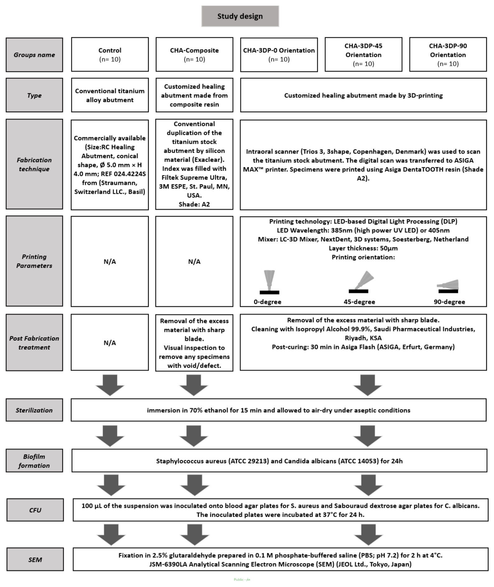
Figure 2.
Colony-forming units of Candida albicans across experimental groups. Mean colony-forming units (CFUs) of Staphylococcus aureus expressed as Log₁₀ values across the five experimental groups (Control, Resin CHA Composite, CHA 3D-Printed-0° Orientation, CHA 3D-Printed-45° Orientation, and CHA 3D-Printed-90° Orientation). Error bars represent 95% confidence intervals. Individual data points are shown as open circles. Individual data points are shown as open circles. Different letters (b,b,c,ab, and a) above bars indicate statistically significant differences between groups (P < 0.05).
Figure 2.
Colony-forming units of Candida albicans across experimental groups. Mean colony-forming units (CFUs) of Staphylococcus aureus expressed as Log₁₀ values across the five experimental groups (Control, Resin CHA Composite, CHA 3D-Printed-0° Orientation, CHA 3D-Printed-45° Orientation, and CHA 3D-Printed-90° Orientation). Error bars represent 95% confidence intervals. Individual data points are shown as open circles. Individual data points are shown as open circles. Different letters (b,b,c,ab, and a) above bars indicate statistically significant differences between groups (P < 0.05).
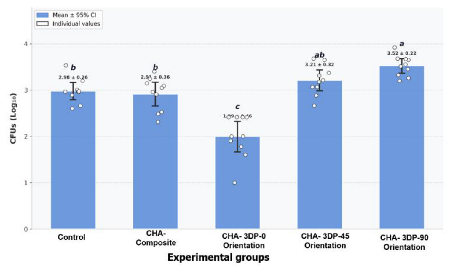
Figure 3.
Colony-forming units of Staphylococcus aureus across experimental groups. Mean colony-forming units (CFUs) of Staphylococcus aureus expressed as Log₁₀ values across the five experimental groups (Control, Resin CHA Composite, CHA 3D-Printed-0° Orientation, CHA 3D-Printed-45° Orientation, and CHA 3D-Printed-90° Orientation). Error bars represent 95% confidence intervals. Individual data points are shown as open circles. Different letters (b,ab, c, a, a) above bars indicate statistically significant differences between groups (P < 0.05).
Figure 3.
Colony-forming units of Staphylococcus aureus across experimental groups. Mean colony-forming units (CFUs) of Staphylococcus aureus expressed as Log₁₀ values across the five experimental groups (Control, Resin CHA Composite, CHA 3D-Printed-0° Orientation, CHA 3D-Printed-45° Orientation, and CHA 3D-Printed-90° Orientation). Error bars represent 95% confidence intervals. Individual data points are shown as open circles. Different letters (b,ab, c, a, a) above bars indicate statistically significant differences between groups (P < 0.05).
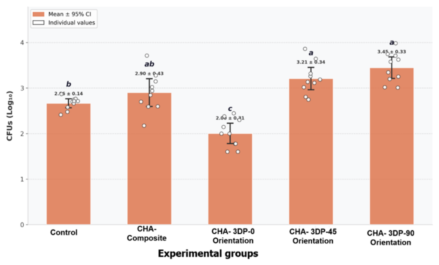
Figure 4.
Scanning electron microscopy (SEM) images of biofilm formation on the surfaces of the five experimental groups: Control, Resin CHA Composite, CHA 3D-Printed-0° Orientation, CHA 3D-Printed-45° Orientation, and CHA 3D-Printed-90° Orientation. Images were captured at magnifications ranging from ×2,500 to ×3,000. Scale bars represent 5–10 µm.
Figure 4.
Scanning electron microscopy (SEM) images of biofilm formation on the surfaces of the five experimental groups: Control, Resin CHA Composite, CHA 3D-Printed-0° Orientation, CHA 3D-Printed-45° Orientation, and CHA 3D-Printed-90° Orientation. Images were captured at magnifications ranging from ×2,500 to ×3,000. Scale bars represent 5–10 µm.
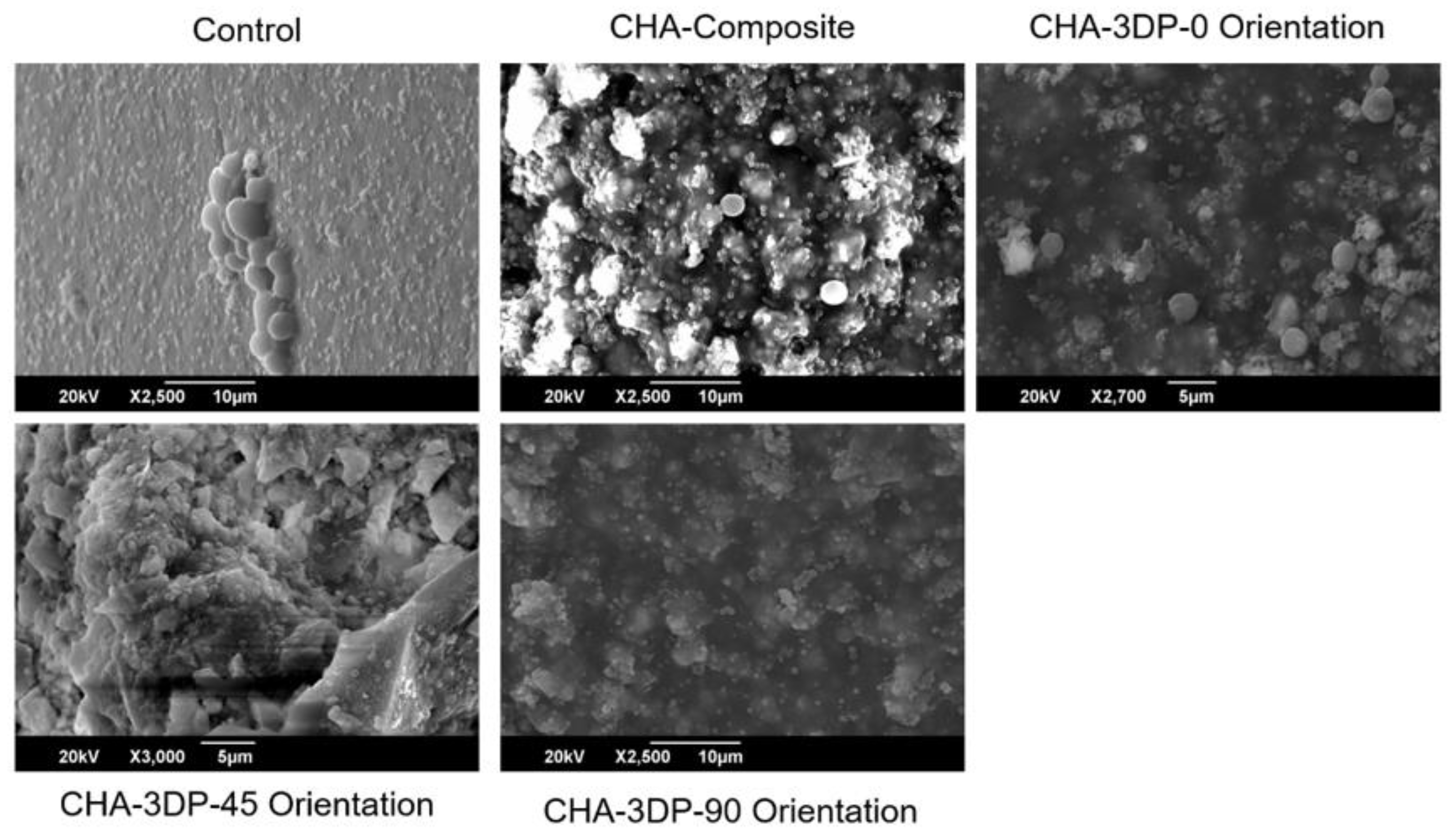

Table 1.
Colony-forming units of Candida albicans across experimental groups.
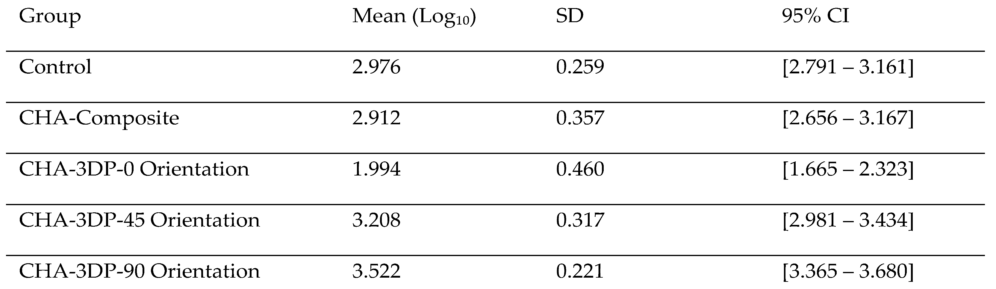 |
SD: Standard deviation; CI: Confidence Interval; CHA: Customized Healing Abutment.
Table 2.
Colony-forming units of Staphylococcus aureus across experimental groups.
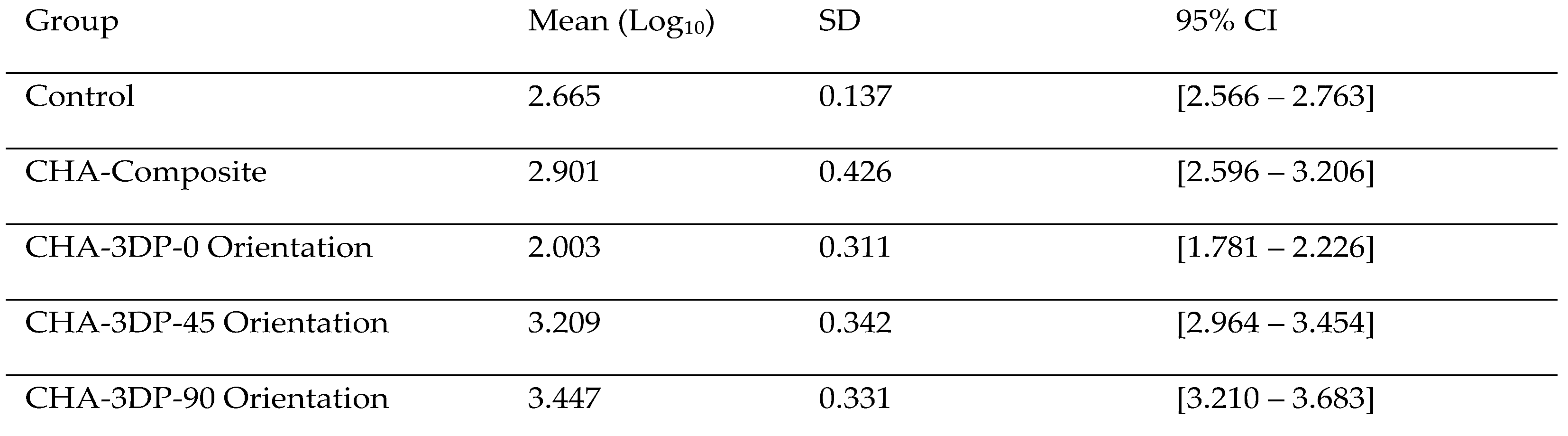 |
SD: Standard deviation; CI: Confidence Interval; CHA: Customized Healing Abutment.
Disclaimer/Publisher’s Note: The statements, opinions and data contained in all publications are solely those of the individual author(s) and contributor(s) and not of MDPI and/or the editor(s). MDPI and/or the editor(s) disclaim responsibility for any injury to people or property resulting from any ideas, methods, instructions or products referred to in the content. |
© 2026 by the authors. Licensee MDPI, Basel, Switzerland. This article is an open access article distributed under the terms and conditions of the Creative Commons Attribution (CC BY) license (http://creativecommons.org/licenses/by/4.0/).
Copyright: This open access article is published under a Creative Commons CC BY 4.0 license, which permit the free download, distribution, and reuse, provided that the author and preprint are cited in any reuse.