
Submitted:
07 August 2026
Posted:
07 August 2026
You are already at the latest version
Abstract
Background/Objectives: Post-traumatic nasal pyramid reconstruction is challenging in patients with severe deformity and exhausted autologous graft options, particularly after multiple failed surgeries. Custom-made titanium implants may offer precise structural support, but data on their mucosal integration and ultrastructural tissue response in nasal reconstruction are lacking. The aim of this study is to describe the first case of nasal pyramid reconstruction using an IPS (Individual Patient Solutions) custom-made titanium implant and to evaluate peri-implant tissue response with transmission electron microscopy (TEM). Methods: A 50-year-old male military personnel with long-standing saddle-nose deformity, nasal obstruction, and prior failed graft-based reconstructions underwent open rhinoplasty with placement of a patient-specific titanium implant designed with three-dimensional virtual surgical planning. The implant reconstructed the nasal dorsum from nasion to tip and was stabilized with cortical screws; an additional thin titanium plate was used for tip support. Intraoperative biopsies of nasal mucosa and adjacent connective tissue were processed for TEM to assess epithelial, stromal, vascular, and implant–tissue interface characteristics. Results: Clinically, nasal airway patency improved immediately after packing removal, with restoration of nasal contour and stable functional and aesthetic outcomes at 24-month follow-up without complications. Ultra-structurally, the respiratory epithelium showed preserved stratification, intact basal and ciliated cells, normal nuclei and mitochondria, and organized extracellular matrix closely opposed to discrete titanium particles, without granulomatous foreign-body reaction, dense inflammatory infiltrate, or fibrotic encapsulation. Focal ciliary damage, cytoplasmic vacuolization, lamina propria thickening, and vascular elastic lamina remodeling were interpreted as adaptive or transient stress-related changes rather than chronic implant toxicity. Conclusions: Custom-made IPS titanium implants can provide stable structural support, satisfactory functional and aesthetic outcomes, and a well-tolerated mucosal interface in complex post-traumatic nasal pyramid reconstruction after failed graft-based surgeries. Ultrastructural analysis with TEM supports their biocompatibility, showing predominantly preserved epithelial and stromal architecture with only focal, adaptive stress-related changes rather than chronic implant-related toxicity.
Keywords:
nasal fracture
; post-traumatic nasal deformity
; nasal pyramid reconstruction
; custom-made titanium implant
; patient-specific implant
; rhinoplasty
; transmission electron microscopy
; ultrastructure
; biocompatibility
; IPS implant
1. Introduction
1.1. Nasal Fracture Reconstruction
Nasal bone fractures are the most common facial skeletal injury, accounting for a significant proportion of trauma-related presentations and affecting both functional and aesthetic outcomes [1,2,3]. Epidemiological data indicate a bimodal age distribution, with the highest incidence predominantly in adolescents and young adults [3,4]. The clinical spectrum of nasal fractures is broad, encompassing simple non-displaced fractures to complex comminuted injuries with septal involvement, each presenting unique diagnostic and therapeutic challenges [2,5,6,7]. Traditional management has centered on closed reduction within two weeks of injury occurrence, which is cost-effective and generally yields satisfactory early results for simple fractures [1,5,6,8]. However, this technique is linked with a substantial rate of secondary deformity, particularly in cases with significant disruption or unfavorable fracture patterns, often necessitating subsequent open interventions [7,8,9]. Open reduction and structural reconstruction are preferred in delayed presentations or when rigid fixation and precise anatomical restoration are required, especially for severe bony deviations, L-strut deformities, and nasal valve compromise [5,8,9,12]. Contemporary systematic reviews and practice management guidelines have sought to standardize the acute and delayed management of these injuries and to reduce the incidence of residual deformity [10].
1.2. Personalized Medicine and Advances in the Current Era
Recent advances in personalized medicine and surgical technology have driven the development of custom-made titanium-based implants for structural craniofacial and nasal reconstruction [13,14,15]. Customized titanium-based implants provide the potential for enhanced stability, biocompatibility, and tailored anatomical corrections in complex or revision cases where conventional methods are not suitable [13,16]. The integration of imaging modalities, computer-assisted design, and additive manufacturing enables precise customization that addresses both functional and cosmetic deficits while minimizing the risk of recurrent deformity and airway obstruction [14,15]. Titanium is one of the most extensively studied biomaterials, with a long record of osseointegration and favorable soft-tissue response across dental, orthopedic, and craniofacial applications [17,18]. As the literature increasingly emphasizes individualized, technology-assisted approaches, custom titanium implants represent a promising frontier in the management of challenging nasal bone fractures and post-traumatic nasal pyramid defects.
2. Case Presentation
2.1. Patient Description
A 50-year-old male patient, military personnel with a history of comminuted nasal bone fractures at an early age, was referred to our clinic at the 424 General Military Hospital, Thessaloniki, Greece, due to nasal obstruction and significant post-traumatic nasal deformity. The patient had undergone prior failed nasal reconstructions in other institutions, utilizing autologous cartilage and bone graft techniques, which unfortunately led to melting and collapse of the nasal bridge, cartilaginous septum, and lateral cartilages.
A detailed clinical and three-dimensional (3D) CT evaluation was performed to assess breathing ability and residual nasal anatomy. Nasal obstruction was mainly due to internal nasal valve collapse [12]. Under-reduction of the nasal dorsal hump had led to significant saddle-nose deformity and nasal tip ptosis due to inadequate structural support [11]. The bony nasal septum and piriform apertures were intact. Furthermore, with the patient declining any further bone and cartilage harvests, reconstructive alternatives were very limited.
We proposed to the patient an innovative solution integrating computer-aided design and a three-dimensional custom-made titanium nasal prosthesis to reconstruct the nasal shape for functional and cosmetic purposes. A reliable reconstruction with a satisfying result was obtained, and the patient resumed his professional activities. No additional surgical procedures were required 24 months post-operatively.
2.2. Surgical Planning
Taking into consideration the patient’s surgical history, nasal reconstruction with an IPS (Individual Patient Solutions) titanium-based implant was proposed. Advantages, disadvantages, and possible complications were carefully explained. The patient underwent cone-beam computed tomography (CT) and plain radiographs so that the amount of available bone and cartilage and the appropriate location for the implant could be evaluated. All digital data were transferred to a DICOM (Digital Imaging and Communications in Medicine) file, which was then imported into the IPS Case Designer software (KLS Martin Manufacturing LLC, Florida, USA). With the help of 3D virtual surgical planning [14,15], a functionalized patient-specific implant was designed, meeting hard- and soft-tissue anatomical requirements. The material of choice for the implant was titanium, a highly biocompatible material [17,18] that had not previously been reported for use in nasal pyramid reconstruction. Titanium screw measurements were 1.2 mm in diameter and 7 mm in length.
2.3. Operative Setting
The procedure was performed at the 424 General Military Hospital of Thessaloniki, Greece. Following induction of general anesthesia and endotracheal intubation, an open rhinoplasty was carried out.
2.4. Surgical Approach
A mid-columellar inverted-V incision was made, which was then transitioned to marginal incisions with the help of skin hooks and sharp dissection (Figure 1). The full-thickness mucocutaneous flap was reflected superiorly to expose the bony and cartilaginous remains. The dome and dorsal part of the lower lateral cartilages, the dorsal part of the septal cartilage, and the upper lateral and sesamoid cartilages were missing.
2.5. Implant Placement
A periosteal elevator was used to dissect the periosteum over the nasal bones up to the nasofrontal angle. After preparation of the implant hole, the implant was placed in the midline, over the nasal bones, covering the distance between the nasion and the tip, forming an artificial nasal dorsum. Small incisions were made at the level of the inner canthus of the eyes bilaterally to ensure correct implant placement. Cortical screws of 1.5 mm × 5 mm were used to stabilize the implant on the frontal and residual nasal bones (Figure 2). To achieve adequate support of the tip, a thin titanium plate was used, anchored between the caudal free edge of the implant and the anterior caudal surface of the vomer bone (Figure 3). A symmetrical small wedge-shaped part of the lower lateral crura was excised bilaterally to create the desired nostril size and base width. Stabilization sutures were placed between the lower lateral cartilages to bring them together and contour the nasal tip. At the end, the mucocutaneous flap was repositioned, and the trans columellar and marginal incisions were closed with interrupted permanent (5-0 nylon) sutures. Adhesive steri-strips were applied horizontally from the nasofrontal angle to the supratip and a larger strip around the infratip lobule, to avoid postoperative edema and support the tip, respectively. A custom-made nasal splint was applied for postoperative stabilization. Finally, sterile petrolatum gauzes were packed in the nasal cavity. A CT scan was obtained postoperatively to ensure the proper position of the nasal implant (Figure 4 and Figure 5).
2.6. Postoperative Recovery
Total hospitalization lasted seven days. Gauzes were removed after five days, while the splint and skin sutures were removed after ten days. As soon as the nasal packing was removed, the patient reported recession of upper airway breathing difficulties and return of normal nasal breathing. Regarding the postoperative cosmetic outcome, the nasal structure was restored without post-traumatic signs, which provided appropriate rehabilitation to improve the patient’s quality of life. At the 6-month follow-up evaluation, during which the patient exhibited no complaints, he was satisfied with the final result.
2.7. Tissue Sampling for Ultrastructural Analysis
Biopsy specimens were collected from the nasal mucosa and connective tissue adjacent to the implant site two years (24 months) after placement of the titanium implant. The tissue sample preparation is further explained in section 3.1.
3. Materials and Methods
3.1. Tissue Sample Collection and Preparation for Transmission Electron Microscopy (TEM)
For the tissue sampling the following procedure was performed:
- The samples were immediately immersed in 2.5% glutaraldehyde in 0.1 M phosphate buffer (pH 7.4) at 4 °C for primary fixation.
- Following rinsing in phosphate buffer, tissues were post-fixed in 1% osmium tetroxide for 1 h to preserve membrane integrity and enhance contrast.
- The specimens were then dehydrated via a graded ethanol series (30%, 50%, 70%, 90%, and 100%) and cleared in propylene oxide.
- For embedding, the samples were infiltrated and embedded in epoxy resin (Epon/Araldite mixture), which was polymerized at 60 °C for 48 h.
- Ultrathin sections (70–90 nm) were cut using an ultramicrotome equipped with a diamond knife and mounted on copper grids. Sections were stained with uranyl acetate and lead citrate to enhance visualization of subcellular structures.
- The prepared grids were examined by TEM operating at 80 to 120 kV.
- Images were captured digitally at varying magnifications to evaluate cellular and subcellular morphology, including nuclei, mitochondria, microvilli, vacuoles, and connective tissue organization.
- Scale bars were included in all micrographs to provide dimensional references.
4. Results
4.1. Ultrastructural Findings
Transmission electron microscopy of the nasal mucosa and underlying connective tissue adjacent to the IPS custom-made titanium implant demonstrated predominantly preserved tissue architecture with focal adaptive and stress-related changes across all examined fields (Figure 6, Figure 7, Figure 8, Figure 9, Figure 10 and Figure 11). In high-magnification views, the lamina propria and intercellular substance appeared thickened, yet the respiratory epithelium maintained an orderly stratification with intact basal and ciliated cells, indicating structural integrity despite local remodeling. Epithelial cells contained numerous secondary lysosomes and phagosomes, reflecting active intracellular processing of extracellular material and suggesting ongoing metabolic adaptation at the peri-implant interface rather than overt cytotoxic damage (Figure 6 and Figure 9).
Vascular structures in the vicinity of the implant showed ultrastructural features compatible with remodeling. Sections of small vessels exhibited pronounced thickening of the internal and external elastic laminae, together with expansion of intercellular spaces within the vessel wall, implying altered mechanical load and increased permeability during the healing phase (Figure 7). These changes were not accompanied by endothelial necrosis or fibrinoid degeneration, supporting a controlled adaptive response rather than vasculitic damage. At the implant–tissue interface, discrete titanium particles were observed embedded within an organized extracellular matrix, with collagen fibers and ground substance closely opposed to the implant surface [19,20]. Notably, there was no evidence of granulomatous foreign-body reaction, dense inflammatory infiltrate, or fibrotic encapsulation, pointing to a stable and biocompatible contact zone (Figure 8).
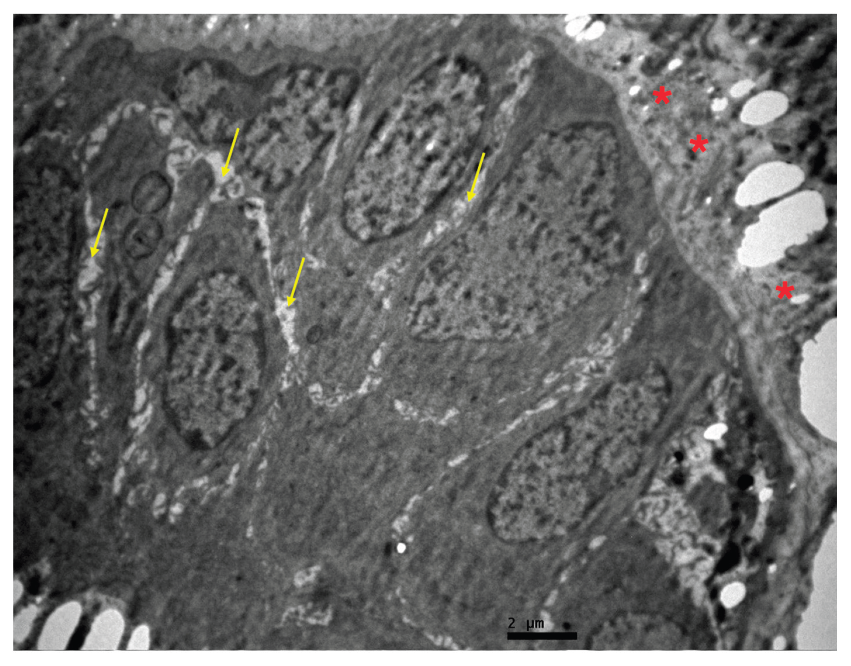
Several fields demonstrated completely preserved lamina propria without pathological lesions, and epithelial cells showed normal nuclei, mitochondria, and cell membranes, consistent with sustained cellular viability. In contrast, localized areas displayed features compatible with transient stress: apical ciliary damage or loss in ciliated columnar cells, cytoplasmic vacuolization, and intercellular oedema, most pronounced at the level of the basal lamina (Figure 9 and Figure 10). These alterations were focal, did not extend throughout the mucosa, and were not associated with widespread apoptosis or necrosis, suggesting reversible injury related to surgical manipulation or early mechanical loading rather than chronic implant toxicity. Overall, the ultrastructural profile across all images supports a predominantly non-inflammatory, well-tolerated interface between the custom titanium implant and adjacent nasal tissues (Figure 10 and Figure 11).
5. Discussion
Post-traumatic nasal reconstruction presents unique challenges, especially in cases with prior failed grafts and compromised soft-tissue integrity [9,11]. In this first reported case of nasal pyramid reconstruction using a custom-made titanium implant, we observed not only favorable clinical outcomes but also compelling ultrastructural evidence of implant biocompatibility and tissue adaptation.
The expanded ultrastructural assessment of the nasal mucosa and peri-implant connective tissue reinforces the clinical impression that the IPS custom-made titanium implant achieves a stable and biocompatible integration in the nasal pyramid. The coexistence of preserved epithelial and stromal architecture, normal organellar morphology, and organized extracellular matrix deposition around titanium particles is in line with the expected behavior of titanium in craniofacial and nasal applications, where it is known to support long-term tissue compatibility without provoking chronic inflammation [13,17,18,21]. The predominance of secondary lysosomes, scattered vacuoles, and limited connective tissue distortion is more suggestive of physiological turnover and adaptive remodeling than of destructive degeneration, particularly in a setting of complex post-traumatic reconstruction and prior failed grafts.
The vascular changes observed-elastic lamina thickening and increased intercellular spacing-can be interpreted as manifestations of vascular remodeling in response to modified mechanical and hemodynamic conditions created by the implant and reconstructed nasal framework. Importantly, these alterations occurred without ultrastructural hallmarks of vasculitis or thrombosis, which supports their adaptive rather than pathological nature. Similarly, focal ciliary damage, microvilli attenuation, and intercellular edema at the epithelial–stromal interface likely reflect transient insult due to surgical elevation of mucosal flaps, periosteal dissection, and early implant loading—phenomena also described in other titanium implant settings where long-term mucosal and osseous integrity is ultimately preserved [18,19].
The absence of necrosis, extensive apoptosis, granulomatous foreign-body reaction, or dense lymphohistiocytic infiltrates across all examined images is particularly reassuring, as these findings would typically be expected in cases of titanium intolerance, surface contamination, or implant-related infection [20]. Coupled with the patient’s complete functional recovery, lack of infectious or inflammatory complications, and stability of the reconstruction at 24-month follow-up, the ultrastructural data strongly support the concept of a harmonious tissue–implant interface in this novel indication. From a reconstructive standpoint, this case suggests that custom-made titanium implants can provide durable structural support and mucosal compatibility in patients with severe post-traumatic deformity, limited autologous graft reserves, or multiple prior failures [13,16,21]. Future investigations with larger series and integration of ultrastructural analysis, immunohistochemistry, and molecular markers of inflammation and remodeling will be essential to define long-term safety, optimize implant design, and refine patient selection criteria for personalized nasal pyramid reconstruction using titanium implants.
This case not only expands the reconstructive options for patients with limited autologous graft availability but also sets a precedent for integrating advanced imaging modalities into postoperative evaluation. Subsequent research could be enhanced by connecting ultrastructural observations with molecular indicators of tissue integration and stress response, thereby further refining the criteria for implant selection and design.
6. Conclusions
In conclusion, the findings of this report indicate that a custom-made titanium implant provides a successful nasal pyramid reconstruction in a patient with severe post-traumatic deformity and failed prior grafts. Clinical outcomes and ultrastructural evaluation confirmed biocompatibility, stability, and long-term safety. Titanium implants may represent a novel solution for challenging nasal reconstructions, expanding the reconstructive armamentarium.
Author Contributions
Conceptualization, S.T., I.C. and T.P.; methodology, I.C., S.T. and S.K.; investigation, S.T., I.C., I.K., A.L., K.P and V.P.; resources, S.T., V.T., K.P and G.-A.S.; data curation, S.T., S.K. and A.S.; writing-original draft preparation, S.T. and S.K.; writing—review and editing, S.T., I.C., A.S., K.P. and T.P.; visualization, S.T..; supervision, T.P. and I.C. All authors have read and agreed to the published version of the manuscript.
Funding
This research received no external funding.
Informed Consent Statement
Written informed consent has been obtained from the patient to publish this paper, including all accompanying images.
Data Availability Statement
The data presented in this study are available on request from the corresponding author. The data are not publicly available due to patient privacy restrictions.
Conflicts of Interest
The authors declare no conflicts of interest.
Abbreviations
The following abbreviations are used in this manuscript:
| CAD | Computer-Aided Design |
| CT | Computed Tomography |
| DICOM | Digital Imaging and Communications in Medicine |
| IPS | Individual Patient Solutions |
| TEM | Transmission Electron Microscopy |
| VSP | Virtual Surgical Planning |
References
- Mondin, V.; Rinaldo, A.; Ferlito, A. Management of Nasal Bone Fractures. Am. J. Otolaryngol. 2005, 26, 181–185. [Google Scholar] [CrossRef] [PubMed]
- Hwang, K.; Yoon, J.M. Analysis of Nasal Bone Fractures: A 17-Year Study of 3785 Patients. J. Craniofac. Surg. 2023, 34, e757–e759. [Google Scholar] [CrossRef] [PubMed]
- Dong, S.X.; Shah, N.; Gupta, A. Epidemiology of Nasal Bone Fractures. Facial Plast. Surg. Aesthet. Med. 2022, 24, 27–33. [Google Scholar] [CrossRef] [PubMed]
- Marston, A.P.; O’Brien, E.K.; Hamilton, G.S. Nasal Injuries in Sports. Clin. Sports Med. 2017, 36, 337–353. [Google Scholar] [CrossRef] [PubMed]
- Hollins, A.; Pyfer, B.; Breeze, J.; Zhang, G.; Lohmeier, S.J.; Powers, D.B. Closed Reduction of Nasoseptal Fractures: Key Concepts for Predictable Results. Br. J. Oral Maxillofac. Surg. 2023, 61, 344–350. [Google Scholar] [CrossRef] [PubMed]
- James, J.G.; Izam, A.S.; Nabil, S.; Rahman, N.A.; Ramli, R. Closed and Open Reduction of Nasal Fractures. J. Craniofac. Surg. 2020, 31, e22–e26. [Google Scholar] [CrossRef] [PubMed]
- Kim, S.W.; Hong, J.P.; Min, W.K.; Seo, D.W.; Chung, Y.K. Accurate, Firm Stabilization Using External Pins: A Proposal for Closed Reduction of Unfavorable Nasal Bone Fractures and Their Simple Classification. Plast. Reconstr. Surg. 2002, 110, 1240–1246. [Google Scholar] [CrossRef] [PubMed]
- Trujillo, O.; Lee, C. Nasal Fractures: Acute, Subacute, and Delayed Management. Otolaryngol. Clin. North Am. 2023, 56, 1089–1099. [Google Scholar] [CrossRef] [PubMed]
- Arnold, M.A.; Yanik, S.C.; Suryadevara, A.C. Septal Fractures Predict Poor Outcomes after Closed Nasal Reduction: Retrospective Review and Survey. Laryngoscope 2019, 129, 1784–1790. [Google Scholar] [CrossRef] [PubMed]
- Paliwoda, E.D.; Newman-Plotnick, H.; Buzzetta, A.J.; Post, N.K.; LaClair, J.R.; Trandafirescu, M.; Gildener-Leapman, N.; Kpodzo, D.S.; Edwards, K.; Tafen, M.; et al. Acute Management of Nasal Bone Fractures: A Systematic Review and Practice Management Guideline. Am. Surg. 2026, 92, 238–245. [Google Scholar] [CrossRef] [PubMed]
- Gadkaree, S.K.; Weitzman, R.E.; Fuller, J.C.; Justicz, N.; Gliklich, R.E. Review of Literature of Saddle Nose Deformity Reconstruction and Presentation of Vomer Onlay Graft. Laryngoscope Investig. Otolaryngol. 2020, 5, 1039–1043. [Google Scholar] [CrossRef] [PubMed]
- Sinkler, M.A.; Wehrle, C.J.; Elphingstone, J.W.; Magidson, E.; Ritter, E.F.; Brown, J.J. Surgical Management of the Internal Nasal Valve: A Review of Surgical Approaches. Aesthetic Plast. Surg. 2021, 45, 1127–1136. [Google Scholar] [CrossRef] [PubMed]
- Toso, S.M.; Menzel, K.; Motzkus, Y.; Adolphs, N.; Hoffmeister, B.; Raguse, J.D. Patient-Specific Implant in Prosthetic Craniofacial Reconstruction: First Report of a Novel Technique with Far-Reaching Perspective. J. Craniofac. Surg. 2015, 26, 2133–2135. [Google Scholar] [CrossRef] [PubMed]
- Vyas, K.; Gibreel, W.; Mardini, S. Virtual Surgical Planning (VSP) in Craniomaxillofacial Reconstruction. Facial Plast. Surg. Clin. North Am. 2022, 30, 239–253. [Google Scholar] [CrossRef] [PubMed]
- Ahmed, K.S.; Ibad, H.; Suchal, Z.A.; Gosain, A.K. Implementation of 3D Printing and Computer-Aided Design and Manufacturing (CAD/CAM) in Craniofacial Reconstruction. J. Craniofac. Surg. 2022, 33, 1714–1719. [Google Scholar] [CrossRef] [PubMed]
- Haroun, F.; Benmoussa, N.; Bidault, F.; Lassau, N.; Moya-Plana, A.; Leymarie, N.; Honart, J.F.; Kolb, F.; Qassemyar, Q.; Gorphe, P. Outcomes of Mandibular Reconstruction Using Three-Dimensional Custom-Made Porous Titanium Prostheses. J. Stomatol. Oral Maxillofac. Surg. 2023, 124, 101281. [Google Scholar] [CrossRef] [PubMed]
- Guglielmotti, M.B.; Olmedo, D.G.; Cabrini, R.L. Research on Implants and Osseointegration. Periodontol. 2000 2019, 79, 178–189. [Google Scholar] [CrossRef] [PubMed]
- Wennerberg, A.; Albrektsson, T. Effects of Titanium Surface Topography on Bone Integration: A Systematic Review. Clin. Oral Implant. Res. 2009, 20 (Suppl. 4), 172–184. [Google Scholar] [CrossRef] [PubMed]
- Palmquist, A.; Grandfield, K.; Norlindh, B.; Mattsson, T.; Brånemark, R.; Thomsen, P. Bone–Titanium Oxide Interface in Humans Revealed by Transmission Electron Microscopy and Electron Tomography. J. R. Soc. Interface 2012, 9, 396–400. [Google Scholar] [CrossRef] [PubMed]
- Mombelli, A.; Hashim, D.; Cionca, N. What Is the Impact of Titanium Particles and Biocorrosion on Implant Survival and Complications? A Critical Review. Clin. Oral Implant. Res. 2018, 29 (Suppl. 18), 37–53. [Google Scholar] [CrossRef] [PubMed]
- Cabraja, M.; Klein, M.; Lehmann, T.N. Long-Term Results Following Titanium Cranioplasty of Large Skull Defects. Neurosurg. Focus 2009, 26, E10. [Google Scholar] [CrossRef] [PubMed]
Figure 1.
Intraoperative view demonstrating the open rhinoplasty approach through a mid-columellar inverted-V incision extended bilaterally into marginal incisions, allowing exposure of the nasal framework.
Figure 1.
Intraoperative view demonstrating the open rhinoplasty approach through a mid-columellar inverted-V incision extended bilaterally into marginal incisions, allowing exposure of the nasal framework.
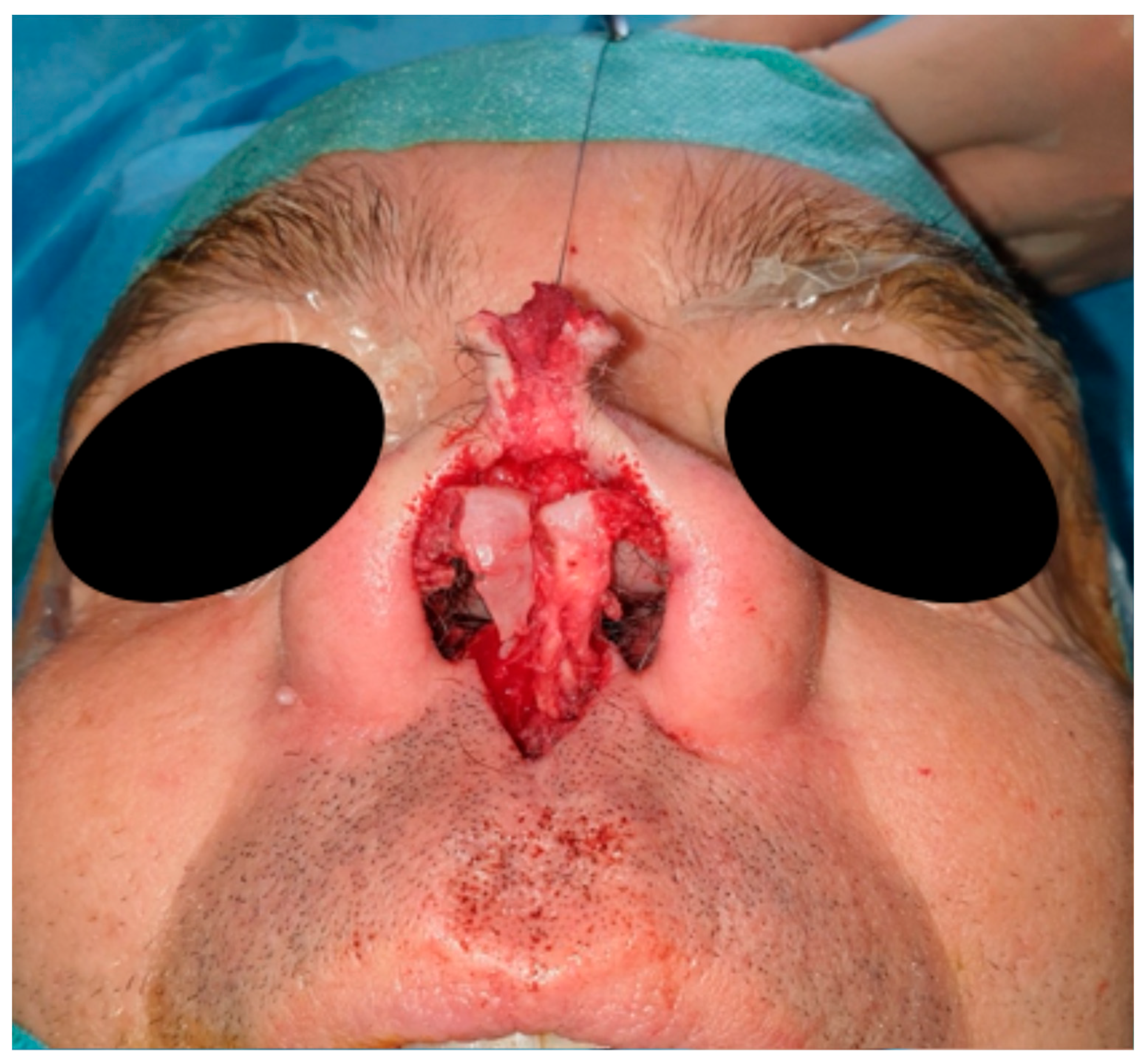
Figure 2.
Intraoperative placement and fixation of the patient-specific nasal implant along the midline, extending from the nasion to the nasal tip. The implant was stabilized using 1.5 mm × 5 mm cortical screws anchored to the frontal and residual nasal bones.
Figure 2.
Intraoperative placement and fixation of the patient-specific nasal implant along the midline, extending from the nasion to the nasal tip. The implant was stabilized using 1.5 mm × 5 mm cortical screws anchored to the frontal and residual nasal bones.
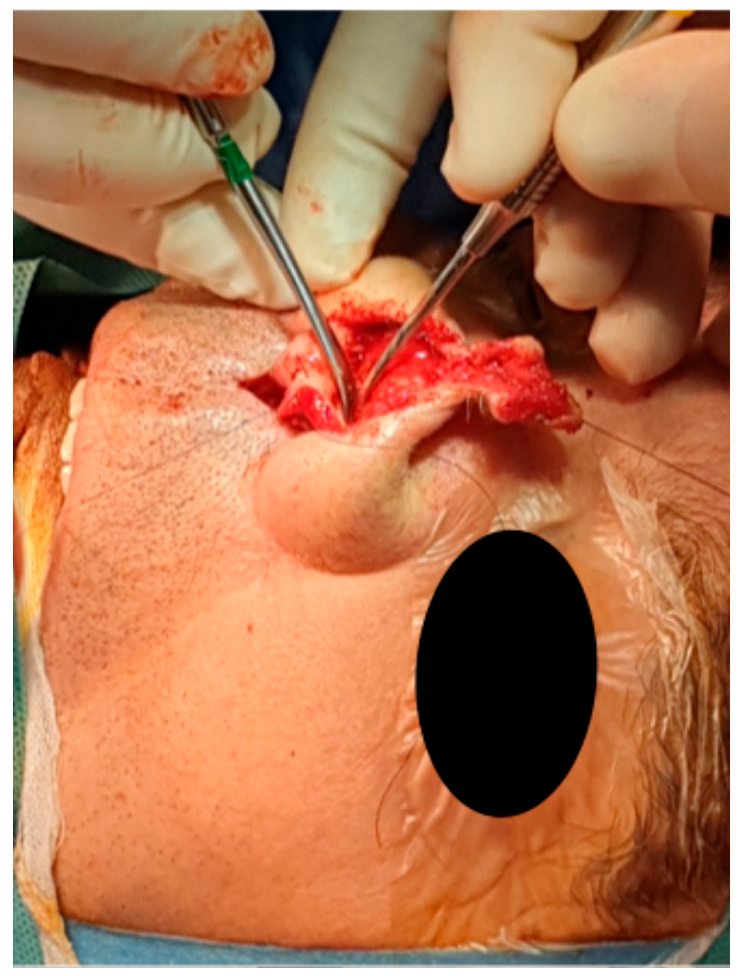
Figure 3.
Intraoperative view of the titanium plate used to provide additional support to the nasal tip, anchored between the caudal edge of the implant and the anterior caudal surface of the vomer bone.
Figure 3.
Intraoperative view of the titanium plate used to provide additional support to the nasal tip, anchored between the caudal edge of the implant and the anterior caudal surface of the vomer bone.
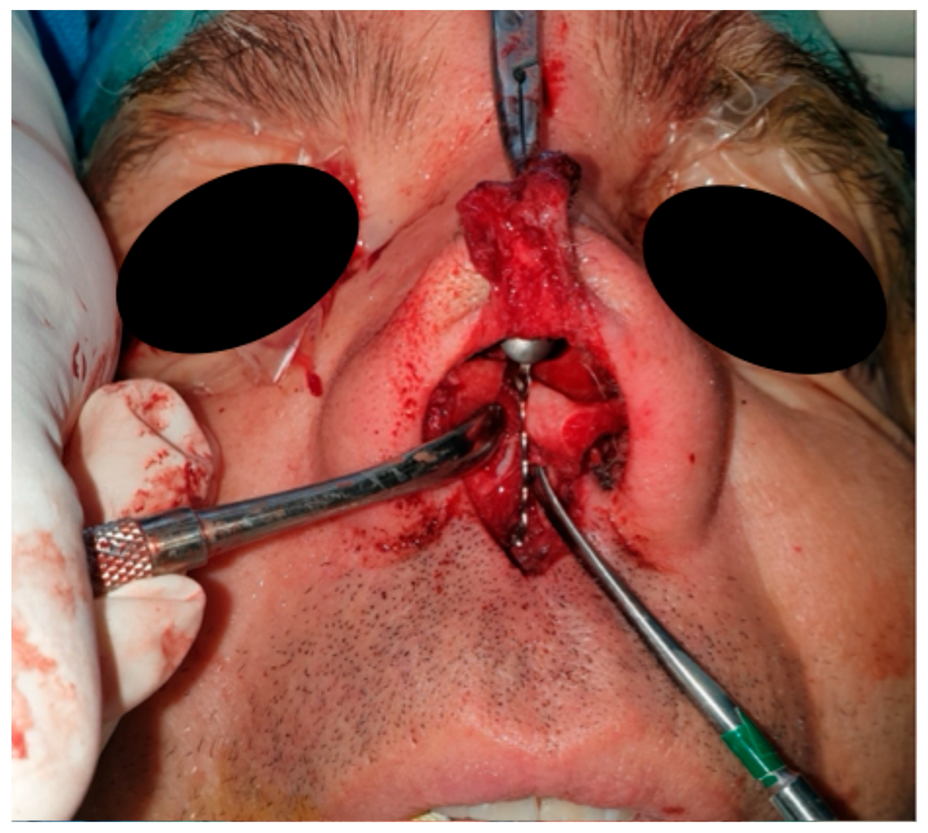
Figure 4.
Postoperative computed tomography scan (CT, frontal aspect) demonstrating appropriate positioning and fixation of the nasal implant.
Figure 4.
Postoperative computed tomography scan (CT, frontal aspect) demonstrating appropriate positioning and fixation of the nasal implant.
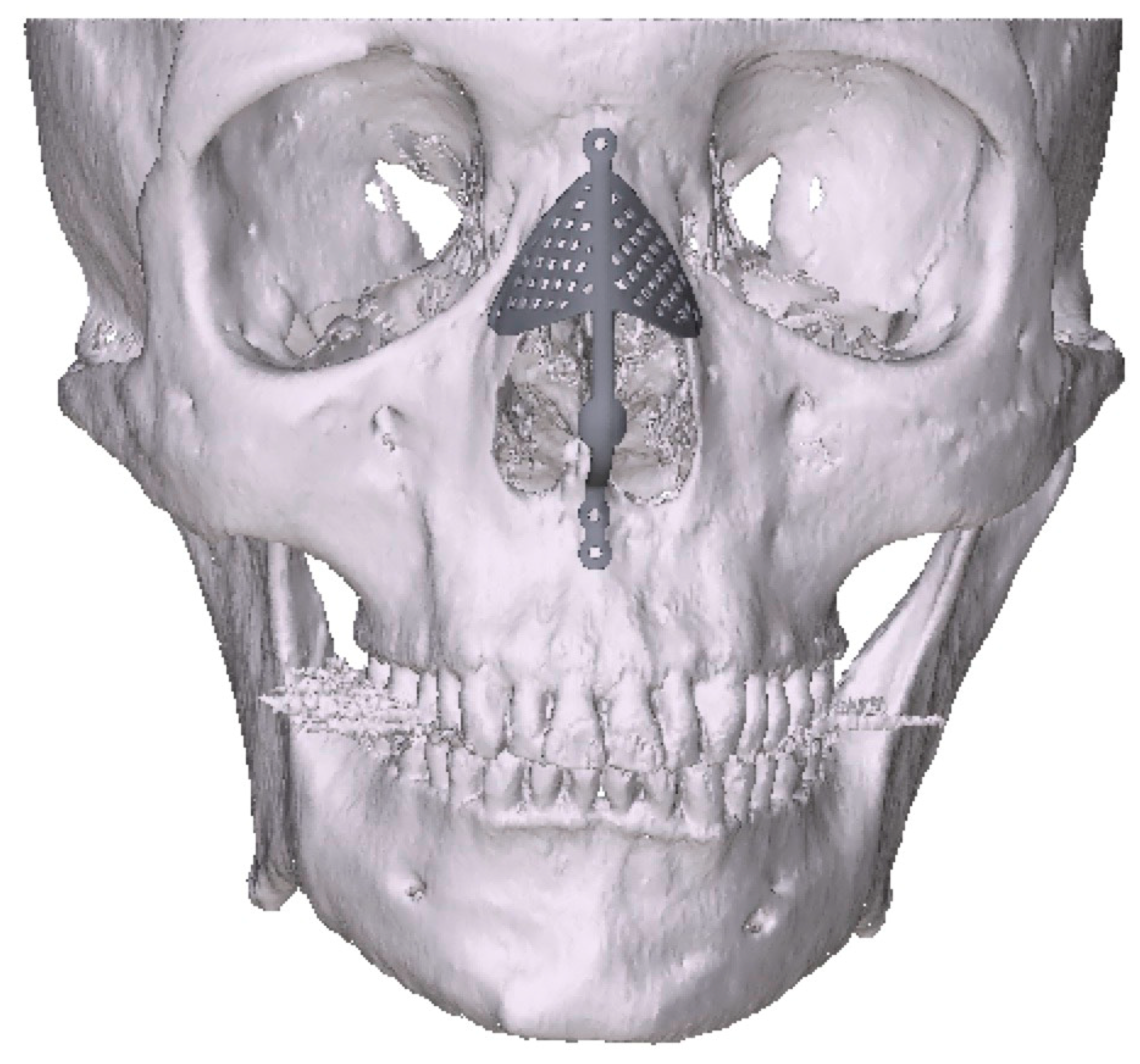
Figure 5.
Postoperative computed tomography scan (CT, lateral aspect) confirming correct alignment and stable positioning of the nasal implant and supporting titanium plate.
Figure 5.
Postoperative computed tomography scan (CT, lateral aspect) confirming correct alignment and stable positioning of the nasal implant and supporting titanium plate.
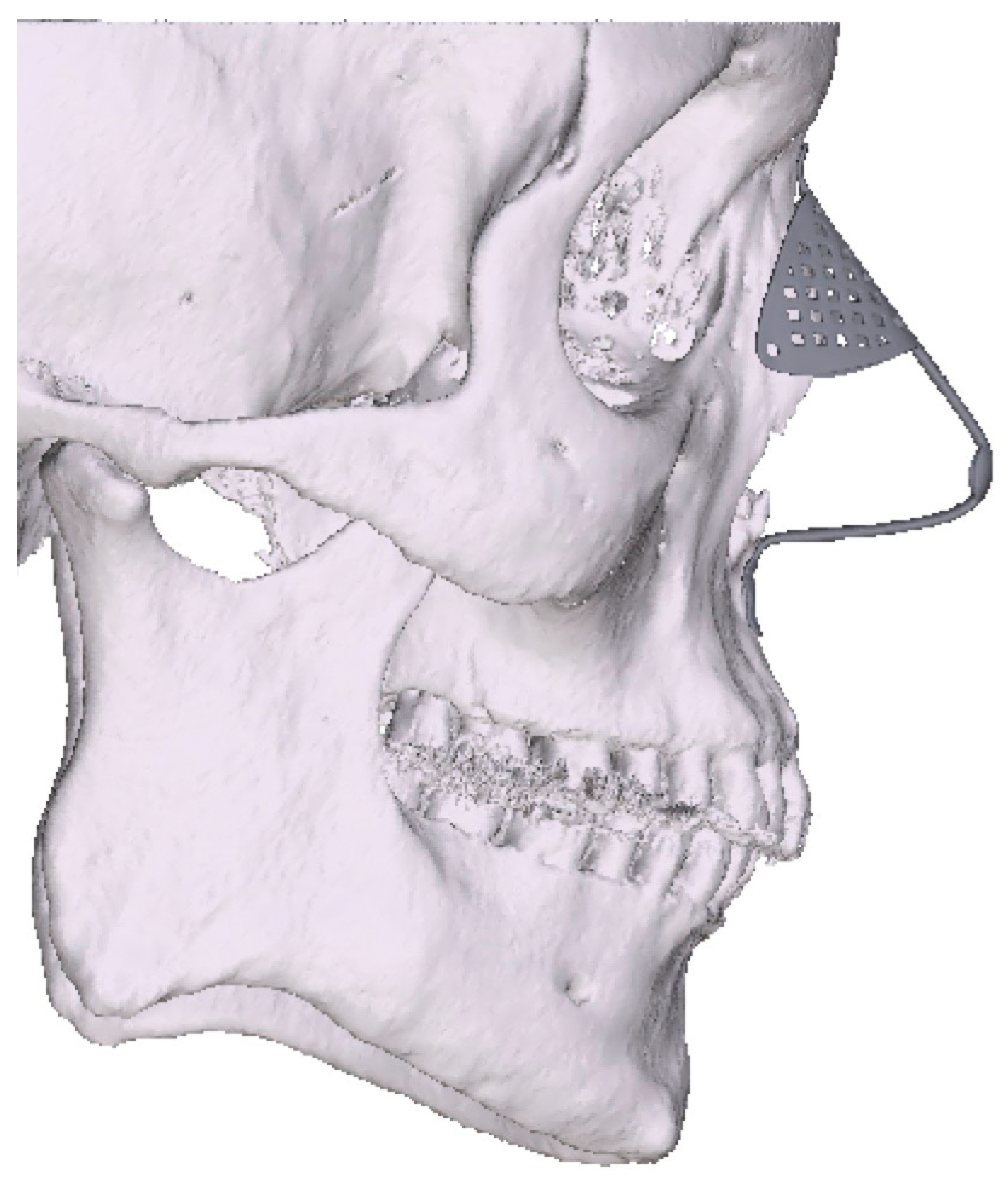
Figure 6.
Transmission electron microscopy image of the nasal mucosa adjacent to the titanium implant. Thickening of the lamina propria (∗) and the intercellular substance (↑) is observed, with preservation of the architecture of the adjacent epithelium. Secondary lysosomes or phagosomes (∗) are present in the epithelial cells. ×12,000.
Figure 6.
Transmission electron microscopy image of the nasal mucosa adjacent to the titanium implant. Thickening of the lamina propria (∗) and the intercellular substance (↑) is observed, with preservation of the architecture of the adjacent epithelium. Secondary lysosomes or phagosomes (∗) are present in the epithelial cells. ×12,000.
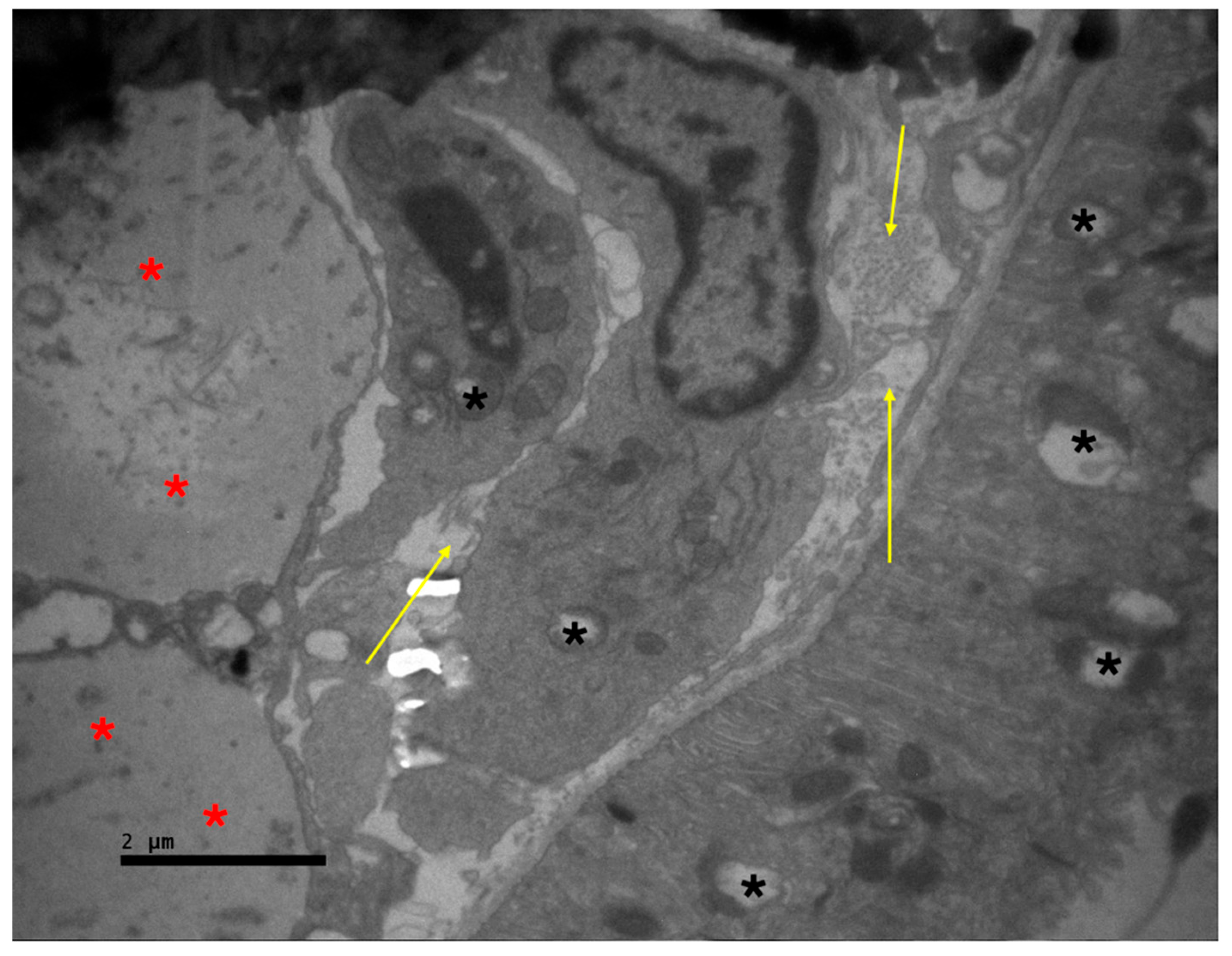
Figure 7.
Transmission electron microscopy image of the nasal mucosa adjacent to the titanium implant. Section of a vessel in the center with increased thickness of the internal (↑) and external elastic laminae (↑). Increased intercellular space in the wall of the vessel is also observed (∗). ×3000.
Figure 7.
Transmission electron microscopy image of the nasal mucosa adjacent to the titanium implant. Section of a vessel in the center with increased thickness of the internal (↑) and external elastic laminae (↑). Increased intercellular space in the wall of the vessel is also observed (∗). ×3000.
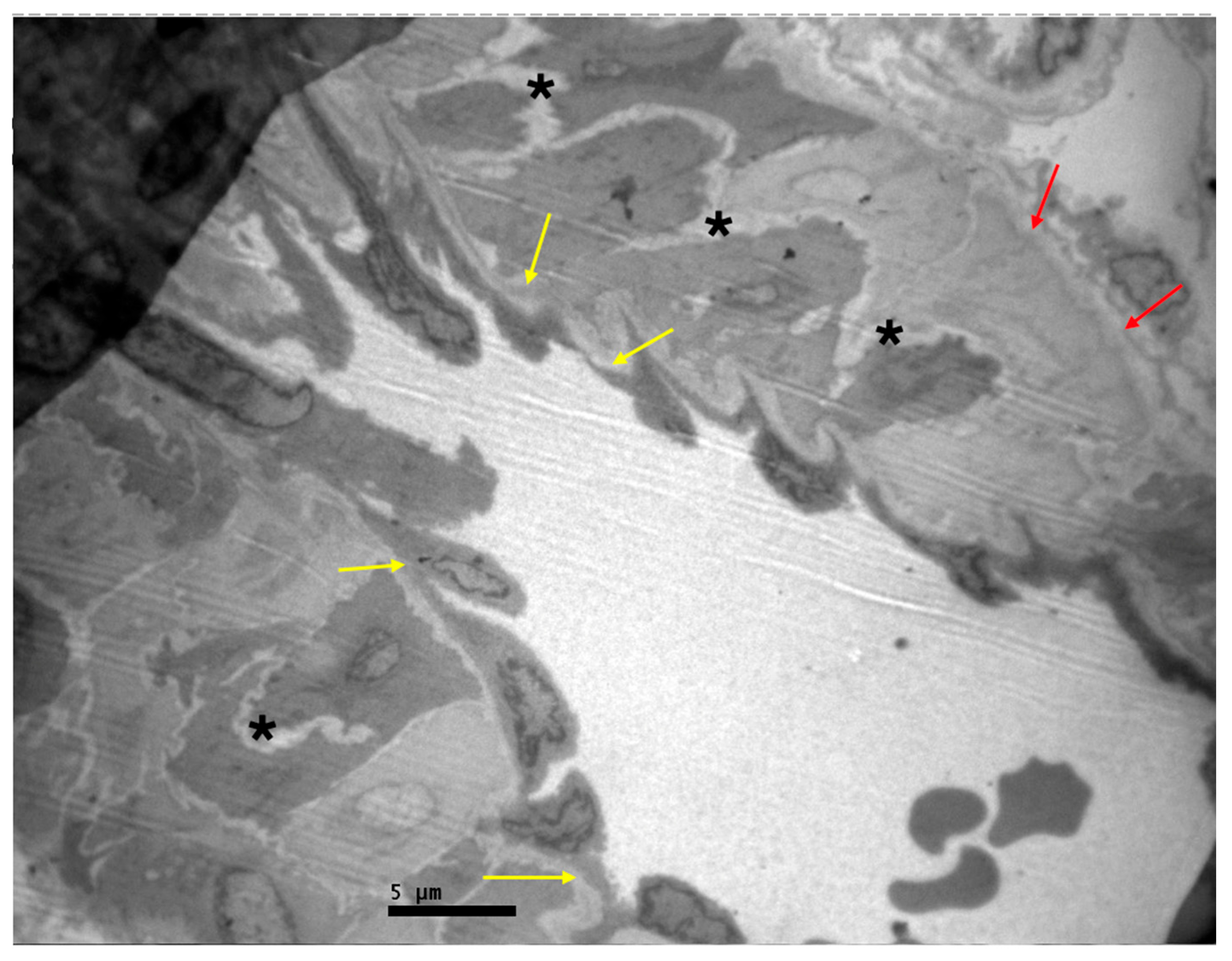
Figure 8.
Transmission electron microscopy image of the nasal mucosa adjacent to the titanium implant. Particles of the implant (∗) are observed, surrounded by extracellular matrix (∗). ×3000.
Figure 8.
Transmission electron microscopy image of the nasal mucosa adjacent to the titanium implant. Particles of the implant (∗) are observed, surrounded by extracellular matrix (∗). ×3000.
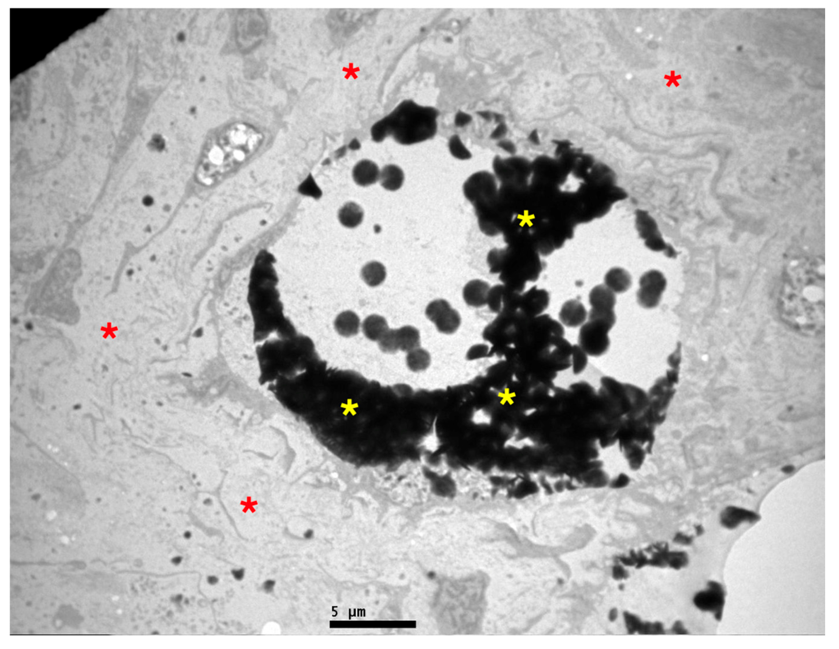
Figure 9.
Transmission electron microscopy image of the nasal mucosa adjacent to the titanium implant. The lamina propria (↑) appears without pathological lesions. The basal cells of the epithelium (∗) are presented with normal architecture, as are the ciliated cells (∗). ×4000.
Figure 9.
Transmission electron microscopy image of the nasal mucosa adjacent to the titanium implant. The lamina propria (↑) appears without pathological lesions. The basal cells of the epithelium (∗) are presented with normal architecture, as are the ciliated cells (∗). ×4000.
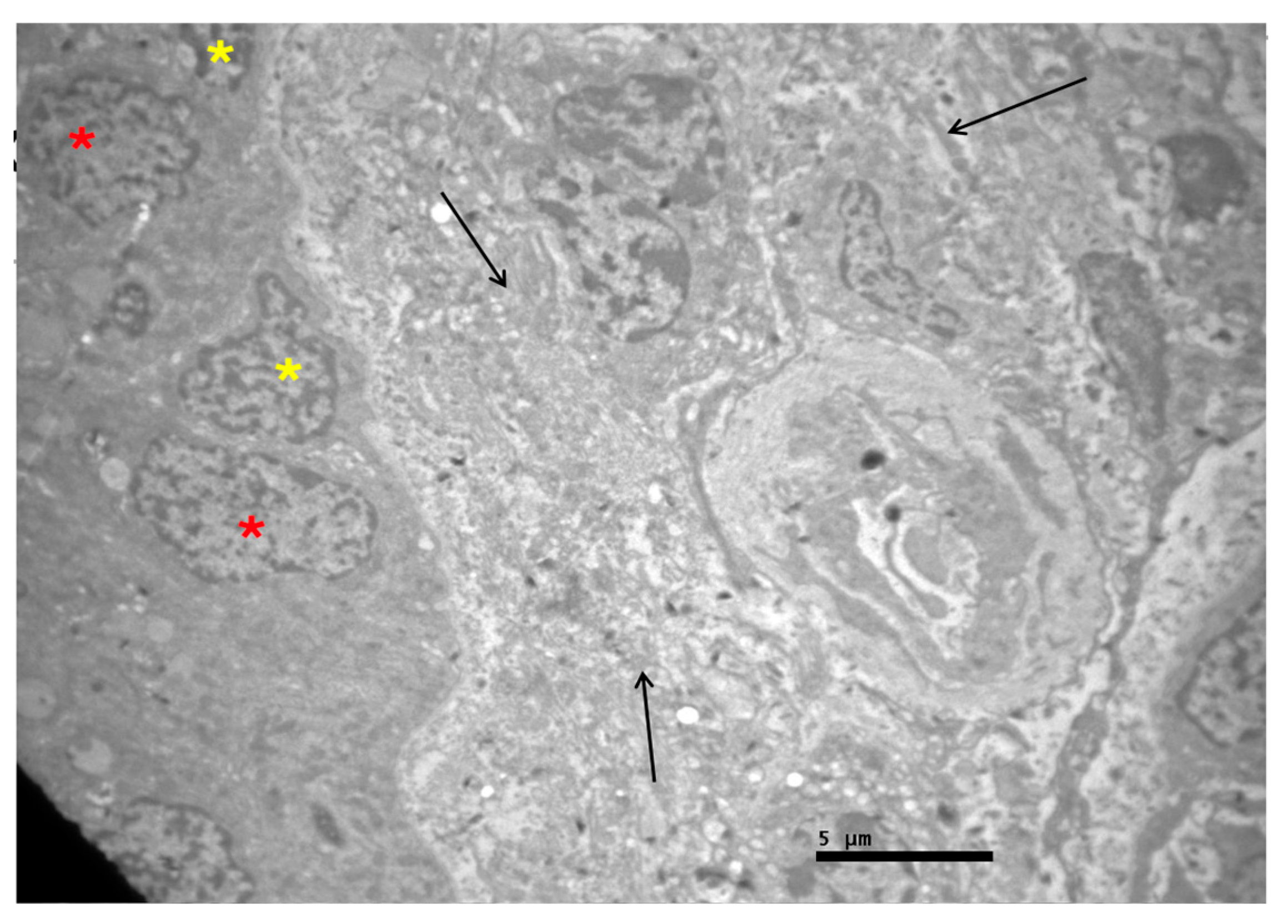
Figure 10.
Transmission electron microscopy image of the nasal mucosa adjacent to the titanium implant. Respiratory epithelium with damaged cilia at the apical surface of the ciliated columnar cells (↑). Intercellular oedema is also observed between the epithelial cells (∗). ×4000.
Figure 10.
Transmission electron microscopy image of the nasal mucosa adjacent to the titanium implant. Respiratory epithelium with damaged cilia at the apical surface of the ciliated columnar cells (↑). Intercellular oedema is also observed between the epithelial cells (∗). ×4000.
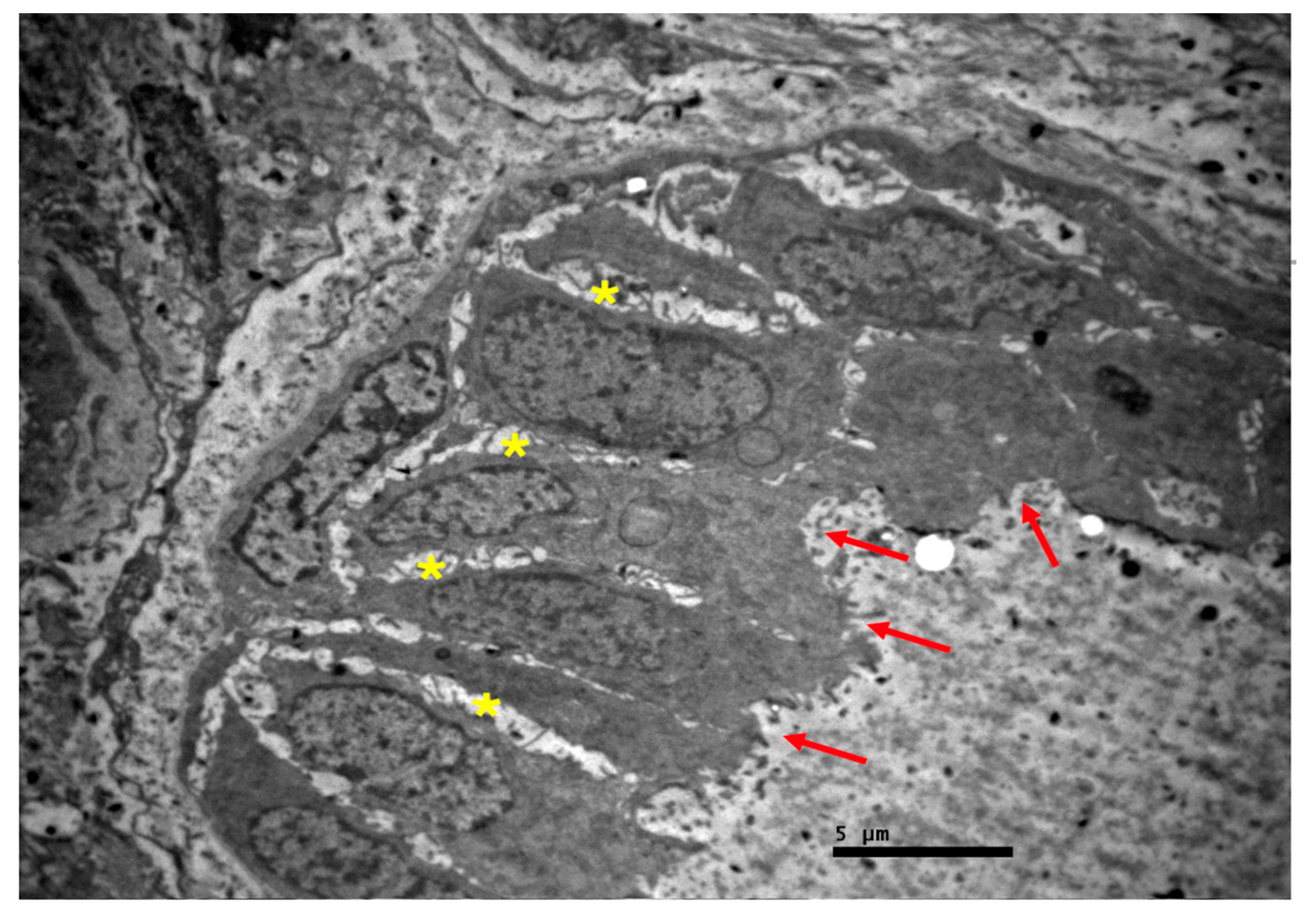
Figure 11.
Transmission electron microscopy image of the nasal mucosa adjacent to the titanium implant. Respiratory epithelium with intercellular oedema (↑), especially an increase of the intercellular space at the site of the basal lamina (∗). ×6000.
Figure 11.
Transmission electron microscopy image of the nasal mucosa adjacent to the titanium implant. Respiratory epithelium with intercellular oedema (↑), especially an increase of the intercellular space at the site of the basal lamina (∗). ×6000.
Disclaimer/Publisher’s Note: The statements, opinions and data contained in all publications are solely those of the individual author(s) and contributor(s) and not of MDPI and/or the editor(s). MDPI and/or the editor(s) disclaim responsibility for any injury to people or property resulting from any ideas, methods, instructions or products referred to in the content. |
© 2026 by the authors. Licensee MDPI, Basel, Switzerland. This article is an open access article distributed under the terms and conditions of the Creative Commons Attribution (CC BY) license (http://creativecommons.org/licenses/by/4.0/).
Copyright: This open access article is published under a Creative Commons CC BY 4.0 license, which permit the free download, distribution, and reuse, provided that the author and preprint are cited in any reuse.


