

Submitted:
06 August 2026
Posted:
07 August 2026
You are already at the latest version
Abstract
Leishmaniasis is a widely distributed vector-borne neglected tropical disease for which current therapeutic options remain suboptimal because of toxicity, emerging drug resistance, and limited induction of long-term protective immunity. Immunotherapeutic approaches that enhance host protective immune responses therefore represent an attractive alternative strategy. In this study, we evaluated the immunotherapeutic efficacy of recombinant Leishmania (L.) infantum eukaryotic initiation factor (LieIF) in BALB/c murine models of cutaneous (Leishmania (L.) major) and visceral (Leishmania (L.) infantum) leishmaniasis. Mice received recombinant LieIF after infection, and disease progression, parasite burden, humoral and cellular immune responses and immune-related gene expression were assessed. LieIF treatment significantly attenuated lesion progression in cutaneous leishmaniasis and reduced parasite burden in visceral disease model. Therapeutic efficacy was associated with enhanced Th1-associated immune responses, including increased frequencies of IFN-γ-producing CD4⁺ lymphocytes, increased IL-12-producing cells, and elevated expression of the Th1-associated genes (transcription factor Tbx21 and IFN-γ). In the cutaneous model, treatment also reduced Leishmania-specific IgG1 responses, whereas antibody isotype responses were largely unchanged in visceral leishmaniasis, indicating that protection was primarily associated with cellular rather than humoral immune modulation. The demonstration of therapeutic efficacy in two distinct experimental models highlights the broad immunomodulatory capacity of LieIF. Collectively, these findings identify LieIF as a promising host-directed immunotherapeutic candidate for leishmaniasis and provide a strong rationale for further studies to optimize treatment regimens, elucidate its mechanisms of action, and evaluate its efficacy in combination with conventional antileishmanial chemotherapy.
Keywords:
leishmaniasis
; Leishmania infantum eukaryotic initiation factor (LieIF)
; host-directed therapy
; antiparasitic immunotherapy
; Th1-type immune response
; immunomodulation
1. Introduction
Leishmaniasis is a complex vector-borne disease caused by digenetic protozoan parasites of the Leishmania genus (family Trypanosomatidae) and represents a major public health concern in approximately one hundred endemic countries across five continents [1]. The disease encompasses a broad spectrum of clinical manifestations, ranging from localized, self-healing cutaneous lesions to disseminated mucosal disease and life-threatening visceral form, with disease outcome determined by the infecting Leishmania species and host immune response [1,2]. Leishmania major is a well-established model organism for cutaneous leishmaniasis (CL), whereas Leishmania infantum is the principal causative agent of visceral leishmaniasis (VL) in the Mediterranean basin and other endemic regions [1,3].
Current chemotherapy relies on pentavalent antimonials, liposomal amphotericin B, miltefosine, and paramomycin [4,5]. However, the use of these chemotherapeutic options is limited by significant toxicity, prolonged treatment regimens, high cost, variable efficacy, and the emergence of drug-resistant parasite strains [6,7,8]. Importantly, conventional antileishmanial chemotherapy does not induce long-lasting protective immunity, resulting in frequent relapse or reinfection [4,5,9]. These limitations highlight the urgent need for alternative therapeutic approaches, including immunotherapy and host-directed interventions.
Immunotherapy aims to modulate the host immune system to enhance protective immune responses and improve disease control, either alone or in combination with conventional antimicrobial treatments. This can be achieved with immunomodulatory agents and/or specially formulated Leishmania antigens. Protective immunity against Leishmania infection is strongly associated with the induction of a T helper 1 (Th1)-type immune response, characterized by the production of interleukin-12 (IL-12) and interferon-γ (IFN-γ), which promotes classical (M1) macrophage activation and intracellular parasite killing [10]. Conversely, T helper 2 (Th2)-type immune response, characterized by the production of IL-4 and IL-13 cytokines, is associated with alternative (M2) macrophage activation, impaired leishmanicidal activity, and parasite persistence, thereby contributing to disease progression, particularly in experimental cutaneous leishmaniasis [10]. Therefore, immunotherapeutic approaches aim to restore or enhance Th1-driven responses to improve parasite clearance and disease control.
Among candidate immunomodulatory molecules, selected parasite-derived initiation factors (eIFs), beyond their canonical role in translation, have been implicated in host–pathogen interactions and immune modulation [11,12]. The Leishmania eukaryotic initiation factor (LeIF), a conserved homolog of the eukaryotic initiation factor eIF4A, expressed in both intracellular amastigote and extracellular promastigote parasite forms, has been identified as an immunomodulatory molecule [12,13,14,15,16,17]. Previous studies have shown that LeIF stimulates the production of the protective Th-1-associated cytokines, IL-12 and IFN-γ, in peripheral blood mononuclear cells from both leishmaniasis patients and healthy donors [13]. In addition, LeIF has been shown to modulate cytokine production in human monocyte-derived macrophages, promoting the secretion of IL-12p70 and TNF-α, while also inducing IL-10, indicative of its pleiotropic immunomodulatory activity [14,18]. It has also been incorporated into the chimeric recombinant vaccine, Leish-111f, which demonstrated protective efficacy in murine and hamster experimental models of leishmaniasis [19,20]. Furthermore, we have previously shown a synergistic effect of recombinant L. infantum eIF (LieIF) and IFN-γ in enhancing intracellular parasite elimination in macrophage infection models, mediated by the induction of antimicrobial effector molecules [15]. LieIF has also been shown to enhance the expression of co-stimulatory molecules in macrophages and bone-marrow derived dendritic cells [16,17], and to promote a local proinflammatory environment following in vivo administration, supporting its potential role as an immunomodulatory candidate [17].
Although LeIF has been investigated as an immunomodulatory and vaccine-associated antigen, its therapeutic activity after infection establishment remains insufficiently defined, particularly across both cutaneous and visceral disease models. In the present study, we investigated the immunotherapeutic potential of recombinant LieIF in murine models of both cutaneous and visceral leishmaniasis. Specifically, we evaluated its impact on parasite burden, humoral immunity, and cellular immune profiles, aiming to determine its capacity to modulate host immunity and its potential as a host-directed immunotherapeutic strategy against leishmaniasis.
2. Results
2.1. LieIF Immunotherapy Attenuates Disease Progression and Reduces Parasite Burden in Experimental Models of Leishmaniasis
To evaluate the immunotherapeutic potential of LieIF in murine models of both cutaneous and visceral leishmaniasis, its recombinant form was expressed and purified by Ni-affinity chromatography and its purity was more than 90% (Figure 1).
In the CL model, footpad lesions developed progressively following L. major infection in both the LieIF-treated and untreated groups. Longitudinal footpad swelling was analyzed using a mixed-effects model (REML), with treatment and time as fixed effects and subject as a random effect to account for repeated measurements and incomplete observations. Significant mean effects of time (F(5,84) = 126.6, P < 0.0001) and treatment (F(1,18) = 15.8, P = 0.0009) were observed. Importantly, a significant treatment × time interaction was also observed (F(5,84) = 4.52, P = 0.0011), indicating that the therapeutic effect of recombinant LieIF protein varied over the course of infection. Šídák-adjusted multiple comparisons revealed no significant differences between the groups during the early stages of infection (weeks 1–4; all adjusted P > 0.05) (Figure 2). In contrast, therapeutic administration of recombinant LieIF protein significantly attenuated lesion progression at later time points, as evidenced by reduced footpad swelling in treated animals at weeks 5 (***adjusted P = 0.0009) and 6 (****adjusted P < 0.0001) post-infection (Figure 2).
Several mice in the untreated control group subsequently developed severe tissue damage and ulcerative lesions during the late stages of infection, preventing accurate footpad swelling measurements at week 5 and 6. These missing observations were accommodated in the mixed-effects model. However, the longitudinal findings should be interpreted with appropriate caution because they were associated with disease progression. Nevertheless, the results indicate that recombinant LieIF treatment mitigates disease severity and delays the progression of cutaneous pathology.
In the VL model, parasite burden in the spleen and liver, the principal target organs of Leishmania infection, was quantified using two complementary methodologies, namely limiting dilution assays and quantitative PCR, to provide robust quantification at 4 and 10 weeks post-infection, representing the early and late stages of disease, respectively. Limiting dilution assay of splenic parasite burden revealed, by two-way ANOVA, a significant interaction between treatment and condition (F(1,15) = 7.84, P = 0.0134), indicating that the effect of treatment depended on the experimental condition (Figure 3). Significant main effects of treatment (F(1,15) = 29.00, P < 0.0001) and condition (F(1,15) = 7.60, P = 0.0147) were also detected. Post hoc Sidak-adjusted multiple comparisons revealed no significant difference between the LielF-treated and control groups at 4 weeks post-infection (P > 0.05), whereas at 10 weeks post-infection, the LielF-treated group exhibited significantly lower splenic parasite load than the untreated control group (mean difference = −35,798, 95% CI: −59,080 to −12,516, P < 0.01) (Figure 3). Notably, LieIF administration also significantly reduced the hepatic parasite burden. Two-way ANOVA of the liver limiting dilution assay revealed a significant interaction between treatment and experimental condition (F(1,12) = 7.44, P = 0.0183), indicating that the effect of treatment varied across the experimental conditions. Significant main effects of treatment (F(1,12) = 22.07, P = 0.0005) and experimental condition (F(1,12) = 16.76, P = 0.0015) were also observed. Post hoc analysis using Sidak’s multiple comparisons test demonstrated that the LielF-treated group had significantly lower limiting dilution values than the untreated control group at 4 weeks post-infection (mean difference = −10,506; 95% CI: −16,067 to −4,945; ***, P < 0.001). In contrast, no significant difference was detected between the groups at week 10 (mean difference = −2,104; 95% CI: −7,665 to 3,457; ns, P > 0.05).
To complement the limiting dilution assay findings, parasite burden was further evaluated using quantitative PCR-based analysis. A two-way ANOVA analysis revealed significant effects of treatment and time post-infection on splenic parasite burden, together with a significant treatment × time interaction (interaction: F(1,16) = 56.37, P < 0.0001). Significant main effects were also observed for time post-infection (F(1,16) = 61.28, P < 0.0001) and treatment (F(1,16) = 59.58, P < 0.0001). Post hoc Sidak’s multiple comparisons demonstrated no significant difference in splenic parasite burden between LielF-treated and untreated control mice at 4 weeks post-infection (P > 0.05) (Figure 4). In contrast, at 10 weeks post-infection, LielF-treated mice exhibited a significantly lower splenic parasite burden than control mice (mean difference = −1.017 × 10⁷; 95% CI, −1.249 × 10⁷ to −7.836 × 10⁶; P < 0.0001), indicating that the effect of treatment became evident during the later stages of infection. The analysis of hepatic parasite burden with a two-way ANOVA revealed no significant interaction between time post-infection and treatment (F(1,16) = 0.2971, P = 0.5933) and no significant main effect of time post-infection (F(1,16) = 0.07699, P = 0.7850). In contrast, quantitative PCR analysis highlighted a significant main effect (F(1,16) = 12.90, P = 0.0024), indicating that LieIF treatment significantly reduced the parasite burden in liver compared to the control group. Sidak’s multiple comparisons showed that this reduction reached statistical significance at 10 weeks post-infection (P < 0.05), whereas no significant difference was detected at 4 weeks post-infection (P > 0.05) (Figure 4). Collectively, these findings demonstrate that LieIF immunotherapy significantly reduces parasite burden in both L. major and L. infantum infection.
2.2. Humoral Immune Profiling of LieIF-Treated Mice
Leishmania-specific IgG isotype responses were evaluated as indicators of T-helper cell polarization. In murine leishamaniasis elevated IgG2a levels are generally associated with a Th1-type immune response driven by IFN-γ, whereas increased IgG1 levels are indicative of a Th2-type response driven by IL-4 production. Leishmania-specific IgG1 and IgG2a antibody levels were compared between groups using the two-tailed Mann–Whitney U test, at the study endpoint.
In the CL model, IgG1 antibody responses were significantly lower in LieIF-treated mice than in untreated control animals, as determined by ELISA AUC analysis (P = 0.0086), whereas no significant difference was observed in IgG2a responses (P = 0.0747), at the end of disease progression assessment (6 weeks post-infection) (Figure 5a).
In contrast, in the VL model, no significant differences in either IgG1 or IgG2a antibody responses were observed between LieIF-treated and control mice at the end of disease progression assessment (10 weeks post-infection; P = 0.0556 and P = 0.2255, respectively) (Figure 5b), indicating that LieIF treatment did not markedly alter systemic Th1/Th2 polarization as reflected by Leishmania-specific antibody isotype responses in this model.
2.3. Cellular Immune Profiling of LieIF-Treated Mice
Host resistance to Leishmania infection depends on a Th1-polarized immune response that promotes macrophage-mediated parasite killing, whereas Th2-associated cytokines are associated with impaired parasite clearance and disease progression [21,22]. In order to determine whether the therapeutic efficacy of LieIF was associated with immune modulation, cellular immune responses were evaluated in LieIF-treated and untreated control mice with CL and VL, at the study endpoint.
In the CL model, flow cytometric analysis of popliteal lymph node cells demonstrated a significant increase in the frequency of CD4⁺IFN-γ⁺ lymphocytes in LieIF-treated mice compared to untreated controls, as determined by two-tailed Mann–Whitney U test (P = 0.0005) (Figure 6a). In contrast, no significant difference was observed in the frequency of CD8⁺IFN-γ⁺ lymphocytes between the experimental groups (P = 0.6182) (data not shown). In the VL model, flow cytometric analysis of splenocytes revealed a significant increase in IL-12–producing cells (P = 0.0224) (Figure 6b), accompanied by an increased frequency of CD4⁺ lymphocytes (P = 0.0119) (Figure 6c), whereas CD8a⁺ T cell frequencies were unchanged (P = 0.1479) (data not shown).
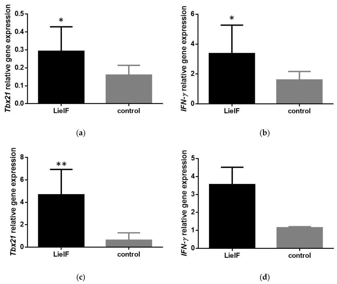
In parallel with the flow cytometric analysis, quantitative real-time PCR was performed to assess the expression of immune-related genes and further characterize the immune response elicited by LieIF treatment. In the CL model, splenocytes from LieIF-treated mice exhibited significantly higher expression of Tbx21 and IFN-γ than untreated controls (P = 0.0128 and P = 0.0118, respectively) (Figure 7a and Figure 7b), whereas the expression of GATA3 remained unchanged (P > 0.05) (data not shown). These transcriptional changes are consistent with a Th1-skewed immune profile following LieIF treatment. In the VL model, no significant differences were observed between LieIF-treated and untreated control mice in the splenic expression of Tbx21, GATA3, or IFN-γ (data not shown). In contrast, lymph node analysis revealed significantly increased expression of Tbx21 and IFN-γ in LieIF-treated mice compared to controls (P = 0.0095 and 0.0159, respectively) (Figure 7c and Figure 7d).
3. Discussion
In the absence of an effective human vaccine and adequate vector control measures, chemotherapy remains the primary strategy for the control of leishmaniasis [5]. Over the past decades, immunotherapy, either as a standalone approach or in combination with conventional chemotherapy (immunochemotherapy), has attracted increasing scientific attention as a promising strategy to combat this disease [21,23]. Unlike conventional antileishmanial drugs that directly target the parasite, immunotherapeutic strategies aim to strengthen or modulate host immune responses, thereby promoting parasite elimination while potentially reducing treatment failure, relapse, and the emergence of drug resistance [24]. Since Leishmania spp. parasites can persist within host cells by evading or manipulating immune mechanisms, the development of a robust and specific immune response is crucial for controlling parasite replication. Therefore, stimulating the immune system through the administration of parasite-derived antigens or immunomodulatory molecules may represent an effective alternative or complementary approach for the treatment of leishmaniasis.
Among Leishmania proteins, LeIF, a well-characterized DEAD-box RNA helicase [25], has emerged as a promising immunomodulatory molecule in the context of leishmaniasis [12,13,14]. Previous studies have shown that recombinant LeIF protein can stimulate pro-inflammatory cytokine production and promote protective Th1-oriented immune responses in both human and experimental models, highlighting its potential as both a vaccine antigen and an immunotherapeutic candidate [12,13,14]. Accordingly, LeIF has been extensively investigated as a vaccine antigen, either alone or in combination with other Leishmania proteins and adjuvants, demonstrating its ability to induce Th1-based cellular immune responses and reduce parasite burden in experimental models of leishmaniasis [19,26]. In addition, LeIF has also been used as part of a cocktail of recombinant proteins to treat a patient having a chronic muco-cutaneous leishmaniasis [27]. Previously, we demonstrated that recombinant LieIF, in the presence of IFN-γ, significantly inhibits L. donovani intracellular growth in murine macrophages by inducing the production of microbicidal molecules [15]. Collectively, these findings suggest that LeIF could be used as an immunotherapeutic molecule against Leishmania infection. However, no published study has yet demonstrated the curative efficacy of LeIF as a standalone immunotherapeutic intervention in an animal model of leishmaniasis.
The results of the present study are consistent with previous observations and further extend them by demonstrating that LieIF retains therapeutic efficacy when administered after the establishment of Leishmania infection. The present findings suggest that LieIF treatment mitigates lesion progression and tissue pathology in L. major-infected mice and reduces parasite burden in L. infantum-infected mice along with enhancing localized Th1-associated cellular immune responses. These results support the potential of LieIF as a promising immunotherapeutic candidate for the control of leishmaniasis. In the CL murine model, LieIF treatment resulted in significantly reduced lesion development, accompanied by the absence of tissue destruction and lower Leishmania-specific IgG1 antibody responses compared with untreated controls, four weeks after treatment termination. Although reduced IgG1 levels may suggest modulation of humoral immunity, the absence of significant changes in IgG2a levels or in the IgG1/IgG2a ratio does not support a clear shift in Th1/Th2 polarization. Instead, the therapeutic effect of LieIF was associated with a significant increase in the frequency of IFN-γ-producing CD4⁺ lymphocytes in the draining popliteal lymph nodes. Since CD4⁺ Th1 cells represent a major source of IFN-γ during protective immunity against Leishmania infection, these findings suggest that LieIF-mediated parasite control is primarily driven by the enhancement of local Th1 effector responses [28]. Consistent with this observation, LieIF treatment increased the expression of Th1-associated transcription factor Tbx21 and the cytokine IFN-γ in splenocytes, further supporting the induction of Th1-oriented cellular immunity. Notably, no significant differences were observed in the expression of GATA3, suggesting that LieIF did not broadly affect Th2-associated immune pathways. Rather, its immunomodulatory activity appears to be predominantly associated with the selective amplification of IFN-γ-producing CD4⁺ T-cell responses, which are critical for effective control of Leishmania infection. In the VL murine model, LieIF treatment significantly reduced parasite burden in the spleen and liver, the principal organs affected during visceral Leishmania infection, suggesting that its immunotherapeutic effect extends beyond local immune activation and contributes to the control of systemic infection. Indeed, compared to untreated controls, the limiting dilution assay highlighted that LieIF treatment induced a significant reduction in parasite burden in the liver at 4 weeks post-infection, and in the spleen at 10 weeks post-infection. These findings are particularly relevant in light of the distinct roles of the liver and spleen during L. infantum infection in BALB/c murine model. Following infection, parasites initially undergo rapid multiplication in the liver, where the development of effective cell-mediated immune responses is essential for parasite clearance [29]. The observed reduction in hepatic parasite burden suggests that LieIF treatment may enhance immune mechanisms involved in the early control of infection within this organ. In contrast, although the spleen serves as an important site for the generation of parasite-specific effector T cells, it progressively becomes a reservoir of persistent infection and is generally considered more susceptible to chronic parasite establishment than the liver. Therefore, the significant reduction in splenic parasite burden observed at 10 weeks post-infection is particularly noteworthy, as it indicates that LieIF-mediated immune responses may also contribute to limiting parasite persistence during the chronic phase of infection. These findings were further corroborated by qPCR-based quantification of parasite burden in both target organs. Assessment of the humoral immune response showed no significant differences in Leishmania-specific IgG1 or IgG2a antibody levels between LieIF-treated and control groups, suggesting that the therapeutic effect was not associated with alterations in antibody class switching or a measurable shift in systemic Th1/Th2-associated humoral profiles at this time point. Analysis of cellular immune parameters revealed a significant increase in IL-12–producing cells in the spleen of LieIF-treated mice, accompanied by an expansion of CD4⁺ cell populations, while CD8α⁺ cell frequencies remained unchanged, at 10 weeks post-infection. At the transcriptional level, no significant differences were observed in splenic expression of the Th1- and Th2-associated transcription factors Tbx21 and GATA3, respectively, or IFN-γ between treated and control groups. In contrast, Tbx21 and IFN-γ expression was significantly upregulated in lymph node samples from LieIF-treated mice compared to controls, at the same time point.
In the present study, the immunotherapeutic activity of LieIF may be explained, at least in part, by its previously described immunomodulatory properties. In earlier work, we demonstrated that LieIF induces a pro-inflammatory environment characterized by the activation of peritoneal exudate cells, recruitment of innate immune cell populations, enhanced expression of co-stimulatory molecules, and increased production of inflammatory mediators [17]. In addition, we subsequently demonstrated that LieIF promotes dendritic cell maturation characterized by increased expression of the co-stimulatory molecules CD40, CD80 and CD86, together with enhanced IL-12 production, thereby favoring the development of Th1-oriented immune responses [16]. The increased frequency of IL-12-producing cells and enhanced IFN-γ-associated responses observed herein are consistent with these earlier observations and further support a role for LieIF in shaping protective cell-mediated immunity against Leishmania infection [12]. In turn, IFN-γ represents a key mediator of resistance to Leishmania infection by promoting macrophages activation and intracellular parasite killing through nitric oxide-dependent and other leishmanicidal mechanisms [30]. Although macrophages activation and effector mechanisms were not directly evaluated in the current study, the induced increase in IL-12-producing cells, the enhanced expression of Tbx21 and IFN-γ, and the expansion of IFN-γ-producing CD4⁺ lymphocytes collectively support a model in which LieIF promotes parasite control through the amplification of protective cell-mediated immunity. Future studies aiming to investigate the innate immune pathways activated by LieIF, including the contribution of dendritic cells, monocytes, and macrophage effector mechanisms, will be important for further elucidating the cellular and molecular basis of its immunotherapeutic activity.
Despite the encouraging results obtained in both experimental models, several limitations should be acknowledged. First, the study was performed exclusively in BALB/c mice, and therefore the extending of these findings to other host backgrounds remains to be established. In addition, only a single treatment regimen was evaluated, precluding conclusions regarding the optimal dose, frequency, and duration of LieIF administration. Furthermore, the durability of LieIF-induced immune responses and their capacity to generate long-term protective immunological memory were not investigated. Although the present findings support the ability of LieIF to induce protective cell-mediated immunity and to promote therapeutic control of Leishmania infection, further studies are warranted to elucidate the underlying immune mechanisms, particularly the contribution of innate immune cells, including dendritic cells, monocytes, and macrophages to its immunotherapeutic activity. In addition, given the growing interest in host-directed therapies for leishmaniasis, future studies should also investigate the therapeutic efficacy of LieIF in combination with conventional antileishmanial drugs. Such immunochemotherapeutic approaches may enhance parasite clearance while reducing drug toxicity and the risk of emergence of drug resistance [31]. Such investigations will be essential for establishing the translational potential of LieIF as a novel host-directed immunotherapeutic intervention against leishmaniasis.
Taken together, the present findings highlight the potential of LieIF as an immunotherapeutic tool capable of modulating host immune responses in favor of parasite control. By targeting immune pathways rather than the parasite directly, LieIF may represent a complementary approach to conventional antileishmanial treatments and contribute to the development of more effective therapeutic strategies against leishmaniasis. Further investigation of its mechanisms of action and therapeutic applications will help define its potential role in future immunotherapeutic and immunochemotherapeutic interventions.
4. Materials and Methods
4.1. Ethical Approval
All experimental procedures were conducted in accordance with the provisions of PD56/2013 and European Directive 2010/63/EU for welfare and ethical use of laboratory animals based on 3 + 1Rs and the guidelines of PREPARE and ARRIVEs. Experimental protocols were reviewed and approved by the Institutional Protocol Evaluation Committee and were performed under the licensed protocol with registered code 6380/11 – 12 – 2017 (date of approval; December 11, 2017) by the Official Veterinary Authorities of Attica Prefecture.
4.2. Expression and Purification of Recombinant LieIF
The LieIF coding sequence was cloned into the NdeI and XhoI sites of pET22-b as previously reported [25]. The expression and purification were performed as previously described [16,25]. The purity and concentration of the recombinant protein were verified on 12% Coomassie stained SDS-PAGE gel. Furthermore, the recombinant protein was tested for endotoxin levels (≤5 EU/mg) using the Limulus amebocyte lysate (LAL) assay (Pierce™ LAL Chromogenic Endotoxin Quantitation Kit, Thermo Scientific).
4.3. In Vitro Culture of Leishmania Parasites
L. major (zymodeme LV39, strain MRHO/SU/59/P) and L. infantum (zymodeme MON-1, strain MCAN/PT/98/IMT244) promastigotes, causative agents of cutaneous and visceral leishmaniasis, respectively, were maintained at 26 °C in complete RPMI-1640 medium (Capricorn Scientific, Germany) supplemented with 2 mM L-glutamine, 10 mM HEPES, 24 mM sodium bicarbonate (NaHCO3), 100 U/mL penicillin, and 100 µg/mL streptomycin (Gibco™, Thermo Fisher Scientific, USA) and 10% (v/v) heat-inactivated fetal bovine serum (FBS; Capricorn Scientific, Germany). Parasites were routinely propagated in tissue culture flasks (Kisker Biotech, Germany).
Soluble Leishmania antigen (SLA) obtained from 109 stationary-phase L. infantum and L. major promastigotes, respectively. Briefly, promastigotes were resuspended in sterile phosphate buffered saline (PBS) and disrupted by three repeated freeze—thaw cycles (freezing at -80 °C and thawing at 37 °C). Lysed parasites were subsequently sonicated 3 times for 1 min each, with 30 s intervals between sonication cycles. The crude lysates were centrifuged at 8000 x g for 30 min at 4 °C, and the resulting supernatants were collected, aliquoted, and stored at -80 °C until use. The protein concentration of SLA preparations was determined using a NanoDrop 2000 spectrophotometer (Thermo Fisher Scientific, USA).
4.4. Murine Experimental Models of Leishmaniasis
Laboratory animals (BALB/c mice) were maintained under specific pathogen-free conditions at the approved establishments of the Department of Animal Models for Biomedical Research in Hellenic Pasteur Institute (facility registration codes: EL25BIOsup011, EL25BIObr012, EL25BIOexp013). Animals were housed under controlled environmental conditions at 22 ± 2 °C, with a relative humidity of 40 – 70%, and a 12 h light/12 h dark cycle, and were provided with food and water ad libitum.
Female BALB/c mice (8 -10 weeks old) were used as experimental models of cutaneous leishmaniasis (CL) and visceral leishmaniasis (VL). For each disease model, mice were randomly assigned to three experimental groups. In the CL model, mice in Group 1 (n = 10) and Group 2 (n = 10) were infected on day 0 by subcutaneous inoculation of 1 x 10⁷ stationary-phase L. major promastigotes (zymodeme LV39, strain MRHO/SU/59/P) into the left hind footpad. In the VL model, mice in Group 1 (n = 10) and Group 2 (n = 10) were infected intravenously with 1 x 10⁷ stationary-phase L. infantum promastigotes (zymodeme MON-1, strain MCAN/PT/98/IMT244). In both experimental models, mice in Group 1 received recombinant LieIF as immunotherapy by intraperitoneal injection (i.p.) at a dose of 10 µg per mouse on days 7 and 14 post-infection, whereas mice in Group 2 served as infected untreated controls. Group 3 (n = 4) consisted of uninfected, untreated mice and served as the naïve control group.
4.5. Assessment of Disease Progression
All animals were monitored throughout the experimental period and subsequently subjected to parasitological, immunological, and molecular analyses. In the CL model, disease progression was assessed by measuring footpad swelling (mm) compared to the uninfected contralateral footpad with a dial gauge caliper (Mitutoyo, Kawasaki, Japan), and lesion development was assessed 6 weeks post-infection. In the VL model, parasite burden was quantified in the liver and spleen at 10 weeks post-infection, using limiting dilution assays and quantitative real-time PCR (qRT-PCR). Humoral immune responses were determined by measuring Leishmania-specific antibody production at the terminal time point, in both experimental models. Cellular immune responses were assessed by flow cytometry (FACS), in regional draining lymph nodes (CL model) and spleen (VL model), respectively. Gene expression profiling was performed by qRT-PCR in the spleen and peripheral lymph nodes in both experimental models at the terminal time point.
4.6. Parasite Burden Quantification by Limiting Dilution
The number of viable parasites was quantified in the spleen and liver, which are the major target organs of Leishmania dissemination in the VL model, by limiting dilution assay. Mice were sacrificed at 4 (n = 5) and 10 weeks (n = 5) post-infection and weighed. The spleen and liver were harvested and homogenized in 5 mL Schneider’s insect medium (Biosera, France) supplemented with 20% FBS (Capricorn Scientific, Germany). Homogenates were subjected to 12 serial 2-fold dilutions and cultured in quadruplicate in sterile 96-well flat-bottom microtiter plates (Sarstedt, Nümbrecht, Germany) at 26 °C for 10 days. The presence of motile parasites was assessed using an inverted microscope (Olympus Corporation, Tokyo, Japan). The number of viable parasites per mg of tissue was calculated as the mean ± standard deviation (SD) based on the highest dilution at which live parasites were detectable, multiplied by the corresponding dilution factor. Total organ parasite burden was estimated by normalizing to the weight of each respective organ.
4.7. Parasite Burden Quantification by Quantitative PCR (qPCR)
Genomic DNA was extracted from homogenized spleen and liver tissue samples using the QIAamp DNA Mini Kit (Qiagen, Netherlands), according to the manufacturer’s instructions. DNA concentration and purity were determined spectrophotometrically by measuring absorbance at 260 nm, and DNA samples were stored at −80 °C until analysis. Quantitative PCR (qPCR) assays were performed using an Exicycler™ 96 Real-Time PCR System (Bioneer Corporation, Daejeon, Republic of Korea).
For the VL model, parasite burden in the spleen and liver was quantified at 4 and 10 weeks post-infection by amplification of L. infantum kinetoplast DNA (kDNA) using the previously described RV1 (5′-CTTTTCTGGTCCTCCGGGTAGG-3′) and RV2 (5′-CCACCCGGCCCTATTTTACACCAA-3′) primers [32,33]. Amplification was performed using a TaqMan assay with the specific probe 5′-FAM-TTTTCGCAGAACGCCCCTACCCGC-TAMRA-3′. All qPCR reactions were carried out in a final volume of 20 µL containing 25 pmol of each primer. For the TaqMan assay, 50 pmol of the RV probe was included in each reaction. Standard curves were generated from 10-fold dilutions of genomic DNA extracted from 5 × 10⁶ L. infantum promastigotes, corresponding to 10⁶ to 1 parasite equivalents per reaction.
To assess the variability in kinetoplast minicircle copy number, parasite quantification was normalized using the single-copy Leishmania DNA polymerase gene, as previously described [32]. Amplification was performed using the forward primer 5′-TGTCGCTTGCAGACCAGATG-3′ (50 pmol), the reverse primer 5′-GCATCGCAGGTGTGAGCAC-3′ (50 pmol), and the TaqMan probe 5′-HEX-CCAGGCTCGAAGTTGTTGCTGCCC-TAMRA-3′ (100 pmol), under the same cycling conditions as those used for kDNA amplification. Standard curves for the DNA polymerase gene were generated using the same serial dilutions of Leishmania genomic DNA, corresponding to 10⁶ to 1 parasite equivalents per reaction.
4.8. Assessment of Leishmania-Specific IgG Responses
The Leishmania-specific IgG isotype profile was determined by enzyme-linked immunosorbent assay (ELISA) using soluble Leishmania antigen (SLA) obtained from L. infantum and L. major promastigotes, as described above. At the experimental endpoint (6 and 10 weeks post-infection for the CL and VL disease model, respectively), serum samples were collected from all experimental mice and analyzed for the presence of Leishmania-specific IgG1 and IgG2a antibodies. Briefly, 96-well microplates (Sarstedt, Nümbrecht, Germany) were coated overnight at 4 °C with 5 µg/mL of SLA prepared from L. major and L.infantum promastigotes, diluted in carbonate coating buffer (15 mM Na2CO3 , 35 mM NaHCO3, pH 9.6). Plates were subsequently blocked with 2% bovine serum albumin (BSA; AppliChem GmbH, Darmstadt, Germany) and serial dilutions of serum samples (1:20—1:40 960) were added. Then, plates were incubated with biotinylated anti-mouse IgG1 (500 ng/mL) or IgG2a (250 ng/mL) (AbD Serotec, Kidlington, Oxford, UK), followed by horseradish peroxidase (HRP)-conjugated streptavidin (1:5000, AbDSerotec, Kidlington, Oxford, UK). Color development was achieved using 3,3′,5,5′-tetramethylbenzidine (TMB) substrate (Thermo Fisher Scientific, USA), and absorbance was measured at 450 nm. Results are expressed as the Area Under Curve (AUC) ± SD.
4.9. Flow Cytometric Analysis of Cellular Immune Responses
Flow cytometric analysis was performed on cells isolated from the draining lymph nodes of mice with CL and from the spleens of mice with VL, at the study endpoint (6 and 10 weeks post-infection for CL and VL, respectively). Single-cell suspensions were prepared and seeded into 48-well plates (Sarstedt, Nümbrecht, Germany) (5 x 10⁵ cells per well), followed by incubation with 2.5 µg/mL brefeldin A (Fluka, Buchs, Switzerland) for 4 h to inhibit cytokine secretion. Cells were then fixed with 2% paraformaldehyde solution in PBS for 10 min at room temperature and permeabilized using FACS buffer (PBS-3% FBS) supplemented with 0.1% (v/v) saponin (Sigma-Aldrich, St. Louis, MO, USA). Permeabilized cells were stained with fluorochrome-conjugated monoclonal antibodies, including anti-CD4 (clone H129.19, PE-conjugated), anti-CD8a (clone 53-6.7, PE—conjugated), anti-IFN-γ (clone XMG1.2, FITC-conjugated), and anti-IL-12p40/p70 (clone C15.6, PE-conjugated), all purchased from BD Biosciences (Erembodegem, Belgium). Unstained samples were processed in parallel and used as controls for fluorescence analysis. Flow cytometric analysis was performed using the corresponding fluorescence parameters to identify the relevant marker-positive populations, following exclusion of non-cellular events. For each sample, 20,000 events were acquired using a FACSCalibur flow cytometer (BD Biosciences, San Jose, CA, USA), and data were analyzed using FlowJo software (version 10.0.8; Tree Star Inc., Ashland, OR, USA).
4.10. Gene Expression Analysis by qRT-PCR
Gene expression analysis was performed on cells isolated from the draining lymph nodes and spleens of experimental mice at the study endpoint (6 and 10 weeks post-infection for CL and VL models, respectively). Total mRNA was isolated with an RNeasy Mini kit (Qiagen, Netherlands), and mRNA concentrations were determined using a NanoDrop 2000 spectrophotometer (Thermo Fisher Scientific, USA). cDNAs from all experimental groups were synthesized using the SuperScript™ II Reverse Transcriptase kit (Invitrogen™, Thermo Fisher Scientific, Carlsbad, CA, USA) and oligo(dT) primers (Promega, Madison, WI, USA). Quantitative real-time PCR (qRT-PCR) was performed using an Exicycler™ 96 real-time PCR system (Bioneer Corporation, Daejeon, Republic of Korea) with the KAPA SYBR® FAST Universal 2× qPCR Master Mix kit (Kapa Biosystems, Wilmington, MA, USA). The specific primers for the interferon-γ (IFN-γ), T-box transcription factor (Tbx21), trans-acting T-cell-specific transcription factor (GATA3), and the glyceraldehyde dehydrogenase of the 3-phosphatease (GAPDH) gene sequences were designed by Qiagen (QuantiTect Primer Assays; Qiagen, Netherlands) and were run in triplicate. The qPCR assays were performed according to the manufacturer’s protocol for the QuantiTect Primer Assays. All gene expression ratios were computed with the ΔΔCt method. All qPCR experiments were performed in replicate for each experimental condition. Expression levels of target genes were normalized to the GAPDH housekeeping gene, and expressed as fold changes relative to the uninfected, untreated control group.
4.11. Statistical Analysis
Longitudinal footpad swelling measurements were analyzed using a mixed-effects model (REML) with treatment and time as fixed effects and subject as a random effect. This approach was chosen because repeated-measures ANOVA cannot accommodate the missing observations that occurred following tissue necrosis. Type III tests of fixed effects were used to evaluate treatment, time, and treatment × time interaction effects. When the interaction was significant, Sidak-adjusted multiple comparisons were used to compare treatment groups at each time point. Statistical analysis of parasite burden was performed using a two-way ANOVA to assess the effects of treatment, experimental condition, and their interaction. This test allowed evaluation of whether parasite load differed by treatment, changed over time, and whether treatment effects varied across time points. When significant effects were detected, Sidak’s multiple comparisons test was applied for post hoc pairwise comparisons between groups at each time point. Adjusted mean differences and 95% confidence intervals were used to report effect sizes and precision. Statistical analysis of Leishmania-specific IgG1 and IgG2a antibody responses, flow cytometry, and quantitative real-time PCR gene expression data was performed using the two-tailed Mann–Whitney U test. Analyses were performed using GraphPad Prism version 11 (GraphPad Software, San Diego, CA, USA), and statistical significance was defined as P < 0.05 (*), P < 0.01 (**), P < 0.001 (***), and P < 0.0001 (****).
Author Contributions
Conceptualization, E.D., I.G., M.B. and O.K.; Methodology, O.K., I.K., M.B., and K.E.-B.; Software, O.K. and I.K.; Validation, O.K., I.K., M.B, I.G. and E. D.; Formal Analysis, O.K., I.K..; Investigation, O.K., I.K. M.B.; Resources, E.D., I.G.; Writing—Original Draft Preparation, O.K., M.B.; Writing—Review & Editing, O.K., I.K., M.B., K.E.-B., I.G., and E.D.; Visualization, O.K., I.K., and M.B..; Supervision, E.D. I.G.; Project Administration, E.D., I.G.; Funding Acquisition, E.D., I.G.
Funding
This study was funded by Research Project No. 426 to IG, within the framework of the PTR (Programmes Transversaux de Recherche) action. This project was supported by the scientific network comprising the Institut Pasteur de Tunis, the Institut Pasteur of Paris, and the Hellenic Pasteur Institute. The study received support from the Research Laboratory Contract Program of the Ministry of Higher Education and Research of the Republic of Tunisia (LR16IPT04). I.G., K.E.-B & M.B. receive support from the Science for Africa Foundation to the Developing Excellence in Leadership, Training and Science in Africa (DELTAS Africa) programme (Del-22-005 to IG) with support from Wellcome Trust and the UK Foreign, Commonwealth & Development Office and is part of the EDCTP2 programme supported by the European Union. MB is an NTD career fellow of the African Leishmaniases Consortium (ALC, Del- 22-005).
Institutional Review Board Statement
The study was conducted in accordance with the Declaration of Helsinki, and approved by the Institutional Protocol Evaluation Committee, and were performed under the licensed protocol with registered code 6380/11 – 12 – 2017 (date of approval; December 11, 2017) by the Official Veterinary Authorities of Attica.
Data Availability Statement
The data supporting the findings of this study are available from the corresponding author upon reasonable request.
Conflicts of Interest
The authors declare no conflict of interest.
Acknowledgments
During the preparation of this manuscript, the authors used ChatGPT (OpenAI) solely for language refinement and improvement of readability. All AI-assisted text was critically reviewed, edited, and validated by the authors, who take full responsibility for the content of the manuscript.
Abbreviations
The following abbreviations are used in this manuscript:
| LieIF | Leishmania infantum eukaryotic initiation factor |
| L. major | Leishmania major |
| L. infantum | Leishmania infantum |
| CL | Cutaneous leishmaniasis |
| VL | Visceral leishmaniasis |
| IL-4 | Interleukin-4 |
| IL-12 | Interleukin-12 |
| IL-13 | Interleukin-13 |
| IFN-γ | Iinterferon-γ |
| FBS | Fetal Bovine Serum |
| SLA | Soluble Leishmania Antigen |
| FACS | Fluorescence-Activated Cell Sorting |
| PE | Phycoerythrin |
| FITC | Fluorescein |
| qPCR | Quantitative Polymerase Chain Reaction |
| RT-PCR | Reverse Transcription Quantitative Polymerase Chain Reaction |
References
- Burza, S.; Croft, S.L.; Boelaert, M. Leishmaniasis. Lancet 2018, 392(10151), 951–970. [Google Scholar] [CrossRef] [PubMed]
- Torres-Guerrero, E.; et al. Leishmaniasis: a review. F1000Res 2017, 6, 750. [Google Scholar] [CrossRef] [PubMed]
- Ready, P.D. Epidemiology of visceral leishmaniasis. Clin. Epidemiol. 2014, 6, 147–54. [Google Scholar] [CrossRef] [PubMed]
- Sundar, S.; et al. Current and emerging therapies for the treatment of leishmaniasis. Expert Opin. Orphan Drugs 2024, 12(1), 19–32. [Google Scholar] [CrossRef]
- Zhang, H.; et al. Progress in antileishmanial drugs: Mechanisms, challenges, and prospects. PLoS Negl. Trop. Dis. 2025, 19(1), e0012735. [Google Scholar] [CrossRef] [PubMed]
- Hefnawy, A.; et al. Exploiting Knowledge on Leishmania Drug Resistance to Support the Quest for New Drugs. Trends Parasitol. 2017, 33(3), 162–174. [Google Scholar] [CrossRef] [PubMed]
- Ghorbani, M.; Farhoudi, R. Leishmaniasis in humans: drug or vaccine therapy? Drug Des. Devel Ther. 2018, 12, 25–40. [Google Scholar] [CrossRef] [PubMed]
- Domagalska, M.A.; Barrett, M.P.; Dujardin, J.C. Opinion Drug resistance in does it really matter? Trends Parasitol. 2023, 39(4), 251–259. [Google Scholar] [CrossRef] [PubMed]
- Kaye, P.; Scott, P. Leishmaniasis: complexity at the host-pathogen interface. Nat. Rev. Microbiol. 2011, 9(8), 604–615. [Google Scholar] [CrossRef] [PubMed]
- Scott, P.; Novais, F.O. Cutaneous leishmaniasis: immune responses in protection and pathogenesis. Nat. Rev. Immunol. 2016, 16(9), 581–92. [Google Scholar] [CrossRef] [PubMed]
- Garde, E.; et al. Analysis of the Antigenic and Prophylactic Properties of the Leishmania Translation Initiation Factors eIF2 and eIF2B in Natural and Experimental Leishmaniasis. Front Cell Infect. Microbiol. 2018, 112. [Google Scholar] [CrossRef] [PubMed]
- Skeiky, Y.A.; et al. LeIF: a recombinant Leishmania protein that induces an IL-12-mediated Th1 cytokine profile. J. Immunol. 1998, 161(11), 6171–9. [Google Scholar] [CrossRef]
- Skeiky, Y.A.; et al. A recombinant Leishmania antigen that stimulates human peripheral blood mononuclear cells to express a Th1-type cytokine profile and to produce interleukin 12. J. Exp. Med. 1995, 181(4), 1527–37. [Google Scholar] [CrossRef] [PubMed]
- Barhoumi, M.; et al. Leishmania infantum LeIF and its recombinant polypeptides modulate interleukin IL-12p70, IL-10 and tumour necrosis factor-alpha production by human monocytes. Parasite Immunol. 2011, 33(10), 583–8. [Google Scholar] [CrossRef] [PubMed]
- Koutsoni, O.; et al. Leishmania eukaryotic initiation factor (LeIF) inhibits parasite growth in murine macrophages. PLoS ONE 2014, 9(5), e97319. [Google Scholar] [CrossRef] [PubMed]
- Barhoumi, M.; et al. Leishmania infantum LeIF and its recombinant polypeptides induce the maturation of dendritic cells in vitro: An insight for dendritic cells based vaccine. Immunol. Lett. 2019, 210, 20–28. [Google Scholar] [CrossRef] [PubMed]
- Koutsoni, O.S.; et al. New Insights on the Adjuvant Properties of the Leishmania infantum Eukaryotic Initiation Factor. J. Immunol. Res. 2019, 9124326. [Google Scholar] [CrossRef] [PubMed]
- Probst, P.; et al. A Leishmania protein that modulates interleukin (IL)-12, IL-10 and tumor necrosis factor-alpha production and expression of B7-1 in human monocyte-derived antigen-presenting cells. Eur. J. Immunol. 1997, 27(10), 2634–42. [Google Scholar] [CrossRef] [PubMed]
- Skeiky, Y.A.; et al. Protective efficacy of a tandemly linked, multi-subunit recombinant leishmanial vaccine (Leish-111f) formulated in MPL adjuvant. Vaccine 2002, 20(27-28), 3292–303. [Google Scholar] [CrossRef] [PubMed]
- Coler, R.N.; et al. Immunization with a polyprotein vaccine consisting of the T-Cell antigens thiol-specific antioxidant, Leishmania major stress-inducible protein 1, and Leishmania elongation initiation factor protects against leishmaniasis. Infect. Immun. 2002, 70(8), 4215–25. [Google Scholar] [CrossRef] [PubMed]
- Okeke, G.N.; et al. Leishmaniasis: pathogenesis, host immunity, and the evolving landscape of immunotherapy and immunochemotherapy. Front Microbiol. 2026. 17, 1861933. [Google Scholar] [CrossRef] [PubMed]
- Ayala, A.; et al. Advances in Leishmania Vaccines: Current Development and Future Prospects. Pathogens 2024, 13(9). [Google Scholar] [CrossRef] [PubMed]
- Espuelas, S.; et al. Immunochemotherapy with Amphotericin B and HisAK70 Vaccine for Cutaneous Leishmaniosis. Int. J. Mol. Sci. 2026, 27(14). [Google Scholar] [CrossRef] [PubMed]
- Lafleur, A.; et al. Immunotherapeutic Strategies as Potential Treatment Options for Cutaneous Leishmaniasis. Vaccines 2024, 12(10). [Google Scholar] [CrossRef] [PubMed]
- Barhoumi, M.; et al. Leishmania infantum LeIF protein is an ATP-dependent RNA helicase and an eIF4A-like factor that inhibits translation in yeast. FEBS J. 2006, 273(22), 5086–100. [Google Scholar] [CrossRef] [PubMed]
- Saini, I.; Joshi, J.; Kaur, S. Leishmania vaccine development: A comprehensive review. Cell Immunol. 2024, 399-400, 104826. [Google Scholar] [CrossRef] [PubMed]
- Badaro, R.; et al. Successful use of a defined antigen/GM-CSF adjuvant vaccine to treat mucosal Leishmaniasis refractory to antimony: A case report. Braz. J. Infect. Dis. 2001, 5(4), 223–32. [Google Scholar] [CrossRef] [PubMed]
- Singh, O.P.; Sundar, S. Immunotherapy and targeted therapies in treatment of visceral leishmaniasis: current status and future prospects. Front Immunol. 2014, 5, 296. [Google Scholar] [CrossRef] [PubMed]
- Nieto, A.; et al. Mechanisms of resistance and susceptibility to experimental visceral leishmaniosis: BALB/c mouse versus Syrian hamster model. Vet. Res. 2011, 42(1), 39. [Google Scholar] [CrossRef] [PubMed]
- Bogdan, C. Macrophages as host, effector and immunoregulatory cells in leishmaniasis: Impact of tissue micro-environment and metabolism. Cytokine X 2020, 2(4), 100041. [Google Scholar] [CrossRef] [PubMed]
- Aruleba, R.T.; et al. Can We Harness Immune Responses to Improve Drug Treatment in Leishmaniasis? Microorganisms 2020, 8(7). [Google Scholar] [CrossRef] [PubMed]
- Mary, C.; et al. Quantification of Leishmania infantum DNA by a real-time PCR assay with high sensitivity. J. Clin. Microbiol. 2004, 42(11), 5249–55. [Google Scholar] [CrossRef] [PubMed]
- Lachaud, L.; et al. Comparison of six PCR methods using peripheral blood for detection of canine visceral leishmaniasis. J. Clin. Microbiol. 2002, 40(1), 210–5. [Google Scholar] [CrossRef] [PubMed]
Figure 1.
Expression and purification of the recombinant LieIF protein. An aliquot of 1.5 µg of purified recombinant LieIF protein was resolved on a 12% SDS-PAGE gel under denaturing conditions and stained with Coomassie Brilliant Blue. Lane 1, molecular weight markers; Lane 2, purified recombinant LieIF protein. The apparent molecular weight of the purified recombinant LieIF protein is 45.3 kDa.
Figure 1.
Expression and purification of the recombinant LieIF protein. An aliquot of 1.5 µg of purified recombinant LieIF protein was resolved on a 12% SDS-PAGE gel under denaturing conditions and stained with Coomassie Brilliant Blue. Lane 1, molecular weight markers; Lane 2, purified recombinant LieIF protein. The apparent molecular weight of the purified recombinant LieIF protein is 45.3 kDa.
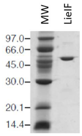
Figure 2.
Therapeutic effect induced by recombinant LieIF in murine experimental model of cutaneous leishmaniasis. BALB/c mice were subcutaneously infected with 1 x 10⁷ stationary-phase L. major promastigotes into the left hind footpad. Seven and fourteen days post-infection, mice received intraperitoneal (i.p.) administration of recombinant LieIF as immunotherapy at a dose of 10 µg per mouse (n = 10), or left untreated (n = 10). Footpad thickness (mm) was measured once per week compared to the uninfected contralateral footpad with a dial gauge caliper, and lesion development was assessed 6 weeks post-infection. Data are presented as mean ± SD for each group at the indicated time points. Statistical comparisons between groups was performed using a mixed-effects model (REML). Sidak’s multiple comparisons test was applied to compare groups at each time point, with P < 0.05 considered statistically significant.
Figure 2.
Therapeutic effect induced by recombinant LieIF in murine experimental model of cutaneous leishmaniasis. BALB/c mice were subcutaneously infected with 1 x 10⁷ stationary-phase L. major promastigotes into the left hind footpad. Seven and fourteen days post-infection, mice received intraperitoneal (i.p.) administration of recombinant LieIF as immunotherapy at a dose of 10 µg per mouse (n = 10), or left untreated (n = 10). Footpad thickness (mm) was measured once per week compared to the uninfected contralateral footpad with a dial gauge caliper, and lesion development was assessed 6 weeks post-infection. Data are presented as mean ± SD for each group at the indicated time points. Statistical comparisons between groups was performed using a mixed-effects model (REML). Sidak’s multiple comparisons test was applied to compare groups at each time point, with P < 0.05 considered statistically significant.
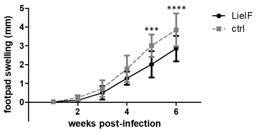
Figure 3.
Estimation of parasite burden in spleen and liver in LieIF-treated and untreated BALB/c mice, following L. infantum infection. BALB/c mice were intravenously infected with 1 x 10⁷ stationary-phase L. infantum promastigotes. At 7 and 14 days post-infection, mice received intraperitoneal administration of recombinant LieIF as immunotherapy at a dose of 10 µg per mouse (n = 10), or were left untreated (n = 10). Parasite burden in the spleen and liver was subsequently assessed by limiting dilution assay. 5 mice/group were sacrificed at week 4 and at week 10, respectively. Results are expressed as mean ± SD for each group at the indicated time points. Statistical comparisons between groups was performed using a two-way ANOVA. Sidak’s multiple comparisons test was applied for post hoc comparisons between groups at each time point. A P value < 0.05 was considered statistically significant.
Figure 3.
Estimation of parasite burden in spleen and liver in LieIF-treated and untreated BALB/c mice, following L. infantum infection. BALB/c mice were intravenously infected with 1 x 10⁷ stationary-phase L. infantum promastigotes. At 7 and 14 days post-infection, mice received intraperitoneal administration of recombinant LieIF as immunotherapy at a dose of 10 µg per mouse (n = 10), or were left untreated (n = 10). Parasite burden in the spleen and liver was subsequently assessed by limiting dilution assay. 5 mice/group were sacrificed at week 4 and at week 10, respectively. Results are expressed as mean ± SD for each group at the indicated time points. Statistical comparisons between groups was performed using a two-way ANOVA. Sidak’s multiple comparisons test was applied for post hoc comparisons between groups at each time point. A P value < 0.05 was considered statistically significant.
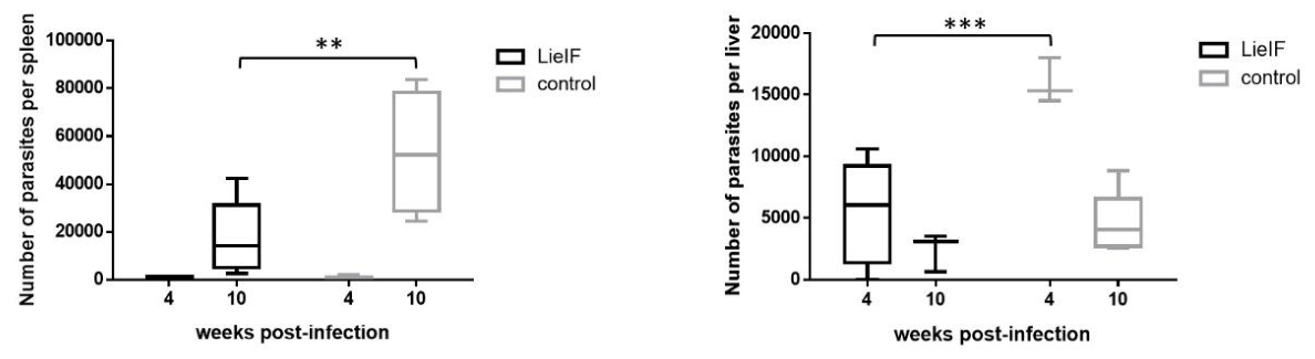
Figure 4.
Quantitative PCR assessment of LieIF-mediated therapeutic efficacy in visceral leishmaniasis in the spleen and liver. BALB/c mice were intravenously infected with 1 x 10⁷ stationary-phase L. infantum promastigotes. At 7 and 14 days post-infection, mice received intraperitoneal administration of recombinant LieIF as immunotherapy at a dose of 10 µg per mouse (n = 10), or were left untreated (n = 10). Results are expressed as mean ± SD for each group at the indicated time points. Statistical comparisons between groups was performed using a two-way ANOVA. Sidak’s multiple comparisons test was applied for post hoc comparisons between groups at each time point. A P value < 0.05 was considered statistically significant.
Figure 4.
Quantitative PCR assessment of LieIF-mediated therapeutic efficacy in visceral leishmaniasis in the spleen and liver. BALB/c mice were intravenously infected with 1 x 10⁷ stationary-phase L. infantum promastigotes. At 7 and 14 days post-infection, mice received intraperitoneal administration of recombinant LieIF as immunotherapy at a dose of 10 µg per mouse (n = 10), or were left untreated (n = 10). Results are expressed as mean ± SD for each group at the indicated time points. Statistical comparisons between groups was performed using a two-way ANOVA. Sidak’s multiple comparisons test was applied for post hoc comparisons between groups at each time point. A P value < 0.05 was considered statistically significant.
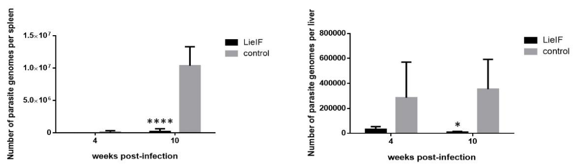
Figure 5.
Leishmania—specific IgG1 and IgG2a antibody responses following LieIF immunotherapy. Serum samples were collected from mice with CL (a) or VL (b) following treatment with LieIF or from untreated infected controls. Leishmania-specific IgG1 and IgG2a antibody levels were determined by ELISA at the study endpoint (6 and 10 weeks post-infection for CL and VL, respectively). Antibody responses are presented as the area under the curve (AUC) ± SD. Statistical analysis was performed using the two-tailed Mann–Whitney U test. Statistical significance was defined as P < 0.05.
Figure 5.
Leishmania—specific IgG1 and IgG2a antibody responses following LieIF immunotherapy. Serum samples were collected from mice with CL (a) or VL (b) following treatment with LieIF or from untreated infected controls. Leishmania-specific IgG1 and IgG2a antibody levels were determined by ELISA at the study endpoint (6 and 10 weeks post-infection for CL and VL, respectively). Antibody responses are presented as the area under the curve (AUC) ± SD. Statistical analysis was performed using the two-tailed Mann–Whitney U test. Statistical significance was defined as P < 0.05.
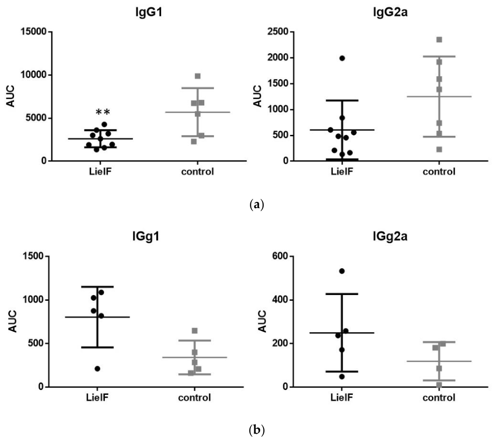
Figure 6.
Flow cytometric analysis of cellular immune responses following LieIF immunotherapy in cutaneous and visceral leishmaniasis. Flow cytometric analysis was performed on cells isolated from the draining lymph nodes of mice with CL (a) and the spleens of mice with VL (b, c), at the study endpoint (6 and 10 weeks post-infection for CL and VL, respectively). (a) Frequency of CD4+IFNγ+ T cells in the draining popliteal lymph nodes of CL mice. (b, c) Frequency of IL-12+ producing cells and CD4+ T cells in the spleens of VL mice, respectively. Data are presented as mean ± SD. Statistical comparisons between LieIF-treated and untreated infected mice were performed using the two-tailed Mann–Whitney U test and significance was defined as P < 0.05.
Figure 6.
Flow cytometric analysis of cellular immune responses following LieIF immunotherapy in cutaneous and visceral leishmaniasis. Flow cytometric analysis was performed on cells isolated from the draining lymph nodes of mice with CL (a) and the spleens of mice with VL (b, c), at the study endpoint (6 and 10 weeks post-infection for CL and VL, respectively). (a) Frequency of CD4+IFNγ+ T cells in the draining popliteal lymph nodes of CL mice. (b, c) Frequency of IL-12+ producing cells and CD4+ T cells in the spleens of VL mice, respectively. Data are presented as mean ± SD. Statistical comparisons between LieIF-treated and untreated infected mice were performed using the two-tailed Mann–Whitney U test and significance was defined as P < 0.05.
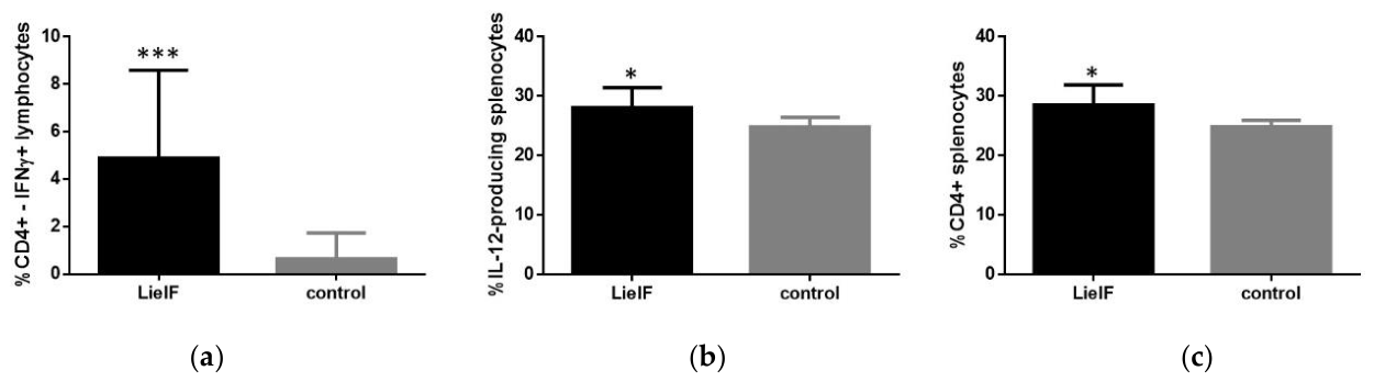
Figure 7.
Quantitative real-time PCR analysis of immune-related gene expression following LieIF immunotherapy in cutaneous and visceral leishmaniasis. Gene expression analysis was performed on cells isolated from the spleens of mice with CL (a, b) and the draining lymph nodes of mice with VL (c, d), at the study endpoint (6 and 10 weeks post-infection for CL and VL, respectively). Expression levels of target genes were normalized to the GAPDH housekeeping gene, and expressed as fold changes relative to the uninfected, untreated control group. Data are presented as mean ± SD. Statistical comparisons between LieIF-treated and untreated infected mice were performed using the two-tailed Mann–Whitney U test. Statistical significance was defined as P < 0.05. .
Figure 7.
Quantitative real-time PCR analysis of immune-related gene expression following LieIF immunotherapy in cutaneous and visceral leishmaniasis. Gene expression analysis was performed on cells isolated from the spleens of mice with CL (a, b) and the draining lymph nodes of mice with VL (c, d), at the study endpoint (6 and 10 weeks post-infection for CL and VL, respectively). Expression levels of target genes were normalized to the GAPDH housekeeping gene, and expressed as fold changes relative to the uninfected, untreated control group. Data are presented as mean ± SD. Statistical comparisons between LieIF-treated and untreated infected mice were performed using the two-tailed Mann–Whitney U test. Statistical significance was defined as P < 0.05. .
Disclaimer/Publisher’s Note: The statements, opinions and data contained in all publications are solely those of the individual author(s) and contributor(s) and not of MDPI and/or the editor(s). MDPI and/or the editor(s) disclaim responsibility for any injury to people or property resulting from any ideas, methods, instructions or products referred to in the content. |
© 2026 by the authors. Licensee MDPI, Basel, Switzerland. This article is an open access article distributed under the terms and conditions of the Creative Commons Attribution (CC BY) license (http://creativecommons.org/licenses/by/4.0/).
Copyright: This open access article is published under a Creative Commons CC BY 4.0 license, which permit the free download, distribution, and reuse, provided that the author and preprint are cited in any reuse.


