

Submitted:
07 August 2026
Posted:
07 August 2026
You are already at the latest version
Abstract
Introduction: Primordial odontogenic tumor (POT) is an exceptionally rare benign mixed odontogenic neoplasm recognized as a distinct entity in the 2017 World Health Organization classification of head and neck tumors. Owing to its rarity and overlapping clinicopathological features with other odontogenic lesions, establishing the diagnosis before definitive treatment remains challenging. Case Report: We report the case of a 2-year-old boy presenting with rapidly progressive swelling of the left maxilla associated with facial asymmetry and orbital displacement. Initial incisional biopsy performed at another institution suggested odontogenic myxoma. Computed tomography and magnetic resonance imaging demonstrated a large, well-defined expansile lesion occupying the left maxillary sinus with extensive bone thinning, cortical perforation and compression of the orbital contents. Because of the lesion extent, ocular involvement, and the presumed diagnosis, subtotal maxillectomy with immediate soft tissue reconstruction using an anterolateral thigh free flap was performed. Histopathological examination of the entire surgical specimen established the final diagnosis of primordial odontogenic tumor. At 24 months of follow-up, no evidence of recurrence was observed, and visual function remained normal. Conclusions: This case illustrates the diagnostic limitations of incisional biopsy in biphasic odontogenic tumors and highlights the importance of integrating clinical, radiological, and histopathological findings. Although POT generally demonstrates benign biological behavior, extensive lesions involving critical anatomical structures may require individualized surgical management.
Keywords:
primordial odontogenic tumor
; rare odontogenic tumor
; pediatric maxillofacial surgery
; maxillary tumor
; odontogenic myxoma
; differential diagnosis
; case report
1. Introduction
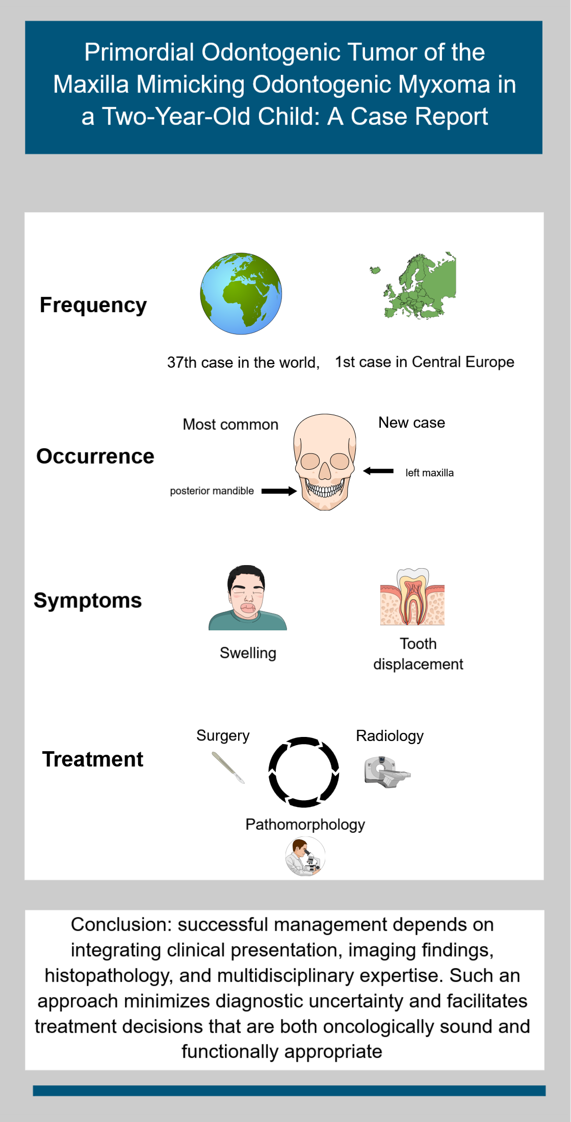
Primordial odontogenic tumor (POT) is a rare benign mixed odontogenic neoplasm composed of odontogenic epithelium and ectomesenchymal tissue resembling the early tooth germ. Since its first description by Mosqueda-Taylor et al. in 2014 [1,2], POT has been recognized as a distinct pathological entity and was subsequently incorporated into the World Health Organization (WHO) Classification of Head and Neck Tumours in 2017. Despite increasing awareness, less than 40 well-documented cases have been published [3], and current knowledge regarding its biological behavior and optimal management remains based almost exclusively on individual case reports.
POT occurs predominantly during the first and second decades of life and most commonly affects the posterior mandible [1,2,3]. Clinically, it usually presents as a painless swelling associated with delayed eruption of an unerupted tooth [2,3]. Radiologically, the lesion is typically well-defined, predominantly unilocular, and closely related to the crown of an unerupted tooth, features that substantially overlap with those of several odontogenic cysts and tumors [4]. Consequently, establishing an accurate preoperative diagnosis may be difficult, particularly when only limited biopsy material is available.
Histopathological diagnosis may also be challenging because the characteristic biphasic architecture of POT is not always represented in small incisional biopsy specimens. In such situations, the lesion may be mistaken for other odontogenic tumors, particularly odontogenic myxoma or ameloblastic fibroma, potentially influencing treatment planning [5,6]. Correlation between clinical presentation, radiological findings, and pathological examination therefore remains essential.
We report a case of a rapidly enlarging primordial odontogenic tumor arising in the maxilla of a two-year-old child. The lesion produced extensive bone destruction, orbital displacement, and was initially diagnosed as odontogenic myxoma on incisional biopsy. This report emphasizes the diagnostic pitfalls associated with limited tissue sampling and discusses the rationale for individualized surgical management in extensive maxillary lesions.
Complete de-identification of patient details was not feasible. This study conforms to CARE guidelines [7]. This study was approved by local bioethics committee on 01/04/2026 (decision number: L.Dz. WMIL-KB/21/2026).
2. Detailed Case Description
A previously healthy 2-year-old and 10-month-old boy was referred to the Department of Maxillofacial, Plastic and Reconstructive Surgery, Regional Specialized Children's Hospital in Olsztyn, Poland, in July 2024 because of rapidly progressive swelling of the left maxilla that had developed over the preceding three months. According to his parents, the first noticeable symptom was erythema of the left infraorbital region, which prompted ophthalmological consultation. Empirical antibiotic therapy was initiated; however, no clinical improvement was observed. As the swelling progressively increased, an odontogenic abscess was suspected. This diagnosis was subsequently excluded by a dentist, and the patient was referred to a local otorhinolaryngology department for further evaluation.
Computed tomography performed at the referring institution demonstrated a well-defined expansile lesion involving the left maxilla, measuring approximately 3.5 × 4.0 × 4.4 cm. An incisional biopsy was performed, and histopathological examination suggested odontogenic myxoma. The specimen, measuring approximately 0.5 cm in diameter, was assessed at another institution, and only the final pathology report was available for review.
The patient was subsequently admitted to our department for definitive management. Physical examination revealed marked facial asymmetry caused by a firm, non-tender enlargement of the left maxilla. Bluish discoloration of the overlying oral mucosa was present, and upward displacement of the left eye was evident on extraoral examination. No neurological deficits were observed.
Computed tomography(CT) and magnetic resonance imaging (MRI) demonstrated a well-defined lesion measuring 3.9 × 4.6 × 5.2 cm. The lesion caused marked thinning and cortical perforation of the adjacent bone, extended into the ipsilateral nasal cavity and nasopharynx, and displaced the orbital floor superiorly with compression of the orbital contents. The tumor was associated with the unerupted left maxillary second primary molar, second premolar, and first permanent molar. Retrospective analysis of the imaging after the final diagnosis acknowledged that radiological findings of well-defined, highly expansive, unilocular tumor associated with an unerupted teeth, with root resorption of adjacent erupted teeth align with the diagnosis of POT much closer than with myxoma. However, given the biopsy results and the absence of imaging findings explicitly suggestive of a different tumor, they did not consider challenging the initial diagnosis. The rarity of POT and the limited general awareness of its existence also contributed to this outcome.
Before final therapeutic decisions, the patient was examined by an ophthalmologist. The examination revealed astigmatism – refraction after mydriasis was +1.5/-0.5 cylindrical diopters (Dcyl) axis (ax) 96 in the right eye and +2.0/-3.0 Dcyl ax 3 in the left eye. Vertical strabismus (hypertropia) of the left eye due to compression was also noted. Eye fundus examination showed no abnormalities.
Considering the rapid clinical progression, extensive destruction of the maxilla, orbital compression, and the preliminary diagnosis of odontogenic myxoma, surgical treatment was indicated. Enucleation was considered technically infeasible because of the severe loss of supporting bone. Therefore, subtotal left maxillectomy was performed. Immediate soft tissue reconstruction was achieved using a free anterolateral thigh (ALT) flap to restore facial contour and minimize postoperative soft tissue contraction. Definitive skeletal reconstruction was deferred until completion of craniofacial growth.
The entire tumor specimen measuring 5 cm in greatest diameter was available for examination. Owing to the size and extent of the lesion, entities such as dental papilla and hyperplastic dental follicle were excluded. Histologically, the tumor exhibited a lobular architecture and was composed almost exclusively of a myxoid mesenchymal stroma with stellate fibroblasts. The lesion was surrounded by a peripheral layer of palisaded odontogenic epithelial cells with areas of lobular infolding. These findings were considered characteristic of POT, particularly in the context of the patient's age and the clinical presentation.
The lesion was well-circumscribed and clearly demarcated from the surrounding bone tissue. No cytological atypia, necrosis, or mitotic activity was identified. Therefore, malignant neoplasms were not considered in the differential diagnosis. Odontogenic fibroma was excluded due to the absence of the characteristic odontogenic epithelial component, typically represented by long, narrow epithelial strands and cords distributed throughout the tumor stroma.
The principal differential diagnosis was odontogenic myxoma. However, odontogenic myxoma occurs predominantly in young adults and is exceedingly uncommon in children as young as 2 years of age. Furthermore, no odontogenic epithelial rests were identified within the lesion. Instead, the epithelial component was limited to palisaded cells, a feature typical of POT. Given the characteristic morphology, additional immunohistochemical studies were not performed in our institution. Immunohistochemistry had already been carried out during the initial diagnostic workup. The tumor cells were negative for pan-cytokeratin (CK Pan), desmin, S100, SMA, β-catenin, MyoD1, synaptophysin, and CD56, while vimentin showed positive staining within the stromal component.
At the one-month postoperative ophthalmological follow-up, visual acuity was normal in both eyes. The previously observed astigmatism had markedly improved, vertical strabismus had resolved completely, and funduscopic examination remained normal. During 24 months of clinical and radiological follow-up, no evidence of local recurrence was detected. Facial symmetry remained satisfactory, with non-hypertrophic surgical scar and expected absence of teeth within the resected maxillary segment. The patient is still under observation by an interdisciplinary team of maxillofacial surgeons, ophthalmologists, dentists, radiologist and speech therapist. Planned duration of observation is until adulthood. Secondary bony reconstruction is planned after completion of craniofacial growth.
Figure 1.
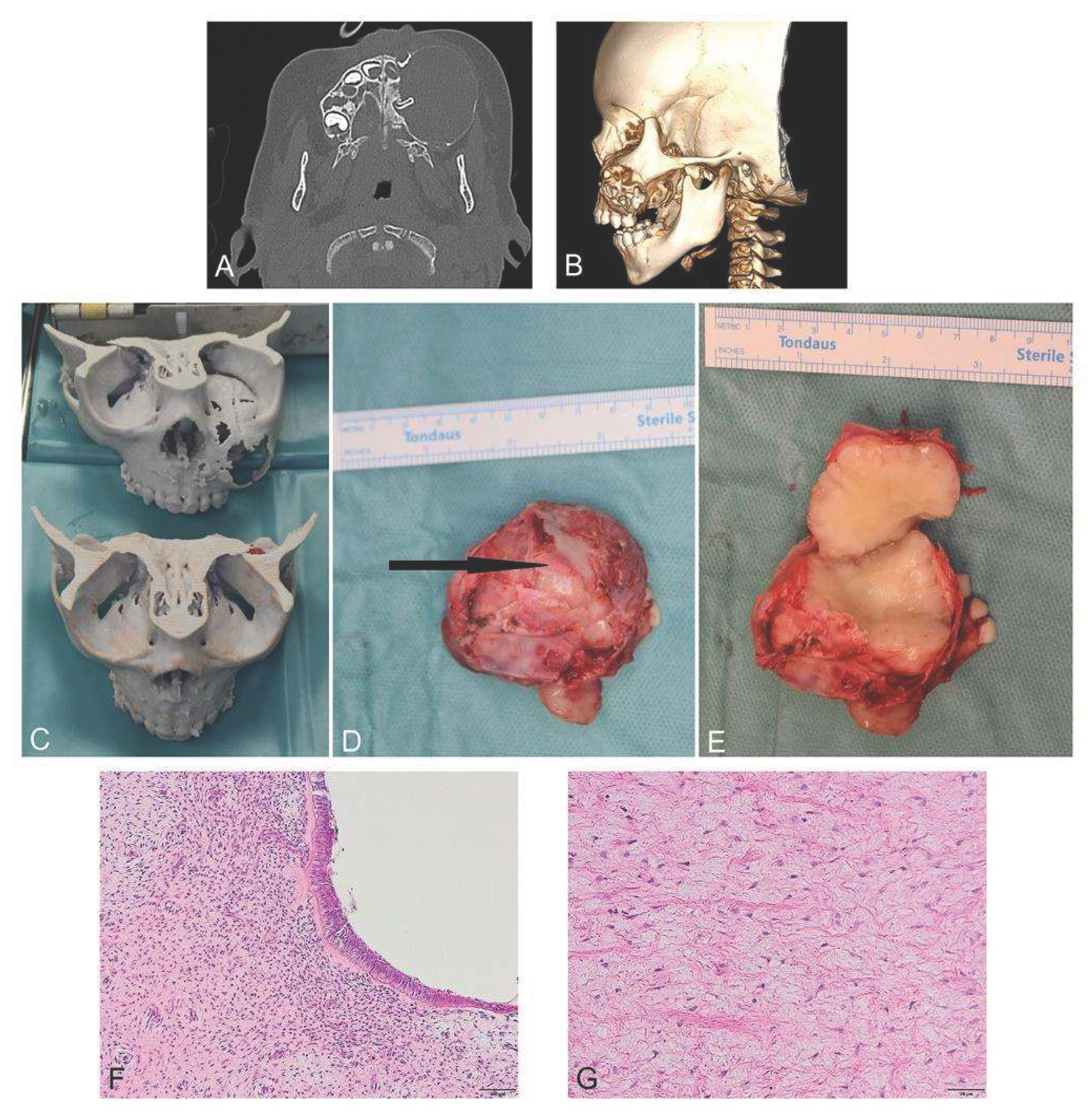
(A) Pretreatment CT, coronal view (B) Pretreatment CT, 3D reconstruction side view (C) 3D printed models used during surgery: upper model with tumor, lower one is designed by mirroring the right side and was used to bend the titanium reconstruction plate for the reconstruction of the floor of the orbit (D) Resected tumor, arrow pointing to the floor of the orbit, which was destroyed by the tumor (E) Cut surface of the tumor was glossy and pale yellow with lobulated on the periphery (F) Histopathologic appearance of primordial odontogenic tumor at low magnification (H&E, 10x), the tumor periphery is lined by a single layer of columnar, palisaded epithelial cells, beneath which are loosely arranged, reticulum-like mesenchymal cells resembling the stellate reticulum. (G) Histopathologic appearance of primordial odontogenic tumor at higher magnification (H&E, 20x), the tumor stroma reveals a fibromyxoid, loose connective tissue composed of stellate and fusiform fibroblasts, embedded within an abundant myxoid matrix;.
Figure 1.
(A) Pretreatment CT, coronal view (B) Pretreatment CT, 3D reconstruction side view (C) 3D printed models used during surgery: upper model with tumor, lower one is designed by mirroring the right side and was used to bend the titanium reconstruction plate for the reconstruction of the floor of the orbit (D) Resected tumor, arrow pointing to the floor of the orbit, which was destroyed by the tumor (E) Cut surface of the tumor was glossy and pale yellow with lobulated on the periphery (F) Histopathologic appearance of primordial odontogenic tumor at low magnification (H&E, 10x), the tumor periphery is lined by a single layer of columnar, palisaded epithelial cells, beneath which are loosely arranged, reticulum-like mesenchymal cells resembling the stellate reticulum. (G) Histopathologic appearance of primordial odontogenic tumor at higher magnification (H&E, 20x), the tumor stroma reveals a fibromyxoid, loose connective tissue composed of stellate and fusiform fibroblasts, embedded within an abundant myxoid matrix;.
3. Discussion
Primordial odontogenic tumor (POT) is an exceptionally rare benign mixed odontogenic neoplasm whose diagnosis remains challenging despite increasing recognition since its inclusion in the 2017 WHO Classification of Head and Neck Tumours [1]. While most published reports have focused on describing its rarity and histopathological characteristics, [1,2,3] the present case highlights several practical issues that are directly relevant to clinical management, demonstrating how diagnostic uncertainty may influence surgical decision-making in a very young child.
The most important lesson from this case concerns the limitations of incisional biopsy. The initial diagnosis of odontogenic myxoma was based on a small tissue specimen composed predominantly of myxoid ectomesenchymal tissue. Examination of the complete surgical specimen subsequently demonstrated the characteristic peripheral odontogenic epithelium required for the diagnosis of POT. This discrepancy should not be interpreted as a diagnostic error but rather as a consequence of sampling limitations inherent to heterogeneous biphasic lesions [8].
This observation has important clinical implications. Although histopathological examination remains the diagnostic gold standard, the reliability of an incisional biopsy depends on the representativeness of the sampled tissue. When clinical presentation, radiological findings, and histopathological features are not fully concordant, clinicians should consider multidisciplinary review, repeat biopsy, or referral to an oral and maxillofacial pathology center before undertaking definitive treatment. In rare odontogenic tumors, diagnosis should therefore be regarded as an integrative process rather than the interpretation of an isolated biopsy specimen. [9,10]
POT is generally considered a slow-growing lesion and most reported patients have undergone conservative surgical treatment [1,3]. However, the present case demonstrates that biological benignity does not always correspond to clinical behavior.
Our patient developed rapid expansion of the maxillary lesion over only a few months, resulting in marked facial asymmetry, displacement of the orbital floor, vertical strabismus, and visual disturbances. These manifestations reflected the anatomical constraints of the maxilla rather than aggressive tumor biology. Even relatively slow tumor growth may produce significant functional impairment when confined within the limited space of the midface [8].
This observation suggests that clinicians should maintain a low threshold for comprehensive radiological assessment whenever POT is suspected in the maxilla. Careful evaluation of orbital, nasal, and skull base involvement is essential because treatment decisions should be based not only on histological diagnosis but also on the functional consequences of tumor expansion.
The extent of surgery performed in the present case deserves particular consideration. At first glance, subtotal maxillectomy may appear excessive for a benign odontogenic tumor. However, the indication for radical resection was determined by the overall clinical scenario rather than by the final diagnosis alone. Several factors influenced treatment planning: the presumed diagnosis of odontogenic myxoma based on biopsy, extensive cortical destruction with loss of structural support, progressive orbital compression. Consequently, subtotal maxillectomy was considered the most appropriate approach to achieve complete tumor removal while preserving ocular function and enabling reconstruction.
This distinction is important because our findings should not be interpreted as supporting aggressive treatment for all cases of POT. On the contrary, they emphasize that surgical management must be individualized according to anatomical extent, radiological characteristics,diagnostic certainty and potential impact on adjacent structures . Multidisciplinary collaboration involving maxillofacial surgeons, radiologists, ophthalmologists, pediatric dentists, and oral pathologists remains essential for selecting the most appropriate therapeutic strategy [2,3,11].
Finally, the favorable postoperative outcome further supports this individualized approach. At 24 months of follow-up, no evidence of recurrence was observed, visual function had completely recovered, and satisfactory facial symmetry was maintained. Although recurrence appears to be uncommon, the limited number of reported cases and the young age of many affected patients justify prolonged clinical and radiological surveillance.
Rather than emphasizing the rarity of POT itself, this case illustrates a broader principle applicable to many uncommon odontogenic lesions: successful management depends on integrating clinical presentation, imaging findings, histopathology, and multidisciplinary expertise. Such an approach minimizes diagnostic uncertainty and facilitates treatment decisions that are both oncologically sound and functionally appropriate [12].
Although POT is exceptionally rare, the diagnostic challenge presented in this case reflects a broader problem encountered in the management of uncommon odontogenic lesions. Histopathological interpretation of limited biopsy material should always be integrated with radiological findings, patient age, lesion location, and clinical behavior. This multidisciplinary diagnostic approach is applicable not only to POT but also to other odontogenic tumors with overlapping clinicopathological features.

Table 1.
Comparison of clinical features of POT and OM with our case.. Key feature leading to misdiagnosis in the present case: incisional biopsy sampled only the myxoid stromal component, without the peripheral odontogenic epithelium characteristic of POT. Overall, most clinicoradiological features were more consistent with POT than with OM despite the misleading incisional biopsy. .
Table 1.
Comparison of clinical features of POT and OM with our case.. Key feature leading to misdiagnosis in the present case: incisional biopsy sampled only the myxoid stromal component, without the peripheral odontogenic epithelium characteristic of POT. Overall, most clinicoradiological features were more consistent with POT than with OM despite the misleading incisional biopsy. .
| TUMOR CLINICAL CHARACTERISTIC | Primordial Odontogenic Tumor | Odontogenic Myxoma | Our case (POT) |
| Age-related prevalence peak | 1st and 2nd decade [1] | 2nd and 3rd decade [13] | 1st decade |
| Typical location | Posterior mandible | Mandible | Maxilla |
| Radiological features | Well-defined, 81% UL [3] | 62% ML [13,14]- “tennis racket”/”honeycomb” | Well-defined, UL |
| Root resorption | 43% [3] | 10% [13,14] | Present |
| Association with unerupted tooth | 100% [1,2] | 5.3% [14] | Yes |
| Biological behavior | Benign | Locally invasive | Marked local expansion-compression of the eye |
| Usual treatment | 50% enucleation [3] | 49% resection [14] | Subtotal maxillectomy with ALT-flap reconstruction |
| Histology | Variably cellular fibrous tissue with areas similar to dental papilla, surrounded by epithelium resembling the internal epithelium of the enamel organ [1,2] | Loose myxoid stroma with a sparse population of stellate to spindle cells [13] | From incisional biopsy-OM Resection specimen- POT |
| Recurrence rate | 2,7% [3] | 14,4% [14] | No recurrence, 24 month follow-up |
The comparison presented in Table.1. demonstrates that several clinicoradiological characteristics favored POT over odontogenic myxoma in retrospect, despite the misleading biopsy findings. This observation underlines the importance of considering the overall diagnostic context rather than relying exclusively on histopathology obtained from limited tissue samples.
From a surgical perspective, this case also illustrates that treatment planning should be driven primarily by the anticipated functional consequences of the lesion rather than by histological diagnosis alone. In pediatric patients, preservation of vision, craniofacial growth, and future reconstructive options may justify treatment strategies that differ from those expected solely on the basis of tumor biology.
4. Conclusions
This case provides three practical messages for clinicians managing pediatric odontogenic tumors.
First, incisional biopsy may be insufficient for establishing the diagnosis of biphasic odontogenic tumors such as primordial odontogenic tumor. Histopathological findings should always be interpreted in conjunction with clinical and radiological information, particularly when biopsy results are inconsistent with the patient's age, imaging characteristics, or clinical course.
Second, benign histopathology should not be equated with benign clinical behavior. Although primordial odontogenic tumor is regarded as a non-aggressive neoplasm, lesions arising in the maxilla may produce rapid functional deterioration because of their proximity to the critical anatomical structures.
Finally, treatment should be individualized through multidisciplinary clinicopathological correlation rather than determined by histopathology alone. Surgical planning should consider not only the pathological diagnosis but also tumor extent, anatomical location, anticipated functional consequences, and diagnostic certainty. In the present case, subtotal maxillectomy was dictated mostly by extensive local destruction and orbital involvement.
As awareness of primordial odontogenic tumor continues to increase, recognition of these diagnostic and therapeutic principles may help optimize patient outcomes while avoiding both under- and overtreatment of this rare lesion.
Author Contributions
Conceptualization, Z.K.S.; methodology, Z.K.S., D.R; validation, K.D.; formal analysis, K.D., M.B., Ł.K.; investigation, Z.K.S., D.R.; resources, A.S.; data curation, Z.K.S., D.R.; writing—original draft preparation, Z.K.S., D.R, M.S.; writing—review and editing, K.D., M.B., Ł.K.; visualization, Z.K.S., D.R.; supervision, K.D.; project administration, Z.K.S.; All authors have read and agreed to the published version of the manuscript.
Funding
This research received no external funding.
Institutional Review Board Statement
A notification regarding the intended publication of this case report with literature review was submitted to the Bioethics Committee of Regional Medical Council of Warmia and Mazury, which approved this study on 01/04/2026 (decision number: L.Dz. WMIL-KB/21/2026).
Informed Consent Statement
Written informed consent has been obtained from the patient’s parents to publish this paper.
Data Availability Statement
The data supporting the findings of this case report are available from the corresponding author on reasonable request.
Acknowledgments
We thank parents of our patient for giving their consent for this publication.
Conflicts of Interest
The authors declare no conflicts of interest.
Abbreviations
The following abbreviations are used in this manuscript:
| ALT | Anterolateral Thigh Flap |
| ax | axis |
| CT | Computed Tomography |
| Dcyl | Cylindrical Diopters |
| MRI | Magnetic Resonanse Imaging |
| ML | Multilocular |
| OM | Odontogenic Myxoma |
| POT | Primordial Odontogenic Tumor |
| UL | Unilocular |
| WHO | World Health Organization |
References
- Mosqueda-Taylor, A.; Neville, B.W. Primordial odontogenic tumour. In WHO Classification of Tumours of the Head and Neck; El-Naggar, A.K.; Chan, J.K.C.; Grandis, J.R.; Takata, T.; Slootweg, P.J., Eds.; IARC Press: Lyon, France, 2017; pp. 223–224.
- Mosqueda-Taylor, A.; Pires, F.R.; Aguirre-Urízar, J.M.; Carlos-Bregni, R.; de la Piedra-Garza, J.M.; Martínez-Conde, R.; Ruíz-Godoy Rivera, L.M.; Delgado-Azañero, W.; de Almeida, O.P. Primordial odontogenic tumour: Clinicopathological analysis of six cases of a previously undescribed entity. Histopathology 2014, 65, 606–612. [CrossRef]
- Almazyad, A.; Alhumaidan, A.; Almarwan, M.; Almadan, N. Primordial odontogenic tumor: A decade post-description systematic review. Head Neck Pathol. 2024, 18, 105. [CrossRef]
- Brierley, D.J.; Speight, P.M.; Hunter, K.D. Odontogenic tumours. Diagn. Histopathol. 2015, 21, 370–379. [CrossRef]
- Poomsawat, S.; Ngamsom, S.; Nonpassopon, N. Primordial odontogenic tumor with prominent calcifications: A rare case report. J. Clin. Exp. Dent. 2019, 11, e952–e956. [CrossRef]
- Figueredo Junior, J.M.; Vanini, J.V.; Carvalho, G.L.; et al. Primordial odontogenic tumor in anterior mandible: A case image. Head Neck Pathol. 2025, 19, 58. [CrossRef]
- Gagnier, J.J.; Kienle, G.; Altman, D.G.; Moher, D.; Sox, H.; Riley, D.; CARE Group. The CARE guidelines: Consensus-based clinical case reporting guideline development. Glob. Adv. Health Med. 2013, 2, 38–43. [CrossRef]
- Hunter, K.D.; Niklander, S. Pitfalls in odontogenic lesions and tumours: A practical guide. Diagn. Histopathol. 2020, 26, 173–180. [CrossRef]
- Strokov, S.; Cardot-Leccia, N.; Raybaud, H.; et al. Cysts of the jaws and how to make their diagnoses under a microscope: A need for a better communication between clinicians and pathologists. J. Oral Med. Oral Surg. 2024, 30, 8. [CrossRef]
- Labib, A.; Adlard, R.E. Odontogenic tumors of the jaws. In StatPearls; StatPearls Publishing: Treasure Island, FL, USA, 2023.
- Azzi, L.; Tettamanti, L.; Di Francesco, A.; Cerati, M.P.; Tagliabue, A.; Farronato, D.; Maurino, V. Primordial odontogenic tumour: A systematic review of the common but also unusual features of this novel entity. J. Stomatol. Oral Maxillofac. Surg. 2020, 121, 408–417. [CrossRef]
- Kansy, K.; Juergens, P.; Krol, Z.; Paulussen, M.; Baumhoer, D.; Bruder, E.; Schneider, J.; Zeilhofer, H.-F.; Schwenzer-Zimmerer, K. Odontogenic myxoma: Diagnostic and therapeutic challenges in paediatric and adult patients—A case series and review of the literature. J. Craniomaxillofac. Surg. 2012, 40, 271–276. [CrossRef]
- WHO Classification of Tumours Editorial Board. Head and Neck Tumours, 5th ed.; International Agency for Research on Cancer: Lyon, France, 2023; Vol. 9. Available online: IARC WHO Head and Neck Tumours Volume (accessed on 29 July 2026).
- Lam-Ubol, A.; Naorungroj, S.; Sappayatosok, K. Management and Recurrence of Intra-Osseous Odontogenic Myxoma: Systematic Review and Individual Participant Data Meta-Analysis. Eur. J. Dent. 2026, Advance online publication. [CrossRef]
Disclaimer/Publisher’s Note: The statements, opinions and data contained in all publications are solely those of the individual author(s) and contributor(s) and not of MDPI and/or the editor(s). MDPI and/or the editor(s) disclaim responsibility for any injury to people or property resulting from any ideas, methods, instructions or products referred to in the content. |
© 2026 by the authors. Licensee MDPI, Basel, Switzerland. This article is an open access article distributed under the terms and conditions of the Creative Commons Attribution (CC BY) license (http://creativecommons.org/licenses/by/4.0/).
Copyright: This open access article is published under a Creative Commons CC BY 4.0 license, which permit the free download, distribution, and reuse, provided that the author and preprint are cited in any reuse.