

Submitted:
05 August 2026
Posted:
07 August 2026
You are already at the latest version
Abstract
Gut microbial metabolites serve as functional mediators of gut–brain communication, linking microbial alterations to neurodegenerative pathology. How metabolite profiles shift during disease progression and interact with host genetic susceptibility remains poorly characterized. This review centers on Alzheimer’s disease (AD) as the primary model, with Parkinson’s disease (PD) and amyotrophic lateral sclerosis (ALS) included for cross-disease comparison. Across the AD continuum, microbial metabolic remodeling features reduced protective metabolites and elevated inflammation-related metabolites. Short-chain fatty acids (SCFAs), particularly butyrate, and indole-derived metabolites are altered from early cognitive impairment to clinical dementia. Trimethylamine N-oxide (TMAO), kynurenine intermediates and abnormal bile acid profiles accumulate and drive neuroinflammatory and metabolic disturbances. Mechanistically, metabolic shifts affect AD pathology through three interrelated pathways. Tryptophan-derived metabolites regulate immune homeostasis via aryl hydrocarbon receptor (AhR) signaling. SCFAs modulate epigenetic processes linked to Aβ and tau lesions. Intestinal and blood-brain barrier damage allows peripheral metabolic and inflammatory signals to reach the central nervous system. APOE4 alters lipid metabolism and systemic inflammation to modify individual metabolic susceptibility. Shared metabolic abnormalities including SCFA loss and barrier damage exist in PD and ALS, alongside disease-specific metabolic changes. Existing data support associations among host factors, the microbiome, and metabolite profiles, but longitudinal target-engagement studies remain limited.
Keywords:
gut-brain axis
; microbial metabolites
; neurodegenerative diseases
; Alzheimer’s disease
; APOE4
1. Introduction
Neurodegenerative diseases place an expanding burden on ageing societies. The number of people living with dementia was estimated at 57.4 million in 2019 and is projected to reach 152.8 million by 2050, largely because of population growth and ageing [1]. Incidence has declined in some high-income cohorts, but this pattern is not geographically uniform and does not offset the projected increase in absolute case numbers [2]. Parkinson’s disease is also becoming more prevalent [3]. Amyotrophic lateral sclerosis is rarer, progresses rapidly, and remains difficult to quantify across regions with unequal registry coverage [4]. These disorders affect different neural systems and express distinct protein pathologies, yet they converge on progressive neuronal loss, protein aggregation, disrupted energy homeostasis, inflammation, and network failure [5]. Alzheimer’s disease provides the main clinical continuum for examining how these processes develop before and after cognitive impairment.
Biomarkers have moved the field closer to that presymptomatic interval. Amyloid and tau imaging, cerebrospinal fluid assays, and blood measurements can detect cerebral pathology before dementia and can support diagnosis, trial enrolment, and treatment monitoring [6]. Parkinson’s disease is undergoing a similar shift as α-synuclein seed amplification assays identify pathological protein in cerebrospinal fluid and other biospecimens, although their prognostic value and sensitivity to progression remain under evaluation [7]. Osteopontin illustrates why timing still complicates interpretation. Cerebrospinal fluid levels were elevated in biomarker-positive individuals without dementia and were associated with faster subsequent cognitive decline. Its relationship with conversion weakened after established AD biomarkers were included [8]. A detectable signal can therefore carry stage-dependent information without defining an independent disease trajectory.
Metabolic measurements face an additional spatial problem. Post-mortem brain tissue offers anatomical detail but represents a late and non-repeatable observation. Cerebrospinal fluid is closer to the central nervous system, whereas blood is easier to sample repeatedly but incorporates metabolism from multiple organs. Paired plasma and cerebrospinal fluid profiling in AD separated systemic disturbances in energy metabolism from central alterations in tryptophan metabolism and creatinine handling. Some peripheral changes reached the central compartment in relation to blood-brain barrier (BBB) permeability, while others remained compartment-specific [9]. No single biospecimen reconstructs the full route from production to neural exposure. Gut microbial metabolism adds an accessible functional layer to this route. Microbes transform dietary and host substrates into short-chain fatty acids, tryptophan derivatives, modified bile acids, and precursors of host-microbial co-metabolites. These products can act in the intestine or enter host circulation, where absorption, hepatic conversion, renal clearance, and barrier passage reshape the measured concentration [10,11].
Human studies now connect this metabolic capacity with AD-related phenotypes. Fecal short-chain fatty acids and propionate-producing bacteria vary with sex and amyloid status. Several associations extend to cerebrospinal fluid markers, plasma phosphorylated tau accumulation, and cognitive change, but the observed relationships do not establish a uniform protective trajectory or direct brain exposure [12]. Taxonomic signatures create a related problem. A large meta-analysis in Parkinson’s disease detected recurrent microbial and functional differences, yet prediction models trained in one study transferred poorly to other cohorts [13]. Diet, medication, geography, intestinal transit, analytical platform, and disease definition all contribute to this instability. Most AD studies also compare different participants at one time point. Group differences across cognitively unimpaired, subjective impairment, mild cognitive impairment, and dementia cannot be assembled into the longitudinal history of one patient.
The central problem is therefore temporal as well as mechanistic. Gut microbiota-related metabolites may change across the AD continuum, but their order, tissue exposure, and pathological meaning remain uncertain. AD is used here to examine stage-associated metabolic patterns, the microbial and host processes that generate them, and the sources of variation in individual transition points. The same evidence is then tested against the requirements for metabolic target engagement and clinical translation. Comparisons with Parkinson’s disease and amyotrophic lateral sclerosis determine whether shared metabolic pathways retain the same effects across distinct proteinopathies and vulnerable neural systems. This approach treats microbial metabolism as a changing interface between disease stage, host physiology, and neurodegenerative pathology rather than as a fixed diagnostic signature.
2. Gut Microbiota-Derived Metabolic Alterations Across the AD Continuum
Alzheimer’s disease develops along a continuum from subjective cognitive decline through mild cognitive impairment to clinically diagnosed dementia [14]. Fermentation-related alterations are already detectable during subjective cognitive decline (SCD) and biomarker-defined preclinical AD. Mild cognitive impairment (MCI) retains these features while adding broader changes in amino acid products and circulating host-microbial co-metabolites. Clinical AD involves bile acid, nitrogen, and lipid metabolism across intestinal and systemic compartments. SCFAs provide a useful reference within this pattern because they are major products of microbial fermentation of dietary substrates [15].
2.1. Early Prodromal Stages
2.1.1. Subjective Cognitive Decline
Direct metabolic evidence in SCD is limited. Biomarker-defined preclinical AD offers an adjacent view before objective cognitive impairment, but the two states are not interchangeable. Cognitively unimpaired adults with amyloid pathology already differ from amyloid-negative controls in microbial composition and predicted function [16]. Combining fecal microbial features with plasma amyloid measures also distinguishes these groups [17]. Neither study defines a metabolic profile unique to SCD.
Fermentation-related changes provide the clearest early signal. Paired fecal and plasma multi-omics detected cognition-associated metabolic alterations in both compartments, with fecal changes appearing before corresponding plasma differences [18]. Fecal propionate and propionate-producing bacteria varied with sex and amyloid status in another human cohort [12]. These measurements capture different biological processes. Feces contain metabolites remaining after local production and absorption. Blood concentrations additionally reflect tissue use and clearance.
Serial perioperative sampling shows how the prodromal gut responds to physiological stress. Before surgery, participants with SCD or amnestic MCI had fewer SCFA-producing bacteria and greater evidence of intestinal barrier disturbance than cognitively normal participants. Surgery widened these differences [19]. This design tests susceptibility to an acute challenge rather than natural progression from SCD to MCI.
Temporal evidence is clearer in animals. Longitudinal sampling of an AD mouse model showed that butyrate-producing bacteria and cecal butyrate declined before overt neuropathology and memory impairment [20]. Human studies place fermentation-related abnormalities near the preclinical phase. The temporal order among microbial change, amyloid deposition, and subjective symptoms remains unresolved at the individual patient level.
2.1.2. Mild Cognitive Impairment
MCI contains a more reproducible fermentation deficit. Faecalibacterium prausnitzii, a prominent butyrate producer, is depleted and correlates with cognitive performance [21]. Lower abundance of other SCFA-producing taxa accompanies higher odds of amyloid and phosphorylated tau positivity [22]. Brain imaging adds another layer. Fewer butyrate producers occur alongside greater free water in MCI and AD [23]. These studies converge on fermentation-related ecology, not on one diagnostic bacterium.
Community differences extend into microbial functions. Shotgun metagenomics identifies coordinated species and pathway alterations related to cognition and AD biomarkers [24]. A smaller comparison found substantial overlap between MCI and AD-associated profiles [25]. Mediterranean cohorts likewise separated cognitively impaired participants from healthy aging, although the contributing taxa differed between populations [26,27]. The recurring case-control separation is more consistent than any universal taxonomic list.
Longitudinal studies narrow the possible direction of change. Microbial genes involved in the urea cycle, polyamine synthesis, and methionine or cysteine metabolism predicted poorer cognitive performance in older adults at risk for AD [28]. Baseline community features also predicted subsequent cognitive and depressive symptoms during a two-year follow-up [29]. These outcomes concern symptom change, not conversion to AD dementia.
Intestinal physiology shapes the same metabolic environment. Infrequent bowel movements were associated with fewer butyrate-producing species and poorer cognition in a large population study [30]. Bowel frequency is not a metabolite biomarker. It influences transit time and the ecological conditions under which microbial fermentation occurs.
Blood profiling adds a systemic layer at MCI. Serum tryptophan products, indoxyl sulfate, choline-related compounds, and cresol metabolites distinguished cognitively healthy adults, subjective impairment, and MCI [31]. Plasma TMAO was higher in participants with MCI in a population with elevated cardiovascular risk [32]. The cardiovascular setting is part of that association. Direct metabolomic comparisons further identified altered SCFAs, lithocholic acid, and tryptophan products across amnestic MCI and AD [33]. The MCI profile therefore extends beyond fermentation without yielding a single stage-specific metabolite.
2.2. Clinical Dementia
In clinical AD, measured differences span the largest number of compartments. Fecal metagenomic functions differ between people with dementia and cognitively unimpaired controls, and selected features correlate with cerebrospinal fluid markers of AD pathology [34]. Blood and cerebrospinal fluid analyses add immune, proteomic, and tryptophan-related signatures linked to amyloid and tau status [35]. The observed profile is no longer confined to intestinal output.
Bile acid composition changes in a relatively consistent direction. Serum analyses show lower primary cholic acid together with higher bacterially modified secondary species and ratios in AD [36]. Individual bile acids correlate with cognition, brain atrophy, glucose metabolism, and cerebrospinal fluid amyloid or phosphorylated tau [37]. Two large biomarker-defined cohorts provide longitudinal evidence. Baseline lithocholic and deoxycholic acids predicted subsequent amyloid and tau progression [38]. Recurrent redistribution of the bile acid pool is better supported than any single late-stage marker.
Other products broaden the systemic profile. Cerebrospinal fluid TMAO is elevated in MCI and AD and correlates with AD biomarkers and neuronal injury [39]. Large-scale metabolic phenotyping links conjugated bile acids, branched-chain amino acids, glutamate-related features, and ammonia homeostasis with cognitive stage and amyloid burden [40]. Reviews report substantial variation in the direction and strength of individual gut-derived compounds [10]. TMAO and ammonia belong to a wider state of host-microbial co-metabolism.
Animal multi-omics helps connect the compartments. Established pathology in APP/PS1 mice coincides with microbiota-associated differences in fecal, serum, and cortical metabolites, including bile acids and unsaturated fatty acids [41]. Transfer of an aged AD-associated community to younger transgenic mice aggravates pathology and reproduces lipid metabolic disruption [42]. These experiments show that dysbiosis can reorganize peripheral and cerebral metabolism together. Whether humans follow the same order has not been tested longitudinally.
2.3. Temporal Metabolic Trajectories and Biomarker Potential
Cross-stage studies yield a relative ordering rather than fixed thresholds. Microbial network organization differs among SCD, MCI, and AD even when individual taxa do not change monotonically [43]. Multi-omics analyses identify progressive links among microbial composition, fecal metabolites, brain structure, and cognition [44]. Multimodal profiles distinguish the clinical stages most effectively when microbiomic and metabolomic features are combined with imaging and clinical variables [45].
The ordering occurs at several measurement levels. Fecal fermentation-related features appear early. MCI adds microbial pathway and circulating metabolite differences. Dementia is accompanied by wider systemic involvement. A large multi-omics study independently connects gut microbial features with cognition, hippocampal volume, serum metabolites, and inflammation, while showing that these data layers need not move in parallel [46]. Fecal and circulating measurements should therefore be treated as complementary rather than interchangeable.
Most human studies compare separate diagnostic groups. Diagnostic definitions, diet, medication, geography, sequencing methods, and metabolomic platforms vary substantially [47,48]. Several mechanistic metabolite findings remain better established in animals than in patients [49]. Small datasets and platform dependence also restrict the portability of multi-omics classifiers [50]. These design features prevent reconstruction of one person’s trajectory from SCD to dementia.
Circulating profiles contain additional host variation. Genetic background, microbial activity, lifestyle, comorbidities, and medication jointly contribute to blood metabolite concentrations [51]. Population averages can still identify a broad metabolic ordering, but they cannot assign a universal transition point. The same stage may therefore contain distinct metabolic states, and similar metabolite profiles may carry different pathological consequences.
3. Microbial Metabolic Mechanisms Across the AD Continuum
Stage-associated profiles raise two mechanistic questions: why do fermentation products, circulating co-metabolites, and bile acids gain prominence at different stages of the AD continuum, and how do these shifts modify pathology? Microbial production is only one determinant. Host conversion, clearance, barrier integrity, and tissue-specific receptor distributions jointly shape brain exposure and cellular response. The interaction of these variables provides a working framework for the relative ordering observed in human cohorts, but it does not define a fixed sequence for individual patients.
3.1. Early Loss of Metabolic Resilience
3.1.1. SCFA-Dependent Homeostasis
SCFAs connect microbial fermentation with epithelial, immune, and cellular metabolism. Acetate, propionate, and butyrate signal through G protein-coupled receptors, inhibit histone deacetylases, support epithelial junctions, and regulate immune activity [52]. SCFA-sensitive pathways also shape microglial maturation and inflammatory function [53]. A decline in fermentation capacity can therefore weaken several homeostatic systems before one circulating metabolite becomes dominant.
Microglial metabolism responds directly to acetate. Germ-free mice showed epigenetic changes in microglial metabolic genes, increased mitochondrial mass, and respiratory-chain dysfunction. Acetate restored metabolic fitness and modified microglial phagocytosis during neurodegeneration [54]. Acetyl-CoA synthetase 2 links acetate availability to chromatin regulation. Acetate replenishment increased histone acetylation, glutamate-receptor expression, synaptic plasticity, and cognition in 5xFAD mice in an ACSS2-dependent manner [55]. SCFAs also strengthened the astrocyte-neuron glutamate-glutamine shuttle and the supply of glutathione precursors, thereby reducing neuronal oxidative injury in experimental systems [56].
Direct glial metabolism does not account for the entire SCFA effect. Propionate also altered the peripheral immune signal reaching the brain. In male APPPS1-21 mice, propionate reduced RORγt-positive CD4+ T cells and IL-17 secretion. IL-17 levels correlated positively with reactive astrocytosis. Depletion of IL-17 showed that the reductions in astrocyte reactivity and Aβ plaques depended on this cytokine pathway [52]. Gut bacterial depletion likewise lowered IL-17A-expressing T cells, brain inflammation, and Aβ, while Il-17a deficiency abolished these effects [57]. A commentary interpreted the propionate study within the gut-brain-immune axis but added no independent experimental replication [58].
The direction of influence is not restricted to gut-to-brain signaling. Once amyloid pathology develops, Aβ can injure the enteric nervous system and alter the intestinal environment. Aβ disrupted synaptic proteins and neuronal connectivity in primary enteric cultures, human intestinal organoids, and colon explants. In SAMP8 mice, butyrate reduced Aβ secretion and limited amyloid accumulation in the gut, plasma, and brain [59]. This reciprocal mechanism is experimentally supported, but its position along the human AD continuum has not been established.
3.1.2. Receptor-Specific Indole Signaling
The microbiota regulates three major branches of tryptophan metabolism that generate indole derivatives, kynurenines, and serotonin [60]. Their products differ in receptor affinity, redox activity, and tissue access [61]. Total tryptophan turnover therefore gives little information about the biological direction of the resulting signal.
Individual indoles recruit distinct cellular programs. Indole-3-lactic acid reduced soluble Aβ in 5xFAD mice through an AhR-dependent response involving microglia and astrocytes [62]. Indole-3-propionic acid crossed the blood-brain barrier and engaged neuronal PXR. Blocking PXR or disrupting IPA synthesis abolished the cognitive and amyloid-related effects in experimental models [63]. Indole-3-acetic acid acted on microglial synaptic engulfment. It reduced CCR4 expression and synapse loss in AD models, while circulating IAA was lower in patients and correlated inversely with cognitive impairment [64].
AhR connects several of these responses. Indole, IAA, and IPA increased AhR activity and suppressed NF-κB signaling, NLRP3 inflammasome assembly, and inflammatory cytokine release in APP/PS1 mice [65]. Neuronal AhR activation also increased neprilysin transcription and Aβ-degrading activity [66]. A broader review links 5-hydroxyindoleacetic acid (5-HIAA) and kynurenic acid with metalloproteinases involved in cerebral Aβ clearance [67].
Multi-metabolite experiments require narrower interpretation. Akkermansia muciniphila administration increased IAA, tryptophan, acetate, and other metabolites in APP/PS1 mice. The shifts accompanied stronger AhR-related signaling, lower neuroinflammation, and less Aβ deposition [68]. IAA therefore forms part of a broader metabolite response whose individual contributions remain unresolved.
3.2. Expansion of Metabolic Injury
3.2.1. Circulating Toxic Metabolites
Imidazole propionate (ImP) combines a human exposure signal with experimental vascular and neuronal effects. Among cognitively unimpaired adults, higher plasma ImP was associated with poorer cognition and ADRD biomarkers in cross-sectional and longitudinal analyses. Chronic exposure aggravated pathology in mice, impaired brain endothelial integrity, and promoted tau phosphorylation in primary neurons. GSK3β inhibition blocked the neuronal effect [69]. Independent structural work shows that GSK3β phosphorylation can catalyze tau assembly into filaments resembling those found in AD brains [70].
TMAO reaches tau pathology through more than one route. TMAO bound HIF1α, inhibited its signaling, increased oxidative stress, and promoted tau phosphorylation in cellular and P301S models [71]. A separate metabolite screen identified TMAO, indoxyl sulfate, and several other microbiome-dependent compounds as promoters of tau seeding. Systemic administration worsened cognition and tau pathology in mice [72].
Human association and experimental mechanism remain distinct. Plasma TMAO was higher in participants with MCI. In rats, chronic choline exposure reduced inhibitory Ser9 phosphorylation of GSK3β and lowered proteins involved in synaptic plasticity. Blocking TMAO production or GSK3β prevented these deficits [73]. The GSK3β-dependent synaptic route is experimentally established, whereas its contribution to human disease progression remains undetermined.
Kynurenine products can act in opposite directions. Oral Porphyromonas gingivalis exposure increased 3-hydroxykynurenine in serum and hippocampus. This metabolite suppressed BCL2 and promoted neuronal apoptosis in mice and cell models [74]. In another AD mouse model, microbial modulation increased hippocampal kynurenic acid. This increase was associated with metabolic changes consistent with enhanced neuronal fatty acid oxidation, reduced lipid accumulation, and suppressed microglial activation [75]. Biological direction depends on the metabolite branch and experimental context, not on kynurenine metabolism as a single category.
3.2.2. Bile Acid and Lipid Remodeling
Human evidence supports redistribution of the circulating bile acid pool. Primary cholic acid is often lower, whereas several conjugated and secondary species are higher in dementia. Longitudinal studies support this pattern, although individual bile acids vary across cohorts [76]. Serum profiling detected stage-related and sex-related differences. Selected changes appeared before clinical AD in men and improved predictive models [77]. A small cerebrospinal fluid study found hydroxybutyrate and bile acid differences among controls, MCI due to AD, and dementia [78]. These studies establish remodeling more firmly than a microbial or receptor-level cause in patients.
TGR5 experiments expose the importance of disease stage and cell identity. Early in AD mice, deoxycholic acid and neuronal TGR5 increased. TGR5 recruited a STAT3-APH1-γ-secretase pathway and promoted amyloidogenic APP processing in excitatory neurons [79]. During middle and late stages, TGR5 declined in medial septal cholinergic neurons. Local activation increased cholinergic activity, hippocampal neurogenesis, and cognition through the medial septal-to-dentate gyrus circuit [80]. The opposing results arise in different cell populations and disease windows. Ligand concentration remains an untested source of variation.
Lipid effects are equally molecule-specific. A Bacteroides ovatus-associated lysophosphatidylcholine activated GPR119, lowered ACSL4 expression, and restrained ferroptosis in a 5xFAD model. Fecal and serum LPC levels were also lower in individuals with AD [81]. Integrated serum and brain metabolomics linked gut microbial changes with glycerophospholipid disruption, glial activation, and neuroinflammation in APP/PS1 mice [82]. Conversely, host-derived 27-hydroxycholesterol disturbed microbial composition, lowered fecal SCFAs, and damaged intestinal junctions [83]. Lipid remodeling can thus transmit gut-to-brain effects or carry brain and host metabolic stress back to the intestine.
3.3. Barrier Failure and Neuroimmune Amplification
Barrier integrity determines tissue exposure to microbial products and metabolites. Mice without a gut microbiota had greater blood-cerebrospinal fluid barrier permeability and disorganized tight junctions. Recolonization or SCFAs restored junctional organization. SCFAs also altered microglial phenotype and reduced Aβ in APP-knock-in mice [84]. Intestinal epithelial Dicer1 deletion changed bacterial abundance, increased LRP1 and ABCB1 at the blood-brain barrier, and lowered cerebral Aβ [57]. This experiment concerns epithelial control and Aβ transport rather than a direct metabolite effect.
M cells, LPS, and bacterial vesicles occupy the exposure arm of this pathway. M-cell depletion changed the gut community and reduced colonic inflammation, amyloid, microglial dysfunction, and memory impairment in 5xFAD mice [85]. LPS is a bacterial structural component, not a metabolite. Its blood and brain levels are elevated in AD, while experimental exposure activates innate immunity and AD-related pathology [86]. LPS-bearing vesicles crossed the blood-brain barrier, activated microglial Piezo1, and triggered C1q-C3-dependent synaptic pruning [87]. Commensal vesicles also reversed the less inflammatory microglial state of germ-free AD mice and increased amyloid pathology [88].
Metabolites shape the response after exposure. Bacteroides fragilis produced 12-hydroxy-heptadecatrienoic acid and prostaglandin E2. Both lipids activated microglia and recruited neuronal C/EBPβ and asparaginyl endopeptidase, followed by Aβ and tau pathology in mice [89]. Microbial GABA has been linked to mucin and tight-junction regulation, although its AD-specific mechanism remains inferential [90]. A mechanistic review integrates microbiota-sensitive microglial responses with neuroinflammation and synaptic dysfunction [91]. A knowledge-driven network prioritized links among microbial metabolites, microglial genes, and AD phenotypes, with SCFAs ranking highly. These computational relationships require experimental confirmation [92].
Human data capture this interface as a combined signature. Fecal microbial features correlate with circulating LPS, endothelial markers, cytokines, amyloid, phosphorylated tau, and neurodegeneration measures [93]. Cross-sectional sampling cannot order them. After barrier crossing, gastric Helicobacter pylori vesicles activated C3-C3aR signaling across astrocytes, microglia, and neurons and aggravated amyloid in mice [94]. This is complement amplification by a structural product, not a metabolic pathway.
3.4. Temporal Dynamics and Reciprocal Feedback
Different metabolite classes follow different production and handling routes. Diet, antibiotics, and microbial metabolism directly influence SCFA availability in the gut and other organs [95]. TMAO requires microbial and host conversion. Enterobacteriaceae reduce TMAO to trimethylamine, which enters the circulation and undergoes hepatic reoxidation [96]. Kidney function further changes systemic exposure. TMAO was not associated with incident dementia in a large population cohort overall, but the association emerged among participants with impaired renal function [97].
Bile acids pass through a longer host-microbial circuit. The liver synthesizes primary species, enterohepatic circulation recycles them, and microbial enzymes generate secondary forms. Diet, antibiotics, and intestinal transit reshape the pool [98]. These routes make unequal stage prominence biologically plausible. They do not prove that SCFAs, TMAO, and bile acids change in a fixed order within the same patient.
Established brain pathology can then alter the metabolic source. Ventricular Aβ injection changed the mouse gut microbiota after four weeks, damaged colonic structure, and suppressed cholinergic anti-inflammatory signaling [99]. In 5xFAD mice, amyloid pathology reorganized the colonic immune compartment and altered the distribution of gut-associated immune cells in the brain and meninges [100]. The intestinal ecosystem is therefore both a source of signals and a target of cerebral pathology.
Oxidative stress can reinforce this loop. Dysbiosis-related oxidative and inflammatory signals can weaken intestinal and brain barriers, expanding peripheral exposure and renewing innate immune activation [101]. Reviews consequently describe the relationship between gut dysbiosis and AD pathology as bidirectional [102]. Longitudinal network analysis supplies an ecological example. During progression in mice, the microbial network shifted from scale-free to random organization and reached marked disequilibrium at the late stage. Functional modeling implicated a hub taxon in quinolinic acid synthesis [103].
Evidence remains uneven. SCFA homeostasis, indole signaling, barrier amplification, and brain-to-gut feedback recur across experimental settings. Human data mainly compare separate clinical groups, and much of the mechanistic literature centers on amyloid [104]. Animal age does not correspond directly to SCD, MCI, or dementia. The temporal order remains a model for population-level stage patterns, not an individual timetable.
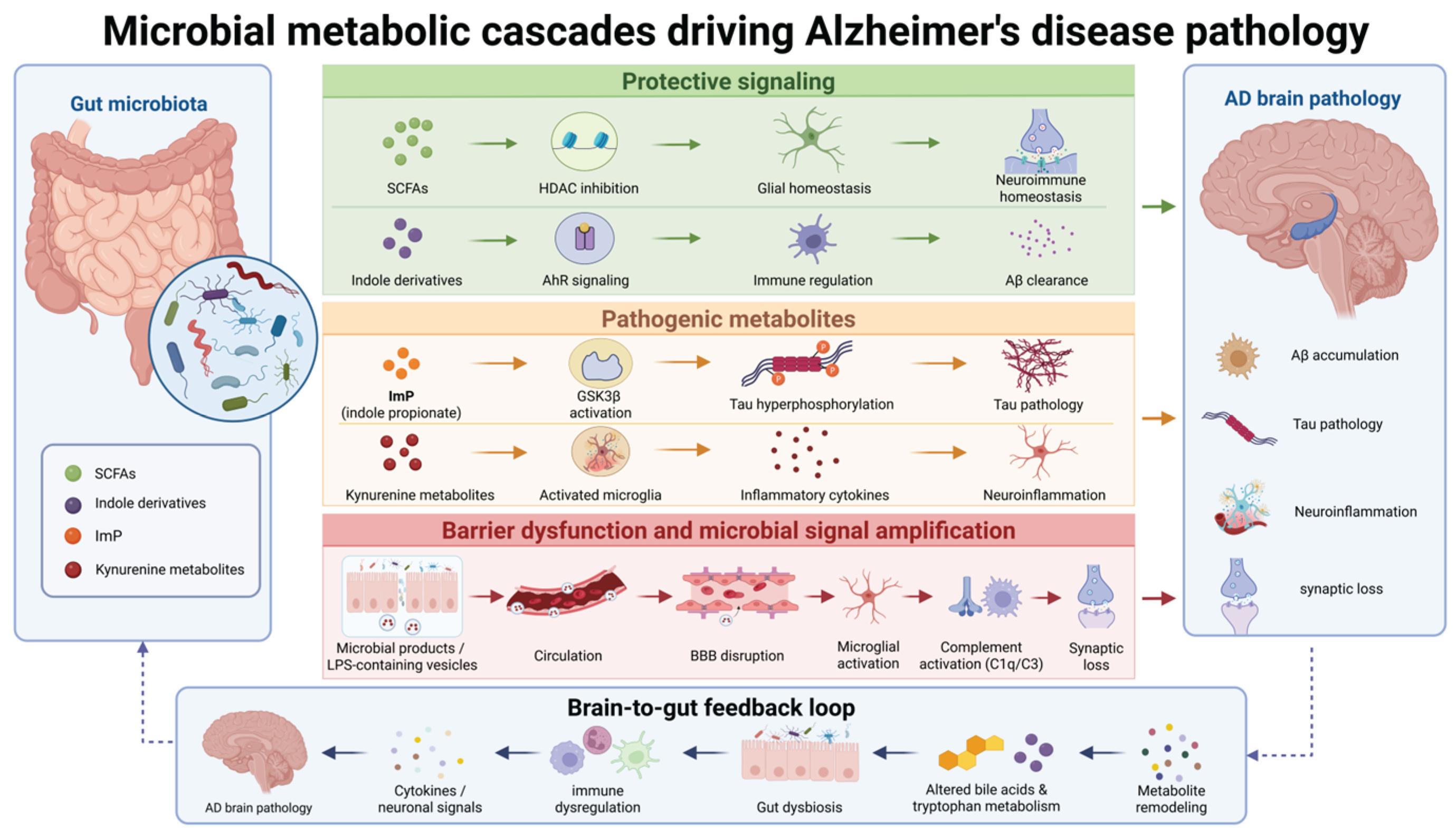
Figure 1.
Microbial metabolic cascades driving Alzheimer’s disease pathology. This schematic illustrates how gut microbial metabolites influence AD pathology through protective signaling, metabolic injury, barrier dysfunction, and gut-brain feedback. SCFAs and indole derivatives maintain neuroimmune homeostasis via HDAC inhibition and AhR signaling, whereas ImP and kynurenine metabolites contribute to tau pathology and neuroinflammation. Barrier impairment amplifies microbial signals, leading to BBB disruption, complement activation, and synaptic loss. Brain pathology further remodels gut immune and metabolic states through a bidirectional feedback loop. Abbreviations: AD, Alzheimer’s disease; SCFAs, short-chain fatty acids; ImP, imidazole propionate; HDAC, histone deacetylase; AhR, aryl hydrocarbon receptor; GSK3β, glycogen synthase kinase 3β; BBB, blood-brain barrier.
Figure 1.
Microbial metabolic cascades driving Alzheimer’s disease pathology. This schematic illustrates how gut microbial metabolites influence AD pathology through protective signaling, metabolic injury, barrier dysfunction, and gut-brain feedback. SCFAs and indole derivatives maintain neuroimmune homeostasis via HDAC inhibition and AhR signaling, whereas ImP and kynurenine metabolites contribute to tau pathology and neuroinflammation. Barrier impairment amplifies microbial signals, leading to BBB disruption, complement activation, and synaptic loss. Brain pathology further remodels gut immune and metabolic states through a bidirectional feedback loop. Abbreviations: AD, Alzheimer’s disease; SCFAs, short-chain fatty acids; ImP, imidazole propionate; HDAC, histone deacetylase; AhR, aryl hydrocarbon receptor; GSK3β, glycogen synthase kinase 3β; BBB, blood-brain barrier.
4. APOE4 and Metabolic Susceptibility
Comparable gut-derived exposures do not carry equal pathological weight in every individual. The apolipoprotein E (APOE) ε4 allele (APOE4) helps explain part of this variation by changing the host environment in which metabolic signals are received. It alters lipid handling, cellular energetics, neurovascular stability, and immune responsiveness. Evidence does not place APOE4 at the universal origin of dysbiosis or microbial metabolite production. Its better-supported role is that of a host response modifier.
4.1. APOE4-Associated Metabolic Changes
APOE isoforms alter lipid transport and storage across cells and tissues [105]. In astrocytes and microglia, APOE4 expression is associated with lipid-droplet accumulation, endolysosomal defects, impaired mitochondrial metabolism, and reactive states [106]. Human tissue, stem-cell models, and targeted-replacement mice further show abnormal cholesterol deposition and reduced myelination in oligodendrocytes [107]. Aged APOE4 mice have lower hippocampal acetyl-CoA and ATP together with reduced citrate synthase activity [108]. These host abnormalities do not directly establish altered microbial metabolite production. They define the metabolic background against which gut-derived signals are processed.
Stable-isotope tracing identifies a related energetic constraint. APOE4 shifts astrocytes toward lactate production, reduces pyruvate entry into the tricarboxylic acid cycle, and limits oxidative flexibility. Young female carriers also showed lower whole-body oxygen consumption after a glucose challenge [109]. Human cerebrovascular lipidomics associated APOE4 with higher phosphatidylethanolamine and lower sphingomyelin [110]. In AD cortex, APOE4 modified the relationships of proinflammatory and proresolving eicosanoids with cognition and plaque burden [111]. An APOE4-expressing neuroglioma model adds changes in glycerophospholipid, sphingolipid, and amino acid metabolism [112]. These results describe a restricted metabolic reserve rather than one APOE4-specific metabolite. Reduced metabolic flexibility may therefore change the pathological effect of a comparable circulating microbial exposure.
Genotype acts within a broader metabolic background. Prospective cohorts found that associations between circulating metabolites, dementia risk, and diet differed across genetic strata [113]. APOE-stratified multi-omics separated a small subset of APOE-specific trends from mitochondrial, inflammatory, and lipid abnormalities that also occurred independently of APOE [114]. Aging altered the brain metabolome in every humanized APOE mouse, whereas APOE2 retained stronger indices of branched-chain amino acid use and energetic resilience [115]. In prospective human data, fatty-acid and acylcarnitine associations with cognitive decline were concentrated in women and APOE4 noncarriers [116]. Network analysis identified sex-specific and genotype-specific signatures, including a phosphatidylcholine-focused profile in APOE4 carriers [117]. Population averages can therefore conceal metabolically distinct subgroups.
A cross-sectional human study found genotype-dependent relationships among dietary fibre, microbial composition, fecal and serum SCFAs, and cognition. Associations with fibre intake and cognitive status were stronger in noncarriers [118]. In a tauopathy model expressing human APOE isoforms, germ-free conditions and antibiotics reduced gliosis, tau pathology, and neurodegeneration to different degrees according to isoform and sex [119]. The human study establishes stratified associations. The mouse experiment shows that APOE background changes the response to whole-microbiota perturbation.
Lipid-centred gut-brain models place microbial metabolism and host lipid processing within one physiological interface [120]. A holobiont perspective likewise treats genetic risk within a combined host-microbial system [121]. Both sources provide a framework rather than metabolite-specific clinical proof. Current direct evidence is strongest for the fibre-SCFA axis and broad microbiota perturbation. Genotype-stratified longitudinal evidence for indoles, TMAO, and bile acids is still absent.
4.2. APOE4-Related Barrier and Immune Alterations
The same host-modifying principle extends to tissue exposure. Neurovascular integrity can influence how circulating gut-derived signals reach neural interfaces and how the brain responds to them. Human imaging associated APOE4 with greater cortical blood-brain barrier permeability and linked entorhinal leakage to microstructural injury [122]. In biologically confirmed AD, insulin resistance correlated with barrier permeability, and APOE4 homozygotes showed the strongest interaction [123]. A meta-analysis of APOE target-replacement mice found consistently lower cerebral blood flow in APOE4 models. Vascular morphology showed only a non-significant trend, and methods varied substantially [124].
Systemic stressors reveal how this vulnerability can alter response. Monomeric C-reactive protein bound endothelial CD31 and produced greater cerebrovascular injury in APOE4 mice [125]. Plasma soluble PDGFRβ, a marker associated with pericyte and barrier injury, was elevated in cognitively impaired APOE4 carriers after adjustment for amyloid and phosphorylated tau [126]. Among dementia-free adults, blood-pressure variability correlated with poorer cognition and altered barrier water exchange. Its association with white-matter hyperintensity volume occurred in APOE4 carriers [127]. None of these studies tested gut-derived metabolites. They establish a genotype-sensitive vascular context in which peripheral exposure can acquire different consequences.
Immune processing varies in parallel. Longitudinal single-cell analysis identified a terminally inflammatory microglial population that expanded with age and APOE4 burden. A related population was present in human AD cortex [128]. In human stem-cell-derived microglia, LPS and interferon-γ blocked autophagic flux in both genotypes. Aβ42 caused greater lysosomal membrane permeabilization in APOE4 cells, while metabolomics detected dysregulated amino acid metabolism, primarily involving L-glutamine [129].
Human immune profiles also differ by genotype. Adaptive immune changes vary between MCI and AD, and APOE4 influences peripheral immune composition [130]. Associations among immunoglobulin A, cognition, inflammation, and neuropathology appeared mainly in noncarriers [131]. Prostaglandin and isoprostane relationships with tau biomarkers differed across APOE strata, although none predicted progression from MCI to dementia [132]. APOE4 modifies response capacity, but these studies do not identify a microbial source for the peripheral signal.
APOE4 brings lipid handling, energy use, vascular access, and immune responsiveness into the interpretation of gut-derived exposure. Variation in these host systems offers a biological explanation for why comparable metabolic signals need not produce the same pathological effect. The strongest gut-specific evidence currently comes from SCFA-related ecology and whole-microbiota perturbation. APOE4 therefore defines a testable axis of metabolic susceptibility that can help explain variation in clinical transition points.
5. Precision Metabolic Intervention and Clinical Translation
The stage-associated metabolic patterns described in previous sections are not uniform across individuals. Host genetic variation, particularly APOE4, introduces substantial interindividual differences in metabolic exposure and tissue response. A clinically useful intervention must therefore match the disease stage, alter a defined microbial or host metabolic step, and verify that the intended metabolite exposure changes. Early metabolic disturbances, including depletion of short-chain fatty acids and alterations in tryptophan-derived metabolites, have been reported during subjective cognitive decline and mild cognitive impairment, whereas disease-associated metabolic alterations, including increased trimethylamine-N-oxide levels and bile acid remodeling, become more evident during later stages of AD progression[133,134]. Through interactions with intestinal barrier integrity, immune homeostasis, and neuronal signaling pathways, microbial metabolites may influence key pathological processes, including neuroinflammation, Aβ accumulation, and tau phosphorylation [49,135].
These observations shift the discussion from descriptive stage-associated patterns to target selection and clinical translation, moving beyond broad microbial modification toward restoration of specific metabolic functions according to disease stage, genetic background, and individual metabolic characteristics.
5.1. Metabolite-Based Therapeutic Targets
5.1.1. Short-Chain Fatty Acid Metabolism
SCFAs, including acetate, propionate, and butyrate, have emerged as one of the earliest altered classes of microbial-derived metabolites reported during AD progression [20,59]. Reduced abundance of SCFA-producing bacteria and impaired microbial capacity for butyrate synthesis have been observed in individuals with early cognitive decline, suggesting that microbial metabolic dysfunction may precede extensive neuropathological changes [18,21,44].
Among SCFAs, butyrate has received considerable attention due to its potential roles in maintaining intestinal homeostasis and regulating neuroimmune responses [136]. Butyrate contributes to epithelial barrier integrity, regulates inflammatory signaling, and modulates microglial activity through mechanisms including histone deacetylase inhibition [137].
Reduced butyrate availability may contribute to increased intestinal permeability and facilitate the translocation of microbial-derived inflammatory molecules, including lipopolysaccharide, thereby promoting systemic inflammation and central nervous system immune activation. These alterations may create an environment favorable for Aβ accumulation and tau-associated pathological processes [138].
Restoring SCFA metabolism therefore represents a potential early intervention strategy. Dietary fiber supplementation, promotion of SCFA-producing microbial populations, and direct metabolite supplementation have demonstrated beneficial effects in preclinical models. However, the relationship between SCFA levels and AD pathology remains complex, as microbial metabolites may exert context-dependent effects influenced by disease stage, microbial composition, and host metabolic status. Whether restoration of SCFA metabolism can modify human AD progression requires further longitudinal clinical investigation.
5.1.2. Tryptophan-Derived Metabolites
Tryptophan metabolism represents an important pathway linking gut microbiota activity with AD progression. Intestinal microorganisms metabolize dietary tryptophan into diverse bioactive compounds, including indole derivatives that participate in immune regulation, barrier maintenance, and neuronal function.
During early AD stages, protective microbial-derived indole metabolites, including indole-3-propionic acid (IPA), indole-3-lactic acid (ILA), and 5-HIAA, have been reported to decrease, accompanied by alterations in the kynurenine pathway. This metabolic imbalance suggests a shift from beneficial microbial signaling toward a potentially inflammatory metabolic state [10,31,139].
Indole metabolites regulate gut-brain communication partly through activation of the aryl hydrocarbon receptor [140,141]. AhR signaling contributes to intestinal immune regulation, epithelial barrier maintenance, and inflammatory control [142]. In addition, microbial-derived indole compounds may regulate Aβ-related pathological processes through modulation of neuroinflammatory pathways and immune homeostasis, suggesting that disruption of tryptophan metabolism may contribute to AD progression [142,143].
Other tryptophan-derived metabolites, including indoxyl sulfate, have been implicated in inflammatory regulation and endothelial dysfunction, suggesting that alterations in tryptophan metabolism may exert diverse effects beyond classical neuroinflammatory pathways [144].
Therefore, restoring the balance between protective indole metabolites and potentially harmful kynurenine-related metabolites may represent a potential therapeutic direction. However, due to the complexity of tryptophan metabolism, future studies should evaluate the functional effects of individual metabolites rather than considering the pathway as a single therapeutic target.
5.1.3. TMAO Metabolism
TMAO represents a microbiota-derived metabolite increasingly implicated in AD-associated metabolic dysfunction. Unlike SCFAs and indole derivatives, which are generally considered protective metabolites, elevated TMAO levels have been associated with cognitive decline and disease progression.
TMAO is generated through microbial metabolism of dietary nutrients, including choline and carnitine, which are converted into trimethylamine (TMA) and subsequently oxidized by hepatic flavin-containing monooxygenases [145]. Increasing evidence suggests that TMAO elevation becomes more prominent during the MCI stage, indicating a potential association with the transition from early cognitive impairment toward clinical AD [146].
Mechanistically, TMAO has been suggested to modulate GSK-3β signaling, which may contribute to synaptic dysfunction and cognitive impairment. Given the established role of GSK-3β in tau phosphorylation, TMAO-induced alterations in this pathway may potentially influence tau-related pathology [73]. Furthermore, TMAO may participate in oxidative stress and inflammatory regulation, which could increase neuronal vulnerability [147].
Targeting TMAO-related metabolic pathways therefore represents a potential therapeutic strategy. However, because TMAO production is influenced by dietary factors, microbial composition, and host metabolism, personalized metabolic assessment will likely be necessary before clinical application.
5.1.4. Bile Acid Remodeling
Bile acid remodeling represents a characteristic metabolic alteration associated with later stages of AD. Although traditionally recognized as regulators of lipid digestion, bile acids also function as signaling molecules involved in immune regulation, energy metabolism, and neuronal homeostasis.
Metabolomic studies have identified altered bile acid profiles in AD patients, including changes in both primary and secondary bile acid composition. Increased levels of microbial-derived secondary bile acids, including deoxycholic acid (DCA) and lithocholic acid (LCA), suggest disrupted interactions between gut microbiota and host metabolic regulation [36,38]. Bile acid signaling is mediated through receptors including farnesoid X receptor (FXR) and Takeda G protein-coupled bile acid receptor 1 (TGR5). Dysregulation of these pathways may influence neuroinflammation, mitochondrial function, and neuronal survival [148].
Because bile acid alterations become increasingly evident during advanced disease stages, modulation of bile acid metabolism may represent a potential intervention strategy for later-stage AD. However, further studies are required to determine whether bile acid alterations represent causal drivers of pathology or secondary responses to neurodegeneration.
5.2. Translational Challenges
5.2.1. Intervention Timing
The stage-dependent trajectory of microbial metabolite alterations highlights the importance of defining optimal intervention windows.
Early metabolic abnormalities, including SCFA depletion and reduced indole metabolites, may provide opportunities for preventive intervention before substantial neuronal damage occurs. During the MCI stage, targeting metabolites associated with disease transition, such as TMAO, may provide opportunities to delay progression. In contrast, modulation of bile acid metabolism may be more relevant during clinical AD stages characterized by extensive metabolic remodeling.
Therefore, future clinical studies should incorporate disease staging and metabolic profiling to determine whether specific interventions are most effective at distinct stages of AD progression.
5.2.2. APOE-Related Stratification
Genetic background represents an important determinant of metabolic vulnerability and therapeutic response. The APOE ε4 allele, the strongest genetic risk factor for late-onset AD, influences lipid metabolism, inflammatory responses, and susceptibility to barrier dysfunction [149].
Emerging evidence suggests that APOE genotype may also influence gut microbiota composition and microbial metabolite profiles. APOE4 carriers may therefore represent a metabolically distinct subgroup with different baseline vulnerabilities and responses to microbiota-targeted interventions [150,151].
Incorporating APOE stratification into future studies may improve patient selection and facilitate personalized metabolite-targeted therapeutic approaches.
5.2.3. Interindividual Variability
A major challenge in translating metabolite-targeted interventions is substantial variability among individuals. Gut microbiota composition and metabolic outputs are influenced by multiple factors, including age, dietary patterns, medication exposure, lifestyle, genetic background, and disease stage.
Consequently, identical interventions may produce heterogeneous outcomes among patients. Identification of reliable metabolic biomarkers capable of predicting therapeutic responses will therefore be essential for precision intervention.
5.3. Precision Intervention Strategies
Given the complexity of gut microbiota-derived metabolic alterations, future AD interventions are likely to shift from broad microbiota manipulation toward precision regulation of microbial metabolic functions.
Several strategies have been proposed to restore metabolic homeostasis, including dietary modulation, probiotics and prebiotics, metabolite supplementation, and microbiota transplantation. Dietary strategies primarily influence microbial metabolic activity by providing substrates that promote beneficial microbial populations and enhance SCFA production. Probiotic and prebiotic approaches aim to restore microbial functions associated with protective metabolite production, particularly SCFAs and indole derivatives. Direct metabolite supplementation provides a more targeted approach by restoring specific metabolic deficiencies. Compared with broad microbiota manipulation, metabolite-based strategies may offer greater mechanistic specificity. Microbiota transplantation represents a broader ecological intervention capable of reshaping microbial communities and metabolic networks. However, its application in AD remains limited, and further studies are required to determine safety, efficacy, and appropriate patient selection [152,153,154].
Importantly, these approaches should not be considered universal solutions. Their therapeutic effects are likely influenced by disease stage, APOE genotype, baseline microbiota composition, and individual metabolic characteristics. Future integration of metagenomics, metabolomics, transcriptomics, and host genetic information will enable identification of metabolic subtypes and facilitate personalized therapeutic strategies.
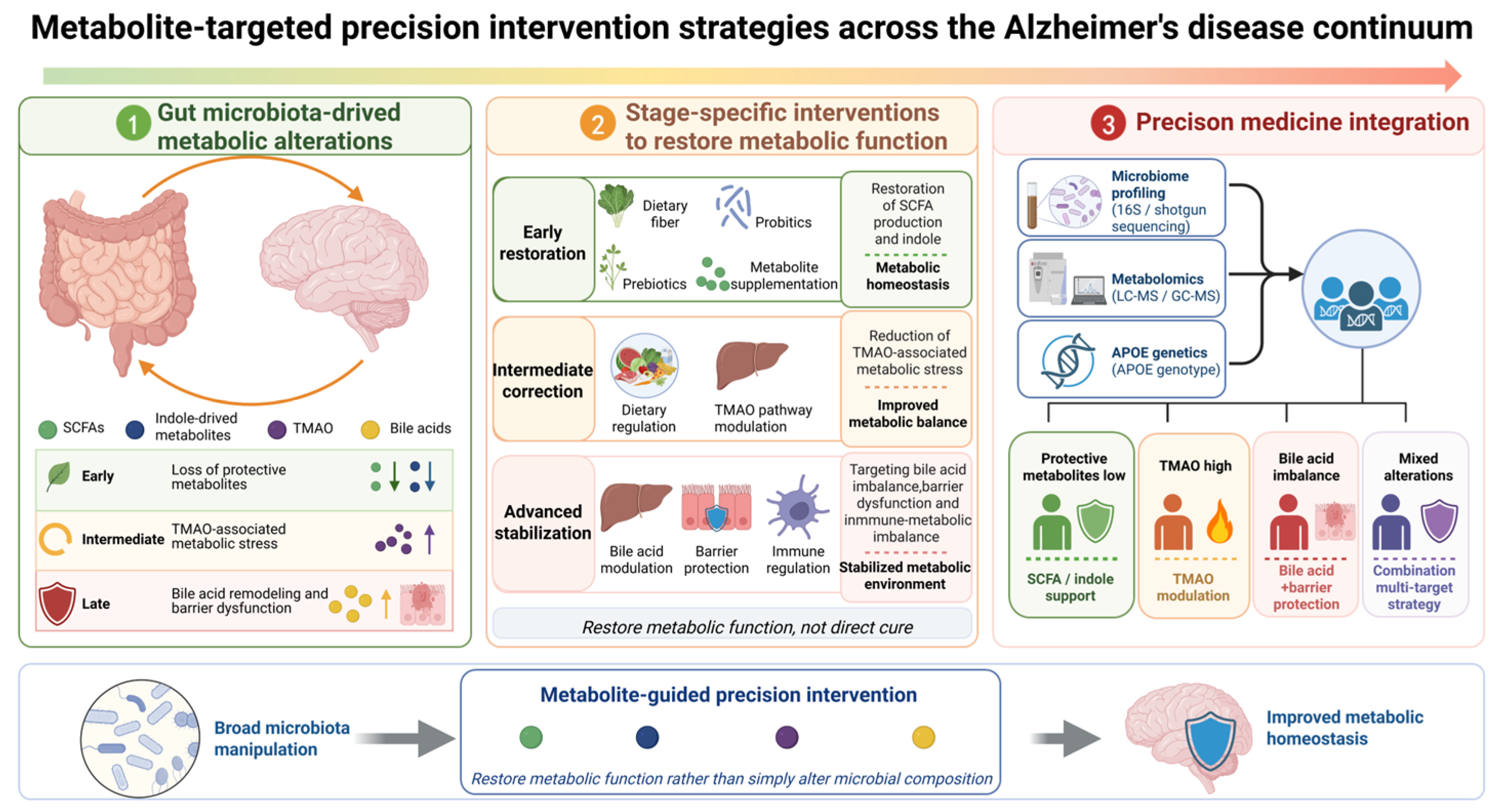
Figure 2.
This schematic presents a stage-oriented framework for targeting microbial metabolic dysfunction during AD progression. Early-stage interventions focus on restoring protective metabolites, including SCFAs and indole-derived metabolites, whereas later-stage strategies address TMAO-associated metabolic stress, bile acid remodeling, barrier dysfunction, and immune-metabolic imbalance. Integration of microbiome profiling, metabolomics, and host genetic information, including APOE genotype, may enable patient stratification and personalized metabolic interventions. This framework emphasizes restoration of metabolic functions rather than nonspecific manipulation of microbial composition. Abbreviations: AD, Alzheimer’s disease; SCFAs, short-chain fatty acids; TMAO, trimethylamine N-oxide; APOE, apolipoprotein E.
Figure 2.
This schematic presents a stage-oriented framework for targeting microbial metabolic dysfunction during AD progression. Early-stage interventions focus on restoring protective metabolites, including SCFAs and indole-derived metabolites, whereas later-stage strategies address TMAO-associated metabolic stress, bile acid remodeling, barrier dysfunction, and immune-metabolic imbalance. Integration of microbiome profiling, metabolomics, and host genetic information, including APOE genotype, may enable patient stratification and personalized metabolic interventions. This framework emphasizes restoration of metabolic functions rather than nonspecific manipulation of microbial composition. Abbreviations: AD, Alzheimer’s disease; SCFAs, short-chain fatty acids; TMAO, trimethylamine N-oxide; APOE, apolipoprotein E.
6. Comparative Metabolic Signatures of Gut Microbiota-Derived Metabolites Across Neurodegenerative Diseases
A target that appears actionable in Alzheimer’s disease cannot be assumed to retain the same effect in another neurodegenerative disease. The previous sections established that gut microbiota-derived metabolites represent dynamic regulators of AD progression by integrating intestinal microbial activity, barrier integrity, immune regulation, and neuronal signaling. Rather than acting as passive biomarkers, microbial metabolites can function as biological mediators that shape neuroinflammatory responses, protein aggregation, and neuronal vulnerability. Increasing evidence suggests that similar metabolic disturbances are also involved in other neurodegenerative diseases, including Parkinson’s disease and amyotrophic lateral sclerosis [155].
However, PD and ALS involve some of the same microbial metabolic pathways but differ in their proteinopathies, vulnerable cells, anatomical starting points, and drug exposures, all of which can redirect the consequences of a shared microbial signal. Disease-specific pathological processes, including α-synuclein accumulation in PD and motor neuron degeneration in ALS, may determine distinct metabolic vulnerabilities and therapeutic responses. Cross-disease comparison therefore tests the limits of metabolic target transfer rather than merely cataloguing shared dysbiosis, and can inform whether therapeutic strategies require disease-specific adaptation.
6.1. Shared Metabolic Vulnerabilities Across Neurodegenerative Diseases
A common feature among AD, PD, and ALS is disruption of microbial metabolic functions involved in maintaining immune and metabolic homeostasis.
SCFAs, particularly acetate, propionate, and butyrate, represent one of the most conserved microbial metabolic pathways across neurodegenerative diseases [156]. SCFAs are produced through microbial fermentation of dietary fibers and regulate intestinal barrier integrity, immune responses, and central nervous system inflammation. In AD, reduced SCFA availability has been associated with impaired microglial homeostasis, altered immune regulation, and increased vulnerability to pathological processes [53]. Similar reductions in SCFA-producing microbial capacity have also been reported in PD and ALS, suggesting that impaired microbial metabolic support may represent a shared vulnerability [157,158].
Loss of SCFA-mediated regulation may promote intestinal barrier dysfunction and increase exposure to microbial-derived inflammatory signals. In AD, barrier disruption facilitates the transmission of peripheral inflammatory signals to the brain, resulting in microglial activation and synaptic dysfunction. Similar mechanisms may contribute to neuroinflammatory amplification in PD and ALS, although disease-specific downstream pathways remain to be fully clarified.
Tryptophan metabolism represents another shared metabolic pathway linking gut microbiota with neuroimmune regulation. Microbial-derived indole metabolites regulate host responses through pathways including AhR signaling. In AD, reduced protective indole metabolites impair AhR-associated immune regulation and may contribute to abnormal inflammatory responses and amyloid-related pathology [159]. Altered tryptophan metabolism has also been observed in PD and ALS, suggesting that microbial regulation of immune-metabolic balance may represent a common mechanism across neurodegenerative disorders [160,161].
Despite these shared abnormalities, microbial metabolites may exert disease-specific effects depending on the pathological environment. Therefore, identifying unique metabolic signatures is essential for developing targeted interventions.
6.2. Disease-Specific Metabolic Features
6.2.1. Parkinson’s Disease: Microbial Metabolites, α-Synuclein Pathology, and Neuroinflammation
Parkinson’s disease is characterized by progressive degeneration of dopaminergic neurons in the substantia nigra and accumulation of pathological α-synuclein aggregates [162]. Among neurodegenerative disorders, PD exhibits a particularly strong association with gastrointestinal dysfunction, including constipation and altered intestinal motility, which frequently precede motor symptoms.
Current evidence indicates that PD is associated with gut microbial dysbiosis, reduced abundance of beneficial microbial taxa, and altered microbial metabolic capacity. Similar to AD, impaired SCFA production may contribute to intestinal barrier dysfunction and inflammatory activation. Reduced microbial-derived metabolic support may weaken intestinal homeostasis and increase peripheral immune activation [157].
However, PD differs from AD through the prominent involvement of α-synuclein-related mechanisms. Microbial-derived inflammatory signals and altered metabolic environments may influence α-synuclein aggregation and microglial activation, thereby contributing to dopaminergic neuronal vulnerability [162]. Experimental studies suggest that gut microbial alterations can modify α-synuclein pathology, although whether microbial changes initiate disease progression or result from neurodegeneration remains unresolved [157].
Compared with AD, where metabolite alterations such as SCFA depletion, TMAO elevation, and bile acid remodeling represent emerging metabolic signatures across disease stages, PD appears to be more strongly associated with interactions among microbial metabolites, intestinal inflammation, and α-synuclein-associated neurodegeneration [162,163].
Therefore, microbiota-derived metabolites may represent potential therapeutic targets in PD. Future studies should determine whether restoring specific metabolic functions can modify disease progression rather than merely alter microbial composition.
6.2.2. Amyotrophic Lateral Sclerosis: Emerging Gut–Muscle Metabolic Axis
Amyotrophic lateral sclerosis is a progressive neurodegenerative disease characterized by degeneration of upper and lower motor neurons, resulting in muscle weakness and progressive atrophy [164]. Compared with AD and PD, the relationship between gut microbiota-derived metabolites and ALS remains less extensively characterized.
Emerging evidence suggests that ALS is associated with alterations in gut microbial composition and metabolic function, including changes in microbial pathways related to SCFA production and intestinal homeostasis [158,161]. Because ALS progression involves substantial systemic metabolic alterations and skeletal muscle degeneration, microbial metabolites may represent a potential link between intestinal microbial activity and muscle metabolic dysfunction.
The concept of the gut–muscle axis extends the traditional gut–brain axis by highlighting the potential role of microbial-derived metabolites in regulating skeletal muscle metabolism, energy homeostasis, and inflammatory responses. Among these metabolites, SCFAs may be particularly relevant because they participate in immune regulation and metabolic homeostasis, processes that are increasingly recognized as important components of ALS pathophysiology [15,158,165].
However, current evidence does not establish whether microbial metabolic alterations directly contribute to motor neuron degeneration or represent secondary consequences of disease-associated changes, including altered nutrition, reduced mobility, and systemic inflammation. Therefore, compared with AD and PD, ALS remains at an earlier stage of mechanistic investigation.
Future studies combining metagenomics, metabolomics, and functional validation are required to identify disease-specific microbial metabolites and determine their potential therapeutic value.
6.3. Implications for Precision Metabolite-Based Intervention
Comparison among AD, PD, and ALS demonstrates that gut microbiota-derived metabolites represent a convergent interface connecting environmental factors, microbial ecology, immune regulation, and neurodegeneration. Shared metabolic abnormalities, including impaired SCFA production, altered tryptophan metabolism, and intestinal barrier dysfunction, suggest common therapeutic opportunities.
However, the distinct metabolic characteristics of each disease highlight the limitations of universal microbiota manipulation. As discussed in AD, effective intervention may require restoration of specific metabolic functions according to disease stage, host genetic background, and individual metabolic characteristics.
For PD, targeting metabolites associated with intestinal inflammation and α-synuclein-related pathology may represent a potential strategy. For ALS, regulation of metabolic pathways involved in systemic inflammation and muscle homeostasis may provide new therapeutic directions. Therefore, future microbiota-based therapies should shift from broad microbial modification toward precision regulation of microbial metabolic functions.
Integration of microbiome profiling, metabolomics, host genetics, and longitudinal clinical information will be essential for identifying disease-specific metabolic signatures and developing personalized interventions across neurodegenerative diseases.
7. Conclusions
Gut microbial metabolism is better represented as a changing host-microbial system than as a fixed signature of AD. Microbial production supplies only the first layer. Substrate availability, intestinal absorption, hepatic conversion, renal clearance, barrier integrity, and tissue responsiveness determine which compounds reach relevant cells and what those cells do with the exposure. This framework gives biological context to the broad ordering observed across the AD continuum. Fermentation-related abnormalities are detectable near the prodromal phase, while wider amino acid, bile acid, lipid, and host-microbial co-metabolic changes accompany cognitive impairment and dementia. The ordering is relative rather than uniform. Its timing may reflect both upstream metabolic change and feedback from established pathology.
No single sample compartment captures this process. Fecal measurements integrate microbial production, local consumption, absorption, and transit. Circulating concentrations add hepatic and renal processing, while cerebrospinal fluid or brain measurements represent another barrier and distribution step. A fall in fecal abundance can therefore coexist with stable or increased systemic exposure. Taxonomic changes create a similar problem. Loss of one producer does not establish loss of a function when other community members carry the same pathway. Functional genes do not prove pathway activity. These distinctions explain part of the disagreement over individual metabolites.
The evidence is strongest where several experimental levels converge. Microbial metabolites can modify epithelial and neurovascular barriers, glial activity, inflammatory signaling, amyloid processing, tau-related pathways, and cellular energetics in animal or cell models. Transfer, depletion, receptor manipulation, and pathway blockade strengthen causal interpretation within those systems. Some studies trace a defined product to a receptor or cellular pathway. Community-wide perturbations cannot assign the effect to one compound. Bidirectional experiments further show that cerebral pathology can reshape the intestine and its microbial ecology. Gut metabolic change may act as a driver, an adaptive response, or both during the same disease process.
Human evidence has a narrower reach. Repeated associations link microbial functions and circulating or fecal metabolites with cognition and AD biomarkers. Fewer longitudinal studies relate baseline metabolic features to later pathological or cognitive change. Most datasets compare different participants at one time point. They cannot reconstruct an individual trajectory or determine whether a metabolite changed before cerebral pathology. Animal age does not map directly onto human clinical stage. Dose, diet, housing, and genetic background also constrain transfer from experimental models. Current evidence supports a dynamic working model, not fixed metabolite thresholds for clinical progression.
Individual variation forms part of that model. Genetic background, age, sex, diet, medication, vascular state, and hepatic or renal function alter both metabolic exposure and tissue response. APOE illustrates how host susceptibility can change the pathological weight of a shared signal without necessarily initiating microbial dysbiosis. The same concentration may therefore carry different implications across patients. Population averages cannot assign a universal transition point. Longitudinal studies with repeated sampling across fecal, blood, and disease-relevant compartments, together with AD biomarkers and clinical outcomes, are essential. Without such data, apparent stage specificity may merely reflect cohort composition or organ function. This boundary also governs intervention. A shift in community composition or cognitive score does not establish metabolic mediation. Precision trials must verify that an intervention changes the intended microbial function, produces the expected metabolite exposure, sustains that exposure, and alters an AD biomarker before clinical benefit is attributed to the pathway. Baseline metabolic state should guide enrollment rather than serve only as a post hoc correlate. Safety assessment must account for dose, tissue, and host processing. Existing human studies seldom complete this chain. The principal gap is target engagement.
Shared metabolic pathways across AD, PD, and ALS do not erase disease specificity. Different vulnerable neurons, protein aggregates, anatomical starting points, receptor distributions, and drug exposures can redirect the same microbial signal. ALS has the least developed human evidence for metabolite-specific regulation, while PD adds direct interactions between microbial metabolism and medication. An AD target must be revalidated before transfer to either disease. Microbial metabolism connects disease stage, host heterogeneity, and neurodegenerative pathology at a functional level. Its clinical value depends on engaging the right target, at the right stage, in patients whose biology can respond.
Author Contributions
Conceptualization, M.N. and T.W.; Writing – Original Draft Preparation, Z.K. and R.M.; Writing – Review & Editing, M.N. and T.W.; Visualization, Z.K., T.W. and R.M.; Supervision, T.W; Funding Acquisition, T.W All authors have read and agreed to the published version of the manuscript.
Funding
This work was supported by the Young Scientists Fund of the National Natural Science Foundation of China (Grant No. 82405557, recipient T.W.); the R&D Program of Beijing Municipal Education Commission (Grant No. KM202410025018, recipient T.W.); the Young Elite Scientists Sponsorship Program of Beijing High-level Innovation Talent Support Plan (Grant No. 20250639, recipient T.W.); and the Young Talent Lifting Project of China Association of Chinese Medicine (Grant No. 2024-QNRC2-B08, recipient T.W.).
Institutional Review Board Statement
Not applicable.
Informed Consent Statement
Not applicable.
Data Availability Statement
No new data were created or analyzed in this study.
Acknowledgments
Figures were created with BioRender.com.
Conflicts of Interest
The authors declare no conflicts of interest.
References
- Nichols, E.; Steinmetz, J.D.; Vollset, S.E.; Fukutaki, K.; Chalek, J.; Abd-Allah, F.; Abdoli, A.; Abualhasan, A.; Abu-Gharbieh, E.; Akram, T.T.; et al. Estimation of the Global Prevalence of Dementia in 2019 and Forecasted Prevalence in 2050: An Analysis for the Global Burden of Disease Study 2019. Lancet Public Health 2022, 7, e105–e125. [Google Scholar] [CrossRef] [PubMed]
- Mukadam, N.; Wolters, F.J.; Walsh, S.; Wallace, L.; Brayne, C.; Matthews, F.E.; Sacuiu, S.; Skoog, I.; Seshadri, S.; Beiser, A.; et al. Changes in Prevalence and Incidence of Dementia and Risk Factors for Dementia: An Analysis from Cohort Studies. Lancet Public Health 2024, 9, e443–e460. [Google Scholar] [CrossRef] [PubMed]
- Zhu, J.; Cui, Y.; Zhang, J.; Yan, R.; Su, D.; Zhao, D.; Wang, A.; Feng, T. Temporal Trends in the Prevalence of Parkinson’s Disease from 1980 to 2023: A Systematic Review and Meta-Analysis. Lancet Healthy Longev. 2024, 5, e464–e479. [Google Scholar] [CrossRef] [PubMed]
- Wolfson, C.; Gauvin, D.E.; Ishola, F.; Oskoui, M. Global Prevalence and Incidence of Amyotrophic Lateral Sclerosis: A Systematic Review. Neurology 2023, 101, e613–e623. [Google Scholar] [CrossRef] [PubMed]
- Wilson, D.M.; Cookson, M.R.; Van Den Bosch, L.; Zetterberg, H.; Holtzman, D.M.; Dewachter, I. Hallmarks of Neurodegenerative Diseases. Cell 2023, 186, 693–714. [Google Scholar] [CrossRef] [PubMed]
- Hansson, O. Biomarkers for Neurodegenerative Diseases. Nat. Med. 2021, 27, 954–963. [Google Scholar] [CrossRef] [PubMed]
- Tolosa, E.; Poewe, W.; Noyce, A.J.; Svenningsson, P.; Mahlknecht, P.; Marti, M.J. Towards Biomarker-Based Diagnosis of Parkinson Disease. Nat. Rev. Neurol. 2026, 22, 139–151. [Google Scholar] [CrossRef] [PubMed]
- Quesnel, M.J.; Labonté, A.; Picard, C.; Bowie, D.C.; Zetterberg, H.; Blennow, K.; Brinkmalm, A.; Villeneuve, S.; Poirier, J.; Alzheimer’s Disease Neuroimaging Initiative; et al. Osteopontin: A Novel Marker of Pre-Symptomatic Sporadic Alzheimer’s Disease. Alzheimers Dement 2024, 20, 6008–6031. [Google Scholar] [CrossRef] [PubMed]
- van der Velpen, V.; Teav, T.; Gallart-Ayala, H.; Mehl, F.; Konz, I.; Clark, C.; Oikonomidi, A.; Peyratout, G.; Henry, H.; Delorenzi, M.; et al. Systemic and Central Nervous System Metabolic Alterations in Alzheimer’s Disease. Alzheimers Res. Ther. 2019, 11, 93. [Google Scholar] [CrossRef] [PubMed]
- Connell, E.; Le Gall, G.; Pontifex, M.G.; Sami, S.; Cryan, J.F.; Clarke, G.; Müller, M.; Vauzour, D. Microbial-Derived Metabolites as a Risk Factor of Age-Related Cognitive Decline and Dementia. Mol. Neurodegener. 2022, 17, 43. [Google Scholar] [CrossRef] [PubMed]
- Loh, J.S.; Mak, W.Q.; Tan, L.K.S.; Ng, C.X.; Chan, H.H.; Yeow, S.H.; Foo, J.B.; Ong, Y.S.; How, C.W.; Khaw, K.Y. Microbiota-Gut-Brain Axis and Its Therapeutic Applications in Neurodegenerative Diseases. Signal Transduct. Target Ther. 2024, 9, 37. [Google Scholar] [CrossRef] [PubMed]
- Kuehn, E.; Lambrecht, N.; Mueller, K.D.; Clark, L.R.; Chin, N.A.; Gleason, C.E.; Bendlin, B.B.; Johnson, S.C.; Rey, F.E. Fecal Short-Chain Fatty Acids Vary by Sex and Amyloid Status. Alzheimer’s Dement. 2025, 21, e14438. [Google Scholar] [CrossRef] [PubMed]
- Romano, S.; Wirbel, J.; Ansorge, R.; Schudoma, C.; Ducarmon, Q.R.; Narbad, A.; Zeller, G. Machine Learning-Based Meta-Analysis Reveals Gut Microbiome Alterations Associated with Parkinson’s Disease. Nat. Commun. 2025, 16, 4227. [Google Scholar] [CrossRef] [PubMed]
- Jack, C.R.; Andrews, J.S.; Beach, T.G.; Buracchio, T.; Dunn, B.; Graf, A.; Hansson, O.; Ho, C.; Jagust, W.; McDade, E.; et al. Revised Criteria for Diagnosis and Staging of Alzheimer’s Disease: Alzheimer’s Association Workgroup. Alzheimers Dement 2024, 20, 5143–5169. [Google Scholar] [CrossRef] [PubMed]
- Dalile, B.; Van Oudenhove, L.; Vervliet, B.; Verbeke, K. The Role of Short-Chain Fatty Acids in Microbiota–Gut–Brain Communication. Nat. Rev. Gastroenterol. Hepatol. 2019, 16, 461–478. [Google Scholar] [CrossRef] [PubMed]
- Ferreiro, A.L.; Choi, J.; Ryou, J.; Newcomer, E.P.; Thompson, R.; Bollinger, R.M.; Hall-Moore, C.; Ndao, I.M.; Sax, L.; Benzinger, T.L.S.; et al. Gut Microbiome Composition May Be an Indicator of Preclinical Alzheimer’s Disease. Sci. Transl. Med. 2023, 15, eabo2984. [Google Scholar] [CrossRef] [PubMed]
- Sheng, C.; Yang, K.; He, B.; Du, W.; Cai, Y.; Han, Y. Combination of Gut Microbiota and Plasma Amyloid-β as a Potential Index for Identifying Preclinical Alzheimer’s Disease: A Cross-Sectional Analysis from the SILCODE Study. Alzheimers Res. Ther. 2022, 14, 35. [Google Scholar] [CrossRef] [PubMed]
- Hosseinkhani, F.; Chevalier, C.; Marizzoni, M.; Park, R.; Bos, S.; Dunjko, A.K.; van Duijn, C.M.; Harms, A.C.; Frisoni, G.B.; Hankemeier, T. Plasma and Feces Multiomics Unveil Cognition-Associated Perturbations of Chronic Inflammatory Pathways of the Gut-Microbiota-Brain Axis. Alzheimers Dement 2025, 21, e70844. [Google Scholar] [CrossRef] [PubMed]
- Liu, F.; Duan, M.; Fu, H.; Zhao, G.; Han, Y.; Lan, F.; Ahmed, Z.; Cao, G.; Li, Z.; Ma, D.; et al. Orthopedic Surgery Causes Gut Microbiome Dysbiosis and Intestinal Barrier Dysfunction in Prodromal Alzheimer Disease Patients: A Prospective Observational Cohort Study. Ann. Surg. 2022, 276, 270–280. [Google Scholar] [CrossRef] [PubMed]
- Chilton, P.M.; Ghare, S.S.; Charpentier, B.T.; Myers, S.A.; Rao, A.V.; Petrosino, J.F.; Hoffman, K.L.; Greenwell, J.C.; Tyagi, N.; Behera, J.; et al. Age-Associated Temporal Decline in Butyrate-Producing Bacteria Plays a Key Pathogenic Role in the Onset and Progression of Neuropathology and Memory Deficits in 3×Tg-AD Mice. Gut Microbes 16, 2389319. [CrossRef] [PubMed]
- Ueda, A.; Shinkai, S.; Shiroma, H.; Taniguchi, Y.; Tsuchida, S.; Kariya, T.; Kawahara, T.; Kobayashi, Y.; Kohda, N.; Ushida, K.; et al. Identification of Faecalibacterium Prausnitzii Strains for Gut Microbiome-Based Intervention in Alzheimer’s-Type Dementia. Cell Rep. Med. 2021, 2, 100398. [Google Scholar] [CrossRef] [PubMed]
- Verhaar, B.J.H.; Hendriksen, H.M.A.; de Leeuw, F.A.; Doorduijn, A.S.; van Leeuwenstijn, M.; Teunissen, C.E.; Barkhof, F.; Scheltens, P.; Kraaij, R.; van Duijn, C.M.; et al. Gut Microbiota Composition Is Related to AD Pathology. Front Immunol. 2022, 12, 794519. [Google Scholar] [CrossRef] [PubMed]
- Yamashiro, K.; Takabayashi, K.; Kamagata, K.; Nishimoto, Y.; Togashi, Y.; Yamauchi, Y.; Ogaki, K.; Li, Y.; Hatano, T.; Motoi, Y.; et al. Free Water in Gray Matter Linked to Gut Microbiota Changes with Decreased Butyrate Producers in Alzheimer’s Disease and Mild Cognitive Impairment. Neurobiol. Dis. 2024. [Google Scholar] [CrossRef] [PubMed]
- Fan, K.-C.; Lin, C.-C.; Chiu, Y.-L.; Koh, S.-H.; Liu, Y.-C.; Chuang, Y.-F. Compositional and Functional Gut Microbiota Alterations in Mild Cognitive Impairment: Links to Alzheimer’s Disease Pathology. Alzheimers Res. Ther. 2025, 17, 122. [Google Scholar] [CrossRef] [PubMed]
- Li, B.; He, Y.; Ma, J.; Huang, P.; Du, J.; Cao, L.; Wang, Y.; Xiao, Q.; Tang, H.; Chen, S. Mild Cognitive Impairment Has Similar Alterations as Alzheimer’s Disease in Gut Microbiota. Alzheimer’s Dement. 2019, 15, 1357–1366. [Google Scholar] [CrossRef] [PubMed]
- Cabrera, C.; Carrión, N.; Mateo, D.; Vicens, P.; Pinzón, A.; Heredia, L.; Forcadell-Ferreres, E.; Pino, M.; Yerga, B.; Zaragoza, J.; et al. Gut Microbiota Characterization in Ageing, Mild Cognitive Impairment, and Alzheimer’s Disease in the Context of Mediterranean Lifestyle in a Spanish Population. Alzheimers Res. Ther. 2025, 17, 211. [Google Scholar] [CrossRef] [PubMed]
- Rouskas, K.; Mamalaki, E.; Ntanasi, E.; Pantoura, M.; Anezaki, M.; Emmanouil, C.; Novau-Ferré, N.; Bulló, M.; Dimas, A.S.; Papandreou, C.; et al. Gut Microbiome Alterations in Mild Cognitive Impairment: Findings from the ALBION Greek Cohort. Microorganisms 2025, 13, 2112. [Google Scholar] [CrossRef] [PubMed]
- Zeamer, A.L.; Lai, Y.; Loew, E.; Sanborn, V.; Tracy, M.; Jo, C.; Ferdinand, D.; Ward, D.V.; Bhattarai, S.K.; Drake, J.; et al. Microbiome Functional Gene Pathways Are Indicative of Cognitive Performance in Older Adults at Risk for Alzheimer’s Disease. Gut Microbes 18, 2676162. [CrossRef] [PubMed]
- Kolobaric, A.; Andreescu, C.; Jašarević, E.; Hong, C.H.; Roh, H.W.; Cheong, J.Y.; Kim, Y.K.; Shin, T.S.; Kang, C.S.; Kwon, C.O.; et al. Gut Microbiome Predicts Cognitive Function and Depressive Symptoms in Late Life. Mol. Psychiatry 2024, 29, 3064–3075. [Google Scholar] [CrossRef] [PubMed]
- Ma, C.; Li, Y.; Mei, Z.; Yuan, C.; Kang, J.H.; Grodstein, F.; Ascherio, A.; Willett, W.C.; Chan, A.T.; Huttenhower, C.; et al. Association Between Bowel Movement Pattern and Cognitive Function. Neurology 2023, 101, e2014–e2025. [Google Scholar] [CrossRef] [PubMed]
- Connell, E.; Sami, S.; Khondoker, M.; Minihane, A.M.; Pontifex, M.G.; Müller, M.; McArthur, S.; Le Gall, G.; Vauzour, D. Circulatory Dietary and Gut-Derived Metabolites Predict Early Cognitive Decline. Gut Microbes 2026, 18, 2649487. [Google Scholar] [CrossRef] [PubMed]
- Buawangpong, N.; Pinyopornpanish, K.; Phrommintikul, A.; Chindapan, N.; Devahastin, S.; Chattipakorn, N.; Chattipakorn, S.C. Increased Plasma Trimethylamine-N-Oxide Levels Are Associated with Mild Cognitive Impairment in High Cardiovascular Risk Elderly Population. Food Funct. 2022. [Google Scholar] [CrossRef] [PubMed]
- Wu, L.; Han, Y.; Zheng, Z.; Peng, G.; Liu, P.; Yue, S.; Zhu, S.; Chen, J.; Lv, H.; Shao, L.; et al. Altered Gut Microbial Metabolites in Amnestic Mild Cognitive Impairment and Alzheimer’s Disease: Signals in Host-Microbe Interplay. Nutrients 2021, 13, 228. [Google Scholar] [CrossRef] [PubMed]
- Kang, J.W.; Khatib, L.A.; Heston, M.B.; Dilmore, A.H.; Labus, J.S.; Deming, Y.; Schimmel, L.; Blach, C.; McDonald, D.; Gonzalez, A.; et al. Gut Microbiome Compositional and Functional Features Associate with Alzheimer’s Disease Pathology. Alzheimers Dement 2025, 21, e70417. [Google Scholar] [CrossRef] [PubMed]
- Wang, M.; Buthut, M.; Meinhardt, J.; Otto, C.; Gallaccio, G.; Fernández-Zapata, C.; Teves, M.; Samol, C.; Dettmer, K.; Heckscher, S.; et al. Immune-Proteo-Metabolomic Changes Link to Aβ and Tau Pathology in Alzheimer Disease. Alzheimer’s Dement. 2026. [Google Scholar] [CrossRef] [PubMed]
- MahmoudianDehkordi, S.; Arnold, M.; Nho, K.; Ahmad, S.; Jia, W.; Xie, G.; Louie, G.; Kueider-Paisley, A.; Arthur Moseley, M.; Will Thompson, J.; et al. Altered Bile Acid Profile Associates with Cognitive Impairment in Alzheimer’s Disease – An Emerging Role for Gut Microbiome. Alzheimers Dement 2019, 15, 76–92. [Google Scholar] [CrossRef] [PubMed]
- Nho, K.; Kueider-Paisley, A.; MahmoudianDehkordi, S.; Arnold, M.; Risacher, S.L.; Louie, G.; Blach, C.; Baillie, R.; Han, X.; Kastenmüller, G.; et al. Altered Bile Acid Profile in Mild Cognitive Impairment and Alzheimer’s Disease: Relationship to Neuroimaging and CSF Biomarkers. Alzheimers Dement 2019, 15, 232–244. [Google Scholar] [CrossRef] [PubMed]
- Fu, W.; Chao, X.; Wang, Y.; Liu, S.; Wang, J.; Huang, Q.; Luan, Y.; Luo, P.; Guan, Y.; Mai, Y.; et al. Bile Acids Are Associated with Baseline and Longitudinal Amyloid and Tau Pathology in Patients with Alzheimer’s Disease. Alzheimers Dement 2026, 22, e71307. [Google Scholar] [CrossRef] [PubMed]
- Vogt, N.M.; Romano, K.A.; Darst, B.F.; Engelman, C.D.; Johnson, S.C.; Carlsson, C.M.; Asthana, S.; Blennow, K.; Zetterberg, H.; Bendlin, B.B.; et al. The Gut Microbiota-Derived Metabolite Trimethylamine N-Oxide Is Elevated in Alzheimer’s Disease. Alz Res. Ther. 2018, 10, 124. [Google Scholar] [CrossRef] [PubMed]
- Chen, T.; Pan, F.; Huang, Q.; Xie, G.; Chao, X.; Wu, L.; Wang, J.; Cui, L.; Sun, T.; Li, M.; et al. Metabolic Phenotyping Reveals an Emerging Role of Ammonia Abnormality in Alzheimer’s Disease. Nat. Commun. 2024, 15, 3796. [Google Scholar] [CrossRef] [PubMed]
- Li, N.; Wang, S.; Li, H.; Liu, Z.; Mo, H.; Luan, J.; Guo, N.; Gou, X.; Wu, Y.; Li, Z. Gut Microbiota-Driven Metabolic Alterations Reveal the Gut-Brain Communication of Ergothioneine in Ameliorating Cognitive Impairment in APP/PS1 Mice. J. Agric. Food Chem. 2025, 73, 16933–16948. [Google Scholar] [CrossRef] [PubMed]
- Qu, C.; Xu, Q.-Q.; Yang, W.; Zhong, M.; Yuan, Q.; Xian, Y.-F.; Lin, Z.-X. Gut Dysbiosis Aggravates Cognitive Deficits, Amyloid Pathology and Lipid Metabolism Dysregulation in a Transgenic Mouse Model of Alzheimer’s Disease. J. Pharm. Anal. 2023, 13, 1526–1547. [Google Scholar] [CrossRef] [PubMed]
- Son, S.J.; Wu, X.; Roh, H.W.; Cho, Y.H.; Hong, S.; Nam, Y.J.; Hong, C.H.; Park, S. Distinct Gut Microbiota Profiles and Network Properties in Older Korean Individuals with Subjective Cognitive Decline, Mild Cognitive Impairment, and Alzheimer’s Disease. Alzheimers Res. Ther. 2025, 17, 187. [Google Scholar] [CrossRef] [PubMed]
- Zhao, H.; Zhou, X.; Song, Y.; Zhao, W.; Sun, Z.; Zhu, J.; Yu, Y. Multi-Omics Analyses Identify Gut Microbiota-Fecal Metabolites-Brain-Cognition Pathways in the Alzheimer’s Disease Continuum. Alzheimers Res. Ther. 2025, 17, 36. [Google Scholar] [CrossRef] [PubMed]
- Lin, Y.; Shi, X.; Mu, J.; Ren, H.; Jiang, X.; Zhu, L.; Cai, X.; Lian, C.; Pei, Z.; Zhang, Y.; et al. Uncovering Stage-specific Neural and Molecular Progression in Alzheimer’s Disease: Implications for Early Screening. Alzheimers Dement 2025, 21, e70182. [Google Scholar] [CrossRef] [PubMed]
- Liang, X.; Fu, Y.; Cao, W.; Wang, Z.; Zhang, K.; Jiang, Z.; Jia, X.; Liu, C.; Lin, H.; Zhong, H.; et al. Gut Microbiome, Cognitive Function and Brain Structure: A Multi-Omics Integration Analysis. Transl. Neurodegener. 2022, 11, 49. [Google Scholar] [CrossRef] [PubMed]
- Warren, A.; Wynia, Z.; Corr, P.G.; Devin, M.F.; Celikkol, Z.; Gordon, L.; Farah, M.; Karam, M.; Villarreal, D.; Jackson, S.A.; et al. The Microbiota–Gut–Brain Axis in Mild Cognitive Impairment and Alzheimer’s Disease: A Scoping Review of Human Studies. Alzheimers Dement 2026, 22, e71023. [Google Scholar] [CrossRef] [PubMed]
- Zhou, X.-P.; Sun, L.-B.; Liu, W.-H.; Zhu, W.-M.; Li, L.-C.; Song, X.-Y.; Xing, J.-P.; Gao, S.-H. The Complex Relationship between Gut Microbiota and Alzheimer’s Disease: A Systematic Review. Ageing Res. Rev. 2025, 104, 102637. [Google Scholar] [CrossRef] [PubMed]
- Chandra, S.; Vassar, R.J. Gut Microbiome-Derived Metabolites in Alzheimer’s Disease: Regulation of Immunity and Potential for Therapeutics. Immunol. Rev. 2024, 327, 33–42. [Google Scholar] [CrossRef] [PubMed]
- Ren, F.; Wei, J.; Chen, Q.; Hu, M.; Yu, L.; Mi, J.; Zhou, X.; Qin, D.; Wu, J.; Wu, A. Artificial Intelligence-Driven Multi-Omics Approaches in Alzheimer’s Disease: Progress, Challenges, and Future Directions. Acta Pharm. Sin. B 2025, 15, 4327–4385. [Google Scholar] [CrossRef] [PubMed]
- Ahmad, S.; Wu, T.; Arnold, M.; Hankemeier, T.; Ghanbari, M.; Roshchupkin, G.; Uitterlinden, A.G.; Borkowski, K.; Neitzel, J.; Kraaij, R.; et al. The Blood Metabolome of Brain Health in Midlife and Influences of Genes, Microbiome and Exposome. Nat. Aging 2026, 6, 1452–1467. [Google Scholar] [CrossRef] [PubMed]
- Chandra, S.; Popovic, J.; Singhal, N.K.; Watkins, E.A.; Dodiya, H.B.; Weigle, I.Q.; Salvo, M.A.; Ramakrishnan, A.; Chen, Z.; Watson, T.; et al. The Gut Microbiome Controls Reactive Astrocytosis during Aβ Amyloidosis via Propionate-Mediated Regulation of IL-17. J. Clin. Invest 2025, 135, e180826. [Google Scholar] [CrossRef] [PubMed]
- Qian, X.; Xie, R.; Liu, X.; Chen, S.; Tang, H. Mechanisms of Short-Chain Fatty Acids Derived from Gut Microbiota in Alzheimer’s Disease. Aging Dis. 2022, 13, 1252–1266. [Google Scholar] [CrossRef] [PubMed]
- Erny, D.; Dokalis, N.; Mezo, C.; Mossad, O.; Staszewski, O.; Winkler, A.; Pohlmeyer, F.; Blank, T.; Gomez de Aguero, M.; Macpherson, A.J.; et al. Microbiota-Derived Acetate Enables the Metabolic Fitness of the Brain Innate Immune System during Health and Disease. Cell Metab. 2021, 33, 2260–2276. [Google Scholar] [CrossRef] [PubMed]
- Lin, Y.; Lin, A.; Cai, L.; Huang, W.; Yan, S.; Wei, Y.; Ruan, X.; Fang, W.; Dai, X.; Cheng, J.; et al. ACSS2-Dependent Histone Acetylation Improves Cognition in Mouse Model of Alzheimer’s Disease. Mol. Neurodegener. 2023. [Google Scholar] [CrossRef] [PubMed]
- Sun, Y.; Zhang, H.; Zhang, X.; Wang, W.; Chen, Y.; Cai, Z.; Wang, Q.; Wang, J.; Shi, Y. Promotion of Astrocyte-Neuron Glutamate-Glutamine Shuttle by SCFA Contributes to the Alleviation of Alzheimer’s Disease. Redox Biol. 2023. [Google Scholar] [CrossRef] [PubMed]
- Hao, W.; Luo, Q.; Szabo, I.M.; Gasparoni, G.; Tierling, S.; Quan, W.; Chang, H.-F.; Wu, G.; Schulze-Hentrich, J.; Liu, Y. Intestinal Epithelial Dicer1 Regulates Gut Microbiome and Alzheimer’s Pathology in App-Knock-in Mice. Alzheimers Res. Ther. 2025, 17, 202. [Google Scholar] [CrossRef] [PubMed]
- Self, W.K.; Holtzman, D.M. Thwarting Amyloidosis: IL-17 as a Disease Modifier along the Gut/Brain Axis. J. Clin. Investig. 2025. [Google Scholar] [CrossRef] [PubMed]
- Brossaud, R.; Oullier, T.; Bessard, A.; Aubert, P.; Brossard, L.; Mahé, M.M.; Caillaud, M.; Delfino, G.; Paillusson, S.; Falentin, H.; et al. The Short-Chain Fatty Acid Butyrate Prevents Gut-Brain Amyloid-β Pathology and Neuroinflammation in an Alzheimer Mouse Model. Mol. Psychiatry 2026, 31, 3910–3928. [Google Scholar] [CrossRef] [PubMed]
- Agus, A.; Planchais, J.; Sokol, H. Gut Microbiota Regulation of Tryptophan Metabolism in Health and Disease. Cell Host Microbe 2018, 23, 716–724. [Google Scholar] [CrossRef] [PubMed]
- Pappolla, M.A.; Perry, G.; Fang, X.; Zagorski, M.; Sambamurti, K.; Poeggeler, B. Indoles as Essential Mediators in the Gut-Brain Axis. Their Role in Alzheimer’s Disease. Neurobiol. Dis. 2021. [Google Scholar] [CrossRef] [PubMed]
- Kim, H.; Lee, E.; Park, M.; Min, K.; Diep, Y.N.; Kim, J.; Ahn, H.; Lee, E.; Kim, S.; Kim, Y.; et al. Microbiome-Derived Indole-3-Lactic Acid Reduces Amyloidopathy through Aryl-Hydrocarbon Receptor Activation. Brain Behav. Immun. 2024, 122, 568–582. [Google Scholar] [CrossRef] [PubMed]
- Li, L.; Jia, M.; Yang, C.; Zhao, Y.; Hu, J.; Zhao, Y.; Hu, X.; Ning, F.; Ding, C.; Li, Q.; et al. Gut Microbial-Derived Indole-3-Propionate Improves Cognitive Function in Alzheimer’s Disease. Sci. Adv. 11, eadw8410. [CrossRef] [PubMed]
- Ye, T.; Lv, X.; Li, Q.; Liao, X.; Zhan, L.; Dai, C.; Xiang, K.; Lin, X.; Sun, J.; Liu, J. Gut Microbial Metabolite Indole-3-Acetic Acid Inhibits Microglia-Mediated Synaptic Loss in Alzheimer’s Disease by Targeting CCR4. Brain Behav. Immun. 2026, 136, 106769. [Google Scholar] [CrossRef] [PubMed]
- Sun, J.; Zhang, Y.; Kong, Y.; Ye, T.; Yu, Q.; Kumaran Satyanarayanan, S.; Su, K.-P.; Liu, J. Microbiota-Derived Metabolite Indoles Induced Aryl Hydrocarbon Receptor Activation and Inhibited Neuroinflammation in APP/PS1 Mice. Brain Behav. Immun. 2022, 106, 76–88. [Google Scholar] [CrossRef] [PubMed]
- Qian, C.; Yang, C.; Lu, M.; Bao, J.; Shen, H.; Deng, B.; Li, S.; Li, W.; Zhang, M.; Cao, C. Activating AhR Alleviates Cognitive Deficits of Alzheimer’s Disease Model Mice by Upregulating Endogenous Aβ Catabolic Enzyme Neprilysin. Theranostics 2021. [Google Scholar] [CrossRef] [PubMed]
- Maitre, M.; Klein, C.; Patte-Mensah, C.; Mensah-Nyagan, A.-G. Tryptophan Metabolites Modify Brain Aβ Peptide Degradation: A Role in Alzheimer’s Disease? Prog. Neurobiol. 2020. [Google Scholar] [CrossRef] [PubMed]
- Wang, B.; Pan, M.; Yang, L.; Xu, J.; Ye, C.; Li, Y.; Gong, Q.; Liu, C.; Li, L.; Qin, J.; et al. Akkermansia Muciniphila Reduces Neuroinflammation and Aβ Deposition via Tryptophan Metabolism in the APP/PS1 Mouse Model of Alzheimer’s Disease. Alzheimers Res. Ther. 2026, 18, 41. [Google Scholar] [CrossRef] [PubMed]
- Vemuganti, R.; Chokkalla, A.K.; Jeong, S.; Mehta, S.L.; Davis, C.K.; Aronowski, J. Gut Bacterial Metabolite Imidazole Propionate Potentiates Alzheimer’s Disease Pathology. Nat. Commun. 2026, 17, 3157. [Google Scholar] [CrossRef]
- Chakraborty, P.; Ibáñez de Opakua, A.; Purslow, J.A.; Fromm, S.A.; Chatterjee, D.; Zachrdla, M.; Zhuang, S.; Puri, S.; Wolozin, B.; Zweckstetter, M. GSK3β Phosphorylation Catalyzes the Aggregation of Tau into Alzheimer’s Disease-like Filaments. Proc. Natl. Acad. Sci. U S A 2024, 121, e2414176121. [Google Scholar] [CrossRef] [PubMed]
- Lv, X.; Ye, T.; Liao, X.; Li, Q.; Fang, Z.; Lin, X.; Chen, M.; Dai, C.; Zhan, L.; Zhuo, L.; et al. Blautia Coccoides-Derived Metabolite Trimethylamine-N-Oxide Exacerbates Alzheimer’s Disease Progression via Targeting HIF1α Signaling. Gut Microbes 18, 2605768. [CrossRef] [PubMed]
- Kazmi, S.A.; Chandra, F.; Wasney, M.; Cheng, J.; Lum, G.R.; Iyer, M.; Di Blasi, D.; Espinoza, A.N.; Lopez-Romero, A.; Yang, X.; et al. Select Microbial Metabolites Promote Tau Aggregation in a Murine Tauopathy Model. Nat. Commun. 2026. [Google Scholar] [CrossRef] [PubMed]
- Shi, Y.; Wang, P.; Deng, J.; Chen, Y.; Wang, F.; Han, Y.; Wang, H.; Li, Y.; Fang, X.; Hui, J.; et al. Gut Microbial Metabolite TMAO Impairs Cognitive Function and Induces Hippocampal Synaptic Plasticity Decline through Modulation of GSK-3β Activity. Alzheimers Res. Ther. 2025, 17, 196. [Google Scholar] [CrossRef] [PubMed]
- Zhu, H.; Huang, C.; Luo, Z.; Wu, L.; Cheng, X.; Wu, H. Porphyromonas Gingivalis Induces Disturbance of Kynurenine Metabolism Through the Gut-Brain Axis: Implications for Alzheimer’s Disease. J. Dent. Res. 2025. [Google Scholar] [CrossRef] [PubMed]
- Choi, H.; Hong, S.B.; Kim, Y.; Joung, H.; Choi, Y.; Cha, J.; Park, J.Y.; Lee, Y.-S.; Choi, H.; Han, J.W.; et al. Tryptophan–Kynurenine Metabolic Reprogramming along the Gut–Brain Axis Alleviates Alzheimer’s Pathology. J. Neuroinflammation 2026, 23, 197. [Google Scholar] [CrossRef] [PubMed]
- Zhang, Y.; Li, J.; Li, J.; Wang, Y.; Cui, M.; Li, W.; Chen, X.; Jiang, Y. Cross-Sectional and Longitudinal Associations of Circulating Bile Acids and Dementia: A Systematic Review. Geroscience 2026, 48, 4789–4804. [Google Scholar] [CrossRef] [PubMed]
- Chen, T.; Wang, L.; Xie, G.; Kristal, B.S.; Zheng, X.; Sun, T.; Arnold, M.; Louie, G.; Li, M.; Wu, L.; et al. Serum Bile Acids Improve Prediction of Alzheimer’s Progression in a Sex-Dependent Manner. Adv. Sci. (Weinh) 2024, 11, e2306576. [Google Scholar] [CrossRef] [PubMed]
- Talavera Andújar, B.; Mary, A.; Venegas, C.; Cheng, T.; Zaslavsky, L.; Bolton, E.E.; Heneka, M.T.; Schymanski, E.L. Can Small Molecules Provide Clues on Disease Progression in Cerebrospinal Fluid from Mild Cognitive Impairment and Alzheimer’s Disease Patients? Env. Sci. Technol. 2024, 58, 4181–4192. [Google Scholar] [CrossRef] [PubMed]
- Li, C.; Wang, L.; Xie, W.; Chen, E.; Chen, Y.; Li, H.; Can, D.; Lei, A.; Wang, Y.; Zhang, J. TGR5 Deficiency in Excitatory Neurons Ameliorates Alzheimer’s Pathology by Regulating APP Processing. Sci. Adv. 2024, 10, eado1855. [Google Scholar] [CrossRef] [PubMed]
- Nie, R.-Z.; Zhang, Q.-L.; Tan, X.-R.; Hu, S.-S.; Zhou, X.-T.; Jiang, W.-K.; Guo, B.-W.; Cao, X.; Yuan, D.-H.; Long, Y.; et al. Cholinergic G Protein-Coupled Bile Acid Receptor 1 (TGR5/GPBA) in the Medial Septal Orchestrates Adult Hippocampal Neurogenesis and Cognition in Alzheimer’s Disease Mice. Br. J. Pharmacol. 2025, 182, 5409–5429. [Google Scholar] [CrossRef] [PubMed]
- Zha, X.; Liu, X.; Wei, M.; Huang, H.; Cao, J.; Liu, S.; Bian, X.; Zhang, Y.; Xiao, F.; Xie, Y.; et al. Microbiota-Derived Lysophosphatidylcholine Alleviates Alzheimer’s Disease Pathology via Suppressing Ferroptosis. Cell Metab. 2025. [Google Scholar] [CrossRef] [PubMed]
- Qian, X.; Hai, W.; Chen, S.; Zhang, M.; Jiang, X.; Tang, H. Multi-Omics Data Reveals Aberrant Gut Microbiota-Host Glycerophospholipid Metabolism in Association with Neuroinflammation in APP/PS1 Mice. Gut Microbes 2023. [Google Scholar] [CrossRef] [PubMed]
- Wang, Y.; An, Y.; Ma, W.; Yu, H.; Lu, Y.; Zhang, X.; Wang, Y.; Liu, W.; Wang, T.; Xiao, R. 27-Hydroxycholesterol Contributes to Cognitive Deficits in APP/PS1 Transgenic Mice through Microbiota Dysbiosis and Intestinal Barrier Dysfunction. J. Neuroinflammation 2020. [Google Scholar] [CrossRef] [PubMed]
- Xie, J.; Bruggeman, A.; De Nolf, C.; Vandendriessche, C.; Van Imschoot, G.; Van Wonterghem, E.; Vereecke, L.; Vandenbroucke, R.E. Gut Microbiota Regulates Blood-Cerebrospinal Fluid Barrier Function and Aβ Pathology. EMBO J. 2023, 42, e111515. [Google Scholar] [CrossRef] [PubMed]
- Kim, N.; Ju, I.G.; Jeon, S.H.; Lee, Y.; Jung, M.-J.; Gee, M.S.; Cho, J.S.; Inn, K.-S.; Garrett-Sinha, L.A.; Oh, M.S.; et al. Inhibition of Microfold Cells Ameliorates Early Pathological Phenotypes by Modulating Microglial Functions in Alzheimer’s Disease Mouse Model. J. Neuroinflammation 2023. [Google Scholar] [CrossRef] [PubMed]
- Brown, G.C.; Heneka, M.T. The Endotoxin Hypothesis of Alzheimer’s Disease. Mol. Neurodegener. 2024, 19, 30. [Google Scholar] [CrossRef] [PubMed]
- Zhao, X.; Yu, J.; Xu, B.; Xu, Z.; Lei, X.; Han, S.; Luo, S.; Zhang, C.; Peng, G.; Li, J.; et al. Gut-Derived Bacterial Vesicles Carrying Lipopolysaccharide Promote Microglia-Mediated Synaptic Pruning. Alzheimers Dement 2025, 21, e70331. [Google Scholar] [CrossRef] [PubMed]
- Xie, J.; Van Hoecke, L.; Van Wonterghem, E.; Van Imschoot, G.; Andries, V.; Vereecke, L.; Vandenbroucke, R.E. The Gut–Brain Axis in Alzheimer’s Disease Is Shaped by Commensal Gut Microbiota Derived Extracellular Vesicles. Gut Microbes 17, 2501193. [CrossRef] [PubMed]
- Xia, Y.; Xiao, Y.; Wang, Z.-H.; Liu, X.; Alam, A.M.; Haran, J.P.; McCormick, B.A.; Shu, X.; Wang, X.; Ye, K. Bacteroides Fragilis in the Gut Microbiomes of Alzheimer’s Disease Activates Microglia and Triggers Pathogenesis in Neuronal C/EBPβ Transgenic Mice. Nat. Commun. 2023. [Google Scholar] [CrossRef] [PubMed]
- Conn, K.A.; Borsom, E.M.; Cope, E.K. Implications of Microbe-Derived ɣ-Aminobutyric Acid (GABA) in Gut and Brain Barrier Integrity and GABAergic Signaling in Alzheimer’s Disease. Gut Microbes 2024. [Google Scholar] [CrossRef] [PubMed]
- Bairamian, D.; Sha, S.; Rolhion, N.; Sokol, H.; Dorothée, G.; Lemere, C.A.; Krantic, S. Microbiota in Neuroinflammation and Synaptic Dysfunction: A Focus on Alzheimer’s Disease. Mol. Neurodegener. 2022. [Google Scholar] [CrossRef] [PubMed]
- Wang, Q.; Davis, P.B.; Qi, X.; Chen, S.G.; Gurney, M.E.; Perry, G.; Doraiswamy, P.M.; Xu, R. Gut-Microbiota-Microglia-Brain Interactions in Alzheimer’s Disease: Knowledge-Based, Multi-Dimensional Characterization. Alzheimer’s Res. Ther. 2021. [Google Scholar] [CrossRef] [PubMed]
- Marizzoni, M.; Mirabelli, P.; Mombelli, E.; Coppola, L.; Festari, C.; Lopizzo, N.; Luongo, D.; Mazzelli, M.; Naviglio, D.; Blouin, J.-L.; et al. A Peripheral Signature of Alzheimer’s Disease Featuring Microbiota-Gut-Brain Axis Markers. Alzheimers Res. Ther. 2023, 15, 101. [Google Scholar] [CrossRef] [PubMed]
- Xie, J.; Cools, L.; Van Imschoot, G.; Van Wonterghem, E.; Pauwels, M.J.; Vlaeminck, I.; De Witte, C.; El Andaloussi, S.; Wierda, K.; De Groef, L.; et al. Helicobacter Pylori-Derived Outer Membrane Vesicles Contribute to Alzheimer’s Disease Pathogenesis via C3-C3aR Signalling. J. Extracell. Vesicles 2023, 12, e12306. [Google Scholar] [CrossRef] [PubMed]
- Mann, E.R.; Lam, Y.K.; Uhlig, H.H. Short-Chain Fatty Acids: Linking Diet, the Microbiome and Immunity. Nat. Rev. Immunol. 2024, 24, 577–595. [Google Scholar] [CrossRef] [PubMed]
- Hoyles, L.; Jiménez-Pranteda, M.L.; Chilloux, J.; Brial, F.; Myridakis, A.; Aranias, T.; Magnan, C.; Gibson, G.R.; Sanderson, J.D.; Nicholson, J.K.; et al. Metabolic Retroconversion of Trimethylamine N-Oxide and the Gut Microbiota. Microbiome 2018, 6, 73. [Google Scholar] [CrossRef] [PubMed]
- Yaqub, A.; Vojinovic, D.; Vernooij, M.W.; Slagboom, P.E.; Ghanbari, M.; Beekman, M.; van der Grond, J.; Hankemeier, T.; van Duijn, C.M.; Ikram, M.A.; et al. Plasma Trimethylamine N-Oxide (TMAO): Associations with Cognition, Neuroimaging, and Dementia. Alzheimers Res. Ther. 2024, 16, 113. [Google Scholar] [CrossRef] [PubMed]
- Collins, S.L.; Stine, J.G.; Bisanz, J.E.; Okafor, C.D.; Patterson, A.D. Bile Acids and the Gut Microbiota: Metabolic Interactions and Impacts on Disease. Nat. Rev. Microbiol. 2023, 21, 236–247. [Google Scholar] [CrossRef] [PubMed]
- Qian, X.-H.; Liu, X.-L.; Chen, G.; Chen, S.; Tang, H.-D. Injection of Amyloid-β to Lateral Ventricle Induces Gut Microbiota Dysbiosis in Association with Inhibition of Cholinergic Anti-Inflammatory Pathways in Alzheimer’s Disease. J. Neuroinflammation 2022, 19, 236. [Google Scholar] [CrossRef] [PubMed]
- Makhijani, P.; Valentino, T.R.; Manwaring-Mueller, M.; Emani, R.; Mu, W.-C.; Aguirre, C.G.; Tan, C.R.; Rane, A.; Wilson, K.A.; Kifle, A.; et al. Amyloid-β-Driven Alzheimer’s Disease Reshapes the Colonic Immune System in Mice. Cell Rep. 2025, 44, 116109. [Google Scholar] [CrossRef] [PubMed]
- Das, T.K.; Ganesh, B.P. Interlink between the Gut Microbiota and Inflammation in the Context of Oxidative Stress in Alzheimer’s Disease Progression. Gut Microbes 15, 2206504. [CrossRef] [PubMed]
- Tarawneh, R.; Penhos, E. The Gut Microbiome and Alzheimer’s Disease: Complex and Bidirectional Interactions. Neurosci. Biobehav Rev. 2022, 141, 104814. [Google Scholar] [CrossRef] [PubMed]
- Li, Y.; Chen, Y.; Fan, Y.; Chen, Y.; Chen, Y. Dynamic Network Modeling of Gut Microbiota during Alzheimer’s Disease Progression in Mice. Gut Microbes 2023, 15, 2172672. [Google Scholar] [CrossRef] [PubMed]
- Liang, Y.; Liu, C.; Cheng, M.; Geng, L.; Li, J.; Du, W.; Song, M.; Chen, N.; Yeleen, T.A.N.; Song, L.; et al. The Link between Gut Microbiome and Alzheimer’s Disease: From the Perspective of New Revised Criteria for Diagnosis and Staging of Alzheimer’s Disease. Alzheimers Dement 2024, 20, 5771–5788. [Google Scholar] [CrossRef] [PubMed]
- Yang, L.G.; March, Z.M.; Stephenson, R.A.; Narayan, P.S. Apolipoprotein E in Lipid Metabolism and Neurodegenerative Disease. Trends Endocrinol. Metab. 2023, 34, 430–445. [Google Scholar] [CrossRef] [PubMed]
- Windham, I.A.; Cohen, S. The Cell Biology of APOE in the Brain. Trends Cell Biol. 2024, 34, 338–348. [Google Scholar] [CrossRef] [PubMed]
- Blanchard, J.W.; Akay, L.A.; Davila-Velderrain, J.; von Maydell, D.; Mathys, H.; Davidson, S.M.; Effenberger, A.; Chen, C.-Y.; Maner-Smith, K.; Hajjar, I.; et al. APOE4 Impairs Myelination via Cholesterol Dysregulation in Oligodendrocytes. Nature 2022, 611, 769–779. [Google Scholar] [CrossRef] [PubMed]
- Lv, S.; Zhang, Y.; Lin, Y.; Fang, W.; Wang, Y.; Li, Z.; Lin, A.; Dai, X.; Ye, Q.; Zhang, J.; et al. ApoE4 Exacerbates the Senescence of Hippocampal Neurons and Spatial Cognitive Impairment by Downregulating Acetyl-CoA Level. Aging Cell 2023, 22, e13932. [Google Scholar] [CrossRef] [PubMed]
- Farmer, B.C.; Williams, H.C.; Devanney, N.A.; Piron, M.A.; Nation, G.K.; Carter, D.J.; Walsh, A.E.; Khanal, R.; Young, L.E.A.; Kluemper, J.C.; et al. APOΕ4 Lowers Energy Expenditure in Females and Impairs Glucose Oxidation by Increasing Flux through Aerobic Glycolysis. Mol. Neurodegener. 2021, 16, 62. [Google Scholar] [CrossRef] [PubMed]
- Inoue, Y.; Wang, H.; Heckman, M.G.; Ren, Y.; White, L.J.; Lu, W.; Wang, P.; Tcw, J.; Koga, S.; Alnobani, A.; et al. Impact of APOE on Cerebrovascular Lipid Profile in Alzheimer’s Disease. Acta Neuropathol. 2025, 150, 39. [Google Scholar] [CrossRef] [PubMed]
- Ebright, B.; Assante, I.; Poblete, R.A.; Wang, S.; Duro, M.V.; Bennett, D.A.; Arvanitakis, Z.; Louie, S.G.; Yassine, H.N. Eicosanoid Lipidome Activation in Post-Mortem Brain Tissues of Individuals with APOE4 and Alzheimer’s Dementia. Alzheimers Res. Ther. 2022, 14, 152. [Google Scholar] [CrossRef] [PubMed]
- Lu, L.; Kotowska, A.M.; Kern, S.; Fang, M.; Rudd, T.R.; Alexander, M.R.; Scurr, D.J.; Zhu, Z. Metabolomic and Proteomic Analysis of ApoE4-Carrying H4 Neuroglioma Cells in Alzheimer’s Disease Using OrbiSIMS and LC-MS/MS. Anal. Chem. 2024, 96, 11760–11770. [Google Scholar] [CrossRef] [PubMed]
- Liu, Y.; Gu, X.; Li, Y.; Wang, F.; Vyas, C.M.; Peng, C.; Dong, D.; Li, Y.; Zhang, Y.; Zhang, Y.; et al. Interplay of Genetic Predisposition, Plasma Metabolome and Mediterranean Diet in Dementia Risk and Cognitive Function. Nat. Med. 2025, 31, 3790–3800. [Google Scholar] [CrossRef] [PubMed]
- Li, F.; Chen, Y.; Western, D.; Ali, M.; Liu, M.; Gong, K.; Xu, Y.; Lowery, J.; Holtzman, D.M.; Robins, C.; et al. APOE-Stratified Proteomic and Metabolomic Analysis Reveals Mitochondrial Dysfunction Inflammation and Lipid Dysregulation in Alzheimer’s Disease. Adv. Sci. (Weinh) 2026, 13, e13872. [Google Scholar] [CrossRef] [PubMed]
- Borkowski, K.; Liang, N.; Zhao, N.; Arnold, M.; Huynh, K.; Karu, N.; Mahmoudiandehkordi, S.; Kueider-Paisley, A.; Kanekiyo, T.; Bu, G.; et al. APOE Genotype Influences on the Brain Metabolome of Aging Mice - Role for Mitochondrial Energetics in Mechanisms of Resilience in APOE2 Genotype. Mol. Neurodegener. 2025, 20, 97. [Google Scholar] [CrossRef] [PubMed]
- González-Domínguez, R.; Castellano-Escuder, P.; Lefèvre-Arbogast, S.; Low, D.Y.; Du Preez, A.; Ruigrok, S.R.; Lee, H.; Helmer, C.; Pallàs, M.; Urpi-Sarda, M.; et al. Apolipoprotein E and Sex Modulate Fatty Acid Metabolism in a Prospective Observational Study of Cognitive Decline. Alzheimers Res. Ther. 2022, 14, 1. [Google Scholar] [CrossRef] [PubMed]
- Chang, R.; Trushina, E.; Zhu, K.; Zaidi, S.S.A.; Lau, B.M.; Kueider-Paisley, A.; Moein, S.; He, Q.; Alamprese, M.L.; Vagnerova, B.; et al. Predictive Metabolic Networks Reveal Sex- and APOE Genotype-Specific Metabolic Signatures and Drivers for Precision Medicine in Alzheimer’s Disease. Alzheimers Dement 2023, 19, 518–531. [Google Scholar] [CrossRef] [PubMed]
- Liaquat, M.; Le Gall, G.; Scholey, A.; Pontifex, M.G.; Bastiaanssen, T.F.S.; Muller, M.; Minihane, A.M.; Vauzour, D. APOE4 Genotype Shapes the Role of Dietary Fibers in Cognitive Health through Gut Microbiota Changes. Gut Microbes 17, 2526133. [CrossRef] [PubMed]
- Seo, D.; O’Donnell, D.; Jain, N.; Ulrich, J.D.; Herz, J.; Li, Y.; Lemieux, M.; Cheng, J.; Hu, H.; Serrano, J.R.; et al. ApoE Isoform and Microbiota-Dependent Progression of Neurodegeneration in a Mouse Model of Tauopathy. Science 2023, 379, eadd1236. [Google Scholar] [CrossRef] [PubMed]
- Luo, Y.-X.; Yang, L.-L.; Yao, X.-Q. Gut Microbiota-Host Lipid Crosstalk in Alzheimer’s Disease: Implications for Disease Progression and Therapeutics. Mol. Neurodegener. 2024, 19, 35. [Google Scholar] [CrossRef] [PubMed]
- Kazmi, S.A.; Hsiao, E.Y. Extending Genetic Risk for Alzheimer’s Disease from Host to Holobiont. Cell 2023, 186, 690–692. [Google Scholar] [CrossRef] [PubMed]
- Reas, E.T.; Solders, S.K.; Tsiknia, A.; Triebswetter, C.; Shen, Q.; Rivera, C.S.; Andrews, M.J.; Alderson-Myers, A.; Brewer, J.B. APOE ε4-Related Blood-Brain Barrier Breakdown Is Associated with Microstructural Abnormalities. Alzheimers Dement 2024, 20, 8615–8624. [Google Scholar] [CrossRef] [PubMed]
- Padovani, A.; Galli, A.; Bazzoli, E.; Tolassi, C.; Caratozzolo, S.; Gumina, B.; Benussi, A.; Libri, I.; Outeiro, T.F.; Pilotto, A. The Role of Insulin Resistance and APOE Genotype on Blood-Brain Barrier Integrity in Alzheimer’s Disease. Alzheimers Dement 2025, 21, e14556. [Google Scholar] [CrossRef] [PubMed]
- Laing, K.K.; Fialova, N.; Wardlaw, J.; Montagne, A. Impact of Apolipoprotein E4 on Blood-Brain Barrier Integrity in Target Replacement Murine Models: A Systematic Review and Meta-Analysis. Alzheimers Res. Ther. 2026, 18. [Google Scholar] [CrossRef] [PubMed]
- Zhang, Z.; Na, H.; Gan, Q.; Tao, Q.; Alekseyev, Y.; Hu, J.; Yan, Z.; Yang, J.B.; Tian, H.; Zhu, S.; et al. Monomeric C-Reactive Protein via Endothelial CD31 for Neurovascular Inflammation in an ApoE Genotype-Dependent Pattern: A Risk Factor for Alzheimer’s Disease? Aging Cell 2021, 20, e13501. [Google Scholar] [CrossRef] [PubMed]
- Sagare, A.P.; Lohman, T.; Ramirez, I.; Joe, E.B.; Harrington, M.G.; Chui, H.C.; Schneider, L.S.; Yassine, H.N.; Ringman, J.M.; Toga, A.W.; et al. Elevated Levels of Circulating Plasma sPDGFRβ in Cognitively Impaired APOE4 Carriers. Geroscience 2026, 48, 3649–3657. [Google Scholar] [CrossRef] [PubMed]
- Gibson, M.; Snelson, M.; Rowsthorn, E.; Cribb, L.; Franks, K.; Mangan, C.; Cavuoto, M.G.; Campbell-Brown, B.; Harding, I.H.; Law, M.; et al. Association of 24-Hour Blood Pressure Variability With Cognition and Brain MRI Markers of Structural Change in Adults in Mid- to Late-Life. Neurology 2026, 106, e214935. [Google Scholar] [CrossRef] [PubMed]
- Millet, A.; Ledo, J.H.; Tavazoie, S. An Exhausted-Like Microglial Population Accumulates in Aged and APOE4 Genotype Alzheimer’s Brains. Immunity 2024, 57, 153–170.e6. [Google Scholar] [CrossRef] [PubMed]
- Hellén, M.; Weert, I.; Müller, S.A.; Räsänen, N.; Kettunen, P.; Lehtonen, Š.; Peitz, M.; Fließbach, K.; Takalo, M.; Koskuvi, M.; et al. Inflammation-Induced Lysosomal Dysfunction in Human iPSC-Derived Microglia Is Exacerbated by APOE 4/4 Genotype. J. Neuroinflammation 2025, 22, 147. [Google Scholar] [CrossRef] [PubMed]
- van Olst, L.; Kamermans, A.; Halters, S.; van der Pol, S.M.A.; Rodriguez, E.; Verberk, I.M.W.; Verberk, S.G.S.; Wessels, D.W.R.; Rodriguez-Mogeda, C.; Verhoeff, J.; et al. Adaptive Immune Changes Associate with Clinical Progression of Alzheimer’s Disease. Mol. Neurodegener. 2024, 19, 38. [Google Scholar] [CrossRef] [PubMed]
- Pocevičiūtė, D.; Nuñez-Diaz, C.; Roth, B.; Janelidze, S.; Giannisis, A.; Hansson, O.; Wennström, M. Increased Plasma and Brain Immunoglobulin A in Alzheimer’s Disease Is Lost in Apolipoprotein E Ε4 Carriers. Alzheimers Res. Ther. 2022, 14, 117. [Google Scholar] [CrossRef] [PubMed]
- Ahmad, S.; Yang, W.; Orellana, A.; Frölich, L.; de Rojas, I.; Cano, A.; Boada, M.; Hernández, I.; Hausner, L.; Harms, A.C.; et al. Association of Oxidative Stress and Inflammatory Metabolites with Alzheimer’s Disease Cerebrospinal Fluid Biomarkers in Mild Cognitive Impairment. Alzheimers Res. Ther. 2024, 16, 171. [Google Scholar] [CrossRef] [PubMed]
- Akhtar, M.; Chen, Y.; Ma, Z.; Zhang, X.; Shi, D.; Khan, J.A.; Liu, H. Gut Microbiota-Derived Short Chain Fatty Acids Are Potential Mediators in Gut Inflammation. Anim. Nutr. 2022, 8, 350–360. [Google Scholar] [CrossRef] [PubMed]
- Scott, S.A.; Fu, J.; Chang, P.V. Microbial Tryptophan Metabolites Regulate Gut Barrier Function via the Aryl Hydrocarbon Receptor. Proc. Natl. Acad. Sci. U S A 2020, 117, 19376–19387. [Google Scholar] [CrossRef] [PubMed]
- Seo, D.-O.; Holtzman, D.M. Current Understanding of the Alzheimer’s Disease-Associated Microbiome and Therapeutic Strategies. Exp. Mol. Med. 2024, 56, 86–94. [Google Scholar] [CrossRef] [PubMed]
- Sun, J.; Xu, J.; Yang, B.; Chen, K.; Kong, Y.; Fang, N.; Gong, T.; Wang, F.; Ling, Z.; Liu, J. Effect of Clostridium Butyricum against Microglia-Mediated Neuroinflammation in Alzheimer’s Disease via Regulating Gut Microbiota and Metabolites Butyrate. Mol. Nutr. Food Res. 2020, 64, e1900636. [Google Scholar] [CrossRef] [PubMed]
- Jiang, Y.; Li, K.; Li, X.; Xu, L.; Yang, Z. Sodium Butyrate Ameliorates the Impairment of Synaptic Plasticity by Inhibiting the Neuroinflammation in 5XFAD Mice. Chem. Biol. Interact. 2021, 341, 109452. [Google Scholar] [CrossRef] [PubMed]
- Mishra, S.P.; Jain, S.; Wang, B.; Wang, S.; Miller, B.C.; Lee, J.Y.; Borlongan, C.V.; Jiang, L.; Pollak, J.; Taraphder, S.; et al. Abnormalities in Microbiota/Butyrate/FFAR3 Signaling in Aging Gut Impair Brain Function. JCI Insight 2024, 9. [Google Scholar] [CrossRef] [PubMed]
- Marizzoni, M.; Mombelli, E.; Alboni, S.; Rosa, M.; Moretti, D.V.; Mirabelli, P.; Coppola, L.; Luongo, D.; Salamone, D.; Saleri, S.; et al. Microbiota-Gut-Brain Axis Dysregulation in Alzheimer’s Disease and Its Modulation through Probiotic Supplementation. Brain Behav. Immun. 2026, 131, 106138. [Google Scholar] [CrossRef] [PubMed]
- Ma, N.; He, T.; Johnston, L.J.; Ma, X. Host-Microbiome Interactions: The Aryl Hydrocarbon Receptor as a Critical Node in Tryptophan Metabolites to Brain Signaling. Gut Microbes 2020, 11, 1203–1219. [Google Scholar] [CrossRef] [PubMed]
- Lee, H.U.; McPherson, Z.E.; Tan, B.; Korecka, A.; Pettersson, S. Host-Microbiome Interactions: The Aryl Hydrocarbon Receptor and the Central Nervous System. J. Mol. Med. (Berl) 2017, 95, 29–39. [Google Scholar] [CrossRef] [PubMed]
- Salminen, A. Activation of Aryl Hydrocarbon Receptor (AhR) in Alzheimer’s Disease: Role of Tryptophan Metabolites Generated by Gut Host-Microbiota. J. Mol. Med. (Berl) 2023, 101, 201–222. [Google Scholar] [CrossRef] [PubMed]
- Zhou, Y.; Chen, Y.; He, H.; Peng, M.; Zeng, M.; Sun, H. The Role of the Indoles in Microbiota-Gut-Brain Axis and Potential Therapeutic Targets: A Focus on Human Neurological and Neuropsychiatric Diseases. Neuropharmacology 2023, 239, 109690. [Google Scholar] [CrossRef] [PubMed]
- Harlacher, E.; Wollenhaupt, J.; Baaten, C.C.F.M.J.; Noels, H. Impact of Uremic Toxins on Endothelial Dysfunction in Chronic Kidney Disease: A Systematic Review. Int. J. Mol. Sci. 2022, 23, 531. [Google Scholar] [CrossRef] [PubMed]
- Wang, Z.; Klipfell, E.; Bennett, B.J.; Koeth, R.; Levison, B.S.; Dugar, B.; Feldstein, A.E.; Britt, E.B.; Fu, X.; Chung, Y.-M.; et al. Gut Flora Metabolism of Phosphatidylcholine Promotes Cardiovascular Disease. Nature 2011, 472, 57–63. [Google Scholar] [CrossRef] [PubMed]
- Bai, H.; Zhang, Y.; Tian, P.; Wu, Y.; Peng, R.; Liang, B.; Ruan, W.; Cai, E.; Lu, Y.; Ma, M.; et al. Serum Trimethylamine N-Oxide and Its Precursors Are Associated with the Occurrence of Mild Cognition Impairment as Well as Changes in Neurocognitive Status. Front Nutr. 2024, 11, 1461942. [Google Scholar] [CrossRef] [PubMed]
- Mudimela, S.; Vishwanath, N.K.; Pillai, A.; Morales, R.; Marrelli, S.P.; Barichello, T.; Giridharan, V.V. Clinical Significance and Potential Role of Trimethylamine N-Oxide in Neurological and Neuropsychiatric Disorders. Drug Discov. Today 2022, 27, 103334. [Google Scholar] [CrossRef] [PubMed]
- Romero-Ramírez, L.; Mey, J. Emerging Roles of Bile Acids and TGR5 in the Central Nervous System: Molecular Functions and Therapeutic Implications. Int. J. Mol. Sci. 2024, 25, 9279. [Google Scholar] [CrossRef] [PubMed]
- Parhizkar, S.; Holtzman, D.M. APOE Mediated Neuroinflammation and Neurodegeneration in Alzheimer’s Disease. Semin Immunol. 2022, 59, 101594. [Google Scholar] [CrossRef] [PubMed]
- Zajac, D.J.; Green, S.J.; Johnson, L.A.; Estus, S. APOE Genetics Influence Murine Gut Microbiome. Sci. Rep. 2022, 12, 1906. [Google Scholar] [CrossRef] [PubMed]
- Cammann, D.; Lu, Y.; Cummings, M.J.; Zhang, M.L.; Cue, J.M.; Do, J.; Ebersole, J.; Chen, X.; Oh, E.C.; Cummings, J.L.; et al. Genetic Correlations between Alzheimer’s Disease and Gut Microbiome Genera. Sci. Rep. 2023, 13, 5258. [Google Scholar] [CrossRef] [PubMed]
- Chen, C.; Wang, G.-Q.; Li, D.; Zhang, F. Microbiota-Gut-Brain Axis in Neurodegenerative Diseases: Molecular Mechanisms and Therapeutic Targets. Mol. BioMed 2025, 6, 64. [Google Scholar] [CrossRef] [PubMed]
- Zhang, T.; Gao, G.; Kwok, L.-Y.; Sun, Z. Gut Microbiome-Targeted Therapies for Alzheimer’s Disease. Gut Microbes 2023, 15, 2271613. [Google Scholar] [CrossRef] [PubMed]
- Varesi, A.; Pierella, E.; Romeo, M.; Piccini, G.B.; Alfano, C.; Bjørklund, G.; Oppong, A.; Ricevuti, G.; Esposito, C.; Chirumbolo, S.; et al. The Potential Role of Gut Microbiota in Alzheimer’s Disease: From Diagnosis to Treatment. Nutrients 2022, 14, 668. [Google Scholar] [CrossRef] [PubMed]
- Czaj, P.V.; Szewczyk-Golec, K.; Nuszkiewicz, J.; Woźniak, A. Gut Dysbiosis and Microbiota-Derived Metabolites in Neurodegenerative Diseases: Molecular and Biochemical Mechanisms Along the Gut-Brain Axis. Molecules 2026, 31, 490. [Google Scholar] [CrossRef] [PubMed]
- Fu, Y.; Gu, Z.; Cao, H.; Zuo, C.; Huang, Y.; Song, Y.; Jiang, Y.; Wang, F. The Role of the Gut Microbiota in Neurodegenerative Diseases Targeting Metabolism. Front Neurosci. 2024, 18, 1432659. [Google Scholar] [CrossRef] [PubMed]
- Sampson, T.R.; Debelius, J.W.; Thron, T.; Janssen, S.; Shastri, G.G.; Ilhan, Z.E.; Challis, C.; Schretter, C.E.; Rocha, S.; Gradinaru, V.; et al. Gut Microbiota Regulate Motor Deficits and Neuroinflammation in a Model of Parkinson’s Disease. Cell 2016, 167, 1469–1480.e12. [Google Scholar] [CrossRef] [PubMed]
- Fontdevila, L.; Povedano, M.; Domínguez, R.; Boada, J.; Serrano, J.C.; Pamplona, R.; Ayala, V.; Portero-Otín, M. Examining the Complex Interplay between Gut Microbiota Abundance and Short-Chain Fatty Acid Production in Amyotrophic Lateral Sclerosis Patients Shortly after Onset of Disease. Sci. Rep. 2024. [Google Scholar] [CrossRef] [PubMed]
- Vaiserman, A.; Koliada, A.; Lushchak, O. Neuroinflammation in Pathogenesis of Alzheimer’s Disease: Phytochemicals as Potential Therapeutics. Mech. Ageing Dev. 2020, 189, 111259. [Google Scholar] [CrossRef] [PubMed]
- Chen, S.-J.; Wu, Y.-J.; Chen, C.-C.; Wu, Y.-W.; Liou, J.-M.; Wu, M.-S.; Kuo, C.-H.; Lin, C.-H. Plasma Metabolites of Aromatic Amino Acids Associate with Clinical Severity and Gut Microbiota of Parkinson’s Disease. npj Park. Dis. 2023, 9, 165. [Google Scholar] [CrossRef] [PubMed]
- Nicholson, K.; Bjornevik, K.; Abu-Ali, G.; Chan, J.; Cortese, M.; Dedi, B.; Jeon, M.; Xavier, R.; Huttenhower, C.; Ascherio, A.; et al. The Human Gut Microbiota in People with Amyotrophic Lateral Sclerosis. Amyotroph. Lateral Scler. Front. Degener. 2021, 22, 186–194. [Google Scholar] [CrossRef] [PubMed]
- Bloem, B.R.; Okun, M.S.; Klein, C. Parkinson’s Disease. Lancet 2021, 397, 2284–2303. [Google Scholar] [CrossRef] [PubMed]
- Morais, L.H.; Boktor, J.C.; MahmoudianDehkordi, S.; Kaddurah-Daouk, R.; Mazmanian, S.K. α-Synuclein Overexpression and the Microbiome Shape the Gut and Brain Metabolome in Mice. npj Park. Dis. 2024, 10, 208. [Google Scholar] [CrossRef] [PubMed]
- Feldman, E.L.; Goutman, S.A.; Petri, S.; Mazzini, L.; Savelieff, M.G.; Shaw, P.J.; Sobue, G. Amyotrophic Lateral Sclerosis. Lancet 2022, 400, 1363–1380. [Google Scholar] [CrossRef] [PubMed]
- Grosicki, G.J.; Fielding, R.A.; Lustgarten, M.S. Gut Microbiota Contribute to Age-Related Changes in Skeletal Muscle Size, Composition, and Function: Biological Basis for a Gut-Muscle Axis. Calcif. Tissue Int. 2018, 102, 433–442. [Google Scholar] [CrossRef] [PubMed]
Disclaimer/Publisher’s Note: The statements, opinions and data contained in all publications are solely those of the individual author(s) and contributor(s) and not of MDPI and/or the editor(s). MDPI and/or the editor(s) disclaim responsibility for any injury to people or property resulting from any ideas, methods, instructions or products referred to in the content. |
© 2026 by the authors. Licensee MDPI, Basel, Switzerland. This article is an open access article distributed under the terms and conditions of the Creative Commons Attribution (CC BY) license (http://creativecommons.org/licenses/by/4.0/).
Copyright: This open access article is published under a Creative Commons CC BY 4.0 license, which permit the free download, distribution, and reuse, provided that the author and preprint are cited in any reuse.