

Submitted:
06 August 2026
Posted:
07 August 2026
You are already at the latest version
Abstract
Central nervous system (CNS) relapse remains a major cause of treatment failure in paediatric acute lymphoblastic leukaemia (ALL). Repeated isolated CNS relapse is particularly rare and associated with poor prognosis, while optimal therapeutic strategies remain insufficiently defined. Intensified CNS-directed therapy may improve disease control but is also associated with substantial long-term neurotoxicity and survivorship burden. We present the case of a boy with favourable-risk B-cell ALL who developed two isolated CNS relapses despite good initial response to frontline therapy and absence of classical CNS relapse risk factors. The second relapse was associated with extensive meningeal involvement and optic nerve infiltration. The patient underwent intensive multimodal CNS-directed therapy including repeated intrathecal chemotherapy, liposomal cytarabine administered according to the IntReALL 2010 protocol, cranial irradiation, and allogeneic hematopoietic stem cell transplantation (alloHSCT) from a matched sibling donor. Durable long-term remission was achieved despite the extremely unfavourable prognosis associated with second isolated CNS relapse. Fifteen-year follow-up extending into early adulthood revealed substantial late complications, including epilepsy, transient ischemic attack, optic nerve injury, endocrinopathies, obesity, secondary thyroid malignancy, neurocognitive difficulties, and depression requiring long-term psychiatric and psychological support. The present case illustrates the complex balance between effective CNS disease control and cumulative treatment-related neurotoxicity in paediatric ALL survivors. It also highlights the cumulative CNS injury and long-term survivorship burden associated with repeated CNS relapse and multimodal CNS-directed therapy.
Keywords:
acute lymphoblastic leukaemia
; relapse
; children
; cytarabine
; paediatric acute lymphoblastic leukaemia
; central nervous system relapse
; liposomal cytarabine
; intrathecal chemotherapy
; allogeneic hematopoietic stem cell transplantation
; neurotoxicity
; survivorship
1. Introduction
Relapse of acute lymphoblastic leukaemia (ALL) remains a major clinical challenge in children treated with contemporary therapeutic protocols. Despite substantial improvement in survival rates over recent decades, recurrent disease continues to represent the leading cause of treatment failure in paediatric ALL [1,2]. Prognosis after relapse depends mainly on the timing and location of recurrence, with isolated late extramedullary relapses generally associated with more favourable outcomes than early bone marrow relapses [2,3,4].
In Poland, approximately 200–250 children are diagnosed with ALL annually, and nearly 90% of patients achieve long-term remission without relapse [1]. Consequently, first relapse of ALL is currently diagnosed in only approximately 20–30 paediatric patients per year nationwide. Repeated isolated CNS relapses therefore represent exceptionally rare and clinically challenging events in paediatric haemato-oncology practice.
The CNS is considered a sanctuary site in ALL because penetration of systemic chemotherapy through the blood–brain and blood–CSF barriers remains limited. As a result, residual leukemic cells may survive within the CNS despite apparently effective systemic treatment [3]. Particularly challenging are patients who develop repeated CNS relapse despite initially favourable-risk disease characteristics and good early response to frontline therapy.
The prognosis after second isolated CNS relapse is particularly unfavourable and therapeutic strategies in this setting remain insufficiently defined [4,5]. In such patients, treatment decisions often require balancing effective CNS disease control against the risk of long-term neurotoxicity. Achieving durable remission frequently requires highly intensive multimodal therapy, which may itself become a major source of long-term morbidity.
Treatment intensification in patients with recurrent CNS disease commonly involves repeated intrathecal chemotherapy, CNS irradiation, and hematopoietic stem cell transplantation (HSCT). However, these approaches are associated with substantial acute and delayed toxicity, particularly affecting the developing central nervous system [2,6,7,8]. Combined exposure to repeated CNS-directed therapies may lead to long-term neurological, vascular, endocrine, neurocognitive, visual, and psychosocial complications.
One proposed approach to improve CNS disease control is the use of liposomal formulations of cytotoxic agents. Liposomal cytarabine was developed to prolong drug half-life within the cerebrospinal fluid (CSF), enhance CNS drug distribution, and maintain sustained cytotoxic activity in the meningeal compartment [9,10]. Compared with conventional cytarabine formulations, liposomal cytarabine enables prolonged therapeutic CSF concentrations and improved drug distribution within the CNS compartment [9,10]. In selected high-risk patients with recurrent CNS disease, this strategy may facilitate temporary CNS disease control and potentially enable subsequent consolidation with allogeneic hematopoietic stem cell transplantation (alloHSCT).
At the time when the presented patient was treated, the role of alloHSCT in children with isolated CNS relapse remained controversial and standardized therapeutic approaches were not clearly established [5,11]. While some treatment groups supported intensive chemotherapy alone in selected patients with local relapse, others considered consolidation with alloHSCT justified in particularly high-risk clinical scenarios. This therapeutic uncertainty further emphasizes the clinical relevance of the present case and its exceptionally long-term follow-up. Nevertheless, prolonged CNS exposure to intrathecal chemotherapy may also increase the risk of neurotoxicity, including seizures, encephalopathy, arachnoiditis, and delayed neurological injury [7,8]. Long-term data describing neurological and psychosocial outcomes in survivors treated with repeated CNS-directed therapy remain limited, particularly in paediatric patients.
The aim of this report was to present a rare case of second isolated CNS relapse in paediatric B-cell acute lymphoblastic leukaemia successfully treated with intrathecal liposomal cytarabine and alloHSCT from a matched sibling donor, and to discuss the long-term neurological sequelae associated with intensive multimodal CNS-directed therapy.
2. Case Report
2.1. Primary Disease
The initial diagnosis was made in a 6-year-old boy in the Department of Paediatrics, Haematology, Oncology and Endocrinology of the Medical University of Gdańsk at the beginning of 2006. The laboratory tests showed significant anaemia (Hb = 10.21 g%), leucopoenia (WBC = 2.62/ mm3), and thrombocytopenia (PLT = 104 000/mm3). The boy complained of generalized weakness and pain. Physical examination revealed skin pallor, generalized lymphadenopathy, and hepatosplenomegaly. Based on the cytomorphological, immunophenotypic, cytogenetic, and cytochemical examination of the collected bone marrow, acute lymphoblastic leukaemia of the common type with favourable prognostic factors was diagnosed. Clinical imaging revealed no extramedullary lesions. The boy was treated in accordance with the ALL-IC 2002 [12] program intended for a standard risk group. No severe toxicities during treatment that would imply a delay of subsequent chemotherapy cycles were reported.
2.2. First ALL Relapse Within the CNS
The first ALL relapse within the CNS was diagnosed in February of 2009. The boy complained of headaches, dizziness, and double vision. An MRI of the head and a neurological examination showed no deviations from the normal condition. Only the lumbar puncture revealed 1370 lymphoblasts in 1 mm3 of cerebrospinal fluid. The boy was diagnosed with late isolated cerebral relapse. He was qualified into the S1 risk group and ALL relapse therapy was introduced on February 25, 2009. CNS remission, i.e., the elimination of cerebrospinal fluid (CSF) blasts, was achieved in the R2 course. This treatment stage induced numerous infectious complications, including invasive pulmonary mycosis, requiring a long withdrawal from therapy. After the 5th course, the boy experienced a seizure of unknown aetiology. Neither ophthalmic and neurological examinations, nor clinical imaging (head CT) revealed any perceptible deviations from the normal condition. Finally, the seizure was explained by fluid and electrolyte metabolism disorders resulting from chemotherapy. Intensive chemotherapy was finished in early September of 2009. On August 24, 2009, the boy started CNS irradiation therapy at a dose of 18 Gy, which was completed as planned. Maintenance therapy was introduced in mid-2009 and ended in mid-2010, according to protocol.
2.3. Second ALL Relapse Within the CNS
The boy began to experience severe headaches 6 months after the end of therapy. The initial fundus examination and EEG showed no alarming changes. Additional tests performed 14 days later revealed fundus stasis and an MRI showed significant thickening of the cerebrospinal meninges. Significant blastic pleocytosis was found in the CSF, confirming the suspicion of another CNS relapse. After consultation with the national coordinator for ALL treatment in children, we started chemotherapy according to the IntReALL 2010 [13] protocol with intrathecal liposomal cytarabine. The boy also qualified for bone marrow transplantation from a family donor (brother). This decision was made based on the particularly poor prognosis associated with a second isolated local relapse. We maintained intrathecal liposomal cytarabine administration, but due to exacerbating neurological symptoms appearing to result from treatment, we withdrew from further intrathecal chemotherapy. The child experienced periodic paroxysmal headaches of unknown aetiology, accompanied by vegetative symptoms in the form of anxiety, tachycardia, severe sweating, and malaise. Complications of intravenous chemotherapy also involved steroid-induced Cushing’s syndrome with diabetes and hypertension, subclinical hypothyroidism, hypercortisolism, and hyperinsulinism.
2.4. Summary of the Intrathecal Treatment
During implementation of the IntReALL 2010 protocol, the patient received 6 intrathecal 35 mg doses of cytarabine with steroid cover. After the 6th dose, the boy experienced alarming symptoms: increasing headaches and severe generalized seizures. Clinical imaging and ophthalmologic and neurologic consultations were performed to exclude another relapse of the primary disease. No leukemic cells were found in the CSF. The ophthalmic exam revealed persistent fundus stasis in both eyes, while the head EEG, MRI, and CT showed no features of relapse. Considering the above, the cytarabine therapy was abandoned; the reported symptoms seemed to be related to complications of intrathecal chemotherapy and radiation therapy carried out after chemical treatment of the first cerebral relapse.
2.5. Further Fate of the Patient
After receiving alloBMT from his brother in 2011, the boy was supervised by the Department of Paediatrics, Haematology, Oncology and Endocrinology of the Medical University of Gdańsk until he reached adulthood. During the observation period he still experienced periodic headaches and dizziness, but weakness and malaise were much rarer. After consulting a paediatric neurologist and increasing his oxcarbazepine dose to mg, with the inclusion of i.v. clonazepam ( mg), which was gradually discontinued, the generalized seizures ceased for a while. His neurological history is still ongoing, he experiences episodes of generalised epileptic seizures (last hospitalization due to that cause took place in 02.2026). The patient also has a parasagittal meningioma (left frontal region) and a vascular malformation in the left occipital lobe. In 2023 he had an episode of transient ischemic anaemia too. His right optic nerve is permanently damaged with partial loss of vision.
The patient later was diagnosed with another neoplasia - the papillary thyroid cancer which was treated by thyroidectomy in 2015, followed by adjuvant radioiodine therapy. In 2016 he underwent the closure of the Botall duct. He remains under the care of outpatient departments in haematology, and endocrinology. The chemotherapy, radiotherapy and complications after allo-BMT (09.2011) caused further side effects: hypergonadotropic hypogonadism, and partial insufficiency of anterior hypophysis. He also suffers from obesity, diabetes mellitus, mixed dyslipidaemia, hypertriglyceridemia (in the course of which he had two episodes of oedematous acute pancreatitis in 2020 and 2025). Currently the patient is 25 years old. Despite the challenges and a long list of medical conditions he graduated from high school and currently is enrolled at the University. Due to his health issues, he struggles with depression.
The detailed timeline of the disease course, therapy, and complications is shown in Figure 1.
3. Discussion
The present case illustrates several uncommon and clinically important aspects of paediatric B-cell acute lymphoblastic leukaemia. The patient developed two isolated CNS relapses despite initially favourable-risk disease characteristics and good response to frontline therapy. Repeated CNS recurrence occurred in the absence of classical high-risk features typically associated with CNS relapse. Despite the extremely unfavourable prognosis after second isolated CNS relapse, durable long-term remission was ultimately achieved. At the same time, successful CNS disease control was accompanied by substantial long-term neurological, vascular, endocrine, visual, metabolic, and psychosocial sequelae related to cumulative CNS-directed therapy. The case therefore reflects the difficult balance between CNS sanctuary-site biology, aggressive salvage therapy, and long-term survivorship burden in paediatric ALL.
3.1. Risk Factors and Epidemiology of Cerebral ALL Relapses
Despite major advances in contemporary therapeutic protocols, relapse remains the leading cause of treatment failure in childhood acute lymphoblastic leukaemia (ALL) [3,5]. CNS relapse occurs in approximately 3–10% of paediatric ALL patients and continues to represent a clinically challenging form of recurrence [3]. Prognosis is generally more favourable in children with late isolated CNS relapse occurring more than 18 months after initial diagnosis and treatment completion [2,3]. Nevertheless, repeated isolated CNS relapse remains associated with poor long-term outcomes [4].
The biological mechanisms underlying CNS relapse in paediatric ALL are still not fully understood. One of the most important recognized risk factors is traumatic lumbar puncture at diagnosis, especially in patients with high peripheral blast counts [3,14,15,16]. CNS-2 status and insufficient CNS-directed therapy have also been associated with increased risk of CNS recurrence in some studies, although the prognostic significance of CNS-2 remains controversial [17].
Established risk factors for CNS relapse include T-cell immunophenotype, hyperleukocytosis, unfavourable cytogenetic abnormalities such as t(9;22) and t(4;11), and initial CNS involvement [3,4]. Additional reported risk factors include male sex, hepatomegaly, CNS-2 status, and age below 2 years or above 6 years [14,15,18]. However, none of these classical risk factors were clearly present in the described patient.
The occurrence of repeated CNS relapse despite the absence of overt CNS involvement at initial diagnosis may support the concept of occult leukemic persistence within CNS sanctuary sites. Limited sensitivity of standard diagnostic methods may allow subclinical leukemic infiltration to remain undetected during frontline therapy. In our patient, optic nerve involvement and meningeal disease may additionally suggest persistence of leukemic cells within anatomically protected CNS compartments characterized by reduced penetration of systemic chemotherapy.
Recent studies have also suggested that individual pharmacogenetic variability may influence CNS relapse risk. Polymorphisms affecting vitamin D receptor pathways, cytochrome P450 activity, P-glycoprotein regulation, or methotrexate metabolism may potentially contribute to reduced efficacy of CNS prophylaxis in selected patients [19]. Although such mechanisms remain speculative in the present case, they may partially explain the occurrence of repeated CNS relapse despite otherwise favourable clinical characteristics.
Severe infectious and toxic complications occurring during treatment of the first CNS relapse resulted in substantial prolongation and modification of therapy. This may also have contributed to insufficient eradication of residual leukemic cells within the CNS compartment.
Potential mechanisms that may have contributed to repeated isolated CNS relapse in the present patient are summarized in Table 1.
3.2. Challenges in Treatment of Second Isolated CNS Relapse
Second isolated CNS relapse in paediatric ALL represents a particularly difficult therapeutic scenario associated with poor prognosis and limited evidence regarding optimal salvage strategies [4,5,20]. Prognosis after second CNS relapse is closely related to the time from primary diagnosis to relapse, as well as to relapse location. Patients with late isolated CNS relapse generally achieve better outcomes than patients with early bone marrow recurrence; however, repeated isolated CNS relapse still carries extremely unfavourable prognosis [2,4,20].
The present case was particularly challenging because another CNS relapse occurred only 6 months after completion of maintenance therapy despite rapid achievement of remission during treatment of the first CNS relapse. Extensive meningeal involvement and optic nerve infiltration may have represented important adverse prognostic factors contributing to repeated CNS recurrence. Clinical manifestations such as double vision and imaging abnormalities involving the optic nerve suggested leukemic infiltration within anatomically protected CNS compartments characterized by limited penetration of systemic chemotherapy. Similar limitations may also apply to therapeutic irradiation, which may not uniformly eradicate residual leukemic cells in selected sanctuary regions.
One strategy aimed at overcoming limited CNS drug penetration involves intensification of intrathecal therapy and the use of modified cytotoxic formulations with improved pharmacokinetic properties [21]. Liposomal cytarabine was developed to prolong drug half-life within the cerebrospinal fluid and maintain sustained cytotoxic concentrations within the meningeal compartment [9,10]. Compared with conventional cytarabine, liposomal formulations may provide more effective drug distribution within the CNS and prolonged exposure of leukemic cells to cytotoxic therapy [9,10]. Cytarabine can even be detected in cerebrospinal fluid by flow cytometry long after administration [22].
Several paediatric studies demonstrated promising efficacy of intrathecal liposomal cytarabine in leukemic CNS relapse, although treatment-related neurotoxicity remains an important limitation [20,23,24,25,26]. In our patient, intrathecal liposomal cytarabine was introduced during treatment according to the IntReALL 2010 protocol in an attempt to improve CNS disease control before alloHSCT. In retrospect, this strategy may have contributed to temporary stabilization of CNS disease before transplantation. However, at the time when the patient was treated, the balance between potential therapeutic benefit and treatment-related neurotoxicity remained highly uncertain.
An important clinical dilemma involved the decision to perform alloHSCT after second isolated CNS relapse. Therapeutic approaches differed substantially between treatment groups and standardized recommendations were lacking [5,11]. Some groups supported intensive chemotherapy alone in selected patients with local relapse, whereas others considered transplantation justified in patients with particularly high-risk disease characteristics. In our patient, repeated CNS relapse, optic nerve involvement, meningeal infiltration, and progressively shorter relapse-free intervals strongly supported treatment intensification with alloHSCT from a matched sibling donor.
Another therapeutic strategy described in the literature involves intraventricular chemotherapy administered through ventricular access devices [3,27]. Nevertheless, all intensified CNS-directed treatment modalities are associated with substantial toxicity risk. The present case illustrates the difficult balance between aggressive salvage therapy required to achieve durable remission and the risk of cumulative long-term CNS injury. At that stage of treatment, concerns regarding late complications seemed secondary to the extremely poor prognosis associated with second isolated CNS relapse.
The present case also reflects the evolving concept of CNS-directed salvage therapy in paediatric ALL. While intensified multimodal treatment strategies may contribute to durable long-term remission even in extremely high-risk clinical scenarios, they may simultaneously increase the risk of delayed neurological, endocrine, vascular, and psychosocial sequelae observed in long-term survivors.
3.3. Neurotoxicity and Long-Term Complications
An important challenge associated with intensified CNS-directed therapy in paediatric ALL involves the risk of acute and delayed neurotoxicity. In patients with repeated CNS relapse, cumulative exposure to intrathecal chemotherapy, cranial irradiation, total body irradiation (TBI), and alloHSCT may result in substantial long-term neurological and systemic morbidity.
In the present case, interpretation of individual treatment-related complications remains particularly difficult because the patient was exposed to multiple sequential CNS-directed interventions over several years. Intensive intrathecal chemotherapy, cranial irradiation administered during treatment of the first CNS relapse, and TBI conditioning before alloHSCT likely contributed synergistically to the observed late complications.
Cranial irradiation is considered one of the major risk factors for delayed neurological injury in survivors of childhood ALL. Radiation-induced vascular damage, white matter injury, neurocognitive dysfunction, endocrinopathies, and secondary malignancies have all been widely described in the literature [7,8]. Similarly, repeated intrathecal chemotherapy may contribute to chemical arachnoiditis, seizures, leukoencephalopathy, and delayed neurotoxicity, particularly in heavily pre-treated patients [7,8].
The spectrum of late complications observed in our patient included epilepsy, transient ischemic attack, optic nerve injury, neurocognitive difficulties, endocrinopathies, obesity, secondary thyroid malignancy, and depression requiring long-term psychiatric and psychological support. These complications likely did not result from a single therapeutic modality but rather from cumulative CNS injury associated with repeated leukemic CNS involvement combined with sequential multimodal CNS-directed therapy.
The present case therefore illustrates the complex balance between achieving durable CNS disease control and limiting long-term survivorship burden. Although intensified salvage therapy contributed to long-term remission, it was also associated with substantial neurological, vascular, endocrine, visual, metabolic, and psychosocial sequelae persisting into early adulthood.
3.4. Genetic and Pharmacogenetic Determinants of Relapse and Treatment-Related Toxicity
Beyond the clinical and anatomical factors discussed above, host and tumour genetics may help to explain why this patient, classified as favourable-risk and lacking classical CNS risk features, experienced repeated CNS recurrence together with an unusually severe burden of treatment-related complications. Contemporary genomic studies indicate that relapse risk in childhood ALL is strongly subtype-dependent and that a substantial proportion of relapses arise in patients initially classified as standard- or favourable-risk. In a large Children’s Oncology Group analysis, specific genomic subtypes and cooperating lesions, such as PAX5-altered ALL and IKZF1plus, were independently associated with relapse despite otherwise favourable presenting features [28]. Although the leukemic genome of the present patient was not characterized at the molecular resolution available today, this observation supports the possibility that an adverse genomic lesion was present beneath a clinically favourable phenotype and contributed to repeated recurrence.
The propensity for isolated CNS relapse, in particular, may reflect intrinsic biological properties of the leukemic clone rather than conventional risk factors alone. Preclinical and translational data indicate that CNS infiltration in B-cell precursor ALL is driven by molecular programmes governing leukemic entry into, and survival within, the leptomeningeal and perivascular niche, including adhesion and cytokine-receptor signalling, such as IL7R, and the capacity of dormant leukemic cells to persist in a sanctuary microenvironment with limited drug penetration [29]. These mechanisms are consistent with the concept of occult CNS persistence proposed earlier and could account for repeated meningeal and optic nerve involvement in a patient without overt CNS disease at initial diagnosis. Constitutional genetic background may further modulate susceptibility; for instance, rare deleterious variants in DNA-repair genes such as NBN have recently been associated with predisposition to childhood B-ALL [30], illustrating how the germline can shape both leukemogenesis and the response of normal tissues to genotoxic therapy.
Inherited pharmacogenetic variation offers a plausible mechanistic link between the patient’s genotype and the severity of CNS-directed treatment toxicity. Inter-individual differences in folate metabolism and drug-transporter function, mediated by polymorphisms in genes including MTHFR, SLCO1B1, ABCB1 and ABCC2, modulate systemic and central nervous system exposure to methotrexate and other cytotoxic agents and have been associated both with reduced efficacy of CNS-directed prophylaxis and with treatment-related neurotoxicity [31]. Variants that delay drug clearance or alter cerebrospinal fluid pharmacokinetics may simultaneously diminish leukemic eradication within the CNS and increase the risk of seizures, encephalopathy and arachnoiditis of the kind observed in this patient. Although the pharmacogenetic profile of the present patient is unknown, such mechanisms may partly explain the coexistence of insufficient CNS disease control and a pronounced neurotoxic response to repeated intrathecal and systemic therapy.
Finally, genetic susceptibility is increasingly recognised as a determinant of late effects and subsequent neoplasms, which is particularly relevant given this patient’s development of papillary thyroid carcinoma and a parasagittal meningioma after cranial irradiation, total body irradiation and radioiodine therapy. Large survivorship cohorts have demonstrated that germline cancer-predisposing variants, pathogenic variants in DNA-repair genes and polygenic risk interact with radiotherapy and chemotherapy exposures to increase substantially the risk of subsequent neoplasms; notably, elevated genetic risk has been estimated to account for a major fraction of radiation-associated thyroid cancers and to contribute to meningioma risk among childhood cancer survivors [32]. This convergence of cumulative treatment exposure and constitutional susceptibility provides a coherent framework for the multiplicity of late complications observed here.
Taken together, these considerations suggest that, in retrospect, integrated germline and somatic genomic profiling combined with pharmacogenetic-guided dosing might have refined both relapse-risk stratification and toxicity prevention in this patient. Although they remain hypothesis-generating in the present historical case, in which such analyses were not available, they underline the potential value of incorporating germline and tumor genetic testing, together with pharmacogenetic screening, into the management and long-term surveillance of children with high-risk or repeatedly relapsing ALL.
3.5. Literature Review of Published Paediatric Cases
To place our patient in a broader clinical context, we reviewed published paediatric reports describing the use of intrathecal liposomal cytarabine in CNS relapse of acute lymphoblastic leukaemia. The PubMed database was searched using the following query:
(((leukaemia) AND (intrathecal)) AND (liposomal)) AND (cytarabine)
The search identified 39 publications. After exclusion of duplicate records, non-English articles, adult studies, animal models, and reports not directly related to paediatric CNS relapse treatment, six paediatric publications were selected for qualitative comparison in Table 3.
Most published reports described the use of liposomal cytarabine in patients with recurrent or refractory CNS leukaemia treated with intensified CNS-directed therapy [20,23,24,25,26]. The analysed studies differed substantially with regard to patient characteristics, extent of CNS involvement, treatment protocols, concomitant therapies, and reported outcomes. Nevertheless, several authors reported good local CNS disease control associated with prolonged cerebrospinal fluid exposure achieved with liposomal formulations [9,10,22,23,24,25,26].
At the same time, neurotoxicity remained an important limitation of treatment. Reported adverse events included headache, arachnoiditis, seizures, encephalopathy, and delayed neurological complications, particularly in heavily pre-treated patients receiving combined CNS-directed modalities [7,8,23,24,25,26].
Most available reports focused mainly on short-term treatment response and acute toxicity. Long-term follow-up data describing neurological, endocrine, vascular, neurocognitive, and psychosocial sequelae remain scarce.
Compared with previously published paediatric cases, our patient represents unusually long-term follow-up after second isolated CNS relapse treated with intrathecal liposomal cytarabine and alloHSCT. The case additionally illustrates the cumulative neurological and psychosocial burden associated with repeated CNS relapse and multimodal CNS-directed therapy.
The case shows several uncommon and clinically important aspects of paediatric B-cell acute lymphoblastic leukaemia. The patient developed two isolated CNS relapses despite initially favourable-risk disease characteristics and good response to frontline therapy. Repeated CNS recurrence occurred in the absence of classical high-risk features typically associated with CNS relapse. Durable long-term remission was achieved despite extremely unfavourable prognosis after second isolated CNS relapse. Finally, successful CNS disease control was accompanied by substantial long-term neurological, vascular, endocrine, visual, metabolic, and psychosocial sequelae resulting from cumulative CNS-directed therapy.
The case therefore highlights the complex interplay between CNS sanctuary-site biology, multimodal salvage therapy, and long-term survivorship burden in paediatric ALL.
4. Conclusions and Clinical Implications
The present case illustrates the rare occurrence of repeated isolated CNS relapse in a patient with initially favourable-risk paediatric B-cell acute lymphoblastic leukaemia and good response to frontline therapy. Despite the extremely unfavourable prognosis associated with second isolated CNS relapse, durable long-term remission was achieved using intensive multimodal CNS-directed therapy, including intrathecal liposomal cytarabine and alloHSCT from a matched sibling donor.
From the current perspective, intrathecal liposomal cytarabine may have contributed to temporary CNS disease control before transplantation, although the exact impact of individual treatment modalities cannot be clearly separated in such heavily pre-treated patients.
At the same time, the case highlights the substantial long-term survivorship burden associated with repeated CNS relapse and cumulative CNS-directed therapy. The observed neurological, endocrine, vascular, visual, cognitive, metabolic, and psychosocial sequelae likely resulted from the combined effects of repeated leukemic CNS involvement, intrathecal chemotherapy, CNS irradiation, total body irradiation, and alloHSCT.
Our observations emphasize the need for prolonged multidisciplinary follow-up in survivors treated with intensified CNS-directed salvage therapy and underline the importance of balancing effective CNS disease control with the risk of cumulative long-term neurotoxicity.
Author Contributions
Conceptualization M.N.; Data curation M.N., A.S., M.L., J.S., M.C., E.W.; Formal analysis M.N., M.L., J.S., M.C., Investigation M.N., M.L., M.C., A.S., E.W., J.S.; Methodology M.N., M.C.; Project administration M.N., M.C.; Resources M.N., M.L., M.C., A.S., E.W., J.S.; Software E.W.; Supervision M.N.; Validation M.N., M.L., M.C., A.S., E.W., J.S.; Visualization E.W.; Writing—original draft M.N., A.S., E.W., J.S.; Writing—review and editing M.N., M.L., M.C.; All authors have read and agreed to the published version of the manuscript.
Funding
The authors thank Mundipharma Polska Sp. z o.o., Warsaw, Poland, for sponsoring this paper. The language assistance in the preparation of this paper was provided by Proper Medical Writing (Infrared group s.c.).
Institutional Review Board Statement
Not applicable
Informed Consent Statement
Written consent has been obtained from both the patient, and from the parents for publication of the report. Documents are available on demand. Please contact the corresponding Author for further details.
Data Availability Statement
Basic data is provided within the manuscript. Other data is available for request. In order to obtain this data, contact the corresponding Author.
Acknowledgments
The authors thank Mundipharma Polska Sp. z o.o., Warsaw, Poland, for sponsoring this paper. The language assistance in the preparation of this paper was provided by Proper Medical Writing (Infrared group s.c.).
Conflicts of Interest
The article was sponsored by Mundipharma Polska Sp. z.o.o., Warsaw Poland. The funders had no role in the design of the study, in the collection, analyses, or interpretation of data; they also did not influence the writing of the manuscript, nor in the decision to publish the results. The Authors have no other conflicts of interest to disclose.
Abbreviations
The following abbreviations are used in this manuscript.
| ALL | Acute Lymphoblastic Leukaemia |
| CNS | Central Nervous System |
| BMT | Bone Marrow Transplantation |
| WBC | White Bloodcell Count |
| HSCT | Hematopoietic Stem Cell Transplant |
| PLT | Platelet Count |
| IntReALL 2010 | International Study for Treatment of Standard Risk Childhood Relapsed ALL 2010 - A Randomized Phase III Study Conducted by the Resistant Disease Committee of the International BFM Study Group |
| ALL-IC 2002 | A Randomized Trial of the I-BFM-SG for the Management of Childhood non-B Acute Lymphoblastic Leukemia (ALL IC-BFM 2002). |
| EEG | Electroencephalography |
| MRI | Magnetic Resonance Imaging |
| CT | Computer Tomography |
| CR | Complete Remission |
| PR | Partial Remission |
| SD | Stable Disease |
| PD | Progression of Disease |
| PRES | Posterior Reversible Encephalopathy Syndrome |
References
- Nadziei, P. Nawrót ostrej białaczki limfoblastycznej (ALL) u dzieci – postępowanie i dostępne opcje terapeutyczne. Retrieved May 5, 2026, from https://przyladeknadziei.pl/nawrot-ostrej-bialaczki-limfoblastycznej-all-u-dzieci-postepowanie-i-dostepne-opcje-terapeutyczne/, 2026.
- Barredo, J.C.; Devidas, M.; Lauer, S.J.; Billett, A.; Kurtzberg, J.; Sather, H.; Pullen, J.; Shuster, J.; Winick, N. Isolated CNS relapse of acute lymphoblastic leukemia treated with intensive systemic chemotherapy and delayed CNS radiation: A Pediatric Oncology Group study. Journal of Clinical Oncology, 24(19), 3142–3149, 2006. [CrossRef]
- Pui, C.H. Central nervous system disease in acute lymphoblastic leukemia: Prophylaxis and treatment. Hematology/Oncology Clinics of North America, American Society of Hematology Education Program, 2006(1), 142–146, 2006. [CrossRef]
- Morris, E.C.; Harrison, G.; Bailey, C.C.; et al. Prognostic factors and outcome for children after second central nervous system relapse of acute lymphoblastic leukaemia. British Journal of Haematology, 120(5), 787–789, 2003. [CrossRef]
- Eapen, M.; Zhang, M.J.; Devidas, M.; Raetz, E.; Barredo, J.C.; Ritchey, A.K.; Sather, H.; Borowitz, M.J.; Billett, A.; Kurtzberg, J.; et al. Outcomes after HLA-matched sibling transplantation or chemotherapy in children with acute lymphoblastic leukemia in a second remission after an isolated central nervous system relapse: A collaborative study of the Children’s Oncology Group and the Center for International Blood and Marrow Transplant Research. Leukemia, 22(2), 281–286, 2008. [CrossRef]
- Relling, M.V.; Rubnitz, J.E.; Rivera, G.K.; Boyett, J.M.; Hancock, M.L.; Felix, C.A.; Kun, L.E.; Walter, A.W.; Evans, W.E.; Pui, C.H. High incidence of secondary brain tumours after radiotherapy and antimetabolites. The Lancet, 354(9172), 34–39, 1999. [CrossRef]
- Hilgendorf, I.; Wolff, D.; Junghanss, C.; Kahl, C.; Leithaeuser, M.; Steiner, B.; Casper, J.; Freund, M. Neurological complications after intrathecal liposomal cytarabine application in patients after allogeneic haematopoietic stem cell transplantation. Annals of Hematology, 87(12), 1009–1012, 2008. [CrossRef]
- Sommer, C.; Lackner, H.; Benesch, M.; Sovinz, P.; Schwinger, W.; Moser, A.; Bergloeff, J.; Gruber, A.; Urban, C. Neuroophthalmological side effects following intrathecal administration of liposomal cytarabine for central nervous system prophylaxis in three adolescents with acute myeloid leukaemia. Annals of Hematology, 87(11), 887–890, 2008. [CrossRef]
- Benesch, M.; Urban, C. Liposomal cytarabine for leukemic and lymphomatous meningitis: Recent developments. Expert Opinion on Pharmacotherapy, 9(2), 301–309, 2008. [CrossRef]
- Levinsen, M.; Harila-Saari, A.; Grell, K.; Jonsson, O.G.; Taskinen, M.; Abrahamsson, J.; Vettenranta, K.; Åsberg, A.; Risteli, J.; Heldrup, J.; et al. Efficacy and toxicity of intrathecal liposomal cytarabine in first-line therapy of childhood acute lymphoblastic leukemia. Journal of Pediatric Hematology/Oncology, 38(8), 602–609, 2016. [CrossRef]
- Harker-Murray, P.D.; Thomas, A.J.; Wagner, J.E.; et al. Allogeneic hematopoietic cell transplantation in children with relapsed acute lymphoblastic leukemia isolated to the central nervous system. Biology of Blood and Marrow Transplantation, 14(6), 685–692, 2008. [CrossRef]
- Stary, J.; Zimmermann, M.; Campbell, M.; Castillo, L.; Dibar, E.; Donska, S.; Gonzalez, A.; Izraeli, S.; Janic, D.; Jazbec, J.; et al. Intensive chemotherapy for childhood acute lymphoblastic leukemia: Results of the randomized intercontinental trial ALL IC-BFM 2002. Journal of Clinical Oncology, 32(3), 174–184, 2014. [CrossRef]
- von Stackelberg, A.; Qi, C.; Saha, V.; Locatelli, F.; Moschandreas, J.; Love, S. IntReALL SR 2010: An international randomised phase III study for the treatment of standard risk childhood relapsed acute lymphoblastic leukaemia. National Cancer Research Institute Cancer Conference, Liverpool, United Kingdom., 2017.
- Nachman, J.; Cherlow, J.; Sather, H.N.; et al. Effect of initial central nervous system (CNS) status on event-free survival (EFS) in children and adolescents with acute lymphoblastic leukemia (ALL) [Abstract SL035]. Medical and Pediatric Oncology, 39, 277., 2002.
- Gilchrist, G.S.; Tubergen, D.G.; Sather, H.N.; Coccia, P.F.; O’Brien, R.T.; Waskerwitz, M.J.; Hammond, G.D.; Albo, V.C. Low numbers of CSF blasts at diagnosis do not predict the development of CNS leukemia in children with intermediate-risk acute lymphoblastic leukemia: A Children’s Cancer Group report. Journal of Clinical Oncology, 12(12), 2594–2600, 1994. [CrossRef]
- te Loo, D.M.W.M.; Kamps, W.A.; Does-van den Berg, A.; van Wering, E.R.; de Graaf, S.S.N.; Veerman, A.J.P. Prognostic significance of blasts in the cerebrospinal fluid without pleiocytosis or a traumatic lumbar puncture in children with acute lymphoblastic leukemia: The experience of the Dutch Childhood Oncology Group. Journal of Clinical Oncology, 24(15), 2332–2336, 2006. [CrossRef]
- Bürger, B.; Zimmermann, M.; Mann, G.; Kühl, J.; Löning, L.; Riehm, H.; Reiter, A.; Schrappe, M. Diagnostic cerebrospinal fluid examination in children with acute lymphoblastic leukemia: Significance of low leukocyte counts with blasts or traumatic lumbar puncture. Journal of Clinical Oncology, 21(2), 184–188, 2003. [CrossRef]
- Matloub, Y.; Lindemulder, S.; Gaynon, P.S.; et al. Intrathecal triple therapy decreases central nervous system relapse but fails to improve event-free survival when compared to intrathecal methotrexate: Results of the Children’s Cancer Group (CCG) 1952 study for standard-risk acute lymphoblastic leukemia. Blood, 108(4), 1165–1173, 2006. [CrossRef]
- Rocha, J.C.C.; Cheng, C.; Liu, W.; et al. Pharmacogenetics of outcome in children with acute lymphoblastic leukemia. Blood, 105(12), 4752–4758, 2005. [CrossRef]
- Bomgaars, L.; Geyer, J.R.; Franklin, J.; et al. Phase I trial of intrathecal liposomal cytarabine in children with neoplastic meningitis. Journal of Clinical Oncology, 22(19), 3916–3921, 2004. [CrossRef]
- Pui, C.H.; Mahmoud, H.H.; Rivera, G.K.; Hancock, M.L.; Sandlund, J.T.; Behm, F.G.; Head, D.R.; Relling, M.V.; Ribeiro, R.C.; Rubnitz, J.E.; et al. Early intensification of intrathecal chemotherapy virtually eliminates central nervous system relapse in children with acute lymphoblastic leukemia. Blood, 92(2), 411–415, 1998. [CrossRef]
- Stacchini, A.; Demurtas, A.; Aliberti, S. Flow cytometric detection of liposomal cytarabine in cerebrospinal fluid of patients treated with intrathecal chemotherapy. Cytometry Part B: Clinical Cytometry, 82B(5), 280–282, 2012. [CrossRef]
- Parasole, R.; Petruzziello, F.; Messina, C.; Barisone, E.; Pession, A.; Locatelli, F.; Poggi, V. Toxicity and efficacy of intrathecal liposomal cytarabine in children with leukemia/lymphoma relapsing in the central nervous system: A retrospective multicenter study. Leukemia & Lymphoma, 56(3), 650–655, 2015. [CrossRef]
- Seif, A.E.; Reilly, A.F.; Rheingold, S.R. Intrathecal liposomal cytarabine in relapsed or refractory infant and pediatric leukemias: The Children’s Hospital of Philadelphia experience and review of the literature. Journal of Pediatric Hematology/Oncology, 32(8), e349–e352, 2010. [CrossRef]
- Parasole, R.; Menna, G.; Marra, N.; Petruzziello, F.; Locatelli, F.; Mangione, A.; Misuraca, A.; Buffardi, S.; Di Cesare-Merlone, A.; Poggi, V. Efficacy and safety of intrathecal liposomal cytarabine for the treatment of meningeal relapse in acute lymphoblastic leukemia: Experience of two pediatric institutions. Leukemia & Lymphoma, 49(8), 1553–1559, 2008. [CrossRef]
- Benesch, M.; Sovinz, P.; Krammer, B.; Lackner, H.; Mann, G.; Schwinger, W.; Gadner, H.; Urban, C. Feasibility and toxicity of intrathecal liposomal cytarabine in 5 children and young adults with refractory neoplastic meningitis. Journal of Pediatric Hematology/Oncology, 29(4), 222–226, 2007. [CrossRef]
- Pui, C.H. Toward optimal central nervous system-directed treatment in childhood acute lymphoblastic leukemia. Journal of Clinical Oncology, 21(2), 179–181, 2003. [CrossRef]
- Chang, T.C.; Chen, W.; Qu, C.; et al. Genomic determinants of outcome in acute lymphoblastic leukemia. Journal of Clinical Oncology, 42(29), 3491–3503, 2024. [CrossRef]
- Lenk, L.; Alsadeq, A.; Schewe, D.M. Involvement of the central nervous system in acute lymphoblastic leukemia: Opinions on molecular mechanisms and clinical implications based on recent data. Cancer and Metastasis Reviews, 39(1), 173–187, 2020. [CrossRef]
- Escherich, C.S.; Chen, W.; Li, Y.; et al. Germline genetic NBN variation and predisposition to B-cell acute lymphoblastic leukemia in children. Blood, 143(22), 2270–2283, 2024. [CrossRef]
- Rahmayanti, S.U.; Amalia, R.; Rusdiana, T. Systematic review: Genetic polymorphisms in the pharmacokinetics of high-dose methotrexate in pediatric acute lymphoblastic leukemia patients. Cancer Chemotherapy and Pharmacology, 94(2), 141–155, 2024. [CrossRef]
- Neupane, A.; et al. Contributions of cancer treatment and genetic predisposition to risk of subsequent neoplasms in long-term survivors of childhood cancer: A report from the St Jude Lifetime Cohort and the Childhood Cancer Survivor Study. The Lancet Oncology, 2025. [CrossRef]
Figure 1.
Timeline of disease course, CNS-directed therapy, late complications, and long-term survivorship following two isolated CNS relapses of B-cell acute lymphoblastic leukaemia.
Figure 1.
Timeline of disease course, CNS-directed therapy, late complications, and long-term survivorship following two isolated CNS relapses of B-cell acute lymphoblastic leukaemia.
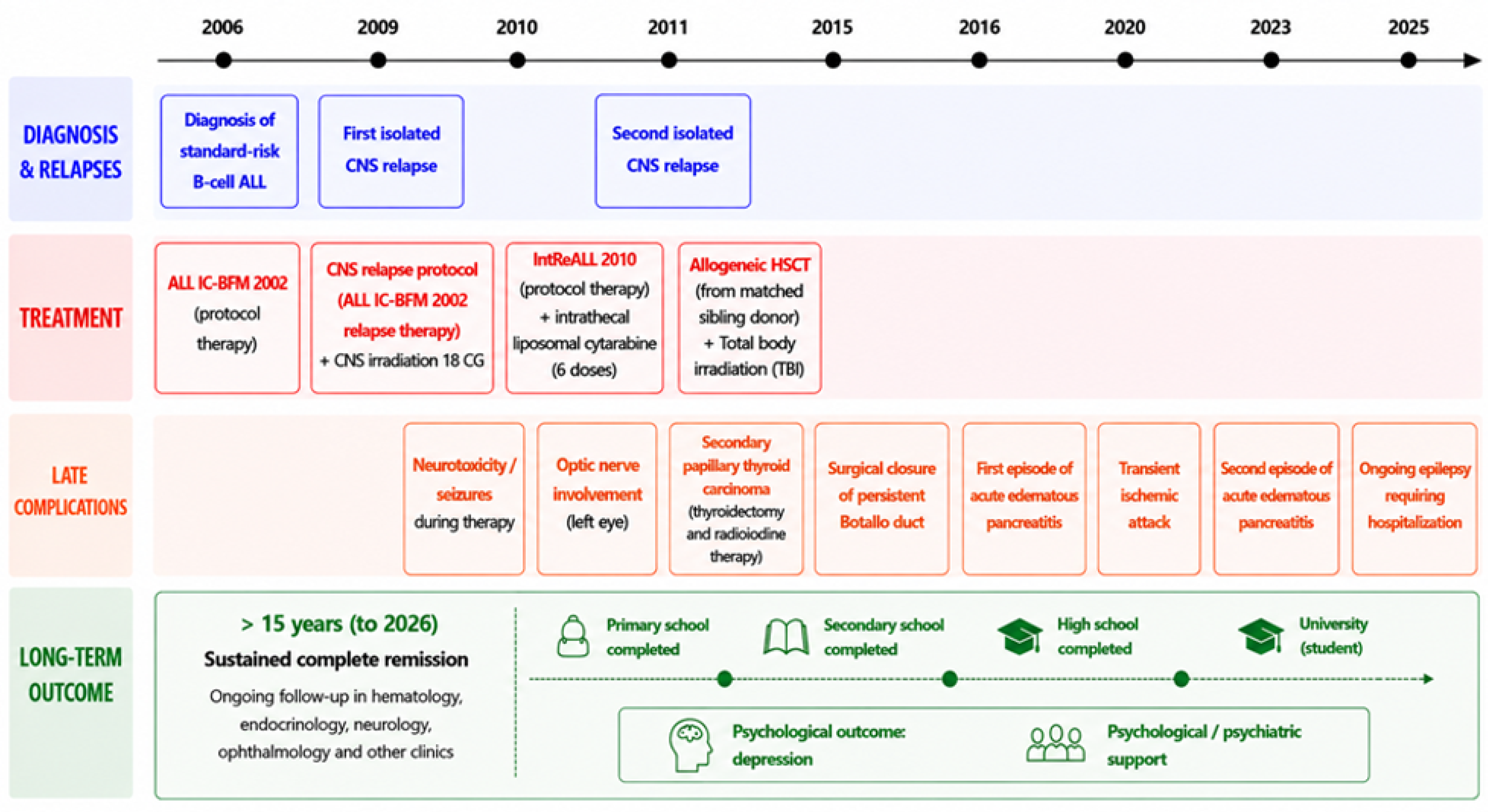
Figure 2.
Conceptual model of cumulative CNS injury associated with repeated isolated CNS relapse and multimodal CNS-directed therapy in paediatric B-cell acute lymphoblastic leukaemia.
Figure 2.
Conceptual model of cumulative CNS injury associated with repeated isolated CNS relapse and multimodal CNS-directed therapy in paediatric B-cell acute lymphoblastic leukaemia.
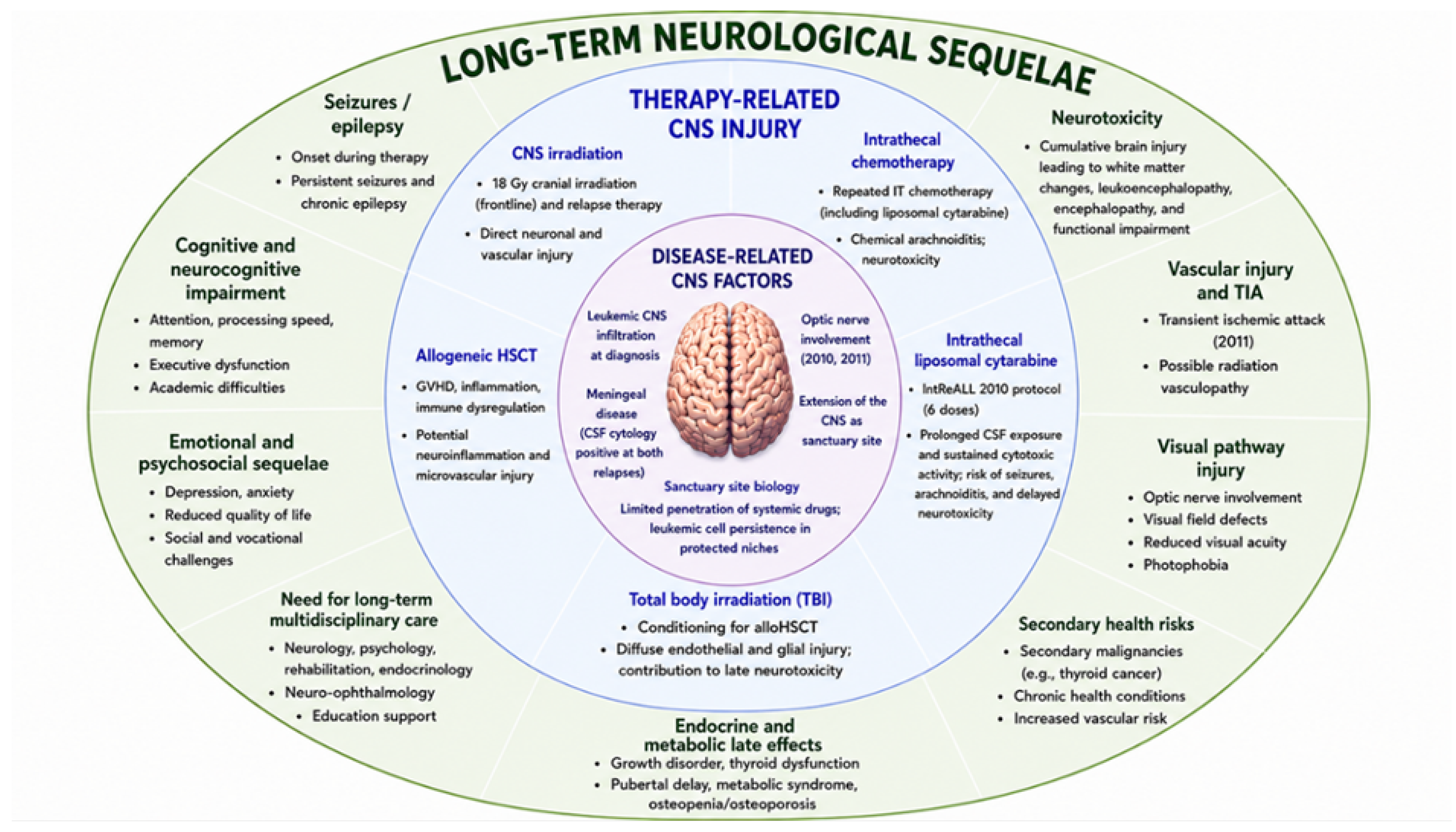

Table 1.
Potential mechanisms contributing to repeated isolated CNS relapse in the present case.
| Potential mechanism | Possible relevance in the present case |
|---|---|
| Sanctuary-site biology | Possible persistence of leukemic cells within CNS compartments |
| Optic nerve involvement | Limited penetration of systemic chemotherapy |
| Meningeal infiltration | Diffuse CNS disease distribution |
| Treatment delays | Prolongation of therapy after infectious complications |
| Pharmacogenetic variability | Possible altered methotrexate response |
| Limited sensitivity of initial diagnostics | Potential occult CNS disease |
Table 2.
Multimodal CNS-directed therapies used during treatment of repeated isolated CNS relapse and their potential contribution to cumulative long-term CNS injury.
Table 2.
Multimodal CNS-directed therapies used during treatment of repeated isolated CNS relapse and their potential contribution to cumulative long-term CNS injury.
| CNS-directed intervention | Therapeutic rationale | Potential contribution to long-term CNS injury |
|---|---|---|
| CNS relapse protocol chemotherapy | Control of isolated CNS relapse and prevention of systemic progression | Cumulative systemic and CNS toxicity; possible contribution to neurocognitive and metabolic burden |
| Cranial irradiation | Local CNS leukaemia control after CNS relapse | Radiation-induced vascular injury, white matter damage, neurocognitive impairment, endocrine dysfunction, and secondary malignancy risk |
| Repeated intrathecal chemotherapy | Direct treatment of leukemic cells within CSF and meningeal compartment | Chemical arachnoiditis, leukoencephalopathy, seizures, and cumulative neurotoxicity |
| Intrathecal liposomal cytarabine | Prolonged cytotoxic exposure within CSF; bridge to disease control before alloHSCT | Seizures, encephalopathy, arachnoiditis, and delayed neurotoxicity, especially in heavily pre-treated patients |
| Total body irradiation | Conditioning before alloHSCT | Diffuse endothelial and glial injury; possible contribution to vascular, endocrine, and neurocognitive late effects |
| Allogenic HSCT | Consolidation strategy after high-risk second isolated CNS relapse | Immune dysregulation, inflammation, GVHD-related effects, and increased burden of late complications |
| Multimodal sequential CNS-directed therapy | Durable CNS disease control through combined therapeutic approaches | Cumulative and potentially synergistic CNS injury with long-term epilepsy, vascular injury, visual pathway damage, and psychosocial sequelae |
Table 3.
Summary of literature.
| Group characteristics | Therapy characteristics | Effects | Side effects | ||||
|---|---|---|---|---|---|---|---|
| N | Age median (y) | Age range (y) | Dose | N of cycles | |||
| [10] | 9 | 7 | – | – | 6 | – | Headache, vomiting, backache |
| [20] | 18 | 10 | 4–19 | 25 → 35 → 50 mg | 11 | CR 4; PR 3; SD 2; PD 5 | Arachnoiditis, headache |
| [23] | 30 (21 male, 9 female) | 9.4 | 0.8–18 | Age-dependent dosing from 20 to 50 mg per cycle | Median 4 doses (2–9) | CNS CR 25 | PRES (1); strabismus and clonus in the lower right limb (1); partial seizures due to haemorrhagic stroke in aplastic phase (1); aphasia, ataxia, left hyposthenia, and chorea (1) |
| [24] | 4 | – | 0.3–17 | 25 mg (2), 35 mg (2) | 2–4 | CR 4 | Pseudotumor cerebri (1), irritability (1) |
| [25] | 6 (5 male, 1 female) | 11 | 2.5–16 | 25 → 35 → 50 mg | Median 6 (3–7) | CR 7 | No side effects; one case of mild headache, but all patients died (3 cases DOD, 2 after HSCT, and 1 from septic shock) |
| [26] | 3 (2 male, 1 female) | – | 5–18 | 20 → 35 → 50 mg | Median 3 (1–9) | CR 3 | Drowsiness, agitation, disorientation, hallucinations (grade III), mild headache, and seizures |
Disclaimer/Publisher’s Note: The statements, opinions and data contained in all publications are solely those of the individual author(s) and contributor(s) and not of MDPI and/or the editor(s). MDPI and/or the editor(s) disclaim responsibility for any injury to people or property resulting from any ideas, methods, instructions or products referred to in the content. |
© 2026 by the authors. Licensee MDPI, Basel, Switzerland. This article is an open access article distributed under the terms and conditions of the Creative Commons Attribution (CC BY) license (http://creativecommons.org/licenses/by/4.0/).
Copyright: This open access article is published under a Creative Commons CC BY 4.0 license, which permit the free download, distribution, and reuse, provided that the author and preprint are cited in any reuse.


