

Submitted:
27 July 2026
Posted:
07 August 2026
You are already at the latest version
Abstract
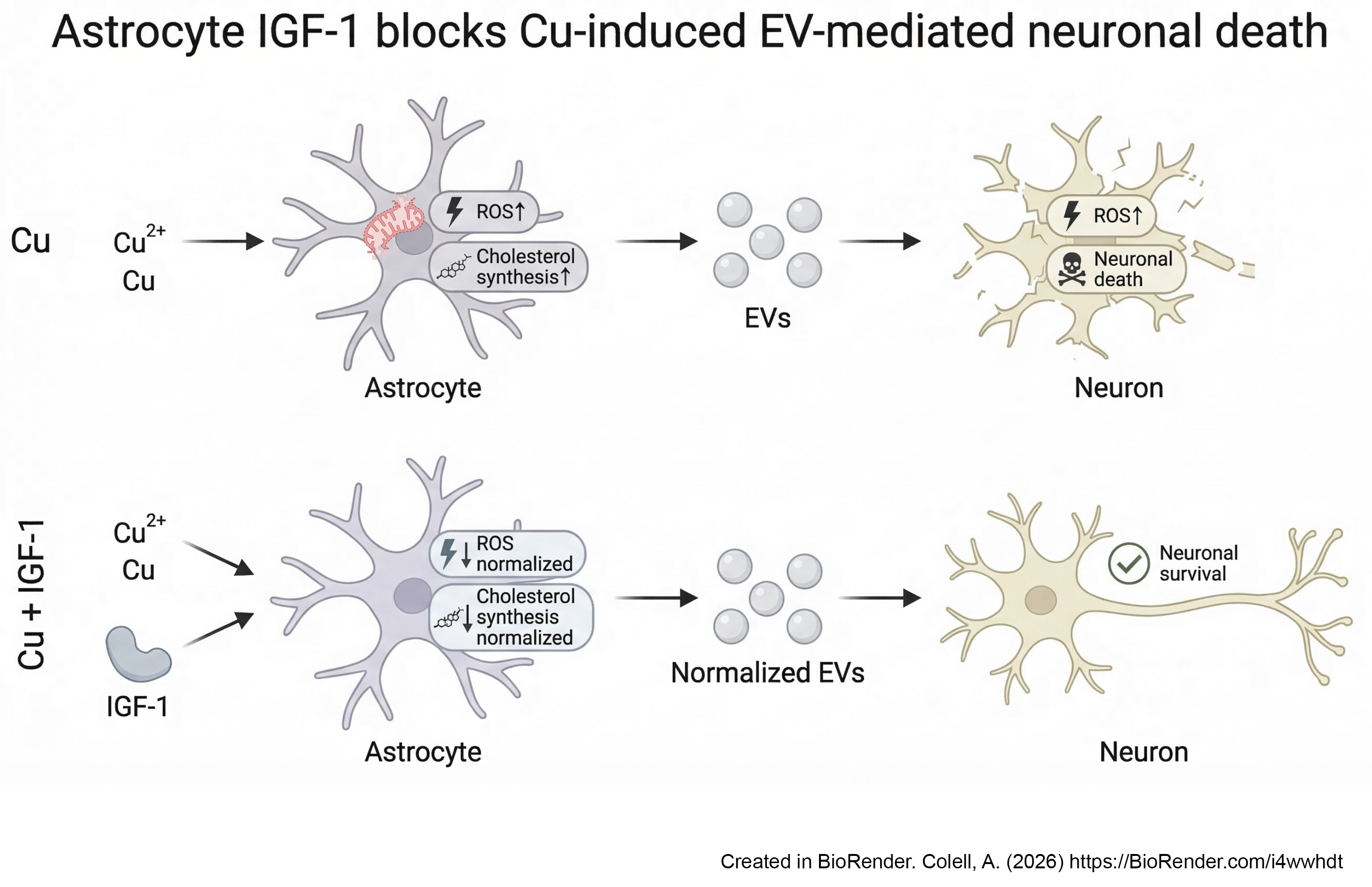
Copper (Cu) is an essential trace metal; however, its dysregulation promotes oxidative stress and contributes to neurodegeneration. Astrocytes are central to neuronal homeostasis, in part through extracellular vesicle (EV)-mediated communication, yet they are particularly vulnerable to metal-induced oxidative damage. Under pathological conditions, astrocyte-derived EVs can acquire neurotoxic properties, thereby amplifying neuronal injury. However, the mechanisms governing neuron–glia crosstalk under Cu overload remain incompletely defined. Here, we show that sublethal Cu exposure induces mitochondrial oxidative stress in astrocytes, leading to redox-dependent reprogramming of cholesterol metabolism and alterations in EV release. EVs derived from Cu-overloaded astrocytes propagate oxidative damage to neurons, establishing a ROS-driven pathway of glia-to-neuron communication. Notably, activation of insulin-like growth factor 1 (IGF-1) signaling—via recombinant protein or adenoviral delivery—attenuates Cu-induced oxidative stress, partially restores cholesterol metabolic homeostasis, and reprograms astrocyte-derived EVs toward a non-cytotoxic phenotype. Collectively, these findings identify astrocyte-derived EVs as key mediators of Cu-induced neuronal injury and highlight IGF-1 signaling as a potential therapeutic strategy to preserve astrocyte oxidative resilience and neuroprotective function.
Keywords:
copper toxicity
; astrocytes
; extracellular vesicles
; insulin-like growth factor 1
; neurotoxicity
; neuron–glia communication
1. Introduction
Copper (Cu) is an essential trace metal required for numerous biological processes, including mitochondrial respiration, antioxidant defense, and neurotransmitter synthesis. However, when present in excess or in poorly regulated forms, Cu can act as a potent pro-oxidant due to its redox-cycling capacity, promoting the generation of reactive oxygen species (ROS) with the consequent oxidative damage to lipids, proteins, and nucleic acids [1]. In this context, redox imbalance has been proposed as a key mechanism linking metal exposure, whether environmental or metabolic, to the development and progression of several neurodegenerative disorders [2].
Increasing evidence suggests that alterations in Cu homeostasis are associated with Alzheimer’s disease (AD) pathology [3,4]. In particular, elevated levels of non-ceruloplasmin-bound (“free”) Cu have been consistently observed in patients with AD. This labile pool of Cu is redox-active and can catalyze ROS formation, thereby promoting oxidative damage in neural tissue [5,6,7].
Within the brain, astrocytes are especially vulnerable to metal-induced oxidative stress, which can disrupt their homeostatic functions and compromise their ability to support neuronal health [8]. Under physiological conditions, astrocytes regulate neuronal physiology through multiple forms of intercellular signaling that provide metabolic substrates, maintain redox balance, and modulate synaptic activity. One mechanism mediating this communication is the release of extracellular vesicles (EVs) [9,10], a heterogeneous population of membrane-bound particles secreted by most cell types [11]. EVs transport bioactive molecules that can influence the physiology of recipient cells, and their molecular cargo often reflects the physiological or pathological state of the donor cell [12,13]. Notably, under cellular stress, astrocyte-derived EVs may become enriched in inflammatory mediators or neurotoxic factors, thereby amplifying neuroinflammatory signaling and potentially exacerbating neuronal damage [14].
Among the factors released by astrocytes during brain injury is insulin-like growth factor 1 (IGF-1) [15]. IGF-1 is a pleiotropic hormone that mediates many of the anabolic and mitogenic effects of growth hormone through autocrine and paracrine actions [16,17]. In the central nervous system (CNS), IGF-1 exerts potent neurotrophic effects that promote neurogenesis by supporting cell proliferation, survival, neurite outgrowth, and neuronal differentiation [18,19]. Importantly, IGF-1 levels in the brain decline with age and are reduced in neurodegenerative disorders [16,20,21]. This molecule can be produced locally in the brain or transported from the peripheral circulation into the CNS in a neuronal activity-dependent manner [22]. Both sources appear to be compromised in AD, as suggested by the reduced cerebrospinal fluid (CSF)/plasma IGF-1 ratio observed in AD patients [23]. In addition, decreased expression of the IGF-1 receptor has been reported in postmortem AD brains [20,21]. In line with its neuroprotective role, strategies aimed at enhancing IGF-1 signaling have demonstrated beneficial effects in experimental models of aging and AD, including modulation of neuroinflammation, increased hippocampal neurogenesis, attenuation of tau pathology, and improved behavioral outcomes [24,25,26]. However, while the effect of IGF-1 on neuronal physiology has been extensively characterized, its role in regulating astrocyte function and neuron–glia crosstalk remains comparatively underexplored.
We hypothesized that sustained IGF-1 signaling in astrocytes could mitigate Cu-induced oxidative stress and metabolic dysfunction. To test this hypothesis, we compared the effects of transient stimulation with recombinant IGF-1 and sustained IGF-1 expression achieved by adenoviral gene transfer in astrocytes exposed to sublethal Cu overload. Our findings uncover a mechanism by which Cu-induced oxidative stress reshapes EV-mediated astrocyte–neuron communication and identify sustained IGF-1 signaling as a promising strategy to preserve astrocytes’ neuroprotective functions under conditions of Cu-induced oxidative injury.
2. Materials and Methods
2.1. Primary Cell Culture
We used postnatal day 1-2 (P1-P2) female and male Sprague-Dawley rats from the animal facility of the Faculty of Medical Sciences at the National University of La Plata and C57BL/6 mice from the University of Barcelona. Animals were housed under standard laboratory conditions with controlled temperature and humidity, a 12 h light/dark cycle, and ad libitum access to food and water. Pups were euthanized, and the brains were aseptically dissected for the preparation of primary astrocyte cultures. Cortical and hippocampal astrocytes were isolated by mechanical dissociation as previously described [26]. Cell suspensions were seeded into 75 cm2 poly-L-lysine–coated flasks and cultured in DMEM/F12 (1:1) (Emeve medium, MicroVet SRL laboratory, Argentina) supplemented with 10% fetal bovine serum (FBS; Natocor, Cordoba, Argentina) and either penicillin-streptomycin mix (Laboratorio Serendipia, Colombia) or Plasmocin (5 μg/mL; InvivoGen, San Diego, CA, USA). Cultures were maintained in a humidified incubator at 37 °C with 5% CO2, and the medium was partially refreshed every 3 days. Upon reaching ~90% confluence, cultures were subjected to overnight agitation in an orbital shaker (Thermo Forma, Marietta, OH, USA) at 37 °C and 5% CO2 to enrich for astrocytes and minimize contamination by oligodendrocytes and microglia. The purity of astrocyte cultures (> 90%) was assessed by immunocytochemistry with antibodies against glial fibrillary acidic protein (GFAP) and ionized calcium-binding adapter molecule 1 (IBA1), markers of astrocytes and microglia, respectively (Supplementary Figure S1A).
Primary neuronal cultures were prepared from embryonic Sprague–Dawley rats and C57BL/6 mice following established protocols with minor modifications [27]. Pregnant dams were euthanized, and embryos were collected under sterile conditions. Cortical and hippocampal tissues were dissected from embryonic day 18 (E18) rat embryos or embryonic day 16–17 (E16–E17) mouse embryos under sterile conditions. Tissues were enzymatically dissociated with trypsin to generate a single-cell suspension. For rat neuronal cultures, dissociation was performed using 0.05% trypsin and 0.06% DNase I (Sigma-Aldrich) for 10 min at 37 °C. Cells were centrifuged (2,000 × g, 5 min), resuspended in DMEM/F12 medium supplemented with 10% fetal bovine serum (FBS), 0.25% glucose (Sigma-Aldrich), 1% vitamin mix (Microvet, Argentina), 2 mM L-glutamine (Sigma-Aldrich), and penicillin–streptomycin, and seeded onto poly-D-lysine-coated 12-mm glass coverslips at a density of 3 × 10⁵ cells/cm2. Cultures were maintained at 37 °C in a humidified atmosphere containing 5% CO2, and cytosine β-D-arabinofuranoside (AraC; 5 μM, Kemex Laboratories) was added on day 4 to inhibit glial proliferation. For mouse neuronal cultures, dissociated cells were seeded onto poly-D-lysine-coated culture plates at a density of 2 × 10⁵ cells/cm2 and maintained in Neurobasal™ medium (Thermo Fisher Scientific) supplemented with 2.5% (v/v) B27 supplement, 0.5 mM L-glutamine (Sigma-Aldrich), and 5 μg/mL Plasmocin™ (InvivoGen). Half of the culture medium was replaced every 3–4 days. Experiments were performed after 10 days in vitro (DIV). Neuronal purity (>95%) was confirmed by immunocytochemistry using the mature neuronal markers microtubule-associated protein 2 (MAP2) and synaptophysin (Supplementary Figure S1B).
Unless otherwise indicated, experiments were performed using untreated cells (CTRL) or cells exposed to the indicated concentrations of CuSO4 in the absence or presence of recombinant mouse IGF-1 (100 ng/mL; R&D Systems). Where indicated, N-acetyl-L-cysteine (NAC; 30 μM, Sigma-Aldrich) or mitoquinone mesylate (MitoQ; 1 μM, MedChemExpress) was included as a pharmacological control to assess the contribution of oxidative stress. In selected experiments, conditioned medium from control and treated astrocytes was collected, and cell debris was removed by centrifugation prior to extracellular vesicle (EV) isolation. Control astrocytes were maintained in culture medium alone.
2.2. Adenoviral Vector-Mediated IGF-1 Expression
Astrocyte cultures from Sprague-Dawley rats (1,500 cells/mm2) were transduced for 24 h with 100 μL of a recombinant adenoviral vector expressing either DsRed (RAd-DsRed; control) or rat IGF-1 (RAd-IGF-1). Both vectors were generated using a modified two-plasmid system as previously described [26,28], with DsRed or rat IGF-1 cDNAs placed under the mouse cytomegalovirus promoter. Recombinant viruses were produced in HEK293 cells and purified by plaque isolation followed by cesium chloride gradient ultracentrifugation. Vectors were used at a final concentration of 300 plaque-forming units/mL (pfu) [26,28]. Transduction efficiency was verified by DsRed fluorescence microscopy (Supplementary Figure S2). After transduction, cells were plated into 24-well plates and cultured until they reached semi-confluence, then exposed to the indicated treatments.
2.3. Viability Assays
Astrocytes were incubated with 0.11 mg/mL AlamarBlue® (Thermo Fisher Sci.) or 0.5 mg/mL 3-(4,5-dimethylthiazol-2-yl)-2,5-diphenyltetrazolium bromide (MTT; Thermo Fisher Sci.) at 37 °C for 2 h. Cell viability was quantified by fluorescence (Ex 535 nm, Em 595 nm) or by absorbance at 570 nm using a Beckman Coulter DTX 880 plate reader.
Primary neuronal viability was assessed using the Live and Dead Cell Assay Kit (Calcein AM, 7-AAD; Abcam) following the manufacturer’s instructions. Viable neurons were identified by intracellular hydrolysis of Calcein AM to fluorescent calcein, whereas non-viable cells were detected with 7-aminoactinomycin D (7-AAD), a membrane-impermeable DNA-binding dye. Images were acquired using an Olympus inverted microscope (TH4-200) with identical settings across conditions. Several random fields per well were captured, and the proportion of 7-AAD–positive cells relative to total cells was quantified. Experiments were performed in at least five independent cultures.
2.4. Determination of ROS, Glutathione (GSH), and Cholesterol Levels
For ROS measurement, cells were centrifuged (900 × g, 5 min) and resuspended in DMEM/F12 without FBS containing 10 mM 2′,7′-dichlorodihydrofluorescein diacetate (DCF-DA; Invitrogen). After incubation for 60 min at 37 °C, cells were washed, and fluorescence was analyzed by flow cytometry using an Accuri C6 Plus (BD). When indicated, superoxide production was assessed using dihydroethidium (DHE; Thermo Fisher Sci). Cells were incubated with 10 μM DHE in Live Cell Imaging Solution (Thermo Fisher Sci.) at 37 °C for 30 min in the dark. After rinsing, the oxidized product 2-hydroxyethidium (Ex 490 nm, Em 580 nm) was detected by fluorimetry.
Mitochondria from astrocytes were isolated by digitonin fractionation as described previously [29]. Cholesterol levels were quantified in whole cells (1 × 10⁶ cells) and mitochondrial fractions (2 × 10⁶ cells) using the Amplex Red Cholesterol Assay Kit (Thermo Fisher Sci.). Briefly, samples were extracted with chloroform: isopropanol: IGEPAL CA-630 (7:11:0.1) and centrifuged (13,000 × g, 10 min). The organic phase was collected, solvents were removed by vacuum centrifugation, and the pellet was reconstituted in 1 × cholesterol reaction buffer. The assay was performed according to the manufacturer’s instructions. The mitochondrial GSH content was assessed in mitochondrial extracts (4 × 10⁶ cells) using the glutathione GSH/GSSG Assay Kit (Sigma-Aldrich), following the manufacturer’s instructions.
2.5. Superoxide Dismutase (SOD) Activity
SOD activity was measured in cell homogenates following the protocol described by Misra and Fridovich [30]. Cells were lysed in a buffer containing 20 mM HEPES (pH 7.4), 100 mM NaCl, 5 mM EDTA, 1% Triton X-100, and protease/phosphatase inhibitors (Thermo Fisher Sci.). 1:5 dilution of the homogenate (30 μL) was added to 970 μL of reaction buffer (62.5 mM Na2CO3/NaHCO3, pH 10.2; 125 μM EDTA) and 20 μL of epinephrine solution. The inhibition of epinephrine autoxidation to adrenochrome was monitored for 2 min at 480 nm using a UV–visible spectrophotometer (Agilent 8453). One unit (U) of SOD activity was defined as the amount of enzyme required to inhibit epinephrine autoxidation by 50% and was expressed as U/min/μg of total protein.
2.6. Lipid Peroxidation (TBARS)
Lipid peroxidation was assessed by measuring thiobarbituric acid–reactive substances (TBARS) according to Yagi [31]. Cell homogenates (67 μL) were mixed with 0.8% thiobarbituric acid (Sigma-Aldrich) and 10% acetic acid (Sigma-Aldrich) and heated at 95 °C for 60 min. Absorbance was measured at 532 nm using a Beckman Coulter DTX 880 plate reader. TBARS levels were calculated from a calibration curve generated with 1,1,3,3-tetramethoxypropane (Sigma-Aldrich) and expressed as mmol TBARS/ μg of total protein.
2.7. Nitrate and Nitrite ([NOx])
[NOx] levels were determined using the Griess reaction [32]. Cell homogenates (400 μL) were mixed with 51 mM Vanadium (III) chloride (Sigma-Aldrich) in 1 M HCl and premixed Griess reagent (2% sulfanilamide in 5% HCl and 0.1% N-[1-naphthyl]-ethylenediamine). After incubation at 37 °C for 30 min, absorbance was measured at 405 nm using a Beckman Coulter DTX 880 plate reader. Concentrations were calculated from a sodium nitrate standard curve (1–500 μM).
2.8. Real-Time PCR Analysis
Total RNA was extracted using TriPure Isolation Reagent (Roche Diagnostics, USA) and reverse-transcribed into cDNA with the iScript™ cDNA Synthesis Kit (Bio-Rad). Quantitative PCR was performed using iQ SYBR Green Supermix (Bio-Rad). The cycling conditions were 95 °C for 3 min, followed by 40 cycles of 95 °C for 15 s and 60 °C for 60 s. Relative gene expression was calculated using the 2^−ΔΔCT method. The oligonucleotide primers used are listed in Table 1.
2.9. EV Isolation
EVs were isolated from astrocyte-conditioned media (using FBS devoid of EVs) by differential centrifugation [33]. Medium was diluted 1:1 with PBS and centrifuged sequentially at 300 × g (10 min), 2,000 × g (10 min), and 12,000 × g (30 min) at 4 °C to remove cells and debris (Beckman JA-25.50 rotor, Avanti J-E centrifuge). The supernatant was then ultracentrifuged at 110,000 × g for 70 min at 4 °C (70.1 Ti rotor, Sorvall). The 12K and 110K pellets were washed with PBS, centrifuged again under the same conditions, resuspended in culture medium supplemented with 10% EV-depleted FBS, pooled, and adjusted to a final volume of 650 μL.
2.10. Transmission Electron Microscopy (TEM)
EV morphology was examined by negative-stain TEM as previously described [34]. Samples were fixed with paraformaldehyde, placed on formvar-carbon-coated nickel grids for 15 min, stained with 3% uranyl acetate for 45 s, and observed in a Zeiss EM 109-T transmission electron microscope at 80 kV.
2.11. Nanoparticle Tracking Analysis (NTA)
EV size distribution and concentration were analyzed by nanoparticle tracking analysis (NTA) using a NanoSight NS300 instrument (Malvern Panalytical, UK). Samples were diluted in PBS to a final volume of 1 mL and analyzed by recording three videos per sample at a camera level of 12 and a syringe pump speed of 30. Videos were processed using NanoSight NTA software (v3.4) with a detection threshold set to 5, following the manufacturer’s recommendations.
2.12. Immunoblotting
Cells and EV fractions were lysed in RIPA buffer containing phenylmethylsulphonyl fluoride (PMSF, 2 mM), sodium orthovanadate (1 mM), and protease inhibitor cocktail (Santa Cruz Biotech.). After centrifugation (16,000 × g, 15 min), equal amounts of protein (30–50 μg) were separated by SDS–PAGE on 4–15% Criterion™ TGX Stain-Free™ gels (Bio-Rad) and transferred to nitrocellulose (Bio-Rad) or Amersham Hybond™ PVDF membranes. Membranes were incubated overnight at 4 °C with primary antibodies against 3-hydroxy-3-methyl-glutharyl-CoA reductase (HMGCR) (Nobus Biol., NBP1-50713; 1:500), β-actin (Sigma-Aldrich, A3854; 1:30,000), flotillin-1 (Santa Cruz Biotech., sc-25506; 1:1,000), CD81 (Thermo Fisher Sci., 10630D; 1:1,000), Alix (Santa Cruz Biotech., sc-43539; 1:1,000), and CD63 (Thermo Fisher Sci., PA5-92370; 1:1,000), followed by HRP-conjugated secondary antibodies. Signals were detected using Clarity™ or Clarity™ Max ECL substrates (Bio-Rad). HMGCR levels were normalized to β-actin in cell lysates, while EV markers (CD63, CD81, Alix) were normalized to flotillin-1. Uncropped scans of representative immunoblots are shown in Supplementary Figure S4.
2.13. Statistical Analysis
Each independent primary cell culture established from a single embryo (neuronal cultures) or a single postnatal pup (astrocyte cultures) was considered one biological replicate (experimental unit). Technical replicates obtained from the same culture were averaged and treated as a single biological replicate for statistical analysis. The number of biological replicates (n) for each experiment is indicated in the corresponding figure legends.
No formal randomization was performed. Independent primary cell cultures were assigned to the experimental groups at the time of treatment. Within each independent culture preparation, all experimental groups were processed simultaneously under identical culture conditions using the same batches of reagents whenever possible. Control and treated cultures were analyzed in parallel to minimize potential confounding effects related to treatment timing, reagent batches, and measurement order.
No blinding was performed during allocation of primary cell cultures to the experimental groups, conduct of the experiments, outcome assessment, or data analysis.
No a priori exclusion criteria were established for biological replicates or data points. All independent cell culture preparations that met predefined quality standards (i.e., successful establishment of viable, contamination-free cultures) were included in the analyses, and no biological replicates or data points were excluded after data collection. No single primary outcome measure was predefined because this was an exploratory mechanistic study. Sample sizes were determined based on previous studies employing similar primary cell culture models and experimental approaches [26].
Data are presented as mean ± SD from independent primary cell culture preparations. Data normality was assessed using the Shapiro–Wilk test. Statistical analyses were performed using an unpaired Student’s t-test or one-way analysis of variance (ANOVA) followed by Bonferroni’s multiple comparisons test, as appropriate, using GraphPad Prism 8 (GraphPad Software). Differences were considered statistically significant at P < 0.05.
No study protocol was formally preregistered before the start of the study. The research question, experimental design, and statistical analysis plan were developed before the experiments were initiated and are described in the Materials and Methods section.
3. Results
3.1. IGF-1 Attenuates Cu-Induced Mitochondrial Oxidative Stress in Astrocytes
To investigate how astrocytes respond to metal-induced oxidative stress, we first established a sublethal Cu exposure model that induces oxidative damage without compromising astrocyte viability. Primary astrocytes derived from C57BL/6 mice were exposed to increasing concentrations of Cu (0–800 μM; Figure 1A) for 24 h, and cell viability was assessed to determine the maximal non-cytotoxic dose. Based on this analysis and in agreement with previous studies [35,36,37], 200 μM Cu was selected as the highest concentration that preserved astrocyte viability and normal cellular morphology (Figure 1A).
Astrocytes were subsequently treated with recombinant IGF-1 (100 ng/mL) either alone or in combination with 200 μM Cu for 24 h, without affecting cell viability (Figure 1B). Intracellular ROS levels were then quantified (Figure 1C). As expected, Cu exposure markedly increased ROS production compared with untreated controls. In contrast, IGF-1 treatment prevented the increase in ROS levels observed in Cu-overloaded cells. Consistent with this observation, the addition of the antioxidant NAC also abolished Cu-induced ROS generation, serving as a positive control for oxidative stress mitigation (Figure 1C). A similar reduction in ROS levels was observed following treatment with the mitochondria-targeted antioxidant MitoQ (1 μM) (Figure 1D), suggesting that Cu-induced oxidative stress in astrocytes arises predominantly from mitochondria. Importantly, unlike antioxidants that promote Cu sequestration or facilitate its cellular efflux [38], MitoQ did not alter mitochondrial Cu accumulation, as mitochondrial Cu levels remained similarly elevated following Cu exposure in the absence (80.6 ± 7.4-fold) or presence (82.4 ± 6.9-fold) of MitoQ [35]. These findings indicate that the antioxidant effect of MitoQ is independent of mitochondrial Cu content and instead reflects its ability to suppress Cu-induced mitochondrial ROS generation.
3.2. IGF-1 Restores Cholesterol Homeostasis in Cu-Loaded Astrocytes by Suppressing SREBF2-HMGCR Signaling
Beyond its direct pro-oxidative effects, Cu dysregulation also perturbs lipid metabolism. We previously showed that sublethal Cu exposure stimulates de novo cholesterol synthesis in neurons and glial cells, leading to increased plasma membrane cholesterol levels [35,36,37]. Elevated membrane cholesterol, in turn, promotes amyloidogenic processing of APP and enhances Aβproduction [37,39].
Consistent with these findings, Cu exposure significantly increased mRNA expression of SREBF2, a key transcriptional regulator of cholesterol biosynthesis, as well as HMGCR, the rate-limiting enzyme of the mevalonate pathway (Figure 2A). Immunoblot analysis confirmed a corresponding increase in HMGCR protein levels following Cu treatment (Figure 2B). This response was suppressed by IGF-1, which blunted Cu-induced upregulation of SREBF2 and HMGCR (Figure 2A and Figure 2B) and prevented intracellular cholesterol accumulation (Figure 2C). Notably, NAC-mediated inhibition of oxidative stress fully recapitulated these effects, implicating ROS as a key driver of Cu-induced activation of cholesterol biosynthesis.
We have previously demonstrated that the induction of cholesterol synthesis is accompanied by mitochondrial cholesterol accumulation, which disrupts mitochondrial GSH transport and amplifies Aβ- and Cu-induced ROS production in neurons and microglia [35,40]. Similarly, Cu-loaded astrocytes exhibited high mitochondrial cholesterol levels (Figure 2D) together with depleted mitochondrial GSH content (Figure 2E). Treatment with MitoQ mimicked the effects of NAC, suppressing SREBF2 and HMGCR expression (Figure 2F). Moreover, MitoQ-mediated downregulation of SREBF2-HMGCR signaling was accompanied by restoration of mitochondrial cholesterol and GSH homeostasis (Figure 2D and Figure 2E), thereby linking lipid remodeling to Cu-induced mitochondrial oxidative stress.
3.3. Cu and IGF-1 Differentially Regulate the Release of Astrocyte-Derived EVs
Given the role of EVs in intercellular communication, we next characterized EVs released by astrocytes under the different experimental conditions. NTA confirmed the presence of EV-sized particles in all 24-h conditioned media. While CTRL EVs showed concentrations within the expected range for basal astrocyte release [41,42], Cu exposure increased EV release by approximately tenfold compared with CTRL cells (Figure 3A). IGF-1 alone moderately increased EV levels, whereas the combination of IGF-1 with Cu yielded the highest EV concentration. Interestingly, NAC treatment failed to reduce EVs release in Cu-treated astrocytes (Figure 3A).
Regarding particle size, EVs derived from CTRL astrocytes displayed a relatively homogeneous population consistent with small EV preparations (D50 ≈ 161 nm) (Figure 3A). In contrast, EVs from Cu-treated astrocytes exhibited a modest rightward shift in particle size distribution, with a median diameter (D50) of approximately 175 nm, which was further increased in EVs derived from IGF-1–treated astrocytes. In this group, the increase in mean diameter and D50 was more pronounced than the increase in modal diameter, indicating a broader particle size distribution enriched in larger EVs rather than a uniform enlargement of the predominant vesicle population. This finding is consistent with the emergence of a distinct subpopulation of larger EVs, potentially including plasma membrane-derived microvesicles. Importantly, co-treatment with the antioxidant NAC did not prevent the increase in EV size in Cu-exposed cells, indicating that the effect occurs independently of Cu-induced oxidative stress (Figure 3A).
To further characterize EV composition, we analyzed the abundance of the tetraspanins CD63 and CD81, together with the endosomal sorting protein Alix, in EV-enriched fractions (Figure 3B). Cu exposure significantly increased CD63 levels, whereas CD81 expression was selectively upregulated in EVs derived from IGF-1- and NAC-treated astrocytes. In contrast, Alix levels were significantly reduced in EVs isolated from IGF-1-treated cells. These findings indicate that Cu exposure and IGF-1 signaling differentially modulate the molecular composition of astrocyte-derived EVs, consistent with treatment-dependent changes in EV biogenesis and/or cargo sorting.
3.4. Impact of Astrocyte-Derived EVs on Neuronal Viability
To assess whether treatment-dependent changes in astrocyte EV release and composition impact neuron–glia communication, we examined how astrocyte-derived EVs influence the viability of primary mouse neuronal cultures. As expected, neuronal survival was unaffected by exposure to EVs from CTRL astrocytes (Figure 4). In contrast, EVs from Cu-loaded astrocytes significantly reduced neuronal viability (Figure 4), indicating that Cu exposure renders astrocyte-derived EVs neurotoxic. Notably, EVs from astrocytes treated with IGF-1, either alone or combined with Cu, preserved neuronal viability at levels similar to CTRL conditions (Figure 4). Meanwhile, NAC treatment did not block the cytotoxic effects of EVs from Cu-loaded astrocytes, suggesting that IGF-1’s protective effects on EV-mediated neuronal outcomes are not solely due to IGF-1’s capacity to prevent Cu-induced oxidative stress.
3.5. Sustained IGF-1 Expression Mitigates Cu-Induced Astrocyte Dysfunction and EV-Mediated Neurotoxicity
Having found that transient IGF-1 co-treatment blocks neurotoxicity of astrocyte-derived EVs, we next investigated whether sustained IGF-1 signaling could further counteract Cu-induced astrocyte dysfunction. This approach was motivated by reports showing reduced IGF-1 receptor expression during neurodegeneration, which may limit IGF-1 signaling and reduce the efficacy of transient IGF-1 exposure [20].
To achieve continuous IGF-1 delivery, astrocytes were transduced with a recombinant adenoviral vector encoding IGF-1 (RAd-IGF-1), enabling persistent intracellular production and secretion of the growth factor as previously reported [26]. Due to the species-specific compatibility requirements of the viral system, these experiments were performed in primary rat astrocytes to ensure efficient transduction and robust IGF-1 expression.
Before evaluating the effects of sustained IGF-1 signaling, we established a Cu exposure model in rat astrocytes. Cells were treated with increasing concentrations of Cu (0–1000 μM) for 24 h, and astrocyte viability was assessed. As shown, Cu treatment led to a concentration-dependent reduction in astrocyte survival (Figure 5A). Based on this dose–response analysis, 400 μM Cu was selected as a sublethal working concentration for subsequent experiments in primary rat astrocytes.
Intracellular ROS production was then evaluated after 24 h of exposure to 400 μM Cu in the presence or absence of RAd-IGF-1. Adenoviral-mediated IGF-1 expression significantly attenuated Cu-induced ROS generation in astrocytes (Figure 5B). To further characterize oxidative stress responses, additional redox biomarkers were analyzed. Cu exposure increased SOD activity compared with CTRL cells (Figure 5C), reflecting activation of antioxidant defenses, whereas RAd-IGF-1 expression partially attenuated this response. Similarly, Cu treatment markedly elevated TBARS (Figure 5D), indicating enhanced lipid peroxidation, and [NOx] (Figure 5E), consistent with enhanced nitrative stress. In both cases, RAd-IGF-1 expression significantly reduced TBARS and [NOx] levels (Figure 5D and Figure 5E). As expected, treatment with the antioxidant NAC resulted in a marked reduction of oxidative markers in Cu-overloaded astrocytes (Figure 5B and Figure 5E).
As observed in murine astrocytes, oxidative stress in Cu-exposed rat astrocytes was also associated with activation of cholesterol synthesis pathways, as evidenced by increased expression of Srebf2 and Hmgcr mRNA (Figure 5F). In parallel, we observed higher Tnf expression levels following Cu treatment (Figure 5G), suggesting activation of inflammatory signaling. Notably, persistent RAd-IGF-1 expression normalized Srebf2 and Hmgcr transcript levels (Figure 5F) and significantly reduced Tnf mRNA expression in Cu-overloaded astrocytes (Figure 5G). A similar downregulation was found following NAC treatment (Figure 5F and Figure 5G). Together, these findings demonstrate that sustained IGF-1 signaling effectively limits Cu-induced oxidative and nitrative stress while preventing the activation of cholesterol biosynthesis and inflammatory pathways in astrocytes, supporting its role in preserving astrocyte function under conditions of metal-induced redox imbalance.
Finally, we examined whether sustained IGF-1 expression could modulate the neurotoxic effects of astrocyte-derived EVs under conditions of Cu-induced redox imbalance. EVs were isolated from 24-h conditioned media and characterized by TEM. This analysis revealed spherical, electron-dense structures with diameters ranging from approximately 70 to 100 nm across all experimental conditions (Figure S3), consistent with the size and morphology of small EVs/exosomes [34]. To assess their functional impact, primary rat neuronal cultures were exposed to EVs derived from CTRL and treated astrocytes. Neurons incubated with EVs from Cu-overloaded astrocytes exhibited significantly increased ROS production (Figure 6A) and reduced cell viability (Figure 6B), further supporting the notion that Cu exposure confers neurotoxic properties on astrocyte-derived EVs. Both oxidative stress and neuronal toxicity were significantly attenuated when astrocytes were transduced with RAd-IGF-1 prior to Cu exposure (Figure 6A and Figure 6B). In contrast, NAC co-treatment reduced EV-induced neuronal ROS generation but was insufficient to prevent the loss of neuronal viability (Figure 6A and Figure 6B).
Together, these findings demonstrate that Cu-induced redox imbalance disrupts astrocyte homeostasis and remodels astrocyte-derived EVs into neurotoxic mediators, whereas sustained IGF-1 signaling preserves astrocyte homeostasis and prevents the acquisition of this neurotoxic EV phenotype.
4. Discussion
In this study, we demonstrate that Cu-stressed astrocytes propagate ROS-dependent neurotoxicity via EVs. Sublethal Cu exposure induces mitochondrial oxidative stress in astrocytes, triggering cholesterol biosynthesis and the release of EVs with detrimental effects on neurons. Importantly, both transient and sustained activation of IGF-1 signaling counteracted these alterations by limiting oxidative stress, normalizing metabolic and inflammatory responses, and restoring the neuroprotective properties of astrocyte-derived EVs. Collectively, these findings indicate that astrocytes are not merely passive targets of metal toxicity but active modulators of redox-driven pathological signaling. In this context, IGF-1 emerges as a key regulator that preserves astrocyte function and supports protective neuron–glial communication under conditions of metal-induced stress.
Disturbances in Cu homeostasis have increasingly been implicated in the pathogenesis of neurodegenerative disorders, including AD. Several studies have reported elevated circulating “free” Cu levels in individuals with AD, which increase with disease severity [5,7]. Environmental and dietary exposure may further contribute to Cu overload in the general population [43,44,45]. Astrocytes, which play a central role in metal buffering and redox regulation in the brain, are particularly vulnerable to such imbalances [46]. Consistent with this view, we found that sublethal Cu exposure induced robust oxidative and nitrative stress in astrocytes, accompanied by activation of antioxidant responses and lipid peroxidation, in agreement with previous studies [47,48]. Previous studies have shown that IGF-1 attenuates oxidative stress through multiple mechanisms, including the upregulation of antioxidant enzymes such as SOD, catalase, and GSH peroxidase, as well as the induction of uncoupling proteins (UCP) 2 and 3 [49,50,51]. Increased UCP expression promotes mild mitochondrial uncoupling, thereby reducing electron leakage from the respiratory chain and limiting mitochondrial ROS production [50]. In our study, the lack of increased SOD activity following IGF-1 treatment suggests that its antioxidant effects may instead be mediated by reduced mitochondrial ROS generation, potentially through UCP-dependent modulation of the mitochondrial membrane potential. Further studies will be necessary to establish whether UCPs contribute to the protective effect of IGF-1 in Cu-exposed astrocytes. Additionally, Cu exposure activated the SREBF2–HMGCR axis and upregulated inflammatory cytokine expression, effects that were prevented after antioxidant treatment, thus linking Cu-induced redox stress to lipid metabolic reprogramming in astrocytes.
Increased expression of SREBF-2 and HMGCR translated into elevated intracellular cholesterol levels and mitochondrial cholesterol enrichment. In contrast, interventions that reduced ROS levels, including IGF-1 and antioxidant treatment, attenuated SREBF2–HMGCR signaling, indicating that oxidative stress regulates cholesterol biosynthesis, as previously reported [52,53]. Particularly relevant is the accumulation of cholesterol in mitochondrial membranes beyond physiological levels, which disrupts mitochondrial function and redox homeostasis [54,55]. Remarkably, Cu-overloaded astrocytes exhibited increased mitochondrial cholesterol, mitochondrial GSH depletion, and enhanced ROS generation; these alterations were prevented by the mitochondria-targeted antioxidant MitoQ. Furthermore, attenuation of mitochondrial oxidative stress reduced SREBF2–HMGCR signaling, supporting a feed-forward loop in which mitochondrial oxidative stress sustains cholesterol biosynthesis under Cu overload. In Cu-enriched microglia, we have shown that cholesterol accumulation and mitochondrial oxidative stress amplify inflammasome-mediated inflammatory responses [35]. Similarly, recent studies have linked Cu-induced mitochondrial ROS in astrocytes to activation of the cyclic GMP–AMP synthase (cGAS)–stimulator of interferon genes (STING)–NOD-, LRR-, and pyrin domain-containing protein 3 (NLRP3) pathway, leading to pyroptosis [56]. Whether the mitochondrial oxidative stress induced by sublethal Cu exposure is sufficient to activate these innate immune pathways, and the extent to which IGF-1 modulates this process, remain to be determined.
A central finding of the present study is that Cu-induced astrocyte dysfunction leads to the release of EVs with neurotoxic properties. EVs are increasingly recognized as critical mediators of neuron–glia communication, allowing astrocytes to transfer proteins, lipids, and nucleic acids to neighboring neurons under both physiological and pathological conditions [57]. Oxidative stress is known to influence EV production and cargo composition, and EV release has been proposed as an adaptive mechanism that allows cells to remove damaged components and modulate intercellular communication under stress conditions [58]. Interestingly, while astrocyte-derived vesicles promote neuronal recovery in acute brain injury [59,60], in neurodegenerative processes, such as amyotrophic lateral sclerosis (ALS) or AD, they seem to adopt a neurotoxic profile [61,62,63]. In the same line, our results demonstrate that Cu exposure markedly increases EV release and alters vesicle composition, generating EVs capable of promoting oxidative stress and reducing neuronal viability. This mechanism may be particularly relevant in AD, where chronic oxidative stress, altered metal homeostasis, and disrupted intercellular communication contribute to the progressive spread of pathology across neural networks. Astrocyte-derived EVs may thus represent an important interface through which redox dysregulation is transmitted within the brain microenvironment.
EVs are enriched in tetraspanins, a family of membrane proteins that organize cholesterol-rich tetraspanin-enriched microdomains involved in EV biogenesis, cargo sorting, and uptake by recipient cells through interactions with a wide range of signaling partners [64,65,66]. Distinct tetraspanins are associated with specific EV subpopulations and are differentially regulated by cellular state and the pathways that drive vesicle biogenesis [67]. CD9 and CD81 are predominantly localized at the plasma membrane and are commonly associated with microvesicles and ectosomes generated by outward plasma membrane budding. In contrast, CD63 is primarily localized to the endosomal compartment, where it is enriched in intraluminal vesicles of multivesicular bodies and is therefore considered a hallmark of endosome-derived exosomes. The increase in CD63 following Cu exposure is consistent with activation of stress-responsive endosomal pathways, a response commonly associated with oxidative imbalance and metal-induced cellular stress [67,68]. Notably, the combined IGF-1 and Cu treatment further increased CD81 levels, suggesting that IGF-1 may modulate EV heterogeneity and subtype composition in a context-dependent manner. This interpretation is supported by the concomitant reduction in Alix, a key ESCRT-associated protein involved in endosomal sorting and intraluminal vesicle formation. Rather than reflecting impaired EV biogenesis, decreased Alix levels, together with increased CD81 abundance, may indicate remodeling of vesicular trafficking and cargo-sorting mechanisms [69,70]. Consistent with this interpretation, EVs released by IGF-1-treated skeletal muscle cells are enriched in CD63 and CD81 and exhibit a distinct lipid composition, despite no change in EVs abundance [71]. Given that the biological effects of IGF-1 are highly dependent on the cellular context and the underlying physiological or pathological state [72], IGF-1 signaling may selectively promote the release of functionally distinct EV subpopulations under Cu-induced stress. Future studies aimed at defining the molecular cargo of these EVs will be essential to identify the factors responsible for their neurotoxic effects and to elucidate the mechanisms by which Cu-induced astrocyte-derived EVs influence neuronal physiology.
Finally, our findings highlight the therapeutic potential of targeting astrocyte signaling pathways to mitigate metal-induced neurotoxicity. IGF-1 is a key regulator of cellular metabolism, redox balance, and survival in the CNS, yet its signaling capacity declines with aging and in neurodegenerative disorders such as AD [18,20,21]. We show that both transient and sustained activation of IGF-1 signaling effectively counteracts Cu-induced astrocyte dysfunction by reducing oxidative and nitrative stress, normalizing metabolic and inflammatory responses, and preventing the cytotoxic effects of astrocyte-derived EVs. While treatment with recombinant IGF-1 represents a direct approach to activate IGF-1 signaling and has demonstrated neuroprotective and anti-inflammatory effects in vitro [26,73], its therapeutic impact is limited by its short half-life and rapid clearance [74]. In contrast, viral-mediated IGF-1 expression enables sustained autocrine and paracrine signaling, resulting in prolonged activation of downstream pathways such as PI3K/Akt (phosphoinositide 3-kinase/protein kinase B) and enhanced regulation of cellular redox and inflammatory responses [26,75,76,77]. Notably, sustained IGF-1 expression provided broader protection than antioxidant NAC treatment alone, suggesting that its beneficial effects extend beyond simple ROS scavenging and involve coordinated regulation of astrocyte homeostasis and intercellular communication.
5. Conclusions
Our findings identify a novel mechanism by which Cu-induced oxidative stress in astrocytes can propagate neuronal damage through EV–mediated signaling. By coupling redox imbalance to metabolic dysregulation and altered EV production, Cu exposure transforms astrocytes into sources of neurotoxic intercellular signals. Importantly, activation of IGF-1 signaling effectively counteracts these alterations, restoring astrocyte homeostasis and preventing EV-mediated neuronal injury. These results highlight astrocyte-derived EVs as potential mediators of metal-induced neurotoxicity and suggest that modulating astrocyte trophic signaling may be a promising strategy to mitigate the effects of metal dyshomeostasis in the brain. Future studies are needed to elucidate how IGF-1-dependent pathways regulate EV biogenesis and cargo selection, and to determine whether enhancing astrocyte IGF-1 signaling can preserve neuron–glia communication and mitigate neurodegeneration in vivo.
Supplementary Materials
The following supporting information can be downloaded at the website of this paper posted on Preprints.org, Figure S1: Characterization of primary astrocyte and neuronal cultures; Figure S2: DsRed fluorescence in transduced astrocytes; Figure S3: Ultrastructural characterization of astrocyte-derived EVs; Figure S4: Uncropped blots from Figure 2 and Figure 3.
Author Contributions
Marlene Zubillaga: Writing – review & editing, Writing – original draft, Methodology, Formal analysis, Data curation, Conceptualization. Xenia Abadin: Methodology, Formal analysis. Elia Ivars: Methodology, Formal analysis. Margalida Puigròs: Methodology, Formal analysis. Ramon Trullas: Writing – review & editing, Resources, Methodology, Funding acquisition, Formal analysis. Julia Tau: Methodology, Formal analysis. Norberto Sanjuan: Methodology, Formal analysis. Daniel Castrogiovanni: Methodology, Formal analysis. M. José Bellini: Writing – review & editing. Anna Colell: Writing – review & editing, Writing – original draft, Resources, Methodology, Funding acquisition, Formal analysis, Data curation, Conceptualization. Nathalie Arnal: Writing – review & editing, Writing – original draft, Resources, Methodology, Funding acquisition, Formal analysis, Conceptualization.
Funding
This work was funded by the MCIN/AEI/10.13039/501100011033, co-funded by NextGenerationEU/PRTR, and by “ERDF A way of making Europe”, grants PID2022-143279OB-100 (A.C.), PID2023-153168OB-100 (R.T.), and RED2024-154130-T (A.C. and R.T.); by the CSIC, grant COOPA20476 (A.C. and N.A.), by the AGAUR, Generalitat de Catalunya, grant 2021-SGR00490 (A.C.), and grant from the National University of La Plata (UNLP) (EM001 - N.A).
Institutional Review Board Statement
All animal procedures were conducted in accordance with the applicable national and institutional guidelines for the care and use of laboratory animals and were approved by the Institutional Animal Care and Use Committee of the School of Medical Sciences, National University of La Plata (Protocol P01-01-2022; approved March 22, 2022) and the Ethics Committee on Animal Experimentation of the University of Barcelona (CEEA-UB 82-22; approved January 10, 2022).
Data Availability Statement
The data presented in this study will be made available upon request.
Acknowledgments
The authors gratefully acknowledge the support of José Amable from the Servicio Científico-Técnico “Soft Materials” at ICMAB-CSIC for assistance with NTA analyses and the Animal Experimentation Unit from the Scientific and Technological Centers (CCiTUB), Universitat de Barcelona, for their support with mouse care. We also thank Mario Ramos for his assistance with figure preparation.
Conflicts of Interest
The authors declare no conflicts of interest. The funders had no role in the design of the study; in the collection, analyses, or interpretation of data; in the writing of the manuscript; or in the decision to publish the results.
Abbreviations
The following abbreviations are used in this manuscript:
| 7-AAD | 7-aminoactinomycin D |
| AD | Alzheimer’s disease |
| Alix | ALG-2-interacting protein X |
| AraC | Cytosine β-D-arabinofuranoside |
| FBS | Fetal bovine serum |
| CNS | Central nervous system |
| CSF | Cerebrospinal fluid |
| Cu | Copper |
| DCF-DA | 2′,7′-dichlorodihydrofluorescein diacetate |
| DHE | Dihydroethidium |
| EVs | Extracellular vesicle |
| ESCRT | Endosomal Sorting Complex Required for Transport |
| GFAP | Glial fibrillary acidic protein |
| GSH | Glutathione |
| HMGCR | 3-Hydroxy-3-methylglutaryl-coenzyme A reductase |
| IBA1 | Ionized calcium-binding adapter molecule 1 |
| IGF-1 | Insulin-like growth factor 1 |
| ILV | Intraluminal vesicle |
| MAP2 | Microtubule-associated protein 2 |
| MitoQ | Mitoquinone mesylate |
| MTT | 3-(4,5-dimethylthiazol-2-yl)-2,5-diphenyltetrazolium bromide |
| MVB | Multivesicular body |
| NAC | N-acetylcysteine |
| NTA | Nanoparticle tracking analysis |
| RAd-IGF-1 | Recombinant adenoviral vector encoding IGF-1 |
| ROS | Reactive oxygen species |
| SOD | Superoxide dismutase |
| SREBF2 | Sterol regulatory element binding transcription factor 2 |
| TBARS | Thiobarbituric acid–reactive substances |
| TNF | Tumor necrosis factor |
| TEM | Transmission electron microscopy |
| UCP | Uncoupling protein |
References
- Tsang, T.; Davis, C.I.; Brady, D.C. Copper Biology. Curr. Biol. 2021, 31, R421–R427. [Google Scholar] [CrossRef] [PubMed]
- Wang, Y.; Li, D.; Xu, K.; Wang, G.; Zhang, F. Copper Homeostasis and Neurodegenerative Diseases. Neural Regen. Res. 2025, 20, 3124–3143. [Google Scholar] [CrossRef] [PubMed]
- Cherny, R.A.; Atwood, C.S.; Xilinas, M.E.; Gray, D.N.; Jones, W.D.; McLean, C.A.; Barnham, K.J.; Volitakis, I.; Fraser, F.W.; Kim, Y.S.; et al. Treatment with a Copper-Zinc Chelator Markedly and Rapidly Inhibits β-Amyloid Accumulation in Alzheimer’s Disease Transgenic Mice. Neuron 2001, 30, 665–676. [Google Scholar] [CrossRef] [PubMed]
- Sparks, D.L.; Schreurs, B.G. Trace Amounts of Copper in Water Induce Beta-Amyloid Plaques and Learning Deficits in a Rabbit Model of Alzheimer’s Disease. Proc. Natl. Acad. Sci. U. S. A. 2003, 100, 11065–11069. [Google Scholar] [CrossRef] [PubMed]
- Squitti, R. Copper Subtype of Alzheimer’s Disease (AD): Meta-Analyses, Genetic Studies and Predictive Value of Non-Ceruloplasmim Copper in Mild Cognitive Impairment Conversion to Full AD. J. Trace Elem. Med. Biol. 2014, 4–7. [Google Scholar] [CrossRef] [PubMed]
- Squitti, R.; Pasqualetti, P.; Dal Forno, G.; Moffa, F.; Cassetta, E.; Lupoi, D.; Vernieri, F.; Rossi, L.; Baldassini, M.; Rossini, P.M. Excess of Serum Copper Not Related to Ceruloplasmin in Alzheimer Disease. Neurology 2005, 64, 1040–1046. [Google Scholar] [CrossRef] [PubMed]
- Arnal, N.; Cristalli, D.O.; de Alaniz, M.J.T.; Marra, C.A. Clinical Utility of Copper, Ceruloplasmin, and Metallothionein Plasma Determinations in Human Neurodegenerative Patients and Their First-Degree Relatives. Brain Res. 2010, 1319. [Google Scholar] [CrossRef] [PubMed]
- Li, B.; Xia, M.; Zorec, R.; Parpura, V.; Verkhratsky, A. Astrocytes in Heavy Metal Neurotoxicity and Neurodegeneration. Brain Res. 2021, 1752. [Google Scholar] [CrossRef] [PubMed]
- Jacquet, A. de R.; Tancredi, J.L.; Lemire, A.L.; Desantis, M.C.; Li, W.P.; O’shea, E.K. The LRRK2 G2019S Mutation Alters Astrocyte-to-Neuron Communication via Extracellular Vesicles and Induces Neuron Atrophy in a Human IPSC-Derived Model of Parkinson’s Disease. Elife 2021, 10. [Google Scholar] [CrossRef] [PubMed]
- Jiang, D.; Gong, F.; Ge, X.; Lv, C.; Huang, C.; Feng, S.; Zhou, Z.; Rong, Y.; Wang, J.; Ji, C.; et al. Neuron-Derived Exosomes-Transmitted MiR-124-3p Protect Traumatically Injured Spinal Cord by Suppressing the Activation of Neurotoxic Microglia and Astrocytes. J. Nanobiotechnology 2020, 18. [Google Scholar] [CrossRef] [PubMed]
- Colombo, M.; Raposo, G.; Théry, C. Biogenesis, Secretion, and Intercellular Interactions of Exosomes and Other Extracellular Vesicles. Annu. Rev. Cell Dev. Biol. 2014, 30, 255–289. [Google Scholar] [CrossRef] [PubMed]
- De Toro, J.; Herschlik, L.; Waldner, C.; Mongini, C. Emerging Roles of Exosomes in Normal and Pathological Conditions: New Insights for Diagnosis and Therapeutic Applications. Front. Immunol. 2015, 6. [Google Scholar] [CrossRef] [PubMed]
- Silverman, J.M.; Christy, D.; Shyu, C.C.; Moon, K.M.; Fernando, S.; Gidden, Z.; Cowan, C.M.; Ban, Y.; Greg Stacey, R.; Grad, L.I.; et al. CNS-Derived Extracellular Vesicles from Superoxide Dismutase 1 (SOD1)G93A ALS Mice Originate from Astrocytes and Neurons and Carry Misfolded SOD1. J. Biol. Chem. 2019, 294, 3744–3759. [Google Scholar] [CrossRef] [PubMed]
- Zhao, S.; Sheng, S.; Wang, Y.; Ding, L.; Xu, X.; Xia, X.; Zheng, J.C. Astrocyte-Derived Extracellular Vesicles: A Double-Edged Sword in Central Nervous System Disorders. Neurosci. Biobehav. Rev. 2021, 125, 148–159. [Google Scholar] [CrossRef] [PubMed]
- Chen, W.; He, B.; Tong, W.; Zeng, J.; Zheng, P. Astrocytic Insulin-Like Growth Factor-1 Protects Neurons Against Excitotoxicity. Front. Cell. Neurosci. 2019, 13. [Google Scholar] [CrossRef] [PubMed]
- Sonntag, W.E.; Ramsey, M.; Carter, C.S. Growth Hormone and Insulin-like Growth Factor-1 (IGF-1) and Their Influence on Cognitive Aging. Ageing Res. Rev. 2005, 4, 195–212. [Google Scholar] [CrossRef] [PubMed]
- Geffken, S.J.; Moon, S.; Smith, C.O.; Tang, S.; Lee, H.H.; Lewis, K.; Wong, C.W.; Huang, Y.; Huang, Q.; Zhao, Y.T.; et al. Insulin and IGF-1 Elicit Robust Transcriptional Regulation to Modulate Autophagy in Astrocytes. Mol. Metab. 2022, 66. [Google Scholar] [CrossRef] [PubMed]
- Sun, L.Y. Hippocampal IGF-1 Expression, Neurogenesis and Slowed Aging: Clues to Longevity from Mutant Mice. Age (Dordr) 2006, 28, 181–189. [Google Scholar] [CrossRef] [PubMed]
- O’Kusky, J.; Ye, P. Neurodevelopmental Effects of Insulin-like Growth Factor Signaling. Front. Neuroendocrinol. 2012, 33, 230–251. [Google Scholar] [CrossRef] [PubMed]
- Steen, E.; Terry, B.M.; Rivera, E.J.; Cannon, J.L.; Neely, T.R.; Tavares, R.; Xu, X.J.; Wands, J.R.; De La Monte, S.M. Impaired Insulin and Insulin-like Growth Factor Expression and Signaling Mechanisms in Alzheimer’s Disease--Is This Type 3 Diabetes? J. Alzheimers Dis. 2005, 7, 63–80. [Google Scholar] [CrossRef] [PubMed]
- Rivera, E.J.; Goldin, A.; Fulmer, N.; Tavares, R.; Wands, J.R.; De La Monte, S.M. Insulin and Insulin-like Growth Factor Expression and Function Deteriorate with Progression of Alzheimer’s Disease: Link to Brain Reductions in Acetylcholine. J. Alzheimers Dis. 2005, 8, 247–268. [Google Scholar] [CrossRef] [PubMed]
- Nishijima, T.; Piriz, J.; Duflot, S.; Fernandez, A.M.; Gaitan, G.; Gomez-Pinedo, U.; Verdugo, J.M.G.; Leroy, F.; Soya, H.; Nuñez, A.; et al. Neuronal Activity Drives Localized Blood-Brain-Barrier Transport of Serum Insulin-like Growth Factor-I into the CNS. Neuron 2010, 67, 834–846. [Google Scholar] [CrossRef] [PubMed]
- Trueba-Sáiz, A.; Cavada, C.; Fernandez, A.M.; Leon, T.; González, D.A.; Fortea Ormaechea, J.; Lleó, A.; Del Ser, T.; Nuñez, A.; Torres-Aleman, I. Loss of Serum IGF-I Input to the Brain as an Early Biomarker of Disease Onset in Alzheimer Mice. Transl. Psychiatry 2013, 3. [Google Scholar] [CrossRef] [PubMed]
- Zappa Villar, M.F.; López Hanotte, J.; Crespo, R.; Pardo, J.; Reggiani, P.C. Insulin-like Growth Factor 1 Gene Transfer for Sporadic Alzheimer’s Disease: New Evidence for Trophic Factor Mediated Hippocampal Neuronal and Synaptic Recovery-Based Behavior Improvement. Hippocampus 2021, 31, 1137–1153. [Google Scholar] [CrossRef] [PubMed]
- Morel, G.R.; León, M.L.; Uriarte, M.; Reggiani, P.C.; Goya, R.G. Therapeutic Potential of IGF-I on Hippocampal Neurogenesis and Function during Aging. Neurogenesis 2016, 4, e1259709. [Google Scholar] [CrossRef] [PubMed]
- Bellini, M.J.; Hereñú, C.B.; Goya, R.G.; Garcia-Segura, L.M. Insulin-like Growth Factor-I Gene Delivery to Astrocytes Reduces Their Inflammatory Response to Lipopolysaccharide. J. Neuroinflammation 2011, 8. [Google Scholar] [CrossRef] [PubMed]
- Echeverria, V.; Greenberg, D.L.; Doré, S. Expression of Prostaglandin E2 Synthases in Mouse Postnatal Cortical Neurons. Ann. N. Y. Acad. Sci. 2005, 1053, 460–471. [Google Scholar] [CrossRef] [PubMed]
- Hereñú, C.B.; Cristina, C.; Rimoldi, O.J.; Becú-Villalobos, D.; Cambiaggi, V.; Portiansky, E.L.; Goya, R.G. Restorative Effect of Insulin-like Growth Factor-I Gene Therapy in the Hypothalamus of Senile Rats with Dopaminergic Dysfunction. Gene Ther. 2007, 14, 237–245. [Google Scholar] [CrossRef] [PubMed]
- Dixit, B.; Vanhoozer, S.; Anti, N.A.; O’Connor, M.S.; Boominathan, A. Rapid Enrichment of Mitochondria from Mammalian Cell Cultures Using Digitonin. MethodsX 2021, 8. [Google Scholar] [CrossRef] [PubMed]
- Misra, H.P.; Fridovich, I. The Role of Superoxide Anion in the Autoxidation of Epinephrine and a Simple Assay for Superoxide Dismutase. J. Biol. Chem. 1972, 247, 3170–3175. [Google Scholar]
- Yagi, K. A Simple Fluorometric Assay for Lipoperoxide in Blood Plasma. Biochem. Med. 1976. [Google Scholar] [CrossRef] [PubMed]
- Miranda, K.M.; Espey, M.G.; Wink, D.A. A Rapid, Simple Spectrophotometric Method for Simultaneous Detection of Nitrate and Nitrite. 2001, 5, 62–71. [Google Scholar] [CrossRef] [PubMed]
- Théry, C.; Amigorena, S.; Raposo, G.; Clayton, A. Isolation and Characterization of Exosomes from Cell Culture Supernatants and Biological Fluids. In Curr. Protoc. Cell Biol.; 2006; Volume Chapter 3. [Google Scholar] [CrossRef] [PubMed]
- Pascucci, L.; Scattini, G. Imaging Extracelluar Vesicles by Transmission Electron Microscopy: Coping with Technical Hurdles and Morphological Interpretation. Biochim. Et. Biophys. Acta (BBA) -General. Subj. 2021, 1865, 129648. [Google Scholar] [CrossRef] [PubMed]
- Zubillaga, M.; Abadin, X.; Ivars, E.; Puigròs, M.; Trullas, R.; Bellini, M.J.; Arnal, N.; Colell, A. Copper Overload Worsens the Inflammatory Response of Microglia to Amyloid Beta (Aβ) by Impairing Phagocytosis and Promoting Mitochondrial DNA-Mediated NLRP3 Inflammasome Activation. Redox Biol. 2025, 87. [Google Scholar] [CrossRef] [PubMed]
- Zubillaga, M.; Tau, J.; Rosa, D.; Bellini, M.J.; Arnal, N. Sex-Dependent Effect of Sublethal Copper Concentrations on de Novo Cholesterol Synthesis in Astrocytes and Their Possible Links to Variations in Cholesterol and Amyloid Precursor Protein Levels in Neuronal Membranes. Biol. Sex. Differ. 2024, 15. [Google Scholar] [CrossRef] [PubMed]
- Zubillaga, M.; Rosa, D.; Astiz, M.; Tricerri, M.A.; Arnal, N. Effect of Sublethal Copper Overload on Cholesterol De Novo Synthesis in Undifferentiated Neuronal Cells. ACS Omega 2022, 7, 25022–25030. [Google Scholar] [CrossRef] [PubMed]
- Bhattacharjee, A.; Chakraborty, K.; Shukla, A. Cellular Copper Homeostasis: Current Concepts on Its Interplay with Glutathione Homeostasis and Its Implication in Physiology and Human Diseases. Metallomics 2017, 9, 1376–1388. [Google Scholar] [CrossRef] [PubMed]
- Wang, H.; Kulas, J.A.; Wang, C.; Holtzman, D.M.; Ferris, H.A.; Hansen, S.B. Regulation of Beta-Amyloid Production in Neurons by Astrocyte-Derived Cholesterol. Proc. Natl. Acad. Sci. U. S. A. 2021, 118. [Google Scholar] [CrossRef] [PubMed]
- Fernández, A.; Llacuna, L.; Fernández-Checa, J.C.; Colell, A. Mitochondrial Cholesterol Loading Exacerbates Amyloid β Peptide-Induced Inflammation and Neurotoxicity. J. Neurosci. 2009, 29, 6394–6405. [Google Scholar] [CrossRef] [PubMed]
- Li, Z.; Moniruzzaman, M.; Dastgheyb, R.M.; Yoo, S.W.; Wang, M.; Hao, H.; Liu, J.; Casaccia, P.; Nogueras-Ortiz, C.; Kapogiannis, D.; et al. Astrocytes Deliver CK1 to Neurons via Extracellular Vesicles in Response to Inflammation Promoting the Translation and Amyloidogenic Processing of APP. J. Extracell. Vesicles 2020, 10. [Google Scholar] [CrossRef] [PubMed]
- Wang, K.; Ye, L.; Lu, H.; Chen, H.; Zhang, Y.; Huang, Y.; Zheng, J.C. TNF-α Promotes Extracellular Vesicle Release in Mouse Astrocytes through Glutaminase. J. Neuroinflammation 2017, 14. [Google Scholar] [CrossRef] [PubMed]
- Brewer, G.J. Copper Toxicity in Alzheimer’s Disease: Cognitive Loss from Ingestion of Inorganic Copper. J. Trace Elem. Med. Biol. 2012, 26, 89–92. [Google Scholar] [CrossRef] [PubMed]
- Arnal, N.; de Alaniz, M.J.T.; Marra, C.A. Alterations in Copper Homeostasis and Oxidative Stress Biomarkers in Women Using the Intrauterine Device TCu380A. Toxicol. Lett. 2010, 192. [Google Scholar] [CrossRef] [PubMed]
- Arnal, N.; Astiz, M.; de Alaniz, M.J.T.; Marra, C.A. Clinical Parameters and Biomarkers of Oxidative Stress in Agricultural Workers Who Applied Copper-Based Pesticides. Ecotoxicol. Environ. Saf. 2011, 74. [Google Scholar] [CrossRef] [PubMed]
- Li, B.; Xia, M.; Zorec, R.; Parpura, V.; Verkhratsky, A. Astrocytes in Heavy Metal Neurotoxicity and Neurodegeneration. Brain Res. 2021, 1752. [Google Scholar] [CrossRef] [PubMed]
- Ferretti, G.; Bacchetti, T.; Moroni, C.; Vignini, A.; Curatola, G. Copper-Induced Oxidative Damage on Astrocytes: Protective Effect Exerted by Human High Density Lipoproteins. Biochim. Biophys. Acta Mol. Cell Biol. Lipids 2003, 1635, 48–54. [Google Scholar] [CrossRef] [PubMed]
- Reddy, P.V.B.; Rama Rao, K. V.; Norenberg, M.D. The Mitochondrial Permeability Transition, and Oxidative and Nitrosative Stress in the Mechanism of Copper Toxicity in Cultured Neurons and Astrocytes. Lab. Investig. 2008, 88, 816–830. [Google Scholar] [CrossRef] [PubMed]
- Gustafsson, H.; Söderdahl, T.; Jönsson, G.; Bratteng, J.O.; Forsby, A. Insulin-like Growth Factor Type 1 Prevents Hyperglycemia-Induced Uncoupling Protein 3 down-Regulation and Oxidative Stress. J. Neurosci. Res. 2004, 77, 285–291. [Google Scholar] [CrossRef] [PubMed]
- Andrews, Z.B.; Liu, Z.W.; Walllingford, N.; Erion, D.M.; Borok, E.; Friedman, J.M.; Tschöp, M.H.; Shanabrough, M.; Cline, G.; Shulman, G.I.; et al. UCP2 Mediates Ghrelin’s Action on NPY/AgRP Neurons by Lowering Free Radicals. Nature 2008, 454, 846–851. [Google Scholar] [CrossRef] [PubMed]
- Herrera, M.L.; Champarini, L.G.; Oliveros, A.L.; Bellini, M.J.; Hereñú, C.B. Potentialities of IGF-1 for Regulating Oxidative Stress in Neuroinflammation and Neurodegeneration: Theoretical Review. Open Explor. 2019 4:6 2024, 4, 442–458. [Google Scholar] [CrossRef]
- Gesquière, L.; Loreau, N.; Minnich, A.; Davignon, J.; Blache, D. Oxidative Stress Leads to Cholesterol Accumulation in Vascular Smooth Muscle Cells. Free Radic. Biol. Med. 1999, 27, 134–145. [Google Scholar] [CrossRef] [PubMed]
- Seo, K.; Shin, S.M. Induction of Lipin1 by ROS-Dependent SREBP-2 Activation. Toxicol. Res. 2017. [Google Scholar] [CrossRef] [PubMed]
- Goicoechea, L.; Conde de la Rosa, L.; Torres, S.; García-Ruiz, C.; Fernández-Checa, J.C. Mitochondrial Cholesterol: Metabolism and Impact on Redox Biology and Disease. Redox Biol. 2023, 61. [Google Scholar] [CrossRef] [PubMed]
- Solsona-Vilarrasa, E.; Fucho, R.; Torres, S.; Nuñez, S.; Nuño-Lámbarri, N.; Enrich, C.; García-Ruiz, C.; Fernández-Checa, J.C. Cholesterol Enrichment in Liver Mitochondria Impairs Oxidative Phosphorylation and Disrupts the Assembly of Respiratory Supercomplexes. Redox Biol. 2019, 24. [Google Scholar] [CrossRef] [PubMed]
- Shi, W.; Zhou, Q.; Lu, L.; Zhang, Y.; Zhang, H.; Pu, Y.; Yin, L. Copper Induced Cytosolic Escape of Mitochondrial DNA and Activation of CGAS-STING-NLRP3 Pathway-Dependent Pyroptosis in C8-D1A Cells. Ecotoxicol. Environ. Saf. 2024, 285. [Google Scholar] [CrossRef] [PubMed]
- Upadhya, R.; Zingg, W.; Shetty, S.; Shetty, A.K. Astrocyte-Derived Extracellular Vesicles: Neuroreparative Properties and Role in the Pathogenesis of Neurodegenerative Disorders. J. Control. Release 2020, 323, 225–239. [Google Scholar] [CrossRef] [PubMed]
- Chiaradia, E.; Tancini, B.; Emiliani, C.; Delo, F.; Pellegrino, R.M.; Tognoloni, A.; Urbanelli, L.; Buratta, S. Extracellular Vesicles under Oxidative Stress Conditions: Biological Properties and Physiological Roles. Cells 2021, 10. [Google Scholar] [CrossRef] [PubMed]
- Zhang, W.; Hong, J.; Zhang, H.; Zheng, W.; Yang, Y. Astrocyte-Derived Exosomes Protect Hippocampal Neurons after Traumatic Brain Injury by Suppressing Mitochondrial Oxidative Stress and Apoptosis. Aging 2021, 13, 21642–21658. [Google Scholar] [CrossRef] [PubMed]
- Wang, X.; Li, A.; Fan, H.; Li, Y.; Yang, N.; Tang, Y. Astrocyte-Derived Extracellular Vesicles for Ischemic Stroke: Therapeutic Potential and Prospective. Aging Dis. 2024, 15, 1227–1254. [Google Scholar] [CrossRef] [PubMed]
- Nogueras-Ortiz, C.J.; Mahairaki, V.; Delgado-Peraza, F.; Das, D.; Avgerinos, K.; Eren, E.; Hentschel, M.; Goetzl, E.J.; Mattson, M.P.; Kapogiannis, D. Astrocyte- and Neuron-Derived Extracellular Vesicles from Alzheimer’s Disease Patients Effect Complement-Mediated Neurotoxicity. Cells 2020, 9. [Google Scholar] [CrossRef] [PubMed]
- Basso, M.; Pozzi, S.; Tortarolo, M.; Fiordaliso, F.; Bisighini, C.; Pasetto, L.; Spaltro, G.; Lidonnici, D.; Gensano, F.; Battaglia, E.; et al. Mutant Copper-Zinc Superoxide Dismutase (SOD1) Induces Protein Secretion Pathway Alterations and Exosome Release in Astrocytes. J. Biol. Chem. 2013, 288, 15699–15711. [Google Scholar] [CrossRef] [PubMed]
- Varcianna, A.; Myszczynska, M.A.; Castelli, L.M.; O’Neill, B.; Kim, Y.; Talbot, J.; Nyberg, S.; Nyamali, I.; Heath, P.R.; Stopford, M.J.; et al. Micro-RNAs Secreted through Astrocyte-Derived Extracellular Vesicles Cause Neuronal Network Degeneration in C9orf72 ALS. EBioMedicine 2019, 40, 626–635. [Google Scholar] [CrossRef] [PubMed]
- Charrin, S.; Manié, S.; Thiele, C.; Billard, M.; Gerlier, D.; Boucheix, C.; Rubinstein, E. A Physical and Functional Link between Cholesterol and Tetraspanins. Eur. J. Immunol. 2003, 33, 2479–2489. [Google Scholar] [CrossRef] [PubMed]
- Kang, T.; Atukorala, I.; Mathivanan, S. Biogenesis of Extracellular Vesicles. Subcell. Biochem. 2021, 97, 19–43. [Google Scholar] [CrossRef] [PubMed]
- Yáñez-Mó, M.; Barreiro, O.; Gordon-Alonso, M.; Sala-Valdés, M.; Sánchez-Madrid, F. Tetraspanin-Enriched Microdomains: A Functional Unit in Cell Plasma Membranes. Trends Cell Biol. 2009, 19, 434–446. [Google Scholar] [CrossRef] [PubMed]
- Hessvik, N.P.; Llorente, A. Current Knowledge on Exosome Biogenesis and Release. Cell. Mol. Life Sci. 2018, 75, 193–208. [Google Scholar] [CrossRef] [PubMed]
- Zhang, W.; Liu, R.; Chen, Y.; Wang, M.; Du, J. Crosstalk between Oxidative Stress and Exosomes. Oxid. Med. Cell. Longev. 2022, 2022. [Google Scholar] [CrossRef] [PubMed]
- Hurley, J.H. ESCRTs Are Everywhere. EMBO J. 2015, 34, 2398–2407. [Google Scholar] [CrossRef] [PubMed]
- Colombo, M.; Raposo, G.; Théry, C. Biogenesis, Secretion, and Intercellular Interactions of Exosomes and Other Extracellular Vesicles. Annu. Rev. Cell Dev. Biol. 2014, 30, 255–289. [Google Scholar] [CrossRef] [PubMed]
- Valentino, T.R.; Rule, B.D.; Mobley, C.B.; Nikolova-Karakashian, M.; Vechetti, I.J. Skeletal Muscle Cell Growth Alters the Lipid Composition of Extracellular Vesicles. Membranes 2021, 11, 619. [Google Scholar] [CrossRef]
- Khan, M.Z.; Zugaza, J.L.; Torres Aleman, I. The Signaling Landscape of Insulin-like Growth Factor 1. J. Biol. Chem. 2025, 301. [Google Scholar] [CrossRef] [PubMed]
- Fernandez, S.; Fernandez, A.M.; Lopez-Lopez, C.; Torres-Aleman, I. Emerging Roles of Insulin-like Growth Factor-I in the Adult Brain. Growth Horm. IGF Res. 2007, 17, 89–95. [Google Scholar] [CrossRef] [PubMed]
- Bailes, J.; Soloviev, M. Insulin-Like Growth Factor-1 (IGF-1) and Its Monitoring in Medical Diagnostic and in Sports. Biomolecules 2021, 11, 1–15. [Google Scholar] [CrossRef] [PubMed]
- Fernandez, A.M.; Hernandez-Garzón, E.; Perez-Domper, P.; Perez-Alvarez, A.; Mederos, S.; Matsui, T.; Santi, A.; Trueba-Saiz, A.; García-Guerra, L.; Pose-Utrilla, J.; et al. Insulin Regulates Astrocytic Glucose Handling Through Cooperation With IGF-I. Diabetes 2017, 66, 64–74. [Google Scholar] [CrossRef] [PubMed]
- Jallali, N.; Ridha, H.; Thrasivoulou, C.; Butler, P.; Cowen, T. Modulation of Intracellular Reactive Oxygen Species Level in Chondrocytes by IGF-1, FGF, and TGF-Beta1. Connect. Tissue Res. 2007, 48, 149–158. [Google Scholar] [CrossRef] [PubMed]
- Hu, W.S.; Liao, W.Y.; Chang, C.H.; Chen, T.S. Paracrine IGF-1 Activates SOD2 Expression and Regulates ROS/P53 Axis in the Treatment of Cardiac Damage in D-Galactose-Induced Aging Rats after Receiving Mesenchymal Stem Cells. J. Clin. Med. 2022, 11. [Google Scholar] [CrossRef] [PubMed]
Figure 1.
IGF-1 protects astrocytes from Cu-induced mitochondrial oxidative stress. (A) Dose-response analysis of mouse astrocyte viability following exposure to increasing concentrations of CuSO4 (Cu, 0–800 μM) for 24 h. Right, representative phase-contrast images of CTRL astrocytes and astrocytes exposed to 200 μM Cu for 24 h. Scale bar, 25 μm. (B) Astrocyte viability after 24 h treatment with IGF-1 (100 ng/ml) alone or in combination with Cu (200 μM). (C-D) Intracellular ROS levels after 24 h exposure to Cu (200 μM) with and without IGF-1(100 ng/ml), NAC (30 μM), or MitoQ (1 μM) treatment. ROS were measured by flow cytometry using DCF-DA (C) and by spectrofluorometry using DHE (D). In (C), the right panels show representative flow cytometry histograms (frequency vs. fluorescence intensity). Results were expressed as the percentage of CTRL. Data are presented as mean ± SD (n = 9-12 independent primary cultures, panel A and B; n = 3 independent primary cultures, panel C and D). Statistical analysis was performed using an unpaired Student’s t-test (panel A) or one-way ANOVA followed by Bonferroni’s multiple comparisons test. Statistical significance is indicated as * P < 0.05, ** P < 0.01, and *** P < 0.001.
Figure 1.
IGF-1 protects astrocytes from Cu-induced mitochondrial oxidative stress. (A) Dose-response analysis of mouse astrocyte viability following exposure to increasing concentrations of CuSO4 (Cu, 0–800 μM) for 24 h. Right, representative phase-contrast images of CTRL astrocytes and astrocytes exposed to 200 μM Cu for 24 h. Scale bar, 25 μm. (B) Astrocyte viability after 24 h treatment with IGF-1 (100 ng/ml) alone or in combination with Cu (200 μM). (C-D) Intracellular ROS levels after 24 h exposure to Cu (200 μM) with and without IGF-1(100 ng/ml), NAC (30 μM), or MitoQ (1 μM) treatment. ROS were measured by flow cytometry using DCF-DA (C) and by spectrofluorometry using DHE (D). In (C), the right panels show representative flow cytometry histograms (frequency vs. fluorescence intensity). Results were expressed as the percentage of CTRL. Data are presented as mean ± SD (n = 9-12 independent primary cultures, panel A and B; n = 3 independent primary cultures, panel C and D). Statistical analysis was performed using an unpaired Student’s t-test (panel A) or one-way ANOVA followed by Bonferroni’s multiple comparisons test. Statistical significance is indicated as * P < 0.05, ** P < 0.01, and *** P < 0.001.
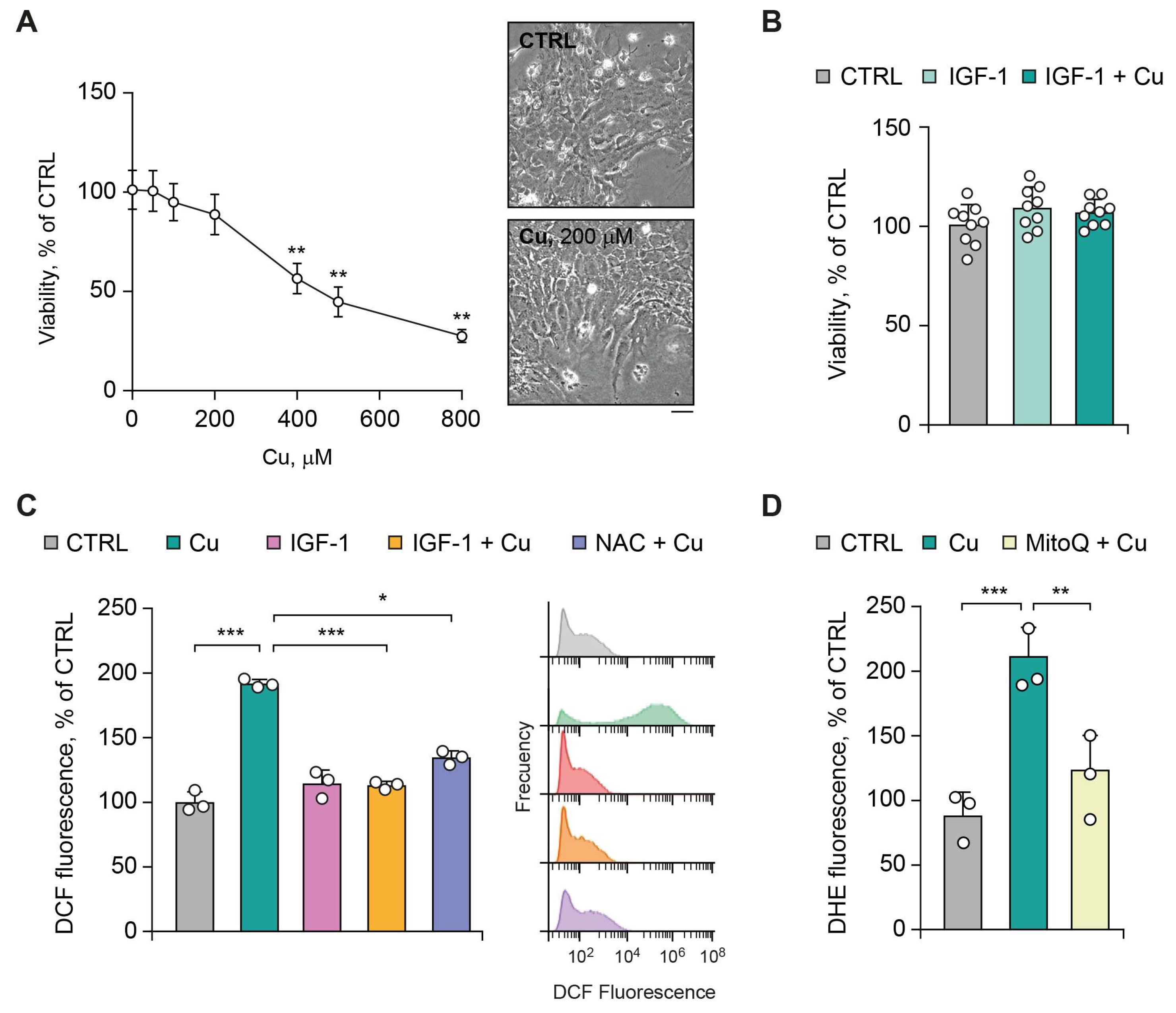
Figure 2.
IGF-1 prevents Cu-induced upregulation of cholesterol biosynthesis in astrocytes. Cells were exposed to CuSO4 (Cu, 200 μM) for 24 h in the presence or absence of IGF-1(100 ng/ml), NAC (30 μM), or MitoQ (1 μM). (A) Relative mRNA expression levels of Hmgcr and Srebf2. (B) Representative immunoblot and densitometric quantification of HMGCR protein levels normalized to β-actin and expressed relative to CTRL. (C) Total intracellular cholesterol levels. (D) Mitochondrial cholesterol (mtCHO) content and (E) total mitochondrial glutathione (mtGSH) levels were measured in mitochondrial fractions from CTRL and Cu-loaded cells with and without MitoQ treatment. (F) Relative mRNA expression levels of Hmgcr and Srebf2 following MitoQ treatment. Data are presented as mean ± SD (n = 3 independent primary cultures). Statistical analysis was performed using one-way ANOVA followed by Bonferroni’s multiple comparisons test. Statistical significance is indicated as * P < 0.05, ** P < 0.01 ***, and P < 0.001. Uncropped scans of representative immunoblots are shown in Supplementary Figure S4.
Figure 2.
IGF-1 prevents Cu-induced upregulation of cholesterol biosynthesis in astrocytes. Cells were exposed to CuSO4 (Cu, 200 μM) for 24 h in the presence or absence of IGF-1(100 ng/ml), NAC (30 μM), or MitoQ (1 μM). (A) Relative mRNA expression levels of Hmgcr and Srebf2. (B) Representative immunoblot and densitometric quantification of HMGCR protein levels normalized to β-actin and expressed relative to CTRL. (C) Total intracellular cholesterol levels. (D) Mitochondrial cholesterol (mtCHO) content and (E) total mitochondrial glutathione (mtGSH) levels were measured in mitochondrial fractions from CTRL and Cu-loaded cells with and without MitoQ treatment. (F) Relative mRNA expression levels of Hmgcr and Srebf2 following MitoQ treatment. Data are presented as mean ± SD (n = 3 independent primary cultures). Statistical analysis was performed using one-way ANOVA followed by Bonferroni’s multiple comparisons test. Statistical significance is indicated as * P < 0.05, ** P < 0.01 ***, and P < 0.001. Uncropped scans of representative immunoblots are shown in Supplementary Figure S4.
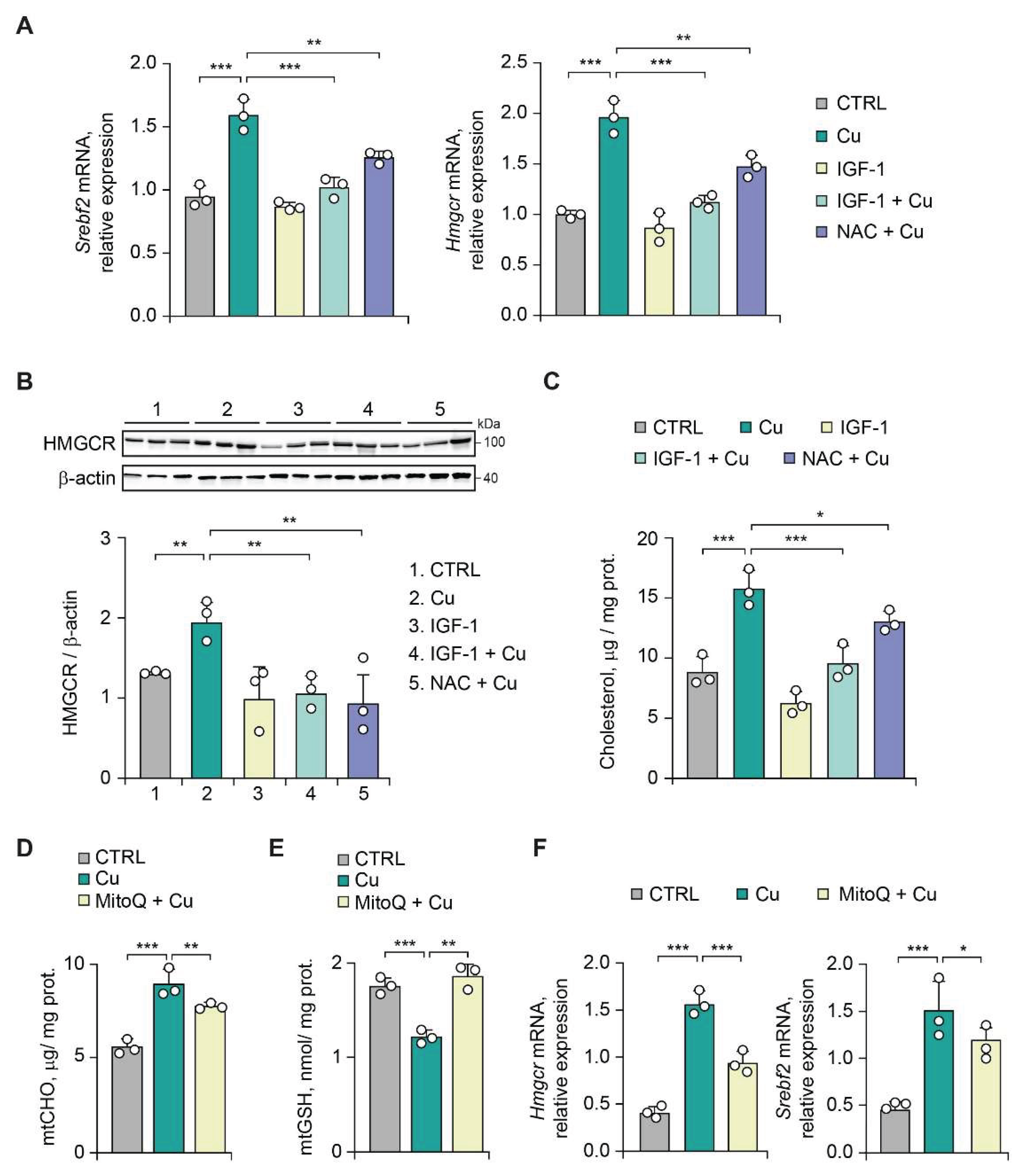
Figure 3.
Cu overload induces the release of EVs from astrocytes through an oxidative stress-dependent mechanism regulated by IGF-1. EVs were isolated from conditioned media collected after 24 h from cells exposed to CuSO4 (Cu, 200 μM) with and without IGF-1(100 ng/ml) or NAC (30 μM) treatment. (A) Left, EV concentration measured by NTA under the indicated experimental conditions. Right, summary of EV size distribution parameters, including mean size, modal size, and D50. Statistical significance is indicated as * P < 0.05 and ** P < 0.01 versus Cu-treated cells and # P < 0.05 and ## P < 0.01 vs CTRL cells. (B) Representative immunoblots showing the expression of EV-associated proteins CD63, CD81, and Alix. Flotillin-1 was used as a loading control. Data are presented as mean ± SD (n = 3 independent primary cultures). Statistical analysis was performed using one-way ANOVA followed by Bonferroni’s multiple comparisons test. Statistical significance is indicated as * P < 0.05 and *** P < 0.01. Uncropped scans of representative immunoblots are shown in Supplementary Figure S4.
Figure 3.
Cu overload induces the release of EVs from astrocytes through an oxidative stress-dependent mechanism regulated by IGF-1. EVs were isolated from conditioned media collected after 24 h from cells exposed to CuSO4 (Cu, 200 μM) with and without IGF-1(100 ng/ml) or NAC (30 μM) treatment. (A) Left, EV concentration measured by NTA under the indicated experimental conditions. Right, summary of EV size distribution parameters, including mean size, modal size, and D50. Statistical significance is indicated as * P < 0.05 and ** P < 0.01 versus Cu-treated cells and # P < 0.05 and ## P < 0.01 vs CTRL cells. (B) Representative immunoblots showing the expression of EV-associated proteins CD63, CD81, and Alix. Flotillin-1 was used as a loading control. Data are presented as mean ± SD (n = 3 independent primary cultures). Statistical analysis was performed using one-way ANOVA followed by Bonferroni’s multiple comparisons test. Statistical significance is indicated as * P < 0.05 and *** P < 0.01. Uncropped scans of representative immunoblots are shown in Supplementary Figure S4.
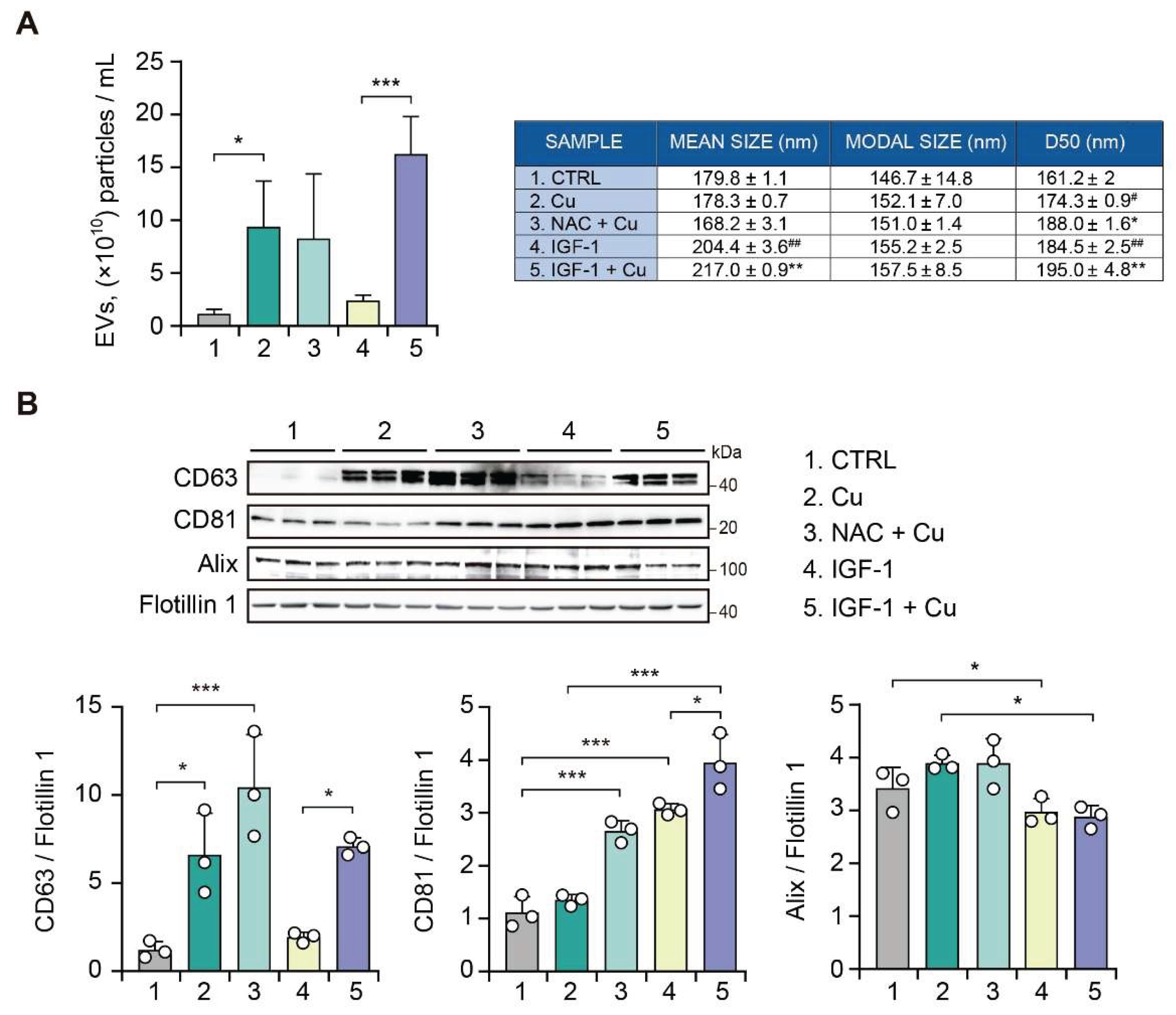
Figure 4.
Neurotoxic effect of EVs from Cu-overloaded astrocytes, counteracted by IGF-1 treatment. Cultured mouse hippocampal and cortical neurons were incubated for 48 h with EVs isolated from conditioned media collected from astrocytes exposed to CuSO4 (Cu, 200 μM) in the presence or absence of IGF-1(100 ng/ml) or NAC (30 μM). Neuronal viability was assessed using the Calcein-AM/7-AAD assay. Representative fluorescence microscopy images are shown, where viable cells are labeled in green (Calcein-AM) and non-viable cells in red (7-AAD). Quantification of neuronal viability is expressed as a percentage relative to the CTRL condition. Data are presented as mean ± SD (n = 5 independent primary cultures). Statistical analysis was performed using one-way ANOVA followed by Bonferroni’s multiple comparisons test. Statistical significance is indicated as ** P < 0.01 and *** P < 0.001.
Figure 4.
Neurotoxic effect of EVs from Cu-overloaded astrocytes, counteracted by IGF-1 treatment. Cultured mouse hippocampal and cortical neurons were incubated for 48 h with EVs isolated from conditioned media collected from astrocytes exposed to CuSO4 (Cu, 200 μM) in the presence or absence of IGF-1(100 ng/ml) or NAC (30 μM). Neuronal viability was assessed using the Calcein-AM/7-AAD assay. Representative fluorescence microscopy images are shown, where viable cells are labeled in green (Calcein-AM) and non-viable cells in red (7-AAD). Quantification of neuronal viability is expressed as a percentage relative to the CTRL condition. Data are presented as mean ± SD (n = 5 independent primary cultures). Statistical analysis was performed using one-way ANOVA followed by Bonferroni’s multiple comparisons test. Statistical significance is indicated as ** P < 0.01 and *** P < 0.001.
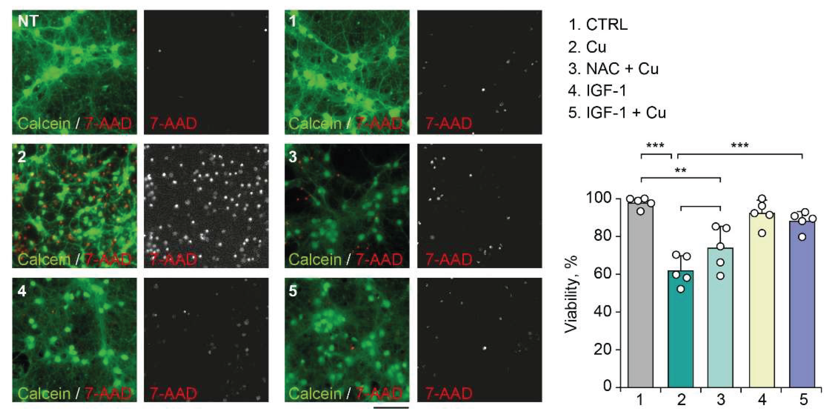
Figure 5.
Sustained IGF-1 expression prevents Cu-induced redox imbalance and upregulation of cholesterol-related and inflammatory proteins. Astrocytes were transduced for 24 h with a recombinant adenoviral vector expressing either DsRed (RAd-DsRed; CTRL) or rat IGF-1 (RAd-IGF-1). (A) Dose-response analysis of astrocyte viability following exposure to increasing concentrations of CuSO4 (Cu, 0–1,000 μM). (B) Intracellular ROS levels after 24 h of exposure to Cu (400 μM) with and without IGF-1 overexpression (RAd-IGF-1) or NAC (30 μM) treatment, measured by flow cytometry using the DCF-DA probe. (C-E) Oxidative stress biomarkers in astrocytes exposed to Cu (400 μM) with or without IGF-1 overexpression or NAC treatment, including superoxide dismutase (SOD) activity (C), lipid peroxidation measured as thiobarbituric acid–reactive substances (TBARS) (D), and Nitrate/nitrite [NOx] levels as an indicator of nitric oxide production (E). (F) Relative mRNA expression levels of Srebf2 and Hmgcr. (G) Relative mRNA expression levels of Tnf. Data are presented as mean ± SD (n = 8 independent primary cultures, panel A,B,C,F and G; n = 18 independent primary cultures, panel D and E). Statistical analysis was performed using one-way ANOVA followed by Bonferroni’s multiple comparisons test. Statistical significance is indicated as ** P < 0.01 and *** P < 0.001.
Figure 5.
Sustained IGF-1 expression prevents Cu-induced redox imbalance and upregulation of cholesterol-related and inflammatory proteins. Astrocytes were transduced for 24 h with a recombinant adenoviral vector expressing either DsRed (RAd-DsRed; CTRL) or rat IGF-1 (RAd-IGF-1). (A) Dose-response analysis of astrocyte viability following exposure to increasing concentrations of CuSO4 (Cu, 0–1,000 μM). (B) Intracellular ROS levels after 24 h of exposure to Cu (400 μM) with and without IGF-1 overexpression (RAd-IGF-1) or NAC (30 μM) treatment, measured by flow cytometry using the DCF-DA probe. (C-E) Oxidative stress biomarkers in astrocytes exposed to Cu (400 μM) with or without IGF-1 overexpression or NAC treatment, including superoxide dismutase (SOD) activity (C), lipid peroxidation measured as thiobarbituric acid–reactive substances (TBARS) (D), and Nitrate/nitrite [NOx] levels as an indicator of nitric oxide production (E). (F) Relative mRNA expression levels of Srebf2 and Hmgcr. (G) Relative mRNA expression levels of Tnf. Data are presented as mean ± SD (n = 8 independent primary cultures, panel A,B,C,F and G; n = 18 independent primary cultures, panel D and E). Statistical analysis was performed using one-way ANOVA followed by Bonferroni’s multiple comparisons test. Statistical significance is indicated as ** P < 0.01 and *** P < 0.001.
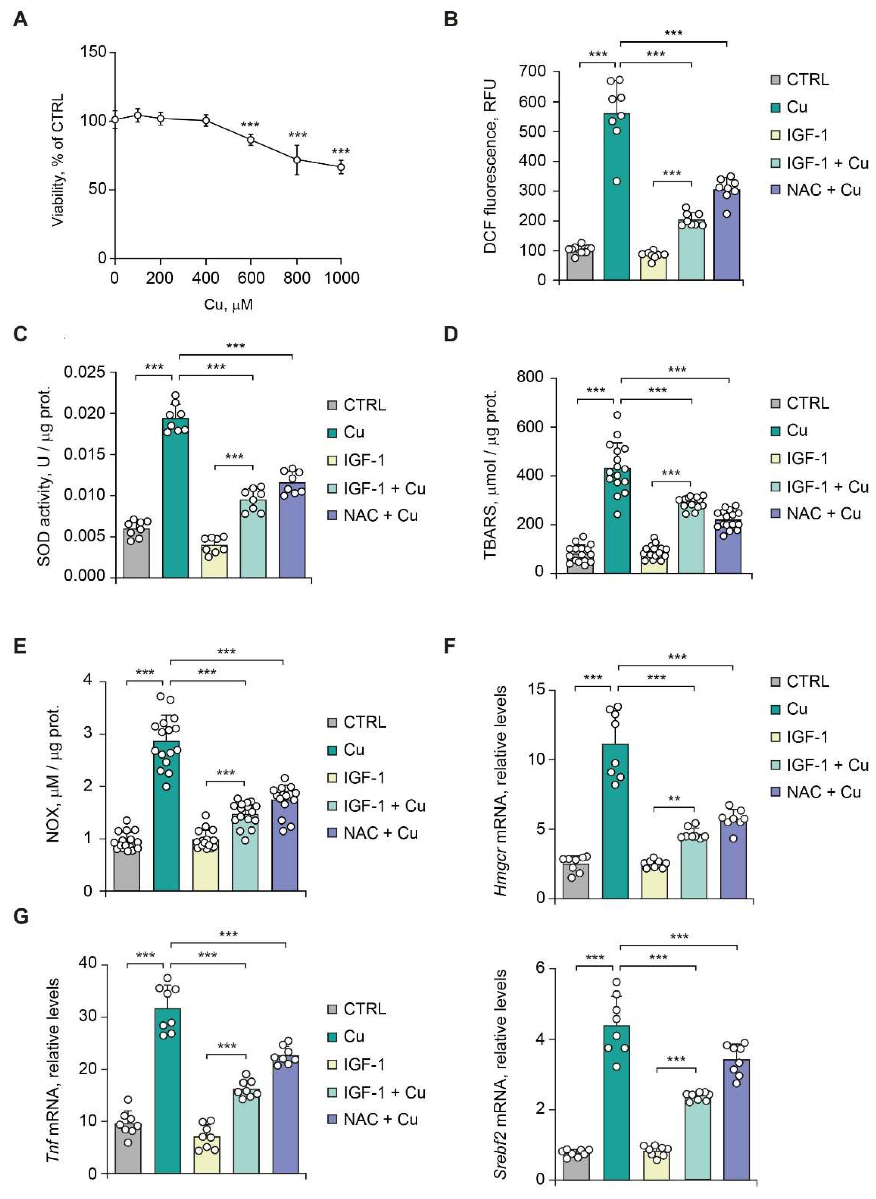
Figure 6.
Sustained IGF-1 expression prevents the neurotoxic features of astrocyte-derived EVs exposed to high Cu load. Astrocytes were transduced for 24 h with a recombinant adenoviral vector expressing either DsRed (RAd-DsRed; control) or rat IGF-1 (RAd-IGF-1). EVs were isolated from conditioned media collected from astrocytes exposed to CuSO4 (Cu, 400 μM) in the presence or absence of IGF-1 overexpression (RAd-IGF-1) or NAC (30 μM). Primary rat neurons were incubated for 24 h with the isolated EVs. (A) Intracellular ROS levels in neurons were determined by flow cytometry using the DCF-DA probe. (B) Neuronal viability was assessed by the Annexin V-FITC assay. Data are presented as mean ± SD (n = 8 independent primary cultures). Statistical analysis was performed using one-way ANOVA followed by Bonferroni’s multiple comparisons test. Statistical significance is indicated as ** P < 0.01 and *** P < 0.001.
Figure 6.
Sustained IGF-1 expression prevents the neurotoxic features of astrocyte-derived EVs exposed to high Cu load. Astrocytes were transduced for 24 h with a recombinant adenoviral vector expressing either DsRed (RAd-DsRed; control) or rat IGF-1 (RAd-IGF-1). EVs were isolated from conditioned media collected from astrocytes exposed to CuSO4 (Cu, 400 μM) in the presence or absence of IGF-1 overexpression (RAd-IGF-1) or NAC (30 μM). Primary rat neurons were incubated for 24 h with the isolated EVs. (A) Intracellular ROS levels in neurons were determined by flow cytometry using the DCF-DA probe. (B) Neuronal viability was assessed by the Annexin V-FITC assay. Data are presented as mean ± SD (n = 8 independent primary cultures). Statistical analysis was performed using one-way ANOVA followed by Bonferroni’s multiple comparisons test. Statistical significance is indicated as ** P < 0.01 and *** P < 0.001.
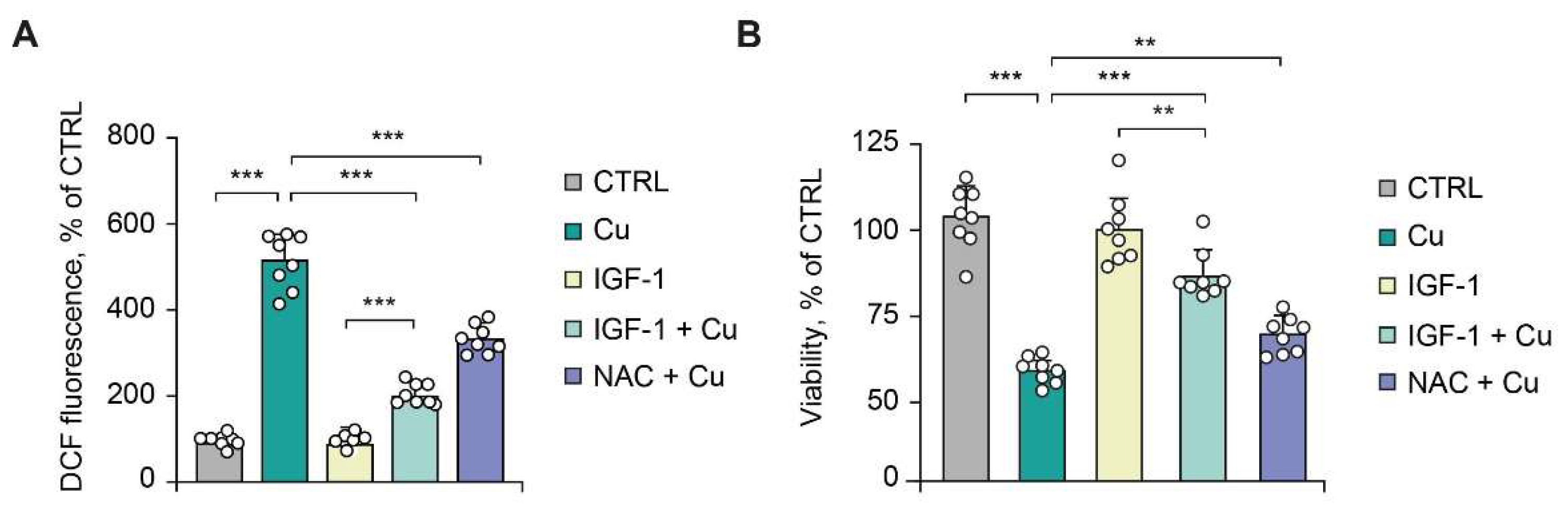

Table 1.
Primer sequence.
| Gene | Forward Primer | Reverse Primer |
| Srebf2 | 5’-AGCATACCGCAAGGTGTTCC-3’ | 5’- CCAGGTGTCTACTTCTCCGTGT-3’ |
| Hmgcr | 5’-GTGGTTACCCTGAGCTTAGC-3’ | 5’-CGGGATGTGCTTAGCATTGA-3’ |
| Tnf | 5′-GAAAAGCAAGCAGCCAACCA-3′ | 5′-CGGATCATGCTTTCTGTGCTC-3′ |
| Actb | 5’-TCTTATTGGTCGAAGGCTCGT-3’ | 5’-ATCTCACTAGAGGCCACCGA-3’ |
Disclaimer/Publisher’s Note: The statements, opinions and data contained in all publications are solely those of the individual author(s) and contributor(s) and not of MDPI and/or the editor(s). MDPI and/or the editor(s) disclaim responsibility for any injury to people or property resulting from any ideas, methods, instructions or products referred to in the content. |
© 2026 by the authors. Licensee MDPI, Basel, Switzerland. This article is an open access article distributed under the terms and conditions of the Creative Commons Attribution (CC BY) license (http://creativecommons.org/licenses/by/4.0/).
Copyright: This open access article is published under a Creative Commons CC BY 4.0 license, which permit the free download, distribution, and reuse, provided that the author and preprint are cited in any reuse.