

Submitted:
05 August 2026
Posted:
06 August 2026
You are already at the latest version
Abstract
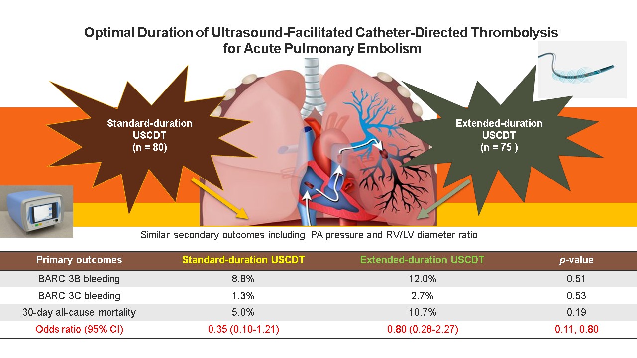
Background/Objectives: The mortality rate for acute high-risk and intermediate-high-risk pulmonary embolism (PE) was high despite treatment with heparin anticoagulation alone. Although adjunctive systemic thrombolysis can reduce mortality by more than 50%, these treatments significantly increase the incidence of major bleeding, particularly intracranial hemorrhage. Ultrasound-facilitated catheter-directed thrombolysis (USCDT) has been shown to reduce the incidence of major bleeding; however, the optimal treatment duration for further reducing mortality and major bleeding remains uncertain. Methods: This retrospective cohort study included 155 hospitalized patients with acute PE who underwent USCDT between January 2017 and June 2025 in the intensive care unit at China Medical University Hospital, Taichung, Taiwan. Patients were categorized into four groups based on average usage time: 12, 24, 36, and 48 hours. The primary endpoints were 30-day all-cause mortality rate and major bleeding. Covariates associated with 30-day all-cause mortality were estimated and adjusted using Cox regression modeling. Results: Across all treatment groups, USCDT significantly reduced pulmonary artery (PA) systolic pressure (26.9%), mean PA pressure (21.0%), fibrinogen levels (25.2%), and right ventricle-to-left ventricle diameter ratio (21.5%) (all p < 0.001). Standard-duration USCDT was associated with a lower odds ratio for 30-day all-cause mortality (OR: 0.35, 95% CI: 0.10–1.21) than extended-duration USCDT. Conclusions: Standard-duration USCDT (12–24 hours) may provide an optimal balance between efficacy and safety by reducing 30-day all-cause mortality and major bleeding in patients with acute PE.
Keywords:
acute pulmonary embolism
; tissue-type plasminogen activator
; ultrasound-facilitated catheter-directed thrombolysis
; EkoSonic Endovascular System
1. Introduction
Acute pulmonary embolism (PE) is characterized by the acute dislodgement of a thrombus formed in a deep vein into the pulmonary artery (PA), resulting in acute hypoxemia and right ventricular strain. The European Society of Cardiology defines high-risk PE as persistent hypotension lasting more than 15 minutes or any signs of shock, such as altered mental status, oliguria, or elevated lactate levels. Intermediate-high-risk PE is defined as right ventricular dysfunction, characterized by a right ventricle-to-left ventricle (RV/LV) diameter ratio greater than 1 on imaging and elevated cardiac troponin I levels [1]. Despite treatment with heparin anticoagulation alone, mortality rates remain high, reaching 30%–50% for high-risk acute PE and 10%–15% for intermediate-high-risk PE [2].
Systemic thrombolytic therapy, supplemented with baseline heparin anticoagulation, has been the primary reperfusion treatment for high-risk PE and a rescue reperfusion strategy for intermediate-high-risk PE for more than a decade [3]. This approach has reduced the mortality rate to 15%–20% in high-risk acute PE and 5%–7% in intermediate-high-risk PE, representing an approximately 50% improvement. However, systemic thrombolysis is associated with an increased risk of major bleeding (10–20%), particularly intracranial hemorrhage, which occurs in 3–5% of cases [4]. The 2026 clinical practice guidelines published by the Joint Committee of the American College of Cardiology and the American Heart Association recommend early systemic thrombolysis for primary reperfusion in any type of shock and for rescue reperfusion in any episode of transient hypotension or other signs of hemodynamic instability [5].
Catheter-directed thrombolysis (CDT), which delivers thrombolytic agents directly into the PA through a Fountain catheter, provides efficacy comparable to that of systemic thrombolysis while reducing the incidence of major bleeding to 7–15% and intracranial hemorrhage to 2–3%, resulting in an overall 30% reduction in bleeding complications [6,7]. Consequently, CDT has become a viable alternative to systemic thrombolysis, particularly for patients with PE who have not experienced cardiac arrest [8]. Ultrasound-facilitated catheter-directed thrombolysis (USCDT), performed using the EkoSonic Endovascular System, further enhances thrombolytic delivery by facilitating more precise administration of tissue-type plasminogen activator (t-PA) and has gradually replaced conventional CDT in recent years [9,10,11]. USCDT has demonstrated efficacy in patients with both high-risk and intermediate-high-risk PE [12,13].
Although low-dose t-PA administered with USCDT has been shown to be effective in the treatment of PE [14], the optimal t-PA dose in combination with the EkoSonic Endovascular System treatment duration remains unclear. Therefore, this study aimed to determine the optimal USCDT treatment duration.
2. Materials and Methods
2.1. Study Design and Population
Data were retrieved from the electronic medical records of China Medical University Hospital. The requirement for written informed consent was waived due to the retrospective study design. The study was approved by the Institutional Review Board of China Medical University Hospital (CMUH114-REC3-201) and was conducted in accordance with its ethical guidelines.
This observational, retrospective cohort study included 155 consecutive adult patients with acute high-risk or intermediate-high-risk PE who underwent USCDT and were admitted to the intensive care unit of China Medical University Hospital, Taichung, Taiwan, between January 2017 and June 2025. Patients with acute low-risk or intermediate-low-risk PE were excluded. Patients who underwent mechanical thrombectomy, surgical embolectomy, systemic thrombolysis, or conventional CDT were also excluded (STROBE flowchart; Figure 1). The remaining 155 patients were categorized into four groups according to USCDT duration: 12, 24, 36, and 48 hours. All patients were followed for 30 days to assess all-cause mortality and major bleeding.
2.2. Definitions of Acute Pulmonary Embolism, Primary Thrombolysis, and Rescue Thrombolysis
Acute PE was defined as symptom onset (e.g., dyspnea or palpitations) occurring within 14 days before presentation and a thrombus observed in the PA confirmed by computed tomography pulmonary angiography (CTPA). Primary thrombolysis was performed within 1–3 hours of diagnosis in patients with acute high-risk PE, whereas rescue thrombolysis was performed within 24–48 hours after initiation of heparin anticoagulation in patients with acute intermediate-high-risk PE.
2.3. Endovascular Ultrasound and Thrombolysis with Tissue-Type Plasminogen Activator
The EkoSonic Endovascular System (Boston Scientific, Marlborough, MA, USA) is a commercially available endovascular ultrasound used for CDT. The system utilizes acoustic microstreaming generated by 200 Hz ultrasound to loosen and separate tightly entangled fibrin mesh fibers, a process known as “unwinding fibrin mesh.” This process exposes additional fibrin-binding sites and enhances thrombolytic drug penetration.
tPA was used to convert plasminogen to plasmin. Plasmin then cleaves the cross-linked fibrin mesh into smaller, soluble fragments, such as d-dimers.
2.4. Anticoagulation with Heparin
Heparin exerts its anticoagulant effect through antithrombin III. After binding to heparin, antithrombin III further binds to coagulation factors XII, XI, IX, and X, as well as thrombin. The heparin/antithrombin III complex inactivates coagulation factor X and thrombin, thereby inhibiting thrombin formation and the conversion of fibrinogen to fibrin. After 5–10 days of continuous heparin infusion, patients were transitioned to novel oral anticoagulants (NOACs), which directly inhibit factor X or thrombin, for 3– 6 months. Patients who could not tolerate NOACs were transitioned to vitamin K antagonists.
2.5. Ultrasound-Facilitated Catheter-Directed Thrombolysis and Anticoagulation Protocol at China Medical University Hospital
USCDT treatment protocol: t-PA (generic name: alteplase) (2–5 mg) was injected into the PA, followed by continuous infusion at a rate of 1 mg/h for 12–48 hours through a Fountain catheter placed in the PA. Fibrinogen levels were measured every 6 hours; if the fibrinogen level fell below 200 mg/L, the infusion of t-PA was suspended. An ultrasound device was connected to the EkoSonic Endovascular System to enhance treatment efficacy.
Anticoagulation treatment regimen: A heparin loading dose of 60 U/kg was administered, followed by continuous infusion to maintain the activated partial thromboplastin time between 50–70 seconds for 5–10 days. When used in combination with t-PA, the target activated partial thromboplastin time was maintained between 45–60 seconds.
2.6. Primary and Secondary Outcomes
The primary outcomes were 30-day all-cause mortality and major bleeding. Major bleeding was defined according to the Bleeding Academic Research Consortium (BARC) Scale as type 3B (hemoglobin decrease > 5 g/dL from baseline or bleeding requiring surgical intervention), type 3C (critical condition, such as intracranial or intraocular hemorrhage), type 4 (bleeding related to coronary artery bypass graft procedures), or type 5 (fatal bleeding confirmed by imaging or autopsy).
The secondary outcomes included 48-hour changes in PA systolic pressure, PA mean pressure, fibrinogen concentration, and the RV/LV diameter ratio after the procedure.
2.7. Statistical Analysis
The sample size was calculated to detect a clinically significant 75% reduction in the primary outcome between the high-risk PE group (20%) and intermediate-high-risk PE group (5%). A total of 150 patients (75 per group) was required to achieve 80% power with a two-sided significance level of 0.05, assuming that 20% of cases would be non-evaluable.
Continuous variables are presented as the mean ± standard deviation and were compared using one-way analysis of variance. Categorical variables are expressed as frequencies and percentages and were compared using Pearson’s chi-square test. Student’s t-test was used for pairwise comparisons, where appropriate. A two-sided p-value of < 0.05 was considered statistically significant. Because of the relatively small sample size, a p-value of < 0.2 in the univariate analysis was considered indicative of a statistical trend and was used for variable selection in the multivariable logistic regression analysis. The odds ratio of 30-day all-cause mortality, 95% confidence intervals (CIs), and related significant values were obtained from the logistic regression analysis. All statistical analyses were performed using SPSS Statistics, version 30.0 (IBM Corp., Armonk, NY, USA).
3. Results
3.1. Baseline Clinical Characteristics
Table 1 summarizes the baseline clinical characteristics of the study participants, categorized according to USCDT treatment duration. Baseline clinical characteristics were similar across all four duration groups (USCDT 12, 24, 36, and 48 hours), with no statistically significant differences. The mean age at diagnosis was 59 ± 19 years, and most patients were female (67.7%). The most prevalent comorbidities (> 10%) were deep vein thrombosis (55.5%), hypertension (32.9%), malignancy (27.7%), diabetes mellitus (20.0%), immobility (19.4%), surgery (13.5%), and autoimmune disease (12.3%).
Regarding PE severity, 44.5% of patients had high-risk PE, and 55.5% had intermediate-high-risk PE. The mean Pulmonary Embolism Severity Index score was 115.6 ± 37.0. Regarding adjunctive procedures, 21.3% of patients received extracorporeal membrane oxygenation (ECMO) because of shock. After thrombolysis or ECMO removal, 66.5% of patients received an inferior vena cava filter for 1–3 months.
3.2. Follow-Up of Laboratory Tests on the Day of Admission
Table 2 shows the laboratory findings on the day of admission. Laboratory findings were similar across all duration groups (USCDT 12, 24, 36, and 48 hours), with no statistically significant differences. For blood counts, the mean white blood cell count was 11.9 ± 6.3 K/μL, hemoglobin was 12.4 ± 2.1 g/dL, and platelet count was 202.5 ± 83.8 K/μL. For biochemical indices, the mean troponin-I level was 0.35 ± 1.13 ng/mL, estimated glomerular filtration was 70.6 ± 33.2 mL/min/1.73 m², and lactate level was 2.2 ± 1.0 mmol/L. Elevated troponin-I (> 0.04 ng/mL) and lactate (> 2.0 mmol/L) levels were observed in 67.7% and 44.5% of patients, respectively.
3.3. Secondary Outcomes Analyses of Treatment Effect on Pulmonary Artery Systolic and Mean Pressure, Fibrinogen Concentration, and the Right Ventricle-to-Left Ventricle Diameter Ratio
Table 3 presents the 48-hour treatment effects of different USCDT treatment durations. The USCDT 12-hour group had a mean treatment duration of 12.2 hours (< 20 hours) and a mean t-PA dose of 14.6 mg. The USCDT 24-hour group had a mean treatment duration of 24.4 hours (20–29 hours) and a mean t-PA dose of 20.3 mg. The USCDT 36-hour group had a mean treatment duration of 36.4 hours (30–39 hours) and a mean t-PA dose of 31.2 mg. The USCDT 48-hour group had a mean treatment duration of 66.9 hours (≥ 40 hours) and a mean t-PA dose of 42.6 mg. All four groups demonstrated favorable secondary outcomes, with no statistically significant differences among the groups. After 48 hours of follow-up, with a mean USCDT treatment duration of 34.2 hours and a mean t-PA dose of 26.8 mg, PA systolic pressure decreased from 48.7 to 35.1 mmHg (26.9%, p < 0.001), PA mean pressure decreased from 31.8 to 24.9 mmHg (21.0%, p < 0.001), fibrinogen concentration decreased from 341.9 to 255.8 mg/dL (25.2%, p < 0.001), and RV/LV diameter ratio decreased from 1.15 to 0.91 (21.5%, p < 0.001). Figure 2 also illustrates these findings.
3.4. Primary Outcomes Analyses of Treatment Effect on Major Bleeding and 30-Day All-Cause Mortality
Table 4 summarizes the primary clinical outcomes, including major bleeding and 30-day all-cause mortality. The standard-duration USCDT group (< 30 hours) comprised the 12-hour and 24-hour groups, whereas the extended-duration USCDT group (≥ 30 hours) comprised the 36-hour and 48-hour groups. Compared with the extended-duration USCDT group, the standard-duration USCDT group had similar rates of major bleeding (10.0% vs. 14.7%, p = 0.38), including BARC type 3B bleeding (8.8% vs. 12.0%, p = 0.51) and BARC type 3C bleeding (1.3% vs. 2.7%, p = 0.53). Compared with the extended USCDT regimen, the standard USCDT regimen showed a trend toward lower 30-day all-cause mortality (5.0% vs. 10.7%, p = 0.19).
Figure 3 illustrates several covariates analyzed as protective factors against 30-day all-cause mortality after thrombolysis. High-risk PE after thrombolysis was used as the reference point for 30-day all-cause mortality. The protective factors between 0.5 and 1 included thrombolysis with an extended-duration USCDT (odds ratio: 0.80, 95% CI: 0.28‒2.27), USCDT 36 h (odds ratio: 0.56, 95% CI: 0.13‒2.33), USCDT 48 h (odds ratio: 1.08, 95% CI: 0.32‒3.63), and all high-risk and intermediate-high-risk PE after thrombolysis (odds ratio: 0.56, 95% CI: 0.22‒1.39). Protective factors below 0.5 include: standard-duration USCDT (odds ratio: 0.35, 95% CI: 0.10‒1.21), USCDT 12 h (odds ratio: 0.36, 95% CI: 0.07‒1.81), USCDT 24 h (odds ratio: 0.34, 95% CI: 0.07‒1.72), and intermediate-high risk PE after thrombolysis (odds ratio: 0.24, 95% CI: 0.06‒0.93).
4. Discussion
4.1. Principal Findings
In this study, we evaluated potential protective factors associated with improved 30-day all-cause mortality after USCDT in patients with acute high-risk or intermediate-high-risk PE. In this retrospective study, the optimal USCDT duration was approximately 12–24 hours, corresponding to a mean t-PA dose of 15–20 mg. Extending the treatment duration to 36–48 hours (30–40 mg of t-PA) did not provide additional clinical benefits and was associated with an upward trend in 30-day all-cause mortality, whereas the incidence of major bleeding remained at a similar level.
4.2. Mechanism of Thrombolysis with Tissue-Type Plasminogen Activator
In acute PE, thrombin catalyzes the conversion of fibrinogen into fibrin monomers. Large amounts of fibrin monomers bind to factor XIII to form X-chain fibrin, known as a fibrin mesh. This fibrin mesh traps red blood cells, forming a red thrombus. Heparin anticoagulation primarily reduces thrombin formation and further promotes endogenous fibrinolysis after 48 hours. However, in critical situations, such as high-risk or intermediate-high-risk PE, t-PA should be administered to trigger plasmin formation within 3 hours for high-risk PE or within 48 hours for intermediate-high-risk PE. Plasmin rapidly breaks down the fibrin mesh, producing large amounts of D-dimers. Importantly, a decrease in fibrinogen concentration has consistently been an indicator of the effectiveness of thrombolytic therapy because this treatment simultaneously reduces both fibrinogen and fibrin concentrations [15]. Fibrinogen is a marker of both thrombosis and bleeding; when fibrinogen levels fall below 200 mg/dL, platelet aggregation becomes impaired. Consequently, damage to blood vessels may result in loss of the coagulation response, leading to major bleeding.
4.3. Right-Heart Function Improved by Ultrasound-Facilitated Catheter-Directed Thrombolysis
USCDT is typically performed using the EkoSonic Endovascular System. Many clinical trials have demonstrated that continuous t-PA infusion for either standard or extended durations reduces PA systolic and mean pressures and the RV/LV diameter ratio, thereby improving right-heart function [15,16,17]. In our study, after 48 hours of USCDT, PA systolic pressure decreased from 48.7 to 35.1 mmHg (26.9%, p < 0.001), PA mean pressure decreased from 31.8 to 24.9 mmHg (21.0%, p < 0.001), and the RV/LV diameter ratio decreased from 1.15 to 0.91 (21.5%, p < 0.001). These findings are highly consistent with those of previous USCDT studies [16,17]. If the thrombus in the PA rapidly dissolves, the hypoxic conditions will improve. As pulmonary vascular resistance decreases rapidly, PA systolic and mean pressures naturally drop. A reduction in PA pressure not only improves RV-PA coupling but also reduces RV afterload. As RV contractility improves, the RV/LV diameter ratio decreases accordingly, facilitating recovery of right-heart function.
4.4. Standard Duration of Ultrasound-Facilitated Catheter-Directed Thrombolysis Confirmed
Ten years ago, clinical trials evaluated the standard-duration USCDT. Different USCDT durations (e.g., 12 hours for bilateral PA treatment and 24 hours for unilateral PA treatment, with a total t-PA dose of 24 mg), as well as different t-PA doses (e.g., 10 mg for unilateral treatment and 20 mg for bilateral treatment during a 15-hour duration), demonstrated similar efficacy and safety [18,19]. Therefore, the standard USCDT regimen typically consists of 12–24 hours and a t-PA dose of 15–20 mg. In recent years, attempts have been made to use shorter durations and lower t-PA doses (e.g., 2–6 hours, with 4–6 mg for unilateral treatment and 8–12 mg for bilateral treatment). Although these regimens demonstrated similar efficacy and safety, they have been shown to be effective primarily in patients with intermediate-high-risk PE [20,21]. This shortened treatment duration and lower-dose regimen does not appear to be suitable for patients with high-risk PE [22].
4.5. Extended Duration of Ultrasound-Facilitated Catheter-Directed Thrombolysis Does Not Increase Major Bleeding
For patients with high-risk or intermediate-high-risk PE, attempts have been made to extend treatment duration and increase the t-PA dose. Extended USCDT typically refers to 36–48 hours with a total t-PA dose of 30–40 mg. One study reported that, despite doubling the t-PA dose, no significant increase in major bleeding occurred [23]. Similarly, in this study, standard-duration USCDT had a similar incidence of major bleeding (10.0% vs. 14.7%, p = 0.38) compared with that performed using extended-duration USCDT, including BARC type 3B bleeding (8.8% vs. 12.0%, p = 0.51) and BARC type 3C bleeding (1.3% vs. 2.7%, p = 0.53). These findings are consistent with previous clinical observations. The incidence of intracranial hemorrhage (BARC type 3C) remained low (1–3%) and appears to be more related to underlying conditions (e.g., history of stroke or uncontrolled blood pressure) rather than t-PA dose [24].
4.6. Extended Duration of Ultrasound-Facilitated Catheter-Directed Thrombolysis Increased the Trend of Mortality
Although the rates of major bleeding were similar between the standard-duration and extended-duration USCDT groups, the additional benefit of reduced mortality is a more important prognostic indicator. We analyzed several covariates—based on the severity or duration of USCDT—as protective factors for 30-day all-cause mortality following thrombolytic therapy. Using high-risk PE as the reference group, intermediate-high-risk PE after thrombolysis was associated with a lower risk of 30-day all-cause mortality (odds ratio: 0.24, 95% CI: 0.06–0.93, p = 0.03), supporting the reliability of this analysis. We further evaluated protective factors based on USCDT duration. Compared with thrombolytic therapy using extended-duration USCDT (odds ratio: 0.80, 95% CI: 0.28–2.27, p = 0.80), thrombolytic therapy using standard-duration USCDT was associated with a lower odds ratio for 30-day all-cause mortality (odds ratio: 0.35, 95% CI: 0.10–1.21, p = 0.11). Although no statistically significant difference was observed, mortality showed an upward trend with extended-duration USCDT, strongly suggesting that the standard-duration USCDT may be preferable to the extended-duration USCDT.
4.7. Reperfusion Injury Increased the Trend of Mortality
Ischemia-reperfusion injury is the primary cause of multiple organ failure and is associated with persistently high mortality rates. Acute myocardial infarction or cardiac arrest primarily affects high-energy-demand organs, such as the heart and brain, where mitochondrial calcium overload is the main mechanism of injury [25]. In contrast, acute PE primarily affects the lungs, which have a unique anatomical structure and physiological characteristics that make them more susceptible to ferroptosis during reperfusion [26,27]. Alveolar epithelial and endothelial cells are directly exposed to high oxygen concentrations; consequently, the local production of reactive oxygen species (ROS) during reperfusion is far more intense than that in other organs. Furthermore, the lungs contain large amounts of pulmonary surfactant and complex cell membrane structures rich in highly sensitive polyunsaturated fatty acids. Under intense ROS attack, these lipids are highly susceptible to chain oxidation reactions. During ischemia-reperfusion injury, pulmonary microcirculation is often accompanied by increased microvascular permeability, red blood cell extravasation, and hemolysis, leading to the release of large amounts of free iron. The combination of high iron and ROS levels rapidly triggers the Fenton reaction, generating highly destructive hydroxyl radicals that directly accelerate the accumulation of lipid hydroperoxides [26,27]. This further illustrates that prolonging the thrombolysis time not only fails to provide additional therapeutic benefits, but also significantly increases reperfusion injury, leading to thrombogenesis [28,29], endothelial dysfunction, and multiple organ failure [30,31].
4.8. Limitations
This study has several limitations. First, the RV/LV diameter ratio was measured using CTPA before USCDT but by standard echocardiography 48 hours after USCDT. The use of different imaging modalities may have introduced measurement variability. Second, this was a single-center study, which may limit the generalizability of the findings. Third, the role of ischemia-reperfusion injury remains hypothetical and requires validation using animal models. Finally, prospective randomized controlled trials are needed to validate these findings and determine the optimal standard-duration USCDT.
5. Conclusions
In this study, standard-duration USCDT (12–24 hours; t-PA dose, 15–20 mg) was associated with a lower odds ratio for 30-day all-cause mortality (odds ratio: 0.35; 95% CI, 0.10–1.21) than extended-duration USCDT (odds ratio: 0.80; 95% CI, 0.28–2.27), although the difference was not statistically significant. These findings suggest that standard-duration USCDT may provide an optimal balance between efficacy and safety in patients with acute high-risk or intermediate-high-risk PE by reducing 30-day all-cause mortality and major bleeding.
Author Contributions
Yu-Kai Lin: Writing – original draft, Methodology, Investigation, Data curation, Conceptualization. Da-Long Chen: Writing – review & editing, Writing – original draft, Visualization, Validation, Methodology, Investigation, Formal analysis, Conceptualization. Chung-Ho Hsu: Writing – review & editing, Methodology, Investigation, Data curation, Conceptualization. Hui-Wen Chang: Methodology, Investigation, Conceptualization. Keng-Yuan Li: Methodology, Investigation, Conceptualization. Li-Chuan Hsieh: Methodology, Investigation, Conceptualization. Chun-Cheng Wang: Methodology, Investigation, Data curation. An-Sheng Lee: Writing – review & editing, Supervision, Resources. Kuan-Cheng Chang: Writing – review & editing, Supervision, Resources. All authors have approved this manuscript and agree to assume corresponding responsibility.
Funding
This study received no additional external funding, except for the AS. L and KC. C. Mackay Medical University [MMU-RD-112-1B-P029 and MMU-RD-113-1B-P018] grant received by AS. L. National Science and Technology Council [NSTC 114-2314-B-039-071] grant received by KC. C.
Institutional Review Board Statement
This study was approved by the Institutional Review Board of China Medical University Hospital (CMUH114-REC3-201) for data collection and analysis.
Informed Consent Statement
Patient consent was waived due to the retrospective nature of the study.
Data Availability Statement
The original contributions presented in this study are included in the article, and further inquiries can be directed to the corresponding authors.
Acknowledgments
All authors thank Ching-Yi Chou, Wen-De Tang, and Kee-Koon Ng at China Medical University Hospital for their excellent clinical team support.
Declaration of Generative AI in Scientific Writing
No applicable.
Conflicts of Interest
The authors have no competing interests.
Abbreviations
| BARC | Bleeding Academic Research Consortium |
| CTPA | Computed tomography pulmonary angiogram |
| PA | Pulmonary artery |
| PE | Pulmonary embolism |
| ROS | Reactive oxygen species |
| RV/LV | Right ventricle to left ventricle |
| t-PA | Tissue-type plasminogen activator |
| USCDT | Ultrasound-facilitated catheter-directed thrombolysis |
References
- Konstantinides, S.V.; Meyer, G.; Becattini, C.; Bueno, H.; Geersing, G.J.; Harjola, V.P.; Huisman, M.V.; Humbert, M.; Jennings, C.S.; Jiménez, D. 2019 ESC Guidelines for the diagnosis and management of acute pulmonary embolism developed in collaboration with the European Respiratory Society (ERS): The task force for the diagnosis and management of acute pulmonary embolism of the European Society of Cardiology (ESC). Eur. Respir. J. 2019, 54, 1901647. [CrossRef]
- Birrenkott, DA.; Kabrhel, C.; Dudzinski, D.M. Intermediate-risk and High-risk pulmonary embolism: Recognition and management: Cardiology clinics: Cardiac Emergencies. Cardiol. Clin. 2024, 42, 215-235. [CrossRef]
- Zuin, M.; Ageno, W.; Vedovati, M.C.; Chopard, R.; Goldhaber, S.Z.; Simioni, P. Robustness and fragility of evidence supporting systemic thrombolysis in acute pulmonary embolism. J. Thromb. Thrombolysis 2026. [CrossRef]
- Mendonça, I.; Teixeira, S.; Ferreira, A.R.; Paiva, J.A. High and intermediate-high risk pulmonary embolism management: A 5-year intensive care unit casuistic review. Port. J. Card. Thorac. Vasc. Surg. 2024, 31, 21-31. [CrossRef]
- Creager, M.A.; Barnes, G.D.; Giri, J.; Mukherjee, D.; Jones, W.S.; Burnett, A.E.; Carman, T.; Casanegra, A.I.; Castellucci, L.A.; Clark, S.M.; et al. 2026 AHA/ACC/ACCP/ACEP/CHEST/SCAI/SHM/SIR/SVM/SVN Guideline for the evaluation and management of acute pulmonary embolism in adults: A report of the American College of Cardiology/American Heart Association Joint Committee on clinical practice guidelines. J. Am. Coll. Cardiol. 2026,87:1626-1710. [CrossRef]
- Zhang, R.S.; Maqsood, M.H.; Sharp, A.S.P.; Postelnicu, R.; Sethi, S.S.; Greco, A.; Alviar, C.; Bangalore, S. Efficacy and safety of anticoagulation, catheter-directed thrombolysis, or systemic thrombolysis in acute pulmonary embolism. JACC Cardiovasc. Interv. 2023, 16, 2644-2651. [CrossRef]
- Planer, D.; Yanko, S.; Matok, I.; Paltiel, O.; Zmiro, R.; Rotshild, V.; Amir, O.; Elbaz-Greener, G.; Raccah, B.H. Catheter-directed thrombolysis compared with systemic thrombolysis and anticoagulation in patients with intermediate- or high-risk pulmonary embolism: systematic review and network meta-analysis. CMAJ. 2023, 195, e833-e843. [CrossRef]
- Stępniewski, J.; Sławek-Szmyt, S.; Karnaś, M.; Chaba-Karnaś, W.; Kurczyna, P.; Magoń, W.; Jonas, K.; Waligóra, M/; Araszkiewicz, A.; Kopeć, G. Outcomes of high-risk pulmonary embolism stratified according to clinical phenotype: Results from 2 specialized pulmonary embolism centres. Can. J. Cardiol. 2026, S0828-282X(26)00069-3. [CrossRef]
- Hadid, B.; Snyder, D.J.; Zilinyi, R.S.; Sethi, S.S.; Parikh, S.A. Ultrasound-assisted catheter-directed thrombolysis for pulmonary embolism: current evidence, limitations, and future directions. Future Cardiol. 2026, 22, 633-657. [CrossRef]
- Elheet, A.A.; Elhadidy, A.F.; Farrag, M.H.; Mahmoud, M.A.; Ibrahim, A.A.; AlAbdali, A.M.; Kazim, H.; Elganainy, M.N. Ultrasound-facilitated, catheter-directed thrombolysis for acute pulmonary embolism. Cureus. 2024, 16, e57345. [CrossRef]
- Rosenfield, K.; Klok, F.A.; Piazza, G.; Sharp, A.S.P.; Áinle, F.N.; Jaff, M.R.; Barco, S.; Goldhaber, S.Z.; Kucher, N.; Lang, I.M.; et al. HI-PEITHO Investigators. Ultrasound-facilitated, catheter-directed fibrinolysis for acute pulmonary embolism. N. Engl. J. Med. 2026, 394, 1979-1990. [CrossRef]
- Ali, M.N.H.E.; Zayat, R.; Hatam, N.; Greven, J.; Nies, J.; Mohammed, A.F.A.; Kersten, A.; Marx, N.; Dreher, M.; Shieba, Y.; et al. Outcomes of EKOS (Ultrasound-Assisted Thrombolysis) for intermediate-high-risk pulmonary embolism: A PERT-guided cohort study. Interdiscip. Cardiovasc. Thorac. Surg. 2026, 41, ivag168. [CrossRef]
- Oudeh, M.; Kirov, H.; Caldonazo, T.; Runkel, A.; Allakkiks, W.; Kögler, K.; Doenst, T. Safety of ultrasound-assisted catheter-directed thrombolysis for central pulmonary embolism in intermediate-high-risk patients with increased bleeding risk in a rural hospital setting. Cardiol. J. 2026, 33, e00226053. [CrossRef]
- Robles, K.E.; Armbruster, A.L.; Austin, S.E.; Baker, J.N. Utilization of EKOS in patients with pulmonary embolism. Innovations (Phila). 2022, 17, 30–36. [CrossRef]
- Graif, A.; Grilli, C.J.; Kimbiris, G.; Paik, H.H.; Leung, D.A. Evaluation of fibrinogen levels during catheter-directed thrombolysis for acute pulmonary embolism. J. Vasc. Interv. Radiol. 2020, 31, 1281-1289. [CrossRef]
- Klein, F.; Möbius-Winkler, S.; Bäz, L.; Pfeifer, R.; Fritzenwanger, M.; Heymel, S.; Franz, M.; Aftanski, P.; Schulze, P.C.; Kretzschmar, D. EKOS jena experience: Safety, feasibility, and midterm outcomes of percutaneous ultrasound-assisted catheter-directed thrombolysis in patients with intermediate-high-risk or high-risk pulmonary embolism. Can. Respir. J. 2022, 7135958. [CrossRef]
- Sayfo, S.; Pickering, T.; Moubarak, G.; McCullough, K.M.; Dorton, C.W.; AboHajar, M.B.; Prasad, T.; Parro, C.; Raghunathan, M.; Bhavikati, P.; et al. Evaluation of long-term key outcomes and safety in pulmonary embolism: The EKOS-PE Study. Cardiovasc. Angiogr. Interv. 2025, 4, 103712. [CrossRef]
- Kucher, N.; Boekstegers, P.; Müller, O.J.; Kupatt, C.; Beyer-Westendorf, J.; Heitzer, T.; Tebbe, U.; Horstkotte, J.; Müller, R.; Blessing, E.; et al. Randomized, controlled trial of ultrasound-assisted catheter-directed thrombolysis for acute intermediate-risk pulmonary embolism. Circulation 2014, 129, 479-486. [CrossRef]
- Piazza G., Hohlfelder B. Jaff, M.R. Ouriel, K. Engelhardt, T.C. Sterling, K.M. Jones, N.J. Gurley, J.C. Bhatheja, R. Kennedy, R.J. et al. SEATTLE II: Investigator. A prospective, single-arm, multicenter trial of ultrasound-facilitated, catheter-directed, low-dose fibrinolysis for acute massive and submassive pulmonary embolism: The SEATTLE II study. JACC. Cardiovasc. Interv. 2015, 8, 1382-1392. [CrossRef]
- Tapson, V.F.; Sterling, K.; Jones, N.; Elder, M.; Tripathy, U.; Brower, J.; Maholic, R.L.; Ross, C.B.; Natarajan,K.; Fong, P.; et al. A randomized trial of the optimum duration of acoustic pulse thrombolysis procedure in acute intermediate-risk pulmonary embolism: The OPTALYSE PE trial. JACC. Cardiovasc. Interv. 2018, 11, 1401-1410. [CrossRef]
- Sterling, K.M.; Goldhaber, S.Z.; Sharp, A.S.P.; Kucher, N.; Jones, N.; Maholic, R.; Meneveau, N.; Zlotnick, D.; Sayfo, S.; Konstantinides, S.V.; et al. Prospective multicenter international registry of ultrasound-facilitated catheter-directed thrombolysis in intermediate-high and high-risk pulmonary embolism (KNOCOUT PE). Circ. Cardiovasc. Interv. 2024, 17, e013448. [CrossRef]
- Laico, A.J.; Tsukagoshi. J.; Sahibzada, O.; Penaloza, N.; Shokrzadeh, C.; Cox, M.W. Catheter-based interventions have little to no benefit in intermediate-low-risk pulmonary embolism. J. Vasc. Surg. 2025, 82, 2226-2233.e23. [CrossRef]
- Graif, A.; Patel, K.D.; Wimmer, N.J.; Kimbiris, G.; Grilli, C.J.; Leung, D.A. Evaluation of the benefit of extended catheter-directed thrombolysis with serial angiography for acute pulmonary embolism. J. Vasc. Interv. Radiol. 2021, 32, 70-79. [CrossRef]
- Chen, J.; Zeng, Z.; Fang, Z.; Ma, F.; Lv, M.; Zhang, J. Risk factors for thrombolysis-related intracranial hemorrhage: a systematic review and meta-analysis. Thromb. J. 2023, 21, 27. [CrossRef]
- Naito, H.; Nojima, T.; Fujisaki, N.; Tsukahara, K.; Yamamoto, H.; Yamada, T.; Aokage, T.; Yumoto, T.; Osako, T.; Nakao, A. Therapeutic strategies for ischemia reperfusion injury in emergency medicine. Acute Med. Surg. 2020, 7, e501. [CrossRef]
- Wei, Y.L.; Cheng, L.; Chen, X.Y.; Qiao, Q.; Ye, X.R.; Wang, D.; Zhang, H.L.; Song, Z.J.; Wang, W.; Zhang, J.J. Ferroptosis in ischemia-reperfusion injury: molecular mechanisms and therapeutic strategies. Am. J. Cardiovasc. Dis. 2025, 15, 405-441. [CrossRef]
- Chen, J.L.; Chao, L.S.; Han, S.Q.; Jia, Z.X.; Chen, W.H.; Liu, Z.W.; Li, J.W.; Yan, X.X.. Temporal transcriptomic profiling of pulmonary thromboembolism reveals persistent NETosis- and ferroptosis-associated gene signatures and enhanced thrombolysis with adjunctive DNase I. PloS. One 2026, 21, e0349853. [CrossRef]
- Ma, H.; Huang, Y.; Tian, W.; Liu, J.; Yan, X.Y.; Ma, L.; Lai, J.H. Endothelial transferrin receptor 1 contributes to thrombogenesis through cascade ferroptosis. Redox. Biol. 2024, 70, 103041. [CrossRef]
- Karbakhsh Ravari F, Ghasemi Gorji M, Rafiei A. From iron-driven cell death to clot formation: The emerging role of ferroptosis in thrombogenesis. Biomed. Pharmacother. 2025, 189, 118328. [CrossRef]
- Maruyama, D.; Doan, T.N.M.; Tian, X.L.; Chaki, T.; Yeh, J.J.; Layden, B.T.; Prakash, A. Gut microbiota-derived lipopolysaccharides and short-chain fatty acids regulate immune responses via FFAR2/FFAR3 in lung ischemia-reperfusion injury. Ann. N. Y. Acad. Sci. 2025, 1553, 449-460. [CrossRef]
- Walsh, D.; Kostyunina, D.S.; Blake, A.; Boylan, J.; McLoughlin, P. Shear stress-induced restoration of pulmonary microvascular endothelial barrier function following ischemia reperfusion injury requires VEGFR2 signaling. Am. J. Physiol. Lung Cell Mol. Physiol. 2025, 328, L389-L404. [CrossRef]
Figure 1.
STROBE flow chart for high-risk or intermediate-high risk PE patients underwent USCDT. A total of 908 adult patients with PE, hospitalized between January 2017 and June 2025, were enrolled for further evaluation. Subsequently, 596 patients with low-risk of intermediate-low risk PE were excluded, and 312 patients of high-risk or intermediate-high risk PE were enrolled. Because of more detailed static analyses to prevent covariables influence, patients received mechanical thrombectomy or surgical embolectomy were excluded, received systemic thrombolysis or conventional CDT were also excluded. Finally, the 155 eligible patients under went USCDT were analyzed. We divided into 4 groups according to the duration of USCDT. Outcomes assessment including mortality and major bleeding after 30-day follow-up. PE, pulmonary embolism; STROBE, Strengthening the Reporting of Observational Studies; USCDT, ultrasound-facilitated, catheter-directed thrombolysis.
Figure 1.
STROBE flow chart for high-risk or intermediate-high risk PE patients underwent USCDT. A total of 908 adult patients with PE, hospitalized between January 2017 and June 2025, were enrolled for further evaluation. Subsequently, 596 patients with low-risk of intermediate-low risk PE were excluded, and 312 patients of high-risk or intermediate-high risk PE were enrolled. Because of more detailed static analyses to prevent covariables influence, patients received mechanical thrombectomy or surgical embolectomy were excluded, received systemic thrombolysis or conventional CDT were also excluded. Finally, the 155 eligible patients under went USCDT were analyzed. We divided into 4 groups according to the duration of USCDT. Outcomes assessment including mortality and major bleeding after 30-day follow-up. PE, pulmonary embolism; STROBE, Strengthening the Reporting of Observational Studies; USCDT, ultrasound-facilitated, catheter-directed thrombolysis.
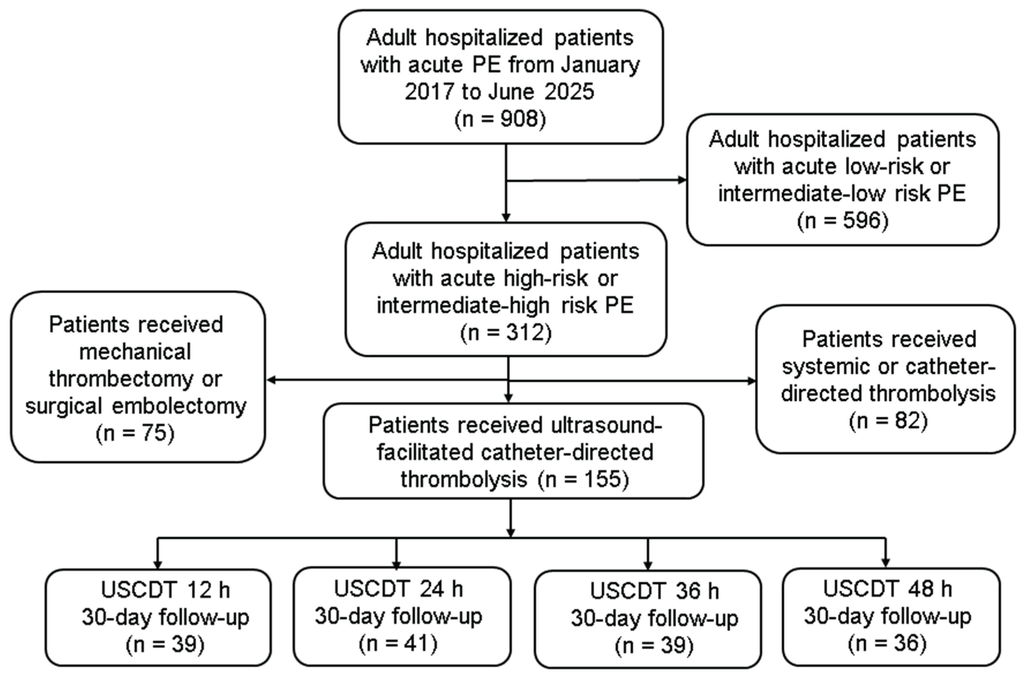
Figure 2.
After 48 hours of follow-up with a mean duration of USCDT 34.2 hours and a mean dose of t-PA 26.8 mg: (A) PA systolic pressure decreased from 48.7 to 35.1 mmHg (a change of 26.9%, p < 0.001); (B) PA mean pressure decreased from 31.8 to 24.9 mmHg (a change of 21.0%, p < 0.001); (C) fibrinogen concentration decreased from 341.9 to 255.8 mg/dL (a change of 25.2%, p < 0.001); and (D) RV/LV diameter ratio decreased from 1.15 to 0.91 (change of 21.5%, p < 0.001). PA, pulmonary artery; RV/LV, right ventricle to left ventricle; t-PA, tissue-type plasminogen activator. Unit of PA pressure: mmHg; Unit of fibrinogen concentration: mg/dL.
Figure 2.
After 48 hours of follow-up with a mean duration of USCDT 34.2 hours and a mean dose of t-PA 26.8 mg: (A) PA systolic pressure decreased from 48.7 to 35.1 mmHg (a change of 26.9%, p < 0.001); (B) PA mean pressure decreased from 31.8 to 24.9 mmHg (a change of 21.0%, p < 0.001); (C) fibrinogen concentration decreased from 341.9 to 255.8 mg/dL (a change of 25.2%, p < 0.001); and (D) RV/LV diameter ratio decreased from 1.15 to 0.91 (change of 21.5%, p < 0.001). PA, pulmonary artery; RV/LV, right ventricle to left ventricle; t-PA, tissue-type plasminogen activator. Unit of PA pressure: mmHg; Unit of fibrinogen concentration: mg/dL.
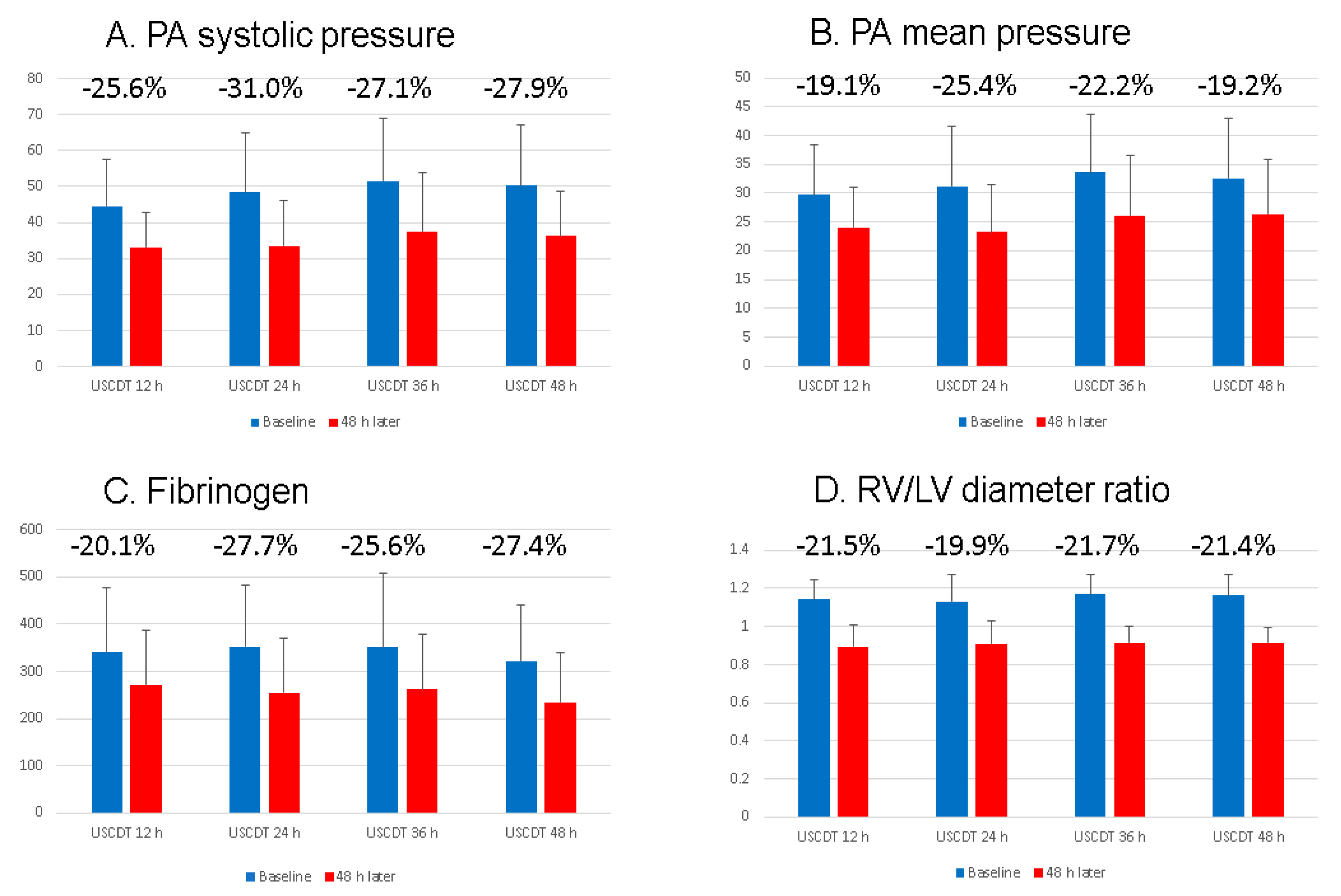
Figure 3.
Thirty-day mortality rate of different PE severity or USCDT duration using logistic regression model. High-risk PE after thrombolysis used as reference point for 30-day mortality. Covariables include all high-risk and intermediate-high risk PE after thrombolysis (odds ratio: 0.56, 95% CI: 0.22‒1.39), intermediate-high risk PE after thrombolysis (odds ratio: 0.24, 95% CI: 0.06‒0.93), USCDT 12 h (odds ratio: 0.36, 95% CI: 0.07‒1.81), USCDT 24 h (odds ratio: 0.34, 95% CI: 0.07‒1.72), USCDT 36 h (odds ratio: 0.56, 95% CI: 0.13‒2.33), USCDT 48 h (odds ratio: 1.08, 95% CI: 0.32‒3.63), standard-duration USCDT (odds ratio: 0.35, 95% CI: 0.10‒1.21), and extended-duration USCDT (odds ratio: 0.80, 95% CI: 0.28‒2.27). CI, Confidence Interval; PE, pulmonary embolism.
Figure 3.
Thirty-day mortality rate of different PE severity or USCDT duration using logistic regression model. High-risk PE after thrombolysis used as reference point for 30-day mortality. Covariables include all high-risk and intermediate-high risk PE after thrombolysis (odds ratio: 0.56, 95% CI: 0.22‒1.39), intermediate-high risk PE after thrombolysis (odds ratio: 0.24, 95% CI: 0.06‒0.93), USCDT 12 h (odds ratio: 0.36, 95% CI: 0.07‒1.81), USCDT 24 h (odds ratio: 0.34, 95% CI: 0.07‒1.72), USCDT 36 h (odds ratio: 0.56, 95% CI: 0.13‒2.33), USCDT 48 h (odds ratio: 1.08, 95% CI: 0.32‒3.63), standard-duration USCDT (odds ratio: 0.35, 95% CI: 0.10‒1.21), and extended-duration USCDT (odds ratio: 0.80, 95% CI: 0.28‒2.27). CI, Confidence Interval; PE, pulmonary embolism.
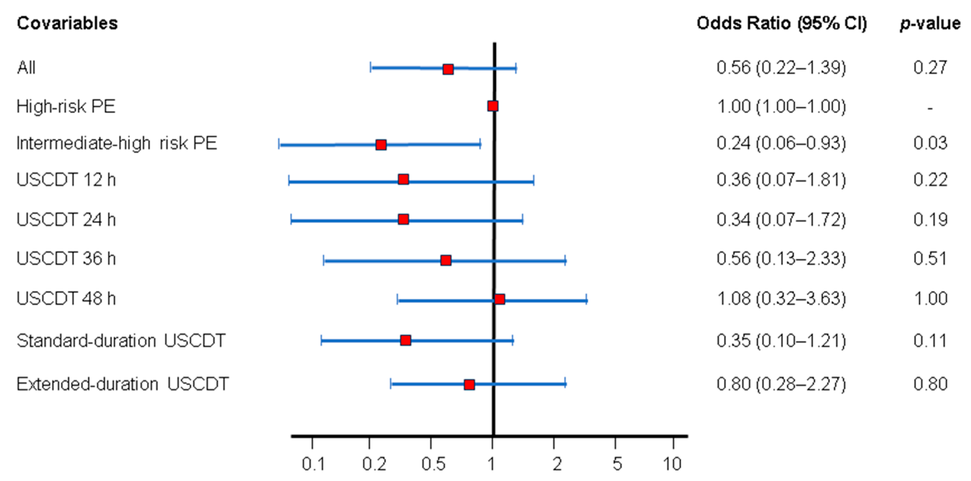

Table 1.
Baseline clinical characteristics of study subjects depending on duration of USCDT.
| USCDT 12 h (n= 39) |
USCDT 24 h (n = 41) |
USCDT 36 h (n = 39) |
USCDT 48 h (n = 36) |
All (n = 155) |
|
|---|---|---|---|---|---|
| Age (y), mean±SD | 63 ± 19 | 58 ± 20 | 57 ± 17 | 58 ± 19 | 59 ± 19 |
| Female sex, n (%) | 31 (79.5) | 23 (56.1) | 25 (64.1) | 26 (72.2) | 105 (67.7) |
|
Body mass index (kg/m2), mean ± SD |
25.3 ± 3.7 | 26.1 ± 5.7 | 26.1 ± 4.8 | 26.8 ± 7.4 | 26.0 ± 5.5 |
| Chronic disease | |||||
| Pregnancy, n (%) | 1 (2.6) | 4 (9.8) | 1 (2.6) | 2 (5.6) | 8 (5.2) |
| Smoking, n (%) | 2 (5.1) | 4 (9.8) | 3 (7.7) | 3 (8.3) | 12 (7.7) |
| Surgery, n (%) | 5 (12.8) | 8 (19.5) | 5 (12.8) | 3 (8.3) | 21 (13.5) |
| Immobility, n (%) | 9 (23.1) | 4 (9.8) | 8 (20.5) | 9 (25.0) | 30 (19.4) |
| Atrial fibrillation, n (%) | 3 (7.7) | 2 (4.9) | 2 (5.1) | 1 (2.8) | 8 (5.2) |
| Diabetes Mellitus, n (%) | 8 (20.5) | 7 (17.1) | 9 (23.1) | 7 (19.4) | 31 (20.0) |
| Hypertension, n (%) | 13 (33.3) | 14 (34.2) | 13 (33.3) | 11 (30.6) | 51 (32.9) |
| Coronary artery disease, n (%) | 1 (2.6) | 1 (2.4) | 3 (7.7) | 2 (5.6) | 7 (4.5) |
| Malignancy, n (%) | 11 (28.2) | 10 (24.4) | 10 (25.6) | 12 (33.3) | 43 (27.7) |
| Heart failure, n (%) | 1 (2.6) | 2 (4.9) | 4 (10.3) | 1 (2.8) | 8 (5.2) |
| Stroke, n (%) | 2 (5.1) | 3 (7.3) | 5 (12.8) | 4 (11.1) | 14 (9.0) |
| Autoimmune disease, n (%) | 2 (5.1) | 5 (12.2) | 5 (12.8) | 7 (19.4) | 19 (12.3) |
| Deep vein thrombosis, n (%) | 18 (46.2) | 23 (56.1) | 20 (51.3) | 25 (69.4) | 86 (55.5) |
| Chronic lung disease, n (%) | 0 (0.0) | 0 (0.0) | 2 (5.1) | 0 (0.0) | 2 (1.3) |
| Severityassessment | |||||
| High-risk, n (%) | 14 (35.9) | 16 (39.0) | 19 (48.7) | 20 (55.6) | 69 (44.5) |
| PESI, mean±SD | 110.9 ± 42.1 | 114.0 ± 32.8 | 118.4 ± 39.3 | 119.3 ± 33.5 | 115.6 ± 37.0 |
| Additional procedure | |||||
| ECMO, n (%) | 8 (20.5) | 5 (12.2) | 10 (25.6) | 10 (27.8) | 33 (21.3) |
| Inferior vena cava filter, n (%) | 26 (66.7) | 32 (78.0) | 24 (61.5) | 21 (58.3) | 103 (66.5) |
ECMO, extracorporeal membrane oxygenation; PESI, Pulmonary Embolism Severity Index; SD, standard deviation; USCDT, ultrasound-facilitated catheter-directed thrombolysis.
Table 2.
Baseline blood counts and biochemical indices of study subjects depending on duration of USCDT.
Table 2.
Baseline blood counts and biochemical indices of study subjects depending on duration of USCDT.
| USCDT 12 h (n= 39) |
USCDT 24 h (n = 41) |
USCDT 36 h (n = 39) |
USCDT 48 h (n = 36) |
All (n = 155) |
||
|---|---|---|---|---|---|---|
| Blood counts | ||||||
| White blood cells (K/μL), mean ± SD | 12.6 ± 8.0 | 11.3 ± 4.5 | 12.0 ± 6.3 | 11.8 ± 5.9 | 11.9 ± 6.3 | |
| Hemoglobin (%), mean ± SD | 12.0 ± 2.2 | 12.9 ± 1.9 | 12.8 ± 2.2 | 12.0 ± 1.9 | 12.4 ± 2.1 | |
| Platelet (K/μL), mean ± SD | 217.8 ± 86.6 | 188.9 ± 59.9 | 192.2 ± 89.5 | 212.6 ± 96.3 | 202.5 ± 83.8 | |
| Biochemical indices | ||||||
| Troponin-I (ng/mL), mean ± SD | 0.24 ± 0.42 | 0.44 ± 1.48 | 0.28 ± 0.73 | 0.45 ± 1.53 | 0.35 ± 1.13 | |
| Troponin-I > 0.04 ng/mL, n (%) | 31 (79.5) | 24 (58.5) | 24 (61.5) | 26 (72.2) | 105 (67.7) | |
| eGFR (mL/min), mean ± SD | 70.8 ± 33.6 | 71.9 ± 35.7 | 66.9 ± 31.5 | 72.9 ± 32.6 | 70.6 ± 33.2 | |
| Lactate (mmol/L), mean ± SD | 2.0 ± 1.0 | 2.1 ± 1.0 | 2.4 ± 1.0 | 2.3 ± 0.9 | 2.2 ± 1.0 | |
| Lactate > 2.0 mmol/L, n (%) | 14 (35.9) | 16 (39.0) | 19 (48.7) | 20 (55.6) | 69 (44.5) | |
eGFR, estimated glomerular filtration rate; USCDT, ultrasound-facilitated catheter-directed thrombolysis.
Table 3.
Different duration of USCDT effect of study subjects of acute high-risk and intermediate-high risk PE.
Table 3.
Different duration of USCDT effect of study subjects of acute high-risk and intermediate-high risk PE.
| USCDT 12 h (n= 39) |
USCDT 24 h (n = 41) |
USCDT 36 h (n = 39) |
USCDT 48 h (n = 36) |
All (n = 155) |
|
|---|---|---|---|---|---|
| Classification of duration (h) | <20 | 20–29 | 30−39 | ≥40 | |
| Duration (h), mean ± SD | 12.2 ± 5.9 | 24.4 ± 3.0 | 36.4 ± 2.8 | 66.9 ± 23.1 | 34.2 ± 23.1 |
| t-PA dose (mg), mean ± SD | 14.6 ± 9.9 | 20.3 ± 11.3 | 31.2 ± 14.4 | 42.6 ± 30.1 | 26.8 ± 20.7 |
| PA systolic pressure | |||||
| PA systolic pressure (before), mean ± SD | 44.5 ± 13.1 | 48.5 ± 16.6 | 51.6 ± 17.3 | 50.2 ± 16.9 | 48.7 ± 16.1 |
| PA systolic pressure (after), mean ± SD | 33.1 ± 9.5 | 33.5 ± 12.5 | 37.6 ± 16.2 | 36.2 ± 12.4 | 35.1 ± 12.9 |
| Change from baseline at 48 h (%) | 25.6 | 31.0 | 27.1 | 27.9 | 26.9 |
| p-value | < 0.001 | < 0.001 | < 0.001 | < 0.001 | < 0.001 |
| PA mean pressure | |||||
| PA mean pressure (before), mean ± SD | 29.7 ± 8.6 | 31.2 ± 10.3 | 33.6 ± 10.2 | 32.6 ± 10.3 | 31.8 ± 9.9 |
| PA mean pressure (after), mean ± SD | 24.1 ± 6.9 | 23.3 ± 8.2 | 26.1 ± 10.4 | 26.3 ± 9.4 | 24.9 ± 8.8 |
| Change from baseline at 48 h (%) | 19.1 | 25.4 | 22.2 | 19.2 | 21.0 |
| p-value | 0.002 | < 0.001 | 0.002 | 0.009 | < 0.001 |
| Fibrinogen | |||||
| Fibrinogen (before), mean ± SD | 339.8 ± 138.2 | 352.6 ± 130.9 | 350.8 ± 157.3 | 322.4 ± 118.9 | 341.9 ± 136.5 |
| Fibrinogen (after), mean ± SD | 271.7 ± 114.3 | 254.9 ± 114.4 | 261.0 ± 117.8 | 234.2 ± 103.9 | 255.8 ± 112.6 |
| Change from baseline at 48 h (%) | 20.1 | 27.7 | 25.6 | 27.4 | 25.2 |
| p-value | 0.02 | 0.001 | 0.006 | 0.001 | < 0.001 |
| RV/LV diameter ratio | |||||
| RV/LV diameter ratio (before), mean ± SD | 1.14 ± 0.11 | 1.13 ± 0.14 | 1.17 ± 0.10 | 1.16 ± 0.11 | 1.15 ± 0.12 |
| RV/LV diameter ratio (after), mean ± SD | 0.89 ± 0.11 | 0.91 ± 0.12 | 0.92 ± 0.09 | 0.92 ± 0.08 | 0.91 ± 0.10 |
| Change from baseline at 48 h (%) | 21.5 | 19.9 | 21.7 | 21.4 | 21.5 |
| p-value | < 0.001 | < 0.001 | < 0.001 | < 0.001 | < 0.001 |
RV/LV, right ventricle-to-left ventricle; SD, standard deviation; t-PA, tissue-type plasminogen activator; USCDT, ultrasound-facilitated catheter-directed thrombolysis. Unit of PA pressure: mmHg; Unit of fibrinogen concentration: mg/dL.
Table 4.
Clinical outcomes of study subjects depending on different duration of USCDT.
| USCDT 12 h (n= 39) |
USCDT 24 h (n = 41) |
USCDT 36 h (n = 39) |
USCDT 48 h (n = 36) |
Standard-duration USCDT (n = 80) |
Extended- duration USCDT (n = 75) |
p-value |
|
|---|---|---|---|---|---|---|---|
| Major complications | |||||||
| Major bleeding, n (%) | 4 (10.3) | 4 (9.8) | 5 (12.8) | 6 (16.7) | 8 (10.0) | 11 (14.7) | 0.38 |
| BARC 3B bleeding, n (%) | 3 (7.7) | 4 (9.8) | 4 (10.3) | 5 (13.9) | 7 (8.8) | 9 (12.0) | 0.51 |
| BARC 3C bleeding, n (%) | 1 (2.6) | 0 (0.0) | 1 (2.6) | 1 (2.8) | 1 (1.3) | 2 (2.7) | 0.53 |
| BARC 4 bleeding, n (%) | 0 (0.0) | 0 (0.0) | 0 (0.0) | 0 (0.0) | 0 (0.0) | 0 (0.0) | - |
| BARC 5 bleeding, n (%) | 0 (0.0) | 0 (0.0) | 0 (0.0) | 0 (0.0) | 0 (0.0) | 0 (0.0) | - |
| Procedure-related advise events, n (%) | 0 (0.0) | 0 (0.0) | 0 (0.0) | 0 (0.0) | 0 (0.0) | 0 (0.0) | - |
| All-cause mortality | |||||||
| 30-day all-cause mortality, n (%) | 2 (5.1) | 2 (4.9) | 3 (7.7) | 5 (13.9) | 4 (5.0) | 8 (10.7) | 0.19 |
BARC, Bleeding Academic Research Consortium; USCDT, ultrasound-facilitated catheter-directed thrombolysis.
Disclaimer/Publisher’s Note: The statements, opinions and data contained in all publications are solely those of the individual author(s) and contributor(s) and not of MDPI and/or the editor(s). MDPI and/or the editor(s) disclaim responsibility for any injury to people or property resulting from any ideas, methods, instructions or products referred to in the content. |
© 2026 by the authors. Licensee MDPI, Basel, Switzerland. This article is an open access article distributed under the terms and conditions of the Creative Commons Attribution (CC BY) license.
Copyright: This open access article is published under a Creative Commons CC BY 4.0 license, which permit the free download, distribution, and reuse, provided that the author and preprint are cited in any reuse.