
Submitted:
05 August 2026
Posted:
06 August 2026
You are already at the latest version
Abstract
Chronic obstructive pulmonary disease (COPD) is a chronic progressive respiratory disease, characterized by respiratory symptoms and airflow limitation, and is one of the leading causes of morbidity and mortality worldwide. The evolution of the disease is associated with episodes of acute exacerbations (AECOPD), characterized by worsening symptoms and accelerated deterioration of lung function. Acute exacerbations of COPD have different severity, being mild, moderate or severe depending on how aggravated the patients’ symptoms are, but also depending on the pulmonary functional and biological changes. Around 70% of AECOPD are infectious in nature, through upper or lower respiratory tract infections. Often, exacerbations associated with lower respiratory tract infections are more severe and have a higher risk of unfavorable outcome compared to AECOPD associated with upper respiratory tract infections. Severe exacerbations require hospital management, most of them being treated in the intensive care unit (ICU). In this narrative review, we evaluated the frequency of different bacterial pathogens involved in severe COPD exacerbations associated with lower respiratory tract infections and the need for treatment in the ICU. Identifying patients with exacerbated COPD at high risk of respiratory infections especially with gram-negative bacilli is extremely important to choose the optimal treatment, thus reducing the risk of death.
Keywords:
chronic obstructive pulmonary disease (COPD)
; severe acute exacerbations
; intensive care unit
1. Introduction
Chronic obstructive pulmonary disease (COPD) is a progressive lung condition secondary to long-term exposure to harmful particles or gases. It is characterized by persistent respiratory symptoms and airflow limitation caused by damage to the airways and alveoli [1]. Worldwide, COPD is a major cause of morbidity and mortality, affecting more than 300 million people and causing over 3 million deaths annually, making it the fourth leading cause of death globally [2].
The evolution of the disease is burdened by acute exacerbations (AECOPD). According to the GOLD strategy, acute exacerbation of COPD is defined as follows: “An exacerbation of COPD is an acute event with symptoms worsening over a few days (up to 14 days) and characterized by increased dyspnea and/or cough and sputum that may be accompanied by tachypnea and/or tachycardia. Exacerbations are often associated with increased local and systemic inflammation caused by airway infection, pollution, or other insults to the lungs”[1].
Depending on the severity, exacerbations can be mild, moderate, or severe. Usually mild to moderate exacerbations can be managed outside the hospital setting, while severe exacerbations require hospital treatment [1,3]. Admission for the management of AECOPD can be done in a clinical ward or in the intensive care unit depending on the clinical condition of the patients and the paraclinical investigations.
In this narrative review of the specialized literature, we aimed to evaluate the distribution of pathogens involved in severe COPD exacerbations that required treatment in the intensive care unit.
2. Profile of Patients with AECOPD Requiring Admission to the ICU
Patients with acute exacerbation of COPD who end up being treated in the intensive care unit (ICU) are usually those with severe exacerbation, complicated by hypercapnic respiratory failure [3]. Patients who experience such AECOPD have a poor prognosis associated with an increased risk of mortality, and either non-invasive or mechanical ventilation is used to correct respiratory failure, depending on the need [3,4]. Approximately 25% of patients with COPD exacerbation require admission to intensive care, and of these, between 26–74% require mechanical ventilator support [5,6,7].
According to the Rome Proposal, GOLD 2026, and Celli BR. et al., severe exacerbation is defined by identifying three of the following parameters - dyspnea on the VAS scale (visual analog dyspnea scale), respiratory rate ≥ 24 breaths/min, heart rate ≥ 95 bpm, resting SaO2 < 92% breathing ambient air or patient`s usual oxygen prescription and/or change > 3%, CRP (C-reactive protein) ≥ 10 mg/L; also, if obtained ABG (arterial blood gases) show new onset/worsening hypercapnia and acidosis (PaCO2 > 45 mmHg and pH < 7.35) [8,9]. Admission to the ICU is for patients with impending or actual respiratory failure, presence of other end-organ dysfunction (shock, renal, liver or neurological disturbance), and/or haemodynamic instability – Figure 1 [10].
The frequency of AECOPD increases with the severity of the disease defined by the degree of airflow obstruction [8,9]. Exacerbations are also associated with disease progression and worsening respiratory dysfunction, leading to decreased quality of life and increased risk of mortality [11,12,13,14,15]. Over time, risk factors that predict relapse of exacerbations have been identified. These include a history of exacerbations, low FEV1 at diagnosis, the need for increased inhaled bronchodilator treatment or the association of corticosteroid therapy, requirement for oxygen when stable, the association of comorbidities like congestive heart failure, chronic renal, coronary artery disease, depression, anxiety or liver failure, and the use of antibiotics in the past – Figure 1 [16,17,18,19,20,21]. However, of these predictive factors, history of exacerbations appears to be the most reliable predictor of future exacerbations [17,22]. As the number of exacerbations in the same patient increases, the severity of the exacerbation associated with the need for hospitalization also increases.
AECOPD increase the economic burden on the healthcare system, especially when they require hospitalization in the intensive care unit. Also, patients who require hospitalization for exacerbation management have a poorer prognosis and an increased risk of death compared to patients without exacerbations or those with mild exacerbations that do not require hospitalization. For these reasons, the prevention of acute events in patients with COPD represents an essential component in the management of this pathology, thus decreasing the risk of hospitalization and admission to the intensive care unit [4,10,22].
3. Bacterial Profile of Patients with AECOPD Admitted to the Intensive Care Unit
COPD exacerbations can be triggered by various factors such as respiratory infections, exposure to pollutants, previous exacerbations, associated comorbidities, or non-compliance with treatment [8,23]. Among these, respiratory infections remain the most common causes of AECOPD, representing between 70–80% of them. According to specialized studies, infectious exacerbations are due to bacteria in approximately 30% of cases, while viruses and bacterial-viral coinfections are incriminated in approximately 25% of cases each [24,25,26].
The bacterial pathogens frequently involved in AECOPD are Haemophilus influenzae, Streptococcus pneumoniae and Moraxella catarrhalis [26,27,28,29]. The bacterial profile changes in patients with AECOPD admitted to the intensive care unit, with them having an increased risk of infections with multidrug-resistant bacteria such as Acinetobacter baumannii, Pseudomonas aeruginosa, Klebsiella spp. and Staphylococcus aureus – Figure 2 [30,31,32]. The risk factors associated with the isolation of Pseudomonas or other gram-negative bacilli in AECOPD are severe form of the disease (FEV1<30%), advanced age, treatment with systemic corticosteroids, administration of antibiotics in the last 3 months, association with bronchiectasis or previous isolation of these microorganisms. Also, the involvement of these bacteria in lower respiratory tract infections in patients with COPD is associated with an increased risk of AECOPD requiring management in the hospital, including in the intensive care unit, as well as an increased risk of death [33,34,35,36,37,38,39,40].
The pathogens listed above can cause AECOPD through upper respiratory tract or lower respiratory tract infections such as pneumonia. Community-acquired pneumonia (CAP) is one of the most common respiratory infections among patients with COPD, leading to exacerbation of the disease. CAP is also a determining criterion for hospitalization of these patients, including in intensive care units, as well as a poor prognostic factor [41,42,43,44]. In patients with COPD, the pulmonary and systemic defense mechanisms are affected, which is why they are at increased risk of pneumonia compared to patients without COPD; moreover, the use of corticosteroid therapy in those with severe disease further increases the risk of respiratory infections [35,45,46].
By researching the studies in the literature, we identified several research papers that aimed to identify the pathogens involved in causing pneumonia in patients with COPD necessiting ICU admission. Most of the research aimed to identify the frequency of these bacteria in the patient groups studied.
In a prospective, multicentere study, conducted by Rello J. et al. it was identified the presence of Streptococcus pneumoniae in 54.1% of patients with COPD and CAP admitted to ICU, followed by Pseudomonas aeruginosa and Legionella pneumophila which were isolated in 13.5% and 4.1% of patients, respectively. Also in the group studied by them, P. aeruginosa predominated in patients with pneumonia and COPD, being isolated in 14.6% of them, compared to the group of patients without COPD, where it was identified in only 0.8% of patients (p˂0.05) [47]. In agreement with this study were the results obtained by Beomsu S. et al regarding the incidence of S. pneumoniae (40.3%) and P. aeruginosa (12.7%) in patients with COPD and CAP. Other pathogens isolated in this study in patients with severe AECOPD were Staphylococcus aureus in 14.2% of patients and Klebsiella pneumoniae in 5.2% of patients [48].
Other studies have identified P. aeruginosa in a smaller percentage of COPD patients. For example, in a retrospective study that pooled data over a 10-year period, P. aeruginosa was isolated in 7% of patients with AECOPD treated in the intensive care unit [49]. In another study, in contrast to the previously mentioned studies, P. aeruginosa was identified in only 2.8% of patients with AECOPD admitted to ICU. The bacteria most frequently involved in COPD infectious exacerbations in this study were Haemophilus influenzae isolated in 12.8% of patients and S. pneumoniae in 10.8% of patients [50]. Hutanu D. et al. identified in their study that Acinetobacter baumanii was the most frequently isolated in patients with COPD, in a percentage of 27.4%, followed by Klebsiella pneumoniae in 10.7% of patients [51].
In Table 1, we have centralized the distribution of pathogens involved in severe infectious EACOPD treated in the ICU according to studies identified in the specialized literature.
In a study that aimed to assess the local epidemiological profile of respiratory infections with Pseudomonas and multidrug-resistant Pseudomonas, Adeniyi J. Idigo et al. identified a 39% higher risk of isolating Pseudomonas in patients diagnosed with COPD compared to those without this diagnosis. However, patients with COPD were not at risk for infections with multidrug-resistant Pseudomonas [53]. It appears that patients with exacerbations requiring hospitalization in the intensive care unit have a higher incidence of gram-negative bacteria such as Pseudomonas and Enterobacteriaceae spp. compared to outpatients with AECOPD [34]. Pengwen Ouyang et al. evaluated in a retrospective, single-center study patients with infectious AECOPD secondary to pneumonia with carbapenem-resistant Klebsiella pneumoniae. They identified a significantly increased mortality rate among these patients compared with patients with pneumonic AECOPD of other bacterial etiology (57.69% vs. 7.69%) [54].
4. Bacterial Profile of Patients with AECOPD and Ventilator-Associated Pneumonia
Between 26-76% of the patients admitted to the intensive care unit for the management of severe AECOPD require invasive mechanical ventilation (IMV) [5,6]. Some cases are complicated by ventilator-associated pneumonia (VAP), which is one of the most common complications among patients treated in intensive care, which is associated with an increased risk of morbidity and mortality, as well as a burden of treatment costs [55,56]. VAP is defined as pneumonia that occurs at least 48 hours after initiation of mechanical ventilation [57].
COPD is also a known risk factor for the occurrence of VAP according to several studies conducted over time in different medical centers [52,58,59,60,61]. There are other studies that have not shown a higher incidence of VAP in COPD patients [62,63], however, most research has shown that VAP increases morbidity and mortality in COPD patients treated in intensive care.
A recent study by Caiden Taowei Lu et al. analyzed the pathogens involved in causing VAP in patients with and without COPD over a 5-year period. The most common bacteria involved in the occurrence of VAP identified in this study were Pseudomonas aeruginosa, Acinetobacter spp., and Klebsiella pneumoniae, without identifying significant differences between patients with and without COPD [64]. In Gursel’s study, the pathogens responsible for VAP with the highest frequency were Pseudomonas aeruginosa, Acinetobacter baumanni and methicillin-resistant Staphylococcus aureus [6]. He also observed that the association of bronchiectasis with COPD increases the frequency of Pseudomonas aeruginosa isolation [65].
In the study conducted by Makris D et al., the bacteria most frequently involved in the occurrence of VAP in patients with AECOPD were P. aeruginosa, S. aureus, Enterobacter species and A. baumannii. Moreover, in the group of patients included in the study, mortality in the intensive care unit was significantly higher among patients with COPD compared to those without COPD (60% vs. 43.3%) [66].
Results consistent with those previously specified were also observed by Koulenti D et al. - the prevalence of P. aeruginosa VAP was higher in patients with COPD compared to those without COPD (29.1% vs. 18.7%). Other pathogens identified in this study with increased frequency in patients with VAP were Enterobacteriaceae, A. baumannii and Staphylococcus aureus. The prevalence of Enterobacteriaceae was also higher in patients with VAP and COPD compared to those without COPD (35.4% vs. 25.9%), while for the other bacteria the differences were not significant between the patient groups [62].
Similar results were also reported by Nseir S et al, with P. aeruginosa (31%), A. baumannii (19%) and S. aureus (14%) being the most common germs isolated in patients with VAP and COPD. Also, the prevalence of multidrug-resistant bacteria in these patients was found to be 41% [67].
In Table 2 we have summarized the frequency of various bacterial agents isolated in patients with COPD and VAP. Regarding the etiology of VAP in patients with COPD, gram-negative bacilli were most frequently involved, and of these, Pseudomonas aeruginosa is the most often isolated.
5. Conclusions
This manuscript reviews the current literature regarding the clinico-bacteriological profile of patients with COPD exacerbations requiring intensive care unit management. The most common bacteria involved in severe AECOPD through lower respiratory tract infections are Streptococcus pneumoniae and Pseudomonas aeruginosa. However, we also identified studies in which Haemophilus influenzae or Acinetobacter spp. had a higher frequency in AECOPD. The disparity between various studies reported in the literature may be due to differences in cohorts, demographics, severity of lung disease, associated comorbidities, and baseline treatments. The prevalence of various bacterial agents involved in infectious exacerbations of COPD may differ by region and time, so a temporal and local assessment is needed.
Regarding the etiology of VAP, gram-negative bacilli have been identified in most studies as determined agents, with Pseudomonas aeruginosa being the most frequently implicated in VAP in patients with COPD. Also, most studies have found an increase in morbidity and mortality in the intensive care unit in COPD patients who developed VAP. Further studies are needed to more comprehensively evaluate patients with severe COPD exacerbations requiring intensive care unit treatment, including studies that stratify patients according to COPD severity relative to ventilatory dysfunction. The goal of these studies could be to identify groups of patients susceptible to developing VAP so that measures to prevent VAP can be implemented.
Author Contributions
Conceptualization DJ, CO and MP; investigation and literature review DJ, PH, FCL and EV; writing—original draft preparation DJ, PH, EV, EP, AMM and OB; writing—review and editing CO, PH, EV and PM; tables and figures DJ, MP, FCL and OB; supervision CO. All authors have read and agreed to the published version of the manuscript.
Funding
This research received no external funding.
Acknowledgments
The publication costs of the present article were covered by “Victor Babes” University of Medicine and Pharmacy Timisoara. The authors used ChatGPT v4.0, an artificial intelligence-based language model developed by OpenAI (San Francisco, CA, USA), exclusively to create the figures based on the data presented in the review. All scientific content, as well as all interpretations and conclusions, represents the authors’ original work.
Conflicts of Interest
The authors declare no conflicts of interest.
Abbreviations
The following abbreviations are used in this manuscript:
| ABG | Arterial blood gases |
| AECOPD | Acute exacerbations of chronic obstructive pulmonary disease |
| COPD | Chronic obstructive pulmonary disease |
| CAP | Community-acquired pneumonia |
| CRP | C-reactive protein |
| GOLD | Global initiative for chronic obstructive lung disease |
| ICU | Intensive care unit |
| IMV | Invasive mechanical ventilation |
| SaO2 | Oxygen saturation |
| VAP | Ventilator-associated pneumonia |
| VAS | Visual analog dyspnea scale |
References
- Langefeld, K.; Agusti, A.G.; Vogelmeier, C. 2026 GOLD Report - Global Initiative for Chronic Obstructive Lung Disease - GOLD. 2026. Available online: https://goldcopd.org/2025-gold-report/.
- World Health Organization. Available online: https://www.who.int/news-room/fact-sheets/detail/the-top-10-causes-of-death.
- Medicine, C.; Steriade, A.T.; Davidoiu, A.; Afrasinei, A.; Tudose, C.; Radu, D.; et al. Predictors of Long-term Mortality after Hospitalization for Severe COPD Exacerbation. 2019, 14(14), 86–92. [Google Scholar] [CrossRef] [PubMed]
- Sárközi, H.; Vultur, M.A.; Sab, A. Analyzing Clinical Parameters and Bacterial Profiles to Uncover the COPD Exacerbations : A Focus on Intensive Care Unit Challenges 2025, 1–11.
- Schmidt, M.; Demoule, A.; Deslandes-boutmy, E.; Chaize, M.; De, Miranda S; Bèle, N.; et al. Intensive care unit admission in chronic obstructive pulmonary disease : patient information and the physician ’ s decision-making process. Crit. Care 2014, 18, R115:1–9. [Google Scholar] [CrossRef] [PubMed]
- Gursel, G. Determinants of the Length of Mechanical Ventilation in Patients with COPD in the Intensive Care Unit. Clin. Investig. 2005, 72, 61–7. [Google Scholar] [CrossRef] [PubMed]
- Esteban, A.; Frutos-Vivar, F.; Muriel, A.; Ferguson, N.; Peñuelas, O.; Abraira, V. Evolution of Mortality over Time in Patients Receiving Mechanical Ventilation. Am. J. Respir. Crit. Care Med. 2013, (188), 220–30. [Google Scholar] [PubMed]
- Langefeld, K.; Agusti, A.G.; Vogelmeier, C. 2026 GOLD Report - Global Initiative for Chronic Obstructive Lung Disease - GOLD [Internet]. 2026. Available online: https://goldcopd.org/2025-gold-report/.
- Celli, B.R.; Barnes, P.J. Exacerbations of chronic obstructive pulmonary disease. Eur. Respir. J. 2007, 29(6), 1224–38. [Google Scholar] [CrossRef] [PubMed]
- Celli, B.R.; Macnee, W. Standards for the diagnosis and treatment of patients with COPD : a summary of the ATS / ERS position paper. 2004, 932–46. [Google Scholar] [CrossRef] [PubMed]
- Dewan, Naresh A.; M.B.B.S.; Salem Rafique, F.C.C.P.; Badar Kanwar, M.D.; Hemant Satpathy, M.; Kay Ryschon, M.D., MS; Glenn S. Tillotson, M.; Niederman, Michael S. M.D. F. Acute Exacerbation of COPD * Factors Associated With Poor Treatment Outcome. Chest [Internet] Available from. 2000, 117(3), 662–71. [Google Scholar] [CrossRef] [PubMed]
- Donaldson, G. C.; Seemungal, T. A. R.; Bhowmik, A. J.A.W. Relationship between exacerbation frequency and lung function decline in chronic obstructive pulmonary disease. Thorax 2002, 57, 847–52. [Google Scholar] [CrossRef] [PubMed]
- Dalal, A.A.; Christensen, L.; Liu, F.; Riedel, A.A. Direct costs of chronic obstructive pulmonary disease among managed care patients. Int. J. Chron. Obstruct Pulmon Dis. 2010, 5, 341–9. [Google Scholar] [CrossRef] [PubMed]
- Balkan, B.; Kaya, E.; Gokoglu, G.; Balkan, A.O.; Yilmaz, G. Management Strategies for Hypercapnic Respiratory Failure in Chronic Obstructive Pulmonary Disease ( COPD ): A Case-Based Discussion Case Presentation. 2025, 17(5). [Google Scholar] [PubMed]
- Cao, Y.; Xing, Z.; Long, H.; Huang, Y.; Zeng, P.; Janssens, J.P. Predictors of mortality in COPD exacerbation cases presenting to the respiratory intensive care unit. Respir Res [Internet]. Available from. 2021, pp. 1–7. [CrossRef]
- MacNee, W.; Anzueto, A.; Agusti, A.; Miller, B.; Calverley, P.; Hurst, J.R.; et al. Susceptibility to Exacerbation in Chronic Obstructive Pulmonary Disease. N Engl. J. Med. 2010, 363(12), 1128–38. [Google Scholar] [CrossRef] [PubMed]
- Donaldson, G. C. J.A.W. COPD exacerbations ? 1 : Epidemiology. Thorax 2006, 61(2), 164–8. [Google Scholar] [CrossRef] [PubMed]
- Miravitlles, M.; Mayordomo, C. Factors Associated with Increased Risk of Exacerbation and Hospital Admission in a Cohort of Ambulatory COPD Patients; 2000; pp. 495–501. [Google Scholar]
- Garcia-aymerich, J.; Monsó, E.; Marrades, R.M.; Escarrabill, J.; Félez, M.A.; Sunyer, J.; et al. Risk Factors for Hospitalization for a Chronic EFRAM STUDY. [CrossRef] [PubMed]
- YOHANNES, A.M.; BALDWIN, R.C.; CONNOLLY, M.J. Depression and anxiety in elderly patients with chronic obstructive pulmonary disease. Age Ageing 2006, 35, 457–9. [Google Scholar] [CrossRef] [PubMed]
- Driscoll, B.R.O.; Neill, J.; Pulakal, S.; Turkington, P.M. A crossover study of short burst oxygen therapy ( SBOT ) for the relief of exercise-induced breathlessness in severe COPD. 2011. [Google Scholar] [CrossRef] [PubMed]
- Hurst, J.R.; Vestbo, J.; Anzueto, A.; Locantore, N.; Müllerova, H.; Tal-Singer, R.; et al. Susceptibility to Exacerbation in Chronic Obstructive Pulmonary Disease. N Engl. J. Med. 2010, 363(12), 1128–38. [Google Scholar] [CrossRef] [PubMed]
- Hogea, S.P.; Tudorache, E.; Fildan, A.P.; Fira-Mladinescu, O.; Marc, M.; Oancea, C. Risk factors of chronic obstructive pulmonary disease exacerbations. Clin. Respir. J. [Internet] 2020, 14(3), 183–97. Available online: https://pubmed.ncbi.nlm.nih.gov/31814260/. [CrossRef] [PubMed]
- Vogelmeier, C.F.; Criner, G.J.; Martinez, F.J.; Anzueto, A.; Barnes, P.J.; Bourbeau, J.; et al. Global Strategy for the Diagnosis, Management, and Prevention of Chronic Obstructive Lung Disease 2017 Report. GOLD Executive Summary. Am. J. Respir. Crit. Care Med. 2017, 195(5), 557–82. [Google Scholar] [CrossRef] [PubMed]
- Miravitlles, M.; Anzueto, A. Chronic Respiratory Infection in Patients with Chronic Obstructive Pulmonary Disease: What Is the Role of Antibiotics? Int. J. Mol. Sci. 2017, 18(7), 1344. [Google Scholar] [CrossRef] [PubMed]
- Papi, A.; Bellettato, C.M.; Braccioni, F.; Romagnoli, M.; Casolari, P.; Caramori, G.; et al. Infections and airway inflammation in chronic obstructive pulmonary disease severe exacerbations. Am. J. Respir. Crit. Care Med. 2006, 173(10), 1114–21. [Google Scholar] [CrossRef] [PubMed]
- Sethi, S. Bacterial Infection in Chronic Obstructive Pulmonary Disease in 2000 : a State-of-the-Art Review. 2001, 14(2), 336–63. [Google Scholar] [CrossRef] [PubMed]
- Sapey, E.; Bafadhel, M.; Bolton, C.E.; Wilkinson, T.; Hurst, J.R.; Quint, J.K. Building toolkits for COPD exacerbations: Lessons from the past and present. Thorax 2019, 74(9), 898–905. [Google Scholar] [CrossRef] [PubMed]
- Brightling, C.E.; Barker, B.L. Phenotyping chronic obstructive pulmonary disease (COPD) exacerbations: realising personalised medicine. Clin. Med. J. R Coll. Physicians Lond. [Internet] Available from. 2012, 12(6), s52–6. [Google Scholar] [CrossRef]
- Scala, R.; Pisani, L. Noninvasive ventilation in acute respiratory failure : which recipe for success ? Available from. 2018. [Google Scholar] [CrossRef] [PubMed]
- Ballouz, T.; Aridi, J.; Afif, C.; Irani, J.; Lakis, C.; Nasreddine, R. Risk Factors, Clinical Presentation, and Outcome of Acinetobacter baumannii Bacteremia. 2017, 7(May), 1–8. [Google Scholar]
- Shorr, A.F.; Haque, N.; Taneja, C.; Zervos, M.; Lamerato, L.; Kothari, S.; et al. Clinical and Economic Outcomes for Patients with Health Care-Associated Staphylococcus aureus Pneumonia ᰔ. 2010, 48(9), 3258–62. [Google Scholar] [CrossRef] [PubMed]
- Chang, K.; Wu, P.; Lee, C.; Lee, Y.; Chen, H. Clinical Features and Antimicrobial Susceptibility of Pseudomonas aeruginosa and Acinetobacter baumannii Complex Isolates in Intensive Care Patients with Chronic Obstructive Pulmonary Disease and Community-Acquired Pneumonia in Taiwan; 2021; pp. 1801–11. [Google Scholar]
- Gupta, N.; Haley, R. Chronic Obstructive Pulmonary Disease in the Intensive Care Unit : Antibiotic Treatment of Severe Chronic Obstructive Pulmonary Disease Exacerbations. 2020. [Google Scholar] [CrossRef] [PubMed]
- Restrepo, M.I.; Mortensen, E.M.; Pugh, J.A.; Anzueto, A. COPD is associated with increased mortality in patients with community- acquired pneumonia. 2006, 28(2), 346–51. [Google Scholar] [CrossRef] [PubMed]
- Huang, Wei-Chang; Lee, Ching-Hsiao; Wu, Ming-Feng; C.-C.H.; Hsu, Cheng-Hui; Chen, Hui-Chen; Hsu, Jeng-Yuan; C.-C.H. Clinical features, bacteriology of endotracheal aspirates and treatment outcomes of patients with chronic obstructive pulmonary disease and community-acquired pneumonia in an intensive care unit in Taiwan with an emphasis on eosinophilia versus non- eosi. BMJ Open 2018, 8(e020341), 1–10. [Google Scholar] [CrossRef] [PubMed]
- Musher, D.M.; Thorner, A.R.; Europe, I. Community-Acquired Pneumonia. 2019, 1619–28. [Google Scholar]
- Cilli, A.; Erdem, H.; Karakurt, Z.; Turkan, H.; Yazicioglu-mocin, O.; Adiguzel, N.; et al. Community-acquired pneumonia in patients with chronic obstructive pulmonary disease requiring admission to the intensive care unit : Risk factors for mortality. 2013, 28, 975–9. [Google Scholar] [CrossRef] [PubMed]
- Sørensen, R.; Ingebrigtsen, T.S.; Sivapalan, P.; Achir, I.; Boel, J.B.; Ekl, J.; et al. Pseudomonas aeruginosa and risk of death and exacerbations in patients with chronic obstructive pulmonary disease : an observational cohort study of 22 053 patients. Clin. Microbiol. Infect. 2020, 26(2), 227–234. [Google Scholar] [CrossRef] [PubMed]
- Ko, F.W.S.; Ip, M.; Chan, P.K.S.; Ng, S.S.S.; Chau, S.S.; Hui, D.S.C. A one-year prospective study of infectious etiology in patients hospitalized with acute exacerbations of COPD and concomitant pneumonia. Respir. Med. 2020, 8(102), 1109–1116. [Google Scholar]
- Zheng, F.; Wang, X. Effect of pneumonia on the outcomes of acute exacerbation of chronic obstructive pulmonary disease : a systematic review and meta-analysis. 2024, 1, 1–14. [Google Scholar] [CrossRef] [PubMed]
- Mullerova, Hana; Chigbo, Chuba; Hagan, Gerry W.; Woodhead, Mark A.; Miravitlles, Marc; Davis, Kourtney J.; J.A.W. The natural history of community-acquired pneumonia in COPD patients : A population database analysis. Respir. Med. 2012, (106), 1124–33. [Google Scholar] [CrossRef] [PubMed]
- Soriano, J.B.; Visick, G.T.; Muellerova, H. Patterns of Comorbidities in Newly Diagnosed COPD and Asthma in Primary Care *; 2012; pp. 2099–107. [Google Scholar]
- Mette, S.; Morten, M.; Løkke, A.; Ole, H.; Sørensen, H.T.; Thomsen, R.W. Incidence and outcomes of patients hospitalized with COPD exacerbation with and without pneumonia. Int. J. Chron. Obs. Pulmon Dis. 2016, 11, 455–65. [Google Scholar] [CrossRef] [PubMed]
- Lu, Z.; Chen, L.; Wang, J. Community-acquired pneumonia and survival of critically ill acute exacerbation of COPD patients in respiratory intensive care units; 2016; pp. 1867–72. [Google Scholar]
- E, F. P.D. S. Incident pneumonia and mortality in patients with chronic obstructive pulmonary disease. A double effect of inhaled corticosteroids? Am. J. Respir. Crit. Care Med. 2015, 191, 141–8. [Google Scholar] [CrossRef] [PubMed]
- Rello, J.; Rodriguez, A.; Torres, A.; Roig, J.; Sirvent, J.M. Implications of COPD in patients admitted to the intensive care unit by community- acquired pneumonia. 2006, 27(6), 1210–6. [Google Scholar] [CrossRef] [PubMed]
- Shin, B.; Kim, S.; Yong, S.J.; Lee, W.; Park, S.; Lee, S.J.; et al. Early readmission and mortality in acute exacerbation of chronic obstructive pulmonary disease with community-acquired pneumonia. 2018. [Google Scholar] [CrossRef] [PubMed]
- Planquette, B.; Péron, J.; Dubuisson, E. T.; Roujansky, A. R.; et al. Antibiotics against Pseudomonas aeruginosa for COPD exacerbation in ICU: a 10-year retrospective study. Int. J. COPD 2015, 10, 379–88. [Google Scholar] [CrossRef] [PubMed]
- Abdallah, G.A.; Diop, S.; Jamme, M.; Legriel, S.; Ferré, A. Respiratory Infection Triggering Severe Acute Exacerbationsfile:///C:/Users/Hogea Patricia/OneDrive/Desktop/articol Dani/descarcate de mine/medicina-61-00669.pdf of Chronic Obstructive Pulmonary Disease. Int. J. Chron. Obstruct Pulmon Dis. 2024, 19(February), 555–565. [Google Scholar]
- Dragos, H.; Sárközi, H.; Vultur, M.A.; Sab, A. Analyzing Clinical Parameters and Bacterial Profiles to Uncover the COPD Exacerbations : A Focus on Intensive Care Unit Challenges. Medicina (B Aires) 2025, 61, 1–11. [Google Scholar] [CrossRef] [PubMed]
- Rogelio, P.M.R.M.F.S.T.F.D.R.; Torres, L.F.-B.G.L.B. A. Impact of COPD in the Outcome of ICU-Acquired Pneumonia With and Without Previous Intubation. Chest 2015, 147(June), 1530–8. [Google Scholar] [CrossRef] [PubMed]
- Idigo, A.J.; Wells, J.M.; Brown, M.L.; Wiener, H.W.; Griffin, R.L.; Cutter, G.; et al. Clinical risk factors for admission with Pseudomonas and multidrug - resistant Pseudomonas community - acquired pneumonia. Antimicrob Resist Infect Control [Internet]. Available from. 2022, pp. 1–10. [CrossRef]
- Ouyang, P.; Zhou, Z.; Pan, C.; Tang, P.; Long, S.; Liao, X.; et al. Acute Exacerbation of Chronic Obstructive Pulmonary Disease Due to Carbapenem-Resistant Klebsiella pneumoniae -Induced Pneumonia : Clinical Features and Prognostic Factors. 2024, (February), 683–93. [Google Scholar] [CrossRef] [PubMed]
- Koulenti, D.; Parisella, F.R. The relationship between ventilator-associated pneumonia and chronic obstructive pulmonary disease : what is the current evidence ? 2019. [Google Scholar] [CrossRef] [PubMed]
- Melsen, W.G.; Rovers, M.M.; Bonten, M.J.M. Ventilator-associated pneumonia and mortality: A systematic review of observational studies*. Crit. Care Med. 2009, 37(10), 2709–2718. [Google Scholar] [CrossRef] [PubMed]
- Kalil, A.C.; Metersky, M.L.; Klompas, M.; Muscedere, J.; Sweeney, D.A.; Palmer, L.B.; et al. Management of Adults With Hospital-acquired and Ventilator-associated Pneumonia : 2016 Clinical Practice Guidelines by the Infectious Diseases Society of America and the American Thoracic Society. Clin. Infect. Dis. 2016, 63, 61–111. [Google Scholar] [CrossRef] [PubMed]
- Hortal, J.; Jesu, M.; Barrio, M. Incidence and risk factors for ventilator- associated pneumonia after major heart surgery. Intensive Care Med. 2009, 35, 1518–25. [Google Scholar] [CrossRef] [PubMed]
- Chang, L.; Dong, Y.; Zhou, P. Investigation on Risk Factors of Ventilator-Associated Pneumonia in Acute Cerebral Hemorrhage Patients in Intensive Care Unit. Can. Respir. J. 2017, 2017, 19–22. [Google Scholar] [CrossRef] [PubMed]
- Karatas, M.; Saylan, S.; Kostakoglu, U.; Yilmaz, G. An assessment of ventilator-associated pneumonias and risk factors identified in the Intensive Care Unit. Pak. J. Med. Sci. 2016, 32(4), 817–22. [Google Scholar] [CrossRef] [PubMed]
- Liu, Y.; Di, Y.; Fu, S. Risk factors for ventilator-associated pneumonia among patients undergoing major oncological surgery for head and neck cancer. Front Med. 2017, 11, 239–46. [Google Scholar] [CrossRef] [PubMed]
- Koulenti, D.; Blot, S.; Dulhunty, J.M.; Papazian, L. COPD patients with ventilator-associated pneumonia : implications for management. Eur. J. Clin. Microbiol. Infect. Dis. 2015, 34, 2403–11. [Google Scholar] [CrossRef] [PubMed]
- Rodríguez, A.; Lisboa, T.; Solé-Violán, J.; Gómez, F.; Roca, O.; Trefl, S.; et al. Impact of nonexacerbated COPD on mortality in critically ill patients. Chest 2011, 139(6), 1354–1360. [Google Scholar] [CrossRef] [PubMed]
- Taowei, C.; Fang, T.; Hung, M.; Lin, Y. Clinical outcomes in ventilator-associated pneumonia patients with and without chronic obstructive pulmonary disease. J. Chin. Med. Assoc. 2025, 88, 65–70. [Google Scholar] [CrossRef] [PubMed]
- Gursel, G. Does coexistence with bronchiectasis influence intensive care unit outcome in patients with chronic obstructive pulmonary disease ? Hear Lung 2006, 35, 58–65. [Google Scholar] [CrossRef] [PubMed]
- Makris, D.; Desrousseaux, B.; Zakynthinos, E.; Durocher, A.; Nseir, S. The impact of COPD on ICU mortality in patients with ventilator-associated pneumonia. Respir. Med. 2011, 105(7), 1022–9. [Google Scholar] [CrossRef] [PubMed]
- Nseir, S.; Di, Pompeo C; Soubrier, S. Impact of ventilatorassociated pneumonia on outcome in patients with COPD. Chest 2005, 128, 1650–1656. [Google Scholar] [CrossRef] [PubMed]
Figure 1.
Risk factors that predict relapse of AECOPD.
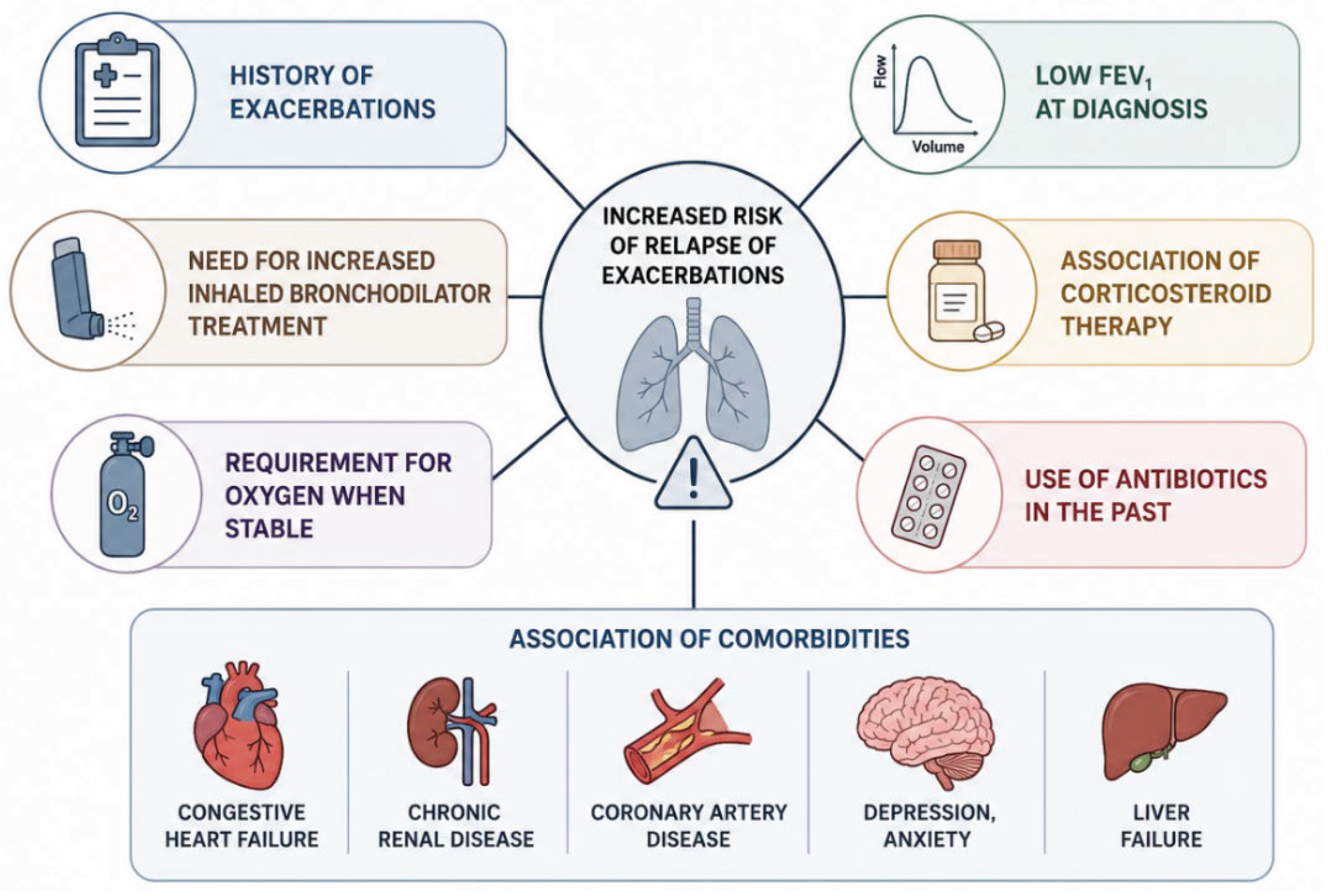
Figure 2.
The bacterial pathogens involved in AECOPD.
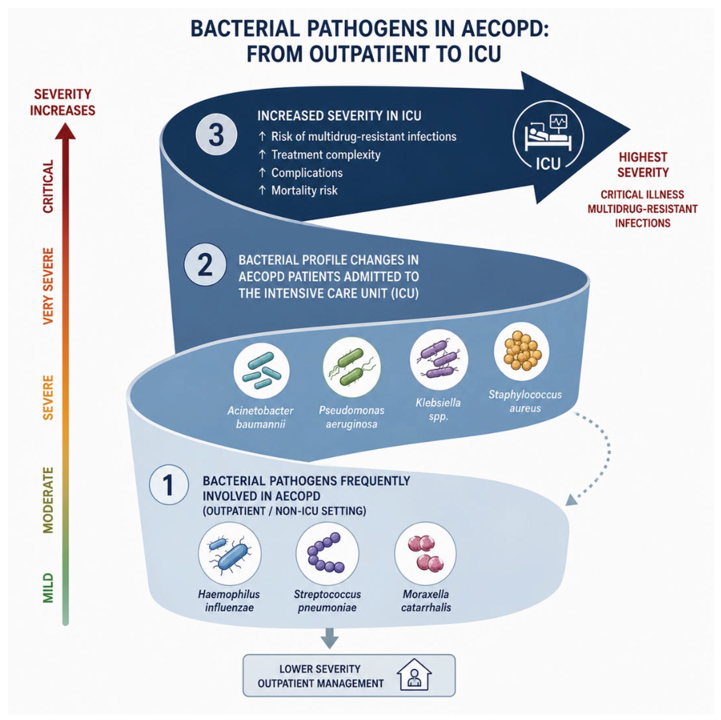

Table 1.
Distribution of pathogens agents’ occurrence (%) in AECOPD with lower respiratory tract infection.
Table 1.
Distribution of pathogens agents’ occurrence (%) in AECOPD with lower respiratory tract infection.
| Distribution of Pathogens Agents’ Occurrence (%) | J. Rello et al. Eur Respir J 2006; 27: 1210–1216 [47] |
Beomsu Shin et al. Chronic Respiratory Disease Volume 16: 1–12 [48] |
Georges Abi Abdallah et al. Inter J of Chronic Obstructive Pulmonary Disease 2024:19 555–565 [50] | Restrepo M.I. et al. Eur Respir J 2006; 28: 346–351 [35] | Hutanu D et al. Medicina 2025, 61, 669 [51] | Mariano Rinaudo et al. CHEST 2015; 147(6): 1530 – 1538 [52] |
| Streptococcus pneumoniae | 54.1 | 40.3 | 10.8 | 6.5 | 6 | 4 |
| Pseudomonas aeruginosa | 13.5 | 12.7 | 2.8 | 5.6 | 6 | 23 |
| Haemophilus influenzae | 11.4 | 0.7 | 12.8 | 3.7 | - | - |
| Legionella pneumophila | 4.1 | 0.7 | 0.3 | - | - | - |
| Acinetobacter spp. | - | 0.7 | - | - | 27.4 | - |
|
Enterobacteriaceae - Klebsiella pneumoniae |
3.1 1 |
- 5.2 |
6.6 - |
1.9 - |
- 10.7 |
8 3 |
|
Staphylococcus aureus |
3.1 | 14.2 | 2.1 | 3.2 | 1.2 | 14 |
|
Mycobacterium tuberculosis |
2.1 | - | - | - | 4.8 | - |
Table 2.
Distribution of pathogens agents’ occurrence (%) in patients with COPD and VAP.
| Distribution of Pathogens Agents’ Occurrence (%) | Taowei C et al. J Chinese Med Assoc. 2025;88:65–70 [64] | Makris D et al. Respir Med. 2011;105(7):1022–9. [66] | Koulenti D et al. Eur J Clin Microbiol Infect Dis. 2015;34:2403–11 [62] | Nseir S et al. Chest. 2005;128:1650–1656 [67] |
| Pseudomonas aeruginosa | 19.4 | 33 | 29.1 | 31 |
| Acinetobacter spp. | 23.3 | 15 | 15.2 | 19 |
|
Enterobacteriaceae - Klebsiella pneumoniae |
- 9.1 |
12 7 |
35.4 - |
4 2 |
|
Staphylococcus aureus |
6.5 | 15 | 19.9 | 13 |
| Stenotrophomonas maltophilia | 12.9 | 4 | 2.5 | 3 |
| Escherichia coli | 2.6 | 9 | - | 5 |
| Streptococcus pneumoniae | - | - | 2.5 | 3 |
Disclaimer/Publisher’s Note: The statements, opinions and data contained in all publications are solely those of the individual author(s) and contributor(s) and not of MDPI and/or the editor(s). MDPI and/or the editor(s) disclaim responsibility for any injury to people or property resulting from any ideas, methods, instructions or products referred to in the content. |
© 2026 by the authors. Licensee MDPI, Basel, Switzerland. This article is an open access article distributed under the terms and conditions of the Creative Commons Attribution (CC BY) license (http://creativecommons.org/licenses/by/4.0/).
Copyright: This open access article is published under a Creative Commons CC BY 4.0 license, which permit the free download, distribution, and reuse, provided that the author and preprint are cited in any reuse.


