

Submitted:
04 August 2026
Posted:
06 August 2026
You are already at the latest version
Abstract
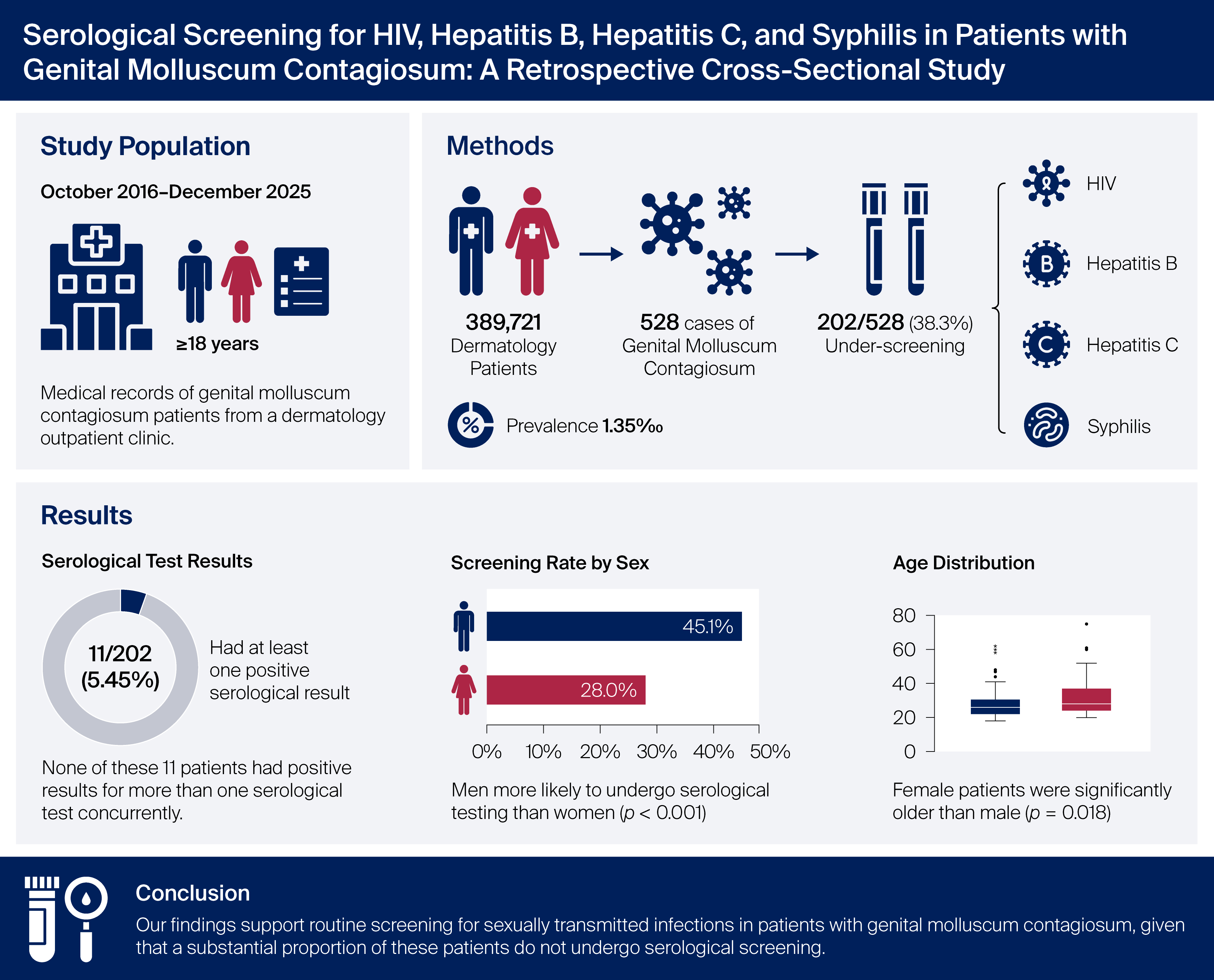
(1) Background: The available literature on sexually transmitted infections in patients with genital molluscum contagiosum is limited. This study aimed to evaluate the serological test results and screening rate for sexually transmitted infections, including human immunodeficiency virus, hepatitis B virus, hepatitis C virus, and syphilis, among patients with genital molluscum contagiosum. (2) Methods: This retrospective cross-sectional study reviewed the medical records of patients aged ≥18 years diagnosed with genital molluscum contagiosum at our dermatology outpatient clinic between October 2016 and December 2025. Age, sex, and serological test results for anti-human immunodeficiency virus, hepatitis B surface antigen, anti-hepatitis C virus, and Venereal Disease Research Laboratory/Rapid Plasma Reagin were recorded. (3) Results: Serological screening was performed in 202 of 528 patients (38.3%), and 11 (5.45%) had positive test results for at least one sexually transmitted infection. (4) Conclusions: Our findings support routine screening for sexually transmitted infections in patients with genital molluscum contagiosum, given that a substantial proportion of these patients do not undergo serological screening.
Keywords:
genital molluscum contagiosum
; serology
; HIV
; hepatitis B virus
; hepatitis C virus
; syphilis
; sexually transmitted infections
1. Introduction
Molluscum contagiosum (MC) is a self-limited, viral, infectious, and cutaneous disease caused by the molluscum contagiosum virus belonging to the Poxviridae family [1]. Molluscum contagiosum virus (MCV) is the only poxvirus with humans as the sole natural hosts following the successful eradication of smallpox [2]. Infection is common, occurring throughout the world with an estimated prevalence of 5-11%, and the incidence appears to be increasing. Viral DNA can be detected on the normal skin of people with molluscum and from objects in their environment, so it is assumed that infection follows contact with infected persons or contaminated objects. It is not known if epidermal injury is important for establishing infection [3]. Sexual or nonsexual direct contact with the infected skin, autoinoculation, and contaminated fomites are the main modes of transmission. Molluscum contagiosum virus is the only representative of the genus Molluscipoxvirus, with approximately 60% of its genes homologous to variola and vaccinia virus genes. MCV exists as four genetic entities: MCV-1, which is responsible for most infections (76-97%), followed by MCV-2, whereas MCV-3 and MCV-4 are extremely rare. To date, complete genomes of only MCV-1 and MCV-2 have been deposited to the GenBank (genetic sequence databank) database; therefore, current MCV subtyping assays can only reliably distinguish infections with these subtypes [2]. There is no clinical relationship between virus type and lesional morphology or anatomical distribution [3]. Patients who are immunocompromised, such as those with HIV (human immunodeficiency virus); who have had organ transplants; or who have cancer are more likely to contract MCV-2, which is responsible for about 60% of all molluscum contagiosum infections in immunocompromised populations [4]. Molluscum contagiosum is usually characterized by round umbilicated papules that vary in size and shape and range from pinkish to skin-colored [1]. Two main clinical presentations are seen: lesions on the face, neck, trunk and arms, seen predominantly in children, and on the genitals, pubic region, lower abdomen, upper thighs and buttocks, which appear to be sexually transmitted [5]. Study of this prevalent skin condition has been relatively limited until recently [4]. In this study, we aimed to evaluate serological test results and the screening rate for sexually transmitted infections, including human immunodeficiency virus, hepatitis B virus, hepatitis C virus, and syphilis, among patients with genital molluscum contagiosum.
2. Materials and Methods
This study was conducted in accordance with the Declaration of Helsinki, and the protocol was approved by the Ethics Committee of Istanbul Medipol University with protocol code E-10840098-202.3.02-3556 on 4 June 2025. As this retrospective study analyzed anonymized routine laboratory data, informed consent for participation was not required in accordance with the Turkish Regulation on Clinical Research, overseen by the Turkish Medicines and Medical Devices Agency, Ministry of Health. The clinical photograph contains no identifiable information, and informed consent for medical photography was obtained from the patient. A retrospective cross-sectional study design was used, wherein the outpatient records of all patients who visited the dermatology outpatient clinic of our hospital between October 2016 and December 2025 were reviewed. Outpatient clinic records were screened for molluscum contagiosum using the ICD-10 (International Classification of Diseases-10th Revision) code B08.1. The medical records of patients aged ≥18 years who were diagnosed with molluscum contagiosum were retrospectively reviewed. As the ICD-10 code B08.1 includes both genital and extragenital molluscum contagiosum, only patients with genital involvement, confirmed through review of the clinical records, were included in the study. Demographic characteristics such as age, gender, and serological test results, including anti-human immunodeficiency virus (anti-HIV), anti-hepatitis C virus (anti-HCV), hepatitis B surface antigen (HBsAg), and Venereal Disease Research Laboratory/Rapid Plasma Reagin (VDRL/RPR) test results, were documented. In cases with reactive VDRL/RPR results, Treponema pallidum hemagglutination assay (TPHA) findings were also documented. Statistical analyses were performed using IBM SPSS Statistics for Windows, Version 30.0 (IBM Corp., Armonk, NY, USA), with continuous variables presented as mean ± standard deviation (SD) and median values, and categorical variables presented as counts (n) and percentages (%). The normality of continuous variables was evaluated using the Shapiro–Wilk test and graphical methods (histograms and Q–Q plots), with comparisons between two groups performed using the Mann–Whitney U test. Genital molluscum contagiosum prevalence, serological testing rates, and seropositivity rates were calculated with 95% confidence intervals (CIs) using the Clopper–Pearson exact binomial method. Categorical variables were compared using Fisher’s exact test. The association between sex and serological testing rate was expressed as relative risk (RR) with the 95% CI. A p-value of < 0.05 was considered statistically significant.
3. Results
During the study period, a total of 389,721 patient visits were documented in the dermatology outpatient clinic. Among these, 528 cases of genital molluscum contagiosum were identified. Accordingly, the proportion of genital molluscum contagiosum among dermatology outpatient visits was 1.35‰ (95% CI: 1.24‰–1.48‰). Serological testing was requested in 202 of the 528 cases (38.3%), resulting in a serological testing rate of 45.1% (143/317) in men and 28.0% (59/211) in women; the likelihood of undergoing serological testing was significantly higher in men than in women (RR = 1.61; 95% CI: 1.25–2.08; p < 0.001) (Figure 1). Among the 202 patients with genital molluscum contagiosum who underwent serological testing, the anti-HIV seropositivity rate was 1.98% (4/202; 95% CI: 0.55%–5.03%); the anti-HCV seropositivity rate was 0.50% (1/202; 95% CI: 0.01%–2.75%); the VDRL/RPR reactivity rate was 0.99% (2/202; 95% CI: 0.12%–3.54%) and all patients with reactive VDRL/RPR results also tested positive for TPHA; and the HBsAg positivity rate was 1.98% (4/202; 95% CI: 0.55%–5.03%). Of the 202 patients who underwent serological testing, 11 had at least one positive serological result, corresponding to an overall seropositivity rate of 5.45% (95% CI: 2.75%–9.53%). None of these 11 patients had positive results for more than one serological test concurrently. The 202 patients included in this study ranged in age from 18 to 65 years, with a mean age of 28.9 ± 8.9 years. Males accounted for 143 (70.8%) and females for 59 (29.2%) of the study population. The demographic and laboratory characteristics of patients with genital molluscum contagiosum are summarized in Table 1. Evaluating age distribution showed that female patients were significantly older than male, with mean ages of 31.7 ± 10.8 and 27.8 ± 7.8 years, respectively (p = 0.018) (Figure 2). There was no significant difference in age between anti-HIV-positive and -negative patients (33.8 ± 11.8 vs. 28.8 ± 8.9 years; p = 0.370). Similarly, no significant association was observed between age and VDRL/RPR or HBsAg positivity (p = 0.927 and p = 0.769, respectively). Because anti-HCV positivity was observed in only a single patient, analyzing age distribution in this group was not feasible. Comparing age between seropositive and -negative patients revealed no statistically significant difference (30.6 ± 8.8 vs. 28.8 ± 9.0 years; p = 0.400), with age distribution by gender and serological status summarized in Table 2. In the cohort of 202 patients who underwent serological testing, anti-HIV positivity was observed in 3.4% of women (2/59) and 1.4% of men (2/143), with no statistically significant difference (p = 0.582). Anti-HCV positivity occurred in only one female patient (1.7%), with no cases observed in male patients (p = 0.292), and VDRL/RPR reactivity occurred solely in male patients (1.4%; 2/143), with no statistically significant difference observed between males and females (p = 0.999). Among male patients, the HBsAg positivity rate was 2.8% (4/143), with none detected in female patients (p = 0.324). Any type of serological test positivity was detected in 5.1% of female patients (3/59) and 5.6% of male patients (8/143), with no statistically significant difference between genders (p = 0.999). The distribution of seropositivity according to gender is summarized in Table 3.
4. Discussion
Molluscum contagiosum was first described in 1817, with a comprehensive article written in 1841 and its viral nature first hypothesized in 1905 [4]. As a member of the poxvirus group, the molluscum contagiosum virus is a large, brick-shaped DNA-containing virus which replicates in the keratinocyte cytoplasm, and is strictly limited to human skin [6]. Molluscum contagiosum is usually characterized by pinkish-to-skin-color and round umbilicated papules varying in size and shape (Figure 3). The diagnosis is usually based on clinical findings [1]. In children, lesions are usually widespread and distributed on the face, torso, and extremities, whereas in immunocompetent adults, in whom the infection is predominantly transmitted sexually, lesions often occur in the anogenital region [2]. Molluscum contagiosum is usually self-limiting after a period of 6-9 months in the majority of cases; however, in rare cases, it may persist for years. Molluscum contagiosum may present atypically with giant, eczematous, and follicular lesions, which may strongly imitate many cutaneous conditions, including warts, basal cell carcinoma, intradermal nevus, keratoacanthoma, cutaneous cryptococcosis, and amelanotic melanoma [1,2]. Oral, ocular, plantar, and subungual regions are unusual localizations for molluscum contagiosum [1]. Congenital molluscum contagiosum occurring via vertical transmission consisting of eyelid or scalp lesions has also been reported [5]. In some cases of molluscum contagiosum, the Koebner phenomenon occurs following trauma or injury, in which lesions appear on areas of skin previously unaffected by the virus [4]. Widespread and refractory molluscum contagiosum on the face is commonly seen in people living with HIV and iatrogenic immunosuppression. Infection is rare under the age of 1 year, perhaps due to maternally transmitted immunity and a long incubation period, which is estimated at between 14 days and 6 months. Molluscum contagiosum virus cannot be grown in tissue culture or eggs. Chronic conjunctivitis and superficial punctate keratitis may complicate lesions on or near the eyelids. Perilesional inflammatory responses may indicate that immunological resolution is likely to occur within a few months rather than the presence of bacterial superinfection. A similar reaction has also been observed in association with COVID-19 (coronavirus disease 2019) vaccination. Typical dermoscopic features include yellow, lobule-like structures around a central, amorphous white nodule with vascular patterns including crown and punctiform patterns (Figure 4). Skin imaging techniques such as reflectance confocal microscopy and lone-field optical coherence tomography have also been proposed to provide an alternative non-invasive approach to diagnosis, which can be confirmed by direct or electron microscopy of the papule contents, histopathology and molecular analysis [3]. A diagnosis can also be obtained by crushing the contents of a lesion on a slide and staining with Wright’s, Giemsa, or Gram stain. Ovoid, homogenous bodies up to 25 μ in diameter are diagnostic [7]. Hematoxylin and eosin (H&E) staining of a molluscum contagiosum lesion typically reveals a crateriform, hyperplastic epidermis or endophytic infundibular hyperplasia that produces a circumscribed cup-shaped pseudotumor containing large cells with granular, eosinophilic, intracytoplasmic molluscum bodies which are also known as inclusion or Henderson–Paterson bodies (Figure 5 and Figure 6). Inclusion bodies are large, measuring up to 35 microns in diameter and made by millions of virions that compress the keratinocyte nuclei [5]. In many instances, therapy is not necessary and spontaneous resolution can be achieved. Treatment may be required if spontaneous clearance is slow, lesions are symptomatic or associated eczema is troublesome. Destructive pro-inflammatory topical, immune-modulating, physical and antiviral treatments are used for the treatment of molluscum contagiosum. Destructive pro-inflammatory topical treatments include cantharidin, salicylic acid, tretinoin, adapalene, 5-10% potassium hydroxide solution, benzoyl peroxide, podophyllotoxin, berdazimer, and lemon myrtle oil. Immune-modulating treatments include topical diphencyprone, imiquimod cream, intralesional or systemic interferon, systemic cimetidine, and intralesional immunotherapies like candida; combined measles, mumps, and rubella vaccine; tuberculin purified protein derivative; and streptococcal substrain OK-432. Physical treatments include cryotherapy, carbon dioxide laser, pulsed dye laser, photodynamic therapy, and surgical removal by curettage. Antiviral treatments include topical, intralesional or intravenous cidofovir [3].
The serological profile of sexually transmitted infections has been examined in patients with condyloma acuminata in the literature [8,9]. But data regarding these infections in patients with genital molluscum contagiosum remain limited, as there have been no studies specifically evaluating this issue in patients with genital molluscum contagiosum. In this study, we aimed to evaluate the serological test results and screening rate for sexually transmitted infections, including human immunodeficiency virus, hepatitis B virus, hepatitis C virus, and syphilis, among patients with genital molluscum contagiosum.
In our study, the prevalence of genital molluscum contagiosum among patients attending the outpatient clinic was 1.35 per 1,000 patients. According to a study conducted in a sexually transmitted infections unit, molluscum contagiosum cases accounted for 1% of patients [10]. To the best of our knowledge, no studies have investigated genital molluscum contagiosum prevalence in dermatology outpatient clinics, nor has the serological testing rate in patients with genital molluscum contagiosum been reported. In our study, serological testing was requested for 202 (38.3%) of the 528 total genital molluscum contagiosum cases, and specifically in 45.1% of male and 28% of female patients, demonstrating a significantly higher likelihood of testing in men. Male patients are more likely to engage in high-risk behaviors [11], which may have prompted clinicians to request serological testing more often in this group. In our study, the overall seropositivity rate was 5.45%, while the anti-HIV seropositivity rate was 1.98%, which is lower than the 6.5% coinfection rate reported in a study conducted in a sexually transmitted infections unit [10]. This lower rate may be explained by the fact that our study was conducted in a general dermatology outpatient clinic rather than in a sexually transmitted infection unit. The anti-HCV seropositivity rate in our study was 0.50%, representing the lowest seropositivity rate among the infections evaluated, which may be due to the highly inefficient sexual transmission of HCV (hepatitis C virus) [12]. In a previous study, the maximum HCV transmission incidence through sexual contact was reported to be 0.07% per year, or approximately 1 per 190,000 sexual contacts [13]. The HBsAg seropositivity rate in our study was 1.98%, identical to the anti-HIV seropositivity rate, which may be explained by the fact that HBV (hepatitis B virus) and HIV share major modes of transmission [14]. HBV is a very resilient organism; in chronically infected individuals, it is present at a high concentration in the blood but is also found in vaginal secretions, semen, breast milk, urine and tears [15]. VDRL/RPR reactivity was detected in 0.99% of patients in our study, and all tested positive for TPHA, confirming a syphilis prevalence of 0.99%. In a 1991 study on patients with genital molluscum contagiosum, syphilis was detected similarly in 0.46% of cases; however, HIV, HBV, and HCV were not included in the screening [16]. In our study, age distribution analysis showed that the mean age of female patients was significantly higher than that of male patients (p = 0.018) (Figure 2). No significant age difference was observed between HIV-positive and -negative patients. Likewise, age was not significantly associated with VDRL/RPR or HBsAg positivity. Since only one patient tested positive for anti-HCV, statistical analysis of the association between anti-HCV positivity and age could not be performed. There was no significant difference in age distribution between seropositive and seronegative patients. In a previous study on trends in molluscum contagiosum in the United States, women with the infection were found to be younger than their male counterparts [17], a finding which contrasts with the results of our study. This finding may be attributable to the greater likelihood of younger women with genital molluscum contagiosum presenting to obstetrics and gynecology clinics, whereas older women may be more likely to seek care in dermatology clinics in our country. Among the 202 patients included in our study, seropositivity rates for anti-HIV, anti-HCV, VDRL/RPR, and HBsAg did not differ significantly between women and men (all p > 0.05), although anti-HCV positivity was detected in only one female patient, whereas VDRL/RPR reactivity and HBsAg positivity were observed only in male patients. Overall seropositivity was 5.1% in women and 5.6% in men, with no statistically significant difference. These findings suggest that there is no significant difference in the prevalence of HIV, hepatitis B, hepatitis C, and syphilis between female and male patients with genital molluscum contagiosum. The mean age of the study population was 28.9 ± 8.9 years, results similar to those of a study performed in a sexually transmitted infections unit, which reported a mean patient age of 29.7 years [10].
It is well established that patients with one sexually transmitted infection are at increased risk of acquiring others [8]. Sexually transmitted molluscum contagiosum usually involves the anogenital area, including the external genital organs, inguinal folds, inner thighs or suprapubic region. The 2020 European guideline on the management of genital molluscum contagiosum recommends that patients presenting with genital molluscum contagiosum be offered screening for other sexually transmitted infections [5]. Our findings support routine screening for sexually transmitted infections in patients with genital molluscum contagiosum and indicate that a proportion of these patients are not screened for concomitant sexually transmitted infections. The under-screening of patients with genital molluscum contagiosum for concomitant sexually transmitted infections has important public health implications and highlights the need for greater physician awareness. The limitations of this study are its single-center design and relatively small sample size. Nevertheless, our study provides valuable real-world insights and emphasizes the need to improve awareness of this issue among physicians managing patients with genital molluscum contagiosum.
5. Conclusions
Molluscum contagiosum is a self-limiting, viral, infectious, and cutaneous disease caused by the molluscum contagiosum virus belonging to the Poxviridae family [1]. Two main clinical presentations are seen: lesions on the face, neck, trunk and arms, seen predominantly in children, and on the genitals, pubic region, lower abdomen, upper thighs and buttocks, which appear to be sexually transmitted [5]. The serological profile of sexually transmitted infections has been examined in patients with condyloma acuminata in the literature [8,9]. But data regarding these infections in patients with genital molluscum contagiosum remain limited, as there have been no studies specifically evaluating this issue in patients with genital molluscum contagiosum. The 2020 European guideline on the management of genital molluscum contagiosum recommends that patients presenting with the disease be offered screening for other sexually transmitted infections [5]. The results obtained in our study lend support to this recommendation and also reveal that a proportion of patients with genital molluscum contagiosum do not undergo screening for concomitant sexually transmitted infections. The under-screening of patients with genital molluscum contagiosum for concomitant sexually transmitted infections has important public health implications and highlights the need for increased awareness among physicians managing these patients.
Author Contributions
Conceptualization, Z.E.; methodology, Z.E.; software, Z.E.; validation, Z.E. and M.T.; formal analysis, Z.E. and M.T.; investigation, Z.E. and M.T.; resources, Z.E.; data curation, Z.E.; writing—original draft preparation, Z.E.; writing—review and editing, Z.E.; visualization, Z.E. and M.T.; supervision, Z.E. and M.T.; project administration, Z.E. All authors have read and agreed to the published version of the manuscript.
Funding
This research received no external funding.
Institutional Review Board Statement
This study was conducted in accordance with the Declaration of Helsinki, and the protocol was approved by the Ethics Committee of Istanbul Medipol University with protocol code E-10840098-202.3.02-3556 on 4 June 2025.
Informed Consent Statement
As this retrospective study analyzed anonymized routine laboratory data, informed consent for participation was not required in accordance with the Turkish Regulation on Clinical Research, overseen by the Turkish Medicines and Medical Devices Agency, Ministry of Health. The clinical photograph contains no identifiable information, and informed consent for medical photography was obtained from the patient.
Data Availability Statement
The raw data supporting the conclusions of this article will be made available by the authors on request.
Acknowledgments
The authors thank biostatistician Alican Sarısaltık for conducting the statistical analyses for this study.
Conflicts of Interest
The authors declare no conflicts of interest.
Abbreviations
The following abbreviations are used in this manuscript:
| MC | Molluscum contagiosum |
| MCV | Molluscum contagiosum virus |
| GenBank | Genetic sequence databank |
| HIV | Human immunodeficiency virus |
| ICD-10 | International Classification of Diseases-10th Revision |
| Anti-HIV | Anti-human immunodeficiency virus |
| Anti-HCV | Anti-hepatitis C virus |
| HbsAg | Hepatitis B surface antigen |
| VDRL/RPR | Venereal Disease Research Laboratory/Rapid Plasma Reagin |
| TPHA | Treponema pallidum hemagglutination assay |
| SD | Standard deviation |
| CI | Confidence interval |
| RR | Relative risk |
| COVID-19 | Coronavirus disease 2019 |
| H&E | Hematoxylin and eosin |
| HCV | Hepatitis C virus |
| HBV | Hepatitis B virus |
References
- Elmas, Ö.F.; Kilitçi, A. Subungual Molluscum Contagiosum: A Rare Presentation. Skin. Appendage Disord. 2020, 6, 55–57. [Google Scholar] [CrossRef] [PubMed]
- Trčko, K.; Hošnjak, L.; Kušar, B.; Zorec, T.M.; Kocjan, B.J.; Križmarić, M.; Seme, K.; Miljković, J.; Luzar, B.; Poljak, M. Clinical, Histopathological, and Virological Evaluation of 203 Patients With a Clinical Diagnosis of Molluscum Contagiosum. Open Forum Infect. Dis. 2018, 5, ofy298. [Google Scholar] [CrossRef] [PubMed]
- Harwood, C.A.; Sterling, J.C. Viral Infections. In Rook’s Textbook of Dermatology, 10th ed.; Griffiths, C., Barker, J., Bleiker, T., Hussain, W., Simpson, R., Eds.; Wiley Blackwell: West Sussex, UK, 2024; Volume 1, pp. 25.15–25.19. [Google Scholar]
- Hebert, A.A.; Bhatia, N.; Del Rosso, J.Q. Molluscum Contagiosum: Epidemiology, Considerations, Treatment Options, and Therapeutic Gaps. J. Clin. Aesthet. Dermatol. 2023, 16, S4–S11. [Google Scholar] [CrossRef] [PubMed]
- Edwards, S.; Boffa, M.J.; Janier, M.; Calzavara-Pinton, P.; Rovati, C.; Salavastru, C.M.; Rongioletti, F.; Wollenberg, A.; Butacu, A.I.; Skerlev, M.; Tiplica, G.S. 2020 European guideline on the management of genital molluscum contagiosum. J. Eur. Acad. Dermatol. Venereol. 2021, 35, 17–26. [Google Scholar] [CrossRef] [PubMed]
- Fonseca, M.E.; Machado, R.D.; Liberto, M.I.; Marcolino, G. Molluscum contagiosum: serology and electron microscopy findings in twenty one patients. Rev. Inst. Med. Trop. Sao Paulo 1987, 29, 86–89. [Google Scholar] [CrossRef] [PubMed]
- Wilkin, J.K. Molluscum contagiosum venereum in a women’s outpatient clinic: a venereally transmitted disease. Am. J. Obste.t Gynecol. 1977, 128, 531–535. [Google Scholar] [CrossRef] [PubMed]
- Ünal, E.; Gönül, M.; Çakmak, S.; Yalçınkaya Iyidal, A.; Kılıç, A.; Gül, Ü.; Doner, P. Serological test results of sexually transmitted diseases in patients with condyloma acuminata. Postep. Dermatol. Alergol. 2015, 32, 286–289. [Google Scholar] [CrossRef] [PubMed]
- Aktaş, H.; Ertuğrul, G.; Benli, A.R. Serological profile of other sexually transmitted diseases in patients with condyloma acuminata: A retrospective view. Arch. Clin. Exp. Med. 2017, 2, 15–17. [Google Scholar] [CrossRef]
- Villa, L.; Varela, J.A.; Otero, L.; Sánchez, C.; Junquera, M.L.; Río, J.S.; Vázquez, F. Molluscum contagiosum: A 20-year study in a sexually transmitted infections unit. Sex. Transm. Dis. 2010, 37, 423–424. [Google Scholar] [CrossRef] [PubMed]
- Balasubramanian, D.; Srinivasan, S.; Paul, P.M.; Ko, N.; Garlapati, S. A Retrospective Study on the Incidence of Kaposi Sarcoma in the United States From 1999 to 2020 Using the Centers for Disease Control and Prevention Wide-Ranging Online Data for Epidemiological Research (CDC WONDER) Database. Cureus 2025, 17, e77213. [Google Scholar] [CrossRef] [PubMed]
- Vandelli, C.; Renzo, F.; Romanò, L.; Tisminetzky, S.; De Palma, M.; Stroffolini, T.; Ventura, E.; Zanetti, A. Lack of evidence of sexual transmission of hepatitis C among monogamous couples: results of a 10-year prospective follow-up study. Am..J Gastroenterol. 2004, 99, 855–859. [Google Scholar] [CrossRef] [PubMed]
- Terrault, N.A.; Dodge, J.L.; Murphy, E.L.; Tavis, J.E.; Kiss, A.; Levin, T.R.; Gish, R.G.; Busch, M.; Reingold, A.L.; Alter, M.J. Sexual transmission of hepatitis C virus among monogamous heterosexual couples: the HCV partners study. Hepatology 2013, 57, 881–889. [Google Scholar] [CrossRef] [PubMed]
- Piot, P.; Goilav, C.; Kegels, E. Hepatitis B: transmission by sexual contact and needle sharing. Vaccine 1990, 8 Suppl, S37–S43. [Google Scholar] [CrossRef] [PubMed]
- Atkins, M.; Nolan, M. Sexual transmission of hepatitis B. Curr. Opin. Infect. Dis. 2005, 18, 67–72. [Google Scholar] [CrossRef] [PubMed]
- Radcliffe, K.W.; Daniels, D.; Evans, B.A. Molluscum contagiosum: a neglected sentinel infection. Int. J. STD AIDS 1991, 2, 416–418. [Google Scholar] [CrossRef] [PubMed]
- Becker, T.M.; Blount, J.H.; Douglas, J.; Judson, F.N. Trends in molluscum contagiosum in the United States, 1966-1983. Sex. Transm. Dis. 1986, 13, 88–92. [Google Scholar] [CrossRef] [PubMed]
Figure 1.
Serology testing rates by gender among patients with genital molluscum contagiosum.
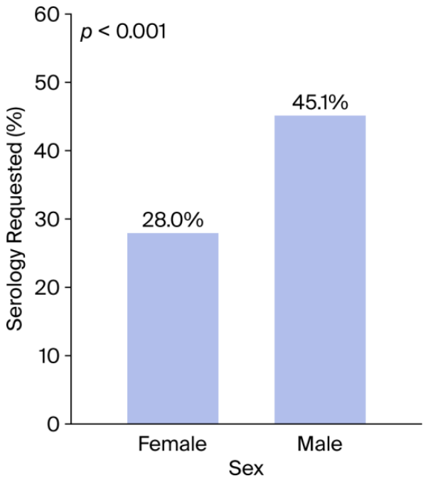
Figure 2.
Distribution of age by gender among patients with genital molluscum contagiosum.
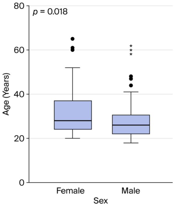
Figure 3.
Multiple molluscum papules on the left antecubital fossa of a 46-year-old man. .
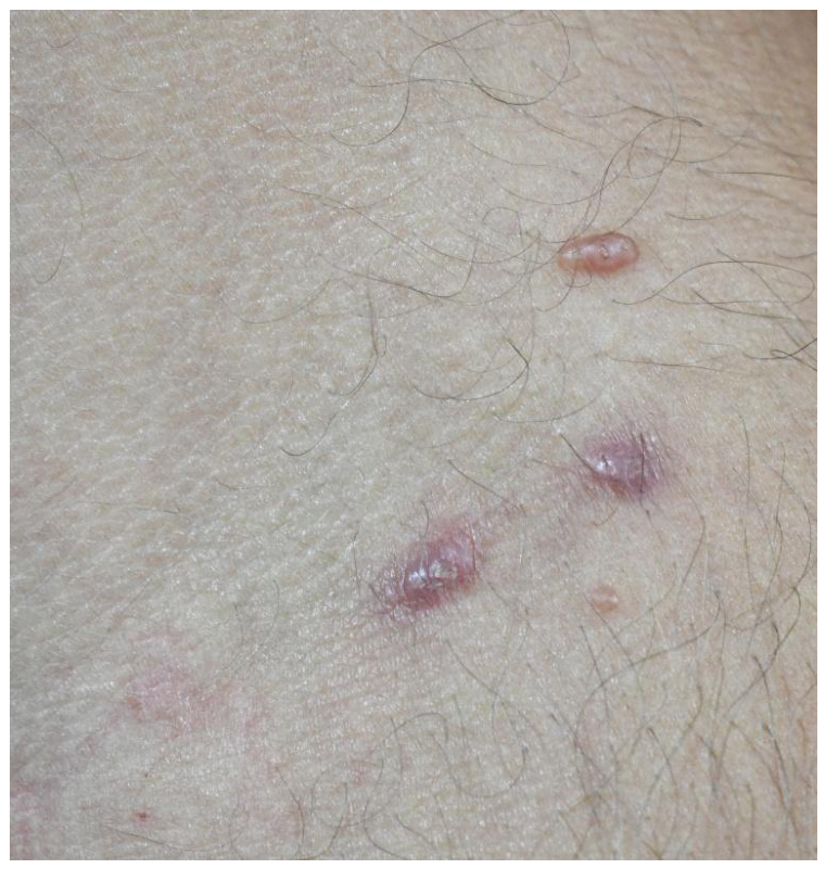
Figure 4.
Dermoscopy of molluscum contagiosum showing white to yellow lobule-like structures with crown and punctiform vessels.
Figure 4.
Dermoscopy of molluscum contagiosum showing white to yellow lobule-like structures with crown and punctiform vessels.
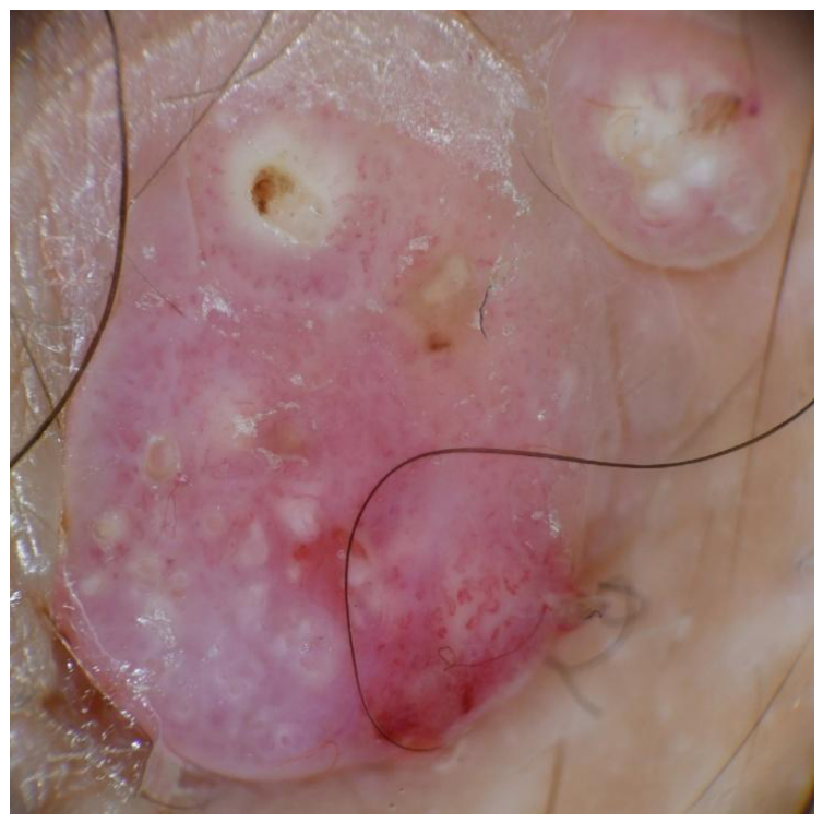
Figure 5.
Histopathology of molluscum contagiosum showing crateriform, hyperplastic epidermis, H&E-stained section (original magnification, x10).
Figure 5.
Histopathology of molluscum contagiosum showing crateriform, hyperplastic epidermis, H&E-stained section (original magnification, x10).
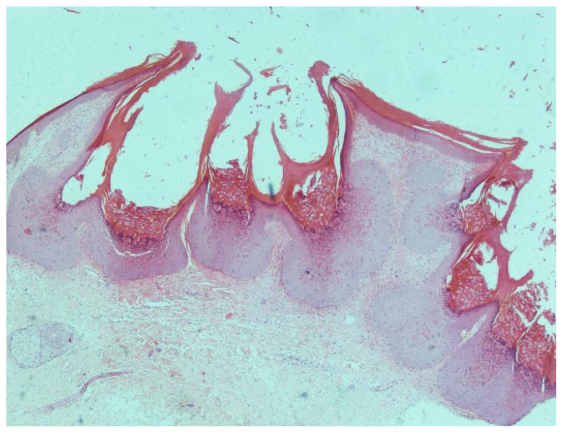
Figure 6.
Histopathology of molluscum contagiosum showing numerous Henderson-Paterson bodies, H&E-stained section (original magnification, x20). .
Figure 6.
Histopathology of molluscum contagiosum showing numerous Henderson-Paterson bodies, H&E-stained section (original magnification, x20). .
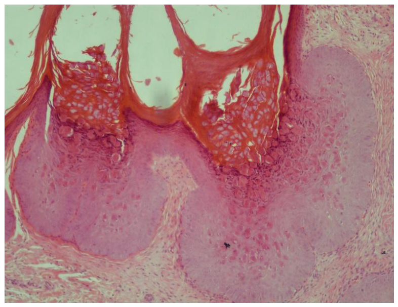

Table 1.
Demographic and laboratory characteristics of patients diagnosed with genital molluscum contagiosum.
Table 1.
Demographic and laboratory characteristics of patients diagnosed with genital molluscum contagiosum.
| N= 202 | |
| Age (years), mean ± standard deviation | 28.9 ± 8.9 |
| Male gender, n (%) | 143 (70.8) |
| Female gender, n (%) | 59 (29.2) |
| Anti-HIV, n (%) | 4 (1.98) |
| Anti-HCV, n (%) | 1 (0.50) |
| VDRL/RPR, n (%) | 2 (0.99) |
| HBsAG, n (%) | 4 (1.98) |
| Any positive serology, n (%) | 11 (5.45) |
Table 2.
Age distribution of gender and serological groups.
|
Age (Years) Mean ± SD/Median |
p* | ||
| Gender | Female | 31.7 ± 10.8/28 | 0.018 |
| Male | 27.8 ± 7.8/26 | ||
| Anti-HIV | Negative | 28.8 ± 8.9/27 | 0.370 |
| Positive | 33.8 ± 11.8/34 | ||
| VDRL/RPR | Negative | 28.9 ± 9.0/27 | 0.927 |
| Positive | 27.5 ± 6.4/27.5 | ||
| HBsAG | Negative | 28.9 ± 9.0/27 | 0.769 |
| Positive | 26.8 ± 5.9/25 | ||
| Any positive serology | Negative | 28.8 ± 9.0/27 | 0.400 |
| Positive | 30.6 ± 8.8/29 | ||
* Mann–Whitney U test.
Table 3.
Distribution of seropositivity by gender.
| Gender | p* | ||
| Female, n (%) | Male, n (%) | ||
| Anti-HIV | 2 (3.4) | 2 (1.4) | 0.582 |
| Anti-HCV | 1 (1.7) | 0 (0) | 0.292 |
| VDRL/RPR | 0 (0) | 2 (1.4) | 0.999 |
| HBsAG | 0 (0) | 4 (2.8) | 0.324 |
| Any positive serology | 3 (5.1) | 8 (5.6) | 0.999 |
* Fisher’s exact test.
Disclaimer/Publisher’s Note: The statements, opinions and data contained in all publications are solely those of the individual author(s) and contributor(s) and not of MDPI and/or the editor(s). MDPI and/or the editor(s) disclaim responsibility for any injury to people or property resulting from any ideas, methods, instructions or products referred to in the content. |
© 2026 by the authors. Licensee MDPI, Basel, Switzerland. This article is an open access article distributed under the terms and conditions of the Creative Commons Attribution (CC BY) license (http://creativecommons.org/licenses/by/4.0/).
Copyright: This open access article is published under a Creative Commons CC BY 4.0 license, which permit the free download, distribution, and reuse, provided that the author and preprint are cited in any reuse.


