
Submitted:
05 August 2026
Posted:
06 August 2026
You are already at the latest version
Abstract
Piscirickettsia salmonis, the etiological agent of piscirickettsiosis (or SRS), represents the most significant health challenge for salmon farming in Chile. While it is generally assumed that fish farms are the epicenter of the disease, the role of migratory wild fish in its ecology and dissemination remains an enigma. This study evaluates the prevalence and shedding routes of P. salmonis in the Chilean silverside (Odontesthes regia), a pelagic fish with extensive latitudinal migration from southern Chile to Peru. Liver, kidney, spleen, and fecal samples were analyzed using nested PCR in individuals captured across five zones of Chile: four in Los Lagos Region, characterized by a high density of salmon farms, with active outbreaks and one in the Maule Region, a pristine area without salmon farming concessions. Results revealed an overall P. salmonis prevalence of 71.6% in O. regia. Remarkably, the highest positivity rate (83%) was detected in the Maule Region, an ecosystem characterized by the absence of salmon farming activities, compared to the Los Lagos region (68.7%), where aquaculture is intensive. These findings suggest that the pathogen is endemic in wild silverside populations regardless of proximity to industrial salmonid cages. P. salmonis was detected in 71,6% of the total samples. The statistical analysis ruled out a significant association between silverside infection and proximity to salmon farms. The pathogen was primarily detected in feces, confirming an active shedding route into the marine environment. These findings challenge the current paradigm, demonstrating that P. salmonis has a broad endemic distribution independent of salmon farming activities. The high rate of fecal shedding, combined with the extensive migratory patterns of O. regia, positions it as a crucial vector capable of transporting the bacteria toward warmer marine ecosystems in the north. This represents a latent risk of horizontal transmission to new geographic areas and other species of aquaculture interest, necessitating a reassessment of biosecurity strategies at a macro-regional level.
Keywords:
wild silverside
; Odontesthes regia
; piscirickettsiosis
; Piscirickettsia salmonis
; epidemiology
1. Introduction
The introduction of exotic species into an ecosystem can have negative impacts on wild animals; it has been established that interactions between them favor disease transmission, with numerous precedents evidencing the consequences of this phenomenon [1,2,3,4,5,6,7]. In light of this scenario, countries with developed aquaculture sectors have sought to determine the risk of horizontal transmission of etiological agents among farmed fish, escaped farmed fish, and wild fish. In the case of Chile, Miranda et al. [8] evaluated the health status of wild fish species in both freshwater and marine environments in the southern zone, where aquaculture concessions for salmonid farming are located, observing the presence of associated pathogens in wild species. Regarding the context of the problem, it should be noted that salmon farming in Chile takes place in the far south of the country, spanning three administrative regions between 41° and 56° South latitude. Given the development of the salmon industry in these regions, studying the interaction between farmed and wild species is of particular interest, as is the case with the Chilean silverside (Odontesthes regia), a native species that frequently interacts with salmon at farm sites.
In relation to the above, empirical evidence shows that the Chilean silverside (O. regia), a native marine species in Chile, enters and subsequently exits salmonid net pens, attracted by feed availability. This phenomenon may constitute a risk factor for the spread of infectious diseases, as these centers often experience outbreaks of a highly contagious and non-specific disease known as piscirickettsiosis. This disease, caused by the bacterium Piscirickettsia salmonis, was first isolated in 1989 [9,10] and is considered endemic in salmonid populations. In this regard, evidence of infection in silversides, the potential excretion of the agent, and the migration of their populations along the entire Chilean coast, from the far south to the northern border with Peru could indicate a potential reservoir for the dissemination of the agent, potentially affecting both wild and farmed species. The occurrence of Piscirickettsia-like organisms (PLOs) and other rickettsial pathogens in wild fish is not a phenomenon unique to the Southern Hemisphere. In the Northern Hemisphere, P. salmonis and related rickettsial agents have been identified in wild salmonids and non-salmonid species across diverse geographical areas, including Norway, Scotland, and the Pacific coast of Canada. For instance, studies in Norway have documented the presence of P. salmonis in wild Atlantic salmon (Salmo salar) and sea trout (Salmo trutta), suggesting that these pathogens maintain a complex ecological presence beyond the confines of industrial aquaculture. These international findings underscore the importance of investigating wild reservoirs to understand the global epidemiology and the potential for bidirectional pathogen transmission in marine ecosystems [11].
This publication addresses new findings regarding the presence of the bacterium in these fish, as well as its excretion routes and potential for dissemination to areas far from salmon farming centers. Experimentally, it has been demonstrated in rainbow trout (Oncorhynchus mykiss) that P. salmonis can be transmitted both vertically and horizontally, being excreted through feces, urine, bile, and seminal and coelomic fluids. Furthermore, national studies refer to the broad host range of this agent, as positivity has been found in free-living fish associated with net pens at farm sites, representing a risk for wild and farmed fish with a probability of maintaining the disease cycle. Additionally, it has been experimentally proven that the bacterium can survive on the shells of certain mollusks [12] that are widely distributed along the Chilean coast. To understand the impact of the infection on the silverside population and better comprehend the disease dynamics in this wild species and its eventual dissemination to other regions, fish were sampled in five zones in southern Chile: four in the Los Lagos Region (high density of salmon farms) and one in the Maule Region, which is characterized by a lack of aquaculture concessions.
2. Materials and Methods
2.1. Sample Collection
Sampling and sample collection. A total of 52 adult specimens of Chilean silverside (Odontesthes regia) with an average length of 25 cm were collected from four Atlantic salmon (Salmo salar) farm sites in the Los Lagos Region, Chile (approximately 42°03'16.1"S 72°45'46.8"W), which were experiencing an active outbreak of piscirickettsiosis, confirmed by official agencies (SERNAPESCA, Chile). The fish were obtained from two types of locations:
- Inside net pens: At two farm sites (zones 1 and 2), 22 specimens were collected directly from inside the net pens, randomly selected from the daily fresh mortality collection carried out at salmon farming centers. None of the sampled fish exhibited macroscopic pathological signs related to the disease.
- Vicinity of farm sites: At another two centers (zones 3 and 4), 24 individuals were captured by direct fishing or with 2-inch gillnets at a distance of approximately 20 meters from the farming structures.
Additionally, six specimens were collected in an area without salmon farming concessions in the Maule Region (35°03'01.1"S 72°13'08.8"W). The sample size was determined considering an expected prevalence of 20% and a 95% confidence interval, according to the results of Miranda et al. [8]. The distribution of individuals by zone is detailed in Table 2, and the geographical location of the sampling sites is presented in Figure 1.
2.2. Sample Processing
2.2.1. Sample Processing Location
Laboratory analysis and sample processing were carried out at the Diagnostic Laboratory of Diseases of Hydrobiological Species, part of the Anatomy Pathology Unit within the Department of Animal Pathology, Faculty of Veterinary and Animal Sciences, University of Chile.
2.2.2. Sample Collection and Processing
The sampled fish were transported to the laboratory and fixed in 70% ethanol for 72 hours. To ensure proper fixation, a ventral incision was made, and the fixative was introduced directly into the abdominal cavity. Subsequently, a necropsy was performed following a protocol adapted from Blazer et al. [13], with specific modifications for the target species. Samples of liver, spleen, kidney, and feces were collected and placed in 5 mL polystyrene tubes containing 2 mL of 70% ethanol. Fecal samples were obtained via mechanical compression of the posterior intestine. Mechanical compression of the posterior intestine was applied gently to extract already formed feces. Furthermore, because the tissues were pre-fixed in ethanol, the risk of bacterial release or cross-contamination from intestinal epithelial cells was minimized, increasing the reliability of the fecal detection. All tubes were stored at 4 °C until further processing. To ensure the absence of cross-contamination between the sampled organs during the necropsy of all Chilean silversides, a negative control using chicken liver was incorporated. This control sample was previously verified to be free of Piscirickettsia salmonis DNA.
2.2.3. Taxonomic Identification of the Target Species
To confirm the identity of the silverside specimens, the artificial identification key for South American silversides from the Southern Cone by Dyer [14] was employed. This remains the only existing identification key to date and was updated by the author, providing a detailed and specific description of silverside species. Through this key, it was verified that the analyzed specimens possessed the attributes that clearly differentiate this species from its congeners.
From an osteological perspective, all examined specimens exhibited the characteristic shape of the urohyal bone and the hemal funnel, which are critical diagnostic characters. Furthermore, based on the same key, detailed observations of external morphology were conducted, including dentition, scaling patterns, and the position of the caudal fin, among others. These findings collectively confirmed the identification of the specimens as Odontesthes regia.
2.3. Sample Processing for Nested PCR (nPCR)
2.3.1. DNA Extraction
Total genomic DNA was extracted from liver, kidney, spleen, and fecal samples, including extraction controls (chicken liver), using the Wizard® Genomic DNA Purification Kit (Promega®), according to the manufacturer’s instructions and a laboratory-standardized protocol. Briefly, 20 mg of each tissue was placed in 1.5 mL microcentrifuge tubes and manually homogenized with pestles in 600 µL of Nuclei Lysis Solution. The lysate was incubated at 65 °C for 20 min and cooled to room temperature for 5 min. Subsequently, 3 µL of RNase Solution was added, and the mixture was incubated at 37 °C for 20 min followed by 5 min on ice. For protein precipitation, 200 µL of Protein Precipitation Solution was added, followed by high-speed vortexing for 20 s and centrifugation at 14,000 x g for 5 min (Super Mini Centrifuge, Lab-tec®).
The supernatant containing the DNA was carefully transferred to a new 1.5 mL tube and mixed with 600 µL of room-temperature isopropanol by gentle inversion until DNA fibrillar structures were visible. The mixture was centrifuged at 14,000 x g for 5 min. The resulting white pellet was washed with 600 µL of 70% ethanol, centrifuged at 14,000 x g for 5 min, and air-dried for 15 min. Finally, the DNA was rehydrated in 100 µL of DNA Rehydration Solution overnight at 4 °C. The DNA extracts were stored at -20 °C until molecular analysis.
2.3.2. DNA Quantification
Following DNA extraction, the samples were quantified by fluorometry using a Quantus™ Fluorometer and the QuantiFluor® dsDNA System (Promega®), following the manufacturer’s instructions. This method was selected to ensure precise measurement of double-stranded DNA (dsDNA) concentration, providing the high sensitivity required for subsequent molecular analysis.
2.4. Nested PCR (nPCR) Analysis
The detection of P. salmonis was performed using nested PCR (nPCR), based on the OIE Manual of Diagnostic Tests for Aquatic Animals [15] and the protocols described by Mauel et al. [16]. All sample processing was conducted inside a Class II biosafety cabinet with a horizontal laminar flow system (BIOBASE, mod. 11231 BBC86). The nPCR was carried out by preparing a working master mix using GoTaq® Colorless Master Mix (containing Taq polymerase, dNTPs, MgCl₂, and reaction buffer) and nuclease-free water. For the first amplification round, universal 16S rDNA bacterial primers were used: EubA (1518R) and EubB (27F). The second round utilized specific primers PS2S (223F) and PS2AS (690R), designed based on the 16S rDNA sequences of five P. salmonis strains (LF-89, EM-90, ATL-4-91, NOR-92, and SLGO-94).
Master mix preparation was performed in DNase-free 1.5 mL tubes, maintaining all reagents on a cooling rack (IsoFreeze®) to ensure the stability of the Taq polymerase. Each reaction consisted of 24 µL of master mix and 1 µL of DNA template in 0.2 mL tubes. The tubes were briefly centrifuged (Capp rondo® model CR68X) for 20 s. For the second amplification, 1 µL of the first PCR product was used as the template. The nPCR included a negative control (nuclease-free water) and a positive control (P. salmonis strain LF-89 DNA extract, derived from a CHSE-214 cell line culture). Amplification was performed in a PCR Touch 960 thermocycler (Zhejiang Scientific Instruments & Materials Import & Export Co., Ltd.®), following the standardized cycling parameters described by Mauel et al. [16],Table 1. The resulting PCR products were stored at -20 °C until electrophoresis.
2.5. Electrophoretic Analysis and Detection of P. salmonis
For the analysis of the nPCR products, electrophoresis was performed on 1.5% agarose gels containing 5 µL of GelRed® nucleic acid stain (Biotium). For sample loading, 5 µL of amplified DNA (previously diluted 1:10 in 1x TBE buffer) was mixed with 2 µL of Orange/Blue Loading Dye 6X (Promega®). A 100 bp to 3,000 bp molecular weight marker (AccuRuler 100 bp DNA RTU Ladder, Maestrogen®) was used as a reference in the first and last wells of each gel, along with 2 µL of loading buffer. Electrophoresis was conducted in a Fermelo® chamber using a Voltronyk reactor-37 power supply (Nyx Technik®) at 80 Volt for 45 min. Band visualization was performed using a BioTop® photo-documentation system (Biosens SC750, Lab-tec®) with UV fluorescence at a wavelength of 312 nm. Samples were considered positive for P. salmonis when a specific 467 base pair (bp) band was clearly visualized.
2.6. Statistical Analysis
To evaluate the association between P. salmonis positivity and the sampling zones, Fisher’s Exact Test [17] was employed, as a low number of observations was expected in some cells of the contingency table. The proportion of positive samples was compared between zones with concessions (inside and in the vicinity of aquaculture farms) and the control zone (no concessions). Additionally, the Odds Ratio [18] was calculated to estimate the relative risk of positivity in zones with concessions. A significance level of p < 0.05 was considered for all analyses. Statistical processing was performed using SPSS software version 23.
3. Results
3.1. Taxonomic Identification of the Target Species
The taxonomic identification process confirmed that the sampled specimens corresponded to the species of interest, O. regia, according to the identification key for South American silversides from the Southern Cone provided by Dyer [14]. In this regard, diagnostic bone and anatomical structures characteristic of the species were identified, including a type 4 hemal funnel and a urohyal bone (SF1, SF2) with an oval-shaped expanded ventral plate. The presence of non-molariform pharyngeal teeth and vomerine teeth (SF1) was also noted. Scale imbrication in the dorsal cranial region was of the normal type; optical microscopy further identified these as cycloid scales with a free posterior margin (SF3). Additionally, the presence of four rows of suborbital scales was observed (SF4).
3.2. Presence of P. salmonis in O. regia via Nested PCR (nPCR)
Of the 52 sampled Chilean silversides, 46 were collected from areas with aquaculture concessions (Z1, Z2, Z3, and Z4), while six were obtained from non-concession areas (Z5). The percentage of P. salmonis positivity for each sampling zone is presented in Table 2. In this study, regarding the tissue distribution of P. salmonis an individual was classified as positive if at least one of its analyzed samples (liver, kidney, spleen, and/or feces) tested positive for the pathogen (Figure 2).

Table 2.
Piscirickettsia salmonis positivity in Chilean silverside (Odontesthes regia) across five sampling zones.
Table 2.
Piscirickettsia salmonis positivity in Chilean silverside (Odontesthes regia) across five sampling zones.
| Sampling Zone | Description | Total Samples (n) | Positivity (%) |
| Zone 1 (Z1) | Inside net pens | 8 | 38 |
| Zone 2 (Z2) | Inside net pens | 14 | 64 |
| Zone 3 (Z3) | Vicinity of farm sites | 14 | 93 |
| Zone 4 (Z4) | Vicinity of farm sites | 10 | 80 |
| Zone 5 (Z5) | Control (No concessions) | 6 | 83 |
| Total | 52 | 71.6 |
As observed, the highest positivity rate for P. salmonis was recorded in Z3 (93%), followed by Z4 (80%); both zones are located in the vicinity of fish farm sites. In areas where specimens were collected from within the net pens, positivity rates were 64% for Z2 and 38% for Z1, resulting in an average prevalence of 68.8% across the four concessioned zones. Notably, in Z5 an area with no history of aquaculture concessions an 83% positivity rate was detected.
Regarding the tissue distribution of P. salmonis in the liver, kidney, spleen, and feces (Figure 2), the highest prevalence was found in the kidney (47.6%) and liver (38.7%), while the spleen showed a lower rate of 23.9%. Furthermore, a significant average fecal positivity of 31.3% was detected across the five zones. This fecal excretion was particularly high in Z3 (42.9%), Z4(40%) and Z5 (66.7%)
3.3. Association Between P. salmonis Positivity and Sampling Zones
As shown in Table 3, no significant association was found between the Chilean silverside specimens that tested positive for P. salmonis and the sampling zones.
3.4. Risk assessment of P. salmonis Positivity in Aquaculture Zones Versus Non-Concessioned Zones
The results obtained from the risk assessment indicate that the probability of P. salmonis occurrence in O. regia is independent of the presence of salmonid aquaculture centers (Table 4).
4. Discussion
4.1. Methodological Considerations and Comparative Analysis
The present study focused on the detection of P. salmonis in Chilean silverside (O. regia) specimens (n=52) collected both within and in the vicinity of net pens, as well as from a control zone without aquaculture concessions. This approach differs from the large-scale study by Miranda et al. [8], who analyzed 791 individuals from areas without reported outbreaks using qPCR. While sample sizes vary, the use of nested PCR (nPCR) in this study provided the high sensitivity required to detect the pathogen in wild populations. Although the sample size in the non-concessioned control zone was relatively small (n=6) compared to the aquaculture zones, the high positivity rate observed serves as a vital qualitative proof of concept. This establishes a baseline detection confirming that the pathogen is present and circulating among wild carriers independently of intensive salmon farming. Nevertheless, future research should integrate larger, seasonally distributed cohorts from both concessioned and non-concessioned zones to elucidate the influence of temperature on the prevalence of this agent.
4.2. Organ Tropism and the Novelty of Fecal Detection
In contrast to OIE [15] recommendations, which prioritize the kidney, liver, and blood for P. salmonis diagnosis, this study analyzed the liver, kidney, spleen, and feces separately. The inclusion of the spleen and feces was instrumental in confirming the widespread presence of the pathogen in both concessioned and non-concessioned zones. Analyzing organs individually revealed a distinct tropism, with the highest positivity found in the kidney (47.6%) and liver (38.7%), followed by feces (31.34%) (Figure 3). Notably, the detection of P. salmonis in the feces of wild fish represents a significant milestone, as this is the first report to date including this matrix in the diagnosis of the pathogen within wild species. The reliability of this novel finding is supported by the sampling methodology. The gentle mechanical compression of ethanol-pre-fixed specimens ensured the extraction of already formed feces while minimizing the risk of bacterial release or cross-contamination from intestinal epithelial cells.
4.3. Horizontal Transmission and Epidemiological Implications
The identification of P. salmonis in the feces of O. regia points to a critical mechanism for environmental dissemination. This finding, when considered alongside the pathogen's documented ability to adhere to and persist within biofilms on mollusk shells, such as Mytilus chilensis [12], helps to complete the epidemiological cycle of the bacterium in wild environments. In this model, the silverside acts as a migratory vector that disperses the pathogen via fecal shedding, while mollusks serve as stable environmental reservoirs. This synergy likely maintains the infective agent in the ecosystem even in the absence of salmonid farming activities. This fecal shedding is of paramount epidemiological importance, as P. salmonis can be excreted via bile, feces, and urine in live fish. In the marine environment, the pathogen can survive for extended periods, facilitating direct horizontal transmission, which is considered a primary route of infection [19,20,21]. Consequently, coprophagy or the ingestion of feces-contaminated feed may serve as a bidirectional transmission route between farmed and wild species. While this study did not establish the interaction with aquaculture centers as a statistically significant risk factor, the evidence of P. salmonis in O. regia suggests a potential impact on the conservation of this native species, given its known interaction with salmonids during outbreaks.
4.4. The Role of Non-Concessioned Zones and Future Directions
The findings in Zone 5 (no aquaculture concessions) are particularly striking: positivity was detected in all analyzed organs, with the highest rate in feces (66.7%) (Figure 2). These results provide the first evidence of the potential dissemination of P. salmonis via feces in a wild species with a broad latitudinal distribution [14]. Future studies should investigate the epidemiological role of the Chilean silverside in the propagation of the agent, specifically focusing on the influence of population densities. While salmonids in intensive systems are subject to high confinement stress, the presence of the agent in free-ranging wild individuals suggests that density-dependent factors in experimental settings should be explored further. Finally, a genetic characterization of P. salmonis in positive silversides from the Maule Region is essential. Previous phylogenetic studies in the Los Lagos Region [6] linked strains in wild fish to the EM-90 strain originally described in Coho salmon. Comparing these strains will determine if the pathogen's distribution has expanded or if it has been historically present but undetected. This is especially relevant considering the historical introduction of salmonid eggs into various regions, including Maule, since the early 20th century [22], often without comprehensive health records.
4.5. Proposed Epidemiological Cycle Based on Study Data
The proposed epidemiological cycle (Figure 3), integrates the high prevalence observed in this study with the biological mechanisms of the vector and environmental persistence. Our data demonstrates an 83% prevalence of P. salmonis in wild populations of Odontesthes regia in areas devoid of salmon farming, such as the Maule Region. This finding serves as the baseline for a cycle that operates independently of aquaculture. The mechanism is driven by fecal shedding, as evidenced by the high bacterial load detected in the feces of wild silversides. We propose that O. regia acts as a migratory bridge, where fecal excretion provides a continuous inoculum of viable bacteria into the water column during the fish's latitudinal movements.
This inoculum facilitates the transition from the pelagic vector to the benthic environment. Once in the benthos, the pathogen adheres to mollusk shells, such as Mytilus chilensis, forming infective biofilms, a mechanism supported by previous experimental evidence [20]. These biofilms serve as environmental anchors or "seed banks," allowing P. salmonis to persist in the marine ecosystem even during periods without active hosts.
In summary, the data supports an open epidemiological cycle where migratory wild fish ensure geographical dissemination via shedding, while benthic organisms provide the necessary substrate for long-term persistence, collectively maintaining the pathogen's endemicity across the Chilean coast.
5. Conclusions
The nested PCR (nPCR) technique successfully detected the presence of P. salmonis DNA in the liver, kidney, spleen, and feces of Chilean silverside (O. regia), indicating a high prevalence of the pathogen in this species.
The use of specific taxonomic identification keys confirmed that all sampled specimens belonged to the species O. regia.
The detection of P. salmonis in silversides from within net pens, in the vicinity of aquaculture sites, and from non-concessioned zones establishes the presence of the agent in areas previously unstudied. This finding is highly relevant given the broad latitudinal distribution of O. regia along the Chilean coast.
The inclusion of fecal samples proved to be a critical diagnostic component, considering the high probability of the agent being excreted via this route. This work represents the first report to include feces in the diagnosis of P. salmonis in wild fish species, providing essential data to better understand the wild cycle of Piscirickettsiosis.
Statistical analysis revealed no significant association between P. salmonis positivity in O. regia and proximity to salmonid aquaculture centers. Consequently, this study could not identify the interaction with farming sites as a determining risk factor for the presence of the pathogen in this native species.
Supplementary Materials
The following supporting information can be downloaded at the website of this paper posted on Preprints.org.
Author Contributions
All authors contributed substantially to the work presented in this manuscript. Conceptualization, L.V.O., J.L., M.A., and P.C.; methodology, L.V.O., J.L., M.A., and P.C.; formal analysis, L.V.O., J.L., M.A., and P.C.; investigation, L.V.O., J.L., M.A., and P.C.; resources, L.V.O. and J.L.; data curation, L.V.O., J.L., M.A., and P.C.; writing—original draft preparation, L.V.O. and J.L.; writing review and editing, L.V.O. and J.L. All authors have read and agreed to the published version of the manuscript.
Funding
This study was funded by Grant Environmental dynamics of P. salmonis FIE-ACUICULTURA V014, Sernapesca, Chile.
Data Availability Statement
Due to privacy restrictions from the funding source, the data are available only upon request.
Acknowledgments
Technical support for this project was provided by: Cristian Contreras (Salmones Humboldt) and Dr. Ricardo Olivares (Department of Animal Pathology, University of Chile).
Conflicts of Interest
The authors declare no conflicts of interest.
References
- Dobson A., J. Foufopoulos . Emerging infectious pathogens of wildlife. Phil Trans R. Soc. Lond. 2001,356(1411),1001-1012. [CrossRef]
- Cleaveland S., G.R. Hess, A.P. Dobson, M.K. Laurenson, H.I. McCallum, M.G. Roberts, R. Woodroffe. The role of pathogens in biological conservation. In The Ecology of Wildlife Diseases; Hudson P.J., Ed.; Oxford University Press, New York, USA, 2002; pp139-150.
- Riley S., J. Foley, B. Chomel. Exposure to feline and canine pathogens in bobcats and gray foxes in urban and rural zones of a national park in California. J. Wildl. Dis. 2004, 40(1),11-22.
- Lobos G., M. Ferres, R. Palma. Presencia de los géneros invasores Mus y Rattus en áreas naturales de Chile: un riesgo ambiental y epidemiológico. Rev. Chilena de Hist. Nat. 2005, 78(1), 113-124. [CrossRef]
- Millan J.& J.C. Casanova. Helminth parasites of the endangered Iberian lynx (Lynx pardinus) and sympatric carnivores. J. Helminthol. 2007, 81(4),377-380. [CrossRef]
- Contreras-Lynch S., P. Olmos, A. Vargas, J. Figueroa, R., González-Stegmaier, R., Enríquez, y A. Romero. Identification and genetic characterization of Piscirickettsia salmonis in native fish from southern Chile. Dis. Aquat. Org. 2015, 115(3), 233-244. [CrossRef]
- Marcos-López M., Ruane, N., Scholz, F., Bolton-Warberg, M., Mitchell, S., Murphy O'Sullivan, S., Irwin Moore, A., & Rodger, H. Piscirickettsia salmonis infection in cultured lumpfish (Cyclopterus lumpus L.). J. Fish Dis. 2017, 40(11), 1625-1634. [CrossRef]
- Miranda P., V. Matus, P. Olmos, F. Shulze. Evaluación y seguimiento sanitario de especies ícticas silvestres en agua dulce y de mar. Informe Final, SUBPESCA(subsecretaria de pesca y acuicultura), IFOP(Instituto de Fomento Pesquero)[Online], 2012;pp 3-257. https://www.researchgate.net/publication/317576168_Evaluacion_de_la_situacion_sanitaria_en_peces_de_agua_dulce_y_mar_en_Chile (accessed June 20, 2025).
- Fryer J, C Lannan, L Garcés, J Larenas, P Smith. Isolation of a rickettsiales-like organism from disease coho salmon (Oncorhynchus kisutch) in Chile. Fish. Pathol. 1990, 25(2), 107-114. [CrossRef]
- Cvitanich J, O. Garate, C. Smith. The Isolation of Rickettsia-like organisms causing disease and mortality in Chilean salmonids and its confirmation by Koch’s postulate. J. Fish Dis.1991,14 (2),121-145. [CrossRef]
- Rozas M, R Enríquez.. Piscirickettsiosis and Piscirickettsia salmonis in fish: a review. J. Fish Dis.2014, 37(3), 163-188. [CrossRef]
- Larenas J, M. Perez, C. Moraleda, M. Godoy, C. Larenas y M. Acuña-Retamar. In vitro adhesion and infectiveness of Piscirickettsia salmonis on mussel shells Mytilus chilensis. Bull. Eur. Ass. Fish Pathol.[Online], 2019,39(3):114-121.https://www.ciba.cl/content/uploads/larenas-et-al.-2019-in-vitro-adhesion-and-infectiveness-of-piscirickettsia-salmonis-on-mussel-shells-mytilus-chilensis.pdf(accessed June 20, 2025).
- Blazer VS, Walsh HL, Braham RP, Smith C. Necropsy-based Wild Fish Health Assessment. J. Vis. Exp.2018,11(139),57946. [CrossRef]
- Dyer B. Revisión sistemática de los pejerreyes de Chile (Teleostei, Atheriniformes). Biocell.[Online], 2006, 30(1),69-88. https://www.scielo.org.ar/scielo.php?script=sci_arttext&pid=S0327-95452006000100010&lng=es&tlng=en (accessed June 20, 2025).
- Manual of diagnostic tests for aquatic animals,Office International des Epizooties in English,5th ed.,Paris, 2006; pp469.
- Mauel MJ, SJ Giovannoni, JL Fryer. Development of polymerase chain reaction assays for detection, identification, and differentiation of Piscirickettsia salmonis. Dis. Aquat. Org. 1996,26, 189-195. [CrossRef]
- Gómez-Biedma S., M. Vivo y E. Soria Pruebas de significación en Bioestadística. Rev. Diagn. Biol.[Online], 2001.50(4), 207-218.http://scielo.isciii.es/scielo.php?script=sci_arttext&pid=S0034-79732001000400008&lng=es&tlng=es (accessed June 20, 2025).
- Cerda J., C. Vera y G. Rada Odds ratio: aspectos teóricos y prácticos. Rev. Méd. Chile 2013,141(10):1329-35. [CrossRef]
- Salinas G., J. Contreras, P. Smith, J. Larenas Horizontal transmission and excretion of Piscirickettsia salmonis in rainbow trout (Oncorhynchus mykiss) in fresh water condition. in: VIII International Conference Diseases of Fish and Shellfish,Abstracts Book. Heriot-Watt University, Edinburgh, European Association of Fish Pathologists,1997; p-057.
- Larenas J., E. Zamorano, P. Smith.Detección de Piscirickettsia salmonis en heces de salmón Coho (Oncorhynchus kisutch) infectados por transmisión vertical. Mon. Electr. Patol. Vet.[Online] 2005, 2(1), 59-67.https://www.researchgate.net/publication/285136074_Deteccion_de_Piscirickettsia_salmonis_en_heces_de_alevines_de_salmon_coho_Oncorhynchus_kitsutch_infectados_por_transmision_vertical (accessed 20 Junio 2014).
- Peirano P. Determinación de la presencia y asociación de P. salmonis, en heces, hígado y riñón de trucha arcoíris (Oncorhynchus mykiss) en condiciones de cultivo en mar. Tesis, Fac. de Cs. Veterinarias y pecuarias, Universidad de Chile , Chile, 2015.https://repositorio.uchile.cl/handle/2250/131926 (Accesed: 20 June 2025).
- Golusda P. La introducción del salmón en Chile. Anales Agronómicos, Sección de Aguas y Bosques del Ministerio de Industria. Santiago de Chile.[Online],1907; pp 31. https://www.memoriachilena.gob.cl/archivos2/pdfs/mc0027366.pdf (Accessed: 20 Junio 2025).
Figure 1.
Referential location of sampling areas: the purple circle includes zones 1 and 2 (inside net pens); green circles correspond to sampling zones 3 and 4 (vicinity of fish farms); and the red circle indicates sampling zone 5 (no maritime concessions).
Figure 1.
Referential location of sampling areas: the purple circle includes zones 1 and 2 (inside net pens); green circles correspond to sampling zones 3 and 4 (vicinity of fish farms); and the red circle indicates sampling zone 5 (no maritime concessions).
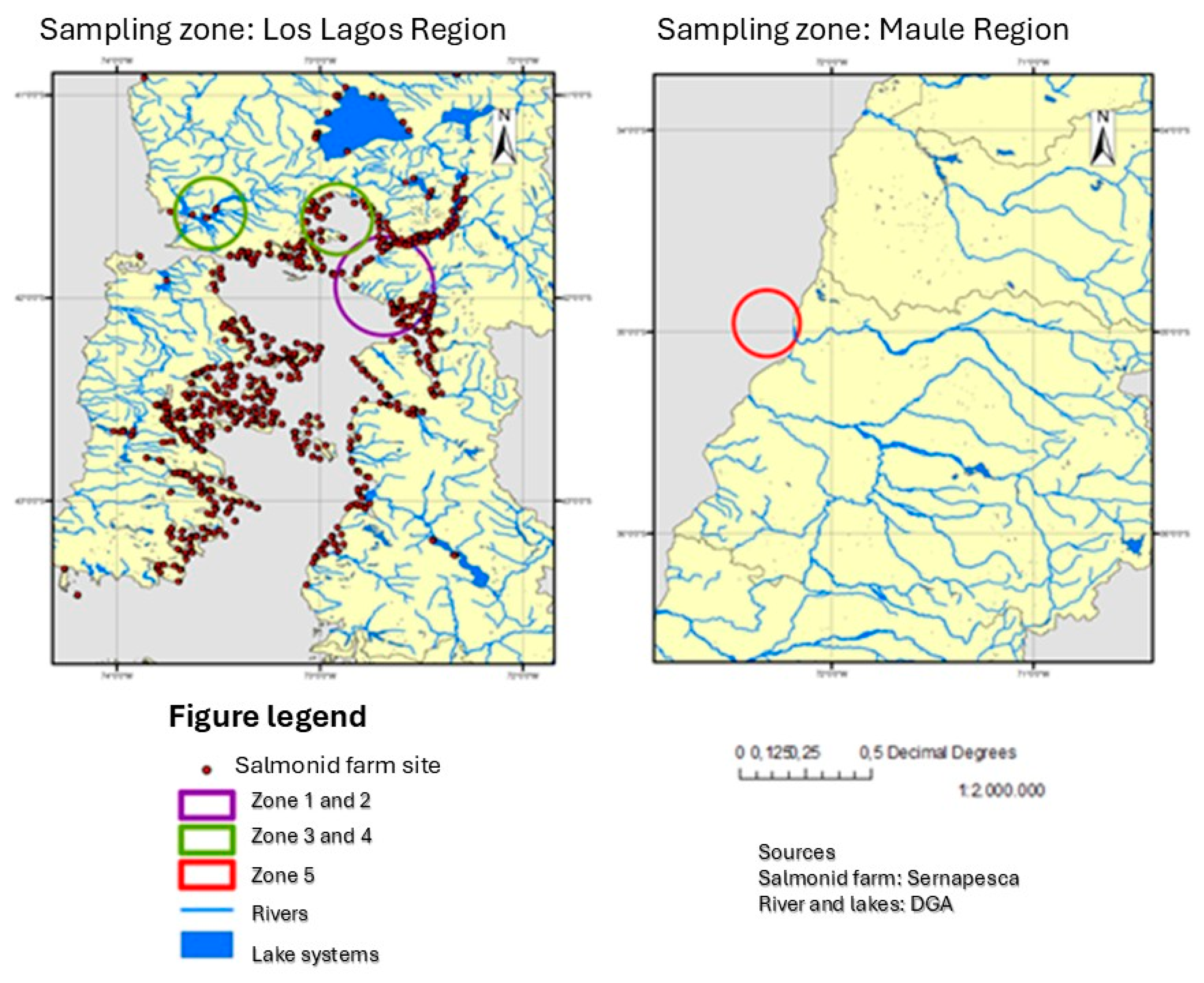
Figure 2.
Detection of Piscirickettsia salmonis in liver, kidney, spleen, and feces of Odontesthes regia categorized by sampling location.
Figure 2.
Detection of Piscirickettsia salmonis in liver, kidney, spleen, and feces of Odontesthes regia categorized by sampling location.
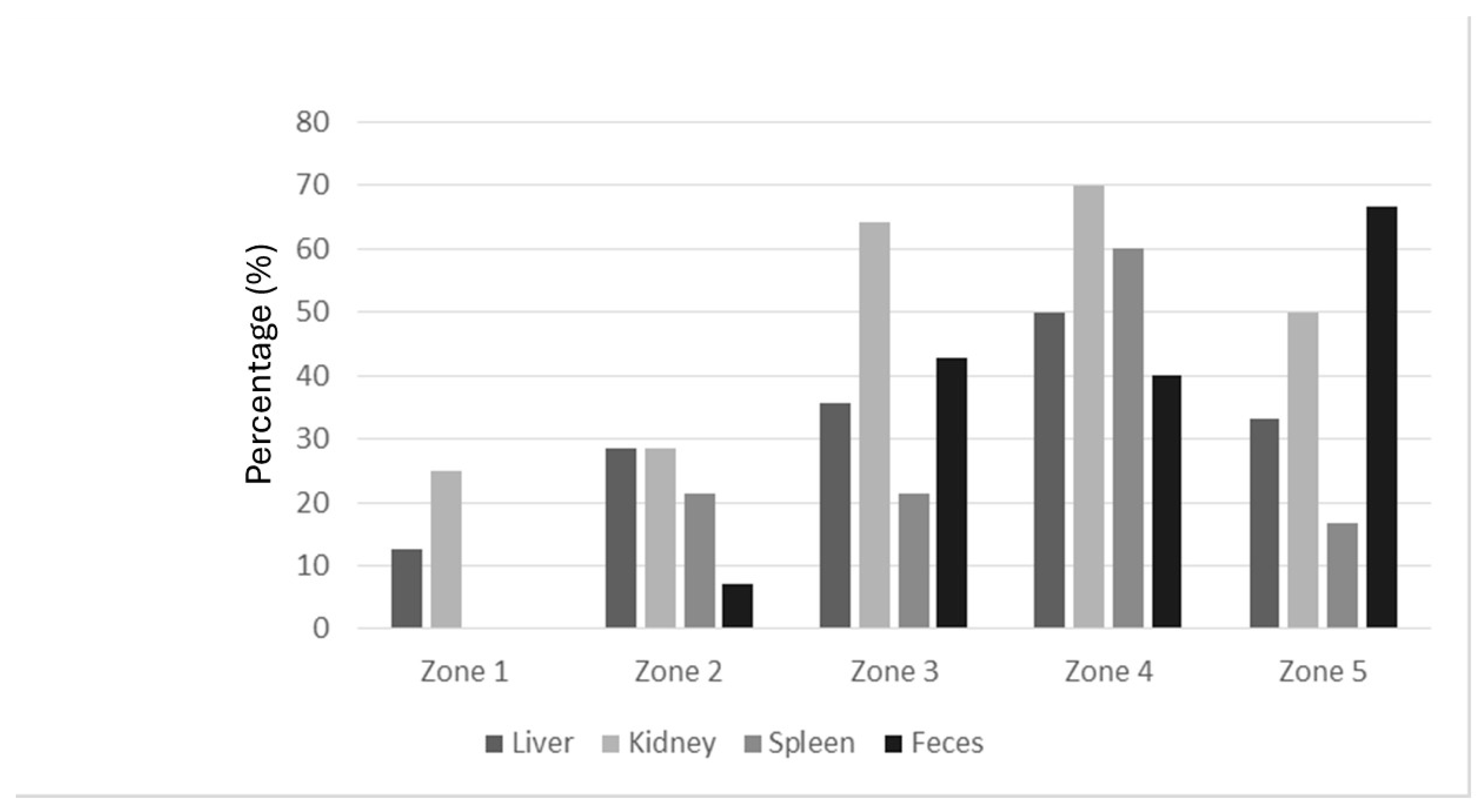
Figure 3.
Conceptual model of the ecology and dissemination of Piscirickettsia salmonis in Chilean marine ecosystems involving Odontesthes regia and environmental reservoirs.
Figure 3.
Conceptual model of the ecology and dissemination of Piscirickettsia salmonis in Chilean marine ecosystems involving Odontesthes regia and environmental reservoirs.
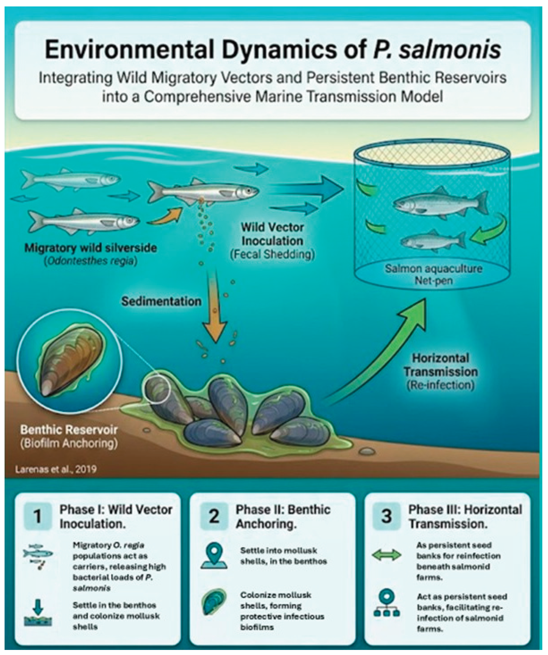
Table 1.
Standardized cycling protocol with respective temperatures and times used in the first and second amplification rounds.
Table 1.
Standardized cycling protocol with respective temperatures and times used in the first and second amplification rounds.
| Round | Step | Process | Temperature (°C) | Time (min) | No. of cycles |
| First amplification | 1 | Initial Denaturation | 94 | 2 | 1 |
| First amplification | 2 | Denaturation | 94 | 1 | 35 |
| First amplification | 3 | Annealing | 60 | 2 | |
| First amplification | 4 | Extension | 72 | 3 | |
| Second amplification | 1 | Initial Denaturation | 94 | 2 | 1 |
| Second amplification | 2 | Denaturation | 94 | 1 | 35 |
| Second amplification | 3 | Annealing | 65 | 2 | |
| Second amplification | 4 | Extension | 72 | 3 |
Table 3.
Association between P. salmonis positivity and sampling zones.
| Sampling Zone | Control Zone | Significance Level | Obtained p-value |
| Z1 | Z5 | p < 0.05 | 0.13753 |
| Z2 | Z5 | p < 0.05 | 0.61262 |
| Z3 | Z5 | p < 0.05 | 0.52105 |
| Z4 | Z5 | p < 0.05 | 1.00000 |
Table 4.
Risk assessment (Odds Ratio) of P. salmonis positivity in aquaculture zones relative to non-concessioned zones.
Table 4.
Risk assessment (Odds Ratio) of P. salmonis positivity in aquaculture zones relative to non-concessioned zones.
| Sampling Zone | Control Zone | OR Value | 95% Confidence Interval (CI) | Significance Level | Obtained p-value |
| Z1 | Z5 | 0.1200 | 0.0091 – 1.5844 | p < 0.05 | 0.1073 |
| Z2 | Z5 | 2.6000 | 0.1351 – 50.0513 | p < 0.05 | 0.5266 |
| Z3 | Z5 | 0.3600 | 0.0324 – 4.0057 | p < 0.05 | 0.4059 |
| Z4 | Z5 | 0.4667 | 0.0369 – 5.9028 | p < 0.05 | 0.5561 |
Disclaimer/Publisher’s Note: The statements, opinions and data contained in all publications are solely those of the individual author(s) and contributor(s) and not of MDPI and/or the editor(s). MDPI and/or the editor(s) disclaim responsibility for any injury to people or property resulting from any ideas, methods, instructions or products referred to in the content. |
© 2026 by the authors. Licensee MDPI, Basel, Switzerland. This article is an open access article distributed under the terms and conditions of the Creative Commons Attribution (CC BY) license (http://creativecommons.org/licenses/by/4.0/).
Copyright: This open access article is published under a Creative Commons CC BY 4.0 license, which permit the free download, distribution, and reuse, provided that the author and preprint are cited in any reuse.