
Submitted:
04 August 2026
Posted:
06 August 2026
You are already at the latest version
Abstract
Gynecologic cancers remain a major cause of cancer-related morbidity and mortality worldwide despite significant advances in surgery, chemotherapy, radiotherapy, tar-geted therapies, and immunotherapy. Signal Transducer and Activator of Transcription 3 (STAT3) is among the most consistently activated signaling pathways across cervical, ovarian and endometrial cancers, where it contributes to malignant transformation, disease progression, metastatic dissemination and therapeutic resistance. Traditionally, STAT3 oncogenic activity has been attributed to tumor-intrinsic functions, including regulation of proliferation, survival, angiogenesis and stemness. However, growing evidence indicates that STAT3 also participates in dynamic interactions between ma-lignant cells and the tumor microenvironment (TME), integrating inflammatory, meta-bolic and stromal signals that collectively shape disease progression. Rather than func-tioning as an isolated pathway, STAT3 operates within an interconnected regulatory network involving cytokines, hypoxia, metabolic intermediates, stromal cells and im-mune populations. Persistent STAT3 activation contributes to an immunosuppressive microenvironment by influencing macrophage polarization, myeloid-derived suppressor cell expansion, dendritic cell dysfunction, regulatory T-cell accumulation and impaired cytotoxic lymphocyte activity. Simultaneously, metabolic alterations characteristic of gynecologic malignancies — including enhanced glycolysis, lactate accumulation and hypoxia — further reinforce STAT3-dependent signaling across cellular compartments. In this review, we discuss STAT3 as a signaling node linking tumor metabolism with immune regulation in gynecologic cancers, summarize its participation in tumor–microenvironment communication, and examine recent advances in STAT3-targeted therapeutic strategies. We further discuss how a broader understanding of STAT3 biology supports biomarker-guided combination therapies and highlights key challenges lim-iting clinical translation, including the absence of validated predictive biomarkers and the need for improved patient stratification.
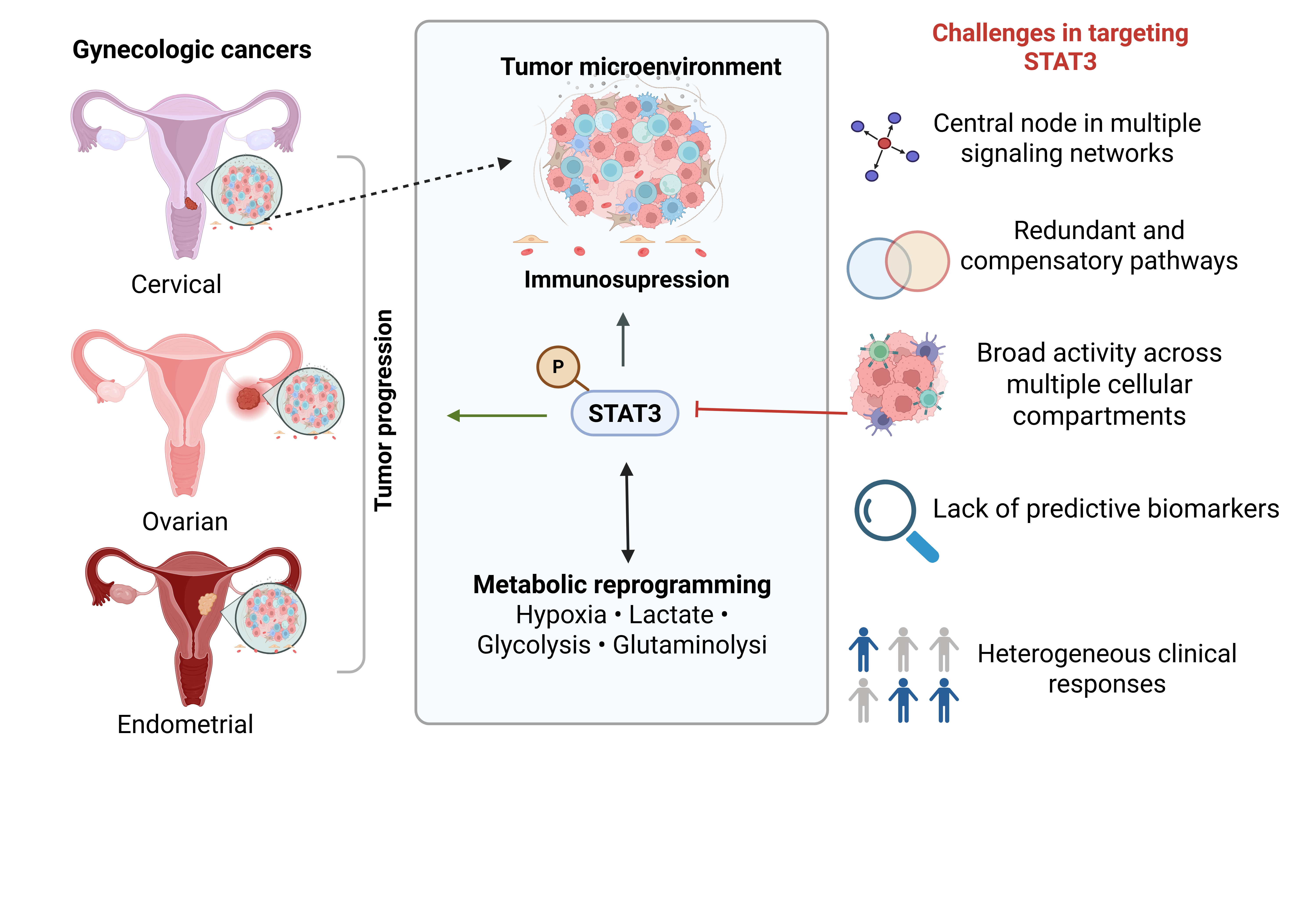
Keywords:
STAT3
; tumor microenvironment
; ovarian cancer
; cervical cancer
; endometrial cancer
; metabolism
; immunotherapy
; immune regulation
1. Introduction
Gynecologic cancers comprise a heterogeneous group of malignancies arising from the female reproductive tract, including cervical, ovarian, endometrial, vulvar and vaginal cancers. Collectively, they represent one of the leading causes of cancer incidence and mortality among women worldwide (Figure 1) [1], although their epidemiology, molecular drivers and clinical behavior differ substantially. Cervical cancer remains one of the most frequently diagnosed malignancies in low- and middle-income countries, where limited access to HPV vaccination and screening programs continues to contribute to high mortality rates [2]. Conversely, endometrial cancer predominates in developed countries and is increasing incidence in parallel with obesity and metabolic disorders [3], whereas ovarian cancer accounts for a disproportionate number of gynecologic cancer-related deaths owing to its asymptomatic progression, late diagnosis and frequent therapeutic resistance [4].
Despite these differences, gynecologic malignancies share several biological features that contribute to disease progression, including chronic inflammation, metabolic adaptation, immune evasion and extensive communication between tumor cells and their surrounding microenvironment. Increasing evidence indicates that these processes do not develop independently but rather emerge from continuous interactions between malignant cells, stromal populations and infiltrating immune cells [5]. Understanding the molecular pathways coordinating these interactions has therefore become a central objective in the development of more effective therapeutic strategies.
Among these pathways, STAT3 (Signal Transducer and Activator of Transcription 3) has attracted considerable attention. Initially characterized as a transcription factor activated downstream of cytokines and growth factors, STAT3 is now recognized as an important regulator of numerous biological processes associated with cancer progression, including cell proliferation, apoptosis resistance, epithelial–mesenchymal transition, angiogenesis, invasion and stemness [6]. Constitutive STAT3 activation is reported in multiple solid tumors and hematological malignancies, where elevated STAT3 activity frequently correlates with poor clinical outcomes and resistance to conventional therapies [7]. The interest in STAT3 as a therapeutic target is therefore well justified. Over the past two decades, substantial efforts have been devoted to developing strategies capable of inhibiting STAT3 signaling through diverse approaches, including upstream kinase inhibition, disruption of STAT3 dimerization, antisense oligonucleotides, and, more recently, targeted protein degradation [7]. Parallel advances in molecular pathology have further demonstrated that persistent STAT3 activation is common in cervical, ovarian and endometrial cancers, supporting its relevance across distinct gynecologic malignancies [8,9,10,11]. Nevertheless, accumulating evidence has also highlighted the remarkable complexity of STAT3 biology. Many of the biological processes influenced by STAT3—including inflammation, metabolic reprogramming, immune suppression and angiogenesis—are regulated by interconnected signaling networks involving NF-κB, PI3K/AKT, HIF-1α, MAPK and TGF-β, among others [12,13]. Rather than functioning independently, these pathways cooperate extensively, generating robust regulatory circuits that enable tumors to adapt to environmental stress while resisting therapeutic interventions. Within this network, STAT3 occupies a particularly interesting position because its activation extends beyond malignant epithelial cells to encompass multiple stromal and immune populations that constitute the tumor microenvironment. This broader perspective has shifted the way STAT3 is viewed on oncology. Instead of considering STAT3 exclusively as a tumor-cell signaling pathway, recent studies increasingly support its participation in reciprocal communication between malignant cells, cancer-associated fibroblasts, endothelial cells, myeloid populations and lymphocytes [14]. The result is a dynamic signaling network in which persistent STAT3 activation contributes to maintaining chronic inflammation, metabolic adaptation and immune dysfunction throughout the tumor microenvironment.
Metabolic reprogramming represents an important component of this network. Although altered glucose metabolism has traditionally been viewed as a hallmark of malignant cells, it is increasingly recognized that metabolites function as signaling molecules capable of influencing immune cell differentiation and function [15]. Lactate accumulation, hypoxia, nutrient competition and alterations in amino acid metabolism collectively shape the immune landscape of tumors through mechanisms involving multiple signaling pathways, including STAT3. Consequently, metabolism should no longer be regarded solely because of malignant transformation but also as an active participant in tumor–microenvironment communication [16].
These observations are particularly relevant for gynecologic cancers, where the composition of the local microenvironment differs substantially among tumor types. Ovarian cancer commonly develops within a peritoneal ecosystem that, in advanced-stage disease, is frequently marked by malignant ascites, abundant myeloid infiltrates and profound metabolic alterations, although ascites is not present in all patients and a substantial proportion present with solid peritoneal or extra-abdominal disease instead [17]. Cervical cancer evolves in the context of persistent HPV infection, chronic inflammation and progressive immune remodeling [2], while endometrial tumors frequently arise within metabolically dysregulated environments associated with obesity and insulin resistance [18]. Despite these differences, persistent STAT3 activation represents a common biological feature capable of integrating signals originating from both malignant and non-malignant compartments.
In this review, we discuss STAT3 within the broader context of the gynecologic tumor microenvironment. We first summarize canonical and non-canonical STAT3 signaling before examining its participation in metabolic adaptation and immune regulation across malignant and stromal compartments. We then discuss current advances in STAT3-targeted therapies and propose that considering STAT3 as an important signaling node linking tumor metabolism, immunity and stromal interactions provides a useful framework for developing rational combination strategies in gynecologic cancers.
2. STAT3: From Signaling Pathway to Integrator of the Tumor Microenvironment
Signal Transducer and Activator of Transcription 3 (STAT3) belong to the STAT family of transcription factors, which comprises seven structurally related proteins (STAT1, STAT2, STAT4, STAT5A, STAT5B and STAT6) that regulate embryonic development, tissue homeostasis, immune responses and cell differentiation [19]. Among them, STAT3 has attracted particular attention in oncology because of its persistent activation in a broad spectrum of solid tumors and hematological malignancies [20]. While transient STAT3 activation is essential for normal physiological responses, constitutive signaling has been consistently associated with tumor progression, immune dysfunction and resistance to therapy [21].
Under physiological conditions, STAT3 activation is initiated by a variety of extracellular stimuli, including cytokines, growth factors and hormones. Members of the IL-6 family represent the best-characterized activators, although interferons, epidermal growth factor (EGF), vascular endothelial growth factor (VEGF), platelet-derived growth factor (PDGF), hepatocyte growth factor (HGF), non-receptor tyrosine kinases and GPCR-associated signaling can also converge on STAT3 activation (Figure 2) [22]. Ligand binding induces receptor oligomerization and activation of Janus kinases (JAKs) or receptor-associated tyrosine kinases, promoting phosphorylation of STAT3 at tyrosine 705. Phosphorylated STAT3 forms homo- or heterodimers through reciprocal SH2-domain interactions and rapidly translocate to the nucleus, where it regulates transcription of genes involved in proliferation, survival, angiogenesis, inflammation and immune regulation [23] (Figure 2). STAT3 signaling is negatively regulated by SOCS proteins, which suppress JAK activity, and by PIAS proteins, which inhibit STAT3-dependent transcription [20].
Beyond this canonical mechanism, STAT3 also exerts important non-canonical functions. Alternative phosphorylation at serine 727, acetylation, methylation and other post-translational modifications influence transcriptional activity, mitochondrial function and interactions with additional signaling pathways [23]. Mitochondrial STAT3 (mtSTAT3) regulates oxidative phosphorylation, reactive oxygen species production and cellular metabolism, whereas unphosphorylated STAT3 (uSTAT3) participates in transcriptional regulation through interactions with transcription factors such as NF-κB (Figure 2) [24,25]. These non-canonical functions expand the biological role of STAT3 beyond conventional gene regulation and connect intracellular bioenergetics with adaptive transcriptional activity.
Collectively, canonical and non-canonical STAT3 signaling converge to regulate multiple hallmarks of cancer. In tumor cells, persistent STAT3 activation promotes proliferation, survival, angiogenesis, metastasis and metabolic reprogramming, whereas in immune cells it contributes to macrophage polarization, dendritic cell dysfunction, expansion of myeloid-derived suppressor cells and impaired antitumor immunity [21] (Figure 2). This functional versatility reflects the ability of STAT3 to integrate signals derived from cytokines, growth factors, metabolic stress and other signaling networks, positioning STAT3 as a central signaling hub within the tumor microenvironment.
In ovarian cancer models, STAT3 activation is positively associated with MMP-9 (Matrix Metalloproteinase-9) and MMP-2 (Matrix Metalloproteinase-2) expression [26,27], while STAT3 inhibition not only reduces cell migration but also decreases the invasive potential of endometrial cancer cells [28]. Constitutive STAT3 activation also induces epithelial–mesenchymal transition (EMT) by downregulating E-cadherin and upregulating mesenchymal markers such as vimentin, which are observed in ovarian, cervical and endometrial cancer [29,30,31]. In addition, STAT3 directly regulates transcriptional activator of vascular endothelial growth factor (VEGF) and hypoxia-inducible factor 1-alpha (HIF-1α) expression, which plays a central role in angiogenesis [32]. Inhibition of STAT3 activity reduces the expression of survivin, VEGF and vimentin, suppresses colony formation, viability, motility, and migration of resistant ovarian cancer cells [33]. Moreover, in vivo assays have shown that IL-6 increases angiogenic activity in human cervical cancer cells, through VEGF upregulation [34].
The extensive crosstalk between STAT3 and other signaling networks is particularly relevant in cancer. Activation of PI3K/AKT, MAPK, NF-κB, TGF-β and HIF-1α pathways frequently occurs alongside STAT3 signaling, generating positive feedback loops that reinforce inflammation, metabolic adaptation and therapeutic resistance [12,13]. IL-6-mediated STAT3 activation, for example, cooperates with NF-κB to maintain chronic inflammatory programs, whereas hypoxia promotes reciprocal activation of HIF-1α and STAT3, enhancing angiogenesis and metabolic remodeling [12]. Likewise, interactions between STAT3 and PI3K/AKT signaling contribute to tumor cell survival, while communication with TGF-β signaling influences epithelial-to-mesenchymal transition, extracellular matrix remodeling and immune suppression [16]. These interactions highlight an important concept for interpreting STAT3 biology in cancer. The biological effects commonly attributed to STAT3—including proliferation, angiogenesis, invasion and immune escape—are not exclusive to this pathway; instead, they emerge from coordinated activity among multiple interconnected signaling networks.
2.1. STAT3 in Metabolic Reprogramming
Metabolic reprogramming is a hallmark of cancer that extends far beyond supporting the energetic and biosynthetic demands of rapidly proliferating tumor cells. Alterations in glucose, glutamine and lipid metabolism continuously reshape the tumor microenvironment (TME), generating conditions characterized by hypoxia, nutrient competition, extracellular acidification and the accumulation of bioactive metabolites. These metabolic changes influence not only malignant cells but also stromal and immune populations, thereby regulating macrophage polarization, dendritic cell function, lymphocyte activity and fibroblast behavior [35,36,37].
STAT3 occupies a central position within this metabolic–immune network by integrating environmental cues into transcriptional and mitochondrial responses that coordinate tumor adaptation. Beyond its canonical transcriptional activity, STAT3 regulates cellular metabolism through both nuclear and non-canonical mitochondrial functions. Mitochondrial STAT3 (mtSTAT3) contributes to oxidative phosphorylation (OXPHOS), reactive oxygen species (ROS) homeostasis and mitochondrial respiration, whereas nuclear STAT3 promotes transcriptional programs involved in glycolysis, hypoxic adaptation and metabolic plasticity [38].
Glutamine metabolism is extensively explored in cancer therapy, and recent evidence highlights a positive regulatory loop between STAT3 and the glutaminolysis pathway in various tumors [16]. A recent study shows that glutamine levels relate to ovarian cancer (OVCA) invasiveness by specifically modulating the STAT3 pathway [39]. In highly invasive OVCA cells, Gln availability, unlike other amino acids, is essential to maintain STAT3 phosphorylation at both Y705 and S727 residues. Notably, the overexpression of constitutively active STAT3 is sufficient to rescue these cells from Gln-starvation-induced growth inhibition, confirming that STAT3 activation is the primary driver of Gln-mediated oncogenes. Furthermore, the non-transcriptional role of STAT3 within the mitochondria is vital for maintaining redox homeostasis in malignant cells [40]. Beyond its genomic activity, STAT3 manages the elevated production of mitochondrion-derived reactive oxygen species (ROS) typical of tumor transformation. While invasive cancers rely on glutamine-derived glutathione (GSH) to neutralize oxidative stress, inhibiting GSH synthesis reveals a synthetic lethal vulnerability strictly dependent on mitochondrial STAT3 [40].
Curiously, investigations in cervical (SiHa and HeLa) and breast cancer (MDA-MB-231) models demonstrate that STAT3 functions as a direct sensor of extracellular glutamine (Gln), coordinating cell proliferation independently of canonical Gln metabolism [41]. While Gln deprivation leads to metabolic collapse and growth arrest in these cells, STAT3 activation remains the limiting step for cellular rescue, primarily by regulating key transcriptional targets such as HIF-1alpha and c-Myc. Consequently, resistance to drugs targeting Gln uptake may be driven by growth factor-mediated STAT3 activation, suggesting that dual inhibition of STAT3 and Gln transporters could provide a more robust therapeutic outcome for these malignancies [41].
Moreover, STAT3 can transactivate genes that promote fatty acid oxidation, which can ameliorate oxidative stress and promote oxidative phosphorylation [42]. These data appear paradoxical when compared with data showing that STAT3 promotes aerobic glycolysis; however, these functions may coexist in a context-dependent manner. Notably, there is still limited evidence regarding the mitochondrial role of STAT3 in gynecological cancers. This gap likely reflects the limited number of studies rather than evidence against a mitochondrial role for STAT3 in gynecologic cancers.
2.1.1. Hypoxia and Metabolic Stress Reinforce Inflammatory Signaling
Progressive tumor growth inevitably generates regions of limited oxygen availability owing to inadequate vascularization and abnormal tissue architecture. Cellular adaptation to hypoxia is primarily coordinated by hypoxia-inducible factors (HIFs), which regulate angiogenesis, glucose metabolism, invasion and cell survival [43]. However, increasing evidence indicates that hypoxic adaptation depends on extensive interactions between HIF signaling and inflammatory pathways, including persistent STAT3 activation [43]. Reciprocal regulation between HIF-1 alpha and STAT3 has been described in several tumor models. STAT3 contributes to HIF-1 alpha transcriptional activity under hypoxic conditions, whereas HIF-dependent cytokine production further amplifies inflammatory signaling, generating positive feedback loops that sustain angiogenesis and metabolic adaptation [16]. This cooperation illustrates how metabolic stress is translated into coordinated transcriptional programs extending well beyond oxygen sensing alone. In ovarian cancer, hypoxia is particularly relevant because rapidly expanding peritoneal implants and malignant ascites generate spatially heterogeneous oxygen gradients that influence both tumor cells and infiltrating leukocytes [44]. Similarly, progressive vascular abnormalities during cervical cancer development contribute to localized hypoxia, further reinforcing inflammatory signaling and immune [45]. Moreover, in endometrial cancer, HIF-1 alpha expression is significantly more frequent in tumor than in normal endometrial tissue and correlates with higher histological grade, lymph node metastasis, myometrial invasion and reduced survival, indicating that hypoxic signaling is also clinically relevant in this tumor type [46]. These observations suggest that hypoxia should be considered not only a consequence of tumor growth but also an active regulator of cellular communication within the tumor microenvironment.
2.1.2. Lactate: From Metabolic By-Product to Immunoregulatory Mediator
Among the metabolites accumulating within tumors, lactate has emerged as one of the most influential regulators of the tumor microenvironment. Traditionally regarded as the product of aerobic glycolysis, lactate is now recognized as an active signaling molecule capable of influencing angiogenesis, extracellular matrix remodeling, immune cell differentiation and therapeutic responses [47].
High glycolytic activity combined with limited perfusion results in substantial lactate accumulation within many solid tumors, including ovarian and cervical cancers. Lactate is transported through monocarboxylate transporters (MCT1 and MCT4), allowing continuous metabolic exchange between neighboring cells while contributing to extracellular acidification [48]. These environmental changes influence virtually every component of the tumor microenvironment, extending the biological consequences of altered metabolism far beyond tumor cells themselves.
Growing evidence demonstrates that lactate affects immune populations through multiple complementary mechanisms. Elevated extracellular lactate impairs dendritic cell differentiation, reduces antigen presentation, limits cytotoxic T-cell activity and favors the development of regulatory immune populations [49]. Macrophages exposed to lactate acquire phenotypic characteristics associated with tissue remodeling and immune suppression, including increased production of IL-10, VEGF and other mediators that further reinforce chronic inflammation [50]. Although these responses involve several signaling pathways, including HIF-1 alpha, GPR81-mediated signaling and NF-κB, persistent STAT3 activation appears to participate in coordinating many of the downstream transcriptional programs associated with immunosuppressive macrophage polarization [47].
These observations are particularly relevant in gynecologic cancers. Ovarian cancer ascites contains high concentrations of lactate together with abundant immunosuppressive myeloid cells, creating conditions favorable for persistent inflammatory signaling and impaired antitumor immunity [51]. In cervical cancer, HPV-driven metabolic reprogramming further contributes to lactate production while chronic inflammation sustains cytokine networks capable of reinforcing STAT3 activity [52]. Furthermore, in endometrial cancer, tumor cells display increased aerobic glycolysis and lactate release, which drives M2 polarization of tumor-associated macrophages; these lactate-stimulated M2 macrophages secrete IL-6, promoting epithelial-mesenchymal transition, angiogenesis and invasiveness, and blockade of IL-6 signaling reduces these effects both in vitro and in vivo [53]. Together, these findings suggest that lactate should not be viewed simply as a metabolic consequence of tumor growth but rather as an important component of the signaling network linking metabolism with immune regulation. Rather than activating STAT3 in isolation, lactate participates in a broader signaling network in which metabolic, inflammatory and stromal pathways cooperate to shape cellular behavior.
3. STAT3, the Tumor Microenvironment and Immune Responses in Gynecologic Cancers
3.1. Stromal Cells Amplify STAT3-Dependent Communication Within the Tumor Microenvironment
The tumor microenvironment is maintained not only by malignant cells and infiltrating leukocytes but also by an extensive stromal compartment that actively contributes to tumor progression. Cancer-associated fibroblasts (CAFs), endothelial cells, mesothelial cells and adipocytes continuously exchange cytokines, growth factors, extracellular matrix components and metabolites with neighboring cells, creating a dynamic signaling network that supports chronic inflammation, tissue remodeling and immune dysfunction [14]. Rather than acting as passive structural elements, stromal cells actively influence tumor evolution and frequently represent important sources of persistent STAT3 activation.
Among stromal populations, CAFs have emerged as major regulators of the inflammatory tumor microenvironment. Activated fibroblasts produce high levels of IL-6, CXCL12, VEGF, TGF-β and matrix-remodeling enzymes, establishing paracrine signaling circuits that promote tumor cell proliferation, epithelial-to-mesenchymal transition (EMT), angiogenesis and therapeutic resistance. IL-6 is particularly relevant because it contributes to sustained JAK/STAT3 activation in both malignant cells and neighboring stromal populations, reinforcing inflammatory feedback loops that become progressively independent of the initiating oncogenic event [54,55] (Figure 3).
The contribution of CAFs appears particularly relevant in ovarian cancer, where progression through the peritoneum drives extensive stromal remodeling, including substantial accumulation of carcinoma-associated fibroblasts, largely arising from mesothelial-to-mesenchymal transition, that accompanies disease progression and metastatic dissemination throughout the peritoneal cavity [56]. In addition to producing inflammatory cytokines, CAFs modify extracellular matrix stiffness and organization, influencing both tumor cell invasion and immune cell trafficking. Similar observations have been reported in cervical cancer, where fibroblast activation contributes to progressive remodeling of the cervical stroma during HPV-associated carcinogenesis [57]. Moreover, in endometrial cancer, a CD146-positive CAF subpopulation similarly sustains STAT3 signaling: CD146+ CAF-derived IL-10 activates the JAK1/STAT3 pathway in tumor cells, promoting angiogenesis and vasculogenic mimicry, being associated with poor prognosis [58]. Although the composition of the stromal compartment differs among gynecologic malignancies, chronic inflammatory signaling involving STAT3 represents a common feature associated with disease progression.
Endothelial cells also participate actively in this communication network. Beyond their role in angiogenesis, activated endothelial cells regulate leukocyte recruitment, vascular permeability and local cytokine gradients. VEGF-driven angiogenesis, frequently associated with persistent STAT3 activation, generates structurally abnormal vasculature that contributes to hypoxia, heterogeneous drug delivery, high interstitial pressure and impaired immune cell infiltration [59]. These vascular abnormalities further reinforce metabolic stress and inflammatory signaling, illustrating the close relationship between stromal remodeling and immune regulation (Figure 3).
Collectively, these observations indicate that stromal cells should not be regarded merely as supporting elements of the tumor microenvironment. Instead, they actively participate in reciprocal signaling circuits that sustain chronic inflammation and influence the composition and function of immune infiltrates. Within these networks, STAT3 contributes to integrating inflammatory and metabolic signals originating from both malignant and stromal compartments.
3.2. Myeloid Cells: A Central Interface Between Metabolism and Immune Suppression
Macrophages, monocytes, dendritic cells, neutrophils and myeloid-derived suppressor cells continuously adapt their phenotype according to cytokine availability, oxygen tension and nutrient composition, making them highly sensitive to changes occurring within the tumor microenvironment. Consequently, these populations occupy a central position linking metabolic adaptation with immune regulation. In the TME, STAT3 promotes M2-like macrophages differentiation, induces the mobilization of myeloid-derived suppressor cells, drives a tolerogenic phenotype in dendritic cells while impairing their maturation, stimulates angiogenesis and epithelial–mesenchymal transition (Figure 3) [20,60]. Moreover, STAT3 transcriptional targets such as arginase 1 [61], PD-L1 (Programmed Death-Ligand 1), IDO (indoleamine-2,3-dioxygenase) and VEGF are part of the mechanisms by which this factor controls the TME [14,32,59,62,63].
Tumor-associated macrophages (TAMs) represent one of the most extensively studied myeloid populations in gynecologic cancers. Rather than existing as discrete M1 or M2 subsets, macrophages display remarkable functional plasticity and frequently acquire intermediate activation states shaped by the local microenvironment. Exposure to IL-6, IL-10, TGF-β, CSF-1, lactate and hypoxia collectively promote phenotypes associated with tissue remodeling, angiogenesis and suppression of adaptive immune responses [14].
In cervical cancer, tumor-derived IL-6 and PGE2 induce M2 polarization, which correlates with PD-L1 expression and poor prognosis [64]. In ovarian cancer, there is evidence that STAT3 is part of a crosstalk mechanism, where tumor cells induce the M2 phenotype, characterized by CD163, IL-6 and IL-10 expression, dependent on STAT3 activity, and macrophages on the other hand promote cell proliferation, through Cyclin D1 expression [17,65]. Furthermore, in ovarian cancer, macrophages frequently constitute one of the dominant immune populations within malignant ascites. These cells produce cytokines and growth factors that support tumor growth while simultaneously limiting effective antitumor immunity. Importantly, these observations reinforce the concept that STAT3 functions within a broader regulatory network rather than acting as an isolated determinant of macrophage behavior.
Myeloid-derived suppressor cells (MDSCs) further contribute to this immunosuppressive landscape. Expansion of MDSCs has been reported in both ovarian and cervical cancers and is associated with advanced disease, impaired T-cell responses and unfavorable clinical outcome [66,67]. Multiple inflammatory mediators contribute to MDSC recruitment and activation, including GM-CSF, G-CSF, IL-6 and VEGF, many of which signal through pathways intersecting with STAT3 (Figure 3). Once established within the tumor microenvironment, MDSCs suppress cytotoxic lymphocyte function through mechanisms involving arginine depletion, reactive oxygen species production, nitric oxide synthesis and secretion of immunosuppressive cytokines [68]. In endometrial cancer, MDSC expansion is similarly associated with a systemic inflammatory response, including thrombocytosis, leukocytosis and neutrophilia, and correlates with impaired antitumor immunity [69].
Dendritic cells are similarly influenced by the metabolic and inflammatory characteristics of the tumor microenvironment. Persistent exposure to IL-6, VEGF and lactate impairs dendritic cell maturation, antigen presentation and migration to draining lymph nodes, reducing the initiation of effective adaptive immune responses. Experimental studies suggest that STAT3 contributes to maintaining immature or tolerogenic dendritic cell phenotypes, although these effects are strongly influenced by additional environmental factors, including hypoxia and metabolic stress [70]. Consequently, alterations in dendritic cell function should be interpreted as the result of integrated signaling rather than STAT3 activation alone [70].
Neutrophils have also attracted increasing attention in gynecologic cancers, particularly in ovarian cancer, where neutrophil infiltration elevated and increased neutrophil-to-lymphocyte ratios correlate with poor prognosis [71]. Like other myeloid populations, neutrophils undergo functional reprogramming in response to inflammatory cytokines and metabolic alterations present within the tumor microenvironment. While the precise contribution of STAT3 to neutrophil biology remains incompletely understood, evidence suggests that STAT3 signaling influences neutrophil survival, recruitment and production of inflammatory mediators, further illustrating its participation in multiple components of the myeloid compartment. Moreover, recent evidence indicates that STAT3 is part of the tolerogenic mechaisms neutrophils play in the TME [72].
Taken together, these observations position myeloid cells as a critical interface connecting metabolic adaptation with immune suppression. Rather than responding independently to isolated cytokines or metabolites, these populations integrate multiple environmental signals that collectively shape antitumor immunity. Because STAT3 participates in several of these interconnected pathways, modulation of its activity has the potential to influence not only tumor cells but also the broader immune landscape.
3.3. Adaptive Immunity: STAT3 Shapes T-Cell Function Within the TME
The effectiveness of antitumor immunity depends on the ability of adaptive immune cells to recognize and eliminate malignant cells. However, T-cell responses develop within a microenvironment characterized by chronic inflammation, metabolic stress, hypoxia and persistent antigen exposure, conditions that progressively impair effector function while favoring immune tolerance. Rather than resulting from a single immunosuppressive mechanism, adaptive immune dysfunction reflects the cumulative influence of multiple stromal, metabolic and inflammatory signals acting simultaneously throughout tumor progression.
Among these mechanisms, chronic activation of cytokine signaling pathways has emerged as an important determinant of T-cell differentiation. Persistent exposure to IL-6, IL-10 and TGF-β alters the balance between effector and regulatory lymphocyte populations, while continuous antigen stimulation promotes progressive functional exhaustion characterized by reduced proliferative capacity, diminished cytokine production and increased expression of inhibitory receptors (Figure 3) [73]. The relationship between STAT3 and regulatory T cells (Tregs) illustrates this complexity. Multiple studies have associated constitutive STAT3 activation with increased recruitment or maintenance of immunosuppressive lymphocyte populations, although the mechanisms involved differ among tumor types and remain incompletely understood [74]. Importantly, Treg accumulation rarely depends on STAT3 signaling alone but instead reflects coordinated activity of inflammatory cytokines, chemokines and metabolic mediators present within the tumor microenvironment.
Cytotoxic CD8+ T lymphocytes are similarly influenced by the metabolic characteristics of the tumor microenvironment. Nutrient deprivation, hypoxia and extracellular acidification collectively impair T-cell activation and effector function while limiting proliferation and cytokine production. Lactate accumulation has received particular attention because elevated extracellular concentrations reduce T-cell motility, interfere with glycolytic metabolism and diminish cytotoxic activity [47]. Although these effects involve multiple molecular pathways, inflammatory signaling mediated by STAT3 contributes to maintaining the immunosuppressive environment that favors T-cell dysfunction (Figure 3).
Natural killer (NK) cells also undergo functional alterations during tumor progression. Reduced cytotoxicity, impaired cytokine production and diminished infiltration have been reported in several gynecologic malignancies, particularly advanced ovarian cancer [75]. Similar to T lymphocytes, NK-cell activity is influenced by cytokines, metabolites and stromal interactions that collectively shape the local immune landscape. Rather than acting directly on NK cells in isolation, STAT3 contributes to the broader inflammatory milieu that determines NK-cell activation and persistence within tumors [76].
These observations highlight an important principle of tumor immunology. Adaptive immune suppression is not generated by individual signaling pathways acting independently but emerges from continuous communication among tumor cells, stromal populations and innate immune cells (Figure 3). Within this network, STAT3 participates in several regulatory circuits that collectively influence lymphocyte recruitment, differentiation and function, reinforcing the interconnected nature of tumor–microenvironment communication.
4. Microbiota and Microbial Metabolites: Emerging Regulators of STAT3 Signaling
Interest in microbiota has expanded considerably over the past decade following the recognition that commensal microorganisms influence not only mucosal homeostasis but also systemic immunity, metabolism and therapeutic responses. Although much of the initial work focused on the intestinal microbiota, increasing evidence indicates that microbial communities associated with the female reproductive tract also contribute to the development and progression of gynecologic malignancies [77,78].
Persistent dysbiosis alters epithelial barrier function, inflammatory signaling and local metabolite production, generating conditions favorable for chronic immune activation. In cervical cancer, disruption of Lactobacillus-dominated microbial communities has been associated with persistent HPV infection, chronic inflammation and progressive cervical dysplasia [77,79]. While the mechanisms linking microbial composition to malignant transformation remain incompletely understood, altered production of microbial metabolites, including short-chain fatty acids and lactate, together with sustained inflammatory cytokine production, appears to contribute to the establishment of a permissive tumor microenvironment.
Vaginal and cervical dysbiosis are associated with cervical intraepithelial neoplasia (CIN, cervicla cancer precursor lesions) progression and persistence of high-risk HPV infection [80]. Dysbiotic microbiota enriched in Gardnerella, Atopobium, Prevotella, and Sneathia species correlates with reduced lactic acid–mediated barrier function, increased local pH, chronic inflammation and neoplastic transformation [81]. These dysbiotic communities promote the production of pro-inflammatory cytokines, including IL-6, IL-1β, and TNF-α, some of which, can activate STAT3 and NF-κB signaling pathways in cervical epithelial and immune cells [79].
In parallel, the endometrium, once considered sterile, has been shown to host distinct microbial communities composed mainly of Bacteroidetes, Firmicutes, Proteobacteria, and Actinobacteria [18]. Recent studies associate alterations in this microbiota with chronic inflammation that may contribute to endometrial carcinogenesis [82,83]. In addition to dysbiosis, excess of adipose tissue, an emerging factor associated with increased endometrial cancer mortality, contributes to increased hyperinsulinemia which stimulates endometrial proliferation and chronic inflammation. This inflammation also promotes STAT3 activation through IL-6 and leptin signaling, as well as NF-κB activation, via TNF-α, creating a feed-forward loop that amplifies cytokine production, insulin resistance, and aromatase activity [18].
According to recent studies, ovarian cancer pathogenesis is influenced by the composition of the vaginal microbiota. A study by Zhou and collaborators demonstrated that ovarian carcinoma tissues significantly reduced microbial diversity and an altered taxonomic composition compared with normal distal fallopian tube tissues, characterized by enrichment of Proteobacteria and depletion of Firmicutes, including Lactococcus-associated taxa. Importantly, these microbial alterations are associated with increased expression of TLR6, MYD88, and NLRP3, as well as activation of inflammation-associated signaling pathways, including cytokine–cytokine receptor interaction, chemokine signaling pathways and the NF-κB signaling pathway [84]. In parallel, case–control studies show that alterations in the cervicovaginal microbiome, particularly reduced Lactobacillus dominance, are associated with increased ovarian cancer risk, especially in genetically susceptible populations such as BRCA1 mutation carriers [85]. Therefore, microbiota imbalance may contribute to gynecological carcinogenesis by promoting chronic inflammation, a key driver of IL-6–dependent STAT3 activation in cancer [86].
5. Clinical Translation of STAT3-Targeted Therapies
The strong biological rationale supporting STAT3 as a therapeutic target has driven extensive drug development efforts over the past two decades. Constitutive STAT3 activation correlates with poor prognosis across multiple malignancies, including gynecologic cancers, and preclinical studies consistently demonstrate that pathway inhibition reduces tumor growth, limits metastatic dissemination and enhances antitumor immunity. Nevertheless, clinical translation has proven challenging, not because of limitations intrinsic to STAT3 itself, but because of the complexity of targeting a pathway active across multiple cellular compartments and physiological contexts, underscoring the need for adequate patient selection, pharmacodynamic assessment and a broader understanding of the tumor microenvironment in which STAT3 operates.
5.1. Therapeutic Strategies Targeting the STAT3 Pathway
Several complementary approaches have been developed to interfere with STAT3 signaling. Early efforts focused on indirect inhibition through blockade of upstream kinases, particularly JAK family members. Although these agents effectively reduce STAT3 phosphorylation, their biological effects extend well beyond the STAT3 pathway because JAK kinases regulate multiple cytokine receptors involved in normal immune homeostasis. Consequently, therapeutic efficacy must be balanced against potential systemic immunological effects [87]. Subsequent strategies sought to inhibit STAT3 more directly by preventing SH2-domain-mediated dimerization, thereby blocking nuclear translocation and transcriptional activity. Although several small molecules demonstrated encouraging preclinical activity, limitations related to specificity, pharmacokinetics and intracellular target engagement slowed clinical translation [87,88] Alternative approaches have included antisense oligonucleotides designed to reduce STAT3 expression and, more recently, targeted protein degraders capable of eliminating STAT3 rather than simply inhibiting its activity. These newer strategies represent an important evolution in the field, reflecting advances in medicinal chemistry and protein degradation technologies while offering potential advantages over first-generation inhibitors [89,90]. Collectively, these therapeutic approaches illustrate the continuous evolution of STAT3-targeted drug development. Rather than representing competing strategies, they provide complementary approaches aimed at overcoming distinct biological and pharmacological challenges associated with modulation of this pathway.
5.2. Lessons Emerging from Clinical Studies
Despite compelling preclinical evidence supporting STAT3 as a therapeutic target, clinical translation has remained disappointing. After nearly three decades of drug development efforts, no STAT3 inhibitor has received FDA approval for cancer treatment, and only a limited number of compounds have advanced to clinical testing [91]. These findings reveal a substantial disconnect between biological rationale and clinical efficacy.
Clinical studies evaluating STAT3-targeted therapies have generally demonstrated acceptable safety profiles and evidence of biological activity, although objective clinical responses have varied considerably among tumor types and treatment settings [87,88]. Importantly, these studies have generated valuable information extending beyond conventional efficacy endpoints by improving our understanding of STAT3 biology in human tumors. Early clinical studies evaluating STAT3-targeting approaches in gynecological and other malignancies have produced modest or negative results. In gynecological cancers, the phase II trial evaluating danvatirsen (AZD9150), an antisense oligonucleotide directed against STAT3, in malignant ascites associated with ovarian and gastrointestinal tumors (NCT02417753) was terminated prematurely because of poor patient accrual and did not generate efficacy data. Similarly, studies performed in other malignancies show limited clinical benefit. In a phase II trial evaluating danvatirsen combined with durvalumab (anti-PD-L1) in advanced solid tumors, including pancreatic ductal adenocarcinoma (PDAC), non-small cell lung cancer (NSCLC), and mismatch repair-deficient colorectal cancer (MRD-CRC) no objective responses were observed. Parallel preclinical analyses unexpectedly showed that danvatirsen did not exclusively suppress tumor-promoting STAT3 signaling. Danvatirsen-mediated STAT3 inhibition unexpectedly altered pancreatic stellate cells (PSCs) and myeloid populations without uniformly reversing the immunosuppressive tumor microenvironment [92]. Likewise, danvatirsen combined with acalabrutinib (irreversible covalent inhibitor of Bruton tyrosine kinase (BTK)) in diffuse large B-cell lymphoma resulted in a modest objective response rate and short median response duration [93].
A central obstacle to the clinical translation of STAT3-targeted therapies lies in the absence of a validated, standardized biomarker for patient stratification. Three candidate markers have been proposed, total STAT3, phosphorylated STAT3 (pSTAT3), and circulating IL-6, yet each reflects a distinct and context-dependent dimension of pathway activity. Beyond prognostic inconsistency, pSTAT3 measured by IHC is plagued by substantial technical variability: differences in antibody clones, fixation protocols, staining platforms, and the absence of a universally validated positivity cutoff generate discordant results across laboratories, making multicenter biomarker harmonization extremely difficult [94]. This instability extends to the tumor itself: Marginean and colaborators evaluated pSTAT3 expression by immunohistochemistry in paired primary colorectal tumors and corresponding liver metastases from 103 patients with metastatic disease. Although pSTAT3 expression was frequently detected in both primary tumors (71%) and metastatic lesions (76%), substantial discordance was observed between paired samples. Differences in pSTAT3 staining patterns were identified in 64% of cases, including loss of expression in 28% and gain of expression in 36% of liver metastases relative to the primary tumor. Furthermore, agreement between primary and metastatic sites was extremely poor, indicating that pSTAT3 activation status may change dynamically during tumor progression and metastatic dissemination [95]. In the same way, a meta-analysis of 4,513 breast cancer patients found no consistent relationship between STAT3 or pSTAT3 expression and overall survival, with paradoxical subgroup findings in which pSTAT3 overexpression associated with improved breast cancer-specific survival, underscoring that these markers carry divergent and tumor-context-dependent prognostic signals [96].
Circulating IL-6, meanwhile, behaves as a dynamic signal rather than a static stratification tool: IL-6 levels rose significantly at disease progression in patients treated with CDK4/6 inhibitors, and STAT3 inhibition was effective only in models where tumor-cell pSTAT3 was already detectable at baseline, highlighting that IL-6 reflects a temporally variable state rather than a fixed biomarker of pathway dependency [97]. Compounding these issues, no validated transcriptomic signature for “STAT3-activated tumors” exists across cancer types, a gap that is even more pronounced in gynecological cancers, where large-scale molecular profiling efforts defining STAT3-dependent subtypes remain absent, leaving clinicians without a reliable tool to identify patients most likely to benefit from pathway-targeted intervention.
Moving forward, it is increasingly clear that isolated pSTAT3 measurements are insufficient to capture the complexity of STAT3 biology in the TME. A composite biomarker panel, integrating tumor-cell pSTAT3, immune-compartment pSTAT3, serum IL-6, and PD-L1 expression, may offer a more biologically faithful strategy for patient stratification. Critically, future trials must address not only which analyte to measure, but in which compartment, by which validated platform, and at which threshold, since these choices have proven to be as consequential as the therapeutic intervention itself.
Other important lesson concerns patient heterogeneity. Constitutive STAT3 activation is not uniformly present across all tumors, nor does it necessarily reflect the same biological processes in different patients. Variability in cytokine production, stromal composition, immune infiltration and metabolic status may substantially influence dependence on STAT3 signaling. Consequently, therapeutic responses are likely to depend on biological context rather than pathway activation alone.
Finally, accumulating clinical experience reinforces an increasingly important concept in oncology: signaling pathways rarely function independently. Tumor progression is sustained by dynamic regulatory networks capable of compensating for inhibition of individual components. Consequently, therapeutic modulation of STAT3 is unlikely to eliminate all mechanisms supporting tumor growth but may substantially influence the broader signaling landscape when combined with complementary therapeutic strategies.
5.3. STAT3, Immunotherapy, and Dual-Mechanism Targeting
Immunotherapy is added to cancer treatment protocols, mostly as adjuvant, but in some cases as neoadjuvant therapy. The pleiotropic nature of STAT3 opens immunotherapy treatment opportunities. Due to its role in tumor cells and immune cells in TME, STAT3 targeting can be used for immune response modulation and tumor progression control [60].
As previously shown, STAT3 regulates immune checkpoint molecules in cancer cells and can enhance the effect of immune checkpoint blockade (ICB) therapy [60]. Contextually, ICB therapy combined with STAT3 inhibition shows promise in cancer therapy. For example, researchers demonstrate that treatment with RNAi-STAT3 and anti-PD-L1 in mouse tumor models coincide with significant tumor regression compared with RNAi-STAT3 treatment alone [98]. In this study, they also show that STAT3 silencing in a variety of immunosuppressive cell types are sufficient to convert a resistant tumor phenotype into “hot” TME with T-cell enriched phenotype [98]. Furthermore, animal models with pancreatic orthotopic tumors, resistant to anti-PD1 antibodies, show sensitivity to the immune checkpoint blockade (ICB) therapy and a significant increase in CD8+T cell infiltration in the TME after the use of JAK inhibitors [99]. They used Ruxolitinib as an inhibitor and showed that this molecule inhibits STAT1 and STAT3 phosphorylation in vivo, proving that the mechanism may be associated with STAT3 pathway in these tumor cells [99]. Although STAT3 inhibition is widely explored as a beneficial immunotherapeutic strategy, its role in CAR-T cell–based therapies appear to be distinct. A recent study using transcriptomic analyses of CD19 CAR-T cells from patients with chronic lymphocytic leukemia demonstrated that enhanced IL-6/STAT3 activity in CAR-T cells correlates with greater expansion, persistence, and anti-tumor activity, indicating that STAT3 signaling is necessary for efficacy of CAR-T cell-based therapy [100]. Still, the correlation between STAT3 signaling and immune checkpoint molecules provides a promising strategy to improve the efficacy of current immunotherapies.
Few studies explore the role of STAT3 inhibition as an immunotherapeutic strategy in gynecological cancers but given the critical influence of the tumor microenvironment (TME) on the development of these malignancies, targeting STAT3 may represent a promising approach to modulate immune responses and improve therapeutic outcomes. In cervical cancer, tumor progression is tightly linked to molecular pathways that drive immune evasion, many of which converge on STAT3. Supporting this, our group demonstrated, in an experimental HPV associated tumor model, that STAT3 inhibition impaired tumor growth and enhanced antitumor specific T lymphocyte responses [101]. In the same way, ovarian tumors display multiple immune evasion mechanisms as recruitment of regulatory T lymphocytes, M2 macrophages and PD-L1 expressions. Based on this, a group analyzed the expression of IL-6, an activator of STAT3, and its blockage in ovarian cancer biopsies and lineages. IL-6 produced by malignant ovarian cancer cells promotes inflammation, angiogenesis, macrophage infiltration, and poor prognosis. The antibody Siltuximab showed modest clinical activity but achieved prolonged disease stabilization in recurrent, platinum-resistant cases. Its effects were indirect, dependent on the tumor stroma, and patients with higher IL-6 expression tended to respond better [102].
Moreover, PARP inhibitors (PARPi), such as Olaparib, are highly effective against BRCA-mutant ovarian cancers, but many patients eventually develop resistance. Using a BRCA1-deficient ovarian cancer mouse model that mimic clinical acquired resistance, the researchers show that Olaparib treatment induces STAT3 phosphorylation and IL-6/JAK/STAT3 activation in ovarian tumor cells, resulting in secretion of chemokines (CCL2, CCL20, CX3CL1, CXCL1) that polarize macrophages toward an immunosuppressive phenotype. This STAT3-driven TAM recruitment and polarization suggested that overcoming immunosuppression could potentially restore PARPi sensitivity. In the same study, in vivo, STING activation increased antigen presentation, type I IFN signaling, dendritic cell activation, and reduced protumor macrophages in ascites and ovarian tumors [10].
Although STAT3 plays an important role in endometrial cancer biology, being associated with enhanced proliferation, resistance to apoptosis, invasion, and the establishment of an immunosuppressive tumor microenvironment, current evidence remains largely limited to mechanistic or correlative studies.
5.4. Metabolic Therapies
The close relationship between metabolism and immune regulation makes metabolic pathways attractive therapeutic partners for STAT3-targeted interventions. Tumor-associated lactate accumulation, hypoxia, glutamine metabolism and lipid remodeling influence both malignant cells and immune populations, creating a metabolically restrictive environment that limits effective antitumor immunity. Metabolic adaptation and inflammatory signaling reinforce one another within the tumor microenvironment, providing a mechanistic rationale for therapeutic strategies that simultaneously target metabolic pathways and STAT3-dependent inflammatory signaling, with the potential to modify multiple components of the tumor microenvironment while reducing the adaptive capacity of tumor cells. Among these approaches, inhibition of lactate transport through monocarboxylate transporters, modulation of glutamine utilization and interventions targeting tumor hypoxia have attracted increasing attention. Although relatively few studies have specifically evaluated these strategies in combination with STAT3 inhibition, their complementary biological effects provide a strong rationale for future investigation.
6. Future Perspectives
The understanding of STAT3 biology has evolved considerably over the past two decades. Initially viewed primarily as a transcription factor regulating tumor cell proliferation and survival, STAT3 is now recognized as an important participant in the reciprocal communication between malignant cells and the tumor microenvironment. This broader perspective has expanded the therapeutic rationale for targeting STAT3 while emphasizing the complexity of the biological networks in which it operates.
Several challenges remain. Greater understanding of the spatial and temporal regulation of STAT3 signaling, improved biomarkers for patient selection and pharmacodynamic monitoring, and integration of metabolic and immune profiling into clinical trial design will all contribute to refining future therapeutic strategies. Advances in spatial transcriptomics, multiplex imaging and single-cell technologies are expected to provide important insights into the cellular context of STAT3 activation across different gynecologic malignancies.
Rather than identifying a single optimal therapeutic strategy, future research should focus on understanding how STAT3 contributes to the biological organization of individual tumors. Such knowledge will facilitate the development of biomarker-guided combination therapies capable of simultaneously targeting malignant cells and the surrounding microenvironment while preserving normal tissue homeostasis.
In conclusion, STAT3 should not be viewed as an isolated oncogenic pathway but as an important signaling node operating within interconnected metabolic, stromal and immune networks. Appreciating this biological context does not diminish the importance of tumor-intrinsic STAT3 signaling; instead, it expands the opportunities for therapeutic intervention by recognizing that durable clinical responses will likely depend on coordinated modulation of the broader tumor microenvironment. This evolving perspective provides a useful framework for future studies aimed at improving the treatment of gynecologic cancers.
Author Contributions
Conceptualization, L.F.M., A.P.L. and F.C.C.; writing—original draft preparation, L.F.M., A.P.L. and F.C.C.; writing—review and editing, L.F.M., A.P.L., F.C.N. and J.A.L.L.; visualization, J.A.L.L.; supervision, A.P.L.; funding acquisition, A.P.L. All authors have read and agreed to the published version of the manuscript.
Funding
This research was funded by the São Paulo Research Foundation (FAPESP), grant number 2022/10388-5, and the National Council for Scientific and Technological Development (CNPq), grant number 310154/2021-9. L.F.M. was supported by a FAPESP scholarship (2024/03962-2), F.C.N. by a FAPESP scholarship 2025/12508-6 and F.C.C. and J.A.L.L. by scholarships from the Coordination for the Improvement of Higher Education Personnel (CAPES).
Institutional Review Board Statement
Not applicable.
Informed Consent Statement
Not applicable.
Data Availability Statement
No new data were created or analyzed in this study. Data sharing is not applicable to this article.
Acknowledgments
Figures were created with BioRender.com. During the preparation of this manuscript, the authors used ChatGPT (OpenAI, GPT-5.5) to assist with language editing, text refinement, and figure planning. The authors reviewed and edited all generated content and take full responsibility for the final version of the manuscript.
Conflicts of Interest
The authors declare no conflicts of interest.
References
- B. Zhu, H. Gu, Z. Mao, N.M. Beeraka, X. Zhao, M.P. Anand, Y. Zheng, R. Zhao, S. Li, P. Manogaran, R. Fan, V.N. Nikolenko, H. Wen, B. Basappa, J. Liu, Global burden of gynaecological cancers in 2022 and projections to 2050, J. Glob. Health 14 (2024). [CrossRef]
- B. Mlynarczyk-Bonikowska, L. Rudnicka, HPV Infections—Classification, Pathogenesis, and Potential New Therapies, Int. J. Mol. Sci. 25 (2024). [CrossRef]
- V.E. Rodriguez, S.P. Tanjasiri, A. Ro, M.A. Hoyt, R.E. Bristow, A.M.W. LeBrón, Trends in endometrial cancer incidence in the United States by race/ethnicity and age of onset from 2000 to 2019, Am. J. Epidemiol. 194 (2025) 103–113. [CrossRef]
- N. Wentzensen, K. Ring, B.K. Erickson, B. Reid, T. O’donnell, D. Check, S.S. Tworoger, P.P. Choudhury, Ovarian cancer risk prediction: a clinical epidemiology perspective, Am. J. Epidemiol. 194 (2025) 1182–1191. [CrossRef]
- P. Garg, S.K. Ramisetty, A. Raghu Subbalakshmi, B.M. Krishna, S. Pareek, A. Mohanty, P. Kulkarni, D. Horne, R. Salgia, S.S. Singhal, Gynecological cancer tumor Microenvironment: Unveiling cellular complexity and therapeutic potential, Biochem. Pharmacol. 229 (2024). [CrossRef]
- H.Q. Wang, Q.W. Man, F.Y. Huo, X. Gao, H. Lin, S.R. Li, J. Wang, F.C. Su, L. Cai, Y. Shi, B. Liu, L.L. Bu, STAT3 pathway in cancers: Past, present, and future, MedComm (Beijing). 3 (2022). [CrossRef]
- T. Zhang, C. Xiaohan, Unveiling the Role of JAK2/STAT3 signaling in chemoresistance of gynecological cancers: From mechanisms to therapeutic implications, Crit. Rev. Oncol. Hematol. 211 (2025). [CrossRef]
- C.L. Chen, F.C. Hsieh, J.C. Lieblein, J. Brown, C. Chan, J.A. Wallace, G. Cheng, B.M. Hall, J. Lin, Stat3 activation in human endometrial and cervical cancers, Br. J. Cancer 96 (2007) 591–599. [CrossRef]
- T.D. Strobel, M. Weber, N. Heber, A. Holzer, K. Hoppe-Seyler, F. Hoppe-Seyler, Revisiting the role of endogenous STAT3 in HPV-positive cervical cancer cells, J. Med. Virol. 95 (2023). [CrossRef]
- L. Ding, Q. Wang, A. Martincuks, M.J. Kearns, T. Jiang, Z. Lin, X. Cheng, C. Qian, S. Xie, H.J. Kim, I.M. Launonen, A. Färkkilä, T.M. Roberts, G.J. Freeman, J.F. Liu, P.A. Konstantinopoulos, U. Matulonis, H. Yu, J.J. Zhao, STING agonism overcomes STAT3-mediated immunosuppression and adaptive resistance to PARP inhibition in ovarian cancer, J. Immunother. Cancer 11 (2023). [CrossRef]
- R. Liang, X. Chen, L. Chen, F. Wan, K. Chen, Y. Sun, X. Zhu, STAT3 signaling in ovarian cancer: A potential therapeutic target, J. Cancer 11 (2020) 837–848. [CrossRef]
- D.E. Johnson, R.A. O’Keefe, J.R. Grandis, Targeting the IL-6/JAK/STAT3 signalling axis in cancer, Nat. Rev. Clin. Oncol. 15 (2018) 234–248. [CrossRef]
- H. Yu, D. Pardoll, R. Jove, STATs in cancer inflammation and immunity: A leading role for STAT3, Nat. Rev. Cancer 9 (2009) 798–809. [CrossRef]
- H. Yu, M. Kortylewski, D. Pardoll, Crosstalk between cancer and immune cells: Role of STAT3 in the tumour microenvironment, Nat. Rev. Immunol. 7 (2007) 41–51. [CrossRef]
- S. Nong, X. Han, Y. Xiang, Y. Qian, Y. Wei, T. Zhang, K. Tian, K. Shen, J. Yang, X. Ma, Metabolic reprogramming in cancer: Mechanisms and therapeutics, MedComm (Beijing). 4 (2023). [CrossRef]
- Y.J. Li, C. Zhang, A. Martincuks, A. Herrmann, H. Yu, STAT proteins in cancer: orchestration of metabolism, Nat. Rev. Cancer 23 (2023) 115–134. [CrossRef]
- S. Reinartz, T. Schumann, F. Finkernagel, A. Wortmann, J.M. Jansen, W. Meissner, M. Krause, A.M. Schwörer, U. Wagner, S. Müller-Brüsselbach, R. Müller, Mixed-polarization phenotype of ascites-associated macrophages in human ovarian carcinoma: Correlation of CD163 expression, cytokine levels and early relapse, Int. J. Cancer 134 (2014) 32–42. [CrossRef]
- W.C. Burkett, A.D. Clontz, T.O. Keku, V. Bae-Jump, The interplay of obesity, microbiome dynamics, and innovative anti-obesity strategies in the context of endometrial cancer progression and therapeutic approaches, Biochim. Biophys. Acta Rev. Cancer 1878 (2023). [CrossRef]
- N. Awasthi, C. Liongue, A.C. Ward, STAT proteins: a kaleidoscope of canonical and non-canonical functions in immunity and cancer, J. Hematol. Oncol. 14 (2021). [CrossRef]
- M.S. Wake, C.J. Watson, STAT3 the oncogene - Still eluding therapy?, FEBS Journal 282 (2015) 2600–2611. [CrossRef]
- S. Zou, Q. Tong, B. Liu, W. Huang, Y. Tian, X. Fu, Targeting stat3 in cancer immunotherapy, Mol. Cancer 19 (2020) 1–19. [CrossRef]
- M.A. Samad, I. Ahmad, A. Hasan, M.H. Alhashmi, A. Ayub, F.A. Al-Abbasi, A. Kumer, S. Tabrez, STAT3 Signaling Pathway in Health and Disease, MedComm (Beijing). 6 (2025). [CrossRef]
- D. Shi, J. Tao, S. Man, N. Zhang, L. Ma, L. Guo, L. Huang, W. Gao, Structure, function, signaling pathways and clinical therapeutics: The translational potential of STAT3 as a target for cancer therapy, Biochim. Biophys. Acta Rev. Cancer 1879 (2024). [CrossRef]
- E. Carbognin, R.M. Betto, M.E. Soriano, A.G. Smith, G. Martello, Stat3 promotes mitochondrial transcription and oxidative respiration during maintenance and induction of naive pluripotency, EMBO J. 35 (2016) 618–634. [CrossRef]
- C.D. Fernando, W.S.N. Jayasekara, C. Inampudi, M.R.J. Kohonen-Corish, W.A. Cooper, T.H. Beilharz, T.M. Josephs, D.J. Garama, D.J. Gough, A STAT3 protein complex required for mitochondrial mRNA stability and cancer, Cell Rep. 42 (2023). [CrossRef]
- Z.H. Jia, Y. Jia, F.J. Guo, J. Chen, X.W. Zhang, M.H. Cui, Phosphorylation of STAT3 at Tyr705 regulates MMP-9 production in epithelial ovarian cancer, PLoS One 12 (2017). [CrossRef]
- J.M. Seo, S. Park, J.H. Kim, Leukotriene B 4 receptor-2 promotes invasiveness and metastasis of ovarian cancer cells through signal transducer and activator of transcription 3 (STAT3)-dependent up-regulation of matrix metalloproteinase 2, Journal of Biological Chemistry 287 (2012) 13840–13849. [CrossRef]
- Q. Che, X. Xiao, M. Liu, Y. Lu, X. Dong, S. Liu, IL-6 promotes endometrial cancer cells invasion and migration through signal transducers and activators of transcription 3 signaling pathway, Pathol. Res. Pract. 215 (2019). [CrossRef]
- G. Zhang, S. Hou, S. Li, Y. Wang, W. Cui, Role of STAT3 in cancer cell epithelial-mesenchymal transition (Review), Int. J. Oncol. 64 (2024). [CrossRef]
- J.W. Miao, L.J. Liu, J. Huang, Interleukin-6-induced epithelial-mesenchymal transition through signal transducer and activator of transcription 3 in human cervical carcinoma, Int. J. Oncol. 45 (2014) 165–176. [CrossRef]
- M. Van Der Zee, A. Sacchetti, M. Cansoy, R. Joosten, M. Teeuwssen, C. Heijmans-Antonissen, P.C. Ewing-Graham, C.W. Burger, L.J. Blok, R. Fodde, IL6/JAK1/STAT3 signaling blockade in endometrial cancer affects the ALDHhi/CD126+ stem-like component and reduces tumor burden, Cancer Res. 75 (2015) 3608–3622. [CrossRef]
- M. Tolomeo, A. Cascio, The multifaced role of stat3 in cancer and its implication for anticancer therapy, Int. J. Mol. Sci. 22 (2021) 1–25. [CrossRef]
- P. Yue, X. Zhang, D. Paladino, B. Sengupta, S. Ahmad, R.W. Holloway, S.B. Ingersoll, J. Turkson, Hyperactive EGF receptor, Jaks and Stat3 signaling promote enhanced colony-forming ability, motility and migration of cisplatin-resistant ovarian cancer cells, Oncogene 31 (2012) 2309–2322. [CrossRef]
- L.-H. Wei, M.-L. Kuo, C.-A. Chen, C.-H. Chou, K.-B. Lai, C.-N. Lee, C.-Y. Hsieh, Interleukin-6 promotes cervical tumor growth by VEGF-dependent angiogenesis via a STAT3 pathway, Oncogene 22 (2003) 1517–1527.
- R.J. DeBerardinis, J.J. Lum, G. Hatzivassiliou, C.B. Thompson, The Biology of Cancer: Metabolic Reprogramming Fuels Cell Growth and Proliferation, Cell Metab. 7 (2008) 11–20. [CrossRef]
- L. Li, Y. Tian, The role of metabolic reprogramming of tumor-associated macrophages in shaping the immunosuppressive tumor microenvironment, Biomedicine and Pharmacotherapy 161 (2023). [CrossRef]
- H. Lv, X. Han, Y. Yang, J. Shen, Y. Yin, Y. Liu, Glutamine: a new strategy for targeted metabolic therapy in the tumor microenvironment, Cell Death Discov. 11 (2025). [CrossRef]
- D.J. Gough, A. Corlett, K. Schlessinger, J. Wegrzyn, A.C. Larner, D.E. Levy, Mitochondrial STAT3 supports RasDependent oncogenic transformation, Science (1979). 324 (2009) 1713–1716. [CrossRef]
- L. Yang, T. Moss, L.S. Mangala, J. Marini, H. Zhao, S. Wahlig, G. Armaiz-Pena, D. Jiang, A. Achreja, J. Win, R. Roopaimoole, C. Rodriguez-Aguayo, I. Mercado-Uribe, G. Lopez-Berestein, J. Liu, T. Tsukamoto, A.K. Sood, P.T. Ram, D. Nagrath, Metabolic shifts toward glutamine regulate tumor growth, invasion and bioenergetics in ovarian cancer, Mol. Syst. Biol. 10 (2014). [CrossRef]
- D.J. Garama, T.J. Harris, C.L. White, F.J. Rossello, M. Abdul-Hay, D.J. Gough, D.E. Levy, A Synthetic Lethal Interaction between Glutathione Synthesis and Mitochondrial Reactive Oxygen Species Provides a Tumor-Specific Vulnerability Dependent on STAT3, Mol. Cell. Biol. 35 (2015) 3646–3656. [CrossRef]
- Cacace, M. Sboarina, T. Vazeille, P. Sonveaux, Glutamine activates STAT3 to control cancer cell proliferation independently of glutamine metabolism, Oncogene 36 (2017) 2074–2084. [CrossRef]
- Tošić, D.A. Frank, STAT3 as a mediator of oncogenic cellular metabolism: Pathogenic and therapeutic implications, Neoplasia (United States) 23 (2021) 1167–1178. [CrossRef]
- G.L. Semenza, HIF-1 mediates metabolic responses to intratumoral hypoxia and oncogenic mutations, Journal of Clinical Investigation 123 (2013) 3664–3671. [CrossRef]
- E. Kipps, D.S.P. Tan, S.B. Kaye, Meeting the challenge of ascites in ovarian cancer: New avenues for therapy and research, Nat. Rev. Cancer 13 (2013) 273–282. [CrossRef]
- Datta, C. West, J.P.B. O’Connor, A. Choudhury, P. Hoskin, Impact of hypoxia on cervical cancer outcomes, Int. J. Gynecol. Cancer 31 (2021) 1459–1470. [CrossRef]
- P. Zhu, L. Shen, Q. Ren, Q. Zeng, X. He, Prognostic and Clinicopathological Significance of Hypoxia-Inducible Factor-1α in Endometrial Cancer: A Meta-Analysis, Front. Oncol. 10 (2020). [CrossRef]
- T. Zhang, W. Zhou, T. Fan, Y. Yuan, X. Tang, Q. Zhang, J. Zou, Y. Li, Lactic acid metabolism: gynecological cancer’s Achilles’ heel, Discover Oncology 16 (2025). [CrossRef]
- C. Manosalva, J. Quiroga, A.I. Hidalgo, P. Alarcón, N. Anseoleaga, M.A. Hidalgo, R.A. Burgos, Role of Lactate in Inflammatory Processes: Friend or Foe, Front. Immunol. 12 (2022). [CrossRef]
- X.Y. Gu, J.L. Yang, R. Lai, Z.J. Zhou, D. Tang, L. Hu, L.J. Zhao, Impact of lactate on immune cell function in the tumor microenvironment: mechanisms and therapeutic perspectives, Front. Immunol. 16 (2025). [CrossRef]
- Y. Gou, H. Wang, T. Wang, H. Wang, B. Wang, N. Jiao, Y. Yu, Y. Cao, H. Wang, Z. Zhang, Ectopic endometriotic stromal cells-derived lactate induces M2 macrophage polarization via Mettl3/Trib1/ERK/STAT3 signalling pathway in endometriosis, Immunology 168 (2023) 389–402. [CrossRef]
- Y. Gong, J. Yang, Y. Wang, L. Xue, J. Wang, Metabolic factors contribute to T-cell inhibition in the ovarian cancer ascites, Int. J. Cancer 147 (2020) 1768–1777. [CrossRef]
- B. Li, L. Sui, Metabolic reprogramming in cervical cancer and metabolomics perspectives, Nutr. Metab. (Lond). 18 (2021). [CrossRef]
- X. Liu, H. Sun, J. Liang, H. Yu, M. Xue, Y. Li, W. Li, Y. Xu, B. Ling, D. Feng, Metabolic interplay between endometrial cancer and tumor-associated macrophages: lactate-induced M2 polarization enhances tumor progression, J. Transl. Med. 23 (2025). [CrossRef]
- Y. Cheng, H. Li, Y. Deng, Y. Tai, K. Zeng, Y. Zhang, W. Liu, Q. Zhang, Y. Yang, Cancer-associated fibroblasts induce PDL1+ neutrophils through the IL6-STAT3 pathway that foster immune suppression in hepatocellular carcinoma, Cell Death Dis. 9 (2018). [CrossRef]
- L. Wang, F. Zhang, J.Y. Cui, L. Chen, Y.T. Chen, B.W. Liu, CAFs enhance paclitaxel resistance by inducing EMT through the IL-6/JAK2/STAT3 pathway, Oncol. Rep. 39 (2018) 2081–2090. [CrossRef]
- L. Pascual-antón, B. Cardeñes, R. Sainz de la Cuesta, L. González-cortijo, M. López-cabrera, C. Cabañas, P. Sandoval, Mesothelial-to-mesenchymal transition and exosomes in peritoneal metastasis of ovarian cancer, Int. J. Mol. Sci. 22 (2021). [CrossRef]
- Fullár, J. Dudás, L. Oláh, P. Hollósi, Z. Papp, G. Sobel, K. Karászi, S. Paku, K. Baghy, I. Kovalszky, Remodeling of extracellular matrix by normal and tumor-associated fibroblasts promotes cervical cancer progression, BMC Cancer 15 (2015). [CrossRef]
- Z. Yu, Q. Zhang, S. Wei, Y. Zhang, T. Zhou, Q. Zhang, R. Shi, D. Zinovkin, Z.I. Pranjol, J. Zhang, H. Wang, CD146+CAFs promote progression of endometrial cancer by inducing angiogenesis and vasculogenic mimicry via IL-10/JAK1/STAT3 pathway, Cell Communication and Signaling 22 (2024). [CrossRef]
- S.-H. Chen, D.A. Murphy, W. Lassoued, G. Thurston, M.D. Feldman, W.M.F. Lee, Activated STAT3 is a mediator and biomarker of VEGF endothelial activation, n.d. www.landesbioscience.com/supplement/ChenCBT7-12-Sup.pdf.
- S. Zou, Q. Tong, B. Liu, W. Huang, Y. Tian, X. Fu, Targeting stat3 in cancer immunotherapy, Mol. Cancer 19 (2020) 1–19. [CrossRef]
- D. Vasquez-Dunddel, F. Pan, Q. Zeng, M. Gorbounov, E. Albesiano, J. Fu, R.L. Blosser, A.J. Tam, T. Bruno, H. Zhang, D. Pardoll, Y. Kim, STAT3 regulates arginase-i in myeloid-derived suppressor cells from cancer patients, Journal of Clinical Investigation 123 (2013) 1580–1589. [CrossRef]
- U.M. Litzenburger, C.A. Opitz, F. Sahm, K.J. Rauschenbach, S. Trump, M. Winter, M. Ott, K. Ochs, C. Lutz, X. Liu, N. Anastasov, I. Lehmann, T. Höfer, A. Von Deimling, W. Wick, M. Platten, Constitutive IDO expression in human cancer is sustained by an autocrine signaling loop involving IL-6, STAT3 and the AHR, n.d. www.impactjournals.com/oncotarget/.
- T. Wang, G. Niu, M. Kortylewski, L. Burdelya, K. Shain, S. Zhang, R. Bhattacharya, D. Gabrilovich, R. Heller, D. Coppola, W. Dalton, R. Jove, D. Pardoll, H. Yu, Regulation of the innate and adaptive immune responses by Stat-3 signaling in tumor cells, Nat. Med. 10 (2004) 48–54. [CrossRef]
- M. Heusinkveld, P.J. de Vos van Steenwijk, R. Goedemans, T.H. Ramwadhdoebe, A. Gorter, M.J.P. Welters, T. van Hall, S.H. van der Burg, M2 Macrophages Induced by Prostaglandin E2 and IL-6 from Cervical Carcinoma Are Switched to Activated M1 Macrophages by CD4+ Th1 Cells, The Journal of Immunology 187 (2011) 1157–1165. [CrossRef]
- K. Takaishi, Y. Komohara, H. Tashiro, H. Ohtake, T. Nakagawa, H. Katabuchi, M. Takeya, Involvement of M2-polarized macrophages in the ascites from advanced epithelial ovarian carcinoma in tumor progression via Stat3 activation, Cancer Sci. 101 (2010) 2128–2136. [CrossRef]
- D. Jianyi, G. Haili, Y. Bo, Y. Meiqin, H. Baoyou, H. Haoran, L. Fang, Z. Qingliang, H. Lingfei, Myeloid-derived suppressor cells cross-talk with B10 cells by BAFF/BAFF-R pathway to promote immunosuppression in cervical cancer, Cancer Immunology, Immunotherapy 72 (2023) 73–85. [CrossRef]
- X. Li, J. Wang, W. Wu, H. Gao, N. Liu, G. Zhan, L. Li, L. Han, X. Guo, Myeloid-derived suppressor cells promote epithelial ovarian cancer cell stemness by inducing the CSF2/p-STAT3 signalling pathway, FEBS Journal 287 (2020) 5218–5235. [CrossRef]
- D.I. Gabrilovich, Myeloid-derived suppressor cells, Cancer Immunol. Res. 5 (2017) 3–8. [CrossRef]
- E. Yokoi, S. Mabuchi, N. Komura, K. Shimura, H. Kuroda, K. Kozasa, R. Takahashi, T. Sasano, M. Kawano, Y. Matsumoto, M. Kodama, K. Hashimoto, K. Sawada, T. Kimura, The role of myeloid-derived suppressor cells in endometrial cancer displaying systemic inflammatory response: clinical and preclinical investigations, Oncoimmunology 8 (2019). [CrossRef]
- Y. Nefedova, M. Huang, S. Kusmartsev, R. Bhattacharya, P. Cheng, R. Salup, R. Jove, D. Gabrilovich, Hyperactivation of STAT3 Is Involved in Abnormal Differentiation of Dendritic Cells in Cancer, 2004. https://academic.oup.com/jimmunol/article/172/1/464/8033598.
- Q. Zhou, L. Hong, M.-Z. Zuo, Z. He, Prognostic significance of neutrophil to lymphocyte ratio in ovarian cancer: evidence from 4,910 patients, 2017. www.impactjournals.com/oncotarget.
- Ozel, G. Sha, A. Będzińska, E. Pylaeva, Y. Naumova, I. Thiel, J. Antczak, A. Squire, M. Gunzer, G. Zelinskyy, C. Kürten, S. Lang, C. Silvestre-Roig, M. Kortylewski, Z. Granot, J. Jablonska, Neutrophil-specific targeting of STAT3 impairs tumor progression via the expansion of cytotoxic CD8+ T cells, Signal Transduct. Target. Ther. 10 (2025). [CrossRef]
- Chaudhry, D. Rudra, P. Treuting, R.M. Samstein, Y. Liang, A. Kas, A.Y. Rudensky, CD4+ regulatory T cells control TH17 responses in a stat3-dependent manner, Science (1979). 326 (2009) 986–991. [CrossRef]
- A.J. Oweida, L. Darragh, A. Phan, D. Binder, S. Bhatia, A. Mueller, B. Van Court, D. Milner, D. Raben, R. Woessner, L. Heasley, R. Nemenoff, E. Clambey, S.D. Karam, STAT3 Modulation of Regulatory T Cells in Response to Radiation Therapy in Head and Neck Cancer, J. Natl. Cancer Inst. 111 (2019) 1339–1349. [CrossRef]
- Ning, L. Yao, Natural killer cell exhaustion in ovarian cancer: from molecular suppression to therapeutic revival, Front. Immunol. 16 (2026). [CrossRef]
- M. Tolomeo, A. Cascio, The multifaced role of stat3 in cancer and its implication for anticancer therapy, Int. J. Mol. Sci. 22 (2021) 1–25. [CrossRef]
- Ravel, P. Gajer, Z. Abdo, G.M. Schneider, S.S.K. Koenig, S.L. McCulle, S. Karlebach, R. Gorle, J. Russell, C.O. Tacket, R.M. Brotman, C.C. Davis, K. Ault, L. Peralta, L.J. Forney, Vaginal microbiome of reproductive-age women, Proc. Natl. Acad. Sci. U. S. A. 108 (2011) 4680–4687. [CrossRef]
- C. Ceccarani, C. Foschi, C. Parolin, A. D’Antuono, V. Gaspari, C. Consolandi, L. Laghi, T. Camboni, B. Vitali, M. Severgnini, A. Marangoni, Diversity of vaginal microbiome and metabolome during genital infections, Sci. Rep. 9 (2019). [CrossRef]
- E. Caselli, M. D’Accolti, E. Santi, I. Soffritti, S. Conzadori, S. Mazzacane, P. Greco, C. Contini, G. Bonaccorsi, Vaginal Microbiota and Cytokine Microenvironment in HPV Clearance/Persistence in Women Surgically Treated for Cervical Intraepithelial Neoplasia: An Observational Prospective Study, Front. Cell. Infect. Microbiol. 10 (2020). [CrossRef]
- Mei, T. Wang, Y. Chen, D. Wei, Y. Zhang, T. Cui, J. Meng, X. Zhang, Y. Liu, L. Ding, X. Niu, Dysbiosis of vaginal microbiota associated with persistent high-risk human papilloma virus infection, J. Transl. Med. 20 (2022). [CrossRef]
- Z.W. Zhou, H.Z. Long, Y. Cheng, H.Y. Luo, D.D. Wen, L.C. Gao, From Microbiome to Inflammation: The Key Drivers of Cervical Cancer, Front. Microbiol. 12 (2021). [CrossRef]
- M.R.S. Walther-António, J. Chen, F. Multinu, A. Hokenstad, T.J. Distad, E.H. Cheek, G.L. Keeney, D.J. Creedon, H. Nelson, A. Mariani, N. Chia, Potential contribution of the uterine microbiome in the development of endometrial cancer, Genome Med. 8 (2016). [CrossRef]
- Moreno, F.M. Codoñer, F. Vilella, D. Valbuena, J.F. Martinez-Blanch, J. Jimenez-Almazán, R. Alonso, P. Alamá, J. Remohí, A. Pellicer, D. Ramon, C. Simon, Evidence that the endometrial microbiota has an effect on implantation success or failure, in: Am. J. Obstet. Gynecol., Mosby Inc., 2016: pp. 684–703. [CrossRef]
- B. Zhou, C. Sun, J. Huang, M. Xia, E. Guo, N. Li, H. Lu, W. Shan, Y. Wu, Y. Li, X. Xu, D. Weng, L. Meng, J. Hu, Q. Gao, D. Ma, G. Chen, The biodiversity Composition of Microbiome in Ovarian Carcinoma Patients, Sci. Rep. 9 (2019). [CrossRef]
- N.R. Nené, D. Reisel, A. Leimbach, D. Franchi, A. Jones, I. Evans, S. Knapp, A. Ryan, S. Ghazali, J.F. Timms, T. Paprotka, L. Bjørge, M. Zikan, D. Cibula, N. Colombo, M. Widschwendter, Association between the cervicovaginal microbiome, BRCA1 mutation status, and risk of ovarian cancer: a case-control study, Lancet Oncol. 20 (2019) 1171–1182. [CrossRef]
- K. Javadi, E. Ferdosi-Shahandashti, M. Rajabnia, M. Khaledi, Vaginal microbiota and gynecological cancers: a complex and evolving relationship, Infect. Agent. Cancer 19 (2024). [CrossRef]
- K. Berkley, J. Zalejski, A. Sharma, Targeting STAT3 for Cancer Therapy: Focusing on Y705, S727, or Dual Inhibition? Cancers (Basel). 17 (2025). [CrossRef]
- A.M. Tsimberidou, D.J. Vining, S.P. Arora, S. de Achaval, J. Larson, J. Kauh, C. Cartwright, R. Avritscher, I. Alibhai, D.J. Tweardy, A.O. Kaseb, Phase I Trial of TTI-101, a First-in-Class Oral Inhibitor of STAT3, in Patients with Advanced Solid Tumors, Clinical Cancer Research 31 (2025) 965–974. [CrossRef]
- R.K. Acharyya, Y. Kothapalli, S. Yarlagadda, C.K. De, S.R. Allu, J. Roy, R.K. Rej, An Update on Clinically Advanced PROTAC Degraders and Their Synthesis, Molecules 31 (2026). [CrossRef]
- R.T.U. Haid, A. Reichel, PK/PD modeling of targeted protein degraders: Charting new waters and navigating the shallows, Drug Discov. Today 30 (2025). [CrossRef]
- Z. Kang, S. Li, Y. Li, J. Song, Y. Peng, Y. Chen, Small molecular inhibitors and degraders targeting STAT3 for cancer therapy: An updated review (from 2022 to 2024), Chinese Chemical Letters 36 (2025). [CrossRef]
- C. Tang, G.P. Hartley, C. Couillault, Y. Yuan, H. Lin, C. Nicholas, A. Srinivasamani, J. Dai, E.E.I. Dumbrava, S. Fu, D.D. Karp, A. Naing, S.A. Piha-Paul, J. Rodon Ahnert, S. Pant, V. Subbiah, T.A. Yap, A.M. Tsimberidou, P. Guerrero, S. Dhebat, T. Proia, M.A. Curran, D.S. Hong, Preclinical study and parallel phase II trial evaluating antisense STAT3 oligonucleotide and checkpoint blockade for advanced pancreatic, non-small cell lung cancer and mismatch repair-deficient colorectal cancer, BMJ Oncology 3 (2024). [CrossRef]
- Roschewski, M.R. Patel, P.M. Reagan, N.S. Saba, G.P. Collins, H.T. Arkenau, S. de Vos, B. Nuttall, M. Acar, K. Burke, R.D. White, M. Udriste, S. Sharma, B. Dougherty, D. Stetson, D. Jenkins, A. Mortlock, A. Forcina, V. Munugalavadla, I. Flinn, Phase I Study of Acalabrutinib Plus Danvatirsen (AZD9150) in Relapsed/Refractory Diffuse Large B-Cell Lymphoma Including Circulating Tumor DNA Biomarker Assessment, Clinical Cancer Research 29 (2023) 3301–3312. [CrossRef]
- M.L. Lerch, H.L. Kenerson, D. Chafin, M. Westerhoff, A. Theiss, M. Otter, R.S. Yeung, G.S. Baird, Effect of immediate cold formalin fixation on phosphoprotein IHC tumor biomarker signal in liver tumors using a cold transport device, Sci. Rep. 10 (2020). [CrossRef]
- E.C. Marginean, J. Gotfrit, H. Marginean, D.W. Yokom, J.J. Bateman, M. Daneshmand, S. Sud, A.M. Gown, D. Jonker, T. Asmis, R.A. Goodwin, Phosphorylated transducer and activator of transcription-3 (pSTAT3) immunohistochemical expression in paired primary and metastatic colorectal cancer, Transl. Oncol. 14 (2021). [CrossRef]
- Y. Liu, J. Huang, W. Li, Y. Chen, X. Liu, J. Wang, Meta-analysis of STAT3 and phospho-STAT3 expression and survival of patients with breast cancer, 2018. www.impactjournals.com/oncotarget/.
- N.M. Kettner, T.N. Bui, J. Navarro-Yepes, S. Ghotbaldini, B. Quintela, C.K. Luo, N. Lam, X. Rao, A.S. Raghavendra, Y. Wang, N. Azizian, T. Kris Eckols, M.M. Kasembeli, K. Evans, M. Yi, H. Wingate, J. Wang, A.A. Sahin, F. Meric-Bernstam, K.K. Hunt, S. Damodaran, D.J. Tweardy, D. Tripathy, K. Keyomarsi, IL-6 predicts CDK4/6 inhibitor resistance, identifying STAT3 as a target in HR + /HER2-negative metastatic breast cancer, NPJ Precis. Oncol. 9 (2025). [CrossRef]
- S. Ganesh, M.J. Kim, J. Lee, X. Feng, K. Ule, A. Mahan, H.S. Krishnan, Z. Wang, M.Y. Anzahaee, G. Singhal, I. Korboukh, J.A. Lockridge, L. Sanftner, R. Rijnbrand, M. Abrams, B.D. Brown, RNAi mediated silencing of STAT3/PD-L1 in tumor-associated immune cells induces robust anti-tumor effects in immunotherapy resistant tumors, Molecular Therapy 32 (2024) 1895–1916. [CrossRef]
- C. Lu, A. Talukder, N.M. Savage, N. Singh, K. Liu, JAK-STAT-mediated chronic inflammation impairs cytotoxic T lymphocyte activation to decrease anti-PD-1 immunotherapy efficacy in pancreatic cancer, Oncoimmunology 6 (2017). [CrossRef]
- J.A. Fraietta, S.F. Lacey, E.J. Orlando, I. Pruteanu-Malinici, M. Gohil, S. Lundh, A.C. Boesteanu, Y. Wang, R.S. O’connor, W.T. Hwang, E. Pequignot, D.E. Ambrose, C. Zhang, N. Wilcox, F. Bedoya, C. Dorfmeier, F. Chen, L. Tian, H. Parakandi, M. Gupta, R.M. Young, F.B. Johnson, I. Kulikovskaya, L. Liu, J. Xu, S.H. Kassim, M.M. Davis, B.L. Levine, N. V. Frey, D.L. Siegel, A.C. Huang, E.J. Wherry, H. Bitter, J.L. Brogdon, D.L. Porter, C.H. June, J.J. Melenhorst, Determinants of response and resistance to CD19 chimeric antigen receptor (CAR) T cell therapy of chronic lymphocytic leukemia, Nat. Med. 24 (2018) 563–571. [CrossRef]
- R.A.M. Rossetti, I.A. da Silva-Junior, G.R. Rodríguez, K.L.F. Alvarez, S.C. Stone, M. Cipelli, C.R.F. Silveira, M.C. Beldi, G.R. Mota, P.F.R. Margarido, E.C. Baracat, M. Uno, L.L. Villa, J.P. Carvalho, K. Yokochi, M.B.S.F. Rosa, N.P. Lorenzi, A.P. Lepique, Local and Systemic STAT3 and p65 NF-KappaB Expression as Progression Markers and Functional Targets for Patients With Cervical Cancer, Front. Oncol. 10 (2020). [CrossRef]
- J. Coward, H. Kulbe, P. Chakravarty, D. Leader, V. Vassileva, D.A. Leinster, R. Thompson, T. Schioppa, J. Nemeth, J. Vermeulen, N. Singh, N. Avril, J. Cummings, E. Rexhepaj, K. Jirström, W.M. Gallagher, D.J. Brennan, I.A. McNeish, F.R. Balkwill, Interleukin-6 as a therapeutic target in human ovarian cancer, Clinical Cancer Research 17 (2011) 6083–6096. [CrossRef]
Figure 1.
Global prevalence, incidence and mortality of major gynecological cancers worldwide. Endometrial cancer is included under the category of corpus uteri. Data adapted from the World Health Organization (WHO), 2022.
Figure 1.
Global prevalence, incidence and mortality of major gynecological cancers worldwide. Endometrial cancer is included under the category of corpus uteri. Data adapted from the World Health Organization (WHO), 2022.
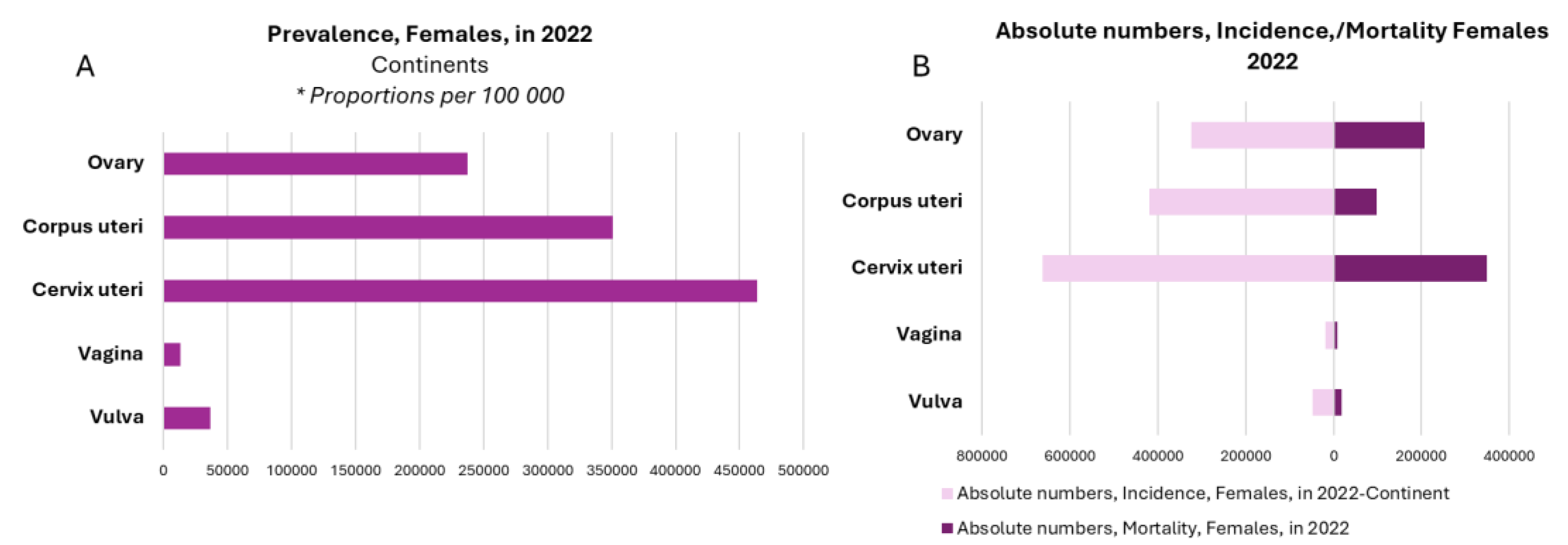
Figure 2.
Schematic of STAT3 pathway and target genes transcription of tumor and immune cells regulation. Cytokines such as interleukin-6 (IL-6), epidermal growth factor (EGF) and G-proteins bind to their receptors that suffer conformation changes and activate Janus kinase (JAK) family. The STAT3 molecules are phosphorylated on Y705 by non-receptor tyrosine kinases (RTKs) such as JAK or can also be activated directly by Src and Abl. The dimerization of STAT3 molecules permits translocation to the nucleus and its functioning as a transcription factor. STAT3 activation causes the transcription of target genes including important molecules for tumor proliferation and survival, angiogenesis and metastasis, in addition to immunosuppression. Non-canonical STAT3 signaling pathway results from three STAT3 forms including Ser727 phosphorylation by serine or threonine kinases such as CDK5, JNKs and MAPKs, mtSTAT3 (mitochondria STAT3), and unphosphorylated STAT3 (uSTAT3). The activation of non-canonical signaling pathways can modify cell metabolism, mitochondrial respiration, NF-κB transcription, and epithelial-mesenchymal transition (EMT) related genes. STAT3 downregulation involves a protein family named PIAS and SOCs. PIAS inhibits STAT3 binding to DNA. SOCs act by inactivating JAK kinase, promoting its degradation or competing with STAT3 in binding to the cytokine receptor. Created in BioRender. Larco, J. (2026) https://BioRender.com/r0t2sc3.
Figure 2.
Schematic of STAT3 pathway and target genes transcription of tumor and immune cells regulation. Cytokines such as interleukin-6 (IL-6), epidermal growth factor (EGF) and G-proteins bind to their receptors that suffer conformation changes and activate Janus kinase (JAK) family. The STAT3 molecules are phosphorylated on Y705 by non-receptor tyrosine kinases (RTKs) such as JAK or can also be activated directly by Src and Abl. The dimerization of STAT3 molecules permits translocation to the nucleus and its functioning as a transcription factor. STAT3 activation causes the transcription of target genes including important molecules for tumor proliferation and survival, angiogenesis and metastasis, in addition to immunosuppression. Non-canonical STAT3 signaling pathway results from three STAT3 forms including Ser727 phosphorylation by serine or threonine kinases such as CDK5, JNKs and MAPKs, mtSTAT3 (mitochondria STAT3), and unphosphorylated STAT3 (uSTAT3). The activation of non-canonical signaling pathways can modify cell metabolism, mitochondrial respiration, NF-κB transcription, and epithelial-mesenchymal transition (EMT) related genes. STAT3 downregulation involves a protein family named PIAS and SOCs. PIAS inhibits STAT3 binding to DNA. SOCs act by inactivating JAK kinase, promoting its degradation or competing with STAT3 in binding to the cytokine receptor. Created in BioRender. Larco, J. (2026) https://BioRender.com/r0t2sc3.
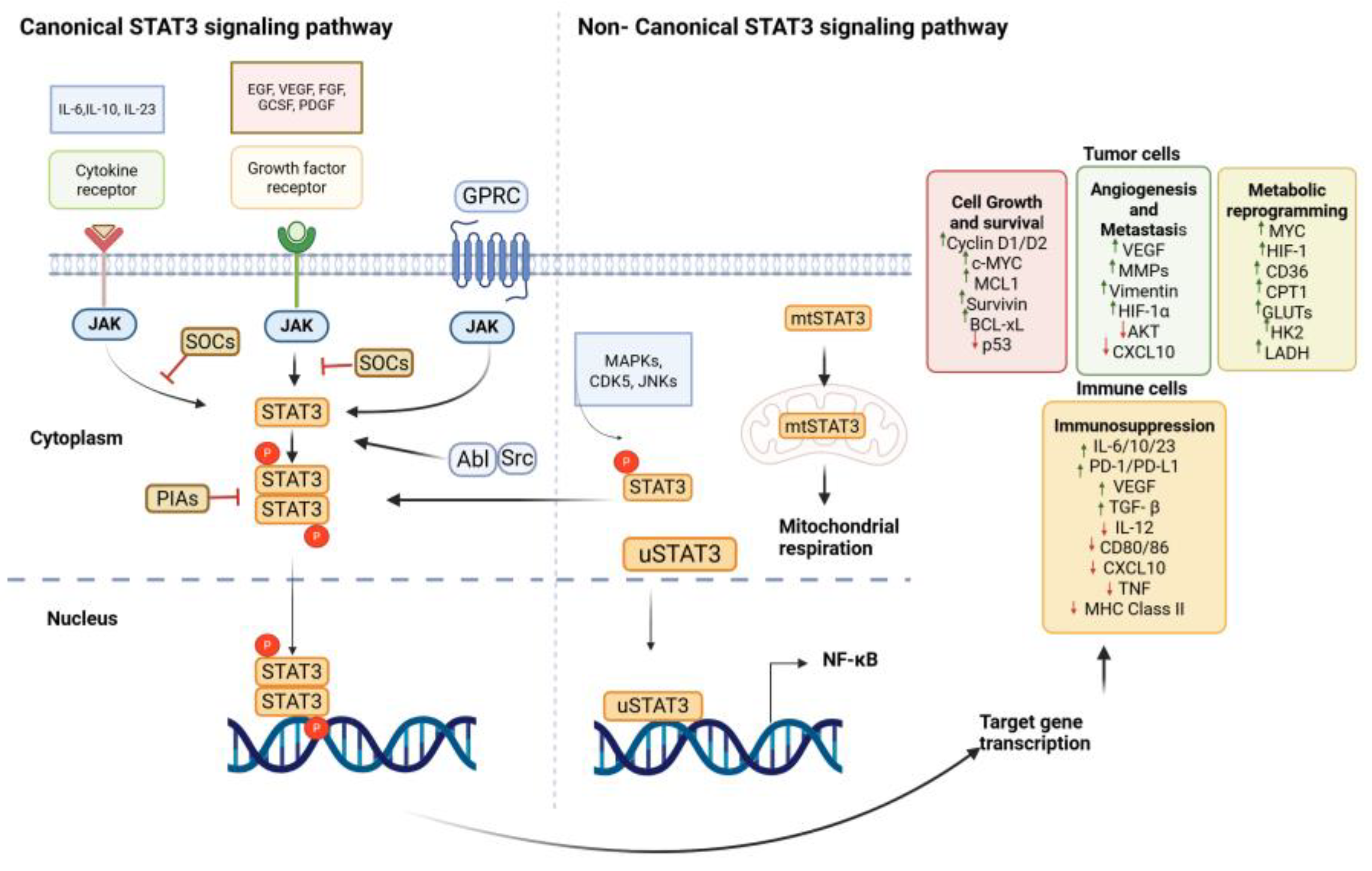
Figure 3.
STAT3-driven immunosuppressive signaling across the tumor microenvironment in gynecologic cancers. Persistent phosphorylation of STAT3 (p-STAT3) in tumor cells orchestrates immunosuppressive communication across myeloid, stromal, and adaptive immune compartments. In the myeloid compartment, STAT3 activation promotes M2 macrophage polarization and MDSC expansion, which collectively secrete IL-10, TGF-β, and IL-6, further sustaining STAT3 signaling in a feed-forward loop. In the stromal compartment, cancer-associated fibroblasts (CAFs) and endothelial cells are activated by STAT3-dependent mediators including IL-6, CXCL12, VEGF, TGF-β, and IL-10, contributing to angiogenesis and stromal remodeling. In the adaptive immune compartment, STAT3-driven lactate accumulation and IL-6 signaling impair dendritic cell maturation, reducing IL-12, MHC class II, and CD40 expression, thereby limiting T cell priming. Simultaneously, IL-6, IL-10, and TGF-β promote Treg accumulation and CD8+ T cell exhaustion, while NK cell cytotoxicity is suppressed. The net result is an immunosuppressive TME characterized by impaired antitumor immunity. Dashed red arrows indicate inhibitory signals; solid green arrows indicate activating signals. Abbreviations: p-STAT3, phosphorylated Signal Transducer and Activator of Transcription 3; IL-6, interleukin-6; IL-10, interleukin-10; IL-12, interleukin-12; TGF-β, transforming growth factor beta; PD-L1, programmed death-ligand 1; VEGF, vascular endothelial growth factor; CXCL12, C-X-C motif chemokine ligand 12; M2 macrophages, alternatively activated macrophages; MDSCs, myeloid-derived suppressor cells; CAFs, cancer-associated fibroblasts; DCs, dendritic cells; MHC class II, major histocompatibility complex class II; CD40, cluster of differentiation 40; CD163, cluster of differentiation 163 (M2 macrophage marker); CD206, cluster of differentiation 206, mannose receptor (M2 macrophage marker); Tregs, regulatory T cells; NK cells, natural killer cells; TME, tumor microenvironment. Created in BioRender. Larco, J. (2026) https://BioRender.com/oiozc6k.
Figure 3.
STAT3-driven immunosuppressive signaling across the tumor microenvironment in gynecologic cancers. Persistent phosphorylation of STAT3 (p-STAT3) in tumor cells orchestrates immunosuppressive communication across myeloid, stromal, and adaptive immune compartments. In the myeloid compartment, STAT3 activation promotes M2 macrophage polarization and MDSC expansion, which collectively secrete IL-10, TGF-β, and IL-6, further sustaining STAT3 signaling in a feed-forward loop. In the stromal compartment, cancer-associated fibroblasts (CAFs) and endothelial cells are activated by STAT3-dependent mediators including IL-6, CXCL12, VEGF, TGF-β, and IL-10, contributing to angiogenesis and stromal remodeling. In the adaptive immune compartment, STAT3-driven lactate accumulation and IL-6 signaling impair dendritic cell maturation, reducing IL-12, MHC class II, and CD40 expression, thereby limiting T cell priming. Simultaneously, IL-6, IL-10, and TGF-β promote Treg accumulation and CD8+ T cell exhaustion, while NK cell cytotoxicity is suppressed. The net result is an immunosuppressive TME characterized by impaired antitumor immunity. Dashed red arrows indicate inhibitory signals; solid green arrows indicate activating signals. Abbreviations: p-STAT3, phosphorylated Signal Transducer and Activator of Transcription 3; IL-6, interleukin-6; IL-10, interleukin-10; IL-12, interleukin-12; TGF-β, transforming growth factor beta; PD-L1, programmed death-ligand 1; VEGF, vascular endothelial growth factor; CXCL12, C-X-C motif chemokine ligand 12; M2 macrophages, alternatively activated macrophages; MDSCs, myeloid-derived suppressor cells; CAFs, cancer-associated fibroblasts; DCs, dendritic cells; MHC class II, major histocompatibility complex class II; CD40, cluster of differentiation 40; CD163, cluster of differentiation 163 (M2 macrophage marker); CD206, cluster of differentiation 206, mannose receptor (M2 macrophage marker); Tregs, regulatory T cells; NK cells, natural killer cells; TME, tumor microenvironment. Created in BioRender. Larco, J. (2026) https://BioRender.com/oiozc6k.
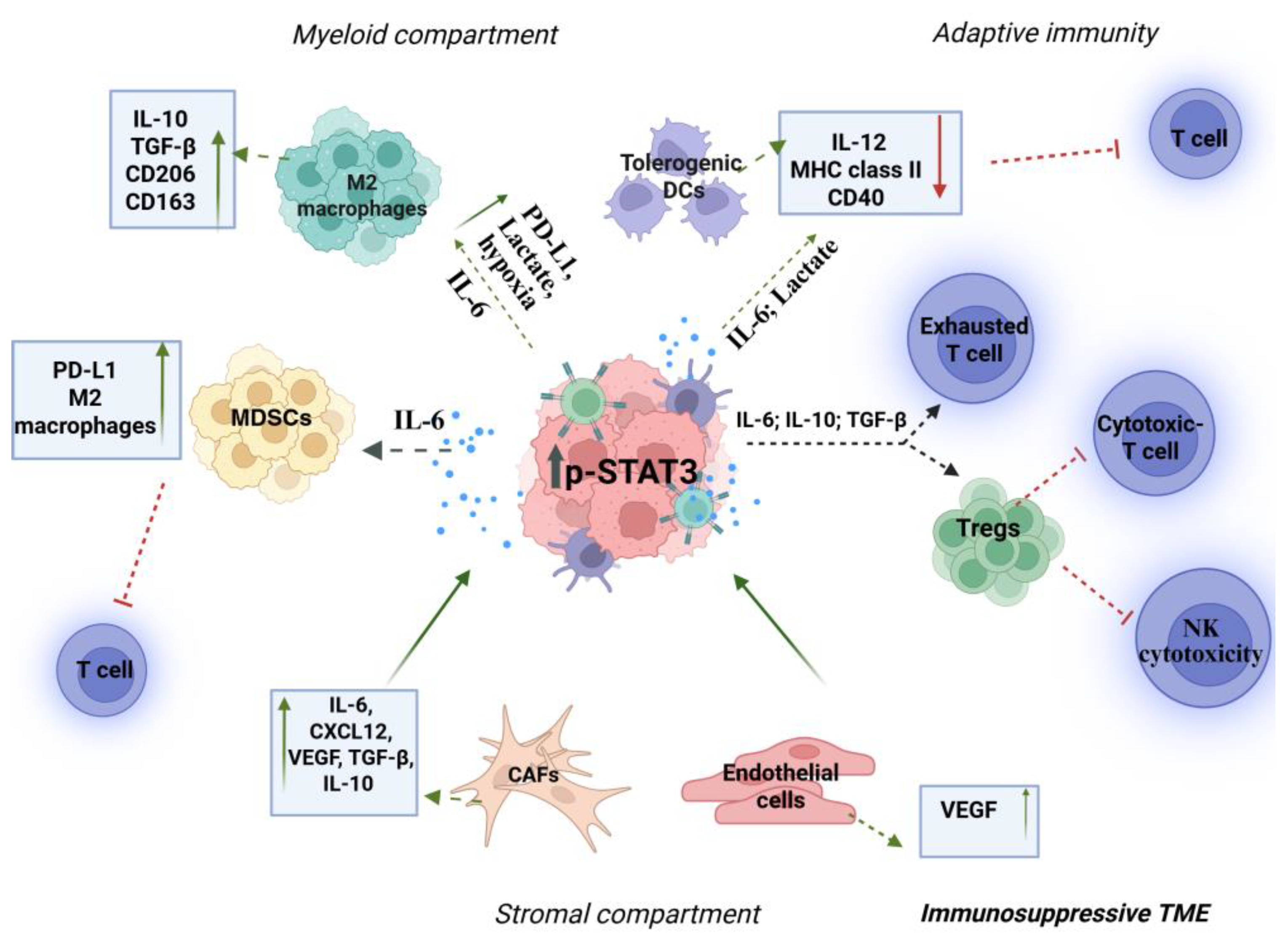
Disclaimer/Publisher’s Note: The statements, opinions and data contained in all publications are solely those of the individual author(s) and contributor(s) and not of MDPI and/or the editor(s). MDPI and/or the editor(s) disclaim responsibility for any injury to people or property resulting from any ideas, methods, instructions or products referred to in the content. |
© 2026 by the authors. Licensee MDPI, Basel, Switzerland. This article is an open access article distributed under the terms and conditions of the Creative Commons Attribution (CC BY) license (http://creativecommons.org/licenses/by/4.0/).
Copyright: This open access article is published under a Creative Commons CC BY 4.0 license, which permit the free download, distribution, and reuse, provided that the author and preprint are cited in any reuse.


