

Submitted:
04 August 2026
Posted:
05 August 2026
You are already at the latest version
Abstract
This study investigated whether the accuracy of video-based kinematic measurements using artificial intelligence (AI) in patients with hip osteoarthritis (OA) is comparable to that in healthy participants, and whether the gait abnormality estimated from video data is comparable to that calculated using optical three-dimensional motion capture (MOCAP) data. This study used open-access datasets of MOCAP and video data including 20 patients with hip OA and 20 healthy participants. Video data were processed using the MYoACT application to extract marker data. The mean absolute error (MAE) of joint angles between MOCAP and MYoACT data was calculated. In patients with hip OA, the modified Gait Abnormality Score (mGAS) for each joint angle was calculated by averaging the absolute differences between each patient’s value and the mean value of healthy participants, divided by the standard deviation of healthy participants, across the entire gait cycle. No significant difference in the MAE was observed between groups. In patients with hip OA, the mGAS showed no significant difference between MYoACT and MOCAP data. These findings suggest that AI-driven video-based kinematic measurements provide comparable accuracy between patients with hip OA and healthy participants, and can identify gait abnormalities at a level comparable to the MOCAP.
Keywords:
hip osteoarthritis
; gait abnormality
; kinematic
; artificial intelligence
; video
; motion capture
1. Introduction
Hip osteoarthritis (OA) causes structural changes in the hip joint [1] and leads to hip pain [2], reduced range of motion [3], and muscle weakness [4]. As a result, hip OA reduces physical function and quality of life (QOL) [5,6]. Because the number of patients with hip OA is expected to increase in the future [7], hip OA is one of the conditions that require further advancements in treatment strategies.
While previous research has identified several factors influencing physical function and QOL in patients with hip OA, such as hip muscle cross-sectional area [8], hip flexion range of motion [9], and walking speed [10], accumulating evidence suggests that gait abnormalities are associated with physical function and QOL in patients with hip OA. Riglet et al. [11] reported that a reduced hip extension angle during gait in patients with hip OA was associated with worsening physical function and QOL after three years. Additionally, Rosenlund et al. [12] quantified gait abnormalities in patients with hip OA using the Gait Deviation Index (GDI) and demonstrated a significant correlation between the GDI and both physical function and QOL in patients with hip OA. Therefore, Quantitative gait analysis of patients with hip OA is crucial for rehabilitation. However, an optical three-dimensional motion capture (MOCAP) has not been easy to implement in clinical practice because it is costly and requires substantial time for data collection and analysis [13].
Recent advances in artificial intelligence (AI) have enabled simple and accurate pose estimations from smartphone videos, leading to high expectations for the clinical application of these technologies. To date, many studies have focused on validating the accuracy of these methods in healthy participants. Viswakumar et al. [14] calculated joint angles using the OpenPose from video data captured with a single smartphone and evaluated the accuracy by calculating the mean absolute error (MAE) relative to the MOCAP. As a result, the MAE between the OpenPose and MOCAP was reported as 7.13° for ankle dorsiflexion, 5.82° for knee flexion, and 7.73° for hip flexion. Furthermore, Uhlrich et al. [15] demonstrated that the average MAE between the OpenCap, which estimates joint angles from videos captured by two or more smartphones, and MOCAP was 4.5° across all joint angles. However, as these studies were conducted on healthy participants, it remains unclear whether similar errors would occur in patients with hip OA who exhibit gait abnormalities [16,17]. Furthermore, it is unknown whether evaluation of gait abnormalities, such as the GDI, show comparable results between the video-based pose estimation and MOCAP. Although the GDI is a composite measure that integrates all joint angles [18], evaluation based on individual joint angles is more practical for guiding interventions in clinical practice. Recently, a standardized method called the modified Gait Abnormality Score (mGAS), which evaluates gait abnormalities based on individual joint angles, has been developed [19]. In clinical practice, the mGAS is more practical than the GDI for gait assessment. Therefore, the aims of this study were twofold: first, to investigate whether the error in AI-driven video-based joint angles relative to the MOCAP in patients with hip OA is comparable to that in healthy participants; and second, to investigate whether the mGAS estimated from video data is comparable to that calculated using the MOCAP.
2. Materials and Methods
2.1. Datasets
This study used open-access motion-capture datasets provided by Bertaux et al. [20] MOCAP and video data from 20 patients with unilateral hip OA (from 30 days to 1 day before total hip arthroplasty) and 20 healthy participants were used as experimental data (Table 1). Bertaux et al. recorded gait data using eight optoelectronic cameras (Vicon MXT40, Vicon Motion Systems Ltd., Oxford, UK) sampled at 100 Hz and a video camera (Basler camera) sampled at 50 Hz in the frontal plane [20]. A total of 35 reflective markers were attached to the whole body based on the Plug-in-Gait model [21]. The participants performed overground walking along a 6-meter walkway as naturally as possible while looking forward.
2.2. Data Processing
MOCAP data were obtained from the recorded C3D files. Frontal plane video data were extracted as C3D files using the MYoACT 2.0.0 (ORGO Inc., Sapporo, Japan) (Figure 1). The MYoACT is an application that enables musculoskeletal analysis from video data using AI, including the estimation of joint angles, ground reaction forces, and joint moments. Subsequently, the C3D files of MOCAP and MYoACT data were processed using CusToM, a MATLAB toolbox, to calculate the hip flexion, hip adduction, hip internal rotation, knee flexion, and ankle dorsiflexion angles [22,23,24]. Additionally, trunk and pelvic obliquity angles were calculated from the marker data. The trunk obliquity angle was defined as the frontal plane projection angle between the vertical axis and the line connecting the midpoints of the bilateral acromioclavicular joints and the midpoints of the bilateral anterior superior iliac spines [25]. The pelvic obliquity angle was defined as the frontal plane projection angle between the horizontal axis and the line connecting the bilateral anterior superior iliac spines [25]. Positive trunk and pelvic obliquity angles indicate an inclination toward the stance limb. Joint angles were analyzed for the affected side in patients with hip OA and a randomly selected side in healthy participants. The following 20 markers were used to calculate joint angles for both MOCAP and MYoACT data: the second metatarsal head, calcaneus, lateral malleolus, flexion-extension axis of the knee, posterior superior iliac spine, anterior superior iliac spine, acromioclavicular joint, spinous processes of the 7th cervical and 10th thoracic vertebrae, back of the head, and temple. All markers were attached bilaterally, except for the spinous processes of the 7th cervical and 10th thoracic vertebrae. Initial contact during gait was identified based on the contralateral peak hip extension angle, which was used to determine the gait cycle for both patients with hip OA and healthy participants [26].
To evaluate the validity of the joint angles obtained from the MYoACT, the mean absolute error (MAE) of MYoACT data relative to MOCAP data over one gait cycle was calculated for both patients with hip OA and healthy participants [27]. The MAE was calculated as follows:
Where; = total number of time points, = each time point, = MOCAP data, and = MYoACT data.
To evaluate gait abnormalities in patients with hip OA, the mGAS was computed separately for MOCAP and MYoACT data based on the concept of z-scores [28]. The mGAS was calculated by averaging the absolute differences between each patient’s kinematic value and the mean kinematic value of healthy participants, divided by the standard deviation of kinematic values in healthy participants, across the entire gait cycle [19]. This formula provides a quantitative assessment of deviations from normal gait patterns. The formula is as follows:
Where; = each patient’s kinematic value, = mean kinematic value of healthy participants, = standard deviation of kinematic values in healthy participants.
2.3. Statistical Analysis
Unpaired t-tests were performed to compare age, height, weight, and MAE between patients with hip OA and healthy participants. Paired t-tests were performed to compare the mGAS between MOCAP and MYoACT data in patients with hip OA. A chi-square test was used to compare the sex distribution between patients with hip OA and healthy participants. Statistical significance was defined as a P value < 0.05.
3. Results
Patients with hip OA were significantly older and heavier than healthy participants, whereas no significant differences were observed in height or sex (Table 1).
The mean kinematic waveforms obtained from MYoACT and MOCAP data for patients with hip OA and healthy participants are shown in Figure 2. The MAE between MYoACT and MOCAP data showed no significant differences between patients with hip OA and healthy participants in any of the evaluated kinematic variables (Table 2). In patients with hip OA, no significant differences were observed in the mGAS between MYoACT and MOCAP data for any of the evaluated kinematic variables (Table 3).
4. Discussion
This study compared the accuracy of AI-driven video data relative to MOCAP data between patients with hip OA and healthy participants, and the mGAS in patients with hip OA between video and MOCAP data. The MAE of all video-based joint angles relative to MOCAP data in patients with hip OA was 5.3°, which was comparable to the 5.5° error observed in healthy participants. Furthermore, no significant differences were found between the mGAS calculated from video data and those calculated from MOCAP data across all joints in patients with hip OA. These results indicate that video-based kinematic measurements using AI can sufficiently identify the characteristics of patients with hip OA. AI-driven video-based kinematic measurements have the potential to become a valuable tool in clinical practice for patients with hip OA.
This is the first study to investigate the accuracy of joint angles and the mGAS during gait in patients with hip OA using video data recorded from a single camera. Viswakumar et al. [14] reported that joint angle errors between the OpenPose using a single camera and MOCAP in healthy participants were 7.13° for ankle dorsiflexion, 5.82° for knee flexion, and 7.73° for hip flexion. Additionally, Uhlrich et al. [15] reported that joint angle errors between the OpenCap using two or more smartphones and MOCAP in healthy participants was 4.5° across all joint angles. Our results for patients with hip OA were similar to those in healthy participants. Our findings suggest that the accuracy of video-based kinematic measurements using AI is sufficiently maintained even in patients with hip OA. Additionally, no significant difference was observed between AI-driven video and MOCAP data in the mGAS evaluating gait abnormalities. AI-driven video-based kinematic measurements may be capable of identifying gait abnormalities in patients with hip OA at a level comparable to the MOCAP.
Exercise therapy is a common conservative treatment for patients with hip OA. Osteoarthritis Research Society International (OARSI) strongly recommends strengthening, cardio, and/or balance training as a core treatment [29]. Similarly, the European Alliance of Associations for Rheumatology (EULAR) recommendations support strength, aerobic, flexibility, or neuromotor exercises [30]. However, gait training (e.g., gait modification) has not been addressed in these guidelines. Riglet et al. [11] and Rosenlund et al. [12] demonstrated that gait patterns in patients with hip OA was associated with both physical function and QOL. Therefore, incorporating gait training in addition to conventionally recommended exercise therapies may further improve clinical outcomes. Although quantitative gait evaluation has conventionally required the MOCAP, its clinical implementation has remain limited due to its high cost and the long time required for data collection and analysis [13]. This study demonstrates the favorable accuracy of AI-driven video-based kinematic measurements in patients with hip OA and suggests its potential utility in extracting gait abnormalities. Consequently, AI-driven video-based kinematic measurements for hip OA patients holds great promise for future clinical practice.
There are several limitations to this study. First, because the participants included only 20 patients with hip OA and 20 healthy participants, the results may not be generalizable. Therefore, future research involving a larger sample size is required. Second, as this study estimated joint angles using only frontal plane video data from the database [20], it remains unclear whether the camera position was optimal. Finally, it should be noted that the hip extension angle was used to identify the initial contact during gait. Although the validity of this method for identifying initial contact has been reported in healthy participants [26], its validity in patients with hip OA remains unclear.
5. Conclusions
The accuracy of AI-driven video-based joint angles relative to the MOCAP in patients with hip OA was comparable to that in healthy participants. Furthermore, regarding the evaluation of gait abnormalities in patients with hip OA, the mGAS calculated from video data using AI showed no substantial difference from that calculated from the MOCAP. These findings suggest that AI-driven video-based kinematic measurements in patients with hip OA have the potential to be useful in rehabilitation.
Author Contributions
Conceptualization, R.U. and H.T.; methodology, K.S. and R.U.; formal analysis, K.S.; writing—original draft preparation, K.S.; writing—review and editing, R.U., M.S. and H.T.; supervision, M.S. and H.T.; project administration, M.S. All authors have read and agreed to the published version of the manuscript. All authors have read and agreed to the published version of the manuscript.
Funding
This research received no external funding.
Institutional Review Board Statement
Ethical review and approval were waived for this study due to open-access datasets.
Informed Consent Statement
Not applicable.
Data Availability Statement
The data presented in this study are available in Figshare at https://doi.org/10.6084/m9.figshare.14420645 and https://doi.org/10.6084/m9.figshare.14420756 [20].
Conflicts of Interest
Kento Sabashi is engaged in research and development for ORGO.inc. Ryo Ueno is a director and head of research and development at ORGO Inc.
Abbreviations
The following abbreviations are used in this manuscript:
| AI | Artificial intelligence |
| OA | Osteoarthritis |
| MOCAP | Motion capture |
| MAE | Mean absolute error |
| mGAS | Modified Gait Abnormality Score |
| QOL | Quality of life |
| GDI | Gait Deviation Index |
| OARSI | Osteoarthritis Research Society International |
| EULAR | European Alliance of Associations for Rheumatology |
References
- Teichtahl, A. J.; Wang, Y.; Smith, S.; Wluka, A. E.; Giles, G. G.; Bennell, K. L.; O’Sullivan, R.; Cicuttini, F. M. Structural Changes of Hip Osteoarthritis Using Magnetic Resonance Imaging. Arthritis Res. Ther. 2014, 16(5), 466. [Google Scholar] [CrossRef] [PubMed]
- Iidaka, T.; Muraki, S.; Akune, T.; Oka, H.; Kodama, R.; Tanaka, S.; Kawaguchi, H.; Nakamura, K.; Yoshimura, N. Prevalence of Radiographic Hip Osteoarthritis and Its Association with Hip Pain in Japanese Men and Women: The ROAD Study. Osteoarthr. Cartil. 2016, 24(1), 117–123. [Google Scholar] [CrossRef] [PubMed]
- Baker, M.; Moreside, J.; Wong, I.; Rutherford, D. J. Passive Hip Movement Measurements Related to Dynamic Motion during Gait in Hip Osteoarthritis. J. Orthop. Res. 2016, 34(10), 1790–1797. [Google Scholar] [CrossRef] [PubMed]
- Loureiro, A.; Constantinou, M.; Diamond, L. E.; Beck, B.; Barrett, R. Individuals with Mild-to-Moderate Hip Osteoarthritis Have Lower Limb Muscle Strength and Volume Deficits. BMC Musculoskelet. Disord. 2018, 19, 303. [Google Scholar] [CrossRef] [PubMed]
- Salaffi, F.; Carotti, M.; Stancati, A.; Grassi, W. Health-Related Quality of Life in Older Adults with Symptomatic Hip and Knee Osteoarthritis: A Comparison with Matched Healthy Controls. Aging Clin. Exp. Res. 2005, 17(4), 255–263. [Google Scholar] [CrossRef] [PubMed]
- Khashan, M.; Mor, A.; Beer, Y.; Rath, E.; Morgensteren, D. R.; Debi, R.; Elbaz, A. Gait Metric Profile and Gender Differences in Hip Osteoarthritis Patients. A Case-Controlled Study. Hip Int. 2014, 24(3), 270–276. [Google Scholar] [CrossRef] [PubMed]
- Gong, J.; Xiang, C.; Zheng, S. Global, Regional, and National Burden of Hip Osteoarthritis (1992-2021) and Predictions of Future Disease Burden Trends: A Systematic Analysis of the Global Burden of Disease Study 2021. BMC Musculoskelet. Disord. 2025, 26, 908. [Google Scholar] [CrossRef] [PubMed]
- Peiris, W. L.; Cicuttini, F. M.; Constantinou, M.; Yaqobi, A.; Hussain, S. M.; Wluka, A. E.; Urquhart, D.; Barrett, R.; Kennedy, B.; Wang, Y. Association between Hip Muscle Cross-Sectional Area and Hip Pain and Function in Individuals with Mild-to-Moderate Hip Osteoarthritis: A Cross-Sectional Study. BMC Musculoskelet. Disord. 2020, 21, 316. [Google Scholar] [CrossRef] [PubMed]
- Tanaka, S.; Robbins, S. M.; Inoue, Y.; Tanaka, R. Factors Associated with Health-Related Quality of Life in Japanese Patients with Hip Osteoarthritis: A Cross-Sectional Study. J. Orthop. Trauma Rehabil. 2020, 27(2), 113–118. [Google Scholar] [CrossRef]
- Tateuchi, H.; Akiyama, H.; Goto, K.; So, K.; Kuroda, Y.; Ichihashi, N. Gait- and Posture-Related Factors Associated With Changes in Hip Pain and Physical Function in Patients With Secondary Hip Osteoarthritis: A Prospective Cohort Study. Arch. Phys. Med. Rehabil. 2019, 100(11), 2053–2062. [Google Scholar] [CrossRef] [PubMed]
- Riglet, L.; Bourredjem, A.; Laroche, D.; Benguella, L.; Binquet, C.; Maillefert, J.; Ornetti, P.; Gueugnon, M. Hip Disability and Osteoarthritis Outcome Score Trajectories and Prognostic Factors for Functional Decline: A 3-Year Follow-up Study. Ann. Phys. Rehabil. Med. 2025, 68(1), 101893. [Google Scholar] [CrossRef] [PubMed]
- Rosenlund, S.; Holsgaard-Larsen, A.; Overgaard, S.; Jensen, C. The Gait Deviation Index Is Associated with Hip Muscle Strength and Patient-Reported Outcome in Patients with Severe Hip Osteoarthritis-A Cross-Sectional Study. PLoS ONE 2016, 11(4), e0153177. [Google Scholar] [CrossRef] [PubMed]
- Akiba, A.; Harato, K.; Yoshihara, H.; Iwama, Y.; Nishizawa, K.; Nagura, T.; Nakamura, M. Development of a Wearable System to Estimate Knee Adduction Moment of Patients with Knee Osteoarthritis during Gait Using a Single Inertial Measurement Unit. J. Jt. Surg. Res. 2025, 3(2), 84–89. [Google Scholar] [CrossRef]
- Viswakumar, A.; Rajagopalan, V.; Ray, T.; Gottipati, P.; Parimi, C. Development of a Robust, Simple, and Affordable Human Gait Analysis System Using Bottom-Up Pose Estimation With a Smartphone Camera. Front. Physiol. 2022, 12, 784865. [Google Scholar] [CrossRef] [PubMed]
- Uhlrich, S. D.; Falisse, A.; Kidziński, Ł.; Muccini, J.; Ko, M.; Chaudhari, A. S.; Hicks, J. L.; Delp, S. L. OpenCap: Human Movement Dynamics from Smartphone Videos. PLoS Comput. Biol. 2023, 19(10), e1011462. [Google Scholar] [CrossRef] [PubMed]
- Porta, M.; Pau, M.; Leban, B.; Deidda, M.; Sorrentino, M.; Arippa, F.; Marongiu, G. Lower Limb Kinematics in Individuals with Hip Osteoarthritis during Gait: A Focus on Adaptative Strategies and Interlimb Symmetry. Bioengineering 2021, 8(4), 47. [Google Scholar] [CrossRef] [PubMed]
- Eitzen, I.; Fernandes, L.; Nordsletten, L.; Risberg, M. A. Sagittal Plane Gait Characteristics in Hip Osteoarthritis Patients with Mild to Moderate Symptoms Compared to Healthy Controls: A Cross-Sectional Study. BMC Musculoskelet. Disord. 2012, 13, 258. [Google Scholar] [CrossRef] [PubMed]
- Schwartz, M. H.; Rozumalski, A. The Gait Deviation Index: A New Comprehensive Index of Gait Pathology. Gait Posture 2008, 28(3), 351–357. [Google Scholar] [CrossRef] [PubMed]
- Sabashi, K.; Ueno, R. Comparison of Quantitative Evaluation Methods for Pathological Gait Abnormalities in Stroke Survivors. Biomechanics 2026, 6(2), 39. [Google Scholar] [CrossRef]
- Bertaux, A.; Gueugnon, M.; Moissenet, F.; Orliac, B.; Martz, P.; Maillefert, J.-F.; Ornetti, P.; Laroche, D. Gait Analysis Dataset of Healthy Volunteers and Patients before and 6 Months after Total Hip Arthroplasty. Sci. Data 2022, 9(1), 399. [Google Scholar] [CrossRef] [PubMed]
- Davis, R. B.; Õunpuu, S.; Tyburski, D.; Gage, J. R. A Gait Analysis Data Collection and Reduction Technique. Hum. Mov. Sci. 1991, 10(5), 575–587. [Google Scholar] [CrossRef]
- Muller, A.; Pontonnier, C.; Puchaud, P.; Dumont, G. CusToM: A Matlab Toolbox for Musculoskeletal Simulation. J. Open Source Softw. 2019, 4(33), 927. [Google Scholar] [CrossRef]
- Ueno, R.; Tsuyuki, Y.; Tohyama, H. Validity of Muscle Activation Estimated with Predicted Ground Reaction Force in Inverse Dynamics Based Musculoskeletal Simulation during Gait. J. Biomech. 2024, 168, 112118. [Google Scholar] [CrossRef] [PubMed]
- Ueno, R. Calibrationless Monocular Vision Musculoskeletal Simulation during Gait. Heliyon 2024, 10(11), e32078. [Google Scholar] [CrossRef] [PubMed]
- Hunt, M. A.; Birmingham, T. B.; Bryant, D.; Jones, I.; Giffin, J. R.; Jenkyn, T. R.; Vandervoort, A. A. Lateral Trunk Lean Explains Variation in Dynamic Knee Joint Load in Patients with Medial Compartment Knee Osteoarthritis. Osteoarthr. Cartil. 2008, 16(5), 591–599. [Google Scholar] [CrossRef] [PubMed]
- De Asha, A. R.; Robinson, M. A.; Barton, G. J. A Marker Based Kinematic Method of Identifying Initial Contact during Gait Suitable for Use in Real-Time Visual Feedback Applications. Gait Posture 2012, 36(3), 650–652. [Google Scholar] [CrossRef] [PubMed]
- Sharma, A. K.; Liu, S.-H.; Zhu, X.; Chen, W. Predicting Gait Parameters of Leg Movement with SEMG and Accelerometer Using CatBoost Machine Learning. Electronics 2024, 13(9), 1791. [Google Scholar] [CrossRef]
- Schober, P.; Mascha, E. J.; Vetter, T. R. Statistics From A (Agreement) to Z (z Score): A Guide to Interpreting Common Measures of Association, Agreement, Diagnostic Accuracy, Effect Size, Heterogeneity, and Reliability in Medical Research. Anesth. Analg. 2021, 133(6), 1633–1641. [Google Scholar] [CrossRef] [PubMed]
- Bannuru, R. R.; Osani, M. C.; Vaysbrot, E. E.; Arden, N. K.; Bennell, K.; Bierma-Zeinstra, S. M. A.; Kraus, V. B.; Lohmander, L. S.; Abbott, J. H.; Bhandari, M.; Blanco, F. J.; Espinosa, R.; Haugen, I. K.; Lin, J.; Mandl, L. A.; Moilanen, E.; Nakamura, N.; Snyder-Mackler, L.; Trojian, T.; Underwood, M.; McAlindon, T. E. OARSI Guidelines for the Non-Surgical Management of Knee, Hip, and Polyarticular Osteoarthritis. Osteoarthr. Cartil. 2019, 27(11), 1578–1589. [Google Scholar] [CrossRef] [PubMed]
- Moseng, T.; Vliet Vlieland, T. P. M.; Battista, S.; Beckwée, D.; Boyadzhieva, V.; Conaghan, P. G.; Costa, D.; Doherty, M.; Finney, A. G.; Georgiev, T.; Gobbo, M.; Kennedy, N.; Kjeken, I.; Kroon, F. P. B.; Lohmander, L. S.; Lund, H.; Mallen, C. D.; Pavelka, K.; Pitsillidou, I. A.; Rayman, M. P.; Tveter, A. T.; Vriezekolk, J. E.; Wiek, D.; Zanoli, G.; Østerås, N. EULAR Recommendations for the Non-Pharmacological Core Management of Hip and Knee Osteoarthritis: 2023 Update. Ann. Rheum. Dis. 2024, 83(6), 730–740. [Google Scholar] [CrossRef] [PubMed]
Figure 1.
MYoACT is an application that enables AI-driven video-based musculoskeletal analysis including joint angles, ground reaction force, and joint moments.
Figure 1.
MYoACT is an application that enables AI-driven video-based musculoskeletal analysis including joint angles, ground reaction force, and joint moments.
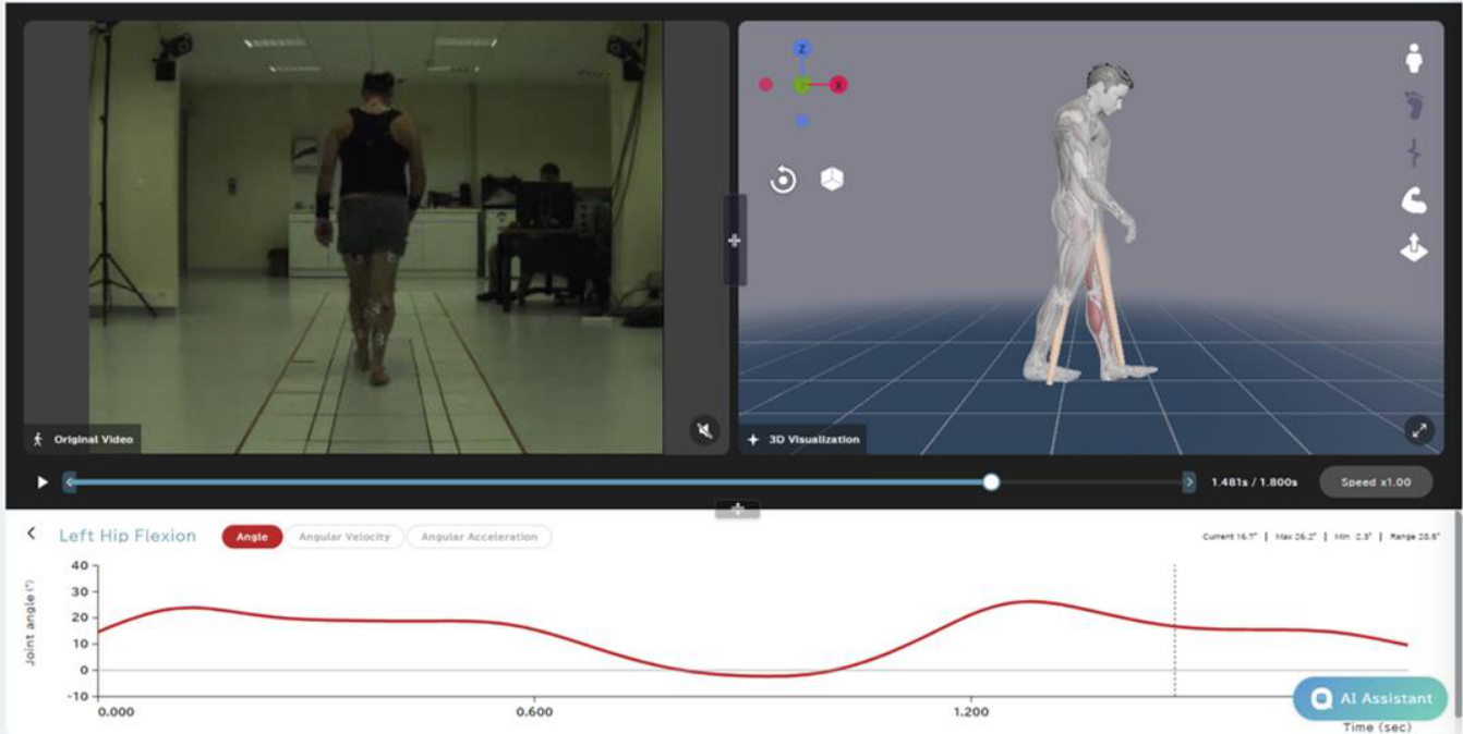
Figure 2.
Mean kinematic waveforms of MYoACT and MOCAP data in patients with hip OA and healthy participants.
Figure 2.
Mean kinematic waveforms of MYoACT and MOCAP data in patients with hip OA and healthy participants.
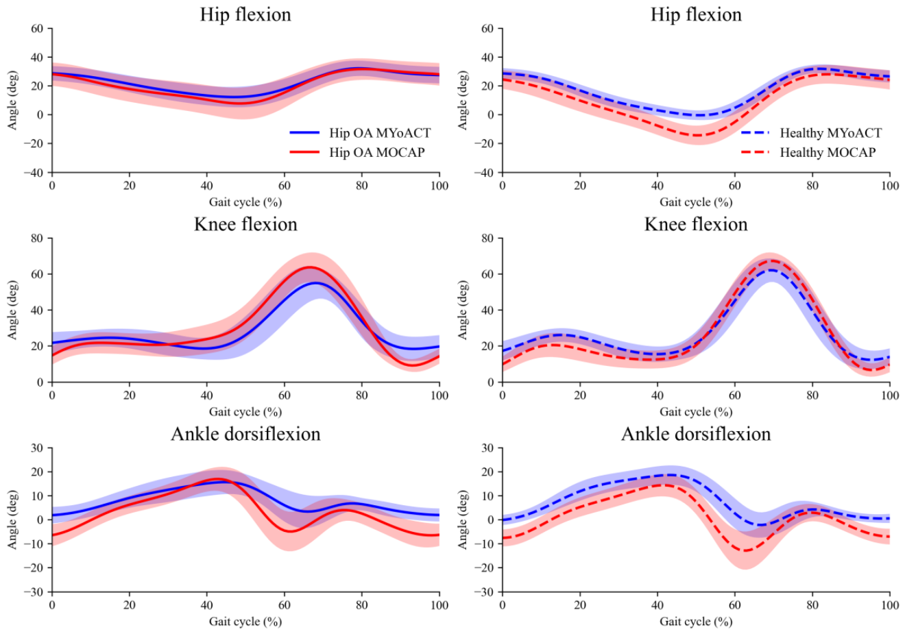

Table 1.
Demographic data of patients with hip OA and healthy participants.
| Hip OA | Healthy | P value | |
| Age, years | 62.0 (9.4) | 51.6 (11.4) | 0.003* |
| Height, cm | 167.5 (10.5) | 167.3 (9.9) | 0.939 |
| Weight, kg | 83.7 (15.5) | 71.9 (15.2) | 0.019* |
| Sex (male/female), n | 12/8 | 8/12 | 0.206 |
| Kellgren–Lawrence grade, n | |||
| Grade 2 | 4 | ||
| Grade 3 | 9 | ||
| Grade 4 | 7 | ||
| * P value < 0.05 |
Table 2.
MAE of joint angles between MYoACT and MOCAP data in patients with hip OA and healthy participants.
Table 2.
MAE of joint angles between MYoACT and MOCAP data in patients with hip OA and healthy participants.
| Hip OA | Healthy | P value | |
| Trunk obliquity, ° | 2.1 (1.1) | 1.8 (1.3) | 0.480 |
| Pelvic obliquity, ° | 2.6 (1.1) | 2.4 (0.8) | 0.513 |
| Hip flexion, ° | 7.5 (4.3) | 8.9 (4.7) | 0.328 |
| Hip adduction, ° | 3.5 (1.2) | 4.2 (1.2) | 0.100 |
| Hip internal rotation, ° | 5.7 (2.9) | 7.5 (3.1) | 0.067 |
| Knee flexion, ° | 8.5 (3.5) | 6.8 (2.6) | 0.096 |
| Ankle dorsiflexion, ° | 6.9 (3.0) | 7.3 (4.4) | 0.764 |
| All joint angles, ° | 5.3 (1.0) | 5.5 (1.9) | 0.533 |
Table 3.
mGAS derived from MYoACT and MOCAP data in patients with hip OA.
| MYoACT | MOCAP | P value | |
| Trunk obliquity | 1.22 (0.49) | 1.02 (0.46) | 0.090 |
| Pelvic obliquity | 0.97 (0.46) | 1.32 (0.83) | 0.106 |
| Hip flexion | 1.94 (0.95) | 1.83 (0.84) | 0.646 |
| Hip adduction | 1.34 (0.83) | 1.15 (0.96) | 0.241 |
| Hip internal rotation | 0.89 (0.57) | 0.95 (0.72) | 0.745 |
| Knee flexion | 1.10 (0.47) | 1.42 (0.64) | 0.087 |
| Ankle dorsiflexion | 1.14 (0.40) | 1.03 (0.50) | 0.434 |
Disclaimer/Publisher’s Note: The statements, opinions and data contained in all publications are solely those of the individual author(s) and contributor(s) and not of MDPI and/or the editor(s). MDPI and/or the editor(s) disclaim responsibility for any injury to people or property resulting from any ideas, methods, instructions or products referred to in the content. |
© 2026 by the authors. Licensee MDPI, Basel, Switzerland. This article is an open access article distributed under the terms and conditions of the Creative Commons Attribution (CC BY) license (http://creativecommons.org/licenses/by/4.0/).
Copyright: This open access article is published under a Creative Commons CC BY 4.0 license, which permit the free download, distribution, and reuse, provided that the author and preprint are cited in any reuse.