
Submitted:
04 August 2026
Posted:
05 August 2026
You are already at the latest version
Abstract
Neonatal sepsis requires microbiological investigation, but blood culture is constrained by small sample volumes, low-density bacteremia, contamination, and delayed results. Closed-tube colorimetric loop-mediated isothermal amplification (LAMP) offers rapid, visually interpretable nucleic acid detection without conventional thermal cycling; however, performance in neonatal blood depends on the diagnostic workflow. This structured narrative review synthesizes evidence on assay design, blood preanalytics, pathogen recovery, amplification inhibition, contamination control, pathogen-specific applications, and clinical translation. Diagnostic performance is influenced by the volume collected, the fraction of the specimen entering the reaction, lysis and extraction efficiency, host-derived inhibitors, and sampling relative to antimicrobial exposure. Sealed colorimetric detection reduces post-amplification handling and amplicon dispersal, but does not prevent pre-amplification contamination or confirm target identity when nonspecific indicators are used. Most evidence derives from isolates, spiked matrices, positive blood-culture bottles, or non-neonatal specimens; prospective diagnostic-accuracy data in neonates remain scarce. Clinical translation will require validated blood-processing methods, compartmentalized target reactions, batch and sample-level controls, predefined invalid-result criteria, and panels adapted to local epidemiology. The method should therefore be evaluated as an adjunct to blood culture, which remains necessary for detecting off-panel organisms and providing phenotypic antimicrobial susceptibility results. Prospective multicenter studies should establish diagnostic accuracy, clinical utility, and safety.
Keywords:
neonatal sepsis
; loop-mediated isothermal amplification
; colorimetric detection
; whole-blood diagnostics
; pre-analytical processing
; contamination control
; diagnostic validation
1. Introduction
Neonatal sepsis remains a major cause of morbidity and mortality during the first 28 days of life, with a disproportionate burden among preterm and low-birth-weight infants and in healthcare settings with limited diagnostic resources [1,2]. Clinical recognition is difficult because the initial signs are often subtle, overlap with non-infectious neonatal conditions, and are interpreted using definitions that remain inconsistent across studies and clinical settings [3,4,5].
Blood culture remains the reference method for confirming bacterial or fungal sepsis [6]. Its diagnostic yield, however, depends strongly on the volume and quality of blood collected [7]. Inadequate inoculation can reduce sensitivity and complicate the interpretation of organisms that may represent contamination rather than true bloodstream infection [6]. Culture results are also rarely available early enough to guide the initial treatment decision [8,9]. Consequently, empirical antibiotics are frequently initiated before microbiological confirmation, exposing many infants without proven infection to unnecessary treatment [10,11].
Molecular assays can provide faster microbial detection [12], but their reported performance in neonatal sepsis remains heterogeneous [13]. A recent diagnostic-accuracy review estimated a summary sensitivity of 0.91 (95% confidence interval [CI], 0.85–0.95) and specificity of 0.88 (95% CI, 0.83–0.92), although the certainty of evidence was low and substantial variation persisted among platforms and study designs [13]. These findings support molecular assays as complementary tests rather than replacements for culture and antimicrobial susceptibility testing [3,13,14].
Loop-mediated isothermal amplification offers a practical alternative because it amplifies nucleic acids at a constant temperature and supports rapid visual detection without conventional thermal cycling [15,16]. Colorimetric readouts simplify result interpretation, while closed-tube detection limits post-amplification handling and reduces the opportunity for amplicon dispersal [17,18]. An extraction-free colorimetric LAMP assay recently detected Streptococcus agalactiae at 2 CFU/mL in spiked neonatal blood, demonstrating analytical promise without yet establishing diagnostic accuracy in prospectively enrolled neonates [19].
The clinical value of closed-tube colorimetric LAMP in neonatal sepsis depends not only on amplification chemistry but also on blood volume, recovery of intact pathogens, matrix-associated inhibition, contamination control, and reliable interpretation of negative and invalid results. Accordingly, this review evaluates the method as an integrated diagnostic pathway rather than as an amplification reaction in isolation. It brings together neonatal-blood preanalytics, assay and control design, pathogen-specific evidence, and clinical implementation while distinguishing analytical and simulated-matrix findings from direct clinical evidence. This framework is used to define the requirements for clinically interpretable testing alongside blood culture and to identify the validation steps needed before routine application in suspected neonatal sepsis.
2. Review Methodology and Evidence Framework
This structured narrative review used a transparent literature-search and evidence-mapping approach. Scopus, Web of Science Core Collection, and PubMed/MEDLINE were searched for English-language publications from January 2000 to 30 July Search terms combined “neonatal sepsis”, “neonatal bloodstream infection”, “loop-mediated isothermal amplification”, “LAMP”, “colorimetric”, “closed-tube”, “whole blood”, “sample preparation”, “inhibition”, “contamination”, and the names of selected neonatal-sepsis pathogens. Reference lists of relevant primary studies and reviews were also screened. Seminal pre-2000 studies were retained when they addressed foundational methods or neonatal blood-volume requirements.
Peer-reviewed studies were considered when they addressed LAMP-based pathogen detection, neonatal blood diagnostics, pre-analytical processing, amplification inhibition, contamination control, or clinical implementation. Animal, food, and environmental studies were included only when they provided an assay-design or contamination-control principle directly transferable to neonatal blood testing. Evidence was grouped as purified-template or isolate studies, spiked-matrix studies, clinical-specimen studies, prospective diagnostic-accuracy studies, or implementation studies. Sample matrix, blood volume, processing method, analytical sensitivity, assay time, comparator, diagnostic performance, and reported limitations were recorded where available.
The review was informed by SANRA principles, particularly transparent searching, appropriate referencing, and explicit differentiation between analytical and clinical evidence [20]. Because the available studies were heterogeneous in population, matrix, assay format, and comparator, findings were synthesized narratively rather than pooled quantitatively [13]. No formal risk-of-bias score was assigned.
3. Closed-Tube Colorimetric LAMP: Principles and Assay Design
Loop-mediated isothermal amplification (LAMP) amplifies a defined nucleic-acid target at a constant temperature using a strand-displacing DNA polymerase and a coordinated set of inner and outer primers [15,21]. The conventional reaction uses forward and backward inner primers, FIP and BIP, together with the outer primers F3 and B3 to recognize six regions of the target sequence and generate dumbbell-shaped intermediates that undergo continuous stem-loop amplification [21]. Loop primers provide additional initiation sites and can accelerate amplification without changing the basic reaction mechanism [22]. Most LAMP assays operate between 60 and 65 °C, although the optimum temperature and incubation period depend on the polymerase, target sequence, and primer set [15,23].
Assay design should begin with a target region that is conserved across the clinically relevant strains intended for detection and sufficiently distinct from closely related or commensal organisms. Whole-genome comparison can improve strain inclusivity and analytical exclusivity by screening candidate primer regions against both target and background genomes [24]. Candidate primer sets should then be evaluated experimentally using diverse target isolates, epidemiologically relevant non-target organisms, and replicated no-template controls because interactions among the multiple LAMP primers can generate nonspecific amplification [23,25,26]. Analytical sensitivity should be established with intact organisms carried through the intended neonatal-blood processing workflow rather than with purified nucleic acid alone [19,27]. A maximum incubation time should be fixed before clinical validation because late amplification may reflect primer-derived or non-template products [23,25]. For a multispecies panel using a common nonspecific color indicator, spatially separated sealed chambers are preferable to pooling multiple primer sets in one reaction, as compartmentalization preserves target attribution while maintaining a closed workflow [28,29].
Colorimetric LAMP converts chemical changes produced during amplification into a visible endpoint. Extensive DNA synthesis generates protons and pyrophosphate, alters the concentration of free magnesium ions, and produces large quantities of double-stranded DNA [16]. These changes can be detected indirectly using pH-sensitive indicators such as phenol red or metal-ion indicators such as hydroxy naphthol blue and calcein [16,30,31]. DNA-binding dyes report the accumulation of double-stranded DNA but remain sequence-independent, whereas sequence-specific probes can provide a signal linked more directly to the intended target [16,32]. pH-based reactions require limited buffering capacity because strongly buffered template solutions may suppress the expected color transition, while acidic sample inputs may alter the initial color independently of amplification [30,33].
In a closed-tube format, the indicator is added before incubation and the result is read without reopening the reaction vessel. This approach reduces post-amplification handling and lowers the opportunity for aerosolized amplicons to contaminate subsequent reactions [18,23]. Closed-tube detection does not prevent contamination introduced during blood processing or reagent preparation, and nonspecific indicators cannot distinguish the intended product from primer-derived or non-template amplification [23]. Carry-over control and amplification specificity must therefore be addressed separately. Colorimetric indicators are not interchangeable because their performance depends on both the signal-generating chemistry and the composition of the processed blood input. Representative readout strategies relevant to a closed-tube neonatal-blood workflow are compared in Table 1.
4. Pre-Analytical Challenges and Contamination Control in Neonatal Blood
4.1. Blood Volume, Sampling Probability, and Specimen Timing
The performance of a neonatal blood LAMP assay is determined before amplification begins. Blood volume is the first constraint because neonatal diagnostic testing must be performed from a limited total blood volume [27,37,38]. In a 2024 neonatal intensive care unit (NICU) cohort, the median blood-culture inoculum was 1.0 mL, but 35% of bottles contained less than 0.9 mL [7]. A separate prospective study found that obtaining at least 1 mL was feasible in most neonates, although inadequate volumes were more frequent during late-onset sepsis evaluations in very-low-birth-weight infants [39]. At low microbial densities, detection becomes probabilistic because the tested aliquot may contain no organism even when bacteremia is present [27,40,41]. Experimental work with common neonatal pathogens showed that small inocula were less reliable when concentrations approached or fell below 1 CFU/mL [40]. For molecular testing, the effective input is not the volume collected but the fraction of the original specimen that finally enters the LAMP tube. Every transfer, split, concentration, and elution step should therefore be considered in the analytical sensitivity claim.
Specimen timing and collection source also influence interpretation. Blood should ideally be obtained before antimicrobial administration because culture requires viable organisms, whereas nucleic-acid tests may remain positive after bacterial killing [27,42]. Direct molecular detection can consequently identify microbial DNA when culture is negative, but such discordance does not by itself establish active bloodstream infection [13,27,43]. Collection from an indwelling catheter may increase the recovery of organisms associated with the line while also complicating distinction between catheter colonization, contamination, and systemic infection [44,45,46]. The collection volume, anatomical source, number of attempts, time relative to antibiotics, and interval to processing should be recorded for every clinical sample.
4.2. Pathogen Recovery and Amplification Inhibition
Whole blood is a difficult amplification matrix. Pathogen nucleic acid is present against a large background of human genomic DNA, leukocytes, erythrocytes, plasma proteins, and other substances that can interfere with microbial recovery or downstream enzymatic reactions [27,47,48]. Increasing the input volume may improve the probability of capturing rare organisms, but it also increases host material and inhibitor load [27,48,49,50]. Efficient processing must lyse both Gram-negative and Gram-positive organisms without losing low-copy DNA during purification [51]. This is particularly relevant for organisms with robust cell walls, including staphylococci and streptococci [27]. Recovery from intact organisms in whole blood is therefore dependent on the sample-processing and DNA-extraction method [27,51,52].
Extraction-free processing reduces handling time and consumable use, but it trades purification for dilution or chemical tolerance. A neonatal-blood group B Streptococcus (GBS) LAMP study demonstrated analytical feasibility in spiked specimens, yet spiking does not reproduce the variable cellular composition, antibiotic exposure, pathogen viability, or microbial burden of prospectively collected sepsis samples [19]. Heat treatment or crude lysis must be validated across clinically relevant hematocrits and bacterial concentrations rather than at a single blood dilution. For pH-based colorimetric LAMP, acidic sample components or extraction buffers may shift the starting color, while strongly buffered inputs may suppress the expected endpoint change [30,33]. The template volume must therefore be chosen jointly with the lysis chemistry, reaction buffering, and the selected sample-control architecture.
4.3. Collection Contamination and Amplicon Carry-Over
Contamination can occur during collection, sample preparation, reagent handling, or amplification [53]. Skin commensals are especially problematic in neonates because coagulase-negative staphylococci may represent either true late-onset sepsis or contamination. In a recent neonatal cohort, time to positivity, gestational age, and clinical findings improved discrimination between these possibilities, showing that organism identity alone is insufficient [54]. Umbilical-cord blood can provide a larger sample for early-onset sepsis testing, but its value depends on a rigorously standardized sterile collection procedure [55]. Low-biomass molecular assays also require attention to bacterial DNA present in extraction kits, water, plasticware, or the laboratory environment because background DNA can become visible when the true target concentration is very low [27,56,57].
LAMP creates a large amount of amplicon, making carry-over contamination a persistent risk [23]. Closed-tube color detection reduces the release of products after amplification, but it cannot correct contamination introduced before the cap is closed and does not prevent nonspecific amplification [18,23]. Incorporating deoxyuridine triphosphate (dUTP) into LAMP products and uracil-DNA glycosylase into subsequent reactions can selectively degrade uracil-containing carry-over amplicons before new amplification begins [58,59]. This strategy has reduced false-positive contamination in both bacterial LAMP and colorimetric RT-LAMP formats [59,60]. It does not remove native microbial DNA introduced during specimen preparation, so enzymatic control must complement physical workflow controls.
A neonatal LAMP workflow should separate reagent preparation, specimen processing, and amplification, with unidirectional movement and sealed post-amplification tubes. Each batch should include an extraction blank, no-template control, and target-positive control [61]. A sample-level process control should be introduced before lysis to assess recovery and inhibition [61]. A post-extraction control assesses inhibition only [61]. With sequence-independent color indicators, the control should occupy a separate sealed chamber or use a distinguishable sequence-specific signal, because target and control amplification cannot otherwise be differentiated [62,63]. A target-negative result is valid only when the control succeeds. Control failure should be reported as invalid [62]. Weak or late color changes should be interpreted within the predefined reaction time. Validation reports should state blood volumes, sample source, anticoagulant, storage conditions, extraction recovery, template input, invalid results, and contamination events.
5. Evidence for Neonatal-Sepsis Pathogens
The pathogen content of a neonatal-sepsis assay cannot be defined by a universal list. A recent global meta-analysis found that Gram-negative organisms accounted for 53% of early-onset and 71% of late-onset bacterial sepsis, although dominant species differed markedly by setting [64]. Streptococcus agalactiae was the leading early-onset pathogen overall, whereas Klebsiella species were prominent in both early- and late-onset disease and were especially important in low- and lower-middle-income countries [64]. In a multicenter Southeast Asian dataset, Klebsiella species represented 27.5% of significant isolates, while Candida species accounted for 8.3% of culture-positive episodes [65]. These differences indicate that target selection should follow local neonatal-unit epidemiology rather than the traditional early-onset versus late-onset classification alone.
The most directly relevant evidence concerns Streptococcus agalactiae. An extraction-free cfb-targeted colorimetric LAMP assay demonstrated analytical detection in spiked neonatal blood, supporting matrix feasibility but not clinical diagnostic performance [19]. In a separate neonatal real-time PCR study, detection differed substantially between cerebrospinal fluid and ethylenediaminetetraacetic acid (EDTA) blood, illustrating the importance of specimen-specific clinical validation [66].
Clinical evidence for Escherichia coli has progressed to testing in blood from young infants with suspected sepsis [67]. This study supports the feasibility of rapid LAMP using a low-volume clinical specimen, but neonatal subgroup performance, multicenter reproducibility, and effects on treatment decisions remain uncertain [67]. A pathogen-specific result would still require culture for off-panel organism recovery and phenotypic antimicrobial susceptibility testing [3,13].
Klebsiella pneumoniae LAMP assays have targeted species-identification and hypervirulence markers [68,69], while carbapenemase determinants have been evaluated in separate LAMP reactions [68]. These findings support target selection and modular resistance-marker testing, but they do not establish direct performance in low-volume neonatal blood [68]. Detection of a resistance gene should be interpreted as a genotypic marker rather than a substitute for phenotypic susceptibility testing [68].
Staphylococcal assays demonstrate that species identification and mecA detection can be combined using lateral-flow or compartmentalized microfluidic systems [28,70]. However, neither approach has been validated directly in unprocessed neonatal blood, and only the microfluidic format maintains physical separation during detection [28,70]. For coagulase-negative staphylococci, molecular positivity cannot independently distinguish true bloodstream infection from collection contamination; interpretation still requires collection source, time to positivity, and clinical context [54,71].
Spiked-blood feasibility has been reported for Enterococcus faecalis using LAMP with lateral-flow detection, but direct neonatal-blood validation and a permanently closed colorimetric readout remain absent [72]. Enterococcal targets may be appropriate in locally adapted panels where epidemiology supports their inclusion, but performance should be established in the intended neonatal matrix rather than extrapolated from simulated specimens [64,72].
Fungal LAMP evidence is divided between rapid identification from positive blood-culture bottles and limited direct-specimen evaluation [73,74,75]. Positive-culture assays can shorten species identification after a bottle signals positive, but they do not provide direct-from-blood diagnosis [73]. Direct-specimen evidence remains limited: one study included only two culture-positive whole-blood samples [74], whereas another evaluated spiked healthy-donor blood rather than clinical candidemia specimens [76]. The available evidence therefore differs in both proximity to neonatal clinical use and alignment with a closed-tube colorimetric workflow, as summarized in Table 2.
Across the selected pathogens, the reviewed literature did not identify a prospectively validated, spatially separated panel operating directly from low-volume neonatal blood. Panel composition should remain adaptable to local epidemiology, and a negative panel result must not exclude an off-panel infection [64,65]. Culture should therefore continue for broad organism recovery and phenotypic antimicrobial susceptibility testing [3,13].
6. Clinical Translation and Research Priorities
6.1. Intended Clinical Use
Closed-tube colorimetric LAMP is most appropriately positioned as a rapid adjunct to blood culture during the initial evaluation of suspected neonatal sepsis. Current molecular assays provide faster pathogen detection but show heterogeneous performance and low-certainty evidence, which precludes their use as stand-alone rule-out tests [13]. Culture should proceed in parallel because a target-specific LAMP panel cannot recover off-panel organisms or provide phenotypic antimicrobial susceptibility results [3,13]. The intended clinical contribution is therefore an earlier pathogen-specific result that may support reassessment of empirical therapy while culture remains in progress. A proposed workflow integrating closed-tube colorimetric LAMP with parallel blood culture and clinical interpretation is summarized in Figure 1.
The intended use should be defined before analytical optimization or clinical validation. In hospital settings, the most defensible role is testing symptomatic neonates or infants undergoing a routine sepsis evaluation, rather than universal screening at birth. The World Health Organization (WHO) neonatal-sepsis target product profile and its recent analysis primarily address triage and risk-stratification tests for infants in a diagnostic gray zone [77]. These documents do not directly specify the performance requirements of a pathogen-identification assay, but their emphasis on affordability, accessibility, rapid turnaround, minimal infrastructure, and context-appropriate deployment provides relevant operational guidance [77]. The assay panel and reporting algorithm should consequently be matched to the healthcare level, local pathogen distribution, and the clinical action expected from a positive, negative, or invalid result.
6.2. Diagnostic-Accuracy Validation
The next validation stage should be a prospective multicenter diagnostic-accuracy study enrolling consecutive neonates undergoing routine evaluation for suspected sepsis. LAMP and blood culture specimens should be obtained during the same clinical episode, with collected volume, processed volume, sampling source, and timing relative to antimicrobial administration documented for both methods. Index-test interpretation should be masked to culture and clinical findings, while positivity thresholds, maximum incubation time, and invalid-result criteria should be defined before enrollment. Culture should remain the primary microbiological comparator, but discordant results require cautious interpretation because low inoculated volume and prior antibiotic exposure may reduce culture yield [13,27]. Predefined secondary assessment using clinical findings and independently obtained microbiological evidence may help characterize discordance, but the index-test result should not itself determine the reference classification. Reports should follow the Standards for Reporting Diagnostic Accuracy Studies (STARD) and present participant selection, reference methods, missing specimens, invalid tests, discordant findings, and complete participant flow [78].
6.3. Clinical Utility and Antimicrobial Stewardship
Diagnostic accuracy should be established before clinical utility is tested. A subsequent randomized or pragmatic add-on study should compare standard sepsis management with standard management plus rapid LAMP reporting [79]. Recommended outcomes include time to pathogen-directed therapy, time to antibiotic narrowing or discontinuation, total antibiotic exposure, repeated blood sampling, length of stay, mortality, and cost per clinically useful result [13,80]. Safety outcomes should include missed or delayed treatment of culture-positive and off-panel infections. LAMP-guided decisions should follow a predefined antimicrobial-stewardship algorithm, and empirical treatment should not be withheld when the clinical indication for immediate therapy is strong [81].
6.4. Technical Standardization and Quality Assurance
Technical translation requires a locked assay design, reproducible manufacturing, reagent stability under intended storage conditions, predefined visual or instrument-assisted interpretation, and external quality assessment. Analytical validation should examine strain inclusivity, cross-reactivity, intact-cell detection, clinically relevant organism concentrations, blood-matrix variation, invalid-result frequency, reader agreement, and lot-to-lot performance [15,23]. A compartmentalized panel should report three operational categories: target detected, target not detected with a valid sample control, and invalid because the sample control failed. With any negative panel result, the report should state that an off-panel bloodstream infection is not excluded. The decisive endpoint is therefore not amplification alone, but a reproducible and clinically interpretable result that can be evaluated for its effect on time-sensitive decisions while preserving culture and antimicrobial stewardship.
7. Conclusions
Closed-tube colorimetric LAMP has the potential to provide earlier pathogen-specific information during the evaluation of suspected neonatal sepsis, but its clinical performance will depend on the entire diagnostic pathway rather than amplification speed alone. Low-volume and low-density bacteremia, variable pathogen recovery, blood-derived inhibition, pre-amplification contamination, and nonspecific color changes remain important sources of analytical and interpretive error. The available evidence is derived mainly from isolates, simulated blood matrices, positive blood-culture bottles, and non-neonatal specimens, and does not yet support routine use as a stand-alone diagnostic or rule-out test. Clinical translation will require validated processing of low-volume neonatal blood, compartmentalized pathogen targets, appropriate process and batch controls, predefined result and invalidity criteria, and panel composition adapted to local epidemiology. Blood culture should continue in parallel to detect off-panel organisms and provide phenotypic antimicrobial susceptibility results. The next priority is prospective multicenter diagnostic-accuracy validation in neonates, followed by clinical-utility studies assessing whether rapid reporting can improve antimicrobial decision-making without compromising patient safety.
Author Contributions
Conceptualization, T.K.S.; methodology, T.K.S.; validation, T.K.S.; formal analysis, T.K.S.; investigation, T.K.S.; resources, T.K.S. and M.P.; data curation, T.K.S.; writing—original draft preparation, T.K.S.; writing—review and editing, P.G. and J.R.; supervision, P.G., J.R., and M.P. All authors have read and agreed to the published version of the manuscript.
Funding
Institute funding (Datta Meghe Institute of Higher Education and Research).
Institutional Review Board Statement
Not Applicable.
Informed Consent Statement
Not Applicable.
Data Availability Statement
No new data were created or analyzed in this study. Data sharing is not applicable to this article.
Conflicts of Interest
The authors declare no conflict of interest.
References
- Coșovanu, E.-T.; Balan, T.A.; Coșovanu, E.-O.; Ionescu, S.; Damian, C.; Petroaie, A.D.; et al. Neonatal Infections Caused by Multidrug-Resistant Bacteria: An Analysis of Prevalence, Risk Factors, and Therapeutic Implications—A Narrative Review. Pathogens 2026, 15, 469. [Google Scholar] [CrossRef] [PubMed]
- Strunk, T.; Molloy, E.J.; Mishra, A.; Bhutta, Z.A. Neonatal bacterial sepsis. The Lancet 2024, 404, 277–93. [Google Scholar] [CrossRef] [PubMed]
- Kosmeri, C.; Giapros, V.; Serbis, A.; Baltogianni, M. Application of Advanced Molecular Methods to Study Early-Onset Neonatal Sepsis. IJMS 2024, 25, 2258. [Google Scholar] [CrossRef] [PubMed]
- Kariniotaki, C.; Thomou, C.; Gkentzi, D.; Panteris, E.; Dimitriou, G.; Hatzidaki, E. Neonatal Sepsis: A Comprehensive Review. Antibiotics 2024, 14, 6. [Google Scholar] [CrossRef] [PubMed]
- Hayes, R.; Hartnett, J.; Semova, G.; Murray, C.; Murphy, K.; Carroll, L.; et al. Neonatal sepsis definitions from randomised clinical trials. Pediatr. Res. 2023, 93, 1141–8. [Google Scholar] [CrossRef] [PubMed]
- De Rose, D.U.; Ronchetti, M.P.; Martini, L.; Rechichi, J.; Iannetta, M.; Dotta, A.; et al. Diagnosis and Management of Neonatal Bacterial Sepsis: Current Challenges and Future Perspectives. TropicalMed 2024, 9, 199. [Google Scholar] [CrossRef]
- Rueda, M.S.; Soghier, L.; Campos, J.; Bahar, B.; Bost, J.E.; Gai, J.; et al. Blood volume collected for cultures in infants with suspected neonatal sepsis. J. Perinatol. 2024, 44, 1800–4. [Google Scholar] [CrossRef]
- Willey, E.; Mitchell, M.; Ehlert, C.; Swoveland, J.; Zembles, T. Time to positive blood cultures in neonatal sepsis evaluations. J. Perinatol. 2025, 45, 993–6. [Google Scholar] [CrossRef]
- Dierikx, T.; Budding, A.; Bos, M.; Van Laerhoven, H.; Van Der Schoor, S.; Niemarkt, H.; et al. Potential of Molecular Culture in Early Onset Neonatal Sepsis Diagnosis: A Proof of Principle Study. Microorganisms 2023, 11, 960. [Google Scholar] [CrossRef]
- Groen, J.; Van Der Kuip, M.; Budding, D.; Bos, M.P.; Benninga, M.A.; Niemarkt, H.J.; et al. Assessing Diagnostic Performance of Molecular Culture for Neonatal Sepsis: Protocol of the CHAMPIONS Study. Diagnostics 2024, 14, 1930. [Google Scholar] [CrossRef]
- Ain Ibrahim, N.; Makmor Bakry, M.; Mohd Tahir, N.A.; Mohd Zaini, N.R.; Mohamed Shah, N. A Prospective Cohort Study of Factors Associated with Empiric Antibiotic De-escalation in Neonates Suspected with Early Onset Sepsis (EOS). Pediatr. Drugs 2020, 22, 321–30. [Google Scholar] [CrossRef]
- Mazzucchelli, I.; Garofoli, F.; Angelini, M.; Tinelli, C.; Tzialla, C.; Decembrino, L. Rapid detection of bacteria in bloodstream infections using a molecular method: a pilot study with a neonatal diagnostic kit. Mol. Biol. Rep. 2020, 47, 363–8. [Google Scholar] [CrossRef]
- Dierikx, T.H.; Visser, D.H.; De Meij, T.; Versalovic, J.; Leeflang, M.M.; Cooper, C.; et al. Molecular assays for the diagnosis of sepsis in neonates: a diagnostic test accuracy review; Service, Cochrane Central Editorial, Ed.; Cochrane Database of Systematic Reviews [Internet]., 2025; p. 2025. [Google Scholar] [CrossRef]
- Caunedo-Jiménez, M.; Fernández-Colomer, B.; Fernández-Suárez, J.; Arias-Llorente, R.P.; Lareu-Vidal, S.; Mantecón-Fernández, L.; et al. Clinical Utility of the FilmArray® Blood Culture Identification (BCID) Panel for the Diagnosis of Neonatal Sepsis. Microorganisms 2023, 11, 732. [Google Scholar] [CrossRef]
- Yang, N.; Zhang, H.; Han, X.; Liu, Z.; Lu, Y. Advancements and applications of loop-mediated isothermal amplification technology: a comprehensive overview. Front Microbiol. 2024, 15, 1406632. [Google Scholar] [CrossRef]
- Selva Sharma, A.; Lee, N.Y. Advancements in visualizing loop-mediated isothermal amplification (LAMP) reactions: A comprehensive review of colorimetric and fluorometric detection strategies for precise diagnosis of infectious diseases. Coord. Chem. Rev. 2024, 509, 215769. [Google Scholar] [CrossRef]
- Saifuddin, S.A.; Rashid, R.; Nor Azmi, N.J.; Mohamad, S. Colorimetric strategies applicable for loop-mediated isothermal amplification. J. Microbiol. Methods 2024, 223, 106981. [Google Scholar] [CrossRef]
- Neshani, A.; Zare, H.; Sadeghian, H.; Safdari, H.; Riahi-Zanjani, B.; Aryan, E. A Comparative Study on Visual Detection of Mycobacterium tuberculosis by Closed Tube Loop-Mediated Isothermal Amplification: Shedding Light on the Use of Eriochrome Black T. Diagnostics 2023, 13, 155. [Google Scholar] [CrossRef]
- Banerjee, S.; K., M.H.; Shastry, R.P. Loop-mediated isothermal amplification (LAMP) assay for early on-site detection of Group B Streptococcus infection in neonatal sepsis blood sample. Mol. Biol. Rep. 2024, 51, 811. [Google Scholar] [CrossRef]
- Baethge, C.; Goldbeck-Wood, S.; Mertens, S. SANRA—a scale for the quality assessment of narrative review articles. Res. Integr. Peer Rev. 2019, 4, 5. [Google Scholar] [CrossRef] [PubMed]
- Notomi, T. Loop-mediated isothermal amplification of DNA. Nucleic Acids Res. 2000, 28, 63e–63. [Google Scholar] [CrossRef]
- Nagamine, K.; Hase, T.; Notomi, T. Accelerated reaction by loop-mediated isothermal amplification using loop primers. Mol. Cell. Probes 2002, 16, 223–9. [Google Scholar] [CrossRef]
- Kim, S.-H.; Lee, S.-Y.; Kim, U.; Oh, S.-W. Diverse methods of reducing and confirming false-positive results of loop-mediated isothermal amplification assays: A review. Anal. Chim. Acta 2023, 1280, 341693. [Google Scholar] [CrossRef]
- Jia, B.; Li, X.; Liu, W.; Lu, C.; Lu, X.; Ma, L.; et al. GLAPD: Whole Genome Based LAMP Primer Design for a Set of Target Genomes. Front Microbiol. 2019, 10, 2860. [Google Scholar] [CrossRef]
- Mo, Y.; Zhu, J.; Huang, Y.; Hu, J.; Su, Z.; Zhang, C. Inner primer Blockers inhibit non-specific amplification in LAMP. Anal. Chim. Acta 2026, 1385, 345020. [Google Scholar] [CrossRef] [PubMed]
- Gao, X.; Sun, B.; Guan, Y. Pullulan reduces the non-specific amplification of loop-mediated isothermal amplification (LAMP). Anal. Bioanal. Chem. 2019, 411, 1211–8. [Google Scholar] [CrossRef] [PubMed]
- Samuel, L. Direct-from-Blood Detection of Pathogens: a Review of Technology and Challenges. In J Clin Microbiol.; Humphries, R.M., Ed.; 2023; Volume 61, p. e00231-21. [Google Scholar] [CrossRef] [PubMed]
- Meng, X.; Zhang, G.; Sun, B.; Liu, S.; Wang, Y.; Gao, M.; et al. Rapid Detection of mecA and femA Genes by Loop-Mediated Isothermal Amplification in a Microfluidic System for Discrimination of Different Staphylococcal Species and Prediction of Methicillin Resistance. Front Microbiol. 2020, 11, 1487. [Google Scholar] [CrossRef] [PubMed]
- Xie, C.; Chen, S.; Zhang, L.; He, X.; Ma, Y.; Wu, H.; et al. Multiplex detection of blood-borne pathogens on a self-driven microfluidic chip using loop-mediated isothermal amplification. Anal. Bioanal. Chem. 2021, 413, 2923–31. [Google Scholar] [CrossRef] [PubMed]
- Tanner, N.A.; Zhang, Y.; Evans, T.C. Visual Detection of Isothermal Nucleic Acid Amplification Using pH-Sensitive Dyes. BioTechniques 2015, 58, 59–68. [Google Scholar] [CrossRef]
- Goto, M.; Honda, E.; Ogura, A.; Nomoto, A.; Hanaki, K.-I. Colorimetric Detection of Loop-Mediated Isothermal Amplification Reaction by Using Hydroxy Naphthol Blue. BioTechniques 2009, 46, 167–72. [Google Scholar] [CrossRef]
- Lee, J.-E.; Kim, S.-A.; Chang, J.-Y.; Mun, H.; Shim, W.-B. A molecular beacon design for a colorimetric loop-mediated isothermal amplification assay. Anal. Bioanal. Chem. 2024, 416, 4029–38. [Google Scholar] [CrossRef]
- Alhamid, G.; Tombuloglu, H.; Al-Suhaimi, E. Development of loop-mediated isothermal amplification (LAMP) assays using five primers reduces the false-positive rate in COVID-19 diagnosis. Sci. Rep. 2023, 13, 5066. [Google Scholar] [CrossRef]
- Xiong, J.; Huang, B.; Xu, J.; Huang, W. A Closed-Tube Loop-Mediated Isothermal Amplification Assay for the Visual Detection of Staphylococcus aureus. Appl. Biochem Biotechnol. 2020, 191, 201–11. [Google Scholar] [CrossRef]
- Tomita, N.; Mori, Y.; Kanda, H.; Notomi, T. Loop-mediated isothermal amplification (LAMP) of gene sequences and simple visual detection of products. Nat. Protoc. 2008, 3, 877–82. [Google Scholar] [CrossRef]
- Szobi, A.; Buranovská, K.; Vojtaššáková, N.; Lovíšek, D.; Özbaşak, H.Ö.; Szeibeczederová, S.; et al. Vivid COVID-19 LAMP is an ultrasensitive, quadruplexed test using LNA-modified primers and a zinc ion and 5-Br-PAPS colorimetric detection system. Commun. Biol. 2023, 6, 233. [Google Scholar] [CrossRef]
- Neal, P.R.; Kleiman, M.B.; Reynolds, J.K.; Allen, S.D.; Lemons, J.A.; Yu, P.L. Volume of blood submitted for culture from neonates. J. Clin. Microbiol. 1986, 24, 353–6. [Google Scholar] [CrossRef] [PubMed]
- Jawaheer, G.; Neal, T.J.; Shaw, N.J. Blood culture volume and detection of coagulase negative staphylococcal septicaemia in neonates. Arch. Dis. Child.-Fetal Neonatal Ed. 1997, 76, F57–8. [Google Scholar] [CrossRef] [PubMed]
- Woodford, E.C.; Dhudasia, M.B.; Puopolo, K.M.; Skerritt, L.A.; Bhavsar, M.; DeLuca, J.; et al. Neonatal blood culture inoculant volume: feasibility and challenges. Pediatr. Res. 2021, 90, 1086–92. [Google Scholar] [CrossRef]
- Schelonka, R.L.; Chai, M.K.; Yoder, B.A.; Hensley, D.; Brockett, R.M.; Ascher, D.P. Volume of blood required to detect common neonatal pathogens. J. Pediatr. 1996, 129, 275–8. [Google Scholar] [CrossRef]
- Stranieri, I.; Kanunfre, K.A.; Rodrigues, J.C.; Yamamoto, L.; Nadaf, M.I.V.; Palmeira, P.; et al. Assessment and comparison of bacterial load levels determined by quantitative amplifications in blood culture-positive and negative neonatal sepsis. Rev. Inst. Med. Trop. S Paulo [Internet] 2018, 60. [Google Scholar] [CrossRef]
- Scheer, C.S.; Fuchs, C.; Gründling, M.; Vollmer, M.; Bast, J.; Bohnert, J.A.; et al. Impact of antibiotic administration on blood culture positivity at the beginning of sepsis: a prospective clinical cohort study. Clin. Microbiol. Infect. 2019, 25, 326–31. [Google Scholar] [CrossRef]
- Lieu, A.; Harrison, L.B.; Harel, J.; Lawandi, A.; Cheng, M.P.; Domingo, M.-C. The microbiological outcomes of culture-negative blood specimens using 16S rRNA broad-range PCR sequencing: a retrospective study in a Canadian province from 2018 to Richter SS, editor. J. Clin. Microbiol. 2024, 62, e01518-23. [Google Scholar] [CrossRef]
- Bunn, J.D.; Cornish, N.E. Blood Culture Contamination and Diagnostic Stewardship: From a Clinical Laboratory Quality Monitor to a National Patient Safety Measure. J. Appl. Lab. Med. 2025, 10, 162–70. [Google Scholar] [CrossRef]
- Everts, R.J.; Vinson, E.N.; Adholla, P.O.; Reller, L.B. Contamination of Catheter-Drawn Blood Cultures. J. Clin. Microbiol. 2001, 39, 3393–4. [Google Scholar] [CrossRef]
- Guerti, K.; Ieven, M.; Mahieu, L. Diagnosis of catheter-related bloodstream infection in neonates: A study on the value of differential time to positivity of paired blood cultures. Pediatr. Crit. Care Med. 2007, 8, 470–5. [Google Scholar] [CrossRef]
- Lu, X.; Qin, Z.; Yu, J.; Li, Y.; Li, Y.; Wang, Y.; et al. An Integrated Rotation-Programmed and Gravity-Driven Microfluidic Platform for Direct Bacterial Detection from Whole Blood. Anal. Chem. 2026, 98, 18456–68. [Google Scholar] [CrossRef]
- Sidstedt, M.; Hedman, J.; Romsos, E.L.; Waitara, L.; Wadsö, L.; Steffen, C.R.; et al. Inhibition mechanisms of hemoglobin, immunoglobulin G, and whole blood in digital and real-time PCR. Anal. Bioanal. Chem. 2018, 410, 2569–83. [Google Scholar] [CrossRef]
- Vutukuru, M.R.; Sharma, D.K.; Mitra, R.N. A rapid, highly sensitive and culture-free detection of pathogens from whole blood by removal of white blood cells using immuno-magnetic beads. J. Microbiol. Methods 2016, 127, 59–61. [Google Scholar] [CrossRef]
- Vutukuru, M.R.; Sharma, D.K.; Ragavendar, M.; Schmolke, S.; Huang, Y.; Gumbrecht, W.; et al. A rapid, highly sensitive and culture-free detection of pathogens from blood by positive enrichment. J. Microbiol. Methods 2016, 131, 105–9. [Google Scholar] [CrossRef]
- Hansen, W.L.J.; Bruggeman, C.A.; Wolffs, P.F.G. Evaluation of New Preanalysis Sample Treatment Tools and DNA Isolation Protocols To Improve Bacterial Pathogen Detection in Whole Blood. J. Clin. Microbiol. 2009, 47, 2629–31. [Google Scholar] [CrossRef]
- Na, B.; Park, J.; Park, S.; Park, E.; Jang, J.; Kim, Y.-H.; et al. Comparison evaluation of bacterial DNA extraction methods for improved molecular diagnostic accuracy of sepsis-causing pathogens in clinical whole blood samples. Sci. Rep. 2025, 15, 4167. [Google Scholar] [CrossRef]
- Allen, E.; Cavallaro, A.; Keir, A.K. A Quality Improvement Initiative to Reduce Blood Culture Contamination in the Neonatal Unit. Pediatr. Qual. Saf. 2021, 6, e413. [Google Scholar] [CrossRef]
- Carbonell-Sahuquillo, S.; Olea, B.; Pérez-Suárez, R.; Giménez, E.; Colomina, J.; Navarro, D.; et al. Time to positivity of Coagulase Negative Staphylococcus In Neonatal Blood Cultures as an adjunct tool to help discriminate between sepsis and contamination. J. Perinatol. 2025, 45, 111–5. [Google Scholar] [CrossRef]
- Quinones Cardona, V.; Lowery, V.; Cooperberg, D.; Anday, E.K.; Carey, A.J. Eliminating Contamination in Umbilical Cord Blood Culture Sampling for Early-Onset Neonatal Sepsis. Front Pediatr. 2021, 9, 794710. [Google Scholar] [CrossRef]
- Salter, S.J.; Cox, M.J.; Turek, E.M.; Calus, S.T.; Cookson, W.O.; Moffatt, M.F.; et al. Reagent and laboratory contamination can critically impact sequence-based microbiome analyses. BMC Biol. 2014, 12, 87. [Google Scholar] [CrossRef] [PubMed]
- Karstens, L.; Asquith, M.; Davin, S.; Fair, D.; Gregory, W.T.; Wolfe, A.J.; et al. Controlling for Contaminants in Low-Biomass 16S rRNA Gene Sequencing Experiments; Gilbert, J.A., Ed.; mSystems, 2019; Volume 4, p. e00290-19. [Google Scholar] [CrossRef]
- Hsieh, K.; Mage, P.L.; Csordas, A.T.; Eisenstein, M.; Tom Soh, H. Simultaneous elimination of carryover contamination and detection of DNA with uracil-DNA-glycosylase-supplemented loop-mediated isothermal amplification (UDG-LAMP). Chem. Commun. 2014, 50, 3747. [Google Scholar] [CrossRef]
- Lai, M.Y.; Bukhari, F.D.M.; Zulkefli, N.Z.; Ismail, I.; Mustapa, N.I.; Soh, T.S.T.; et al. Colorimetric detection of SARS-CoV-2 by uracil-DNA glycosylase (UDG) reverse transcription loop-mediated isothermal amplification (RT-LAMP). Int. J. Infect. Dis. 2022, 120, 132–4. [Google Scholar] [CrossRef]
- Zeng, Y.; Liu, M.; Xia, Y.; Jiang, X. Uracil-DNA-glycosylase-assisted loop-mediated isothermal amplification for detection of bacteria from urine samples with reduced contamination. Analyst 2020, 145, 7048–55. [Google Scholar] [CrossRef]
- Burd, E.M. Validation of Laboratory-Developed Molecular Assays for Infectious Diseases. Clin. Microbiol. Rev. 2010, 23, 550–76. [Google Scholar] [CrossRef]
- De Oliveira Coelho, B.; Sanchuki, H.B.S.; Zanette, D.L.; Nardin, J.M.; Morales, H.M.P.; Fornazari, B.; et al. Essential properties and pitfalls of colorimetric Reverse Transcription Loop-mediated Isothermal Amplification as a point-of-care test for SARS-CoV-2 diagnosis. Mol. Med. 2021, 27, 30. [Google Scholar] [CrossRef]
- Chen, Y.; Zhu, Y.; Du, J.; Peng, C.; Wang, X.; Wu, J.; et al. A new simplified sequence-dependent loop-mediated isothermal amplification (LAMP) detection method. Anal. Bioanal. Chem. 2024, 416, 4143–52. [Google Scholar] [CrossRef]
- Harrison, M.L.; Dickson, B.F.R.; Sharland, M.; Williams, P.C.M. Beyond Early- and Late-onset Neonatal Sepsis Definitions: What are the Current Causes of Neonatal Sepsis Globally? A Systematic Review and Meta-analysis of the Evidence. Pediatr. Infect. Dis. J. 2024, 43, 1182–90. [Google Scholar] [CrossRef]
- Dickson, B.F.R.; Harrison, M.; Villanueva-Uy, M.E.T.; Putri, N.D.; Adrizain, R.; Kartina, L.; et al. Pathogen distribution and antimicrobial resistance among neonatal bloodstream infections in Southeast Asia: results from NeoSEAP, a multicentre retrospective study. Lancet Reg. Health-West. Pac. 2025, 62, 101617. [Google Scholar] [CrossRef]
- De Zoysa, A.; Edwards, K.; Gharbia, S.; Underwood, A.; Charlett, A.; Efstratiou, A. Non-culture detection of Streptococcus agalactiae (Lancefield group B Streptococcus) in clinical samples by real-time PCR. J. Med. Microbiol. 2012, 61, 1086–90. [Google Scholar] [CrossRef]
- Banerjee, S.; Mithun, H.K.; Shastry, R.P. Evaluation and Validation of a Loop-Mediated Isothermal Amplification (LAMP) Assay for Detection of Escherichia coli from Blood Samples of Young Infants with Sepsis. Indian J. Pediatr. 2026, 93, 631–3. [Google Scholar] [CrossRef]
- Poirier, A.C.; Kuang, D.; Siedler, B.S.; Borah, K.; Mehat, J.W.; Liu, J.; et al. Development of Loop-Mediated Isothermal Amplification Rapid Diagnostic Assays for the Detection of Klebsiella pneumoniae and Carbapenemase Genes in Clinical Samples. Front Mol. Biosci. 2022, 8, 794961. [Google Scholar] [CrossRef]
- Cai, Y.; Wang, W.; Liang, H.; Huang, Q.; Qin, J.; Guo, Z.; et al. Sensitive and specific LAMP and multiplex qRT-PCR assays for detection of hypervirulent Klebsiella pneumoniae. Diagn. Microbiol. Infect. Dis. 2025, 111, 116684. [Google Scholar] [CrossRef]
- Chen, X.; Ma, K.; Yi, X.; Xiong, L.; Wang, Y.; Li, S. The rapid and visual detection of methicillin-susceptible and methicillin-resistant Staphylococcus aureus using multiplex loop-mediated isothermal amplification linked to a nanoparticle-based lateral flow biosensor. Antimicrob. Resist Infect. Control 2020, 9, 111. [Google Scholar] [CrossRef]
- Healy, C.M.; Baker, C.J.; Palazzi, D.L.; Campbell, J.R.; Edwards, M.S. Distinguishing true coagulase-negative Staphylococcus infections from contaminants in the neonatal intensive care unit. J. Perinatol. 2013, 33, 52–8. [Google Scholar] [CrossRef]
- Wang, Y.; Li, H.; Wang, Y.; Zhang, L.; Xu, J.; Ye, C. Loop-Mediated Isothermal Amplification Label-Based Gold Nanoparticles Lateral Flow Biosensor for Detection of Enterococcus faecalis and Staphylococcus aureus. Front Microbiol. [Internet] 2017, 8. [Google Scholar] [CrossRef]
- Berlau, A.; Stoll, S.; Edel, B.; Löffler, B.; Rödel, J. Evaluation of the Eazyplex® Candida ID LAMP Assay for the Rapid Diagnosis of Positive Blood Cultures. Diagnostics 2024, 14, 2125. [Google Scholar] [CrossRef]
- Wang, Y.; Zhao, X.; Zhou, Y.; Lu, J.; Yu, H.; Li, S. Establishment and application of loop-mediated isothermal amplification coupled with nanoparticle-based lateral flow biosensor (LAMP-LFB) for visual and rapid diagnosis of Candida albicans in clinical samples. Front Bioeng. Biotechnol. 2022, 10, 1025083. [Google Scholar] [CrossRef]
- Lim, D.H.; Jee, H.; Moon, K.C.; Lim, C.S.; Jang, W.S. Development of a Simple DNA Extraction Method and Candida Pan Loop-Mediated Isothermal Amplification Assay for Diagnosis of Candidemia. Pathogens 2022, 11, 111. [Google Scholar] [CrossRef]
- Prompunt, E.; Jamnai, K.; Marome, N.; Phutthawong, N.; Sumphanapai, T.; Kamseng, P.; et al. LAMP-Based Detection of Candida albicans: A Potential Tool for Candidemia Diagnosis. In Canadian Journal of Infectious Diseases and Medical Microbiology; Uhanova, J., Ed.; 2026; Volume 2026, p. 1980977. [Google Scholar] [CrossRef]
- Gleeson, B.; Blumel, B.; Ibarz Pavon, A.B.; Kirby, R.P.; Hansen, C.; Erkosar, B.; et al. Aligning neonatal sepsis triage diagnostics with WHO target product profiles in low- and middle-income countries. BMJ Glob. Health 2026, 11, e023363. [Google Scholar] [CrossRef]
- Bossuyt, P.M.; Reitsma, J.B.; Bruns, D.E.; Gatsonis, C.A.; Glasziou, P.P.; Irwig, L.; et al. STARD 2015: an updated list of essential items for reporting diagnostic accuracy studies. BMJ 2015, h5527. [Google Scholar] [CrossRef]
- Bhat, B.V.; Prasad, P.; Ravi Kumar, V.B.; Harish, B.N.; Krishnakumari, K.; Rekha, A.; et al. Syndrome Evaluation System (SES) versus Blood Culture (BACTEC) in the Diagnosis and Management of Neonatal Sepsis - A Randomized Controlled Trial. Indian J. Pediatr. 2016, 83, 370–9. [Google Scholar] [CrossRef]
- Wawrzoniak, T.; Romańska, J. Effect of Serial Clinical Observation Complemented by Point-of-Care Blood Culture Volume Verification on Antibiotic Exposure in Newborns. Glob. Pediatr. Health 2024, 11, 2333794X231226057. [Google Scholar] [CrossRef]
- Peri, A.M.; Chatfield, M.D.; Ling, W.; Furuya-Kanamori, L.; Harris, P.N.A.; Paterson, D.L. Rapid Diagnostic Tests and Antimicrobial Stewardship Programs for the Management of Bloodstream Infection: What Is Their Relative Contribution to Improving Clinical Outcomes? A Systematic Review and Network Meta-analysis. Clin. Infect. Dis. 2024, 79, 502–15. [Google Scholar] [CrossRef]
Figure 1.
Proposed diagnostic workflow for closed-tube colorimetric LAMP in suspected neonatal sepsis.
Figure 1.
Proposed diagnostic workflow for closed-tube colorimetric LAMP in suspected neonatal sepsis.
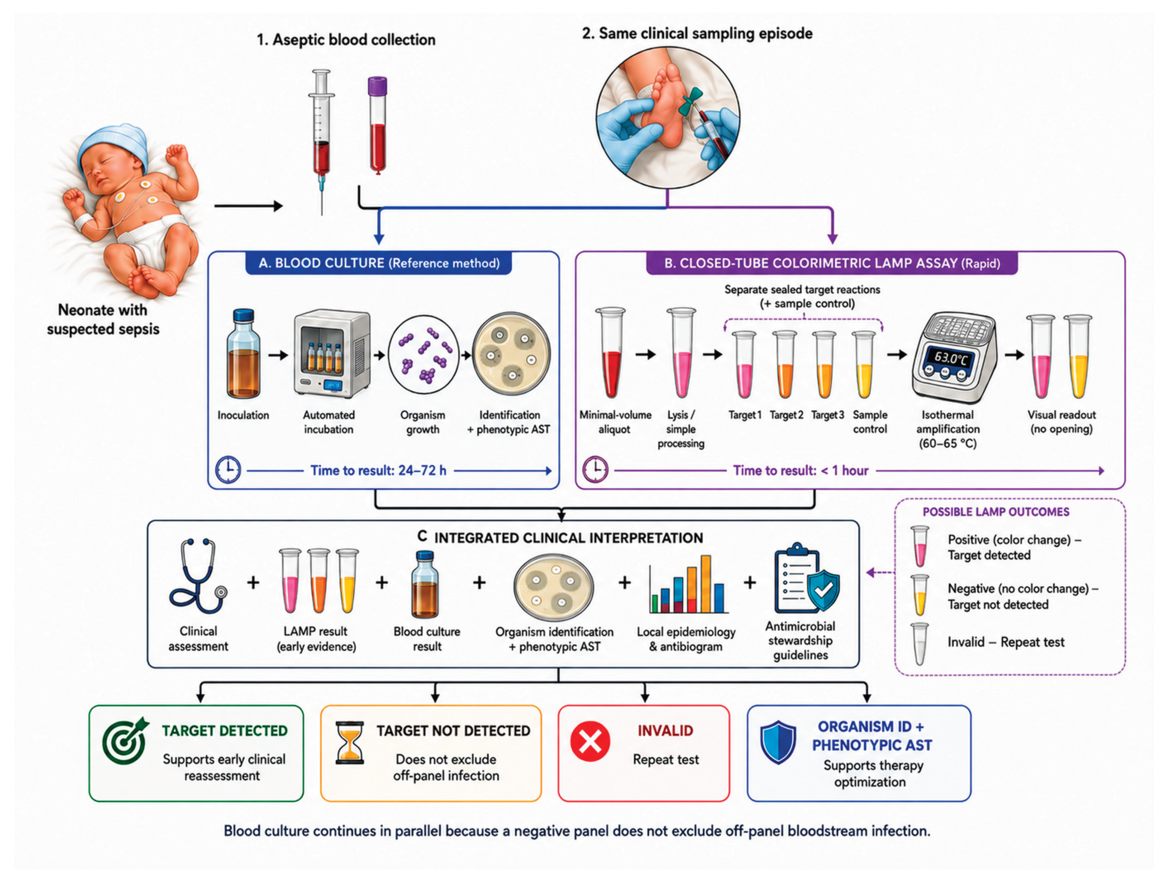

Table 1.
Representative pre-added colorimetric indicators for closed-tube LAMP and their relevance to neonatal-blood pathogen detection.
Table 1.
Representative pre-added colorimetric indicators for closed-tube LAMP and their relevance to neonatal-blood pathogen detection.
| Readout chemistry | Amplification-linked signal and visual endpoint | Principal value in a closed-tube assay | Critical consideration for neonatal-blood testing | Evidence closest to the intended use | References |
|---|---|---|---|---|---|
| pH-sensitive indicators (phenol red or cresol red) | Proton generation during DNA synthesis; pink/red or violet to yellow | Pre-added indicator; instrument-free endpoint | Sensitive to starting pH, buffering capacity, extraction-buffer carry-over, and crude-lysate volume | Established LAMP chemistry and non-neonatal closed-tube pathogen assays; no direct comparison in neonatal blood | [30,34] |
| Hydroxy naphthol blue (HNB) | Reduction in free Mg²⁺ during magnesium-pyrophosphate formation; violet to sky blue | Pre-added indicator; less directly dependent on sample pH | Requires coordinated optimization of HNB, Mg²⁺, and dNTPs concentrations; weak reactions may produce intermediate colors | Foundational method and non-neonatal clinical comparison; no neonatal-blood head-to-head evaluation | [18,31] |
| Calcein–Mn²⁺ system | Amplification-generated pyrophosphate removes Mn²⁺-mediated quenching; orange to green with increased fluorescence | Sealed-tube visual readout with optional fluorescence confirmation | More complex metal-ion balance; hemolysis or residual blood color may impair naked-eye interpretation | Foundational protocol and non-neonatal comparative studies; neonatal-blood performance has not been established | [18,35] |
| Zn²⁺/5-Br-PAPS system | Amplification-generated pyrophosphate disrupts the Zn²⁺–dye complex; magenta to orange-yellow | Strong visual contrast; avoids dependence on reaction acidification and free-Mg²⁺ depletion | Zn²⁺ and dye concentrations must remain compatible with amplification; reproducibility across blood-processing methods and pathogen targets is unknown | Demonstrated in extraction-free RT-LAMP using crude respiratory specimens; bacterial-pathogen and neonatal-blood validation has not been reported | [36] |
Table 2.
Representative LAMP evidence for selected neonatal-sepsis pathogens.
| Pathogen | Assay target and readout | Closest evaluated matrix or population | Key reported result | Alignment with the proposed neonatal workflow | Reference |
|---|---|---|---|---|---|
| Streptococcus agalactiae (GBS) | cfb; extraction-free colorimetric LAMP | GBS-spiked neonatal blood | LoD: 2 colony-forming units (CFU)/mL | Direct colorimetric and neonatal-matrix match; clinical diagnostic accuracy untested | [19] |
| Escherichia coli | uidA; colorimetric LAMP | Blood from 66 young infants evaluated for sepsis | 10 µL input; LoD: 70 CFU/mL; result in <1 h | Closest clinical-blood evidence; neonatal subgroup and multicenter utility unresolved | [67] |
| Klebsiella pneumoniae | yhaI, epsL, and xcpW; colorimetric or fluorescent LAMP | 319 isolates, 40 sputum samples, and spiked sheep blood | yhaI sensitivity 100% and specificity 91% in sputum | Colorimetric feasibility demonstrated; evidence remains indirect for neonatal blood | [68] |
| Staphylococcus aureus and MRSA | femA and mecA; multiplex LAMP–lateral flow | 63 non-neonatal whole-blood samples | Results concordant with culture for 16 MSSA and 12 MRSA samples | Whole-blood evidence available; lateral-flow detection requires post-amplification handling | [70] |
| Coagulase-negative staphylococci | Species-specific femA targets plus mecA; microfluidic LAMP | 102 positive cerebrospinal-fluid cultures | LoD: 20–200 CFU/reaction; species and resistance results agreed with conventional methods | Closed compartmentalized detection; culture enrichment and contamination interpretation remain limitations | [28] |
| Enterococcus faecalis | Ef0027; multiplex LAMP–lateral flow | Artificially contaminated blood | LoD: 710 CFU/mL; total workflow approximately 75 min | Spiked-blood feasibility only; no closed colorimetric or neonatal clinical validation | [72] |
| Candida albicans | ITS2; LAMP–lateral flow | 330 clinical specimens, including 30 whole-blood samples | 2 of 30 whole-blood samples were culture positive; workflow completed within 85 min | Direct blood included, but positive-case numbers were insufficient for neonatal extrapolation | [74] |
CFU, colony-forming unit; GBS, group B Streptococcus; ITS2, internal transcribed spacer 2; LAMP, loop-mediated isothermal amplification; LoD, limit of detection; MRSA, methicillin-resistant Staphylococcus aureus; MSSA, methicillin-susceptible Staphylococcus aureus.
Disclaimer/Publisher’s Note: The statements, opinions and data contained in all publications are solely those of the individual author(s) and contributor(s) and not of MDPI and/or the editor(s). MDPI and/or the editor(s) disclaim responsibility for any injury to people or property resulting from any ideas, methods, instructions or products referred to in the content. |
© 2026 by the authors. Licensee MDPI, Basel, Switzerland. This article is an open access article distributed under the terms and conditions of the Creative Commons Attribution (CC BY) license (http://creativecommons.org/licenses/by/4.0/).
Copyright: This open access article is published under a Creative Commons CC BY 4.0 license, which permit the free download, distribution, and reuse, provided that the author and preprint are cited in any reuse.


