
Submitted:
04 August 2026
Posted:
05 August 2026
You are already at the latest version
Abstract
Urinary continence in healthy women is not a strictly two-body elastic collision but a viscoelastic process of energy storage and release involving neuromuscular regulation, pelvic floor soft-tissue response, and fascial and ligamentous constraint. During surges in intra-abdominal pressure, the pelvic floor muscles contract in anticipation, the soft tissues deform reversibly, and elastic potential energy is transiently stored, while the pelvic support system supplies the structural constraint required for functional closure of the urethra and bladder neck; once the pressure subsides, the stored energy restores a configuration closely resembling the initial state. Building on this analogy, we propose three sequential principles for physiological reconstruction: (1) reconstructing the perineal body and the levator hiatus plane to restore a stable fulcrum; (2) reconstructing functional closure of the bladder neck and proximal urethra under appropriate indications, while minimizing abnormal tension and the tendency to gape; and (3) restoring continence to a structural–functional state governed primarily by autologous tissue, supplemented by limited synthetic material when necessary. In stress urinary incontinence with moderate-to-severe cystocele, native support is often extensively damaged and autologous repair alone may carry a long-term recurrence risk; judicious synthetic reinforcement after careful evaluation of indications, material risks and patient benefit is therefore a reasonable option. Preliminary clinical experience suggests improved total continence without compromising voiding safety; standardized trials are required.
Keywords:
stress urinary incontinence
; elastic collision analogy
; viscoelasticity
; physiological reconstruction
; functional closure of the bladder neck
; structural constraint
; synthetic mesh
1. Introduction
The history of surgical intervention for female stress urinary incontinence (SUI) mirrors the continual evolution of theories on urethral support, their surgical implementation, and their ongoing clinical reappraisal. Mid-urethral sling (MUS) surgery remains a primary first-line option for female SUI. Its theoretical basis derives largely from the Integral Theory and the Hammock Hypothesis: the former emphasizes the synergistic interplay among the vagina, fascia, ligaments and pelvic floor muscles, whereas the latter highlights the critical role of the supportive tissues beneath the urethra in compressing the urethra and maintaining urethral coaptation during rises in intra-abdominal pressure [1,2]. This paradigm has long dominated clinical practice and has yielded favorable outcomes in a substantial number of patients; however, its limitations are becoming increasingly evident.
Although MUS can achieve relatively satisfactory medium- and long-term efficacy in some patients [3,4], the therapeutic effect is not durable in all cases. Several studies report that the long-term recurrence rate of incontinence after MUS may approach 25% [5], with urinary dysfunction occurring in 1.47–3.5% of cases, mesh exposure in 0.2–1.9%, and a reoperation rate as high as 9% [6]. Large-scale population-based cohort studies indicate that, while the absolute incidence of these complications is generally low, the cumulative risk becomes considerable with extended follow-up [7]. These non-negligible safety concerns prompted the U.S. Food and Drug Administration to issue multiple warnings beginning in 2008 and to completely ban transvaginal mesh in 2019 [8]; several countries have also prohibited or heavily restricted its use [9].
The clinical question at hand is not whether MUS has entirely failed, but rather whether merely augmenting suburethral support is insufficient to restore stable continence and may even introduce new injury. In a separate study, our group employed an elastic-collision analogy to explore the mechanical limitations of MUS: in certain individuals, permanent non-absorbable material may alter stress distribution around the urethra, create localized constriction and provoke adverse tissue reactions, thereby contributing to risks such as voiding dysfunction, pain, mesh exposure and recurrence [10]. The aim of the present article is to propose an alternative perspective, arguing that physiological reconstruction—centred on pelvic floor viscoelastic energy storage and release, structural constraint and functional closure—may represent an important therapeutic direction for patients with SUI and concomitant pelvic floor support defects.
2. The Elastic Collision Analogy of the Pelvic Floor Continence Mechanism
It should be clarified that the term “elastic collision” adopted in this paper does not denote a classical two-body collision in the mechanical sense; rather, it serves as a conceptual analogy for the viscoelastic behaviour of the pelvic floor soft tissues during female urinary control. From a biomechanical standpoint, urinary control is more accurately characterized as a process of viscoelastic energy storage and release under structural constraint. During surges in intra-abdominal pressure, the pelvic floor muscles actively contract, causing soft-tissue deformation that achieves structural closure and converts the kinetic energy of the impact into stored viscoelastic potential energy (energy storage). After the pressure subsides, the stored potential energy drives the structures back toward their initial configuration (energy release). Pelvic ligaments, fascia and muscles provide the necessary structural constraint, ensuring that energy conversion proceeds efficiently along a defined pathway. The coefficient of restitution e of this model approaches 1 under physiological conditions (Equation (1)), indicating that the deformation is reversible and that no permanent structural damage occurs. To facilitate comprehension among clinical readers, the term “elastic collision” is retained in the following discussion. Within this analogical framework, urinary control in healthy women can be understood as a highly reversible viscoelastic response system. When intra-abdominal pressure rises abruptly, the posterior fornix functions as a critical fulcrum, while the levator plate, perineal body, bladder and uterus collectively participate in stress transmission and deformation control, thereby effecting closure of the bladder neck and vagina [11]. Concurrently, the synergistic action of the pelvic floor support system and its musculature helps preserve the physiological curvature of the mid-urethra and a geometric locking effect [12]. As the pressure surge diminishes, the pelvic floor muscles relax in a coordinated fashion, and the stored potential energy, together with the structural constraint, restores local anatomical relationships to a state closely resembling the initial condition. The key feature of this process is not absolute “zero energy dissipation”, but rather that the deformation is recoverable, closure is maintained and no lasting structural damage ensues. The correspondence between the elastic collision analogy and physiological urinary control is summarized in Figure 1.
This physiological cycle depends on three essential elements. First, a stable mechanical fulcrum and a continuous, stable support system are required to enable temporary storage, distribution and release of energy, to restrict excessive displacement and to keep tissue deformation within a reversible range. Second, the bladder neck and proximal urethra must sustain functional closure during abdominal pressure surges, meaning that the urethral lumen remains effectively coapted under pressure. Third, surgical intervention should aim to restore, to the greatest extent possible, the dynamic regulatory function of the patient’s own tissues under physiological conditions. In patients with SUI complicated by moderate or severe bladder prolapse, the prolapse itself often indicates extensive native tissue damage. From the perspective of long-term efficacy, repair relying solely on native tissue may carry a risk of recurrence. Therefore, the judicious use of synthetic materials under strict indications can provide appropriate reinforcement of the compromised supportive structures.
3. Physiological Reconstruction
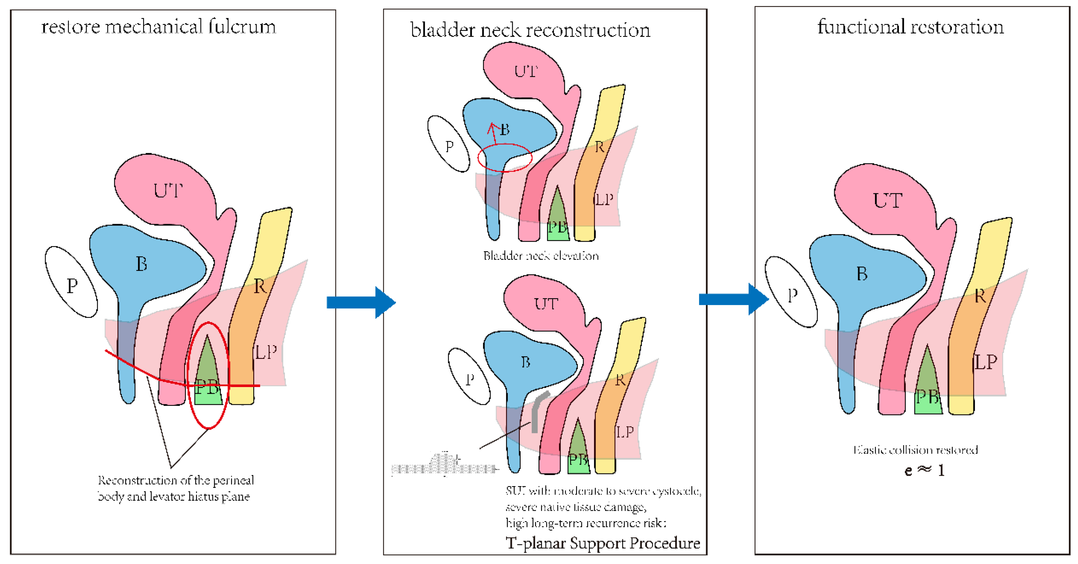
Based on the biomechanical analogy described above, physiological reconstruction comprises three essential steps (Figure 2).
3.1. Step 1: Reconstruction of the Perineal Body and the Levator Hiatus Plane—Restoring the Mechanical Fulcrum for Viscoelastic Energy Storage and Release
The levator plate–perineal body complex plays a critical role in directing stress transmission, limiting deformation and facilitating structural realignment during fluctuations in abdominal pressure. Birth trauma, chronic prolapse or degenerative tissue changes can disrupt this support system, altering pelvic floor load-transfer pathways and increasing the risk of abnormal local displacement and impaired closure. Accordingly, the first priority in physiological reconstruction is to repair the perineal body and the levator hiatus plane, thereby re-establishing continuity and stability of the fulcrum.
3.2. Step 2: Bladder Neck Reconstruction—Achieving Functional Closure and Restoring Structural Constraint
During the storage phase, the continence system must maintain effective closure of the urethra and bladder neck to prevent visible leakage when abdominal pressure rises. The biomechanical prerequisite is that the bladder neck and proximal urethra sustain adequate closure pressure, tissue coaptation and appropriate mobility under stress, rather than being pulled open abnormally.
Clinically, bladder neck reconstruction can be accomplished through two technical approaches: the T-planar support procedure and mechanical bladder neck reconstruction. It must be emphasized that the use of synthetic materials in the T-planar support procedure should be reserved for patients with SUI complicated by moderate or severe bladder prolapse, in whom native tissue damage is extensive and autologous repair alone carries a high long-term recurrence risk. The intent is not to replace pelvic floor function with foreign material, but to provide essential reinforcement for critically compromised support structures under strict indications. Mechanical bladder neck reconstruction, by contrast, involves dissecting the space between the bladder and the vagina and then elevating and reshaping the bladder neck region to enhance its capacity for functional closure. The shared goal of both approaches is to reduce abnormal lateral tension and the tendency of the bladder base–trigone and peri-bladder-neck tissues to gape open, to strengthen structural constraint in key areas, to protect the bladder neck from excessive pressure and displacement, and to indirectly augment the functional urethral length.
3.3. Step 3: Functional Restoration—Returning To a Recoverable Structural–Functional State
The core message conveyed by the elastic collision analogy is not the absolute absence of energy dissipation, but rather the recoverability of deformation, the stability of supporting structures and the maintenance of functional closure. MUS achieves continence through purely mechanical compression; however, the permanently implanted material carries risks of voiding difficulty, pain, exposure or recurrence. In contrast to strategies that rely solely on mid-urethral support, physiological reconstruction aims to restore the overall pelvic floor support network, to achieve functional closure of the bladder neck and to ensure even stress distribution. By re-establishing the biomechanical foundation through posterior pelvic floor reconstruction and recovering functional closure via bladder neck reconstruction, the continence function of the pelvic floor can be expected to return to a structural–functional state that more closely approximates the physiological norm. This is the fundamental distinction between physiological reconstruction and single-point mechanical support: it allows continence to be governed, to the greatest extent possible, by autologous tissue, with synthetic reinforcement applied only in a limited fashion when native tissue damage is severe.
4. Preliminary Clinical Observations Supporting Physiological Reconstruction
Our team has adopted and reported this proactive physiological reconstruction paradigm (posterior pelvic floor reconstruction combined with bladder neck reconstruction) for the treatment of bladder prolapse complicated by SUI. Preliminary imaging observations suggest that postoperative pelvic floor anatomy and certain dynamic parameters may be improved [13]. In addition, this reconstructive procedure has been associated with an increased rate of complete urinary control and a low incidence of voiding difficulty. These findings indicate that structure–function reconstruction may optimize both continence and voiding safety in selected patient populations, although further validation with larger case numbers, longer follow-up, objective measures and controlled study designs is warranted.
This represents not merely a technical refinement but a fundamental shift in the logic guiding surgical design. The passive support paradigm may, in certain patients, increase the risk of complications owing to prolonged material–tissue contact, local constriction or excessive tension. By contrast, the proactive physiological reconstruction paradigm seeks to restore the biomechanical foundation through posterior pelvic floor reconstruction and to achieve functional closure via bladder neck reconstruction. In cases of moderate-to-severe cystocele with SUI and extensive damage to autologous tissue, synthetic materials are used in a limited fashion for structural reinforcement. The potential advantages of this approach stem from more anatomical restoration, improved stress distribution and refined patient selection.
It should be noted that the present paper focuses on establishing the biomechanical rationale for physiological reconstruction, drawing on an elastic collision analogy and a viscoelastic energy storage–release model; the clinical results cited serve only as preliminary supporting evidence. Detailed clinical data on T-planar support bladder neck reconstruction, mechanical bladder neck reconstruction, and perineal body and levator hiatus plane reconstruction—including indications, case numbers, follow-up duration, efficacy endpoints and definitions of complications—will be reported in forthcoming original research articles.
5. Conclusions
The shift from passive support to functional reconstruction represents a direction worthy of further investigation in the surgical treatment of female SUI. At the core of this transition is a move away from sole reliance on static extrinsic support toward restoring the pelvic floor’s intrinsic capacities for viscoelastic energy storage and release, structural constraint and functional closure of the bladder neck. Reconstruction of the perineal body and the levator hiatus plane to restore the mechanical fulcrum, together with bladder neck reconstruction to achieve functional closure while reducing abnormal tension and the tendency to gape open, constitutes a key technical pathway within physiological reconstruction. For patients with SUI complicated by moderate or severe bladder prolapse, in whom native tissues are often severely compromised and autologous repair alone may carry a risk of long-term recurrence, the judicious use of synthetic materials may serve as an appropriate reinforcement.
This is not simply a technical modification; it represents a clinical strategy update grounded in biomechanical reasoning. At present, “physiological reconstruction” is best defined as a therapeutic framework that possesses biomechanical plausibility and is open to clinical testing. Its clinical value needs to be validated through standardized patient classification, dynamic imaging, urodynamic evaluation and long-term controlled trials. Ultimately, the pelvic floor needs to be reconceptualized—not as a static structure requiring mere passive support, but as a complex mechanical system capable of viscoelastic response, structural constraint and dynamic regulation.
Author Contributions
Conceptualization, JI-Hong Shen; writing—original draft preparation, Shuyi Zhang, Ling Li; writing—review and editing, Shuyi Zhang, Ling Li; visualization, Shuyi Zhang. All authors have read and agreed to the published version of the manuscript.
Institutional Review Board Statement
Not applicable.
Informed Consent Statement
Not applicable.
Data Availability Statement
No new data were created or analyzed in this study.
Acknowledgments
During the preparation of this manuscript, the authors used GPT for language polishing. The authors have reviewed and edited the output and take full responsibility for the content of this publication.
Conflicts of Interest
The authors declare no conflict of interest.
Abbreviations
| SUI | Stress urinary incontinence |
| MUS | Mid-urethral sling |
| IAP | Intra-abdominal pressure |
| FDA | U.S. Food and Drug Administration |
| LP | Levator plate |
| PB | Perineal body |
References
- Pe P, Ui U. An integral theory of female urinary incontinence. Experimental and clinical considerations[J]. Acta obstetricia et gynecologica Scandinavica. Supplement, 1990, 153.
- Jo D. Structural support of the urethra as it relates to stress urinary incontinence: the hammock hypothesis[J]. American journal of obstetrics and gynecology, 1994, 170(6): 1713-1720; discussion 1720-1723.
- Aa F, L R, Jd C, et al. Mid-urethral sling operations for stress urinary incontinence in women[J]. The Cochrane database of systematic reviews, 2017, 7(7).
- Nilsson C G, Palva K, Aarnio R, et al. Seventeen years’ follow-up of the tension-free vaginal tape procedure for female stress urinary incontinence[J]. International Urogynecology Journal, 2013, 24(8): 1265-1269. [CrossRef]
- Bb M, Ee P, Sal van L, et al. Persistent or recurrent urinary incontinence 8 years after midurethral sling surgery: a retrospective cohort study[J]. International urogynecology journal, 2025, 36(12): 2401-2410.
- Farag F, Osman N I, Pang K H, et al. Complications of synthetic midurethral slings: is there a relevant discrepancy between observational data and clinical trials?[J]. European Urology Focus, 2024, 10(4): 535-550. [CrossRef]
- P M, I G U, J van der M, et al. Risk of reoperation 10 years after surgical treatment for stress urinary incontinence: a national population-based cohort study[J]. American journal of obstetrics and gynecology, 2021, 225(6).
- Pycek M, Zarzecka J M, Majkusiak W, et al. Current approach to the use of transvaginal mesh systems in pelvic organ prolapse[J]. Ginekologia Polska, 2025, 96(5): 414-419. [CrossRef]
- Ng-Stollmann N, Fünfgeld C, Gabriel B, et al. The international discussion and the new regulations concerning transvaginal mesh implants in pelvic organ prolapse surgery[J]. International Urogynecology Journal, 2020, 31(10): 1997-2002. [CrossRef]
- Shen J H, Li H, Tian D M, et al. Why the Mid-Urethral Sling Cannot Be a Permanent Solution? A Physics-Based Critique of the Hammock Theory and the Case for Physiological Continence Reconstruction[A]. 2026.
- Tian D, Luo Q, Wang X, et al. Dynamic biomechanical equilibrium in pelvic organ prolapse: from mechanistic insights to precision reconstruction[J]. Frontiers in Medicine, 2025, 12: 1637133. 1637133, . [CrossRef]
- Tian D, Li L, Wen Y, et al. Female urinary continence mechanism: a dynamic biomechanical balance[J]. Medical Hypotheses, 2025, 201: 111708. [CrossRef]
- Li L, Tian D M, Wang X Q, et al. T-shaped sling with modified posterior pelvic reconstruction: a technical note with video vignette[J]. Videosurgery and Other Miniinvasive Techniques, 2025: 1-4.
Figure 1.
Correspondence between the elastic collision analogy and physiological urinary control in women. P, pubic symphysis; B, bladder; UT, uterus; R, rectum; PB, perineal body; LP, levator plate; IAP, intra-abdominal pressure. (a) Abdominal pressure impact: storage of elastic potential energy. (b) Abdominal pressure unloading: the elastic potential energy is completely released and the structure returns to its initial form; the entire cycle shows minimal energy dissipation and no permanent deformation, with a coefficient of restitution e approaching 1. (c) At rest: the bladder neck is closed and the pelvic floor muscle groups maintain their basal tension. (d) Abdominal pressure impact during the storage phase: the levator plate and perineal body actively contract upward and forward (red arrows), the bladder neck is fastened behind the pubic bone and a geometric torsion is formed at the mid-urethra; the kinetic energy of the external impact is temporarily stored as elastic potential energy of the soft tissues. (e) Voiding phase: the pelvic floor muscle groups relax in a coordinated manner (blue arrows) and the elastic potential energy is fully released, with the structure returning to its initial form.
Figure 1.
Correspondence between the elastic collision analogy and physiological urinary control in women. P, pubic symphysis; B, bladder; UT, uterus; R, rectum; PB, perineal body; LP, levator plate; IAP, intra-abdominal pressure. (a) Abdominal pressure impact: storage of elastic potential energy. (b) Abdominal pressure unloading: the elastic potential energy is completely released and the structure returns to its initial form; the entire cycle shows minimal energy dissipation and no permanent deformation, with a coefficient of restitution e approaching 1. (c) At rest: the bladder neck is closed and the pelvic floor muscle groups maintain their basal tension. (d) Abdominal pressure impact during the storage phase: the levator plate and perineal body actively contract upward and forward (red arrows), the bladder neck is fastened behind the pubic bone and a geometric torsion is formed at the mid-urethra; the kinetic energy of the external impact is temporarily stored as elastic potential energy of the soft tissues. (e) Voiding phase: the pelvic floor muscle groups relax in a coordinated manner (blue arrows) and the elastic potential energy is fully released, with the structure returning to its initial form.
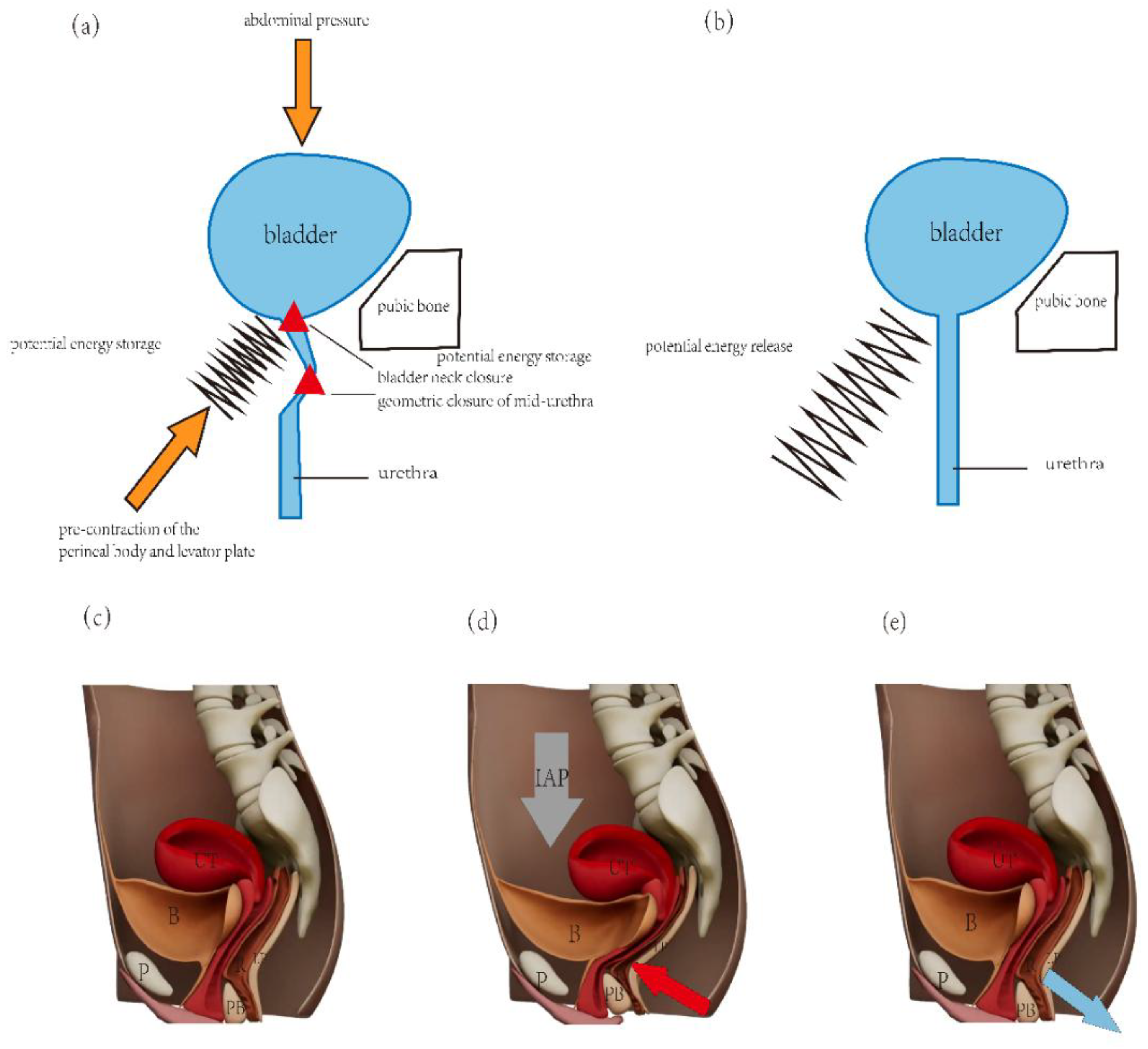
Figure 2.
The three mechanical links of physiological reconstruction. P, pubic symphysis; B, bladder; UT, uterus; R, rectum; PB, perineal body; LP, levator plate.
Figure 2.
The three mechanical links of physiological reconstruction. P, pubic symphysis; B, bladder; UT, uterus; R, rectum; PB, perineal body; LP, levator plate.
Disclaimer/Publisher’s Note: The statements, opinions and data contained in all publications are solely those of the individual author(s) and contributor(s) and not of MDPI and/or the editor(s). MDPI and/or the editor(s) disclaim responsibility for any injury to people or property resulting from any ideas, methods, instructions or products referred to in the content. |
© 2026 by the authors. Licensee MDPI, Basel, Switzerland. This article is an open access article distributed under the terms and conditions of the Creative Commons Attribution (CC BY) license (http://creativecommons.org/licenses/by/4.0/).
Copyright: This open access article is published under a Creative Commons CC BY 4.0 license, which permit the free download, distribution, and reuse, provided that the author and preprint are cited in any reuse.