

Submitted:
04 August 2026
Posted:
05 August 2026
You are already at the latest version
Abstract
The maintenance of calcium and phosphate homeostasis is governed by a highly regulated and complex hormonal network involving parathyroid hormone (PTH), fibroblast growth factor 23 (FGF23), and activated vitamin D (calcitriol). As kidney function declines, the capacity to excrete phosphate and absorb calcium both diminish, resulting in progressive disturbances in bone and mineral metabolism. This disruption promotes an imbalance in calcium–phosphate homeostasis that favors dissolution of bone with simultaneous deposition of calcium–phosphate complexes in soft tissues and the vasculature. Clinically, these derangements manifest as a broad and heterogeneous spectrum of disease, including bone pathology, vascular and valvular calcification, tumoral calcinosis, and calciphylaxis. Notably, these markedly different clinical phenotypes can present in patients with advanced chronic kidney disease (CKD) and End Stage Kidney Disease (ESKD) who exhibit seemingly similar biochemical and hormonal profiles. The determinants of this tissue-specific susceptibility remain incompletely understood and likely reflect a complex interplay between local tissue biology, systemic regulators of mineral metabolism, and environmental influences. This review will systematically present the diverse manifestations of calcium–phosphate imbalance in advanced CKD and explore the underlying pathophysiologic mechanisms that may determine why certain patients develop specific complications while others do not.
Keywords:
chronic kidney disease
; CKD-MBD
; vascular calcification
; hyperphosphatemia
; renal osteodystrophy
; calciphylaxis
; FGF23
; ectopic calcification
Introduction
Calcium and phosphate homeostasis represents one of the most tightly regulated and physiologically consequential systems in human biology, where even minor perturbations carry a profound effect on skeletal integrity, cardiovascular health, and overall survival. Approximately 99% of total body calcium and 85–90% of phosphate are stored in bone as hydroxyapatite crystals which serves as a dynamic reservoir that buffers extracellular mineral concentrations. The remaining fractions of these two minerals circulate in the extracellular and intracellular compartments, tightly regulated within narrow physiological ranges [5,6]. Homeostasis of calcium and phosphate is maintained through coordinated interactions among the intestine, bone, and kidney, under the control of a hormonal network involving parathyroid hormone (PTH), activated vitamin D (calcitriol or 1,25-dihydroxyvitamin D), and fibroblast growth factor 23 (FGF23) [5,18,54].
Intestine–Bone–Kidney Axis
Calcium absorption in the small intestine occurs via both passive paracellular diffusion and active transcellular transport, the latter being strongly upregulated by calcitriol through increased expression of TRPV6 channels, calbindin, and Ca²⁺ATPase pumps. Phosphate is absorbed mainly through paracellular diffusion and also by sodium-dependent cotransport (NaPi-IIb), upregulated by calcitriol. PTH indirectly enhances the intestinal absorption of both calcium and phosphate by stimulating renal 1-alpha-hydroxylase activity and subsequently increasing calcitriol production. FGF23 opposes this effect by suppressing calcitriol synthesis, thereby reducing intestinal mineral uptake [5,11,18,62].
Bone is the primary reservoir for calcium and phosphate, undergoing continuous remodelling through the coordinated activity of osteoblasts and osteoclasts. PTH is central here, promoting osteoclast-mediated bone resorption, mobilizing calcium and phosphate into the circulation, while also enhancing bone formation through its actions on osteoblasts. Osteocytes secrete FGF23 in response to phosphate load and increased calcitriol signaling [54,62].
The kidney serves as the principal regulator in this axis. Approximately 80–90% of filtered phosphate is reabsorbed in the proximal tubule via NaPi-IIa and NaPi-IIc co-transporters, which are downregulated by PTH and FGF23 binding to klotho receptors, promoting phosphaturia. Calcium reabsorption occurs predominantly through passive paracellular mechanisms in the proximal tubule and the thick ascending limb, with fine calibration in the distal convoluted tubule through the action of PTH. The proximal tubule also contains the above mentioned 1-alpha-hydroxylase which synthesizes calcitriol from 25-OH vitamin D, a process upregulated by PTH and inhibited by FGF23; calcitriol in turn allows for feedback inhibition of PTH release from the parathyroid gland [5,11,62].
Phosphate in CKD and the Trade-Off Hypothesis
The tendency to retain phosphate is one of the earliest bone and mineral metabolism disturbances in CKD, but it is at least partially prevented by compensatory increases in phosphaturic hormones that maintain serum phosphate levels within the normal range until late stages [9,26,62]. Originally described by Neal S. Bricker, the trade-off hypothesis provides a unifying framework to explain how maintenance of normal serum phosphate occurs at the cost of secondary hyperparathyroidism [9]. As glomerular filtration declines, phosphate retention is initially prevented by increased FGF23 activity, which reduces proximal tubular phosphate reabsorption and suppresses calcitriol synthesis. The resulting decrease in calcitriol leads to reduced intestinal absorption of calcium, contributing to a subtle decline in serum calcium that stimulates the parathyroid glands to increase PTH secretion. Elevated PTH partially restores mineral homeostasis by enhancing renal calcium reabsorption, and stimulating calcitriol production, thereby increasing intestinal calcium absorption, and further decreasing renal tubular reabsorption of phosphate [5,18]. Although these adaptations initially preserve near-normal serum calcium and phosphate concentrations, they become progressively maladaptive over time, with sustained elevations in FGF23 and PTH driving disordered bone remodeling, renal osteodystrophy, and broader systemic complications collectively known as CKD mineral and bone disorder (CKD-MBD) [41].
Early experimental work by Eduardo Slatopolsky and colleagues demonstrated that phosphate retention is a primary driver of secondary hyperparathyroidism in CKD. In both animal models and human studies, dietary phosphate restriction prevented the rise in PTH despite reduced GFR, directly supporting the hypothesis that phosphate burden, not loss of kidney function alone, stimulates parathyroid activity [63,64]. Subsequent human studies evaluating graded reductions in GFR showed that serum phosphate remains within the normal range across a wide spectrum of kidney function, while fractional excretion of phosphate progressively increases, reflecting adaptive nephron-level changes. Importantly, these studies also demonstrated that PTH levels rise early in CKD, well before overt hyperphosphatemia develops, reinforcing the trade off hypothesis concept that hormonal adaptations precede measurable changes in serum phosphate [64,65].
More recent work has refined this model by identifying FGF23 as an even earlier mediator of phosphate homeostasis. In large observational studies such as the chronic renal insufficiency cohort (CRIC), FGF23 levels increase early in CKD, often before PTH rises and well before serum phosphate increases [26]. This supports an updated trade-off paradigm in which FGF23 represents the initial adaptive response to phosphate retention, followed by secondary increases in PTH as calcitriol levels decline.
Major Clinical Syndromes
1. Renal Osteodystrophy
Renal osteodystrophy (ROD) represents the skeletal manifestations of CKD-MBD and itself encompasses a heterogeneous group of abnormalities in bone turnover, mineralization, and volume that arise as kidney function declines [38,41]. It represents one of the major contributors to the markedly increased fracture risk, disability, and mortality observed in patients with advanced CKD. Historically considered synonymous with secondary hyperparathyroid bone disease, ROD is now recognized as a spectrum of histological phenotypes hence requiring a bone biopsy for precise diagnosis [17,38,41].
Pathophysiology
ROD is broadly categorized by abnormalities in bone turnover, mineralization, and volume (TMV classification) and clinically separated into high-turnover and low-turnover phenotypes [38,41].
The basis of high-turnover disease is described by the trade-off hypothesis above, eventually leading to secondary hyperparathyroidism [14,64]. Sustained elevations in PTH stimulate osteoblastic and osteoclastic activity, accelerating bone remodeling and increasing skeletal turnover. High-turnover ROD includes secondary hyperparathyroid bone disease with osteitis fibrosa cystica (OF) representing the most severe histological phenotype. OF is characterized by intense osteoclastic resorption, peritrabecular and marrow fibrosis, cystic skeletal lesions, and in advanced cases brown tumors [14,38].
Low-turnover ROD includes adynamic bone disease (AD) and often develops in the setting of relative PTH deficiency or oversuppression in the setting of treatment with calcitriol, calcimimetics, or parathyroidectomy [7]. In these patients, bone remodelling is markedly reduced due to suppressed osteoblastic and osteoclastic activity. Diabetes is strongly associated with AD through impaired PTH responsiveness, suppression of osteoblast function, and altered skeletal remodeling. Bone buffering capacity in AD is also markedly reduced, hence calcium is less readily incorporated into the skeleton, predisposing patients to hypercalcemia and extraskeletal calcification [7].
Within the spectrum of low-turnover ROD is also osteomalacia (OM), a disorder defined primarily by defective mineralization of newly formed osteoid. In CKD, osteomalacia may reflect calcitriol deficiency, nutritional vitamin D deficiency, chronic acidosis, aluminum exposure, and other uremic factors, and may occur despite normal or elevated serum phosphate concentrations [38,41]. Distinction between these forms of ROD in CKD or ESKD generally requires transiliac bone biopsy with tetracycline-labeled histomorphometry, as biochemical markers and imaging lack the sensitivity and specificity for detection [17,38,41].
Clinical Presentation
The clinical manifestations of ROD are often subtle and may be poorly correlated with the underlying histologic subtype. Across the spectrum of disease, including both high-turnover and low-turnover phenotypes, many patients remain asymptomatic for prolonged periods and are identified only through abnormalities in biochemical markers or imaging. When symptoms occur, they are frequently nonspecific and may include diffuse bone pain, proximal muscle weakness, reduced exercise tolerance, skeletal tenderness, or chronic musculoskeletal discomfort [14,41]. High-turnover lesions related to secondary hyperparathyroidism may additionally present with bone pain, pruritus, tendon rupture, or rarely focal skeletal lesions such as brown tumors.
Regardless of the specific subtype, fracture represents the most clinically important endpoint of ROD, reflecting cumulative deficits in bone quality, turnover, mineralization, and strength. Patients with CKD have a substantially greater fracture burden than the general population, and fracture risk rises progressively with worsening kidney function [27,44]. Hip fracture incidence across the spectrum of CKD has been reported to be 2- to 4-fold higher than in age- and sex-matched individuals without CKD, while patients receiving dialysis may have an even greater excess risk [27,44]. Large cohort studies have demonstrated an approximately 4-fold higher risk of hip fracture in ESKD patients on dialysis compared with the general population, with some meta-analyses suggesting risks exceeding 5-fold [27,44]. Hip fractures in this population also occur at younger ages and are associated with prolonged hospitalization, loss of independence, and increased mortality [27].
Vertebral and non-vertebral fragility fractures are likewise common but frequently underrecognized because they may present only with height loss, kyphosis, chronic pain, or functional decline. Importantly, fracture risk in advanced CKD reflects not only osteoporosis, but also the combined effects of CKD-MBD, sarcopenia, frailty, neuropathy, falls, and dialysis-related comorbidity. Thus, in patients with advanced CKD or ESKD, otherwise unexplained bone pain, declining mobility, or low-trauma fracture should prompt consideration of underlying ROD.
Treatment
Treatment of ROD is aimed at correcting the underlying abnormalities of CKD-MBD, reducing fracture risk, preserving bone strength, and minimizing extraskeletal calcification [14,30].
Across the spectrum of disease, prevention of phosphate retention remains a central therapeutic target. This includes restriction of dietary phosphate intake, particularly highly absorbable phosphate additives found in processed foods, use of phosphate binders and blockers of GI phosphate absorption when needed, and optimization of dialysis clearance in patients receiving renal replacement therapies.
High-turnover ROD treatment is directed at lowering excess PTH while avoiding oversuppression of PTH or bone turnover if bone biopsy is available [14,30]. Control of hyperphosphatemia and correction of vitamin D deficiency are essential adjunctive therapies. Active vitamin D therapies such as calcitriol or vitamin D receptor activators can suppress PTH secretion, while calcimimetics such as Cinacalcet or Etelcalcetide are particularly useful in dialysis patients with more severe disease. Patients with refractory hyperparathyroidism despite medical therapy may ultimately require parathyroidectomy [14,30].
Low-turnover ROD on bone biopsy or suggested by oversuppressed PTH with hypercalcemia, particularly AD, is managed by reducing factors that suppress normal bone remodeling [7]. This may include limiting calcium loading, reducing calcitriol or calcimimetic exposure, and allowing PTH to rise into a more physiologic range. Exercise, nutritional optimization, diabetes management, and fall prevention are also important supportive measures. When osteomalacia is present on bone biopsy, treatment is focused on correction of impaired mineralization through replacement of nutritional vitamin D deficiency, restoration of calcitriol activity when indicated, treatment of chronic metabolic acidosis, and removal of contributing toxins such as aluminum when relevant [7,14,38].
2. Vascular Calcification
Vascular Calcification (VC) in advanced CKD is a highly prevalent, cell-mediated process that is a major driver of cardiovascular mortality and morbidity [21,42,61]. The development of VC requires both a permissive systemic biochemical milieu characterized by calcium-phosphate supersaturation and an active cellular program that promotes osteogenic transformation of vascular smooth muscle cells [42,61]. VC is classified into intimal calcification, closely associated with atherosclerotic plaque and chronic vascular inflammation, and medial (Mönckeberg) calcification, characterized by diffuse and continuous deposition of hydroxyapatite in the tunica media. Hence, CKD confers a specific risk for medial calcification, although both types are found in advanced CKD since the same risk factors for atherosclerotic disease exist in adult CKD patients [42,61]. CT coronary artery calcium scan remains the gold standard to identify and prognosticate vascular calcification, while newer diagnostic tools such as pulse wave velocity testing strongly correlates with advanced CKD and vascular calcification [4,22,68].
Pathophysiology of Vascular Calcification in Advanced CKD
The hallmark of VC in CKD is the active osteogenic transformation of vascular smooth muscle cells (VSMCs), rather than passive precipitation of calcium–phosphate complexes. As kidney function declines, chronic phosphate retention, oxidative stress, uremic toxins, and persistent inflammation create a pro-calcific environment that drives VSMCs to lose their normal contractile phenotype and acquire characteristics of osteoblast-like cells. Elevated phosphate directly promotes this phenotypic transition by activating NF-κB signaling and upregulating the osteogenic transcription factor RUNX2, leading to expression of bone-associated proteins such as osteocalcin and osteopontin and ultimately promoting hydroxyapatite deposition within the vascular wall [21,28,61]. Hyperphosphatemia also increases tissue non-specific alkaline phosphatase (TNAP), which degrades pyrophosphate, a potent endogenous inhibitor of calcification, thereby further favoring mineral deposition [21,61]. Although hyperphosphatemia is considered the principal driver of osteogenic differentiation, experimental evidence also suggests that elevated PTH may directly contribute by activating PTH1 receptors expressed on VSMCs, increasing expression of RUNX2 and Osterix and further promoting osteogenic transformation [51].
Under physiological conditions, the vasculature possesses several endogenous defense mechanisms that actively inhibit ectopic mineralization. Calcification inhibitors including matrix Gla protein (MGP), Gla-rich protein (GRP), fetuin-A, and pyrophosphate bind calcium-phosphate complexes, suppress osteogenic differentiation, and facilitate the safe transport and clearance of mineral particles [31,58,71]. In CKD, however, these protective systems become progressively impaired through vitamin K deficiency, chronic inflammation, reduced protein activity, and an overwhelming mineral burden. In addition, vascular smooth muscle cells express the calcium-sensing receptor (CaSR), whose activation maintains the contractile phenotype and suppresses osteogenic signaling. Reduced CaSR expression observed in advanced CKD further shifts the balance toward vascular mineralization by promoting loss of the contractile phenotype and increasing RUNX2-mediated osteogenic differentiation [1].
Importantly, systemic disturbances in calcium and phosphate homeostasis alone do not fully explain the marked variability in vascular calcification observed among patients with advanced CKD. Many individuals with similar serum phosphate, calcium, PTH, and FGF23 concentrations develop vastly different degrees of vascular calcification, suggesting that local vascular biology plays an equally important role. Differences in vascular inflammation, oxidative stress, extracellular matrix composition, expression of calcification inhibitors, and susceptibility of VSMCs to osteogenic transformation likely determine whether hydroxyapatite deposition occurs once a permissive systemic mineral environment has been established [21,42,61]. This concept of tissue-specific susceptibility provides one explanation for the remarkable clinical heterogeneity of calcium-phosphate disorders observed in CKD.
Clinical Manifestation of Vascular Calcification
Medial calcification is particularly prominent in CKD and leads to progressive loss of arterial compliance [4,42,68]. As elastic arteries stiffen, there is a rise in systolic blood pressure with a concomitant fall in diastolic pressure, resulting in widened pulse pressure. This altered hemodynamic profile increases left ventricular afterload and promotes the development of LVH and diastolic dysfunction [36,68]. In addition, arterial stiffening accelerates pulse wave velocity and causes early return of reflected waves during systole, further augmenting systolic pressure and myocardial workload [36,68]. The reduction in diastolic pressure impairs coronary perfusion, predisposing to subendocardial ischemia, even in the absence of obstructive coronary artery disease. Clinically, patients may present with difficult-to-control systolic hypertension, exertional dyspnea, or features of heart failure with preserved ejection fraction.
Intimal calcification contributes to luminal narrowing and plaque burden [42,61]. In CKD, calcified plaques are common and may be more diffuse and rapidly progressive. Coronary artery calcification is strongly associated with coronary artery disease, including stable angina, acute coronary syndromes, and myocardial infarction [22,56]. Similarly, calcification within the carotid and cerebral vasculature increases the risk of ischemic stroke and transient ischemic attack. Notably, CKD patients frequently exhibit a combination of medial and intimal calcification, resulting in both impaired vascular compliance and atherosclerotic complications, which together amplify cardiovascular risk [42,61]. Vascular calcification in peripheral arteries contributes to peripheral arterial disease, manifesting as claudication, reduced exercise tolerance, and in advanced cases, critical limb ischemia [47]. Impaired perfusion, combined with microvascular dysfunction and uremia-related factors, contributes to poor wound healing, ulcer formation, and increased risk of limb amputation [47].
Treatment of Vascular Calcification
The treatment of vascular calcification in advanced CKD is primarily directed at reducing phosphate burden and controlling secondary hyperparathyroidism, similar to the management of CKD-MBD as above [30,42]. Emerging strategies targeting calcification inhibitors, such as vitamin K repletion, and novel agents that inhibit hydroxyapatite formation show promise but remain under investigation [55,58]. Despite these approaches, no therapy has been definitively shown to reverse established vascular calcification, and current management focuses on slowing progression and mitigating cardiovascular risk [42,55,61]. Coronary calcification presence justifies aggressive cardiovascular risk reduction with statin-based therapies and lower blood pressure targets [2]. An important consideration to keep in mind is the role of magnesium in opposing vascular calcification. Interestingly, however, hypomagnesemia is common in CKD and dialysis patients and a role for magnesium supplementation in this scenario has been debated [75].
3. Valvular Calcification
Valvular calcification exists on a continuum with vascular calcification but possesses unique structural and biomechanical characteristics that influence its pathogenesis and clinical manifestations. The aortic valve and the mitral annulus are most commonly affected. When compared to the non-CKD population, patients with advanced CKD develop valvular calcification earlier, more rapidly, and with greater severity [37,57,72]. The prevalence of valvular calcification is 8 times greater in patients undergoing hemodialysis than the general population [57,72]. Diagnosis involves non-invasive imaging, usually echocardiography as first line showing defects in leaflet thickening, echogenic calcifications, or reduced mobility; this modality is also used to assess the functional consequences (aortic stenosis or mitral regurgitation) [3].
Pathophysiology of Valvular Calcification
The basic pathophysiology of valvular calcification in advanced CKD is analogous to CKD-MBD maladaptive osteogenic transformation described above in vascular tissue. Phosphate retention, elevated PTH, increased FGF23 and reduced calcitriol, chronic inflammation and oxidative stress all promote osteogenic signalling and mineral deposition through osteogenic differentiation of valvular interstitial cells (VICs), the principal resident cells of the cardiac valves [43,49,57]. What makes valves uniquely susceptible is their exposure to mechanical stress, especially the aortic valve [43,49]. Repetitive shear and pressure, along with a rich extracellular matrix of collagen and elastin, promote activation of inflammatory pathways and osteogenic differentiation of VICs, making the aortic valve and the mitral annulus more prone to mineral deposition [43,49]. The resulting valve injury perpetuates local inflammation, fibrosis, and progressive mineral deposition, driving the development of clinically significant valvular disease. The avascular structure of valves limit the regenerative capacity once injury and inflammation have progressed [43,49].
Clinical Manifestation of Valvular Calcification
Calcification of the aortic valve leads to progressive leaflet thickening, reduced mobility, and ultimately aortic stenosis, characterized by obstruction to left ventricular outflow [3,49]. In early stages, patients may be asymptomatic or present with subtle exercise intolerance. As disease progresses, the classic clinical triad of exertional dyspnea, angina, and syncope emerges [3].
Mitral annular calcification (MAC) involves deposition of calcium within the fibrous annulus of the mitral valve, most commonly affecting the posterior annulus [16,20]. While often asymptomatic in early stages, progressive calcification can impair valvular function and disrupt adjacent cardiac structures. The most common functional consequence is mitral regurgitation, resulting from impaired leaflet coaptation due to annular rigidity. Less commonly, extensive calcification may encroach on the valve orifice and produce mitral stenosis [20]. Beyond valvular dysfunction, MAC has important electrophysiologic implications due to its proximity to the cardiac conduction system. Patients may develop conduction abnormalities, including varying degrees of atrioventricular block, as well as atrial arrhythmias, particularly atrial fibrillation [16,20]. Hemodynamically significant mitral valve disease may lead to left atrial enlargement, pulmonary congestion, and symptoms of heart failure.
Treatment of Valvular Calcification
Medical management of valvular calcification in advanced CKD follows the same tenets of CKD-MBD, involving phosphate control, management of PTH, and avoiding hypercalcemia [30,35]. In this population, given the increased risk of surgical intervention, transcatheter aortic valve replacement is preferred, while surgical mitral valve repair or replacement remains the standard of practice for mitral valve disease [67,70]. Emerging therapies include targeting mineralization pathways (hydroxyapatite inhibitors), Vitamin K repletion (similar to vascular calcification), and anti-inflammatory strategies [55,58].
4. Calciphylaxis (Calcific Uremic Arteriolopathy)
Calciphylaxis is a rare and life-threatening thrombotic vasculopathy that results from the calcification of the arteriolar microvasculature of the deep dermis and subcutaneous adipose tissue in central (visceral) or peripheral presentations. Mortality remains exceptionally high, with reported one-year mortality rates approaching 40–80%, largely driven by infectious complications and sepsis arising from progressive skin ulceration and tissue necrosis [8,39,46].
The diagnosis of calciphylaxis is often clinical and based on the presence of characteristic painful violaceous plaques, retiform purpura, or necrotic ulcerative lesions in patients with advanced CKD or ESKD [45,46]. Definitive diagnosis remains histopathologic confirmation on skin biopsy demonstrating medial calcification of small dermal and subcutaneous arterioles with associated microvascular thrombosis, fat necrosis, and septal panniculitis. Additional findings may include intimal hyperplasia and endothelial injury [23,45]. However, biopsy is frequently avoided because of the risks of poor wound healing, ulcer propagation, infection, and worsening necrosis [8,46].
Imaging may provide important supportive evidence when biopsy is deferred. Plain radiographs and CT imaging can demonstrate subcutaneous calcification, while technetium-99m bone scintigraphy has demonstrated high sensitivity for calciphylaxis and frequently shows increased tracer uptake in affected tissues [19,50].
Calciphylaxis Pathophysiology
Although calciphylaxis shares many of the osteogenic mechanisms described in vascular calcification, including phosphate-driven vascular smooth muscle cell transformation and upregulation of osteogenic transcriptional pathways, it is increasingly recognized as a pathophysiologically distinct entity rather than simply an advanced form of vascular mineral deposition [8,46,73]. In contrast to conventional vascular calcification, which primarily affects large and medium-sized arteries and manifests clinically through arterial stiffness and cardiovascular disease, calciphylaxis preferentially involves the dermal and subcutaneous microvasculature and is characterized by a profound superimposed vasculopathic and thrombotic component resulting in tissue ischemia and necrosis [23,46,73].
The osteogenic transformation pathways implicated in vascular calcification, including activation of RUNX2, BMP-2, alkaline phosphatase, and osteocalcin signaling, are similarly observed in calciphylaxis and contribute to medial calcification of small dermal arterioles [21,28]. However, calciphylaxis appears to involve a more severe disruption in endogenous anti-calcification defenses. Deficiencies in matrix Gla protein (MGP), fetuin-A, and pyrophosphate are thought to play particularly important roles, with the strong association between calciphylaxis and warfarin exposure highlighting the importance of impaired vitamin K-dependent activation of MGP [31,59,73]. Reduced fetuin-A levels, frequently observed in inflammation, malnutrition, and advanced CKD, may further impair buffering of calcium-phosphate nanocrystals and promote uncontrolled microvascular calcification [31].
A major distinguishing feature of calciphylaxis is the central role of endothelial dysfunction and thrombosis. Whereas vascular calcification may progress over years without overt tissue infarction, calciphylaxis lesions demonstrate endothelial injury, fibrointimal hyperplasia, luminal narrowing, and fibrin thrombi within small dermal and subcutaneous arterioles [23,45]. Hypercoagulability appears to contribute substantially to disease progression, with reported associations including protein C deficiency, protein S deficiency, antiphospholipid antibodies, systemic inflammation, and hypoalbuminemia [8,46]. This thrombotic microangiopathy-like process likely explains the severe pain, rapid lesion progression, and ischemic necrosis characteristic of calciphylaxis. Proposed "second-hit" mechanisms including local trauma, injections, surgery, infection, and hypotension may precipitate tissue infarction in an already sensitized calcified microvasculature [46,73].
Another defining feature of visceral calciphylaxis is its striking tissue tropism toward adipose-rich regions such as the abdomen, thighs, buttocks, and breasts. This distribution is not typical of conventional vascular calcification and suggests that local tissue-specific factors contribute importantly to disease pathogenesis. Obesity is among the strongest identified risk factors for calciphylaxis, particularly for proximal lesions [45,46]. Several mechanisms have been proposed to explain this fat predilection. Adipose tissue is relatively hypovascular and prone to local hypoxia, potentially increasing susceptibility to ischemic injury once microvascular compromise develops. Mechanical stress and tension within subcutaneous septa may further predispose to vascular injury in areas of increased adiposity. In addition, adipocytes produce inflammatory cytokines including TNF-α and IL-6, which may amplify endothelial dysfunction, oxidative stress, and osteogenic signaling locally within affected tissues [73]. Experimental studies have also demonstrated that adipocytes exposed to elevated phosphate concentrations may themselves undergo calcific transformation, suggesting that adipose tissue may actively participate in the calcific milieu rather than serving solely as a passive target [73]. Furthermore, adipocytes express MGP, raising the possibility that regional dysregulation of local calcification inhibitors contributes to selective tissue vulnerability [59,73].
Treatment of Calciphylaxis
Treatment of calciphylaxis focuses on interrupting the calcific-thrombotic cascade, limiting tissue ischemia, preventing infection, and optimizing wound healing [46,69].
Warfarin discontinuation is a key intervention whenever feasible given its inhibition of vitamin K–dependent activation of matrix Gla protein (MGP), an important endogenous inhibitor of vascular calcification [59]. This association further supports the concept that calciphylaxis represents profound local failure of anti-calcification defense mechanisms within the microvasculature.
Parenteral administration of sodium thiosulfate (STS) remains the most widely used pharmacologic therapy [12,69]. Proposed mechanisms for STS benefits include calcium chelation, antioxidant effects, vasodilation, and inhibition of hydroxyapatite formation [12,69]. Early case series suggested improvements in pain and wound healing, though more recent observational studies and meta-analyses have not consistently demonstrated clear mortality or wound-healing benefit [69]. Nevertheless, because of biologic plausibility and limited alternatives, STS remains standard adjunctive therapy in many centers. Common adverse effects include nausea, metabolic acidosis, and volume overload that can be addressed with anti-emetics and intensification of dialysis therapy. BEAT-Calci is an ongoing randomized, adaptive, multi-center, platform trial that will evaluate multiple interventions, including STS and other drug therapies as well as different dialysis regimens to establish high-quality evidence on the effect of a range of interventions in patients with ESRD and newly diagnosed calciphylaxis [24].
Newer therapies target calcification more directly. SNF472 (myo-inositol hexaphosphate) inhibits hydroxyapatite crystal growth, thereby targeting the final common pathway of ectopic calcification [55]. Magnesium-based therapies and vitamin K supplementation similarly aim to restore endogenous anti-calcification pathways and reduce crystal formation [55,59]. Despite these emerging approaches, treatment remains largely supportive, with meticulous wound care, hyperbaric oxygen, infection control, and pain management remaining central to outcomes.
5. Tumoral Calcinosis
Tumoral calcinosis (TC) is an uncommon but striking manifestation of severe CKD-MBD characterized by the development of large periarticular calcium-phosphate deposits within soft tissues [40,48,66]. In patients with advanced CKD or ESKD, tumoral calcinosis represents a form of metastatic calcification driven primarily by chronic hyperphosphatemia, elevated calcium-phosphate product, and severe secondary hyperparathyroidism [40,66]. Although historically described as a hereditary disorder involving FGF23 pathway abnormalities in patients with normal kidney function, tumoral calcinosis in CKD occurs as an acquired complication of dysregulated mineral metabolism and prolonged dialysis exposure [48,66].
Pathophysiology of Tumoral Calcinosis
The pathophysiology of tumoral calcinosis in advanced CKD reflects profound disturbances in phosphate homeostasis and failure of normal inhibitory mechanisms that prevent extraskeletal calcium-phosphate deposition [40,66]. Persistent phosphate retention due to declining renal clearance serves as the principal initiating abnormality. As serum phosphate concentrations rise, the calcium-phosphate product exceeds its solubility threshold, favoring precipitation of hydroxyapatite crystals within periarticular soft tissues [13,40].
At the molecular level, CKD-associated tumoral calcinosis shares many osteogenic pathways observed in vascular and valvular calcification as above. Unlike vascular calcification, which develops within the vessel wall, tumoral calcinosis demonstrates a striking predilection for periarticular soft tissues subjected to repetitive mechanical stress. Recurrent microtrauma may provide a nidus for hydroxyapatite crystal nucleation and progressive enlargement of calcific masses. Once established, these lesions incite a chronic foreign-body inflammatory response characterized by macrophages, multinucleated giant cells, and fibrous encapsulation, contributing to continued lesion expansion [48,66]. Histologically, lesions consist of lobulated calcific masses containing hydroxyapatite crystals separated by fibrous septae with surrounding macrophages, multinucleated giant cells, and chronic inflammatory infiltrates [40,48].
Clinical Manifestations of Tumoral Calcinosis
Tumoral calcinosis typically presents as a slowly enlarging, firm periarticular tumor-like masses that may initially be painless but progressively cause pain, stiffness, reduced mobility, and functional impairment depending on lesion size and location [40,48,66]. The hips, shoulders, and elbows are among the most frequently involved sites. Lesions may become massive and mechanically limit joint movement or compress adjacent neurovascular structures. In severe cases, patients may develop difficulty ambulating, impaired upper extremity mobility, or debilitating pain.
Overlying skin may become erythematous, ulcerated, or inflamed, particularly when lesions enlarge rapidly or undergo secondary infection [40,66]. Spontaneous extrusion of chalk-like calcium material through the skin has also been described. Imaging typically demonstrates large multilobulated periarticular calcified masses with cystic or "cloud-like" calcium deposition patterns on radiography or CT imaging [48]. Bone involvement is usually absent, helping distinguish tumoral calcinosis from primary osseous neoplasms.
Treatment of Tumoral Calcinosis
Successful treatment depends on sustained reduction of phosphate burden and regression of the calcium-phosphate product, although complete resolution may require prolonged therapy. Intensification of dialysis, strict dietary phosphate restriction, optimization of phosphate binder therapy, and control of secondary hyperparathyroidism have all been associated with regression of lesions in selected patients [40,66].
Surgical excision may be considered for patients with severe pain, neurovascular compression, recurrent infection, or major functional impairment [48,66]. However, recurrence is common if the underlying metabolic abnormalities remain uncontrolled. Because lesions are often highly vascular and poorly circumscribed, surgery may also be technically challenging. Emerging therapies targeting hydroxyapatite formation and calcification signaling pathways remain investigational. Consequently, sustained correction of phosphate homeostasis through optimization of dialysis, phosphate-lowering therapy, and control of secondary hyperparathyroidism remains the cornerstone of management.
6. Ocular Calcifications (Corneal and Conjunctival)
Clinical Manifestations
Ocular calcification is an underrecognized manifestation of CKD-MBD resulting from metastatic calcium-phosphate deposition within the conjunctiva and cornea. Although frequently asymptomatic, these lesions may cause significant ocular discomfort and visual impairment when advanced [52,53]. The most common manifestations are conjunctival calcification and corneal calcification (band keratopathy). Many patients are identified incidentally on slit-lamp examination, where lesions appear as small, punctate, chalky-white plaques. As calcification progresses, patients may develop blurred vision, foreign-body sensation, ocular pain, and photophobia. When accompanied by ocular surface inflammation, the classic finding of the "red eye of uremia" may produce painful conjunctival irritation that clinically mimics conjunctivitis [52,53].
Pathophysiology
The systemic biochemical milieu underlying ocular calcification is similar to that responsible for other manifestations of CKD-MBD, with chronic calcium-phosphate supersaturation promoting metastatic mineral deposition. However, local ocular factors appear to determine tissue susceptibility. Localized alkalosis within the interpalpebral zone, chronic ocular surface irritation, and minor epithelial injury related to dialysis-associated volume depletion promote hydroxyapatite precipitation within the conjunctiva and cornea [52,53]. Iatrogenic factors may further contribute, as many patients with advanced CKD suffer from dry eye disease and frequently use topical ophthalmic preparations. Some artificial tears and glaucoma medications contain phosphate-containing buffers or preservatives, increasing the local phosphate burden and further promoting hydroxyapatite deposition [53].
Treatment
Management consists of optimization of systemic mineral metabolism together with local ophthalmologic therapy. The primary treatment for symptomatic band keratopathy is epithelial debridement followed by EDTA chelation, resulting in improvement in visual acuity in approximately 80% of patients [53]. Phototherapeutic keratectomy has also been utilized for selected patients, although it may produce corneal surface irregularity and further studies are needed to better define its efficacy and long-term outcomes [52,53].
7. Cutaneous Calcification and Pruritus
Clinical Manifestations
Cutaneous calcification represents an uncommon manifestation of metastatic calcification in advanced CKD and may contribute to localized skin irritation and pruritus. More broadly, chronic kidney disease-associated pruritus (CKD-aP) is one of the most common symptoms experienced by patients receiving dialysis, affecting quality of life, sleep, and psychological well-being. Although many patients have no visible skin calcification, abnormalities in mineral metabolism have been associated with an increased risk of pruritus. In the Chronic Renal Insufficiency Cohort (CRIC), elevated PTH concentrations (>65 pg/mL) were associated with a higher incidence of pruritus, whereas lower serum calcium concentrations (<9 mg/dL) were associated with a lower risk, supporting a contribution of CKD-MBD to symptom development [74].
Pathophysiology
The pathogenesis of CKD-associated pruritus is multifactorial and remains incompletely understood. Calcium-phosphate deposition within the skin has historically been proposed to irritate cutaneous nerve endings and contribute to itching in patients with severe CKD-MBD. However, current evidence suggests that mineral metabolism represents only one component of a broader process involving systemic inflammation, immune dysregulation, peripheral neuropathy, and alterations in opioid receptor signaling. Consequently, disturbances in calcium-phosphate homeostasis may amplify rather than solely cause pruritus.
Treatment
Management includes optimization of CKD-MBD parameters, particularly control of hyperphosphatemia and secondary hyperparathyroidism, in patients with evidence of mineral dysregulation. However, because CKD-associated pruritus is multifactorial, symptom-directed therapies including emollients, gabapentinoids, κ-opioid receptor agonists, ultraviolet B phototherapy, and optimization of dialysis adequacy are frequently required.
Progression of Kidney Disease and Kidney Stones
Beyond its role in CKD-MBD, increasing evidence suggests that phosphate retention itself may contribute to progression of kidney disease. Extensive experimental studies have demonstrated that high dietary phosphate intake accelerates kidney injury and fibrosis, whereas dietary phosphate restriction attenuates progression of established CKD [25,29,34]. In humans, there have been a few epidemiological studies that show an association between serum phosphate levels and rate of progression of kidney disease in CKD patients [10,60]. Although these studies are associations that are hypothesis generating, no interventional studies have shown reducing serum phosphate improves kidney function. Dietary intake of ultraprocessed foods associated with high phosphate intake is a potential unifying explanation for these findings. Nutritional management of CKD with a healthy diet, emphasizing plant-based patterns with low phosphate and ultra processed food intake, could prevent the development of CKD-MBD and all the manifestations of calcification presented.
The majority of idiopathic kidney stones in the Western world are made up of calcium oxalate and only a smaller fraction contain phosphate, but calcium phosphate may be important as a predisposing factor for calcium oxalate kidney stone formation. Idiopathic calcium oxalate stones are thought to form from Randall plaques, which are subepithelial deposits of calcium phosphate in the renal papilla [15,32,33]. It has been postulated that deposition of calcium phosphate in the renal interstitium, and its growth into a Randall plaque formation, is similar to pathological biomineralization seen elsewhere in the body. Once this plaque is exposed to urine, which is metastable with respect to calcium oxalate, calcium oxalate stone formation begins.
Discussion
Integrative Perspective
The diverse clinical syndromes described in this review are best understood not as distinct disease processes, but as different phenotypic expressions of the same underlying disorder: chronic kidney disease–mineral and bone disorder (CKD-MBD). Rather than representing isolated complications, renal osteodystrophy, vascular and valvular calcification, calciphylaxis, tumoral calcinosis, and ocular calcification all arise from a common disturbance in calcium-phosphate homeostasis. Their distribution and severity reflect the balance between systemic promoters of mineralization and tissue-specific mechanisms that either promote or inhibit hydroxyapatite deposition.
A central concept is that phosphate cannot accumulate indefinitely within the circulation. As kidney function declines, phosphate retention develops and is initially counterbalanced by compensatory increases in FGF23 and PTH, preserving near-normal serum phosphate concentrations. However, these adaptations come at the expense of secondary hyperparathyroidism and increased skeletal calcium mobilization. Because advanced CKD is also characterized by reduced calcitriol production and impaired intestinal calcium absorption, much of the calcium required for extraskeletal calcium-phosphate deposition is ultimately derived from bone. In this framework, CKD-MBD may be viewed as a disorder of maladaptive mineral redistribution, whereby hydroxyapatite, the normal mineral constituent of bone, is progressively redistributed to blood vessels, cardiac valves, skin, periarticular soft tissues, and the eye.
Importantly, systemic mineral abnormalities alone do not adequately explain the remarkable heterogeneity observed among patients with advanced CKD. Individuals with similar serum calcium, phosphate, PTH, and FGF23 concentrations frequently develop strikingly different clinical phenotypes. This observation suggests that while systemic phosphate burden establishes a permissive biochemical environment for calcification, local tissue biology ultimately determines where pathological mineralization occurs. Tissue-specific differences in mechanical stress, inflammatory signaling, extracellular matrix composition, vascular smooth muscle or valvular interstitial cell responses, endogenous calcification inhibitors, microvascular susceptibility, and regenerative capacity likely explain why some patients predominantly develop vascular disease, others severe skeletal remodeling, and still others calciphylaxis or tumoral calcinosis.
This conceptual framework also highlights an important limitation of current CKD-MBD management. Contemporary biochemical markers principally reflect systemic mineral homeostasis but provide relatively little information regarding tissue-specific susceptibility to calcification. Consequently, patients with similar laboratory values may carry markedly different risks for individual complications. Future investigation should focus on identifying biomarkers and imaging strategies that better characterize local calcification activity and improve prediction of organ-specific disease. Such approaches may ultimately facilitate more individualized treatment strategies tailored to the predominant phenotype of CKD-MBD rather than relying solely on serum biochemical targets.
Although numerous pharmacologic therapies have been developed to control phosphate, PTH, and calcium homeostasis, none has consistently demonstrated reversal of established extraskeletal calcification. This underscores the importance of prevention through early recognition of phosphate retention and sustained reduction of phosphate burden. Dietary strategies emphasizing minimally processed, plant-predominant foods and avoidance of highly bioavailable phosphate additives represent an important but often underappreciated component of CKD-MBD management, with potential benefits extending beyond bone health to the prevention of vascular and soft tissue calcification.
Ultimately, CKD-MBD should be viewed not simply as a disorder of abnormal laboratory values, but as a systemic disease of pathological biomineralization. Understanding how systemic phosphate toxicity interacts with local tissue susceptibility provides a unifying explanation for the broad clinical heterogeneity observed in advanced CKD and may inform future therapies aimed not only at correcting mineral metabolism but also at preventing tissue-specific calcification before irreversible organ injury occurs.
Figure 1.
Conceptual framework for tissue-specific manifestations of calcium–phosphate deposition in chronic kidney disease. A permissive systemic biochemical milieu, characterized by an elevated calcium–phosphate product, increased fibroblast growth factor 23 (FGF23), and altered parathyroid hormone (PTH) homeostasis, provides the substrate for ectopic calcium–phosphate deposition. Tissue-specific determinants including local tissue biology, hemodynamic and mechanical forces, inflammation and oxidative stress, circulating calcification inhibitors, genetic and epigenetic susceptibility, and secondary clinical modifiers, govern where and how calcification develops, resulting in distinct clinical phenotypes.
Figure 1.
Conceptual framework for tissue-specific manifestations of calcium–phosphate deposition in chronic kidney disease. A permissive systemic biochemical milieu, characterized by an elevated calcium–phosphate product, increased fibroblast growth factor 23 (FGF23), and altered parathyroid hormone (PTH) homeostasis, provides the substrate for ectopic calcium–phosphate deposition. Tissue-specific determinants including local tissue biology, hemodynamic and mechanical forces, inflammation and oxidative stress, circulating calcification inhibitors, genetic and epigenetic susceptibility, and secondary clinical modifiers, govern where and how calcification develops, resulting in distinct clinical phenotypes.
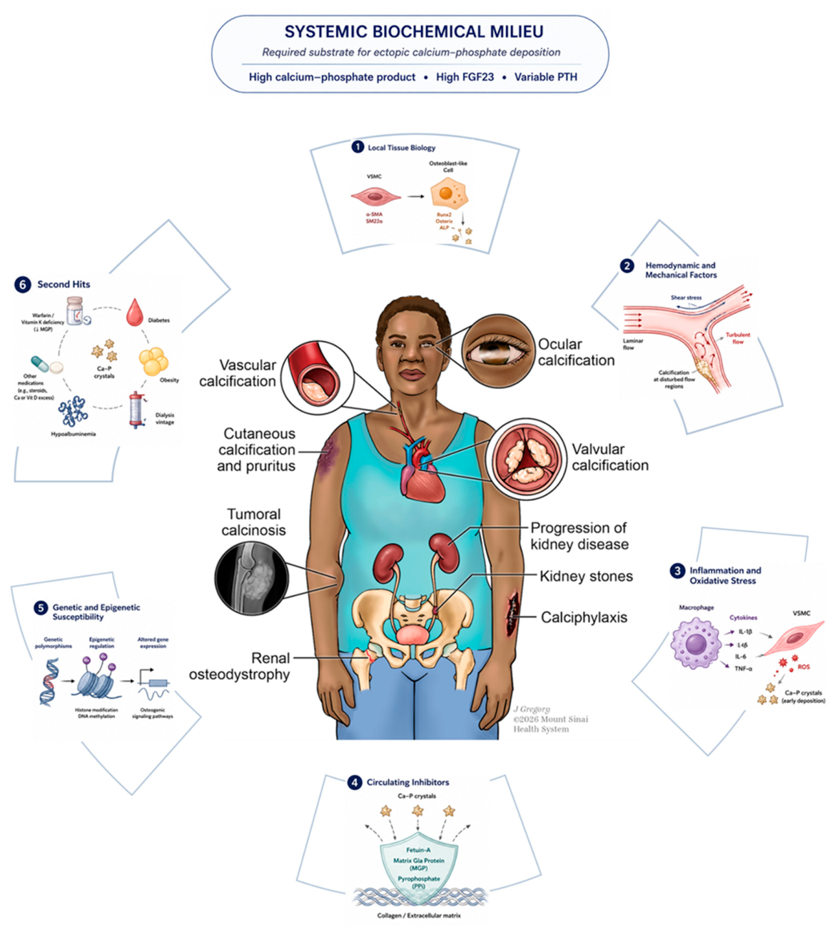

Table 1.
Clinical heterogeneity of calcium–phosphate disorders in advanced CKD.Comparison of the major calcification phenotypes of CKD-MBD by presentation, defining pathophysiology, laboratory/imaging clues, and treatment.
Table 1.
Clinical heterogeneity of calcium–phosphate disorders in advanced CKD.Comparison of the major calcification phenotypes of CKD-MBD by presentation, defining pathophysiology, laboratory/imaging clues, and treatment.
| Clinical syndrome | Presentation | Key pathophysiologic distinction | Biomarkers / Diagnosis | Treatment highlights |
|---|---|---|---|---|
|
Renal osteodystrophy Skeletal manifestation of CKD-MBD |
Often subtle or asymptomatic; nonspecific diffuse bone pain, proximal muscle weakness, skeletal tenderness. Fracture is the key endpoint: hip fracture ~2–4× general population (higher on dialysis), at younger ages; vertebral fragility fractures often missed. |
Spectrum graded by turnover, mineralization, volume (TMV). High-turnover (osteitis fibrosa) from secondary hyperparathyroidism vs low-turnover adynamic bone disease from PTH deficiency/oversuppression; osteomalacia = defective mineralization. | PTH high in high-turnover; low/oversuppressed in adynamic (often with hypercalcemia). Bone-specific ALP tracks turnover. Definitive subtype needs transiliac bone biopsy with tetracycline labeling; biomarkers/imaging lack sensitivity/specificity. |
Phosphate restriction, binders, dialysis clearance. High-turnover: active vitamin D/VDRAs, calcimimetics (cinacalcet, etelcalcetide), parathyroidectomy if refractory — avoid oversuppression. Adynamic: cut calcium/calcitriol/calcimimetic load, let PTH rise. Osteomalacia: vitamin D, correct acidosis, remove aluminum. |
|
Vascular calcification Intimal + medial (Mönckeberg) |
Medial: arterial stiffening, widened pulse pressure, systolic hypertension, LVH, diastolic dysfunction/HFpEF, impaired coronary perfusion. Intimal: accelerated atherosclerosis — CAD/ACS/MI, stroke/TIA, PAD with claudication and critical limb ischemia. |
Cell-mediated osteogenic transformation of vascular smooth muscle cells. CKD specifically drives medial calcification; intimal mirrors atherosclerosis. Phosphate → NF-κB/RUNX2, raised tissue-nonspecific ALP degrading pyrophosphate, reduced CaSR; loss of circulating inhibitors. | High phosphate and Ca×P product; PTH variable; reduced fetuin-A; inactive (uncarboxylated) MGP from vitamin K deficiency. Imaging: CT coronary artery calcium score (gold standard); pulse wave velocity for arterial stiffness. |
Lower phosphate burden and control secondary hyperparathyroidism (CKD-MBD). Statins and lower BP targets for cardiovascular risk. No therapy reverses established disease — goal is slowing progression. Emerging: vitamin K repletion, hydroxyapatite inhibitors (SNF472). |
|
Valvular calcification Aortic valve, mitral annulus |
Aortic: leaflet thickening → aortic stenosis (classic triad of exertional dyspnea, angina, syncope). Mitral annular calcification → mitral regurgitation (rarely stenosis), AV block, atrial fibrillation, left atrial enlargement, heart failure. ~8× more prevalent on hemodialysis. |
Same osteogenic CKD-MBD biology as vascular tissue, but valves are uniquely vulnerable: repetitive shear/pressure stress, collagen/elastin-rich matrix, and an avascular structure that limits repair once injured. | Same CKD-MBD profile (phosphate, PTH, FGF23, low calcitriol); no distinctive single marker. Echocardiography is first-line — leaflet thickening, echogenic foci, reduced mobility; quantifies AS/MR. |
CKD-MBD control (phosphate, PTH, avoid hypercalcemia). TAVR preferred for aortic disease given high surgical risk; surgical repair/replacement remains standard for mitral disease. Emerging: hydroxyapatite inhibitors, vitamin K, anti-inflammatory strategies. |
|
Calciphylaxis Calcific uremic arteriolopathy |
Painful violaceous plaques, retiform purpura, necrotic ulcers; tropism for adipose-rich sites (abdomen, thighs, buttocks, breasts). Very high 1-year mortality (~40–80%) driven by infection/sepsis. Obesity is a strong risk factor. |
Distinct entity, not just advanced vascular calcification: microvascular (dermal/subcutaneous arteriolar) involvement with a superimposed thrombotic vasculopathy — endothelial injury, fibrointimal hyperplasia, fibrin thrombi — plus profound failure of anti-calcification defenses. | Reduced fetuin-A, hypoalbuminemia; hypercoagulable associations (protein C/S deficiency, antiphospholipid antibodies). Warfarin exposure is a notable link (impaired MGP activation). Diagnosis mainly clinical; skin biopsy confirmatory but often avoided. Tc-99m bone scintigraphy is highly sensitive. |
CKD-MBD measures plus calciphylaxis-specific care: discontinue warfarin. Sodium thiosulfate is the most-used agent (inconsistent mortality/wound benefit). SNF472, magnesium, vitamin K investigational. Meticulous wound care, infection control, pain management, hyperbaric oxygen. |
|
Tumoral calcinosis Metastatic periarticular deposition |
Slowly enlarging firm periarticular masses (hips, shoulders, elbows), initially painless then painful with stiffness and reduced mobility; may compress neurovascular structures. Overlying skin erythema/ulceration; chalk-like material may extrude. Bone is usually spared. |
Metastatic calcification from chronic, severe phosphate excess. In CKD it is acquired (vs hereditary FGF23-related forms), with tissue tropism for periarticular regions subject to microtrauma. Shared osteogenic pathways; lobulated hydroxyapatite masses with giant-cell reaction. | Markedly elevated phosphate and Ca×P product with severe secondary hyperparathyroidism (high PTH), the most overtly abnormal mineral profile of the group. Imaging: large multilobulated, “cloud-like” periarticular calcified masses on radiograph/CT. |
Aggressive, sustained correction of phosphate and PTH is the mainstay. Surgical excision for severe pain, compression, or infection; recurrence is common if metabolic abnormalities persist, and lesions are technically difficult to resect. |
|
Ocular calcification Corneal (band keratopathy) & conjunctival |
Often asymptomatic, found incidentally on slit-lamp exam (punctate chalky-white plaques). Progression brings visual disturbance, foreign-body sensation/pain, photophobia; inflamed cases give the “red eye of uremia.” |
Metastatic calcification on the same Ca×P supersaturation basis, with ocular-specific “second hits”: localized interpalpebral alkalosis, minor injury from dialysis-related volume depletion, and iatrogenic phosphate-buffered eye drops raising local phosphate burden. | Systemic Ca×P supersaturation; no eye-specific lab. Diagnosis is clinical via slit-lamp examination. |
Optimize CKD-MBD parameters. EDTA chelation after epithelial debridement improves visual acuity in ~80%; phototherapeutic keratectomy is an option but may cause surface irregularity. |
|
Cutaneous calcification & pruritus Skin manifestation |
Pruritus is common in dialysis patients; multifactorial, frequently distressing. | Pruritus is multifactorial, but deposition of calcium/phosphate salts in skin irritating nerve endings, secondary to a high Ca×P product is thought to play a major role. | Elevated Ca×P product; no specific marker. Diagnosis is clinical. |
Lower the Ca×P product through the full set of CKD-MBD interventions above. |
ACS, acute coronary syndrome; ALP, alkaline phosphatase; AS, aortic stenosis; AV, atrioventricular; CAD, coronary artery disease; Ca×P, calcium–phosphate product; CaSR, calcium-sensing receptor; CKD-MBD, chronic kidney disease–mineral and bone disorder; EDTA, ethylenediaminetetraacetic acid; FGF23, fibroblast growth factor 23; HFpEF, heart failure with preserved ejection fraction; LVH, left ventricular hypertrophy; MGP, matrix Gla protein; MR, mitral regurgitation; PAD, peripheral arterial disease; PTH, parathyroid hormone; TAVR, transcatheter aortic valve replacement; TIA, transient ischemic attack; VDRA, vitamin D receptor activator.
Author Contributions
Kishan Rao conceived, organized, and drafted the manuscript. Joseph A. Vassalotti and Jaime Uribarri contributed to the manuscript's conceptual development, critically revised the manuscript for important intellectual content, and supervised the work. All authors reviewed and approved the final manuscript.
Use of Artificial Intelligence
Generative artificial intelligence (ChatGPT, OpenAI, Claude) was used to assist with language editing, organization, and review of the manuscript for clarity and consistency. All scientific content, interpretation, reference selection, and final editorial decisions were independently reviewed and approved by the authors, who take full responsibility for the content.
Acknowledgments
The authors thank Jill Gregory for creating Figure 1.
References
- Alam, M.U.; Kirton, J.P.; Wilkinson, F.L.; et al. Calcification is associated with loss of functional calcium-sensing receptor in vascular smooth muscle cells. Cardiovasc. Res. 2009, 81, 260–268. [Google Scholar] [PubMed]
- Arnett, D.K.; et al. 2019 ACC/AHA Guideline on the Primary Prevention of Cardiovascular Disease: A Report of the American College of Cardiology/American Heart Association Task Force on Clinical Practice Guidelines. Circulation 2019, 140, e596–e646. [Google Scholar] [CrossRef] [PubMed]
- Baumgartner, H.; Falk, V.; Bax, J.J.; et al. 2017 ESC/EACTS Guidelines for the management of valvular heart disease. Eur. Heart J. 2017, 38, 2739–2791. [Google Scholar] [CrossRef] [PubMed]
- Blacher, J.; Guerin, A.P.; Pannier, B.; Marchais, S.J.; London, G.M. Arterial calcifications, arterial stiffness, and cardiovascular risk in end-stage renal disease. Hypertension 2001, 38, 938–942. [Google Scholar] [CrossRef] [PubMed]
- Blaine, J.; Chonchol, M.; Levi, M. Renal control of calcium, phosphate, and magnesium homeostasis. Clin. J. Am. Soc. Nephrol. 2015, 10, 1257–1272. [Google Scholar] [CrossRef] [PubMed]
- Boron, W.F.; Boulpaep, E.L. Medical Physiology, 3rd ed.; Elsevier: Philadelphia, PA, USA, 2017. [Google Scholar]
- Brandenburg, V.M.; Floege, J. Adynamic bone disease—bone and beyond. NDT Plus 2008, 1, 135–147. [Google Scholar] [CrossRef] [PubMed]
- Brandenburg, V.M.; Kramann, R.; Rothe, H.; et al. Calcific uraemic arteriolopathy (calciphylaxis): data from a large nationwide registry. Nephrol. Dial. Transplant. 2017, 32, 126–132. [Google Scholar] [PubMed]
- Bricker, N.S. On the pathogenesis of the uremic state. An exposition of the “trade-off hypothesis.”. N. Engl. J. Med. 1972, 286, 1093–1099. [Google Scholar] [CrossRef] [PubMed]
- Chang, A.R.; Anderson, C. Dietary phosphorus intake and the kidney. Annu. Rev. Nutr. 2017, 37, 321–346. [Google Scholar] [CrossRef] [PubMed]
- Christakos, S. Vitamin D: metabolism and mechanisms of action. Physiol. Rev. 2016, 96, 365–408. [Google Scholar] [CrossRef] [PubMed]
- Cicone, J.S.; Petronis, J.B.; Embert, C.D.; Spector, D.A. Successful treatment of calciphylaxis with intravenous sodium thiosulfate. Am. J. Kidney Dis. 2004, 43, 1104–1108. [Google Scholar] [CrossRef] [PubMed]
- Cozzolino, M.; Galassi, A.; Biondi, M.L.; et al. Pathogenesis of vascular calcification in chronic kidney disease. Kidney Int. 2008, 74, 429–435. [Google Scholar] [CrossRef] [PubMed]
- Cunningham, J.; Locatelli, F.; Rodriguez, M. Secondary hyperparathyroidism: pathogenesis, disease progression, and therapeutic options. Clin. J. Am. Soc. Nephrol. 2011, 6, 913–921. [Google Scholar] [CrossRef] [PubMed]
- Daudon, M.; Traxer, O.; Lechevallier, E.; Saussine, C. Epidemiology of urolithiasis. Prog. Urol. 2018, 28, 625–631. [Google Scholar]
- Elmariah, S.; Budoff, M.J.; Delaney, J.A.; et al. Risk factors associated with the incidence and progression of mitral annulus calcification: the Multi-Ethnic Study of Atherosclerosis. Am. Heart J. 2013, 166, 904–912. [Google Scholar] [CrossRef] [PubMed]
- Evenepoel, P.; Behets, G.J.; Laurent, M.R.; D’Haese, P.C. Update on the role of bone biopsy in the management of patients with CKD-MBD. J. Nephrol. 2017, 30, 645–652. [Google Scholar] [CrossRef] [PubMed]
- Felsenfeld, A.J.; Levine, B.S.; Rodriguez, M. Pathophysiology of calcium, phosphorus, and magnesium dysregulation in chronic kidney disease. Semin. Dial. 2015, 28, 564–577. [Google Scholar] [CrossRef] [PubMed]
- Fine, A.; Zacharias, J. Calciphylaxis is usually non-ulcerating: risk factors, outcome and therapy. Kidney Int. 2002, 61, 2210–2217. [Google Scholar] [CrossRef] [PubMed]
- Fox, C.S.; Vasan, R.S.; Parise, H.; et al. Mitral annular calcification predicts cardiovascular morbidity and mortality: the Framingham Heart Study. Circulation 2003, 107, 1492–1496. [Google Scholar] [CrossRef] [PubMed]
- Giachelli, C.M. The emerging role of phosphate in vascular calcification. Kidney Int. 2009, 75, 890–897. [Google Scholar] [CrossRef] [PubMed]
- Goodman, W.G.; Goldin, J.; Kuizon, B.D.; et al. Coronary-artery calcification in young adults with end-stage renal disease who are undergoing dialysis. N. Engl. J. Med. 2000, 342, 1478–1483. [Google Scholar] [CrossRef] [PubMed]
- Hayden, M.R.; Goldsmith, D.; Sowers, J.R.; Khanna, R. Calciphylaxis: pathobiology of vascular calcification and thrombosis. Kidney Int. 2005, 68, 1800–1808. [Google Scholar]
- Huish, S.; Moore, S.; Alfieri, C.; et al. Calciphylaxis diagnosis, management and future directions: a comprehensive update on behalf of the European Renal Association CKD-MBD Working Group. Clin. Kidney J. 2025, 18, sfaf338. [Google Scholar] [CrossRef] [PubMed]
- Ibels, L.S.; Alfrey, A.C.; Haut, L.; Huffer, W.E. Preservation of function in experimental renal disease by dietary restriction of phosphate. N. Engl. J. Med. 1978, 298, 122–126. [Google Scholar] [CrossRef] [PubMed]
- Isakova, T.; Wahl, P.; Vargas, G.S.; et al. Fibroblast growth factor 23 is elevated before parathyroid hormone and phosphate in chronic kidney disease. Kidney Int. 2011, 79, 1370–1378. [Google Scholar] [CrossRef] [PubMed]
- Jadoul, M.; Albert, J.M.; Akiba, T.; et al. Incidence and risk factors for hip or other bone fractures among hemodialysis patients in the Dialysis Outcomes and Practice Patterns Study. Kidney Int. 2006, 70, 1358–1366. [Google Scholar] [CrossRef] [PubMed]
- Jono, S.; McKee, M.D.; Murry, C.E.; et al. Phosphate regulation of vascular smooth muscle cell calcification. Circ. Res. 2000, 87, E10–E17. [Google Scholar] [CrossRef] [PubMed]
- Karlinsky, M.L.; Haut, L.; Buddington, B.; Schrier, N.; Alfrey, A.C. Preservation of renal function in experimental glomerulonephritis by phosphate depletion. Kidney Int. 1980, 17, 293–302. [Google Scholar] [CrossRef] [PubMed]
- 30. KDIGO CKD-MBD Update Work Group. KDIGO 2017 Clinical Practice Guideline Update for the Diagnosis, Evaluation, Prevention, and Treatment of Chronic Kidney Disease–Mineral and Bone Disorder (CKD-MBD). Kidney Int. Suppl. 2017, 7, 1–59. [CrossRef] [PubMed]
- Ketteler, M.; Bongartz, P.; Westenfeld, R.; et al. Association of low fetuin-A levels with cardiovascular mortality in patients on dialysis. Lancet 2003, 361, 827–833. [Google Scholar] [CrossRef] [PubMed]
- Khan, S.R.; Canales, B.K.; Dominguez-Gutierrez, P.R. Randall’s plaque and calcium oxalate stone formation: role for immunity and inflammation. World J. Urol. 2011, 29, 65–71. [Google Scholar] [CrossRef] [PubMed]
- Khan, S.R.; Pearle, M.S.; Robertson, W.G.; et al. Kidney stones. Nat. Rev. Dis. Prim. 2016, 2, 16008. [Google Scholar] [CrossRef] [PubMed]
- Loghman-Adham, M. Role of phosphate retention in progressive renal failure. Kidney Int. Suppl. 1993, 41, S13–S21. [Google Scholar] [CrossRef] [PubMed]
- London, G.M. Cardiovascular calcifications in uremic patients: clinical impact on cardiovascular function. J. Am. Soc. Nephrol. 2003, 14 (Suppl. 4), S305–S309. [Google Scholar] [CrossRef] [PubMed]
- London, G.M.; Marchais, S.J.; Guérin, A.P.; Métivier, F. Cardiac hypertrophy and arterial alterations in end-stage renal disease: hemodynamic factors. Kidney Int. Suppl. 1993, 41, S42–S49. [Google Scholar] [PubMed]
- Maher, E.R.; Young, G.; Smyth-Walsh, B.; Pugh, S.; Curtis, J.R. Aortic and mitral valve calcification in patients with end-stage renal disease. Lancet 1987, 2, 875–877. [Google Scholar] [CrossRef] [PubMed]
- Malluche, H.H.; Monier-Faugere, M.C. Renal bone disease 1990: an unmet challenge for the nephrologist. Kidney Int. 1990, 38, 193–211. [Google Scholar] [CrossRef] [PubMed]
- McCarthy, J.T.; El-Azhary, R.A.; Patzelt, M.T.; et al. Survival, risk factors, and effect of treatment in 101 patients with calciphylaxis. Mayo Clin. Proc. 2016, 91, 1384–1394. [Google Scholar] [CrossRef] [PubMed]
- Fathi, I.; Sakr, M. Review of tumoral calcinosis: A rare clinico-pathological entity. World J. Clin. Cases 2014, 2(9), 409–414. [Google Scholar] [CrossRef] [PubMed]
- Moe, S.; Drüeke, T.; Cunningham, J.; et al. Definition, evaluation, and classification of renal osteodystrophy: a position statement from Kidney Disease: Improving Global Outcomes (KDIGO). Kidney Int. 2006, 69, 1945–1953. [Google Scholar] [CrossRef] [PubMed]
- Moe, S.M.; Chen, N.X. Pathophysiology of vascular calcification in chronic kidney disease. Circ. Res. 2004, 95, 560–567. [Google Scholar] [CrossRef] [PubMed]
- Rutkovskiy, A.; Malashicheva, A.; Sullivan, G.; et al. Valve Interstitial Cells: The Key to Understanding the Pathophysiology of Heart Valve Calcification. J. Am. Heart Assoc. Published. 2017, 6(9), e006339. [Google Scholar] [CrossRef] [PubMed]
- Naylor, K.L.; McArthur, E.; Leslie, W.D.; et al. The three-year incidence of fracture in chronic kidney disease. Kidney Int. 2014, 86, 810–818. [Google Scholar] [CrossRef] [PubMed]
- Nigwekar, S.U.; Kroshinsky, D.; Nazarian, R.M.; et al. Calciphylaxis: risk factors, diagnosis, and treatment. Am. J. Kidney Dis. 2015, 66, 133–146. [Google Scholar] [CrossRef] [PubMed]
- Nigwekar, S.U.; Thadhani, R.; Brandenburg, V.M. Calciphylaxis. N. Engl. J. Med. 2018, 378, 1704–1714. [Google Scholar] [CrossRef] [PubMed]
- O’Hare, A.M.; Hsu, C.Y.; Bacchetti, P.; Johansen, K.L. Peripheral vascular disease risk factors among patients undergoing hemodialysis. J. Am. Soc. Nephrol. 2002, 13, 497–503. [Google Scholar] [CrossRef] [PubMed]
- Olsen, K.M.; Chew, F.S. Tumoral calcinosis: pearls, polemics, and alternative possibilities. Radiographics 2006, 26, 871–885. [Google Scholar] [CrossRef] [PubMed]
- Lindman, B.R.; Clavel, M.A.; Mathieu, P.; et al. Calcific aortic stenosis. Nat. Rev. Dis. Prim. Published. 2016, 2, 16006. [Google Scholar] [CrossRef] [PubMed]
- Paul, S.; Rabito, C.A.; Vedak, P.; et al. The role of bone scintigraphy in the diagnosis of calciphylaxis. JAMA Dermatol. 2017, 153, 101–103. [Google Scholar] [CrossRef] [PubMed]
- Perkovic, V.; Hewitson, T.D.; Kelynack, K.J.; et al. Parathyroid hormone has a prosclerotic effect on vascular smooth muscle cells. Kidney Blood Press. Res. 2003, 26, 27–33. [Google Scholar] [CrossRef] [PubMed]
- Porter, R.; Crombie, A.L. Band keratopathy and conjunctival calcification in chronic renal failure. Br. J. Ophthalmol. 1994, 78, 693–695. [Google Scholar]
- Porter, S.M.; Crombie, A.L.; Gardner, J.; et al. Band keratopathy in chronic kidney disease and dialysis patients. Semin. Ophthalmol. 2024, 39, 145–154. [Google Scholar]
- Quarles, L.D. Endocrine functions of bone in mineral metabolism regulation. J. Clin. Invest. 2008, 118, 3820–3828. [Google Scholar] [CrossRef] [PubMed]
- Raggi, P.; Bellasi, A.; Bushinsky, D.; et al. Slowing progression of cardiovascular calcification with SNF472 in patients on hemodialysis. Circulation 2020, 141, 728–739. [Google Scholar] [CrossRef] [PubMed]
- Raggi, P.; Boulay, A.; Chasan-Taber, S.; et al. Cardiac calcification in adult hemodialysis patients. J. Am. Coll. Cardiol. 2002, 39, 695–701. [Google Scholar] [CrossRef] [PubMed]
- Ribeiro, S.; Ramos, A.; Brandão, A.; et al. Cardiac valve calcification in haemodialysis patients: role of calcium-phosphate metabolism. Nephrol. Dial. Transplant. 1998, 13, 2037–2040. [Google Scholar] [CrossRef] [PubMed]
- Schurgers, L.J.; Barreto, D.V.; Barreto, F.C.; Liabeuf, S.; Renard, C.; Magdeleyns, E.J.; Vermeer, C.; Choukroun, G.; Massy, Z.A. The circulating inactive form of matrix Gla protein is a surrogate marker for vascular calcification in chronic kidney disease: A preliminary report. Clin. J. Am. Soc. Nephrol. 2010, 5, 568–575. [Google Scholar] [CrossRef] [PubMed]
- Schurgers, L.J.; Spronk, H.M.H.; Skepper, J.N.; et al. Post-translational modifications regulate matrix Gla protein function: importance for inhibition of vascular smooth muscle cell calcification. J. Thromb. Haemost. 2007, 5, 2503–2511. [Google Scholar] [CrossRef] [PubMed]
- Schwarz, S.; Trivedi, B.K.; Kalantar-Zadeh, K.; Kovesdy, C.P. Association of disorders in mineral metabolism with progression of chronic kidney disease. Clin. J. Am. Soc. Nephrol. 2006, 1, 825–831. [Google Scholar] [CrossRef] [PubMed]
- Shanahan, C.M.; Crouthamel, M.H.; Kapustin, A.; Giachelli, C.M. Arterial calcification in chronic kidney disease: key roles for calcium and phosphate. Circ. Res. 2011, 109, 697–711. [Google Scholar] [CrossRef] [PubMed]
- Shimada, T.; Hasegawa, H.; Yamazaki, Y.; et al. FGF-23 is a potent regulator of vitamin D metabolism and phosphate homeostasis. J. Bone Min. Res. 2004, 19, 429–435. [Google Scholar] [CrossRef] [PubMed]
- Slatopolsky, E.; Bricker, N.S. The role of phosphorus restriction in the prevention of secondary hyperparathyroidism in chronic renal disease. Kidney Int. 1973, 4, 141–145. [Google Scholar] [CrossRef] [PubMed]
- Slatopolsky, E.; Caglar, S.; Pennell, J.P.; et al. On the pathogenesis of secondary hyperparathyroidism in chronic experimental renal insufficiency. J. Clin. Invest. 1971, 50, 492–499. [Google Scholar] [CrossRef] [PubMed]
- Slatopolsky, E.; Delmez, J.A. Pathogenesis of secondary hyperparathyroidism. Am. J. Kidney Dis. 1994, 23, 229–236. [Google Scholar] [CrossRef] [PubMed]
- Smack, D.; Norton, S.A.; Fitzpatrick, J.E. Proposal for a pathogenesis-based classification of tumoral calcinosis. Int. J. Dermatol. 1996, 35, 265–271. [Google Scholar] [CrossRef] [PubMed]
- Thourani, V.H.; Forcillo, J.; Beohar, N.; et al. Early and late outcomes after transcatheter aortic valve replacement in patients with chronic kidney disease. J. Am. Coll. Cardiol. 2016, 67, 2829–2838. [Google Scholar] [CrossRef]
- Townsend, R.R.; Wimmer, N.J.; Chirinos, J.A.; et al. Aortic PWV in chronic kidney disease: a CRIC ancillary study. Am. J. Hypertens. 2010, 23, 282–289. [Google Scholar] [CrossRef] [PubMed]
- Udomkarnjananun, S.; Kongnatthasate, K.; Praditpornsilpa, K.; et al. Treatment of calciphylaxis in CKD: a systematic review and meta-analysis. Kidney Int. Rep. 2019, 4, 231–244. [Google Scholar] [CrossRef] [PubMed]
- Urena, M.; Vahanian, A.; Melehy, A.; et al. Current treatment options for mitral valve disease in chronic kidney disease patients. Nat. Rev. Nephrol. 2015, 11, 715–724. [Google Scholar]
- Viegas, C.S.B.; Santos, L.; Macedo, A.L.; et al. Chronic kidney disease circulating calciprotein particles and extracellular vesicles promote vascular calcification: a role for GRP (Gla-rich protein). Arterioscler. Thromb. Vasc. Biol. 2018, 38, 575–587. [Google Scholar] [PubMed]
- Wang, A.Y.; Lam, C.W.; Wang, M.; et al. Prognostic value of cardiac valvular calcification in end-stage renal disease: a prospective study. J. Am. Soc. Nephrol. 2003, 14, 159–168. [Google Scholar] [PubMed]
- Weenig, R.H. Pathogenesis of calciphylaxis: Hans Selye to nuclear factor κ-B. J. Am. Acad. Dermatol. 2008, 58, 458–471. [Google Scholar] [CrossRef] [PubMed]
- Wulczyn, K.E.; Rhee, E.P.; Myint, L.; Kalim, S.; Shafi, T. Incidence and risk factors for pruritus in patients with nondialysis CKD. Clin. J. Am. Soc. Nephrol. 2023, 18, 193–203. [Google Scholar] [CrossRef] [PubMed]
- Zaslow, S.J.; Oliveira-Paula, G.H.; Chen, W. Magnesium and vascular calcification in chronic kidney disease: current insights. Int. J. Mol. Sci. 2024, 25, 1155. [Google Scholar] [CrossRef] [PubMed]
Disclaimer/Publisher’s Note: The statements, opinions and data contained in all publications are solely those of the individual author(s) and contributor(s) and not of MDPI and/or the editor(s). MDPI and/or the editor(s) disclaim responsibility for any injury to people or property resulting from any ideas, methods, instructions or products referred to in the content. |
© 2026 by the authors. Licensee MDPI, Basel, Switzerland. This article is an open access article distributed under the terms and conditions of the Creative Commons Attribution (CC BY) license (http://creativecommons.org/licenses/by/4.0/).
Copyright: This open access article is published under a Creative Commons CC BY 4.0 license, which permit the free download, distribution, and reuse, provided that the author and preprint are cited in any reuse.