

Submitted:
03 August 2026
Posted:
05 August 2026
You are already at the latest version
Abstract
Diabetic wound healing is characterised by persistent inflammation, impaired angiogenesis, extracellular matrix (ECM) dysregulation, and defective tissue remodelling, frequently resulting in chronic wounds such as diabetic foot ulcers (DFUs). Fibrosis, characterised by excessive ECM deposition and sustained fibroblast activation, is increasingly recognised as an important barrier to effective wound repair in patients with diabetes. Emerging evidence suggests that glucocorticoid signalling, particularly through the stress hormone cortisol and its activating enzyme 11β-hydroxysteroid dehydrogenase type 1 (11β-HSD1), may contribute to pathological and fibrotic changes in diabetic skin by altering inflammation, extracellular matrix turnover, angiogenesis, and cellular behaviour. In parallel, profibrotic mediators such as connective tissue growth factor (CTGF) and insulin-like growth factor binding protein-5 (IGFBP-5) have been implicated in fibroblast activation and fibrosis across multiple tissues, including skin. However, the interactions between cortisol signalling and these downstream pathways remain poorly understood. This review examines current evidence regarding the role of 11β-HSD1-mediated cortisol signalling in fibrosis and impaired diabetic wound healing, with particular emphasis on potential interactions with CTGF and IGFBP-5. Evidence from preclinical and early clinical studies suggests that inhibition of 11β-HSD1 may improve wound repair and tissue integrity; however, many existing studies rely on acute wound models and do not fully reflect the chronic inflammatory and fibrotic environment of diabetic wounds.
Keywords:
diabetic wound healing
; 11β-HSD1
; cortisol
; fibrosis
; diabetic foot ulcer
; glucocorticoids
1. Introduction
Diabetes is a chronic metabolic disorder characterised by elevated blood glucose levels (hyperglycaemia) due to impaired insulin production or action. It is broadly classified into type 1 diabetes (T1D), resulting from autoimmune destruction of insulin-producing beta cells, and type 2 diabetes (T2D), the most common form, which is associated with insulin resistance and often exacerbated by obesity and lifestyle factors [1]. T1D accounts for around 8% of diabetes cases, while T2D represents approximately 90%, with rarer diabetes types making up the remaining 2% of those affected [2]. In 2022, diabetes affected over 830 million people globally, with increasing prevalence in low- and middle-income countries [3]. In the UK alone, over 5.8 million people are now estimated to be living with diabetes, including almost 4.6 million with a formal diagnosis and around 1.3 million undiagnosed cases. This prevalence is predicted to continue to rise.
Diabetes affects multiple organ systems, including the cardiovascular, renal, gastrointestinal, reproductive, nervous, and integumentary systems, leading to increase comorbidities [4]. A common morbidity in diabetes is skin ulcers. Particularly foot ulcers, which affect 19–34% of people with diabetes. Once present they pose a substantial burden to the individuals and healthcare systems. The quality of life in patients with diabetic foot ulcers (DFUs) has been reported to be significantly reduced [5] and the health care costs of a diabetic foot ulcer have been estimated to be between £2,140 and £8,800 per healed and unhealed DFU, respectively, and £16,900 per amputation. This translates to an annual NHS cost attributable to managing these DFUs of between £524.6 million and £728.0 million [6].
Diabetic wound healing is characterised by a failure to progress through the normal stages of repair (i.e., homeostasis, inflammation, proliferation and remodelling), resulting in a prolonged inflammatory state, impaired angiogenesis, and defective tissue remodelling. Rather than resolving, these wounds often become trapped in a pathological state marked by persistent inflammation and extracellular matrix (ECM) dysregulation [7,8]. The dysregulated ECM then continues to actively regulate cellular behaviour through altered growth factor signalling and cell–matrix interactions, preventing tissue repair [9]. A key feature of ECM dysregulation is fibrosis, defined by excessive accumulation and glycation of ECM components such as collagen and fibronectin, leading to increased tissue stiffness, reduced cellular migration, and impaired regenerative capacity. Although transient fibrosis is an essential component of normal wound repair, persistent ECM remodelling and scar formation prevent restoration of normal tissue architecture, contributing to incomplete healing and chronic tissue dysfunction [9,10].
The molecular mechanisms driving fibrosis in diabetic wounds are complex and involve multiple interacting pathways, including transforming growth factor-beta (TGF-β) signalling and downstream mediators such as connective tissue growth factor (CTGF) [11] and insulin-like growth factor binding protein 5 (IGFBP-5) [12]. These factors promote fibroblast activation, myofibroblast differentiation, and sustained ECM deposition, reinforcing a fibrotic wound environment. However, the upstream regulators of these pathways remain incompletely understood, particularly in the context of chronic metabolic disease.
Emerging evidence suggests that glucocorticoid signalling, particularly through the stress hormone cortisol, plays a critical role in regulating skin homeostasis, inflammation, and tissue repair [13,14]. While cortisol is essential for controlling acute inflammatory responses, chronic elevation as observed in some cases of diabetes [15], ageing [16], and stress [17], has been associated with impaired wound healing and altered ECM remodelling. Local cortisol availability within tissues is tightly regulated by the enzyme 11β-hydroxysteroid dehydrogenase type 1 (11β-HSD1), which converts inactive cortisone into active cortisol. Increased expression and activity of 11β-HSD1 in diabetic skin may therefore amplify local glucocorticoid signalling, potentially contributing to wound chronicity and fibrosis [18,19].
Despite growing recognition of the roles of cortisol, 11β-HSD1, and profibrotic mediators such as CTGF and IGFBP-5, the interaction between these pathways remains poorly understood. While cortisol signalling and CTGF/IGFBP-5-mediated fibrosis have individually been associated with impaired wound healing [12,20], it remains unclear whether local cortisol activation through 11β-HSD1 directly regulates these downstream mediators in human diabetic skin. Addressing this gap may help determine whether glucocorticoid metabolism functions as an upstream driver of fibrosis and represents a viable therapeutic target.
This review aims to examine the current understanding of glucocorticoid signalling in skin, with a particular focus on the role of 11β-HSD1-mediated cortisol activation in fibrosis and impaired diabetic wound healing. It further explores the potential interaction between cortisol signalling and key profibrotic mediators, highlighting current knowledge gaps and identifying directions for future research.
2. Wound Healing and Fibrosis: Challenges in Diabetes
2.1. Normal Wound Healing
Wound healing is a dynamic and tightly regulated process consisting of four overlapping phases: haemostasis, inflammation, proliferation, and remodelling. In acute wounds, these phases occur in a coordinated sequence to restore tissue integrity [21].
Haemostasis starts immediately following injury, it involves clot formation and vasoconstriction to prevent blood loss and provide a provisional matrix for cell migration. This phase occurs within minutes to hours following tissue injury and is rapidly followed by the inflammatory phase, which typically predominates during the first 1–4 days of healing. This is followed by the inflammatory phase, during which immune cells such as neutrophils and monocytes clear debris and pathogens while releasing cytokines and growth factors that initiate the repair process. The proliferative phase generally occurs between days 4 and 21 and is characterised by fibroblast activation, angiogenesis, re-epithelialisation, and ECM deposition, leading to the formation of granulation tissue. Finally, during the remodelling phase, which may begin approximately 2–3 weeks after injury and continue for months to years, the ECM is reorganised and strengthened through collagen maturation and controlled degradation, restoring tissue function and tensile strength [22].
Under normal conditions, this process is self-limiting and resolves once tissue repair is complete, with coordinated regulation of inflammation, cellular activity, and matrix turnover ensuring effective healing.
2.2. Dysregulated Wound Healing in Diabetes
A wound that fails to progress through this healing sequence within 4 to 8 weeks is classified as a chronic wound (Figure 1). In diabetes, wound healing becomes chronically dysregulated due to sustained metabolic and vascular abnormalities and can take months to heal or remain functionally un-healed. Persistent hyperglycaemia maintains inflammation and delays the transition from the inflammatory to the proliferative phase, disrupting the normal sequence of repair. This impairs fibroblast migration, angiogenesis, keratinocyte function, and ECM remodelling, leading to delayed or incomplete wound closure [8]. In T2D, insulin resistance and compensatory hyperinsulinemia further contribute to impaired healing by reducing the cellular responsiveness to insulin. This affects keratinocyte migration, inhibits angiogenesis, and alters fibroblast signalling pathways involved in tissue regeneration [8,23]. By contrast, T1D, which involves absolute insulin deficiency rather than insulin resistance, may impair healing through mechanisms including reduced availability of insulin for anabolic processes and delayed immune resolution. Although the initiating mechanisms differ, both conditions expose tissues to chronic hyperglycaemia, oxidative stress and persistent inflammation, all of which contribute to impaired wound healing. However, direct comparisons between wound-healing mechanisms in T1D and T2D remain limited, and much of the current understanding is derived from studies of T2D and diabetic foot ulcers or separate experimental models [7,24].
Chronic hyperglycaemia promotes the generation of advanced glycation end products (AGEs), which modify ECM proteins and alter tissue architecture. AGEs irreversibly bind to structural proteins such as collagen, producing shortened and disorganised fibrils that reduce tissue elasticity and impair matrix turnover [25]. Additionally, AGEs interact with the receptor for advanced glycation end-products (RAGE), which is expressed on fibroblasts, keratinocytes, endothelial cells, and immune cells. Activation of AGE–RAGE signalling promotes oxidative stress and sustained inflammation, impairing cellular responsiveness and reinforcing a chronic wound environment [25].
Angiogenesis, which is essential for supplying oxygen and nutrients to healing tissue, is also impaired in diabetic wounds. Reduced vascular endothelial growth factor (VEGF) expression and endothelial dysfunction contribute to local hypoxia and delayed tissue repair. Simultaneously, persistent inflammation and altered macrophage polarisation disrupt the normal resolution phase of healing. Traditionally, diabetic wounds have been characterised by prolonged M1 macrophage activity, which maintains a pro-inflammatory environment through secretion of cytokines such as tumour necrosis factor-alpha (TNF-α) and interleukin-6 (IL-6) [26]. However, recent single-cell and spatial transcriptomic studies suggest that successful healing requires a timely inflammatory response, with healing diabetic foot ulcers exhibiting a higher proportion of M1 macrophages, whereas non-healing ulcers display a shift towards M2 macrophage populations [27]. Emerging evidence suggests that persistent or dysregulated activation of M2 macrophage populations, particularly CD163+ M2c macrophages, may contribute to pathological tissue remodelling and fibrosis through continued secretion of profibrotic mediators such as TGF-β and promotion of ECM remodelling [28].
Keratinocytes, which are essential for re-epithelialisation and communication with immune and stromal cells during wound healing, are similarly affected by hyperglycaemic conditions. Keratinocytes from people with diabetes exhibit impaired proliferation, delayed migration, and reduced responsiveness to wound-edge signals, thereby limiting their regenerative capacity [29]. These changes are further compounded by increased secretion of inflammatory mediators and reactive oxygen species, contributing to chronic tissue damage.
Recent single-cell transcriptomic analyses [27] have revealed significant heterogeneity within fibroblast populations in diabetic wounds, identifying a distinct subset of “healing-enriched fibroblasts” characterised by increased expression of ECM-remodelling and inflammatory genes such as matrix metalloproteinase-1 (MMP1), matrix metalloproteinase-3 (MMP3), and chitinase-3-like protein 1 (CHI3L1). These cells appear to localise to the wound bed and are associated with successful healing outcomes. In contrast, non-healing diabetic foot ulcers contain fewer healing-enriched fibroblasts, suggesting a reduced capacity for ECM remodelling, cell migration and inflammatory responses. This suggests that fibroblast function in DFUs is not uniformly impaired, but instead depends on the abundance of distinct fibroblast phenotypes.
2.3. Fibrosis as a Barrier to Healing
Diabetic wounds also frequently exhibit features of pathological fibrosis. Fibrosis is a hallmark of chronic wound pathology, characterised by the excessive accumulation of ECM components, particularly collagen and fibronectin, which leads to tissue stiffening, reduced elasticity, and impaired cellular mobility [30]. While matrix deposition is a necessary feature of normal wound healing, enabling mechanical stability and tissue closure, its dysregulation results in excessive scarring rather than regeneration. In chronic wounds, fibrosis persists rather than resolves, becoming a barrier to effective healing [30].
At the centre of fibrotic signalling is TGF-β1, a key cytokine that promotes fibroblast activation, differentiation into α-smooth muscle actin (α-SMA)-positive myofibroblasts and increased ECM synthesis. While transient activation of these pathways is essential for normal tissue repair, sustained TGF-β1 signalling in chronic wounds promotes persistent matrix deposition and impaired tissue remodelling. This prolonged fibrotic response is further reinforced by downstream mediators such as CTGF and IGFBP-5, both of which have been associated with fibroblast activation and extracellular matrix accumulation [11].
The chronicity of fibrosis in chronic wounds is further sustained by prolonged myofibroblast survival, persistent inflammation, and an imbalance between ECM synthesis and degradation. Normally, once repair is complete, myofibroblasts undergo apoptosis to allow tissue remodelling. This process is triggered primarily by the loss of mechanical tension and reduced pro-survival signalling once wound closure is achieved. However, in fibrotic wounds, this resolution phase is impaired. Factors such as persistent mechanical stress and elevated TGF-β1 levels promote myofibroblast survival via antiapoptotic mechanisms, notably through Protein Kinase B (AKT/PKB) signalling [31]. The continued presence of myofibroblasts drives excessive matrix production and contraction, resulting in functionally compromised scar tissue.
Another important contributor to matrix dysregulation is the altered activity of matrix metalloproteinases (MMPs), zinc-dependent enzymes that degrade ECM components during wound remodelling. Under normal conditions, MMPs are tightly regulated by tissue inhibitors of metalloproteinases (TIMPs), ensuring balance between degradation and synthesis. In chronic wounds, however, sustained inflammation and hyperglycaemia disrupt this balance, leading to dysregulated MMP activity. This degrades not only the ECM, but also growth factors and cell surface receptors required for migration and proliferation, further impairing healing and remodelling [32].
Importantly, the role of MMPs in fibrosis is complex and highly dependent on tissue context and timing. While excessive MMP activity, particularly MMP-9, has frequently been associated with chronic inflammation and impaired wound healing [33,34], certain MMPs are essential for physiological tissue repair and matrix remodelling. For example, MMPs contribute to degradation of provisional matrix components and facilitate cell migration during wound healing, whereas reduced expression of anti-fibrotic MMPs such as MMP-1 and MMP-3 has been linked to excessive matrix accumulation and tissue stiffening in fibrotic conditions [35]. Consequently, pathological wound healing may arise not simply from increased MMP activity, but from disruption of the balance between specific MMPs and TIMPs, ultimately contributing to persistent fibrosis and impaired tissue repair.
3. Key Molecular Mediators in Fibrosis and Diabetic Wound Healing: Cortisol, 11β-HSD1, IGFBP-5, and CTGF
Among the molecular regulators implicated in fibrosis, CTGF and IGFBP-5 have emerged as prominent fibrotic mediators and potential therapeutic targets, due to their involvement in ECM deposition and fibroblast activation [12,36]. More recently, the stress hormone cortisol and its activating enzyme 11β-HSD1 have emerged as potential regulators of fibrotic processes, particularly in the skin [18,37]. These molecules have been implicated in the pathogenesis of DFUs and other fibrotic conditions, yet the precise relationships between them remain poorly understood. In particular, it is not yet clear whether cortisol activity through 11β-HSD1 contributes to the regulation of CTGF and IGFBP-5, or whether these pathways act independently in driving fibrosis.
3.1. Function of Cortisol and 11β-HSD1
Cortisol, the principal glucocorticoid in humans, plays a crucial role in modulating inflammation, immune responses, and tissue homeostasis. Under physiological conditions, systemic cortisol levels are tightly regulated by the hypothalamic-pituitary-adrenal (HPA) axis. However, in diabetes, this balance is disrupted. Chronic hyperglycaemia and insulin resistance are associated with HPA axis dysregulation, leading to elevated systemic cortisol levels and altered local cortisol metabolism [38]. Conversely, prolonged glucocorticoid excess may itself contribute to the development of metabolic dysfunction. Patients with Cushing’s syndrome, a condition characterised by chronic cortisol excess, exhibit increased risk of insulin resistance, impaired glucose tolerance, diabetes, and delayed wound healing suggesting a bidirectional relationship between glucocorticoid signalling and metabolic disease [39].
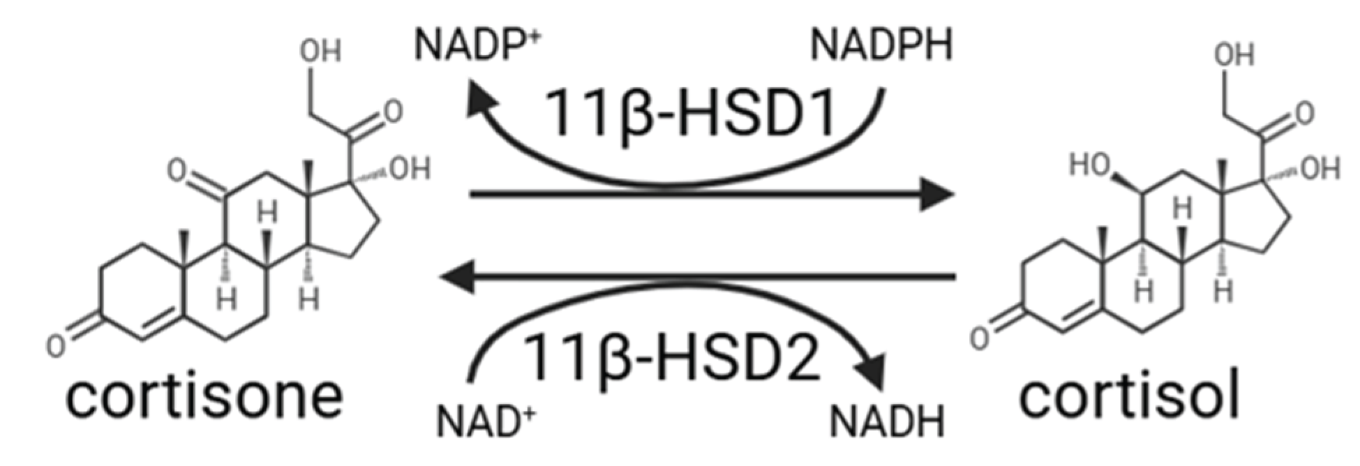
Local activation of cortisol in tissues is primarily controlled by 11β-HSD1, an enzyme that converts inactive cortisone into active cortisol (Figure 2). In diabetic skin and wounds, 11β-HSD1 expression is upregulated, resulting in increased local cortisol concentrations [14]. This enhances the effects of cortisol within the wound environment, where it can influence cell proliferation, ECM deposition, and inflammatory responses. However, the full extent of how diabetes alters 11β-HSD1 activity and local cortisol regulation in chronic wounds remains underexplored, representing a significant research gap.
3.2. Cortisol Effects on Wound Healing and Fibrosis
Cortisol plays a multifaceted and context-dependent role in skin physiology and wound healing, although chronic elevation, as observed in physiological and psychological stress, diabetes, and ageing, has been associated with impaired tissue repair [13,14,18,40]. Cortisol exerts its effects primarily through intracellular glucocorticoid receptors, which regulate the expression of genes involved in inflammation, cellular proliferation, extracellular matrix remodelling, and differentiation pathways.
Chronic psychological stress has increasingly been recognised as a contributor to impaired wound healing, particularly in individuals with diabetes and DFUs. Activation of the HPA axis during chronic stress results in prolonged glucocorticoid release, which has been associated with delayed wound closure, altered inflammatory responses, and impaired immune function. Clinical studies in DFUs further suggest that psychological distress may contribute to poorer wound outcomes, although current evidence remains limited and of variable quality [41,42].
In the skin, prolonged glucocorticoid signalling suppresses fibroblast and keratinocyte proliferation and impairs re-epithelialisation and collagen synthesis, processes essential for effective wound healing [13,18]. In human dermal fibroblasts, In human dermal fibroblasts, cortisol downregulates genes involved in collagen bio-synthesis and maturation, including prolyl and lysyl hydroxylases (e.g., LEPREL1, P4HA2, and PLOD2), the collagen chaperone HSP47 (SERPINH1), lysyl oxidases and lysyl oxidase-like proteins (LOX, LOXL1–4), which are required for the assembly and cross-linking of stable collagen fibrils [18]. Consequently, prolonged cortisol exposure may result in structurally disorganised collagen, reduced collagen density, and dermal thinning, ultimately compromising tissue strength and repair capacity.
Glucocorticoids are also emerging as important regulators of extracellular matrix remodelling through their effects on matrix metalloproteinases. Experimental wound healing studies have demonstrated that activation of the HPA axis influences MMP expression, with higher plasma cortisol levels associated with lower MMP-2 protein expression in human blister wounds, while sympathetic activation was associated with increased MMP-2 levels [43]. Similarly, acute psychological stress has been associated with increased MMP-9 release, while studies have linked altered cortisol responses and HPA-axis dysregulation to elevated circulating MMP-9 levels [44,45]. In chronic diabetic wounds, however, elevated expression of MMPs including MMP-1, MMP-2, MMP-3, MMP-9 and MMP-13 has been associated with sustained inflammatory signalling and excessive proteolytic activity, resulting in degradation of newly deposited matrix components and impaired tissue repair [46,47]. Conversely, evidence from skin fibrosis studies suggests that reduced expression of specific collagen-remodelling MMPs, such as MMP-1 and MMP-3, may also promote pathological matrix accumulation, while increased MMP-9 has been associated with enhanced inflammatory and pro-fibrotic signalling [35]. Together, these findings suggest that MMP function depends strongly on tissue context and disease stage, with effective repair requiring a tightly regulated balance between MMP activity and inhibition. Direct investigation of cortisol-mediated regulation of MMP activity in diabetic skin wounds, however, remains limited.
Moreover, prolonged glucocorticoid signalling may impair angiogenesis, a process essential for supplying oxygen and nutrients to healing tissue. Glucocorticoids have been reported to suppress angiogenic pathways, including VEGF signalling, thereby limiting neovascularisation and potentially exacerbating local tissue hypoxia. Although direct evidence in diabetic skin wound healing remains limited, studies in skin-associated cell systems suggest that cortisol can reduce VEGF expression, supporting a potential role for local glucocorticoid signalling in impaired tissue vascularisation [48].
Cortisol also impairs keratinocyte migration and re-epithelialisation, processes required for restoration of epidermal integrity following injury. Studies suggest that glucocorticoid signalling promotes a less migratory keratinocyte phenotype through alterations in cytoskeletal organisation and cell-signalling pathways involved in cellular movement. Proposed mechanisms include reduced PI3K/AKT signalling and suppression of wound-associated keratins such as keratin 6 (KRT6) and keratin 16 (KRT16), both of which normally increase during active repair and support keratinocyte migration [13]. The PI3K/AKT pathway acts as a central regulator of wound repair, controlling processes including cell proliferation, migration, angiogenesis, and cell survival, and normally becomes activated during the inflammatory and proliferative phases of healing. Impaired PI3K/AKT signalling has been widely associated with diabetic wound pathology and contributes to reduced cellular migration, diminished growth factor signalling, and delayed tissue repair [49]. Local stress hormone signalling within the epidermis could therefore be an important contributor to impaired re-epithelialisation and delayed wound closure.
Importantly, in addition to circulating cortisol, local cortisol synthesis has also been reported in the skin. The enzyme cytochrome P450 11β-hydroxylase (CYP11B1), which synthesises new cortisol from 11-deoxycortisol, is inducible in keratinocytes in response to injury and pro-inflammatory cytokines such as interleukin-1 beta (IL-1β). This suggests that the epidermis may act as an extra-adrenal source of cortisol during acute or chronic wound stress. While this local synthesis is normally tightly regulated, its dysregulation may contribute to excessive glucocorticoid signalling in chronic wounds [13]. However, the physiological contribution of de novo cortisol synthesis remains uncertain, with evidence from mouse studies suggesting that local glucocorticoid regeneration by 11β-HSD1 may represent the predominant source of active cortisol in the skin during wound healing [50].
The role of cortisol in fibrosis also extends to immune modulation. It promotes the polarisation of macrophages toward an M2-like phenotype, particularly CD163+ M2c macrophages, through glucocorticoid receptor-mediated transcriptional effects [51]. These macrophages are associated with pro-fibrotic activity via secretion of TGF-β1, interleukin-10 (IL-10), and other ECM-inducing factors. Thus, although cortisol is generally anti-inflammatory, it paradoxically contributes to chronic inflammation and fibrosis by skewing immune responses toward a persistent wound state [51,52].
3.3. 11β-HSD1 and Wound Healing in Diabetes
Dysregulated expression of 11β-HSD1 in the skin contributes to excessive local cortisol signalling and impaired wound healing. In human and murine studies, 11β-HSD1 expression is upregulated in aged, diabetic, and photo-exposed skin, where it contributes to dermal thinning, delayed wound healing, and disrupted collagen architecture [14,18].
Clinical and preclinical evidence suggests that 11β-HSD1 is pathologically upregulated in type 2 diabetes. In skin from patients with diabetes, increased enzyme activity has been shown to correlate with fasting glucose levels and metabolic dysfunction, even in lean individuals, suggesting that 11β-HSD1 plays a role beyond obesity or systemic glucocorticoid excess [53]. Additionally, insulin has been shown to inhibit 11β-HSD1 expression, suggesting that insulin resistance or deficiency in diabetes may further disinhibit local cortisol activation in skin [37].
Inhibiting 11β-HSD1 activity has been demonstrated in multiple models to accelerate wound closure, restore epidermal structure, and improve collagen density. In aged 11β-HSD1 knockout mice, Tiganescu et al. [18] observed reversal of dermal atrophy and improved collagen organisation compared to wild-type littermates. Similarly, topical 11β-HSD1 inhibitors led to faster re-epithelialisation and increased matrix integrity in murine wounds. These findings have been extended to human skin. In a clinical trial, AZD4017 (a selective 11β-HSD1 inhibitor) treatment reduced cortisol generation in diabetic skin explants and showed a trend toward improved wound healing parameters, such as epidermal thickness and reduced dermal glucocorticoid target gene expression [19,54].
Despite these promising findings, the precise mechanisms by which 11β-HSD1-driven cortisol activity promotes fibrosis remain incompletely defined. It is hypothesised that elevated local cortisol may induce fibroblast-to-myofibroblast differentiation and modulate expression of profibrotic mediators such as IGFBP-5 and CTGF, but direct evidence remains limited [14].
3.4. Connective Tissue Growth Factor (CTGF)
CTGF, also known as CCN2, is a matricellular protein that plays a central role in tissue remodelling and fibrosis. CTGF is a mitogen and modulates a wide range of cellular behaviours, including fibroblast activation, myofibroblast differentiation, ECM deposition, and angiogenesis. These effects are mediated through its interaction with multiple signalling pathways, including TGF-β, integrins, VEGF, and IGF-1 [20]. Elevated CTGF expression has been observed in various fibrotic conditions, and its inhibition in preclinical models has been shown not only to prevent but also to reverse fibrosis across multiple tissues, including the liver, lungs, heart, and vasculature [20].
In both animal models of diabetic wound healing and human DFUs, CTGF expression is dysregulated [55,56]. Topical administration of recombinant human CTGF to diabetic wounds in rats accelerates wound closure, enhances granulation tissue formation, and increases expression of ECM proteins including fibronectin and collagen I [56]. These findings suggest that while CTGF is associated with fibrosis, it may also have reparative functions when precisely regulated. This duality may reflect the context-dependent role of CTGF, where its effects vary according to the timing, dose, and microenvironmental cues within the wound.
Notably, glucocorticoids have also been shown to induce CTGF expression in human lung epithelial cells and fibroblasts, indicating that CTGF may be a downstream mediator of cortisol-induced fibrotic changes [57]. In this study, cortisol increased CTGF mRNA and protein expression in a dose-dependent manner, and this effect was blocked by glucocorticoid receptor antagonism, suggesting transcriptional regulation via the classic glucocorticoid receptor pathway. Additionally, a recent report investigating the effects of the selective 11β-HSD1 inhibitor AZD4017 in primary human dermal fibroblasts and skin biopsies from people with type 2 diabetes identified CTGF as a downstream target of 11β-HSD1 inhibition, alongside changes in extracellular matrix organisation and TGF-β signalling pathways [58]. Thus, CTGF could be a possible effector molecule linking prolonged cortisol exposure to fibrotic tissue remodelling. However, emerging evidence suggests that regulation of CTGF signalling may be more complex than changes in expression alone. Reduced expression of low-density lipoprotein receptor-related protein 1 (LRP1), a receptor involved in CTGF-mediated signalling, has been associated with impaired extracellular matrix synthesis and reduced downstream pathway activation in non-healing tissues [59]. Interestingly, glucocorticoid signalling has also been shown to suppress LRP1 expression through glucocorticoid receptor-dependent mechanisms [60]. Together, these findings raise the possibility that glucocorticoids may regulate CTGF signalling at multiple levels, affecting both CTGF expression and cellular responsiveness to CTGF, although this relationship remains unexplored in diabetic wounds.
So, while the influence of cortisol on CTGF is becoming clearer, the reverse relationship, whether CTGF modulates cortisol signalling or influences 11β-HSD1 expression, has not been explored. No current studies have examined whether CTGF impacts local cortisol metabolism or feeds back on glucocorticoid signalling pathways. Likewise, there is a lack of research investigating the interplay between CTGF and 11β-HSD1 in skin, fibrosis, or diabetes. Given the central role of both molecules in wound healing and fibrosis, and its potentially contradictory role, further investigation into their interaction may uncover novel therapeutic targets or mechanistic insights into chronic wound pathology.
3.5. Insulin-like Growth Factor Binding Protein-5 (IGFBP-5)
IGFBP-5 is a multifunctional protein known for its roles in regulating insulin-like growth factors (IGFs), particularly by limiting the bioavailability of IGF-1, a key mediator of tissue repair and regeneration. IGFBP-5 is highly expressed in the skin, where it plays important roles in both normal tissue homeostasis and pathological fibrosis. IGF-1 itself has anti-inflammatory and pro-angiogenic effects [61], so elevated IGFBP-5 in diabetes may contribute to increased inflammation and reduced angiogenesis by sequestering IGF-1, reducing its bioavailability, and consequently suppressing downstream signalling pathways including AKT activation. In addition to modulating IGF signalling, IGFBP-5 also exerts IGF-independent effects that significantly influence fibrosis and wound repair. It is strongly associated with fibrotic disease across multiple tissues, including skin, lung, and liver, where it promotes fibroblast activation, myofibroblast differentiation, and excess ECM [12,62]. In particular, IGFBP-5 upregulates its own expression as well as that of pro-fibrotic mediators such as CTGF, LOX, and collagen I, establishing a positive feedback loop that perpetuates fibrotic signalling [12]. In vivo, overexpression of IGFBP-5 in murine skin results in increased dermal thickness, enhanced collagen bundle density, and elevated fibronectin expression, establishing it as a potent inducer of dermal fibrosis [62].
Despite its well-established fibrotic role, research into IGFBP-5 in diabetic wound healing remains limited. The preliminary results of a study by Were et al. [63] reported that IGFBP-5 is upregulated in human diabetic dermal fibroblasts and contributes to delayed wound closure in vitro. Treatment of diabetic fibroblasts with IGFBP-5 further reduced wound closure rates, increased ECM production, and promoted a more adhesive and less migratory fibroblast phenotype. IGFBP-5 also induced α-SMA expression and increased levels of MMP-1, MMP-9, and TIMP-1, supporting its role in myofibroblast transition and matrix remodelling. This effect may partly occur through IGFBP-5 enhancing TGF-β signalling and interacting directly with extracellular matrix components, thereby activating pro-fibrotic pathways that drive myofibroblast differentiation and ECM remodelling. These findings suggest that elevated IGFBP-5 may contribute to persistent fibrosis and impaired wound healing in diabetic skin by reinforcing a pathological matrix environment. Similar dysregulation has been observed in other diabetic tissues. Simon et al. [64] reported marked upregulation of IGFBP-5 in peripheral nerves from patients with diabetic neuropathy, suggesting that increased IGFBP-5 expression may represent a broader pathological response. This is further supported by findings in keratinocyte and organotypic skin wound models, where IGFBP-5 inhibited keratinocyte migration and delayed wound closure, suggesting that elevated IGFBP-5 may impair wound repair through effects on multiple skin cell types [65].
The relationship between IGFBP-5 and cortisol remains poorly characterised, particularly in the context of skin and diabetes. One study investigating cortisol dysregulation in psychiatric disorders observed a positive association between IGFBP-5 levels and serum cortisol in patients with schizophrenia and bipolar disorder [66]. This finding raises the possibility that IGFBP-5 may be responsive to glucocorticoid signalling or regulated by cortisol, but published evidence directly examining glucocorticoid regulation of IGFBP-5 in skin or dermal fibroblasts remains limited. Additionally, most existing research on IGF-binding proteins and cortisol has focused on IGFBP-1 and IGFBP-3, while IGFBP-5 remains underexplored in this context.
Moreover, there is no research to date examining whether IGFBP-5 feeds back to regulate cortisol production or influences 11β-HSD1 expression or activity, either in skin or in fibrotic or diabetic models. Further investigation into their potential crosstalk, particularly in the context of local glucocorticoid signalling, could offer new insight into the persistence of fibrosis and impaired healing in diabetic skin.
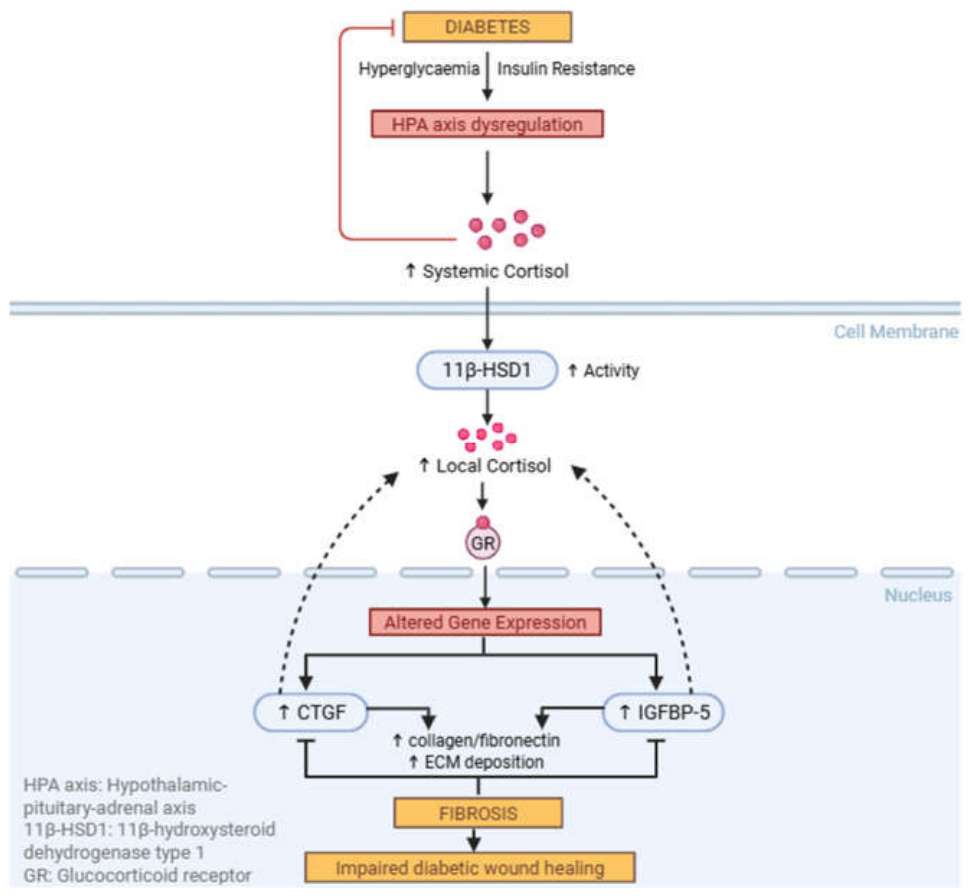
Figure 3.
Proposed interactions between diabetes-associated cortisol signalling and profibrotic pathways in impaired wound healing. Diabetes-associated metabolic disturbances contribute to HPA axis dysregulation and increased cortisol signalling. Local cortisol activation by 11β-HSD1 alters glucocorticoid receptor (GR)-mediated gene expression and may influence profibrotic pathways involving CTGF and IGFBP-5, contributing to ECM accumulation, fibrosis, and impaired diabetic wound healing. Solid arrows indicate established interactions, while dashed arrows represent hypothesised or incompletely characterised relationships. Created in BioRender. Lao, C. (2026) https://BioRender.com/mpozh11.
Figure 3.
Proposed interactions between diabetes-associated cortisol signalling and profibrotic pathways in impaired wound healing. Diabetes-associated metabolic disturbances contribute to HPA axis dysregulation and increased cortisol signalling. Local cortisol activation by 11β-HSD1 alters glucocorticoid receptor (GR)-mediated gene expression and may influence profibrotic pathways involving CTGF and IGFBP-5, contributing to ECM accumulation, fibrosis, and impaired diabetic wound healing. Solid arrows indicate established interactions, while dashed arrows represent hypothesised or incompletely characterised relationships. Created in BioRender. Lao, C. (2026) https://BioRender.com/mpozh11.
4. Therapeutic Targets
4.1. 11β-HSD1 Inhibitors
Inhibiting 11β-HSD1 has emerged as a promising therapeutic strategy to modulate local cortisol levels in chronic wounds, alongside its investigation as a therapeutic target in other disorders associated with excessive local glucocorticoid activation, including metabolic and cardiovascular disease [67,68,69,70]. Pharmacological inhibition with compounds such as AZD4017, a small-molecule selective inhibitor of 11β-HSD1, has shown beneficial effects in both preclinical and early human studies, improving epidermal structure, reducing dermal atrophy, and accelerating wound closure [18,19]. By preventing the intracellular conversion of inactive cortisone to active cortisol, AZD4017 reduces local glucocorticoid levels in the skin and limits cortisol-driven tissue damage. These effects are attributed to reduced cortisol signalling within the wound microenvironment, which relieves cortisol-mediated suppression of keratinocyte and fibroblast function and may reduce pro-fibrotic activation [14,18].
While most data to date derive from models of ageing and UV damage [14,18], recent findings indicate that 11β-HSD1 inhibition could also be effective in the diabetic wound environment [19]. More recent preclinical work has further supported the therapeutic potential of targeting local glucocorticoid activation in diabetic wounds [71]. In a type 1 diabetic mouse model, topical inhibition of local glucocorticoid synthesis using metyrapone, a dual inhibitor of 11β-HSD1 and CYP11B1, accelerated wound closure, restored collagen deposition and α-SMA expression, reduced inflammatory mediators, and enhanced fibroblast migration without altering systemic glucocorticoid levels. These findings suggest that targeting local glucocorticoid production may improve multiple aspects of tissue repair, including inflammation, extracellular matrix organisation, and fibroblast function.
These findings are derived from a non-peer-reviewed preclinical study using a dual inhibitor rather than selective 11β-HSD1 inhibition and therefore require validation in human tissues and clinically relevant chronic wound models. These studies have often relied on animal and acute wound models, which may not fully replicate the chronic inflammation, hypoxia, and matrix dysregulation seen in DFUs. Further research in clinically relevant chronic wound models is needed to evaluate the long-term antifibrotic efficacy of 11β-HSD1 inhibitors and their effects on ECM composition, macrophage polarisation, and fibrosis resolution.
Beyond local effects on skin repair, inhibition of 11β-HSD1 may also provide broader metabolic benefits in patients with diabetes. Tissue-specific amplification of glucocorticoid signalling by 11β-HSD1 contributes to insulin resistance, hepatic glucose production, adipose dysfunction and chronic inflammation, even in the absence of elevated circulating cortisol levels. This has been described as a state of functional hypercortisolism, in which local cortisol availability becomes dysregulated despite normal systemic concentrations [72]. Consequently, inhibition of 11β-HSD1 has the potential to improve not only wound healing outcomes but also the wider metabolic disturbances associated with diabetes. However, clinical studies have shown more modest benefits than preclinical models despite strong target engagement, suggesting that patient heterogeneity and tissue-specific regulation may influence therapeutic response. A critical consideration for evaluating future 11β-HSD1 inhibitor efficacy is controlling for systemic cortisol (and hence cortisone) availability, which has been largely overlooked in previous clinical studies.
4.2. Targeting CTGF and IGFBP-5
Direct inhibition of downstream pro-fibrotic molecules such as CTGF and IGFBP-5 offers an alternative or complementary strategy. CTGF antagonists and neutralising antibodies have shown efficacy in systemic fibrotic diseases (e.g., systemic sclerosis, pulmonary fibrosis), but their application in chronic wound healing, particularly in diabetes, remains underexplored [20]. Similarly, while IGFBP-5 has been shown to promote fibrosis and delay wound closure in diabetic fibroblasts, it is not yet established as a therapeutic target in skin, and current research has primarily focused on IGF-I rather than IGFBP-5 itself [63,73].
Given that CTGF and IGFBP-5 may act downstream of cortisol signalling [57,66], tracking changes in their expression may also serve as biomarkers of response to 11β-HSD1 inhibition. This could support both mechanistic understanding and therapeutic optimisation of antifibrotic strategies in diabetic skin.
4.3. Other Emerging Approaches
A number of complementary therapeutic strategies are also under investigation for chronic wound management. These include anti-inflammatory agents designed to restore immune balance by promoting macrophage polarisation from a pro-inflammatory M1 phenotype to a reparative M2 phenotype, thereby helping to resolve persistent inflammation [74,75]. MMP modulators are being explored to correct imbalances in ECM turnover and prevent excessive tissue degradation or fibrosis [76]. In addition, stem cell [77,78] and growth factor-based therapies [79] aim to stimulate tissue regeneration by enhancing revascularisation, promoting fibroblast and keratinocyte migration, and improving granulation tissue formation. These approaches may offer the greatest benefit when used in combination with antifibrotic agents, targeting the multiple, interconnected drivers of chronic wound pathology [80].
Alongside pharmacological and regenerative strategies, non-drug approaches that address psychosocial and behavioural aspects to poor wound healing are increasingly being explored. Social prescribing interventions aim to connect patients with community-based support, physical activity, welfare advice, peer support and other non-medical resources. thereby addressing social isolation, anxiety, lifestyle behaviours, and the broader factors that influence health [81]. This may also reduce chronic psychological stress and improve cortisol regulation. In T2D, social prescribing interventions have been associated with improvements in glycaemic control and may help address wider social determinants of health by supporting lifestyle modification, self-management and engagement with community resources [81]. However, current evidence remains limited and inconsistent, with relatively few large-scale randomised controlled trials evaluating their effectiveness [82]. Furthermore, their effects on cortisol regulation and wound healing outcomes remain largely unexplored.
5. State of the Research and Future Directions
Although each of these molecules has been studied individually in models of fibrosis, diabetes, and wound healing, there remains a substantial gap in understanding how these pathways interact within the fibrotic microenvironment of chronic diabetic wounds. This has limited our understanding of how local glucocorticoid metabolism influences inflammation, ECM remodelling, fibroblast behaviour, and ultimately fibrosis during chronic wound healing. Progress has been further hindered by the limitations of existing models, as most studies have relied on animal systems that do not fully recapitulate the cellular behaviour of human skin [54,83]. Furthermore, many investigations of 11β-HSD1 have focused on ultraviolet-induced skin damage and ageing rather than diabetes or wound repair [84], while studies examining diabetic wound healing frequently utilise acute wound models that fail to reproduce the persistent inflammation and fibrosis characteristic of chronic diabetic wounds [19,37]. Consequently, the role of cortisol and 11β-HSD1 in regulating fibrosis within chronic diabetic wounds remains poorly understood.
Importantly, while many studies address individual aspects of fibrosis (e.g., ECM deposition, fibroblast activity, angiogenesis), few consider how these processes interact, or how they may be influenced by hormonal regulation. In particular, it is not known whether cortisol activation via 11β-HSD1 directly regulates key profibrotic mediators such as CTGF and IGFBP-5 in human skin. While CTGF and IGFBP-5 are individually associated with fibrosis and poor wound repair [12,62], it remains unclear whether they are regulated by local cortisol activation in skin, or whether they themselves influence cortisol signalling or 11β-HSD1 expression. Most studies to date have examined glucocorticoid signalling [57,66] and fibrosis in isolation, and research on IGFBP-5 in diabetic wounds is particularly scarce [63]. A deeper understanding of these interactions is needed to clarify whether targeting 11β-HSD1 alone is sufficient to reduce fibrosis and relieve its inhibitory effects on wound healing, or whether co-targeting downstream effectors like CTGF and IGFBP-5 will offer a more comprehensive therapeutic strategy.
Future research should prioritise the use of clinically relevant human models, including primary diabetic fibroblasts, full-thickness skin explants, and chronic wound tissue, to better capture the complex inflammatory and fibrotic environment present in diabetic wounds. There is also a need for more physiologically relevant in vitro systems, including three-dimensional (3D) skin models and co-culture systems incorporating multiple cell types such as fibroblasts, keratinocytes, immune cells, and extracellular matrix components. Traditional two-dimensional monolayer cultures often fail to replicate the spatial organisation, cell-cell interactions, mechanical cues, and persistent inflammatory environment characteristic of chronic diabetic wounds. Incorporating features of the diabetic foot ulcer microenvironment, including hyperglycaemia, hypoxia, fibrosis, and altered immune signalling, may provide more representative models for investigating disease mechanisms and evaluating therapeutic responses. In addition to investigating individual pathways, studies should adopt more integrated approaches to determine how cortisol signalling interacts with CTGF, IGFBP-5, macrophage phenotypes, and extracellular matrix remodelling over time. Ultimately, improved understanding of these interactions will dissect the pathways which will inform the development of targeted therapeutic strategies, including selective 11β-HSD1 inhibition alone or in combination with downstream antifibrotic targets.
Author Contributions
Writing—original draft preparation, Lao C.; writing—review and editing, Alcacer-Pitarch B., Patterson S., Tiganescu A. & Wright C.S.; supervision, Patterson S. & Wright C.S.. All authors have read and agreed to the published version of the manuscript.
Funding
This research was funded by a grant from the British Skin Foundation, grant number 004_S_23.
Conflicts of Interest
The authors declare no conflicts of interest.
References
- Tesauro M, Mazzotta FA. Pathophysiology of diabetes. In: Transplantation, Bioengineering, and Regeneration of the Endocrine Pancreas [Internet]. Elsevier; 2020 [cited 2026 Jun 25]. p. 37–47. Available from: https://linkinghub.elsevier.com/retrieve/pii/B9780128148334000034. [CrossRef]
- Diabetes UK [Internet]. [cited 2026 Jun 25]. How many people in the UK have diabetes? Available from: https://www.diabetes.org.uk/about-us/about-the-charity/our-strategy/statistics.
- Diabetes [Internet]. [cited 2026 Jun 25]. Available from: https://www.who.int/news-room/fact-sheets/detail/diabetes.
- Bodke H, Wagh V, Kakar G. Diabetes Mellitus and Prevalence of Other Comorbid Conditions: A Systematic Review. Cureus. 2023 Nov 24. [CrossRef]
- Vileikyte L, Pouwer F, Gonzalez JS. Psychosocial research in the diabetic foot: Are we making progress? Diabetes Metab Res Rev. 2020 Mar;36 Suppl 1:e3257. PubMed PMID: 31850665. [CrossRef]
- Guest JF, Fuller GW, Vowden P. Diabetic foot ulcer management in clinical practice in the UK: costs and outcomes. Int Wound J. 2018 Feb;15(1):43–52. PubMed PMID: 29243399; PubMed Central PMCID: PMC7950039. [CrossRef]
- Burgess JL, Wyant WA, Abdo Abujamra B, Kirsner RS, Jozic I. Diabetic Wound-Healing Science. Medicina (Mex). 2021 Oct 8;57(10):1072. [CrossRef]
- Dasari N, Jiang A, Skochdopole A, Chung J, Reece EM, Vorstenbosch J, et al. Updates in Diabetic Wound Healing, Inflammation, and Scarring. Semin Plast Surg. 2021 Aug;35(03):153–8. [CrossRef]
- Mayorca-Guiliani AE, Leeming DJ, Henriksen K, Mortensen JH, Nielsen SH, Anstee QM, et al. ECM formation and degradation during fibrosis, repair, and regeneration. Npj Metab Health Dis. 2025 Jun 10;3(1):25. [CrossRef]
- Tuleta I, Frangogiannis NG. Diabetic fibrosis. Biochim Biophys Acta BBA - Mol Basis Dis. 2021 Apr;1867(4):166044. [CrossRef]
- Kim KK, Sheppard D, Chapman HA. TGF-β1 Signaling and Tissue Fibrosis. Cold Spring Harb Perspect Biol. 2018 Apr;10(4):a022293. [CrossRef]
- Nguyen XX, Muhammad L, Nietert PJ, Feghali-Bostwick C. IGFBP-5 Promotes Fibrosis via Increasing Its Own Expression and That of Other Pro-fibrotic Mediators. Front Endocrinol. 2018 Oct 15;9. [CrossRef]
- Stojadinovic O, Gordon KA, Lebrun E, Tomic-Canic M. Stress-Induced Hormones Cortisol and Epinephrine Impair Wound Epithelization. Adv Wound Care. 2012 Feb;1(1):29–35. [CrossRef]
- Hall L, Hart R. Role of corticosteroids in skin physiology and therapeutic potential of an 11β-HSD1 inhibitor: A review. Int J Dermatol. 2024 Apr;63(4):443–54. [CrossRef]
- Aroda VR, Umpierrez GE, Edelman SV, Einhorn D. Cortisol’s role in difficult-to-control type 2 diabetes: Insights from the CATALYST study—a plain language review. Curr Med Res Opin. 2026 Mar 4;42(3):415–26. PubMed PMID: 42126387. [CrossRef]
- Stamou MI, Colling C, Dichtel LE. Adrenal aging and its effects on the stress response and immunosenescence. Maturitas. 2023 Feb 1;168:13–9. [CrossRef]
- Mochel K, Bronte J, Kasaba M, Vempati A, Tam C, Hazany S. The Impact of Psychological Stress on Wound Healing: Implications for Neocollagenesis and Scar Treatment Efficacy. Clin Cosmet Investig Dermatol. 2025 Jun 30;18:1625–37. PubMed PMID: 40620896; PubMed Central PMCID: PMC12227520. [CrossRef]
- Tiganescu A, Tahrani AA, Morgan SA, Otranto M, Desmoulière A, Abrahams L, et al. 11β-Hydroxysteroid dehydrogenase blockade prevents age-induced skin structure and function defects. J Clin Invest. 2013 Jul 1;123(7):3051–60. [CrossRef]
- Ajjan RA, Hensor EMA, Del Galdo F, Shams K, Abbas A, Fairclough RJ, et al. Oral 11β-HSD1 inhibitor AZD4017 improves wound healing and skin integrity in adults with type 2 diabetes mellitus: a pilot randomized controlled trial. Eur J Endocrinol. 2022 Apr 1;186(4):441–55. [CrossRef]
- Lipson KE, Wong C, Teng Y, Spong S. CTGF is a central mediator of tissue remodeling and fibrosis and its inhibition can reverse the process of fibrosis. Fibrogenesis Tissue Repair. 2012 Dec;5(S1):S24. [CrossRef]
- Velnar T, Bailey T, Smrkolj V. The Wound Healing Process: An Overview of the Cellular and Molecular Mechanisms. J Int Med Res. 2009 Oct;37(5):1528–42. [CrossRef]
- Las Heras K, Igartua M, Santos-Vizcaino E, Hernandez RM. Chronic wounds: Current status, available strategies and emerging therapeutic solutions. J Controlled Release. 2020 Dec;328:532–50. [CrossRef]
- Falanga V. Wound healing and its impairment in the diabetic foot. The Lancet. 2005 Nov 12;366(9498):1736–43. PubMed PMID: 16291068. [CrossRef]
- Chen VY, Siegfried LG, Tomic-Canic M, Stone RC, Pastar I. Cutaneous changes in diabetic patients: Primed for aberrant healing? Wound Repair Regen. 2023 Sep 1;31(5):700–12. [CrossRef]
- Van Putte L, De Schrijver S, Moortgat P. The effects of advanced glycation end products (AGEs) on dermal wound healing and scar formation: a systematic review. Scars Burns Heal. 2016 Jan 1;2:2059513116676828. [CrossRef]
- Krzyszczyk P, Schloss R, Palmer A, Berthiaume F. The Role of Macrophages in Acute and Chronic Wound Healing and Interventions to Promote Pro-wound Healing Phenotypes. Front Physiol. 2018 May 1;9:419. [CrossRef]
- Theocharidis G, Thomas BE, Sarkar D, Mumme HL, Pilcher WJR, Dwivedi B, et al. Single cell transcriptomic landscape of diabetic foot ulcers. Nat Commun. 2022 Jan 10;13(1):181. [CrossRef]
- Wang L, Yang K, Xie X, Wang S, Gan H, Wang X, et al. Macrophages as Multifaceted Orchestrators of Tissue Repair: Bridging Inflammation, Regeneration, and Therapeutic Innovation. J Inflamm Res. 2025 Jul 8;18:8945–59. [CrossRef]
- Hosseini Mansoub N. The role of keratinocyte function on the defected diabetic wound healing. Clin Diabetol. 2022 Jan 31;VM/OJS/J/85499. [CrossRef]
- Do NN, Eming SA. Skin fibrosis: Models and mechanisms. Curr Res Transl Med. 2016 Oct 1;Guerlain symposium – Biological repair mechanisms: the brain-skin connection64(4):185–93. [CrossRef]
- Darby IA, Laverdet B, Bonté F, Desmoulière A. Fibroblasts and myofibroblasts in wound healing. Clin Cosmet Investig Dermatol. 2014 Nov 6;7:301–11. [CrossRef]
- Fu K, Zheng X, Chen Y, Wu L, Yang Z, Chen X, et al. Role of matrix metalloproteinases in diabetic foot ulcers: Potential therapeutic targets. Front Pharmacol. 2022 Oct 20;13:1050630. [CrossRef]
- Ladwig GP, Robson MC, Liu R, Kuhn MA, Muir DF, Schultz GS. Ratios of activated matrix metalloproteinase-9 to tissue inhibitor of matrix metalloproteinase-1 in wound fluids are inversely correlated with healing of pressure ulcers. Wound Repair Regen Off Publ Wound Heal Soc Eur Tissue Repair Soc. 2002;10(1):26–37. PubMed PMID: 11983004. [CrossRef]
- Liu Y, Min D, Bolton T, Nubé V, Twigg SM, Yue DK, et al. Increased matrix metalloproteinase-9 predicts poor wound healing in diabetic foot ulcers. Diabetes Care. 2009 Jan;32(1):117–9. PubMed PMID: 18835949; PubMed Central PMCID: PMC2606842. [CrossRef]
- Leong E, Bezuhly M, Marshall JS. Distinct Metalloproteinase Expression and Functions in Systemic Sclerosis and Fibrosis: What We Know and the Potential for Intervention. Front Physiol. 2021 Aug 27;12:727451. [CrossRef]
- Moinzadeh P, Denton CP, Abraham D, Ong V, Hunzelmann N, Eckes B, et al. Biomarkers for skin involvement and fibrotic activity in scleroderma. J Eur Acad Dermatol Venereol JEADV. 2012 Mar;26(3):267–76. PubMed PMID: 21819452. [CrossRef]
- Brazel CB, Simon JC, Tuckermann JP, Saalbach A. Inhibition of 11β-HSD1 Expression by Insulin in Skin: Impact for Diabetic Wound Healing. J Clin Med. 2020 Nov 28;9(12):3878. [CrossRef]
- Joseph JJ, Golden SH. Cortisol dysregulation: the bidirectional link between stress, depression, and type 2 diabetes mellitus. Ann N Y Acad Sci. 2017;1391(1):20–34. [CrossRef]
- Barbot M, Ceccato F, Scaroni C. Diabetes Mellitus Secondary to Cushing’s Disease. Front Endocrinol. 2018 Jun 5;9. [CrossRef]
- Tiganescu A. A Double-blind, Randomized, Placebo-controlled Phase II Pilot Trial Investigating Efficacy, Safety and Feasibility of 11β-hydroxysteroid Dehydrogenase Type 1 Inhibition by AZD4017 to Improve Skin Function and Wound Healing in Patients With Type 2 Diabetes [Clinical trial registration] [Internet]. clinicaltrials.gov; 2019 Mar [cited 2026 Jun 29]. Clinical trial registration no.: NCT03313297. Available from: https://clinicaltrials.gov/study/NCT03313297.
- Al-Otaibi ATA, Almuqati BFA, Al-Mutairi ABMA, Alanazi AH, Alharbi AMM, Albawardi HM, et al. The Impact of Psychological Stress on Wound Healing: A Clinical Nursing Perspective. J Med Life Sci. 2025 Dec 14;7(4):946–62. [CrossRef]
- Alasfour L, Alboloushi A, Kirwan E, McIntosh C, MacGilchrist C, Hurst JE. Mapping the evidence to determine the influence of stress, anxiety, and depression on wound healing in patients with diabetes-related foot ulcers: A scoping review. J Tissue Viability. 2025 Aug;34(3):100935. [CrossRef]
- Yang EV, Bane CM, MacCallum RC, Kiecolt-Glaser JK, Malarkey WB, Glaser R. Stress-related modulation of matrix metalloproteinase expression. J Neuroimmunol. 2002 Dec 1;133(1):144–50. [CrossRef]
- Lundberg AK, Jönsson S, Stenmark J, Kristenson M, Jonasson L. Stress-induced release of matrix metalloproteinase-9 in patients with coronary artery disease: The possible influence of cortisol. Psychoneuroendocrinology. 2016 Nov 1;73:117–24. [CrossRef]
- Szymanowski A, Nijm J, Kristenson M, Jonasson L. Elevated levels of circulating matrix metalloproteinase-9 are associated with a dysregulated cortisol rhythm—A case-control study of coronary artery disease. Psychoneuroendocrinology. 2011 Jan 1;36(1):139–43. [CrossRef]
- Al-Rikabi AHA, Tobin DJ, Riches-Suman K, Thornton MJ. Dermal fibroblasts cultured from donors with type 2 diabetes mellitus retain an epigenetic memory associated with poor wound healing responses. Sci Rep. 2021 Jan 14;11(1):1474. [CrossRef]
- Kandhwal M, Behl T, Singh S, Sharma N, Arora S, Bhatia S, et al. Role of matrix metalloproteinase in wound healing.
- Logie JJ, Ali S, Marshall KM, Heck MMS, Walker BR, Hadoke PWF. Glucocorticoid-Mediated Inhibition of Angiogenic Changes in Human Endothelial Cells Is Not Caused by Reductions in Cell Proliferation or Migration. Reitsma PH, editor. PLoS ONE. 2010 Dec 31;5(12):e14476. [CrossRef]
- Jere SW, Houreld NN, Abrahamse H. Role of the PI3K/AKT (mTOR and GSK3β) signalling pathway and photobiomodulation in diabetic wound healing. Cytokine Growth Factor Rev. 2019 Dec;50:52–9. [CrossRef]
- Tiganescu A, Hupe M, Uchida Y, Mauro T, Elias PM, Holleran WM. Increased glucocorticoid activation during mouse skin wound healing. J Endocrinol. 2014 Apr;221(1):51–61. PubMed PMID: 24464022. [CrossRef]
- Huang X, Li Y, Fu M, Xin HB. Polarizing Macrophages In Vitro. In: Rousselet G, editor. Macrophages [Internet]. New York, NY: Springer New York; 2018 [cited 2026 Jun 25]. p. 119–26. (Methods in Molecular Biology). Available from: http://link.springer.com/10.1007/978-1-4939-7837-3_12. [CrossRef]
- Tsai CH, Liao CW, Wu XM, Chen ZW, Pan CT, Chang YY, et al. Autonomous cortisol secretion is associated with worse arterial stiffness and vascular fibrosis in primary aldosteronism: a cross-sectional study with follow-up data. Eur J Endocrinol. 2022 Jul 1;187(1):197–208. [CrossRef]
- Shukla R, Basu AK, Mandal B, Mukhopadhyay P, Maity A, Chakraborty S, et al. 11β Hydroxysteroid dehydrogenase – 1 activity in type 2 diabetes mellitus: a comparative study. BMC Endocr Disord. 2019 Dec;19(1):15. [CrossRef]
- Tiganescu A, Hupe M, Uchida Y, Mauro T, Elias PM, Holleran WM. Topical 11β-Hydroxysteroid Dehydrogenase Type 1 Inhibition Corrects Cutaneous Features of Systemic Glucocorticoid Excess in Female Mice. Endocrinology. 2018 Jan 1;159(1):547–56. [CrossRef]
- Twigg SM. Regulation and bioactivity of the CCN family of genes and proteins in obesity and diabetes. J Cell Commun Signal. 2018;12(1):173. [CrossRef]
- Henshaw FR, Boughton P, Lo L, McLennan SV, Twigg SM. Topically Applied Connective Tissue Growth Factor/CCN2 Improves Diabetic Preclinical Cutaneous Wound Healing: Potential Role for CTGF in Human Diabetic Foot Ulcer Healing. J Diabetes Res. 2015;2015:1–10. [CrossRef]
- Fehrholz M, Glaser K, Speer CP, Seidenspinner S, Ottensmeier B, Kunzmann S. Caffeine modulates glucocorticoid-induced expression of CTGF in lung epithelial cells and fibroblasts. Respir Res. 2017 Dec;18(1):51. [CrossRef]
- Wilton-Waddell A, Farraj LA, Vasconcelos EJ, Byrne E, Taylor AE, Freeman A, et al. 11β-HSD1 inhibitor efficacy in type 2 diabetes is cortisol-dependent [Internet]. Endocrinology (including Diabetes Mellitus and Metabolic Disease); 2024 [cited 2026 Jun 25]. Available from: http://medrxiv.org/lookup/doi/10.1101/2024.05.10.24307180. [CrossRef]
- He C, Ge S, Xia W, Wang Y, Yan L, Sun J, et al. Role of CTGF-LRP1 in impaired healing of cesarean section incisions. Nat Commun. 2026 Feb 28;17(1):3278. [CrossRef]
- Wu Y, Luo X, Zhou Q, Gong H, Gao H, Liu T, et al. The disbalance of LRP1 and SIRPα by psychological stress dampens the clearance of tumor cells by macrophages. Acta Pharm Sin B. 2022 Jan;12(1):197–209. [CrossRef]
- Zhang CY, Wang B, Hua XT, Fan K, Li YF. Serum vascular endothelial growth factor and cortisol expression to predict prognosis of patients with hypertensive cerebral hemorrhage. World J Clin Cases. 2023 Aug 16;11(23):5455–61. [CrossRef]
- Yasuoka H, Jukic DM, Zhou Z, Choi AMK, Feghali-Bostwick CA. Insulin-like growth factor binding protein 5 induces skin fibrosis: A novel murine model for dermal fibrosis. Arthritis Rheum. 2006 Sep;54(9):3001–10. [CrossRef]
- Were S, Wilson T, Martin P, Wright C. P22 Fibrosis in delayed wound healing in diabetes: the role of insulin-like growth factor binding proteins. Br J Dermatol. 2023 Jul 7;189(1):e22–3. [CrossRef]
- Simon CM, Rauskolb S, Gunnersen JM, Holtmann B, Drepper C, Dombert B, et al. Dysregulated IGFBP5 expression causes axon degeneration and motoneuron loss in diabetic neuropathy. Acta Neuropathol (Berl). 2015 Sep;130(3):373–87. [CrossRef]
- Wright CS, Berends RF, Flint DJ, Martin PEM. Cell motility in models of wounded human skin is improved by Gap27 despite raised glucose, insulin and IGFBP-5. Exp Cell Res. 2013 Feb;319(4):390–401. [CrossRef]
- Fernández-Pereira C, Agís-Balboa RC. The Insulin-like Growth Factor Family as a Potential Peripheral Biomarker in Psychiatric Disorders: A Systematic Review. Int J Mol Sci. 2025 Mar 12;26(6):2561. [CrossRef]
- Kupczyk D, Bilski R, Kozakiewicz M, Studzińska R, Kędziora-Kornatowska K, Kosmalski T, et al. 11β-HSD as a New Target in Pharmacotherapy of Metabolic Diseases. Int J Mol Sci. 2022 Aug 11;23(16):8984. PubMed PMID: 36012251; PubMed Central PMCID: PMC9409048. [CrossRef]
- Kupczyk D, Studzińska R, Kołodziejska R, Baumgart S, Modrzejewska M, Woźniak A. 11β-Hydroxysteroid Dehydrogenase Type 1 as a Potential Treatment Target in Cardiovascular Diseases. J Clin Med. 2022 Oct 20;11(20):6190. PubMed PMID: 36294507; PubMed Central PMCID: PMC9605099. [CrossRef]
- Gregory S, Hill D, Grey B, Ketelbey W, Miller T, Muniz-Terrera G, et al. 11β-hydroxysteroid dehydrogenase type 1 inhibitor use in human disease-a systematic review and narrative synthesis. Metabolism. 2020 Jul;108:154246. PubMed PMID: 32333937. [CrossRef]
- Tsai KH, Cooper MS. Recycling glucocorticoids: therapeutic implications of the 11β-HSD1 enzyme system. J Endocrinol. 2023 Sep 1;258(3):e220289. PubMed PMID: 37335193. [CrossRef]
- Fraga-Junior V, Waclawiak I, Ribeiro WR, Oliveira-Junior EJD, Silva JKADO, Palazzo M, et al. Inhibition of skin glucocorticoid synthesis by topical metyrapone administration supports wound healing in diabetic mice [Internet]. 2026 [cited 2026 Jun 25]. Available from: https://www.authorea.com/doi/full/10.22541/au.177314987.71327230/v1. [CrossRef]
- Joshi P, Mazumder A, Pentela B, Debnath A. Advances in 11-HSD1 Inhibitor Development for Addressing Insulin Resistance and Beta-Cell Dysfunction in Type 2 diabetes mellitus. J Pharm Innov. 2026 Aug;21(4):380. [CrossRef]
- Garoufalia Z, Papadopetraki A, Karatza E, Vardakostas D, Philippou A, Kouraklis G, et al. Insulin-like growth factor-I and wound healing, a potential answer to non-healing wounds: A systematic review of the literature and future perspectives. Biomed Rep. 2021 Jun 8;15(2):66. [CrossRef]
- Maruyama K, Asai J, Ii M, Thorne T, Losordo DW, D’Amore PA. Decreased macrophage number and activation lead to reduced lymphatic vessel formation and contribute to impaired diabetic wound healing. Am J Pathol. 2007 Apr;170(4):1178–91. PubMed PMID: 17392158; PubMed Central PMCID: PMC1829452. [CrossRef]
- Takeuchi T, Ito M, Yamaguchi S, Watanabe S, Honda M, Imahashi T, et al. Hydrocolloid dressing improves wound healing by increasing M2 macrophage polarization in mice with diabetes. Nagoya J Med Sci. 2020 Aug;82(3):487–98. PubMed PMID: 33132433; PubMed Central PMCID: PMC7548260. [CrossRef]
- Gooyit M, Peng Z, Wolter WR, Pi H, Ding D, Hesek D, et al. A Chemical Biological Strategy to Facilitate Diabetic Wound Healing. ACS Chem Biol. 2014 Jan 17;9(1):105–10. [CrossRef]
- Li X, Xie X, Lian W, Shi R, Han S, Zhang H, et al. Exosomes from adipose-derived stem cells overexpressing Nrf2 accelerate cutaneous wound healing by promoting vascularization in a diabetic foot ulcer rat model. Exp Mol Med. 2018 Apr;50(4):1–14. [CrossRef]
- Ouyang L, Qiu D, Fu X, Wu A, Yang P, Yang Z, et al. Overexpressing HPGDS in adipose-derived mesenchymal stem cells reduces inflammatory state and improves wound healing in type 2 diabetic mice. Stem Cell Res Ther. 2022 Aug 3;13(1):395. [CrossRef]
- Wieman TJ. Clinical efficacy of becaplermin (rhPDGF-BB) gel. Am J Surg. 1998 Aug 1;176(2, Supplement 1):74S-79S. [CrossRef]
- Yadav JP, Singh AK, Grishina M, Pathak P, Verma A, Kumar V, et al. Insights into the mechanisms of diabetic wounds: pathophysiology, molecular targets, and treatment strategies through conventional and alternative therapies. Inflammopharmacology. 2024 Feb;32(1):149–228. [CrossRef]
- Wildman J, Wildman JM. Evaluation of a Community Health Worker Social Prescribing Program Among UK Patients With Type 2 Diabetes. JAMA Netw Open. 2021 Sep 1;4(9):e2126236. [CrossRef]
- Calderón-Larrañaga S, Clinch M, Greenhalgh T, Finer S. Could social prescribing contribute to type 2 diabetes prevention in people at high risk? Protocol for a realist, multilevel, mixed methods review and evaluation. BMJ Open. 2021 Apr;11(4):e042303. [CrossRef]
- Terao M, Katayama I. Local cortisol/corticosterone activation in skin physiology and pathology. J Dermatol Sci. 2016 Oct;84(1):11–6. [CrossRef]
- Kim BJ, Lee NR, Lee CH, Lee YB, Choe SJ, Lee S, et al. Increased Expression of 11β-Hydroxysteroid Dehydrogenase Type 1 Contributes to Epidermal Permeability Barrier Dysfunction in Aged Skin. Int J Mol Sci. 2021 May 27;22(11):5750. [CrossRef]
Figure 1.
Comparison of healing in normal versus chronic diabetic wounds. In normal wound healing, coordinated signalling by cytokines and growth factors promotes fibroblast activation, balanced ECM remodelling, angiogenesis, and efficient tissue repair. Chronic diabetic wounds are characterised by persistent hyperglycaemia, altered fibroblast and immune cell function, excessive collagen deposition, MMP/TIMP imbalance, and sustained production of pro-inflammatory cytokines, resulting in persistent inflammation, fibrosis, and delayed wound closure. Created in BioRender. Lao, C. (2026) https://BioRender.com/l56gges.
Figure 1.
Comparison of healing in normal versus chronic diabetic wounds. In normal wound healing, coordinated signalling by cytokines and growth factors promotes fibroblast activation, balanced ECM remodelling, angiogenesis, and efficient tissue repair. Chronic diabetic wounds are characterised by persistent hyperglycaemia, altered fibroblast and immune cell function, excessive collagen deposition, MMP/TIMP imbalance, and sustained production of pro-inflammatory cytokines, resulting in persistent inflammation, fibrosis, and delayed wound closure. Created in BioRender. Lao, C. (2026) https://BioRender.com/l56gges.
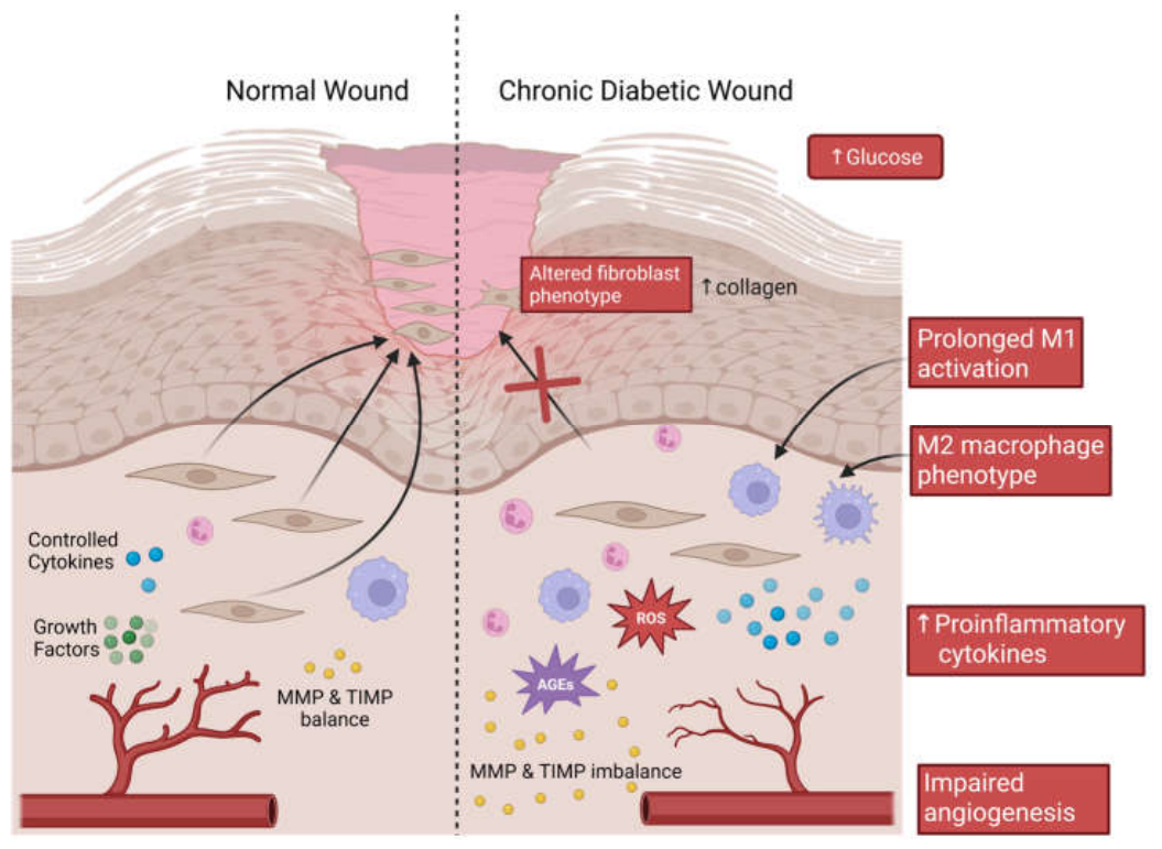
Figure 2.
Interconversion of cortisone and cortisol by 11β-HSD enzymes. Cortisone is converted to the biologically active glucocorticoid cortisol via 11β-HSD1 in an NADPH-dependent reduction. 11β-hydroxysteroid dehydrogenase type 2 (11β-HSD2) catalyses the reverse reaction, converting cortisol back to cortisone. Created in BioRender. Lao, C. (2026) https://BioRender.com/ek55uob.
Figure 2.
Interconversion of cortisone and cortisol by 11β-HSD enzymes. Cortisone is converted to the biologically active glucocorticoid cortisol via 11β-HSD1 in an NADPH-dependent reduction. 11β-hydroxysteroid dehydrogenase type 2 (11β-HSD2) catalyses the reverse reaction, converting cortisol back to cortisone. Created in BioRender. Lao, C. (2026) https://BioRender.com/ek55uob.
Disclaimer/Publisher’s Note: The statements, opinions and data contained in all publications are solely those of the individual author(s) and contributor(s) and not of MDPI and/or the editor(s). MDPI and/or the editor(s) disclaim responsibility for any injury to people or property resulting from any ideas, methods, instructions or products referred to in the content. |
© 2026 by the authors. Licensee MDPI, Basel, Switzerland. This article is an open access article distributed under the terms and conditions of the Creative Commons Attribution (CC BY) license (http://creativecommons.org/licenses/by/4.0/).
Copyright: This open access article is published under a Creative Commons CC BY 4.0 license, which permit the free download, distribution, and reuse, provided that the author and preprint are cited in any reuse.