

Submitted:
04 August 2026
Posted:
04 August 2026
You are already at the latest version
Abstract
Background and Objectives: Delayed contour irregularities may occasionally become apparent years after otherwise successful autologous rhinoplasty, particularly in patients with thin nasal skin. While thin-skinned rhinoplasty, autologous graft behavior, and nasal aging have been extensively discussed individually, their potential interaction has received limited attention. This review evaluates current evidence regarding aging-related nasal soft tissue changes, thin skin, autologous graft behavior, and their potential influence on long-term aesthetic outcomes after rhinoplasty. Materials and Methods: A literature review was performed using PubMed, Scopus, and Google Scholar databases. Relevant studies addressing rhinoplasty, autologous cartilage grafts, thin nasal skin, nasal aging, contour irregularities, preservation rhinoplasty, and soft tissue management were reviewed. Original studies, systematic reviews, and clinically relevant review articles were included. The available evidence was critically evaluated with particular attention to long-term outcomes and limitations of current knowledge. Results: Thin nasal skin is a recognized risk factor for graft visibility and contour irregularities following rhinoplasty. Aging-related changes, including dermal thinning, loss of collagen and elastin, decreased skin elasticity, and soft tissue atrophy, may further reduce the ability of the nasal soft tissue envelope to camouflage underlying structural features. However, direct longitudinal evidence linking aging-related soft tissue changes to delayed contour irregularities remains limited. Current literature consists predominantly of retrospective studies, expert opinion, and short- to medium-term outcome analyses. Preservation rhinoplasty and soft tissue camouflage techniques have been proposed to improve contour stability, although their long-term effectiveness in preventing aging-related contour visibility remains uncertain. Conclusions: Current evidence supports the biologic plausibility that progressive soft tissue aging may influence the long-term visibility of underlying grafts after rhinoplasty. Nevertheless, direct clinical evidence remains limited, and the relationship should currently be regarded as a hypothesis-generating concept rather than a proven causal mechanism. Future longitudinal studies incorporating objective assessment of skin quality, soft tissue thickness, and three-dimensional surface imaging are needed to better define the interaction between aging and long-term rhinoplasty outcomes.
Keywords:
rhinoplasty
; aging
; thin nasal skin
; autologous cartilage graft
; contour irregularity
; soft tissue aging
1. Introduction
Autologous cartilage grafts remain the foundation of modern rhinoplasty because of their excellent biocompatibility, low risk of infection and extrusion, and favorable long-term structural stability.[1] Septal, auricular, and costal cartilage grafts are widely used to restore nasal contour, improve support, and achieve durable aesthetic outcomes.[2] Although most studies report satisfactory long-term results, delayed contour irregularities, graft visibility, translucency, and subtle asymmetries continue to represent challenging postoperative concerns, particularly in patients with thin nasal skin.[3]
Thin nasal skin has long been recognized as a major determinant of rhinoplasty outcomes because it provides limited soft tissue coverage over the underlying framework.[3] Even minor graft imperfections, contour irregularities, or surface asymmetries may become visible through a thin soft tissue envelope.[4] Consequently, considerable attention has been directed toward graft refinement, soft tissue preservation, and camouflage techniques designed to optimize postoperative contour smoothness.
At the same time, the nasal soft tissue envelope undergoes progressive aging-related changes throughout adulthood. Histologic and clinical studies have demonstrated reductions in dermal thickness, collagen content, skin elasticity, and soft tissue volume, accompanied by changes in nasal support structures and external contour.[5] These processes may influence the long-term appearance of previously reconstructed nasal structures. However, despite growing interest in nasal aging, relatively little attention has been devoted to understanding how progressive soft tissue changes may interact with underlying autologous grafts years after surgery.(Figure 1)
Current rhinoplasty literature has extensively examined thin-skinned rhinoplasty, autologous graft behavior, and age-related nasal changes as separate topics. However, few studies have explored their potential interaction or evaluated whether aging-related soft tissue alterations may contribute to delayed contour visibility after initially successful rhinoplasty. Furthermore, the available evidence is heterogeneous and largely based on retrospective studies, expert opinion, and clinical observation, with limited long-term prospective data.
The purpose of this review is to critically evaluate current evidence regarding thin nasal skin, aging-related nasal soft tissue changes, autologous graft behavior, and preservation-oriented rhinoplasty strategies. Particular attention is given to the potential relationship between progressive soft tissue aging and delayed contour irregularities following autologous rhinoplasty, while highlighting current limitations in the literature and identifying areas for future research.
2. Methods of Literature Search
A literature search was conducted using PubMed, Scopus, and Google Scholar databases to identify relevant studies published through June 2026. Search terms included combinations of “rhinoplasty,” “autologous cartilage graft,” “thin nasal skin,” “nasal aging,” “aging nose,” “contour irregularity,” “graft visibility,” “preservation rhinoplasty,” “soft tissue envelope,” and “nasal soft tissue.”
Articles were selected based on their relevance to aging-related changes of the nasal soft tissue envelope, long-term outcomes of autologous rhinoplasty, thin-skinned rhinoplasty, contour irregularities, graft behavior, and preservation-oriented surgical techniques. Original studies, systematic reviews, narrative reviews, and landmark articles that contributed to the understanding of long-term aesthetic outcomes after rhinoplasty were included. Priority was given to contemporary literature published within the last 10 years, while historically important studies were included when necessary to provide clinical context and illustrate the evolution of current concepts.
The retrieved literature was qualitatively reviewed and synthesized to evaluate current evidence, identify limitations in the existing literature, and explore potential mechanisms linking aging-related soft tissue changes with delayed contour irregularities following autologous rhinoplasty.
3. Autologous Graft Materials and Long-Term Complications
Autologous graft materials remain the preferred option in rhinoplasty because of their favorable biocompatibility, low risk of infection, and minimal likelihood of extrusion compared with alloplastic implants.[1] Septal cartilage, auricular cartilage, and costal cartilage are the most commonly used autologous graft sources, each possessing distinct structural characteristics and long-term behavior.[6] Costal cartilage provides abundant volume and strong structural support but has been associated with warping, graft visibility, and contour irregularities, particularly in patients with thin soft tissue coverage[7]. Septal cartilage offers excellent integration and stability but may be limited in quantity, whereas auricular cartilage provides flexibility at the expense of structural rigidity.[8]
Several systematic reviews have demonstrated relatively low rates of major complications following autologous cartilage grafting. However, the reported outcomes are heterogeneous because of differences in surgical techniques, graft preparation methods, follow-up duration, and definitions of complications.[9] Most published studies focus on short- to mid-term outcomes, while data regarding aesthetic changes occurring more than 5–10 years after surgery remain limited.
Minor contour irregularities, graft edge visibility, translucency, and subtle asymmetries may not become clinically apparent during the early postoperative period.[3] These changes may emerge gradually and can be influenced by factors beyond graft behavior alone, including alterations of the overlying soft tissue envelope. Although graft warping, resorption, and scar contracture have been recognized as potential causes of delayed contour changes, their relative contribution remains difficult to quantify because of the lack of long-term prospective studies.[10]
Current evidence supports the structural reliability of autologous grafts; however, there is limited direct evidence regarding how aging-related soft tissue changes influence the long-term visibility of otherwise stable grafts.[11] This represents an important knowledge gap in rhinoplasty literature and highlights the need for future longitudinal studies evaluating both graft stability and age-related changes in soft tissue camouflage.(Table 1)
4. Thin Nasal Skin as a Risk Factor for Contour Irregularity
Thin nasal skin has long been recognized as one of the most important factors influencing aesthetic outcomes after rhinoplasty.[12] Unlike thick skin, which can partially camouflage minor underlying structural imperfections, thin skin provides limited soft tissue coverage and may increase the visibility of subtle contour irregularities, graft edges, dorsal asymmetries, and surface deformities.[13] Consequently, surgical planning in patients with thin nasal skin often requires greater attention to graft shaping, contour refinement, and soft tissue management.[14]
Several authors have emphasized that thin-skinned patients are at increased risk for postoperative graft visibility and contour abnormalities, particularly following dorsal augmentation procedures. Clinical strategies such as meticulous graft carving, fascia grafting, diced cartilage techniques, and preservation of the soft tissue envelope have therefore been recommended to improve contour camouflage.[15] However, much of the available evidence is derived from retrospective studies, expert opinion, and surgical experience rather than high-level comparative research.
Importantly, thin skin alone may not fully explain delayed contour irregularities observed years after surgery. Most studies evaluate outcomes within relatively short follow-up periods and focus primarily on immediate or early postoperative aesthetics. There is limited longitudinal evidence assessing whether progressive changes in skin thickness, elasticity, and soft tissue quality alter the long-term visibility of underlying grafts.
Furthermore, not all patients with thin nasal skin develop clinically significant contour irregularities, suggesting that additional factors—including graft material, graft position, scar formation, surgical technique, and individual aging patterns—may contribute to postoperative outcomes. The relative importance of these variables remains poorly defined.
Current evidence supports thin nasal skin as a recognized risk factor for contour visibility and contour irregularity after rhinoplasty. Nevertheless, the quality of available evidence remains limited, and direct studies examining the interaction between thin skin, aging-related soft tissue changes, and delayed contour deformities are largely absent. This lack of longitudinal data represents a significant knowledge gap and underscores the need for future prospective studies evaluating long-term aesthetic aging after rhinoplasty.
5. Aging-Related Nasal Soft Tissue Changes
Aging produces progressive structural and functional changes throughout the facial soft tissue envelope, including the nose.[16] Histologic studies have demonstrated age-related reductions in dermal thickness, collagen content, elastin integrity, and subcutaneous soft tissue volume.[17] These changes contribute to decreased skin elasticity, reduced tissue support, and alterations in surface contour. Similar processes are believed to affect the nasal soft tissue envelope[18], potentially influencing the long-term appearance of previously reconstructed nasal structures.
In addition to cutaneous changes, aging may affect the ligamentous and fibro-fatty support structures of the nose.[19] Progressive weakening of connective tissue support can contribute to tip ptosis, alar instability, and changes in nasal projection.[20] Previous studies examining the aging nose have reported characteristic findings including tip descent, increased nasal length, and alterations in tip rotation.[21] These observations suggest that the nasal framework and its overlying soft tissue envelope continue to evolve throughout adulthood.
Despite widespread clinical recognition of nasal aging, relatively few studies have specifically investigated how age-related soft tissue changes influence long-term rhinoplasty outcomes. Most available evidence is derived from cross-sectional observations or extrapolated from broader studies of facial aging. Direct measurements of postoperative changes in skin thickness, elasticity, or soft tissue camouflage following rhinoplasty remain scarce.
Importantly, aging-related soft tissue changes may not affect all patients equally. Genetic factors, ultraviolet light exposure, smoking history, skin quality, and individual patterns of facial aging may influence the degree of dermal thinning and soft tissue atrophy.[22] Consequently, the long-term visibility of underlying grafts may vary substantially among individuals despite similar surgical techniques and graft materials.[23]
Current evidence supports the concept that aging alters the structural properties of the nasal soft tissue envelope.[24] However, direct longitudinal evidence linking these changes to delayed contour irregularities after rhinoplasty remains limited. The relationship between progressive soft tissue aging and long-term graft visibility therefore remains largely hypothetical and represents an important area for future investigation.(Figure 2)
6. Interaction Between Aging Skin and Autologous Graft Behavior
The long-term aesthetic outcome of rhinoplasty depends not only on the stability of the reconstructed framework but also on the characteristics of the overlying soft tissue envelope. While autologous cartilage grafts generally demonstrate excellent long-term biocompatibility and structural durability, the visibility of these grafts may be influenced by progressive changes in the surrounding soft tissue environment.
Aging-related reductions in dermal thickness, collagen content, skin elasticity, and subcutaneous soft tissue volume may decrease the ability of the nasal soft tissue envelope to camouflage underlying structural irregularities.[17] As a result, contour deformities that remain clinically unapparent during the early postoperative period may become increasingly visible over time.[25] This phenomenon may be particularly relevant in patients with thin nasal skin, where even minor graft edges or subtle surface asymmetries can be more readily detected.
Several mechanisms may contribute to delayed contour visibility. Progressive soft tissue thinning may reduce coverage over graft margins, while age-related changes in skin elasticity could alter the way light is reflected from the nasal surface,[26] making minor irregularities more perceptible.[27] In addition, scar maturation, graft remodeling, and gradual changes in ligamentous support may further influence long-term contour appearance.[1] However, the relative contribution of each factor remains uncertain.
Importantly, current evidence does not demonstrate that autologous grafts themselves become inherently less stable with aging. Rather, available data suggest that changes in the surrounding soft tissue envelope may alter the visual perception of otherwise stable graft structures.[3] This distinction is clinically important because delayed contour irregularities may reflect changes in soft tissue camouflage rather than true graft failure.
Despite the biologic plausibility of this concept, direct evidence linking age-related soft tissue changes to delayed contour irregularities after rhinoplasty remains limited. No large prospective studies have systematically evaluated the interaction between long-term graft behavior, skin aging, and aesthetic outcomes. Consequently, the proposed relationship should currently be regarded as a hypothesis-generating framework rather than a proven causal mechanism.(Figure 3)
Future longitudinal studies incorporating objective assessment of skin thickness, soft tissue quality, three-dimensional surface imaging, and patient-reported aesthetic outcomes may help clarify the extent to which aging influences long-term contour visibility after autologous rhinoplasty.
7. Preservation and Camouflage Strategies
Recognition of the potential interaction between thin skin, soft tissue aging, and delayed contour visibility has important implications for rhinoplasty planning. Contemporary rhinoplasty increasingly emphasizes preservation of the soft tissue envelope and minimization of structural irregularities that may become more apparent over time.[28] These concepts have contributed to growing interest in preservation rhinoplasty and soft tissue camouflage techniques.
Preservation rhinoplasty aims to maintain native anatomical structures whenever possible, thereby reducing disruption of the osseocartilaginous framework and surrounding soft tissues. [29] Advocates suggest that preservation techniques may promote more natural contour transitions and decrease the risk of visible postoperative irregularities. However, although short- and medium-term outcomes have been encouraging, high-quality long-term comparative studies remain limited.
Various camouflage strategies have also been proposed to improve contour smoothness in patients with thin nasal skin.[30] Commonly used techniques include temporalis fascia grafts, fascia lata grafts, diced cartilage wrapped in fascia, soft tissue augmentation, and meticulous graft edge refinement.[31,32] These approaches seek to increase soft tissue coverage over structural grafts and reduce the likelihood of graft visibility or surface irregularities. Clinical experience generally supports their effectiveness, although objective comparative evidence remains relatively sparse.
Meticulous graft preparation remains particularly important in thin-skinned patients. Careful carving, avoidance of sharp graft edges, precise graft positioning, and minimization of structural overcorrection may help reduce the risk of delayed contour abnormalities.[33] In addition, surgeons should recognize that aesthetic outcomes may continue to evolve as patients age, even when immediate postoperative results appear satisfactory.[34]
Despite widespread adoption of preservation and camouflage techniques, there is currently limited evidence regarding their ability to prevent aging-related delayed contour irregularities. Most published studies focus on early aesthetic outcomes rather than long-term changes occurring years after surgery. Consequently, whether these techniques truly modify the long-term interaction between graft behavior and soft tissue aging remains uncertain.
Future research should evaluate long-term outcomes using objective measures such as three-dimensional surface imaging, quantitative skin assessment, and patient-reported outcome measures. Such studies may help determine which preservation and camouflage strategies provide the most durable aesthetic results in patients with thin or progressively aging nasal skin.(Table 2)
8. Current Evidence Gaps and Future Research
Despite increasing interest in long-term rhinoplasty outcomes, substantial gaps remain in the current literature regarding the relationship between aging-related soft tissue changes and delayed contour irregularities after autologous rhinoplasty. Although thin nasal skin, graft visibility, and nasal aging have each been studied individually, few investigations have examined how these factors interact over time.[35]
One of the most important limitations of the existing literature is the lack of long-term prospective studies. Most rhinoplasty outcome studies focus on short- or medium-term follow-up, typically ranging from several months to a few years after surgery. Consequently, delayed aesthetic changes that may emerge during the aging process remain poorly characterized. Furthermore, many published reports rely on subjective clinical observations rather than objective measurements of skin thickness, soft tissue quality, or contour changes.
Another important limitation is the heterogeneity of currently available studies. Differences in patient populations, graft materials, surgical techniques, follow-up duration, and outcome assessment methods make direct comparison difficult. In addition, few studies specifically evaluate elderly patients or investigate age-related changes occurring decades after rhinoplasty.[36]
Current evidence also provides limited insight into the relative contribution of graft-related factors versus soft tissue-related factors. Delayed contour irregularities may result from graft warping, resorption, scar contracture, progressive skin thinning, or a combination of these mechanisms.[37] However, the extent to which each factor influences long-term aesthetic outcomes remains uncertain.
Future research should focus on longitudinal evaluation of rhinoplasty patients using objective assessment tools such as high-resolution ultrasonography, skin thickness measurements, three-dimensional surface imaging, and patient-reported outcome measures.[38] Advanced computational techniques, including artificial intelligence-based facial analysis and automated surface contour assessment, may further improve the ability to detect subtle long-term aesthetic changes.[39,40]
A better understanding of the interaction between aging-related soft tissue changes and autologous graft behavior may ultimately help surgeons optimize graft selection, improve soft tissue management, refine preservation techniques, and enhance the durability of rhinoplasty outcomes across the aging process.(Table 3)
9. Conclusions
Autologous grafts remain the foundation of contemporary rhinoplasty because of their favorable biocompatibility, structural reliability, and long-term stability. However, long-term aesthetic outcomes are influenced not only by graft behavior but also by the characteristics of the overlying soft tissue envelope. Thin nasal skin is a recognized risk factor for contour visibility and postoperative irregularities, while aging-related changes may further reduce the ability of soft tissues to camouflage underlying structural features.
Current evidence supports the biologic plausibility of an interaction between progressive soft tissue aging and delayed contour visibility after rhinoplasty. Nevertheless, direct longitudinal evidence remains limited, and the relationship between aging-related skin changes and long-term graft visibility has not been definitively established. Consequently, this concept should currently be regarded as a hypothesis-generating framework rather than a proven causal mechanism.
Recognition of potential age-related changes may improve surgical planning, graft selection, soft tissue management, and patient counseling, particularly in individuals with thin nasal skin. Future prospective studies incorporating objective assessment of skin quality, soft tissue thickness, three-dimensional surface imaging, and patient-reported outcomes are needed to clarify the long-term interaction between aging and rhinoplasty outcomes. A better understanding of these factors may ultimately contribute to more durable and predictable aesthetic results throughout the aging process.
Author Contributions
E.L.: conceptualization; investigation; resources; data curation; writing original draft, review, and editing; visualization; S.K.: conceptualization; data curation;, review, and editing; visualization; supervision; C.C.: conceptualization; methodology; validation; review, and editing; visualization, supervision.
Funding
This research received no external funding.
Institutional Review Board Statement
Not applicable.
Informed Consent Statement
Not applicable.
Data Availability Statement
No new data were created or analyzed in this study.
Conflicts of Interest
The authors declare no conflict of interest.
References
- Dermody SM, Lindsay RW, Justicz N. Considerations for Optimal Grafting in Rhinoplasty. Facial Plast Surg. 2023;39:625–629.
- Chi JCY, Lee SD, Lee CY, Liu SY, Ting H, Tsai YJ. Endonasal Septoplasty Using a Septal Cartilaginous Batten Graft for Managing Caudal Septal Deviation. Healthcare (Basel). 2022;10(9).
- Daoud RM, Alelyani AA, Bakhamees BH, Almutairi RF, AlSufyani N, Ayoub AY, et al. Thin Skin in Rhinoplasty: Considerations for Camouflaging Dorsal Nasal Irregularities. Cureus. 2024;16(8).
- Sajjadian A, Guyuron B. Management of the thin-skinned nose in rhinoplasty. ClinPlast Surg. 2010;37(2):367-377.
- Navaratnam AV, Stoenchev K, Acharya V, et al. The ageing nose: challenges and solutions. CurrOtorhinolaryngol Rep. 2022;10:253-261.
- Gryskiewicz JM. Waste not, want not: the use of autogenous fascia grafts in rhinoplasty. ClinPlast Surg. 2010;37(2):313-330.
- Prasetyo AT, Sundoro A, Kusumadiningrat VM. Secondary Open Structural Rhinoplasty with Costal Cartilage Grafts. Maxillofac Plast Reconstr Surg. 2025;47:18.
- Li Y, Guo H, Tang D, Xin Z, Yang W, Yao P. Application of Auricular Cartilage Scaffold Combined With L-Shaped Prosthesis in Asian Rhinoplasty. J Craniofac Surg. 2023;34(6):1661-1665.
- Daniel RK. Preservation rhinoplasty and soft tissue dynamics: redefining natural nasal aesthetics. Aesthetic Surg J. 2018;38(2).
- Karaaltin MV, Demirel BM, Akpinar AC, et al. Fascia lata graft for nasal dorsal contouring in rhinoplasty. J Craniofac Surg. 2009;20(6):1797-1802.
- Farrior EH. Nuances of the nasal tip: rhinoplasty of the thin-skinned nose. Facial Plast Surg. 2012;28(3):295-302.
- Rohrich RJ, Ahmad J. Thin Skin Rhinoplasty: Aesthetic Considerations and Surgical Approach. In: Dallas Rhinoplasty: Nasal Surgery by the Masters.
- Wigley C, Sutton J, Eynon-Lewis N, Uppal R. Optimising rhinoplasty outcomes in patients with thick skin. JPRAS Open. 2025;45:260–270.
- Dey JK, Recker C, Olson MD, et al. Predicting Nasal Soft Tissue Envelope Thickness for Rhinoplasty: A Model Based on Visual Examination of the Nose. Ann Otol Rhinol Laryngol. 2021;130(1):60–66.
- Gunter JP, Rohrich RJ. External approach for secondary rhinoplasty. PlastReconstr Surg. 1987;80(2):161-174.
- Cakmak O, Ergin T. The aging nose and its surgical management. Aesthetic Plast Surg. 2011;35(5):831-840.
- Charoenlux P, Supanakorn V. Nasal Skin Thickness and Its Correlation with Age. J Med Assoc Thai. 2024;107(6):452–457.
- Jacob K. Dey, Chelsey A. Recker, Michael D. Olson, et al. Predicting Nasal Soft Tissue Envelope Thickness for Rhinoplasty. Annals of Otology, Rhinology & Laryngology. 2021, Vol. 130(1) 60–66.
- Rohrich RJ, Hollier LH Jr, Janis JE, Kim J. Rhinoplasty with advancing age. Plast Reconstr Surg. 2004;114(7):1936–1944.
- Karaaltin MV, Demirel BM, Akpinar AC, et al. Fascia lata graft for nasal dorsal contouring in rhinoplasty. J Craniofac Surg. 2009;20(6):1797-1802.
- Rollin K Daniel , Peter Palhazi , The Nasal Ligaments and Tip Support in Rhinoplasty: An Anatomical Study. Aesthet Surg J. 2018 Mar 14;38(4):357-368.
- Farage MA, Miller KW, Elsner P, Maibach HI. Intrinsic and extrinsic factors in skin ageing: a review. Int J Cosmet Sci. 2008;30(2):87–95.
- SAMAN Saman M, Saban Y. Long-Term Follow-Up with Dorsal Preservation Rhinoplasty. Facial Plast Surg Clin North Am. 2023;31:13–24.
- Rohrich RJ, Hollier LH Jr, Janis JE, Kim J. Rhinoplasty with Advancing Age. Plast Reconstr Surg. 2004;114(7):1936–1944.
- Coleman SR, Grover R. The anatomy of the aging face: volume loss and changes in 3-dimensional topography. AesthetSurg J. 2006;26(Suppl 1).
- Rohrich RJ, Muzaffar AR, Janis JE. Component dorsal hump reduction: the importance of maintaining dorsal aesthetic lines in rhinoplasty. PlastReconstr Surg. 2004;114(5):1298-1308.
- Masuda T, Uchi Y, Ryu S, Makishima K. Estimation of nasal deformity by the light-reflex method. Nippon Jibiinkoka Gakkai Kaiho. 1988;91(7):1129–1131.
- Nicole G. DeSisto,Tyler S. Okland, Priyesh N. Patel, et al. State of the Evidence for Preservation Rhinoplasty: A Systematic Review. Facial Plast Surg. 2023 August ; 39(4).
- Saban Y, Daniel RK, Polselli R, et al. Preservation rhinoplasty: the push down technique. Facial PlastSurgClin North Am. 2018;26(3):341-356.
- HANCI Hanci D, et al. Camouflage of the Nasal Dorsum in Thin-Skinned Patients with Diced Cartilage Combined with a New Cross-Linked Hyaluronan Gel and Blood. Aesthetic Plastic Surgery. 2019;43:786–792.
- Kim DW, Shah AR, Toriumi DM. Various Applications of Deep Temporal Fascia in Rhinoplasty. Yonsei Med J. 2015;56(1):167–173.
- Nikparto N, Yari A, Hasani Mehraban S, Bigdelou M, Asadi A, Arabi Darehdor A, et al. The current techniques in dorsal augmentation rhinoplasty: a comprehensive review. Ann Med Surg (Lond). 2024;86(7):3945–3962.
- Baker SR. Principles of nasal soft tissue management in rhinoplasty. Facial PlastSurgClin North Am. 2015;23(1):1-8.
- Kim DW, Toriumi DM. Management of post-rhinoplasty contour irregularities. Arch Facial Plast Surg. 2004;6(4):235-239.
- Foda HM. Management of the thin-skinned nose in rhinoplasty. Facial PlastSurgClin North Am. 2010;18(1):103-111.
- Warren R, Gartstein V, Kligman AM, et al. Age, sunlight, and facial skin: a histologic and quantitative study. J Am AcadDermatol. 1991;25(5):751-760.
- Li J, Sang W, Zhang Y, et al. Long-Term Complications from Diced Cartilage in Rhinoplasty: A Meta-analysis. Facial Plast Surg Aesthet Med. 2022.
- Liu Y, et al. Artificial intelligence in facial aesthetic analysis and surgical planning: current applications and future directions. AesthetSurg J. 2023;43(5).
- Guarin DL, Yunusova Y, Taati B, et al. Toward an automatic system for computer-aided assessment in facial plastic surgery. Facial PlastSurgAesthet Med. 2020;22(1):42-49.
- Wang D, Xiong S, Wu Y, et al. Aging of the Nose: A Quantitative Analysis of Nasal Soft Tissue and Bone on Computed Tomography. Plast Reconstr Surg. 2022.
Figure 1.
Conceptual framework of delayed contour visibility after autologous rhinoplasty. Proposed mechanism illustrating the development of delayed contour visibility after autologous rhinoplasty. Progressive aging reduces soft tissue camouflage despite long-term graft stability, leading to gradual visibility of graft edges or contour irregularities.
Figure 1.
Conceptual framework of delayed contour visibility after autologous rhinoplasty. Proposed mechanism illustrating the development of delayed contour visibility after autologous rhinoplasty. Progressive aging reduces soft tissue camouflage despite long-term graft stability, leading to gradual visibility of graft edges or contour irregularities.
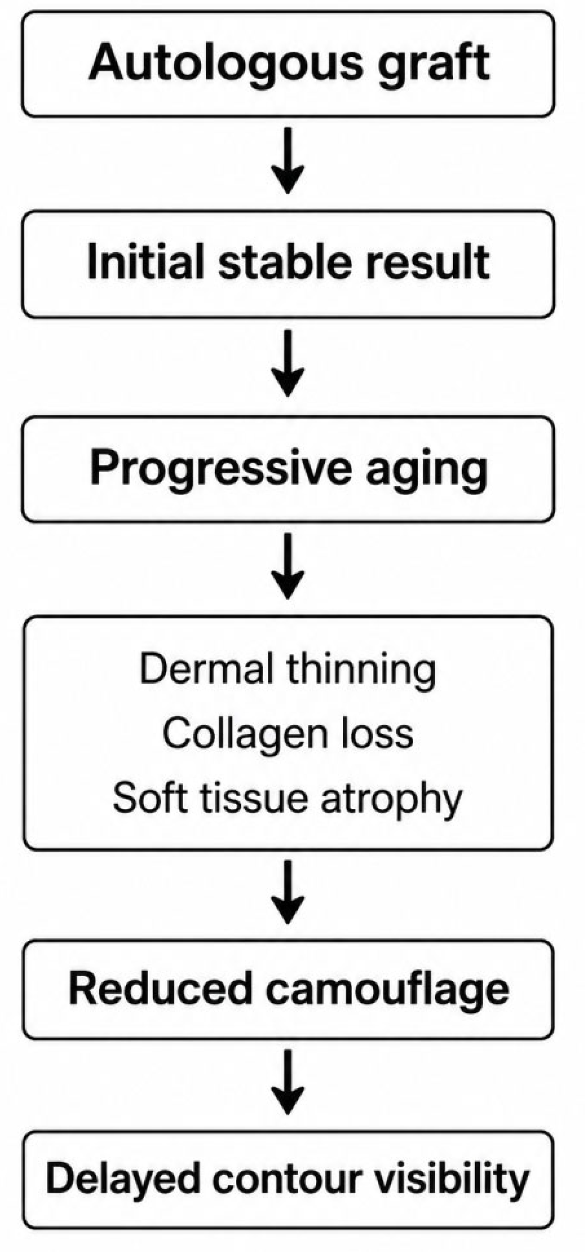
Figure 2.
Age-related changes of the nasal soft tissue envelope. Schematic illustration of age-related thinning of the nasal soft tissue envelope, including the dermis, SMAS, and subcutaneous tissue, resulting in reduced soft tissue coverage over the cartilaginous framework.
Figure 2.
Age-related changes of the nasal soft tissue envelope. Schematic illustration of age-related thinning of the nasal soft tissue envelope, including the dermis, SMAS, and subcutaneous tissue, resulting in reduced soft tissue coverage over the cartilaginous framework.
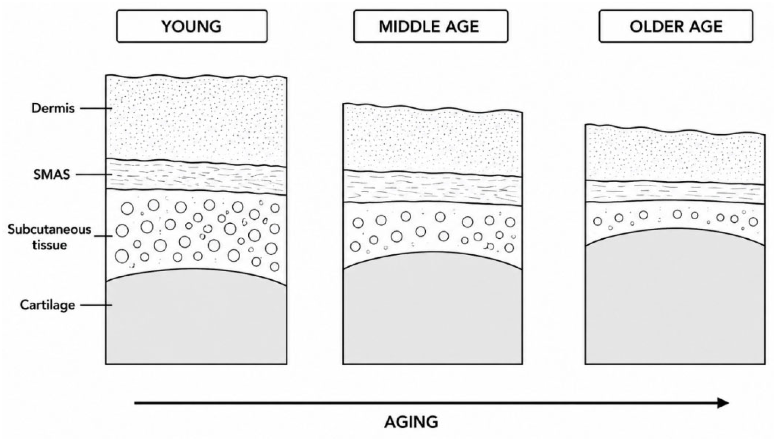
Figure 3.
Interaction between aging-related soft tissue changes and autologous graft behavior leading to delayed contour visibility. Stable autologous grafts may become progressively visible as age-related soft tissue thinning reduces camouflage capacity, resulting in delayed contour visibility.
Figure 3.
Interaction between aging-related soft tissue changes and autologous graft behavior leading to delayed contour visibility. Stable autologous grafts may become progressively visible as age-related soft tissue thinning reduces camouflage capacity, resulting in delayed contour visibility.
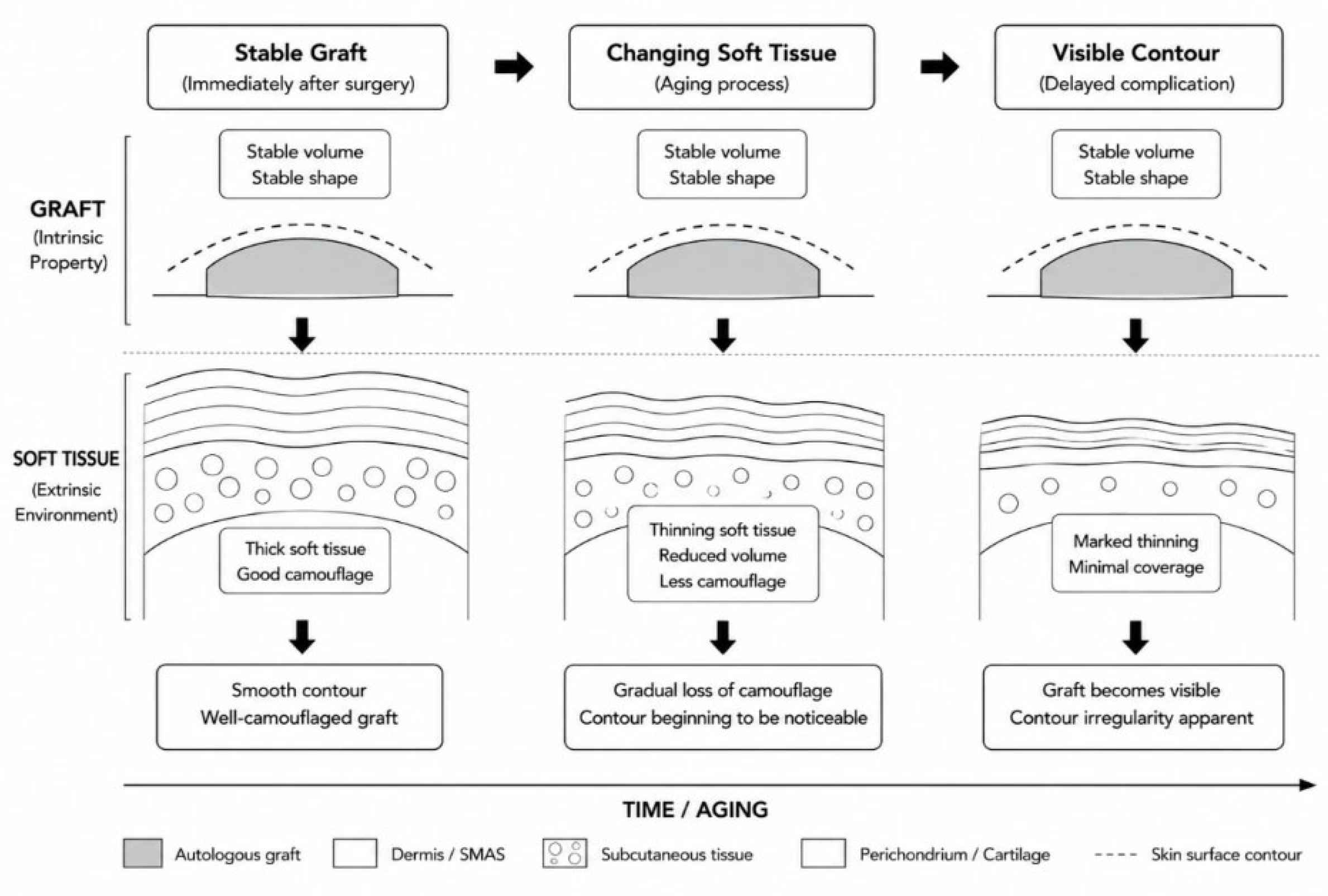

Table 1.
Comparative characteristics of commonly used autologous graft materials in rhinoplasty.
| Graft Material | Structural Support | Long-term Stability | Camouflage Potential | Consistency of Current Evidence | Typical Clinical Applications |
| Septal cartilage | Moderate | Excellent | Moderate | Consistent evidence |
Primary rhinoplasty, spreader grafts, tip support |
| Auricular cartilage | Low–Moderate | Good | Good | Consistent evidence |
Alar batten grafts, tip refinement |
| Costal cartilage | High | Excellent | Limited | Moderately consistent evidence | Revision rhinoplasty, major dorsal augmentation |
| Temporalis fascia | Minimal | Good | Excellent | Moderately consistent evidence | Camouflage in thin-skinned patients |
| Fascia lata | Minimal | Good | Excellent | Limited evidence | Soft-tissue augmentation |
| Diced cartilage wrapped in fascia | Moderate | Good | Excellent | Moderately consistent evidence | Smooth dorsal augmentation |
Comparison of commonly used autologous graft materials with respect to structural support, long-term stability, camouflage potential, evidence consistency, and clinical applications.
Table 2.
Clinical factors associated with delayed contour visibility after autologous rhinoplasty.
| Clinical Factor | Proposed Mechanism | Consistency of Current Evidence | Clinical Recommendations |
| Thin nasal skin | Reduced soft tissue camouflage over the osteocartilaginous framework | Consistent evidence |
Meticulous graft shaping and adjunctive camouflage techniques should be considered. |
| Aging-related dermal thinning | Progressive loss of dermal thickness and collagen content | Moderately consistent evidence |
Long-term follow-up is recommended, particularly in elderly patients. |
| Loss of subcutaneous tissue | Reduced soft tissue coverage over cartilage grafts | Moderately consistent evidence |
Preserve soft tissue volume whenever feasible. |
| Costal cartilage graft | High rigidity beneath a thin soft tissue envelope | Moderately consistent evidence |
Precise carving and fascia coverage may reduce delayed graft visibility. |
| Scar contracture / postoperative remodeling | Progressive tissue contraction alters surface contour | Limited evidence |
Minimize soft tissue trauma and monitor long-term remodeling. |
| Preservation of the soft tissue envelope | Maintains tissue thickness and camouflage capacity | Moderately consistent evidence |
Preservation techniques should be incorporated whenever possible. |
Summary of clinical factors, proposed mechanisms, evidence consistency, and practical recommendations related to delayed contour visibility after autologous rhinoplasty.
Table 3.
Current evidence, limitations, and future research priorities regarding delayed contour visibility after autologous rhinoplasty.
Table 3.
Current evidence, limitations, and future research priorities regarding delayed contour visibility after autologous rhinoplasty.
| Research Topic | Consistency of Current Evidence | Major Limitations | Future Research Priorities |
| Thin nasal skin | Consistent evidence | Mostly retrospective studies | Prospective multicenter studies with standardized outcomes |
| Aging-related soft tissue changes | Moderately consistent evidence | Limited longitudinal clinical data | Serial imaging and histologic correlation |
| Autologous graft behavior | Consistent evidence | Few studies on delayed visibility | Material-specific long-term comparisons |
| Interaction between aging and graft visibility | Limited evidence | Mechanism largely hypothetical | Prospective validation studies |
| Camouflage techniques | Moderately consistent evidence | Lack of comparative trials | Comparative effectiveness research |
| Outcome assessment | Limited evidence | Patient-reported outcomes underused | Develop validated patient-centered outcome measures |
Overview of the current evidence, major limitations, and future research priorities regarding delayed contour visibility after autologous rhinoplasty.
Disclaimer/Publisher’s Note: The statements, opinions and data contained in all publications are solely those of the individual author(s) and contributor(s) and not of MDPI and/or the editor(s). MDPI and/or the editor(s) disclaim responsibility for any injury to people or property resulting from any ideas, methods, instructions or products referred to in the content. |
© 2026 by the authors. Licensee MDPI, Basel, Switzerland. This article is an open access article distributed under the terms and conditions of the Creative Commons Attribution (CC BY) license (http://creativecommons.org/licenses/by/4.0/).
Copyright: This open access article is published under a Creative Commons CC BY 4.0 license, which permit the free download, distribution, and reuse, provided that the author and preprint are cited in any reuse.


