
Submitted:
03 August 2026
Posted:
04 August 2026
You are already at the latest version
Abstract
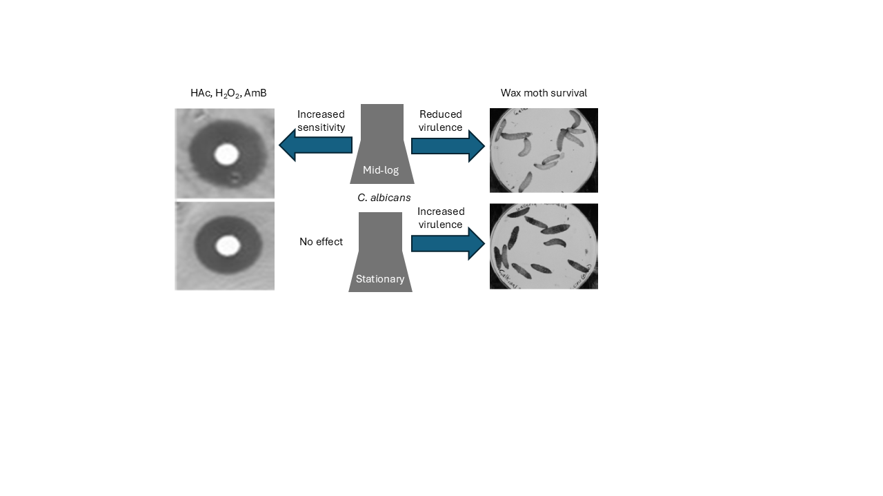
Metacaspases are cysteine proteases, found in plants, fungi, and protists. They are structural orthologs of caspases, which orchestrate apoptosis in metazoa. However, metacaspases differ from caspases in that they cleave after arginine or lysine instead of aspartate, most are activated by calcium, and they do not target the same range of proteins as caspases. The Candida albicans metacaspase, Mca1p mediates cell death in response to various stresses but also possesses pro-life functions such as proteostasis. This research report highlights differences in the effect of MCA1 deletion on cell death and virulence, depending on whether cells originate from exponential or stationary phase culture.
Keywords:
Candida albicans
; metacaspase
; MCA1
; hydrogen peroxide
; acetic acid
; amphotericin B
; RCD
; cell death
; wax moth
; Galleria mellonella
; stationary
; exponential
1. Introduction
Caspases orchestrate regulated cell death (RCD), development, differentiation, inflammation and positive and negative selection of immune cells in mammals, as well as non-death processes [1,2,3,4,5,6,7,8,9]. Distant orthologs of caspases were identified in animals, plants, fungi, archaea and bacteria [10,11,12,13,14,15,16,17]. These orthologs are called paracaspases, metacaspases and orthocaspases and there was a protracted debate regarding whether metacaspases in Saccharomyces cerevisiae and Candida albicans (for example) were true functional orthologs of caspases. It is now known that they have different substrate specificities, usually require calcium for activation of proteolysis and that many have only pro-survival functions, though several appear to have pro-survival (proteostasis, extension of lifespan etc.) and pro-death functions (e.g. RCD with characteristics of apoptosis, pyroptosis, autophagic cell death or necroptosis) [17,18,19,20,21,22].
At the end of the 20th century and beginning of the 21st, Madeo and coworkers showed that S. cerevisiae undergoes regulated cell death in response to mutations in the cell cycle machinery and certain stresses, that this cell death resembles metazoan apoptosis and involves new protein biosynthesis and that hydrogen peroxide-induced cell death is dependent on the yeast metacaspase Mca1p/Yca1p [23,24,25].
Phillips et al. [26] demonstrated that 5-10 mM hydrogen peroxide, 40-60 mM acetic acid or 4-8 μg/mL amphotericin B induced a form of cell death in C. albicans that resembled apoptosis e.g. phosphatidylserine flipped from the inner to outer cell membrane, DNA strand breaks occurred and reactive oxygen species (ROS) accumulated in the cell. Cao et al. [27] showed that deleting MCA1 in C. albicans reduced sensitivity to cell death caused by oxidative stress, reduced intracellular ROS accumulation, increased trehalose accumulation, lowered ATP levels and lowered mitochondrial membrane potential. They suggested that more sugar phosphates were being fed into trehalose synthesis rather than oxidative phosphorylation, leading to reduced ATP synthesis and reduced reactive oxygen species (ROS) production by mitochondria.
The identification of regulated cell death and orthologs of the metazoan cell death machinery in C. albicans is exciting as candidemia is one of the top four hospital-acquired bloodstream infections, C. albicans is the main agent of candidemia and mortality among candidemia patients may be over 40% [28,29,30,31,32]. Targeting components of the fungal cell death machinery could lead to the development of novel classes of antifungal drugs [26].
In this study, it is shown that deleting the C. albicans metacaspase gene MCA1 affects both the rate of stress-induced cell death and virulence in an insect model. This is consistent with reports in the literature that metacaspases reduce stress resistance and virulence by promoting stress- or host immune system-induced cell death [21,23,24,25,27,33,34,35,36,37]. However, it is not consistent with reports of metacaspase pro-survival roles [21,36,37,38,39]. What was unexpected in this study was that using cells from stationary phase, rather than exponential phase, culture either abrogated or reversed these effects. Possible reasons for this phenomenon are discussed.
2. Materials and Methods
Media components were from Formedium (Swaffham, Norfolk, UK) and other reagents from Fisher Scientific (Pittsburgh, Pennsylvania, USA) unless stated otherwise. It should be noted that when the word “prototrophy” is used here, it refers to prototrophy in histidine, leucine and uridine and does not consider arginine trophic status since all growth media contained arginine.
2.1. Culture and Storage
Candida albicans strains were grown in liquid culture in YPD (1% (w/v) yeast extract, 2% (w/v) peptone, 2% (w/v) D-glucose) and an aliquot of culture was mixed with an equal volume of 50% (v/v) glycerol/water for long term storage at ‒80 °C. When needed, frozen stock was streaked onto YPD agar (YPD + 2% (w/v) agar) using a sterile pipette tip and incubated at 30 °C for two days. Overnight cultures were prepared by adding one colony from the agar plate to 10 mL YPD using a sterile pipette tip and incubating at 30 °C overnight. For transformations and to produce exponential (mid-log phase) cells for virulence testing, 50 mL fresh YPD was inoculated with 1 mL overnight culture and incubated at 30 °C for 4 hours. To produce stationary phase cells, the 50 mL liquid culture was incubated at 30 °C for 2 days. For selective culture, 200 µL cell suspension was spread onto selective agar (6.9 g/L yeast nitrogen base with ammonium sulphate but without amino acids; 2 % (w/v) D-glucose; 2 % (w/v) agar No 2; 0.72 g/L amino acid dropout mix without leucine, histidine or uridine; 50 mg/L leucine and/or histidine and/or uridine as appropriate). Escherichia coli was grown in lysogeny broth (LB: 1% (w/v) tryptone, 0.5% (w/v) yeast extract, 1% (w/v) sodium chloride) and aliquots were mixed with equal volumes of 50% (v/v) glycerol/water and stored at ‒80 °C until needed. E. coli was streaked onto LB agar (LB + 2% (w/v) agar) and incubated at 37 °C overnight and 10 mL LB was inoculated with a colony taken from the agar plate and incubated at 37 °C. Where required, kanamycin or ampicillin was added to LB or LB agar at a final concentration of 50 and 100 µg/mL respectively.
2.2. C. albicans Transformation
Candida albicans cells were transformed with cassettes made via polymerase chain reaction (PCR) amplification of auxotrophy markers on plasmids [40] or with linearized plasmids containing auxotrophy markers [41]. The transformation protocol was adapted from that of Gietz and Woods [42]. Cells from a 50 mL mid-log phase culture (above) were washed with 50 mL LATE (10 mM Lithium Acetate, 10 mM Tris, 1mM EDTA pH 8.0 in sterile milliQ water) and resuspended in 1 mL LATE, then 100 µL aliquots were incubated overnight with 0.7 mL PLATE (40% (w/v) PEG in LATE), 80 µL DNA and 5 µL boiled herring sperm (10 mg/mL). The next day, cells were heat shocked for one hour at 42 °C then cells were washed in water, resuspended in 400 µL of water, spread on two selective agar plates and incubated at 30 °C for 2-3 days, until colonies appeared.
2.3. E. coli Transformation
50 µL of frozen competent cells were defrosted on ice, 5 µL of chilled DNA suspension was added and the tube tapped to encourage mixing. The tube was left on ice for half an hour then cells were heat shocked at 42 °C for 45 seconds and placed on ice for a further 2 minutes. 1 mL of LB, pre-heated to 42 °C, was added to the cells, the cell suspension was incubated for 60 mins at 37 °C with shaking (200 rpm). Two aliquots (50 µL and 500 µL) were spread on LB agar (LB + 2% agar) plates containing 100 µg/mL ampicillin or 50 µg/mL kanamycin, as appropriate and incubated overnight at 37 °C.
2.4. Miniprep
Plasmid harvesting from E. coli was carried out using a miniprep kit (Quiagen, Crawley, West Sussex, UK). 1 mL of overnight culture was centrifuged, and cells resuspended in 100 µL of resuspension buffer (50 mM tris(hydroxymethyl)aminomethane-HCl (Tris-HCl) pH 8.0, 10 mM ethylenediaminetetraacetic acid (EDTA), 100 mg/mL RNase A). 200 µL of lysis buffer (200 mM sodium hydroxide, 1% (w/v) sodium dodecyl sulfate (SDS) was added and mixed by inversion. Then 350 µL of neutralization buffer (4.2 M guanidine hydrochloride, 0.9 M potassium acetate, pH 4.8) was added and the tube inverted six times. After centrifugation, the supernatant was applied to a spin tube and centrifuged at 10,000 rpm for 2 minutes. The flow-through was discarded and 500 µL of wash buffer (4.2 M guanidine hydrochloride, 0.9 M potassium acetate, pH 4.8) added before centrifugation at 10,000 for a further 2 minutes. This step was repeated once more, the flow-through discarded and the column centrifuged at 10,000 rpm for 1 minute, the flow-through discarded and the column removed and placed in a clean Eppendorf tube. 50 µL of elution buffer was added to the column, incubated for 2 minutes and the tube centrifuged for 1 minute at 10,000 rpm to collect the plasmid suspension.
2.5. Gel Electrophoresis
An electrophoresis gel was prepared, containing 0.7% agarose and 0.5 µg/mL ethidium bromide in TAE buffer (40 mM Tris acetate, 1 mM EDTA, pH 8.5). After the gel had set, the comb was removed and the gel was placed in an electrophoresis tank and covered in TAE buffer. PCR reaction products or DNA components used in cloning were pipetted into the wells, and a DNA size ladder was pipetted into a spare well then a current of 100 V was applied for 20 to 30 minutes. A Gbox gel imager (Syngene, Cambridge, UK) was used to visualize DNA bands in the gel. Images were recorded on thermal print paper and saved to file as appropriate.
2.6. Gel Extraction
Under a UV light the appropriate DNA band was cut out of the gel with a scalpel and the gel slice placed in a clean Eppendorf tube. Gel purification was carried out with a gel extraction kit (Qiagen, Crawley, West Sussex, UK). The gel slice was added to an equal volume of chaotropic buffer (5 M guanidinium chloride, 30% isopropanol) and incubated at 50 °C until dissolved. The solution was applied to a spin column, incubated for 2 minutes and centrifuged for 2 minutes at 10,000 rpm. After two washes with 500 μL wash buffer (10 mM Tris-HCl pH 7.5, 80 % ethanol) the column was centrifuged at 10,000 rpm for a further minute then transferred to a clean, sterilized Eppendorf tube. 50 μL of elution buffer (2 mM Tris-HCl pH 8-8.5) was added, incubated for 2 minutes then centrifuged at 10,000 rpm for 1 minute to collect the DNA suspension
2.7. PCR
25 μL of PCR Master Mix (Thermo Scientific, Waltham, Massachusetts, USA) (final concentration: 0.625 units Taq DNA polymerase, 75 mM Tris-HCl [pH 8.8], 20 mM ammonium sulphate, 1.5 mM magnesium chloride, 0.01% Tween ® 20, 0.2 mM of each dNTP), 22 μL of sterile nuclease-free water, 1 μL of forward primer (0.2 μM), 1 μL of reverse primer (0.2 μM) and 1 μL (0.5 to 125 ng) of DNA template were mixed in a PCR tube and polymerase chain amplification carried out in a Thermal Hybaid thermal cycler (Franklin, Massachusetts, USA) using the following cycle: initial denaturation at 95 °C for 5 minutes; 30 cycles of i) denaturation at 95 °C for 30 seconds, ii) annealing at 53 °C for 1 minute and iii) extension at 72 °C for 3 minutes; followed by a final extension at 72 °C for 5 minutes.
2.8. Colony PCR
A single colony was resuspended in 25 µL of sterile milliQ water. 5 µL of cell suspension was streaked on fresh agar (LB or YPD, as appropriate) and incubated at 37 °C overnight (E. coli) or 2 days at 30 °C (C. albicans) to prepare stock for freezing. Meanwhile, the same E. coli cell suspension was used directly as DNA for PCR amplification, but the C. albicans cell wall had to be digested first. 10 mg/mL of lyticase (Thermo Scientific, Waltham, Massachusetts, USA) was added to the tube of C. albicans cell suspension and incubated at 37 °C for 10 minutes, followed by freezing at ‒80 °C for 5 minutes and thawing at room temperature. The treated suspension was then applied to PCR amplification.
2.9. Strain and Plasmid Creation
The Candida albicans parental strain used in this study was SN78, developed by Noble and Johnson [40], which is derived from C. albicans clinical isolate SC5314 and was deleted for both copies of the HIS1, LEU2 and URA3 genes and was therefore auxotrophic for histidine, leucine and uridine (Table 1). Noble and Johnson also developed plasmids to be used in gene knockouts (Table 2: pSN52 and pSN40). Since the plasmids contained the HIS1 and LEU2 genes respectively, transformants could be selected on YPD agar plates lacking histidine and/or leucine. Each strain was restored to prototrophy by incorporating the appropriate linearized plasmid (Cip10 [41], Cip10-MCA1 or Cip40 [Table 2]) into the RPS1 locus.
2.9.1. Prototrophic Wild Type (WT)
Plasmid Clp30 [41]: a gift from Dr. Steve Bates) contains the HIS1, URA3 and ARG4 genes while C. albicans strain SN78 [40] lacks the HIS1, LEU2 and URA3 genes. To restore prototrophy, a new plasmid, Clp40, containing HIS1, LEU2, URA3 and ARG4 was created from Clp30 and integrated into the RPS1 locus of SN78. First the LEU2 gene of plasmid pSN40 was amplified by PCR, using primers 1 and 2 (Table 3), the DNA was precipitated, washed and resuspended in water then digested with restriction endonuclease BsaBI (New England Biolabs, Ipswich, MA, USA), A-tailed with PCR Mix and purified using a gel extraction kit (NBS Biologicals, Huntingdon, UK). DNA was then cloned into Strataclone vector pSC-A amp/kan and transformed into Strataclone SoloPack competent E. coli cells. The presence and orientation of LEU2 in the pSC-A plasmid was confirmed via SpeI digestion and gel elecreophoresis. In the E. coli transformant the LEU2 gene was flanked by two SpeI restriction sites. The SpeI restriction fragment was ligated into XbaI-digested Clp30 to yield Clp40. The Clp40 plasmid was linearized by digestion with StuI and transformed into strain SN78. Thus, the auxotrophic markers were incorporated into the RPS1 locus to restore prototrophy and yield strain WT (Table 1). Transformants were selected on agar plates lacking histidine, leucine and uridine and successful transformation confirmed via PCR with primers 10 and 11 and gel electrophoresis.
2.9.2. Prototrophic Double MCA1 Deletion Strain (Mut)
Two rounds of gene deletion were carried out, replacing one copy of MCA1 with the HIS1 gene and the second copy with the LEU2 gene. The Cip10 plasmid, incorporating the URA3 gene, was used to restore prototrophy to the mutant strain. The HIS1 gene of plasmid pSN52 and the LEU2 gene of plasmid pSN40 (Table 2) were amplified by PCR using primers 1 and 2 (Table 3), which each have 70 bp regions of homology with the sequences just upstream and downstream (respectively) of MCA1. Successive rounds of transformation using MCA1 knockout cassettes, created from pSN52 and pSN40, yielded a strain with both histidine and leucine prototrophy. First round transformants were selected on agar plates lacking histidine and second round transformants (double knockouts) on plates that lacked both histidine and leucine. Replacement of the first MCA1 gene with HIS1 and the second with LEU2 was confirmed by PCR amplification using primer pairs 3/4 and 3/5 respectively, followed by gel electrophoresis. The absence of any MCA1 gene in the double knockout strain was confirmed by PCR with primers 6 and 7 followed by gel electrophoresis. The CIp10 plasmid was then linearized via digestion with StuI and was transformed into the double knockout strain. The URA3 gene of CIp10 was therefore incorporated into the RPS1 locus of the double deletion strain and transformants were selected on plates lacking histidine, leucine and uridine. The resulting prototrophic (for histidine, leucine and uridine) double deletion strain was named Mut (Table 1).
2.9.3. Prototrophic MCA1 Reintegrant Strain (Reint)
To be sure that any differences observed between strains WT and Mut were due to MCA1 deletion and not to some other random mutation, the MCA1 gene was reintegrated into the Mut strain to check that this restored the WT phenotype. Lyticase treatment, freezing (at ‒80 °C) and thawing of strain SN78 were used to obtain cell contents and 1 µL was used for PCR amplification of MCA1 with primers 8 and 9 to produce copies of the MCA1 gene, flanked by XhoI restriction sites. The XhoI digestion fragment was ligated into XhoI-digested Cip10 (which had been treated with Antarctic phosphatase to remove hydroxyl groups and prevent self-ligation). Gel electrophoresis and gel extraction was used to purify the resulting CIp10-MCA1 plasmid, which was transformed into DH5α competent E. coli cells (Invitrogen, Waltham, Massachusetts). Successful transformation (and the presence of only one copy of MCA1) was confirmed by XhoI digestion and gel electrophoresis. CIp10-MCA1 was harvested from E. coli and linearized with StuI before transformation into the Mut strain to yield strain Reint (Table 1), in which a functional copy of MCA1 was incorporated into the C. albicans genome at the RPS1 locus. Transformants were selected on agar plates lacking uridine and successful integration of CIp-MCA1 into the RPS1 locus was confirmed by PCR with primers 10 and 11 (a gift from Dr. Steve Bates), followed by gel electrophoresis.
2.10. Stress-Induced Cell Death
The WT, Mut and Reint strains were tested for sensitivity to regulated cell death (RCD)-inducing levels of hydrogen peroxide (H2O2), acetic acid (HAc) and amphotericin B (AmB). C. albicans cells from mid-log phase (4 hour old) or stationary phase (48 hour old) culture were centrifuged, washed in water and resuspended at 1 x 10⁵ cells/mL in water. 1 mL of cell suspension was spread on a YPD agar plate and dried by stacking upside down beneath a Bunsen flame. A sterile 5 mm filter paper disc was placed in the center of each seeded plate. 5 μL of 7.5 M HAc, 2.5 M H2O2 or 2 mg/mL AmB was pipetted onto the disc and the plate incubated at 30 °C for 1 day. The diameter of the zone of fungal growth inhibition around the disc was measured. The assay was carried out three times on separate occasions.
2.11. Virulence in An Insect Model
The WT, Mut and Reint strains were tested for virulence in wax moth (Galleria mellonella) larvae. Cells from mid-log phase (overnight) or stationary phase (48 hour old) YPD liquid cultures were centrifuged, washed twice with phosphate buffered saline (PBS: 136.89 mM sodium chloride; 2.68 mM potassium chloride; 10.14 mM dibasic sodium phosphate; 1.8 mM monobasic potassium phosphate; pH 7.4) and resuspended in PBS at 3 x 10⁷ cells/mL. 10 µL cell suspension or 10 µL of PBS was injected into the left, front proleg of each wax moth larva. Ten larvae were injected with cell suspension from mid-log phase cultures of each strain, ten with cell suspension from stationary phase cultures of each strain and ten with PBS. Each group of ten larvae was placed in a petri dish, lined with filter paper and incubated at 37 °C. Live and dead larvae were counted every 6 hours. The assay was carried out three times on separate occasions.
2.12. Effect of Mca1p on Serum-Induced Filamentation
Cells from stationary (48 hour-old) cultures of WT, Mut and Reint were centrifuged, washed in water and resuspended at a density of 1 x 10⁷ cells/mL in YPD + 10% (v/v) fetal calf serum (FCS). Cell cultures were incubated with shaking (200 rpm) at 37 °C and samples examined at regular intervals beneath a light microscope. For each strain, 400 cells in 4 fields of vision were examined and the numbers of yeast and hyphae were counted. The experiment was carried out three times on different occasions.
2.13. Effect of Mca1p on Growth Rate
Cells from mid-log phase cultures of the WT, Mut and Reint strains were centrifuged, washed and resuspended in synthetic complete medium (6.9 g/L yeast nitrogen base with ammonium sulphate and amino acids; 2 % (w/v) D-glucose; 2 % (w/v) agar No 2) to achieve an absorbance at 650 nm (A650) of 0.2. Then 10 µL of SC pH3 was added to each well of a microtiter plate and media in successive columns included 240 mM, 120 mM, 60 mM, 30 mM, 15 mM and 0 mM acetic acid. 100 µL cell culture was added to each well. The top 3 rows contained WT cells, the next 3 contained Mut cells and the final 3 rows contained Reint cells and the final concentration of acetic acid in each column was 120 mM, 60 mM, 30 mM, 7.5 mM and 0 mM. The plate was placed in a VersaMax automatic plate reader with Softmax Pro 5.4.1 software (Molecular Devices, Sunnyvale, California, USA) and incubated at 30 °C for 24 hours. Absorbance at 650 nm was read and recorded every 3 minutes. Raw data was exported to and processed in Excel. Mean absorbance for each triplicate reading was calculated and normalized to the mean absorbance for that strain at time zero. Absorbances were plotted against time since HAc treatment and natural logs of absorbances were also calculated and plotted as semi-logarithmic graphs. The linear portion of each semi-logarithmic curve was used to calculate the doubling time (generation time, G) using the formula
where Δt = time passed and ΔLnAb = change in natural log of absorbance
G = Δt/(1.443 x ΔLnAb)
2.14. Effect of Mca1p on Acetic Acid-Induced Cell Death in Yeast and Hyphae
Cells from stationary phase cultures of WT, Mut and Reint were centrifuged, washed and resuspended at 1 x 10⁷ cells/mL in SC pH3 + 10% (v/v) FCS and either 80 mM HAc or an equal volume of water. After incubation for 3 hours at 37 °C with shaking (200 rpm) cells were centrifuged, washed and resuspended in 10 µg/mL propidium iodide in PBS and incubated at 37 °C for half an hour. Cells were washed and resuspended in PBS then examined using a fluorescence microscope (Leica DMLB, Wetzlar, Hesse, Germany) with an excitation energy of 488 nm and a band pass filter of 562-588 nm. Dead (red PI-stained) and live (unstained) cells were counted. Treatment with FCS at 37 °C induced germination and mother (yeast) cells had attached daughter hyphae. The numbers of live mother/live daughter, live mother/dead daughter, dead mother/live daughter and dead mother/dead daughter pairs were counted, as was the overall number of live and dead mother and daughter cells.
3. Results
3.1. Mca1p Mediates Cell Death in C. albicans Cells from Mid-Log Phase Culture
The wild type (WT), mca1Δ/mca1Δ double knockout mutant (Mut) and MCA1 reintegrant (Reint) strains were spread on agar plates and dried. Filter paper discs were placed in the centers of the plates and treated with RCD-inducing concentrations of H2O2, HAc or AmB. After incubation at 30 °C for 24 hours, the diameter of the zone of inhibition (area of no fungal growth) around each disc was measured. The means and standard deviations of three independent experiments are shown (Figure 1). A student’s t-test was carried out to compare results for the Mut strain with those for WT and Reint. There were significant differences (p < 0.01) between WT and Mut and between Reint and Mut but not between WT and Reint regarding sensitivity to HAc (Figure 1A), H2O2 (Figure 1C) and AmB (Figure 1E) when spread cells were derived from mid-log phase cultures. In each case, the MCA1 double knockout strain Mut was less sensitive to the cell death-inducing stressor than either of the strains (WT and Reint) with functional MCA1 genes.
3.2. Mca1p Does Not Mediate Cell Death in C. albicans Cells from Stationary Phase Culture
When the cell death induction assay was repeated with cells from stationary phase (48 hours old) cultures (Figure 1B, D and F) there was no significant difference (p > 0.01) between the effect of the stressors on mutant (Mut), wild type (WT) or reintegrant (Reint) strains.
3.3. Mca1p Reduces the Virulence of Mid-Log Phase-Derived C. albicans Cells
To investigate the role of Mca1p in C. albicans virulence, cells from log-phase cultures of WT, Mut and Reint were washed and resuspended in PBS. Cell suspensions (or PBS alone in the control) were injected into wax moth larvae and the larvae incubated at 37 °C. The numbers of live and dead larvae were counted at 6 hourly intervals and percentage survival calculated. The means and standard deviations of three independent experiments are shown (Figure 2A). Generally, Mut killed larvae more rapidly than WT or Reint but students t-testing indicated that differences were not significant (p > 0.01) in most cases. However, at 18 and 24 h after inoculation, larval survival was significantly higher when injected with the Mut strain (green) than the WT (brown) or Reint (purple) strain (p < 0.01). None of the control larva, injected with PBS died during the 36-hour period of observation. All larvae died within 36 h of inoculation — but larvae died more slowly when injected with C. albicans that lacked Mca1p.
3.4. Mca1p Increases the Virulence of Stationary Phase-Derived C. albicans Cells
The virulence assay was repeated with C. albicans cells from stationary phase culture (Figure 2B). Again, all PBS-injected larvae survived for 36 hours after inoculation. In contrast with the previous experiment, Mut appeared to kill larvae more slowly than WT or Reint, but the differences were not significant (p > 0.01) at most time points. At 12 and 18 h after infection, survival of Mut-injected larvae differed significantly from survival of larvae injected with WT or Reint and, unexpectedly, injection with Mut reduced larval survival at two time points, rather than increasing it. All larvae died within 36 h of inoculation — but larvae died more rapidly when injected with C. albicans that lacked Mca1p.
3.5. Mca1p Has No Effect on Serum-Induced Filamentation
Mca1p appeared to affect the virulence of C. albicans in wax moth larvae. A major virulence factor of C. albicans is its ability to switch from yeast to hyphal form and escape from innate immune cells [44,45]. To test whether Mca1p influenced C. albicans virulence via altered filamentation, cells from stationary phase cultures of each of the three strains (WT, Mut and Reint) were grown at 37 °C and 200 rpm in YPD plus 10% FCS and the numbers of yeast and hyphae counted at 15-30 min intervals (Figure 3). There was no significant difference (p > 0.01 in a student’s t-test) in filamentation of any of the strains, compared with any of the others at any time point.
3.6. Mca1p Has No Effect on Growth Rate During Acetic Acid Stress
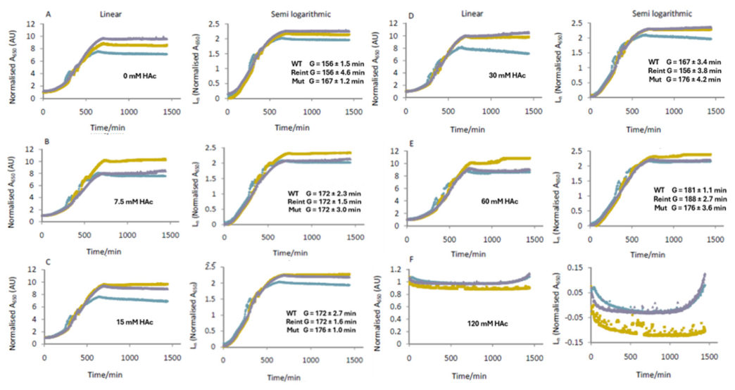
Cao et al. [27] showed that knocking out the C. albicans MCA1 gene reduced the rate of growth. S. cerevisiae Mca1p appears to play a role in the cell cycle [46]. Other fungal metacaspases may help to maintain a higher growth rate under stress (Richie et al., 2006). To test whether the effects of Mca1p on stress-induced RCD and virulence in this study were due to MCa1p-mediated changes in growth rate, the growth rates of each strain in SC at pH3 (to ensure dissociation of HAc) with and without acetic acid, were assayed. The generation (doubling) time G generally increased as the acetic acid concentration increased. The Mut strain reached peak generation time at 15 mM acetic acid and did not increase further while the WT and Reint strains peaked at 7.5 mM, dipped at 30 mM then reached an even higher peak at 60 mM. There was no growth at 120 mM acetic acid. Despite apparent differences in growth patterns, student’s t-testing indicated that the growth rates did not significantly differ between Mut and either WT or Reint (p > 0.01).
Figure 4.
Effect of Mca1p on growth rate during acetic acid-induced stress. Cells from mid-log phase cultures of the wild type (WT: blue), double MCA1 deletion mutant (Mut: purple) and MCA1 reintegrant (Reint: yellow) were washed, resuspended in synthetic complete (SC) medium pH3 and pipetted into a 96-well microtiter plate and treated with 0 mM (A), 7.5 mM (B), 15 mM (C), 30 mM (D), 60 mM (E) or 120 mM (F) of acetic acid then incubated at 30 °C. Absorbance at 650 nm of culture in each well was measured at 3 minute intervals. Mean absorbance for each set of three readings was calculated and normalized to mean absorbance at time zero. Then absorbance and natural log of absorbance were plotted against time since HAc treatment. The linear portions of the semi-logarithmic plots were used to calculate generation (doubling) times G. Since there was no growth in 120 mM acetic acid, no generation time was calculated.
Figure 4.
Effect of Mca1p on growth rate during acetic acid-induced stress. Cells from mid-log phase cultures of the wild type (WT: blue), double MCA1 deletion mutant (Mut: purple) and MCA1 reintegrant (Reint: yellow) were washed, resuspended in synthetic complete (SC) medium pH3 and pipetted into a 96-well microtiter plate and treated with 0 mM (A), 7.5 mM (B), 15 mM (C), 30 mM (D), 60 mM (E) or 120 mM (F) of acetic acid then incubated at 30 °C. Absorbance at 650 nm of culture in each well was measured at 3 minute intervals. Mean absorbance for each set of three readings was calculated and normalized to mean absorbance at time zero. Then absorbance and natural log of absorbance were plotted against time since HAc treatment. The linear portions of the semi-logarithmic plots were used to calculate generation (doubling) times G. Since there was no growth in 120 mM acetic acid, no generation time was calculated.
3.7. Mca1p Has No Effect on the Percentages of Yeast or Hyphae That Die During Acetic Acid Treatment
One of the possible explanations for differences in virulence between cells with and without a functional MCA1 gene could be different rates of filamentation and different sensitivities in yeast and hyphae to stresses, encountered in the host. Therefore, yeast of all three strains (WT, Mut and Reint) were induced to undergo filamentation by treatment with 10% FCS at 37 °C in the presence or absence of 80 mM acetic acid. Then propidium iodide was used to stain dead cells and a fluorescence microscope used to count live and dead cells (mother yeast and/or daughter hyphae). The results were plotted as bar charts (Figure 5). Chi squared testing was carried out to identify significant differences in percentage cell death among the 3 strains. Cell death was significantly higher among treated than untreated hyphae (p < 0.0001) and among treated than untreated yeast (p < 0.0001). There was no significant difference between Mut and WT cell death among treated mother cells (p = 0.34) or treated hyphae (p = 0.48). However, a significantly greater proportion of Mut hyphae and yeast died than those of WT or Reint (p < 0.0001) when not treated with acetic acid.
4. Discussion
Three RCD-inducing stressors (hydrogen peroxide, acetic acid and amphotericin B) were tested against strains with (WT and Reint) and without (Mut) a functional MCA1 gene. Mca1p mediated RCD in response to each stressor when cells were derived from log-phase culture (Figure 1A, C and E) but played no such role in cells, derived from stationary phase culture (Figure 1B, D and F). Other studies have demonstrated a role for Mca1p in fungal cell death, induced by hydrogen peroxide, acetic acid and amphotericin B [23,27,47,48]. Possible reasons for the absence of this pro-death function in cells from stationary phase, compared with mid-log phase, culture include differences in Mca1p expression, differences in Mca1p proteolytic (or other) activity and stress adaptations in stationary phase culture that counteract the pro-death function of Mca1p. It was previously shown that Mca1p expression is highly induced by RCD-inducing concentrations (160 mM) of acetic acid but only modestly induced by lower, stress-inducing (20 mM) concentrations [49]. It is possible that differing conditions within the cell during log-phase and stationary phase growth lead to differing levels of stress and modulate expression of Mca1p.
The sensitivity of the three strains to other stresses was also tested (results not shown) via ten-fold seral dilution of C. albicans cell suspensions on YPD agar containing the stressor. These stressors were sodium chloride (0.5-2 M), menadione (1.2 mM), sorbitol (1.2 M), cadmium chloride (0.75 mM) and calcofluor white (20 µg/mL). Mca1p had no effect on sensitivity to these stressors at the concentrations tested.
The decreased rate of larval death when injected with strains with a functional MCA1 gene (WT and Reint) rather than a strain with no MCA1 gene (Mut) is consistent with a role for Mca1p in the mediation of cell death, induced by elements of the host immune system. Insect hemocytes phagocytose microbes and use low pH, reactive oxygen species and other means to kill fungi and bacteria [50]. If Mca1p mediates acid- and peroxide-induced cell death, it is logical to expect that C. albicans cells with a functional MCA1 gene would be more sensitive to wax moth immune attack than cells with no MCA1 gene. It has been shown that metacaspases mediate stress-induced cell death in pathogenic protists and reduce virulence in animal hosts [51,52,53]. Mechanisms by which metacaspases affect virulence could include altered growth rate, increased sensitivity to immune attack, changes in the rate at which cells switch morphology and differences in the sensitivity of morphotypes.
There did not appear to be a difference in growth rate either without acetic acid or in the presence of increasing concentrations of acetic acid (Figure 4). The effects of MCA1 deletion on growth during AmB- or H2O2-induced stress were not tested. There was no difference in the rate of serum-induced switching from yeast to hyphal form in the three strains (Figure 3) so Mca1p appears to play no role in this process and morphology changes may not be the reason behind altered virulence. In acetic acid-treated hypha or yeast (Figure 5), there were no significant differences in the percentages of cells that died. However, among untreated cells, hyphal and yeast cell death was higher in the mutant (Mut) strain than the wild type (WT) or reintegrant (Reint). Mca1p has been shown previously to extend lifespan in aging yeast cells [54]. The effects of hydrogen peroxide and other ROS on cell death among hyphae or yeast were not tested, so it cannot be ruled out that differences in the virulence of strains with and without a functional MCA1 gene were due to differences in either hyphal or yeast sensitivity to host hemocyte ROS. However, this would explain the decreased virulence of the Mut strain when cells were derived from mid-log phase culture but not the increased virulence when cells were derived from stationary phase. It has been shown previously that Candida albicans hyphae were more resistant to AmB-than yeast and that this phenomenon was dependent on metacaspase Mca1p [55].
While the loss of a pro-death function in cells, derived from stationary phase, rather than mid-log phase, culture is interesting (Figure 1), the reversal of Mca1p’s role in wax moth larvae from an antagonist to an agonist of virulence when fungal cells were derived from stationary phase, rather than mid-log phase, culture (Figure 2) is remarkable. There have been several reports of a pro-survival role for various metacaspases [21,36,51,56,57,58,59] and it has been proposed that some functions are dependent on the catalytic activity of metacaspase while others are not [47] and may be dependent instead on the metacaspase prodomain [60]. It has also been suggested that metacaspases switch between the two roles in response to cellular signaling. The Saccharomyces cerevisiae metacaspase Mca1 mediates pro survival and pro-death functions, and Mca1p-mediated cell death may be dependent on or independent of its proteolytic activity [21,47,54,61,62,63]. There is evidence that under some death-inducing conditions, C. albicans Mca1p cleaves many cellular proteins that are important for survival, including heat shock proteins and that there is a preference for arginine or lysine in the P1 position and aspartate or glutamate in the P2 position [64]. One means by which Mca1p could be shifted from a pro-survival to a pro-death function is via the calcium-dependent cleavage of the prodomain and resulting activation of proteolytic activity [21]. It has been shown that when calmodulin binds to the prodomain of Mc1p it blocks cleavage/activation of proteolytic cactivity and promotes pro-survival over pro-death function [60]. Interestingly, the interaction of Mca1p with aggregated proteins is dependent on its prodomain, which is cleaved to promote its catalytic activity. However, activation of the proteolytic activity of Mca1p can also be pro-survival, helping to extend lifespan [54]. It is also possible that Mca1p does not directly promote cell death but that deleting MCA1 leads to a compensatory increase in chaperones that do directly promote cell survival under stressful conditions [21,46,61]. Lee et al. [61] pointed out that protein aggregates build up in cells that lack functional Mca1p during log phase. It has been shown that metacaspase MC5 expression and activity in the marine diatom Phaeodactylum tricornutum are higher during stationary phase than log phase and MC5 proteolytic activity is activated by ROS as well as calcium [65]. However, PtMC5 is a type III metacaspase with a rare Cys/Cys catalytic dyad that is oxidized by ROS. Beside calcium binding [66,67] modifications of metacaspases that alter their activity include nitrosylation [68,69], ubiquitylation [70] and phosphorylation [71]. Experiments have shown that fungi can adapt to stressful conditions, such as increased acetic acid concentration, that this adaptation includes upregulated expression of proteins such as catalase and that adaptation protects against stress-induced RCD [72]
5. Conclusions
Mca1p mediates RCD induced by acetic acid, hydrogen peroxide and amphotericin B in C. albicans but this effect is absent in C. albicans cells from stationary culture. Mca1p also reduces virulence of C. albicans in wax moth larvae, possibly via increased sensitivity to elements of the host immune system. This phenomenon is reversed when C. albicans cells are derived from stationary phase culture, which is consistent with previous reports of dual pro-death and pro-survival roles. Mca1p does not appear to affect growth rate or changes in morphology. The switch between the pro-survival and pro-death roles may involve the part played by the prodomain in protein aggregate clearance and the cleavage of the prodomain under death-inducing conditions. It has been suggested that Mca1p may be a pro-survival protein and that its apparent promotion of cell death may stem from upregulated expression of chaperones and other pro-survival proteins when MCA1 has been deleted, compensating for the loss of Mca1p. More research needs to be carried out to establish why Mca1p appears to switch behavior in mid-log phase and stationary phase cells. It would be useful to establish whether there are differences in the percentage of metacaspase molecules that are cleaved or uncleaved in mid-log phase and stationary phase C. albicans cells. A comparison of the relative abundances of Mca1p in log phase and stationary phase cultures and the proportion of Mca1p molecules that is cleaved and/or localized to stress granules and/or protein aggregates would also be useful. The rate of killing by mammalian phagocytes of cells from log phase and stationary phase culture and testing of cell death among yeast and hyphae under different stresses and different levels of those stresses may help to identify the stresses within a host that exert different levels of death-induction on C. albicans cells from different culture states. The effects of stress adaptation on metacaspases could be further researched as this could explain the loss of Mca1p function in stress-induced RCD. Finally, screening a gene knockout library for sensitivity to cell death of log phase and stationary phase C. albicans cells could help to identify the pathways that regulate cell death and the precise role of Mca1p and therefore potential targets for antifungal drugs.
Author Contributions
DW carried out the research, analyzed the results and wrote the manuscript.
Funding
This research was funded by UK Biotechnology and Biological Sciences Research Council (BBSRC), grant numbers BB/C501176/1 and CUF_Y3_11 and a BBSRC studentship.
Institutional Review Board Statement
Not applicable.
Informed Consent Statement
Not applicable.
Data Availability Statement
All data are included here.
Acknowledgments
I acknowledge the patient, helpful supervision of Prof. Mark Ramsdale at Exeter University, where I carried out the research. I would also like to thank Dr. Steve Bates for the kind gift of plasmids and PCR primers.
Conflicts of Interest
The author declares there are no conflicts of interest.
Abbreviations
The following abbreviations are used in this manuscript:
| RCD | Regulated cell death |
| ROS | Reactive oxygen species |
| WT | Wild type |
| Mut | Mutant (double MCA1 deletion strain) |
| Reint | MCA1 reintegrant (Mut with MCA1 integrated into the RPS1 locus) |
| PCR | Polymerase chain reaction |
| HAc | Acetic acid |
| H2O2 | Hydrogen peroxide |
| AmB | Amphotericin B |
| FCS | Fetal calf serum |
| YPD | Yeast extract/peptone/dextrose |
| LB | Lysogeny broth |
References
- Dho, S. H.; Cho, M.; Woo, W.; Jeong, S.; Kim, L. K. Caspases as master regulators of programmed cell death: apoptosis, pyroptosis and beyond. Exp. Mol. Med. 2025, 57((6)), 1121–1132. [Google Scholar] [CrossRef] [PubMed]
- Kumar, S.; Dorstyn, L. The role of caspases as executioners of apoptosis. Biochem. Soc. Trans. 2022, 50((1)). [Google Scholar] [PubMed]
- Sahoo, G.; Samal, D.; Khandayataray, P.; Murthy, M. K. A review on caspases: key regulators of biological activities and apoptosis. Mol. Neurobiol. 2023, 60((10)), 5805–5837. [Google Scholar] [CrossRef] [PubMed]
- Nadendla, E. K.; Tweedell, R. E.; Kasof, G.; Kanneganti, T.-D. Caspases: structural and molecular mechanisms and functions in cell death, innate immunity, and disease. Cell Discov. 2025, 11((1)), 42. [Google Scholar] [CrossRef] [PubMed]
- Van Opdenbosch, N.; Lamkanfi, M. Caspases in cell death, inflammation, and disease. Immunity 2019, 50((6)), 1352–1364. [Google Scholar] [CrossRef] [PubMed]
- Nguyen, T. T. M.; Gillet, G.; Popgeorgiev, N. Caspases in the developing central nervous system: apoptosis and beyond. Front. Cell Dev. Biol. 2021, 9, 702404. [Google Scholar] [CrossRef] [PubMed]
- Ramirez, M. L. G.; Salvesen, G. S. A primer on caspase mechanisms. Semin Cell Dev. Biol. 2018, 82, 79–85. [Google Scholar] [CrossRef] [PubMed]
- Beltrán-Visiedo, M.; Soler-Agesta, R.; Sarosiek, K. A.; Green, D. R.; Galluzzi, L. Regulation of inflammatory processes by caspases. Nat. Rev. Mol. Cell Biol. 2025, 26((11)), 884–901. [Google Scholar] [CrossRef] [PubMed]
- McIlwain, D. R.; Berger, T.; Mak, T. W. Caspase functions in cell death and disease. Cold Spring Harb. Perspect. Biol. 2013, 5((4)), a008656. [Google Scholar] [CrossRef] [PubMed]
- Uren, A. G.; O'Rourke, K.; Aravind, L. A.; Pisabarro, M. T.; Seshagiri, S.; Koonin, E. V.; Dixit, V. M. Identification of paracaspases and metacaspases: two ancient families of caspase-like proteins, one of which plays a key role in MALT lymphoma. Mol. Cell 2000, 6((4)), 961–967. [Google Scholar] [CrossRef] [PubMed]
- Minina, E. A.; Staal, J.; Alvarez, V. E.; Berges, J. A.; Berman-Frank, I.; Beyaert, R.; Bidle, K. D.; Bornancin, F.; Casanova, M.; Cazzulo, J. J. Classification and nomenclature of metacaspases and paracaspases: no more confusion with caspases. Mol. Cell 2020, 77((5)), 927–929. [Google Scholar] [CrossRef] [PubMed]
- Klemenčič, M.; Funk, C. Structural and functional diversity of caspase homologues in non-metazoan organisms. Protoplasma 2018, 255((1)), 387–397. [Google Scholar] [PubMed]
- Bhattacharjee, S.; Mishra, A. K. The tale of caspase homologues and their evolutionary outlook: deciphering programmed cell death in cyanobacteria. J. Exp. Bot. 2020, 71((16)), 4639–4657. [Google Scholar] [CrossRef] [PubMed]
- Klemenčič, M.; Novinec, M.; Dolinar, M. Orthocaspases are proteolytically active prokaryotic caspase homologues: the case of M icrocystis aeruginosa. Mol. Microbiol. 2015, 98((1)), 142–150. [Google Scholar] [CrossRef] [PubMed]
- Choi, C.; Berges, J. New types of metacaspases in phytoplankton reveal diverse origins of cell death proteases. Cell Death Dis. 2013, 4((2)), e490–e490. [Google Scholar] [CrossRef] [PubMed]
- Carmona-Gutierrez, D.; Fröhlich, K.; Kroemer, G.; Madeo, F. Metacaspases are caspases. Doubt no more. Cell Death Differ. 2010, 17((3)), 377–378. [Google Scholar] [CrossRef] [PubMed]
- Enoksson, M.; Salvesen, G. Metacaspases are not caspases–always doubt. Cell Death Differ. 2010, 17((8)), 1221–1221. [Google Scholar] [CrossRef] [PubMed]
- Vercammen, D.; Declercq, W.; Vandenabeele, P.; Van Breusegem, F. Are metacaspases caspases? J. Cell Biol. 2007, 179((3)), 375–380. [Google Scholar] [CrossRef] [PubMed]
- Hardwick, J. M. Do fungi undergo apoptosis-like programmed cell death? MBio 2018, 9((4)). [Google Scholar] [CrossRef] [PubMed]
- Palková, Z.; Váchová, L. Cell differentiation, aging, and death in spatially organized yeast communities: mechanisms and consequences. Cell Death Differ. 2025, 1–13. [Google Scholar]
- Hill, S. M.; Nyström, T. The dual role of a yeast metacaspase: What doesn't kill you makes you stronger. Bioessays 2015, 37((5)), 525–531. [Google Scholar] [PubMed]
- Wilkinson, D. Regulated Cell Death in Fungi, the Role of Metacaspases and Assay Techniques. Methods Protoc. 2026, 9((3)), 83. [Google Scholar] [CrossRef] [PubMed]
- Madeo, F.; Herker, E.; Maldener, C.; Wissing, S.; Lächelt, S.; Herlan, M.; Fehr, M.; Lauber, K.; Sigrist, S. J.; Wesselborg, S. A caspase-related protease regulates apoptosis in yeast. Mol. Cell 2002, 9((4)), 911–917. [Google Scholar] [PubMed]
- Madeo, F.; Fröhlich, E.; Fröhlich, K.-U. A yeast mutant showing diagnostic markers of early and late apoptosis. J. Cell Biol. 1997, 139((3)), 729–734. [Google Scholar] [CrossRef] [PubMed]
- Madeo, F.; Fröhlich, E.; Ligr, M.; Grey, M.; Sigrist, S. J.; Wolf, D. H.; Fröhlich, K.-U. Oxygen stress: a regulator of apoptosis in yeast. J. Cell Biol. 1999, 145((4)), 757–767. [Google Scholar] [CrossRef] [PubMed]
- Phillips, A. J.; Sudbery, I.; Ramsdale, M. Apoptosis induced by environmental stresses and amphotericin B in Candida albicans. Proc. Natl. Acad. Sci. 2003, 100((24)), 14327–14332. [Google Scholar] [CrossRef] [PubMed]
- Cao, Y.; Huang, S.; Dai, B.; Zhu, Z.; Lu, H.; Dong, L.; Cao, Y.; Wang, Y.; Gao, P.; Chai, Y. Candida albicans cells lacking CaMCA1-encoded metacaspase show resistance to oxidative stress-induced death and change in energy metabolism. Fungal Genet. Biol. 2009, 46((2)), 183–189. [Google Scholar] [CrossRef] [PubMed]
- Pfaller, M. A. Nosocomial candidiasis: emerging species, reservoirs, and modes of transmission. Clin. Infect. Dis. 1996, 22 (Supplement_2), S89–S94. [Google Scholar] [CrossRef] [PubMed]
- Fridkin, S. K.; Jarvis, W. R. Epidemiology of nosocomial fungal infections. Clin. Microbiol. Rev. 1996, 9((4)), 499–511. [Google Scholar] [CrossRef] [PubMed]
- Odds, F. C. Epidemiological shifts in opportunistic and nosocomial Candida infections: mycological aspects. Int. J. Antimicrob. Agents 1996, 6((3)), 141–144. [Google Scholar] [CrossRef] [PubMed]
- Odds, F. C.; Hanson, M. F.; Davidson, A. D.; Jacobsen, M. D.; Wright, P.; Whyte, J. A.; Gow, N. A.; Jones, B. L. One year prospective survey of Candida bloodstream infections in Scotland. J. Med. Microbiol. 2007, 56((8)), 1066–1075. [Google Scholar] [CrossRef] [PubMed]
- Bongomin, F.; Gago, S.; Oladele, R. O.; Denning, D. W. Global and multi-national prevalence of fungal diseases—estimate precision. J. Fungi 2017, 3((4)), 57. [Google Scholar] [CrossRef] [PubMed]
- Li, M.; Liu, J.; Wu, Y.; Wu, Y.; Sun, X.; Fu, Y.; Zhang, X.; Liu, Q. Requirement of Toxoplasma gondii metacaspases for IMC1 maturation, endodyogeny and virulence in mice. Parasit. Vectors 2021, 14((1)), 400. [Google Scholar] [CrossRef] [PubMed]
- Li, M.; Wang, H.; Liu, J.; Hao, P.; Ma, L.; Liu, Q. The Apoptotic Role of Metacaspase in Toxoplasma gondii. Front Microbiol. 2015, 6, 1560. [Google Scholar] [PubMed]
- Berenguer, E.; Minina, E. A.; Carneros, E.; Bárány, I.; Bozhkov, P. V.; Testillano, P. S. Suppression of metacaspase-and autophagy-dependent cell death improves stress-induced microspore embryogenesis in Brassica napus. Plant Cell Physiol. 2020, 61((12)), 2097–2110. [Google Scholar] [CrossRef] [PubMed]
- Salas-Gómez, M.; Ruiz-Solaní, N.; Armengot, L.; Coll, N. S. The role of plant metacaspases in cell death and survival. Cell Death Differ. 2026, 33((2)), 259–266. [Google Scholar] [PubMed]
- Coll, N. S.; Smidler, A.; Puigvert, M.; Popa, C.; Valls, M.; Dangl, J. L. The plant metacaspase AtMC1 in pathogen-triggered programmed cell death and aging: functional linkage with autophagy. Cell Death Differ. 2014, 21((9)), 1399–1408. [Google Scholar] [CrossRef] [PubMed]
- Sadeh, M.; Schatz, D.; Ben-Dor, S.; Mizrachi, A.; Graff van Creveld, S.; Zafrin, A.; Vardi, A. Metacaspases contribute to the cellular response to heat stress in a marine diatom. New Phytol. 2026, 250((4)), 2357–2367. [Google Scholar] [CrossRef] [PubMed]
- Pei, J.; Zhao, M.; Zhang, L.; Wu, X. The Metacaspase gene PoMCA 1 enhances the mycelial heat stress tolerance and regulates the fruiting body development of Pleurotus ostreatus. Horticulturae 2024, 10((2)), 116. [Google Scholar] [CrossRef]
- Noble, S. M.; Johnson, A. D. Strains and strategies for large-scale gene deletion studies of the diploid human fungal pathogen Candida albicans. Eukaryot. Cell 2005, 4((2)), 298–309. [Google Scholar] [CrossRef] [PubMed]
- Dennison, P. M.; Ramsdale, M.; Manson, C. L.; Brown, A. J. Gene disruption in Candida albicans using a synthetic, codon-optimised Cre-loxP system. Fungal Genet. Biol. 2005, 42((9)), 737–748. [Google Scholar] [CrossRef] [PubMed]
- Gietz, R. D.; Woods, R. A. Transformation of yeast by lithium acetate/single-stranded carrier DNA/polyethylene glycol method. In Methods in enzymology; Elsevier, 2002; Vol. 350, pp. 87–96. [Google Scholar]
- Murad, A. M. A.; Lee, P. R.; Broadbent, I. D.; Barelle, C. J.; Brown, A. J. CIp10, an efficient and convenient integrating vector for Candida albicans. Yeast 2000, 16((4)), 325–327. [Google Scholar] [CrossRef] [PubMed]
- Erwig, L. P.; Gow, N. A. Interactions of fungal pathogens with phagocytes. Nat. Rev. Microbiol. 2016, 14((3)), 163–176. [Google Scholar] [CrossRef] [PubMed]
- Sonnberger, J.; Kasper, L.; Lange, T.; Brunke, S.; Hube, B. “We've got to get out”—Strategies of human pathogenic fungi to escape from phagocytes. Mol. Microbiol. 2024, 121((3)), 341–358. [Google Scholar] [CrossRef] [PubMed]
- Lee, R. E.; Puente, L. G.; Kaern, M.; Megeney, L. A. A non-death role of the yeast metacaspase: Yca1p alters cell cycle dynamics. PLoS ONE 2008, 3((8)), e2956. [Google Scholar] [CrossRef] [PubMed]
- Guaragnella, N.; Pereira, C.; Sousa, M. J.; Antonacci, L.; Passarella, S.; Côrte-Real, M.; Marra, E.; Giannattasio, S. YCA1 participates in the acetic acid induced yeast programmed cell death also in a manner unrelated to its caspase-like activity. FEBS Lett. 2006, 580((30)), 6880–6884. [Google Scholar] [CrossRef] [PubMed]
- Fu, Z.; Lu, H.; Zhu, Z.; Yan, L.; Jiang, Y.; Cao, Y. Combination of baicalein and Amphotericin B accelerates Candida albicans apoptosis. Biol. Pharm. Bull. 2011, 34((2)), 214–218. [Google Scholar] [CrossRef] [PubMed]
- Wilkinson, D.; Ramsdale, M. Proteases and caspase-like activity in the yeast Saccharomyces cerevisiae. Biochem. Soc. Trans. 2011, 39((5)), 1502–1508. [Google Scholar] [CrossRef] [PubMed]
- Browne, N.; Heelan, M.; Kavanagh, K. An analysis of the structural and functional similarities of insect hemocytes and mammalian phagocytes. Virulence 2013, 4((7)), 597–603. [Google Scholar] [CrossRef] [PubMed]
- Duarte, A. C.; Trujilho, M. N.; Valdivia, K. S.; Araujo, A. F.; de Lorena, L. M.; de Oliveira, V. H.; Pereira, B. V.; Leme, E. R.; Júdice, W. A.; Machado, M. F. The structural, functional, and therapeutic potential of metacaspases in fungi and protozoa. FEBS J. 2026, 293((4)), 948–958. [Google Scholar] [CrossRef] [PubMed]
- Zalila, H.; González, I. J.; El-Fadili, A. K.; Delgado, M. B.; Desponds, C.; Schaff, C.; Fasel, N. Processing of metacaspase into a cytoplasmic catalytic domain mediating cell death in Leishmania major. Mol. Microbiol. 2011, 79((1)), 222–39. [Google Scholar] [CrossRef] [PubMed]
- Casanova, M.; Gonzalez, I. J.; Sprissler, C.; Zalila, H.; Dacher, M.; Basmaciyan, L.; Späth, G. F.; Azas, N.; Fasel, N. Implication of different domains of the Leishmania major metacaspase in cell death and autophagy. Cell Death Dis. 2015, 6((10)), e1933. [Google Scholar] [CrossRef] [PubMed]
- Hill, S. M.; Hao, X.; Liu, B.; Nyström, T. Life-span extension by a metacaspase in the yeast Saccharomyces cerevisiae. Science 2014, 344((6190)), 1389–92. [Google Scholar] [CrossRef] [PubMed]
- Laprade, D. J.; Brown, M. S.; McCarthy, M. L.; Ritch, J. J.; Austriaco, N. Filamentation protects Candida albicans from amphotericin B-induced programmed cell death via a mechanism involving the yeast metacaspase, MCA1. Microb. Cell 2016, 3((7)), 285–292. [Google Scholar] [CrossRef] [PubMed]
- Tsiatsiani, L.; Van Breusegem, F.; Gallois, P.; Zavialov, A.; Lam, E.; Bozhkov, P. Metacaspases. Cell Death Differ. 2011, 18((8)), 1279–1288. [Google Scholar] [CrossRef] [PubMed]
- Lam, D. K.; Sherlock, G. Yca1 metacaspase: diverse functions determine how yeast live and let die. FEMS Yeast Res. 2023, 23, foad022. [Google Scholar] [CrossRef] [PubMed]
- Minina, E. A.; Coll, N. S.; Tuominen, H.; Bozhkov, P. V. Metacaspases versus caspases in development and cell fate regulation. Cell Death Differ. 2017, 24((8)), 1314–1325. [Google Scholar] [CrossRef] [PubMed]
- Ambit, A.; Fasel, N.; Coombs, G.; Mottram, J. An essential role for the Leishmania major metacaspase in cell cycle progression. Cell Death Differ. 2008, 15((1)), 113–122. [Google Scholar] [CrossRef] [PubMed]
- Eisele-Bürger, A. M.; Eisele, F.; Hill, S. M.; Hao, X.; Schneider, K. L.; Imamoglu, R.; Balchin, D.; Liu, B.; Hartl, F. U.; Bozhkov, P. V. Calmodulin regulates protease versus co-chaperone activity of a metacaspase. Cell Rep. 2023, 42, 11. [Google Scholar] [CrossRef] [PubMed]
- Lee, R. E.; Brunette, S.; Puente, L. G.; Megeney, L. A. Metacaspase Yca1 is required for clearance of insoluble protein aggregates. Proc. Natl. Acad. Sci. U S A 2010, 107((30)), 13348–53. [Google Scholar] [CrossRef] [PubMed]
- Shrestha, A.; Puente, L. G.; Brunette, S.; Megeney, L. A. The role of Yca1 in proteostasis. Yca1 regulates the composition of the insoluble proteome. J. Proteom. 2013, 81, 24–30. [Google Scholar] [CrossRef] [PubMed]
- Dick, S. A.; Megeney, L. A. Cell death proteins: an evolutionary role in cellular adaptation before the advent of apoptosis. Bioessays 2013, 35((11)), 974–83. [Google Scholar] [CrossRef] [PubMed]
- Leger, T.; Garcia, C.; Ounissi, M.; Lelandais, G.; Camadro, J.-M. The metacaspase (Mca1p) has a dual role in farnesol-induced apoptosis in Candida albicans. Mol. Cell. Proteom. 2015, 14((1)), 93–108. [Google Scholar] [CrossRef] [PubMed]
- Graff van Creveld, S.; Ben-Dor, S.; Mizrachi, A.; Alcolombri, U.; Hopes, A.; Mock, T.; Rosenwasser, S.; Vardi, A. Biochemical characterization of a novel redox-regulated metacaspase in a marine diatom. Front. Microbiol. 2021, 12, 688199. [Google Scholar] [CrossRef] [PubMed]
- Machado, M. F.; Marcondes, M. F.; Juliano, M. A.; McLuskey, K.; Mottram, J. C.; Moss, C. X.; Juliano, L.; Oliveira, V. Substrate specificity and the effect of calcium on Trypanosoma brucei metacaspase 2. FEBS J. 2013, 280((11)), 2608–2621. [Google Scholar] [CrossRef] [PubMed]
- Piszczek, E.; Dudkiewicz, M.; Mielecki, M. Biochemical and bioinformatic characterization of type II metacaspase protein (TaeMCAII) from wheat. Plant Mol. Biol. Report. 2012, 30((6)), 1338–1347. [Google Scholar] [CrossRef] [PubMed]
- Belenghi, B.; Romero-Puertas, M. C.; Vercammen, D.; Brackenier, A.; Inzé, D.; Delledonne, M.; Van Breusegem, F. Metacaspase activity of Arabidopsis thaliana is regulated by S-nitrosylation of a critical cysteine residue. J. Biol. Chem. 2007, 282((2)), 1352–1358. [Google Scholar] [CrossRef] [PubMed]
- Hong, J. K.; Yun, B.-W.; Kang, J.-G.; Raja, M. U.; Kwon, E.; Sorhagen, K.; Chu, C.; Wang, Y.; Loake, G. J. Nitric oxide function and signalling in plant disease resistance. J. Exp. Bot. 2008, 59((2)), 147–154. [Google Scholar] [CrossRef] [PubMed]
- Shrestha, A.; Brunette, S.; Stanford, W. L.; Megeney, L. A. The metacaspase Yca1 maintains proteostasis through multiple interactions with the ubiquitin system. Cell Discov. 2019, 5((1)), 6. [Google Scholar] [CrossRef] [PubMed]
- Liu, J.; Cai, W.; Fang, X.; Wang, X.; Li, G. Virus-induced apoptosis and phosphorylation form of metacaspase in the marine coccolithophorid Emiliania huxleyi. Arch. Microbiol. 2018, 200((3)), 413–422. [Google Scholar] [CrossRef] [PubMed]
- Giannattasio, S.; Guaragnella, N.; Corte-Real, M.; Passarella, S.; Marra, E. Acid stress adaptation protects Saccharomyces cerevisiae from acetic acid-induced programmed cell death. Gene 2005, 354, 93–98. [Google Scholar] [CrossRef] [PubMed]
Figure 1.
Role of Mca1p in cell death induction by HAc, H2O2 and AmB. Cells from mid-log (A, C, D) or stationary (B, D, F) phase cultures of C. albicans wild type (WT: brown), double MCA1 knockout (Mut: green) or MCA1 reintegrant (Reint: purple) strains were spread on agar and dried. A 5 mm diameter filter paper disc was placed in the centre of each plate and treated with 5 µL of 7.5 M HAc (A and B), 2.5 M H2O2 (C and D) or 2 mg/mL AmB (E and F). After 24 h incubation at 30 °C the size of the zone of inhibition around each disc was measured. Three independent experiments were conducted and the means (bars) and standard deviations (error bars) are shown above. Student’s t-testing was carried out. An asterisk (*) indicates a significant difference (p < 0.01) in zone of inhibition size for Mut, compared with those for WT or Reint.
Figure 1.
Role of Mca1p in cell death induction by HAc, H2O2 and AmB. Cells from mid-log (A, C, D) or stationary (B, D, F) phase cultures of C. albicans wild type (WT: brown), double MCA1 knockout (Mut: green) or MCA1 reintegrant (Reint: purple) strains were spread on agar and dried. A 5 mm diameter filter paper disc was placed in the centre of each plate and treated with 5 µL of 7.5 M HAc (A and B), 2.5 M H2O2 (C and D) or 2 mg/mL AmB (E and F). After 24 h incubation at 30 °C the size of the zone of inhibition around each disc was measured. Three independent experiments were conducted and the means (bars) and standard deviations (error bars) are shown above. Student’s t-testing was carried out. An asterisk (*) indicates a significant difference (p < 0.01) in zone of inhibition size for Mut, compared with those for WT or Reint.
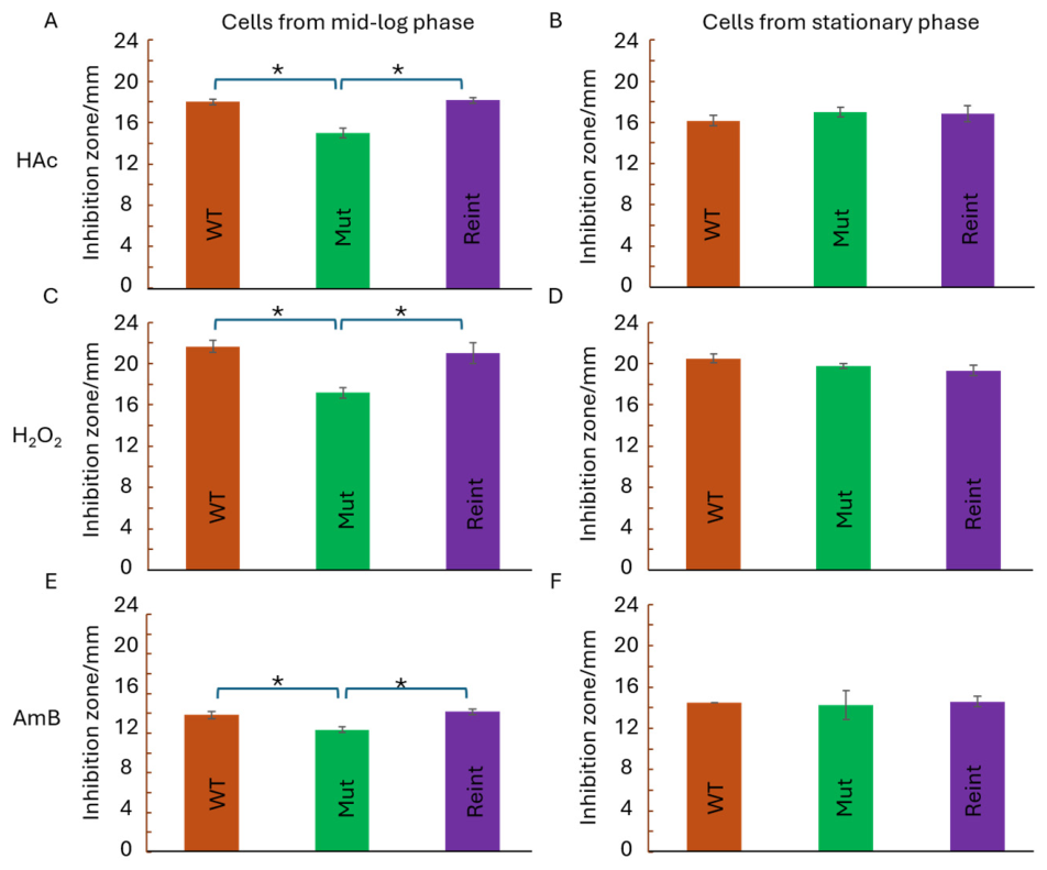
Figure 2.
Role of Mca1p in virulence. Cells from mid-log (A) or stationary (B) phase cultures of C. albicans wild type (WT: brown), double MCA1 knockout (Mut: green) or MCA1 reintegrant (Reint: purple) strains were suspended in PBS and injected into wax moth larvae and incubated at 37 °C. Control larvae were injected with PBS alone (red). Larval survival was checked every 6 hours until all C. albicans-injected larvae had died. Three independent experiments were conducted and the means (bars) and standard deviations(error bars) are shown above. Student’s t-testing was carried out. An asterisk (*) indicates a significant difference (p < 0.01) in larval survival when injected with Mut, compared with survival when injected with WT or Reint.
Figure 2.
Role of Mca1p in virulence. Cells from mid-log (A) or stationary (B) phase cultures of C. albicans wild type (WT: brown), double MCA1 knockout (Mut: green) or MCA1 reintegrant (Reint: purple) strains were suspended in PBS and injected into wax moth larvae and incubated at 37 °C. Control larvae were injected with PBS alone (red). Larval survival was checked every 6 hours until all C. albicans-injected larvae had died. Three independent experiments were conducted and the means (bars) and standard deviations(error bars) are shown above. Student’s t-testing was carried out. An asterisk (*) indicates a significant difference (p < 0.01) in larval survival when injected with Mut, compared with survival when injected with WT or Reint.
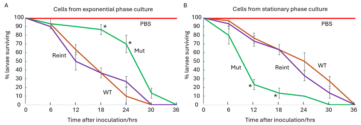
Figure 3.
Role of Mca1p in serum-induced filamentation. Cells from stationary phase cultures of the WT (A), Mut (B) and Reint (C) strains were cultured in YPD + 10% FCS. At 15-30 min intervals, samples were examined beneath a microscope and the numbers of yeast and hyphae counted. At least 400 cells in 4 fields of vision were examined. Three independent experiments were carried out, and a representative result is shown above.
Figure 3.
Role of Mca1p in serum-induced filamentation. Cells from stationary phase cultures of the WT (A), Mut (B) and Reint (C) strains were cultured in YPD + 10% FCS. At 15-30 min intervals, samples were examined beneath a microscope and the numbers of yeast and hyphae counted. At least 400 cells in 4 fields of vision were examined. Three independent experiments were carried out, and a representative result is shown above.
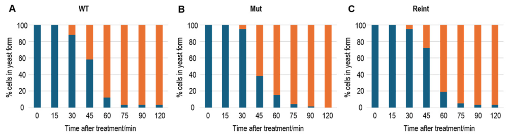
Figure 5.
Cell death of yeast and hyphal cells in acetic acid-treated (D, E, F) and untreated (A, B, C), Candida albicans strains WT (A, D), Mut (B, E) and Reint (C, F) during serum-induced filamentation. Cells from stationary phase culture were washed and resuspended at 107 cells/mL in synthetic complete (SC) medium with 10% FCS with either 80 mM HAc or an equal volume of water. After 3 hours incubation at 37 °C with shaking (200 rpm) the cells were washed in PBS and resuspended in 10 µg/mL propidium iodide and incubated at 37 °C for 30 min. After washing and resuspending in PBS, cells were examined using a fluorescence microscope. Live (non-fluorescent) and dead (red, fluorescent) hyphal daughter and yeast mother cells were counted, as were combinations of stained or unstained mother and stained or unstained daughter cells. M+: stained (dead) mother (yeast) cell; M–: unstained (living) mother (yeast) cell; H+: stained (dead) hyphae (daughter cells); H–: unstained (living) hyphae (daughter cells). Asterisk (*): significant difference (p < 0.0001) between Mut and WT and between Mut and Reint.
Figure 5.
Cell death of yeast and hyphal cells in acetic acid-treated (D, E, F) and untreated (A, B, C), Candida albicans strains WT (A, D), Mut (B, E) and Reint (C, F) during serum-induced filamentation. Cells from stationary phase culture were washed and resuspended at 107 cells/mL in synthetic complete (SC) medium with 10% FCS with either 80 mM HAc or an equal volume of water. After 3 hours incubation at 37 °C with shaking (200 rpm) the cells were washed in PBS and resuspended in 10 µg/mL propidium iodide and incubated at 37 °C for 30 min. After washing and resuspending in PBS, cells were examined using a fluorescence microscope. Live (non-fluorescent) and dead (red, fluorescent) hyphal daughter and yeast mother cells were counted, as were combinations of stained or unstained mother and stained or unstained daughter cells. M+: stained (dead) mother (yeast) cell; M–: unstained (living) mother (yeast) cell; H+: stained (dead) hyphae (daughter cells); H–: unstained (living) hyphae (daughter cells). Asterisk (*): significant difference (p < 0.0001) between Mut and WT and between Mut and Reint.
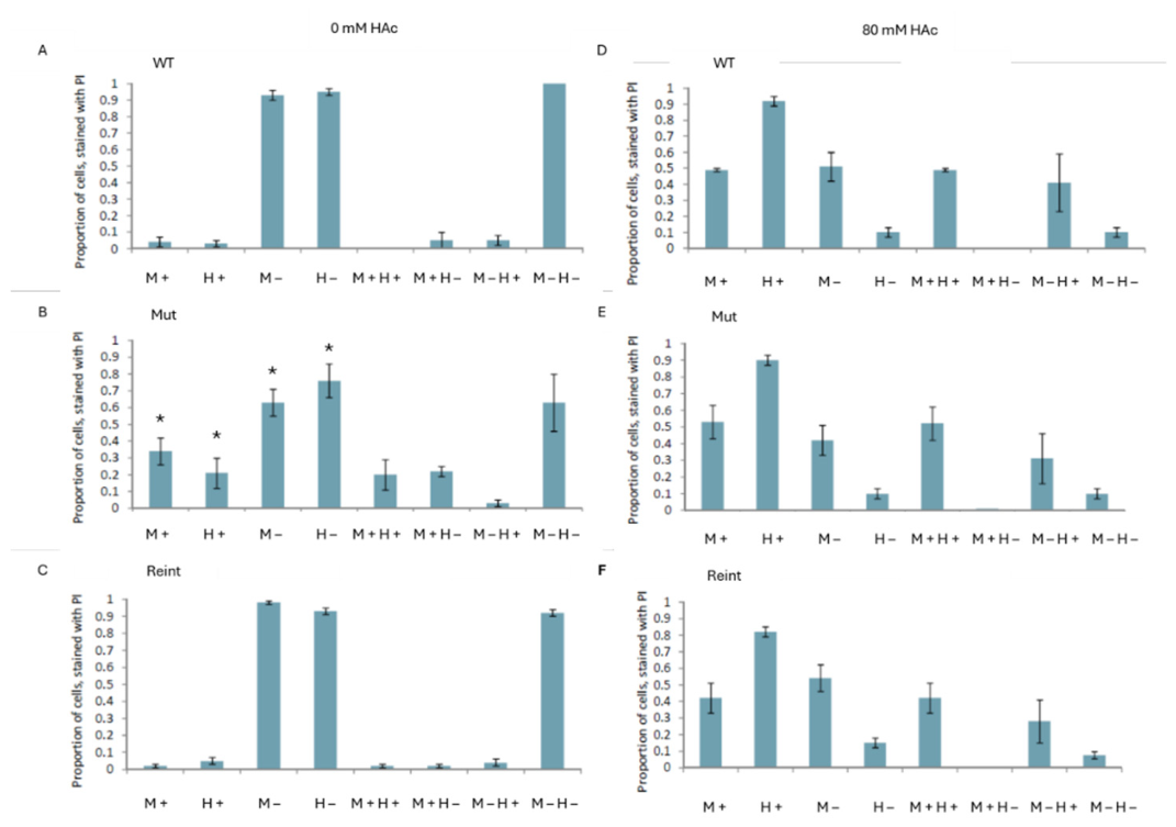

Table 1.
C. albicans strains used in this study.
| Strain | Relevant genotype | Reference |
|---|---|---|
| SN78 | MCA1/MCA1.his1Δ/his1Δ. leu2Δ/leu2Δ. ura3Δ/ura3Δ | [40] |
| WT | MCA1/MCA1.RPS1/rps1Δ::Clp40-HIS1-LEU2- URA3-ARG4.his1Δ/his1Δ. leu2Δ/leu2Δ. ura3Δ/ura3Δ | This study |
| Mut | mca1Δ::LEU2.mca1Δ::HIS1.RPS1/rps1Δ::Cip10-URA3. his1Δ/his1Δ. leu2Δ/leu2Δ. ura3Δ/ura3Δ | This study |
| Reint | mca1Δ::LEU2.mca1Δ::HIS1.RPS1/rps1Δ::Cip10-MCA1-URA3. his1Δ/his1Δ. leu2Δ/leu2Δ. ura3Δ/ura3Δ | This study |
Table 2.
Plasmids used in this study.
| Plasmid | Auxotrophy marker | Resistance marker | Reference |
|---|---|---|---|
| pSN52 | HIS1 | KanR1 | [40] |
| pSN40 | LEU2 | KanR1 | [40] |
| Cip10 | URA3 | AmpR2 | [43] |
| Clp30 | HIS1-URA3-ARG4 | AmpR2 | [41] |
| Clp40 | HIS1-LEU2-URA3-ARG4 | AmpR2 | This study |
| Cip10-MCA1 | URA3 | AmpR2 | This study |
1 Kanamycin resistance. 2 Ampicillin resistance.
Table 3.
PCR primers used in this study.
| Primer | Sequence | Description |
|---|---|---|
| 1 | GGA CAA CAG TCC AAT TAT AGT AAT CAA CAA CAG GGT TAC GAC CAA GGG TAT AAC CAA GGT TAC GGC CAA GGC TCG GAT CCA CTA GTA ACG |
MCA1 knockout forward primer |
| 2 | CAC CAA GAC CCA GAA GCA TAC GAC TTC TAT CAC CTG TTG CAT AAG CCA TGG CAG ATT GCA ATA ACC CTG CCC AGT GTG ATG GAT ATC TGC |
MCA1 knockout reverse primer |
| 3 | GAT AAC TCC GAG CAG AGA AAG | Forward check primer upstream of MCA1 |
| 4 | CTC GTC TCT TGA TGT ATA TGG | Reverse check primer within HIS1 |
| 5 | TGC TTC AGT GGT GAA TCT ACC | Reverse check primer within LEU2 |
| 6 | CAA CAG GGT TAC GAC CAA GG | Forward check primer within MCA1 |
| 7 | ACA ATG CAT CAT TAG GAC GAG | Reverse check primer within MCA1 |
| 8 | ACT GAC TCG AGG GAT TGT GGA ATG AAT TTG TGA ATT TAC TAG TG |
Forward primer upstream of MCA1 with XhoI site |
| 9 | ACT GAC TCG AGA ATG TAA TGC CAA AAG CAT CCA TCC TTT GTT GAG |
Reverse primer downstream of MCA1 with XhoI site |
| 10 | GTA CAT TCC TAC TCC GTT CG | Forward check primer within RPS1 |
| 11 | GAT ATC GAA TTC ACG CGT TAG | Reverse check primer within CIp10 |
Disclaimer/Publisher’s Note: The statements, opinions and data contained in all publications are solely those of the individual author(s) and contributor(s) and not of MDPI and/or the editor(s). MDPI and/or the editor(s) disclaim responsibility for any injury to people or property resulting from any ideas, methods, instructions or products referred to in the content. |
© 2026 by the authors. Licensee MDPI, Basel, Switzerland. This article is an open access article distributed under the terms and conditions of the Creative Commons Attribution (CC BY) license (http://creativecommons.org/licenses/by/4.0/).
Copyright: This open access article is published under a Creative Commons CC BY 4.0 license, which permit the free download, distribution, and reuse, provided that the author and preprint are cited in any reuse.


