

Submitted:
02 August 2026
Posted:
03 August 2026
You are already at the latest version
Abstract
Hydrogen (H2) is fermented by the intestinal microbiota, transferred to the blood according to the pressure gradient, and exhaled. Since H2 is not produced or metabolized in human cells, alveolar H2 saturates the entire human body, including cellular organelles such as mitochondria, owing to gas exchange in the lungs. The electron transport chain comprises a series of redox enzymes in the mitochondria of human cells that drive adenosine triphosphate (ATP) synthesis. The catalytic activity of electron-transport enzymes is optimized at certain electrochemical potentials, as is the hydrogen ion activity (pH). However, the human body is an aqueous solution, which must be electrically neutral. Membrane potentials exist between the inside and outside of human cells because of the unequal distribution of ions across the membrane. The single-electrode potential can only be assessed relative to that of another electrode (a reference electrode). We measured the electrochemical potentials relative to a standard hydrogen electrode (SHE), and found that H2 partial pressure was a fundamental factor affecting the SHE, pH, and a reversible hydrogen electrode (RHE). The H2 partial pressure is not a unit in the human body, therefore, breath H2 is an external variable of the redox environment in the human body.
Keywords:
breath hydrogen
; electrochemical potential
; reversible hydrogen electrode
; reduction-oxidation enzyme
; potential-current kinetics
1. Introduction
Molecular hydrogen (H2) is the second most abundant reduced element on Earth following methane, and has a relatively uniform concentration of 530 parts per billion (ppb) in the atmosphere owing to large-scale atmospheric circulation. [1] Photochemical oxidation of methane and non-methane organic compounds in the atmosphere is considered the dominant source of H2, while the primary sinks include soil uptake and reactions with hydroxyl radicals occurring in the atmosphere [2,3]. For a long time, H2 was thought to be a “biologically inert gas” which could not react with biomolecules under normal pressure [4]. In cultured cells under acute oxidative stress, H2 selectively reduced hydroxyl radicals and effectively protected the cells; however, H2 did not react with other reactive oxygen species that possess physiological roles [5]. The reaction of the hydroxyl radical with H2 in aqueous solution was reported as the rate constant of 3.4×10⁷ dm3 mol−1 s−1 at 20℃ [6]. Interestingly, H2 can be measured in flatus and exhaled breath since it is fermented by the gut microbiomes. Before ingesting a carbohydrate, healthy fasting breath H2 concentration is typically less than 16 parts per million (ppm), with a mean value of approximately 7 ppm [7]. Breath H2 test does not measure the blood H2 nor colonic H2 concentrations. However, when the blood leaves the lungs, arterial H2 partial pressure (PaH2) is in equilibrium with alveolar gas and alveolar H2 partial pressure (PAH2). [8]
PaH2 = PAH2
The H2 partial pressure in the human body can be mathematically estimated using breath (end-tidal) H2 concentration. As H2 is not electrically charged and is not biologically metabolized in human cells, it can diffuse from the intestinal space to the blood, organs, and alveolar space [9]. According to Henry’s law, the amount of gas dissolved in a solution is proportional to its partial pressure, above that of the solution at equilibrium. However, the Bunsen absorption coefficient for H2 in the physiological saline solution is 0.015 at 298K and 0.015 ml of H2 can be dissolved in 1ml of solution under 1 atm (1.01325 bar) of H2 partial pressure [10]. The low concentration and solubility of H2 result in uncertainty regarding its physiological functions. Nevertheless, increasing evidence suggests that H2 may influence biological processes by modulating the redox environment. The redox environment is defined as an interconnected network of redox couples found in biological fluids, organelles, cells, and the human body overall [11]. Against this background, this review summarizes recent findings on the medical applications of H2, with particular emphasis on its potential role in homeostasis.
2. Endogenous H2 Absorption, Diffusion, and Excretions
The intestines have unique anatomical features that facilitate anaerobic states and gut microbiome colonization from birth [12]. Intestinal gas profiles vary among individuals and their diets. Flatus volumes ranged from 33 to 125 ml, and the mean production rate was 40.25 ml/h [13]. The primary constituents of the intestinal gases are nitrogen (N2), oxygen (O2), carbon dioxide, H2 and methane. The concentration of N2 was 65.1 ± 20.89% and that of H2 was 2.9 ± 0.7% in the meta-analysis. Both N2 and H2 are inert gases which do not react with other substances in the human body. Further, if less than 76 ml/6 h of H2 accumulated in the colon, it was absorbed [14]. Although H2 solubility is low in the blood, H2 diffuses from the intestinal space to the blood and from the blood to the alveolar air according to its partial pressure [15]. Therefore, the concentration of H2 decreases from the intraintestinal space to the blood, and finally to the alveolar space. If the concentration of H2 in the colon was 2.9%, that in the alveolar gas would be calculated using Henry’s law in Figure 1.
[H2 concentration in alveolar air] ≈ 0.029×0.015×0.015 = 6.5×10⁻⁶ (6.5 ppm)
The alveolar air was excreted via lung ventilation. The breath H2 test measures the end-tidal H2 concentration that equilibrates alveolar air. Under atmospheric conditions, the human body becomes saturated with N2. In diving medicine, the human body is classified into five theoretical tissue compartments depending on the N2 half-saturation time [16]. The half-saturation time of the colon is approximately 90 min, whereas that of the whole human body is approximately 30 min. Therefore, H2 in the alveolar air saturates the whole human body with the partial pressure of H2 in the alveolar air, 6.5×10⁻⁶ bar. If the H2 partial pressure in the alveolar air decreases, H2 diffuses from the human body to the alveolar air until a dynamic equilibrium is reached. According to Henry’s constant of 7.8×10⁻⁴ (mol/L bar), the concentration of H2 in the human body is calculated as 5.1×10⁻⁹ (mol/L) under 6.5×10⁻⁶ bar-H2 [17]. The concentration of H2 in the human body is extremely low but it is similar to the hydrogen ion concentration of the artery, pH 7.4; 10−7.4 = 39×10−9 (mol/L).
3. pH and H2 Partial Pressure in the Human Body Affects the Electrochemical Potential
The human body consists of 60% aqueous solution that has to be electrically neutral [18]. Accordingly, a membrane potential exists between the inside and outside of human cells and organelles, as illustrated in Figure 2.
The unequal distribution of ions across the membrane creates a resting potential of approximately 70 mV [19]. An action potential occurs due to the opening and closing of ion channels on the cell membrane [20]. In the mitochondria, the pH difference between the mitochondrial matrix and the intermembrane space is approximately 0.4 units, which corresponds to roughly a 2.5-fold difference in hydrogen ion activity [21]. Further, the mitochondria membrane potential is generally around −180 mV, which is equivalent to a 1000-fold difference in hydrogen ion activity. However, the single-electrode potential can only be assessed relative to that of another electrode (a reference electrode). Therefore, changes in the electrochemical potential were calculated using the reference electrode difference.
Electrochemical potential was measured relative to the standard hydrogen electrode (SHE), which is defined as 0.00 V using a platinum electrode in an acidic solution (pH 0) under 1 bar-H2 [22]. The pH level was measured using the electrochemical potential difference between the sample solution and the standard pH solution using a glass electrode [23]. The primary pH standard solution of the Harned cell was corrected using the H2 partial pressure according to IUPAC recommendations [24]. In the human body neither H2 nor H+ are not a unit, so the reversible hydrogen electrode (RHE) is used as a reference electrode at 37 ℃ (310K) [25]:
ERHE = −0.059 × pH − 0.029 × log(H2 partial pressure/1 bar) (V)
Under atmospheric conditions, the concentration of H2, C(H2), can be easily converted to the ratio of the H2 partial pressure/1 bar. pH2 is defined as the negative logarithm of the fugacity of H2. [26]
pH2 = −log[H2 partial pressure/1 bar] = −log[C(H2)]
Under physiological conditions, acid-base homeostasis is maintained in the human body. When 100 ppm-H2 gas was bubbled into the phosphate buffer solution, the pH shifted significantly from 7.08 to 7.18 due to the action of the buffer, and the ORP decreased by 56 mV [27]. Based on the Henderson-Hasselbalch equation,
a change in pH value of 0.1 suggested that the [HPO4²⁻]/[H2PO4⁻] ratio of the 10 mM phosphate buffer changed by a factor of 10−0.1 (approximately 1.259). Although the potential in the phosphate buffer changed according to the Nernst equation, this suggests that the redox state in vivo is also buffered in addition to the pH, yet the ratio of redox couples within the body undergoes changes.
pH = pKa + log10([A−]/[HA])
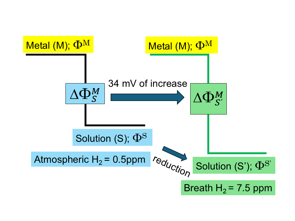
Changes in the electrochemical potential of the human body can be calculated using RHE, especially the value of pH2. Although H2 partial pressure is extremely low in the human body, the average of H2 concentration is already 15-fold relative to the atmosphere (7.5 ppm/0.5 ppm), which is 1.18 units of pH2 and approximately 34 mV converted to RHE potential. Since hydrogen ion concentration is extremely low in the neutral fluids, pH 7 = 1×10⁻⁷ (mol/L), pH can sensitively detect acid-base balance. For instance, the pH of the mitochondrial matrix is 7.4 and that in the intermembrane space is 7.8, which results in a difference in the hydrogen ion concentration [28].
Δ[H+] = 10⁻⁷.⁴ − 10⁻⁷.⁸ = 2.3 × 10⁻⁸ mol/L
Meanwhile, there are high concentration of redox couples in the cytoplasm and the mitochondria, for instance, 10 mM-glutathione disulfide /glutathione in the cytoplasm and 250 μM-nicotinamide adenine dinucleotide (NAD+)/ nicotinamide adenine dinucleotide, reduced form (NADH) in the mitochondrial matrix [29,30]. The electrochemical potentials of these redox couples and redox enzymes are measured relative to the reference electrode. When an RHE is used as the reference electrode, the determination of the potential constitutes a form of electrochemical potential measurement. Therefore, just as with the primary standard pH solution, it is considered necessary to apply corrections based on the H₂ partial pressure when dealing with electrochemical potentials in vivo. Moreover, proton-coupled electron transfer (PCET) reactions are essential for respiration, and the PCET rate depends on the redox species, temperature, pH, and overpotential in the electrochemical processes, including redox cofactors [31].
4. Redox Enzymes’ Potentials and Their Reaction Rates
The electron transport chain (ETC) in the inner mitochondrial membrane comprises a series of protein complexes that transport electrons from NADH to O2 and generate adenosine triphosphate (ATP). Each complex contains redox cofactors, such as a metal complex (iron-sulfur clusters and a Cu complex) or an organic molecule (flavin and quinone). Redox cofactors within the enzymes exhibit different thermodynamics and kinetics in the ETC and exhibit different electrochemical potentials spread over a potential window of water, from −320 mV of NAD+/NADH to +820 mV of O2/H2O related to the standard hydrogen electrode (Figure 3) [32]. Since the catalytic cofactor is deeply buried in the protein structure of the enzymes, the electrochemical potentials of these enzymes relative to the SHE do not change through natural electron transfer.
where Ecofactor is the in situ potential of the redox cofactor and E⁰cofactor is its standard potential relative to the SHE. If the H2 partial pressure is changed, the electrochemical potential of each complex changes relative to RHE as the reference electrode.
Ecofactor = E0cofactor − ERHE,
In electrochemistry, the immobilization of redox enzymes on an electrode is an analytical tool for investigating the reaction rate involved in electron transfer [33]. When the enzyme substrate is present, the electrode specifically collects electrons released from the substrate. Subsequently, the current was recorded at each applied potential using cyclic voltammetry. The resulting curve was quasi-sigmoidal, and the limiting plateau current was proportional to the catalytic rate constant Kcat of the enzyme. The Kcat values for oxidation and reduction are given by the Butler–Volmer equation [34].
where E0RDS is the redox potential of rate determining step, while k0 is the standard rate constant and α is the transfer coefficient at rate determining step. In a biosensor electrode applied at a certain potential, the catalytic current should be proportional to the analyte concentration, such as glucose, in the range where the enzyme is not saturated, as indicated by Michaelis-Menten kinetics. [35]
Kox = k0exp[(1 − α)F(E − E0RDS)/RT] and, Kred = k0exp[−αF(E − E0RDS)/RT],
the current ∝ Kcat × [substrate]
Although it is impossible to apply electrochemical potential to the human body, changes in the potential of the RHE can be measured using breath H2 as a reference electrode. Despite the complexity of redox reactions within the ETC, its catalytic constant can be evaluated using breath H2 concentration as an external variable, as illustrated in Figure 4.
5. H2 Partial Pressure Affects Redox Reactions and Metabolic Reactions
H2 is an inert gas that does not react with biological substrates, even in hyperbaric environments. During the HYDRA 10 experimental human dive (71 atm/7200 kPa), H2 was introduced during compression from days 4 to 33, and ultimately reached a partial pressure of 20 atm [36]. Electrocardiogram indicated a marked global diminution of voltage during decompression, which caused the formation of microbubbles as a physical barrier between the heart and the electrode. In 2007, Ohsawa was the first to report the physiological effects to scavenge hydroxy radical using 2% H2 gas for cerebral ischemia-reperfusion injury treatment in rats. However, the rate constant between the hydroxyl radical and H2 is slower than that of most radical-radical reactions, and the targets of hydroxyl radicals are abundant membrane lipids and thiols [37]. The therapeutic window was addressed prior to reperfusion. Thereafter, many studies demonstrated the protective and therapeutic effects of H2 [38]. Further research is needed to elucidate the mechanisms meditating the physiological effects of H2, particularly its role in modulating oxidative stress.
The pH of a solution affects the activity of electrochemical processes such as hydrogen oxidation, oxygen reduction, and carbon reduction [39]. However, the mechanisms underlying the effects of pH on electrochemical reactions are considered in terms of mass transport, electrochemical double-layer structure, and surface adsorption. In certain redox enzymes, a change in the pH value by a factor of two leads to a 10-fold change in the electron transfer rate constants, according to the outer spherical electron transfer theory [40]. If the RHE potential decreases due to an increase in pH or H2 partial pressure, the oxidation rate constant of the redox enzyme might increase with an increase in the electrochemical potential between the redox enzyme and the substrate in the solution.
When H2 was added to isolated mitochondria, electron transport by succinate was reversed from NAD+ reduction to NADH oxidation, and reactive oxygen species generation was suppressed [41]. In rat model long-term H2 inhalation reduced body weight and visceral fat mass [42]. Inhalation of H2 using a nasal cannula (300 ml/min) significantly increases resting fat oxidation, as evidenced by a decreased respiratory exchange ratio, particularly in individuals with higher body fat percentages [43]. Breath acetone is a the ketone body produced in the hepatic mitochondria during lipid metabolism. Inhalation of 1%-H2 during exercise augmented breath acetone and enhanced O2 uptake compared with the control gas, without affecting oxidative stress or antioxidant activity in human participants [44].
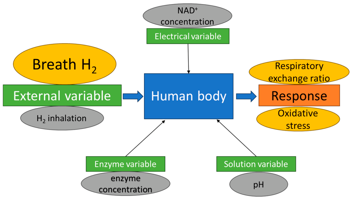
Human breath is a predictive mixture of volatile organic compounds and inorganic gas species for noninvasive diagnosis of common diseases [45]. Breath H2 reflects carbohydrate fermentation by the gut microbiomes and follows a circadian pattern due to dietary fiber intake [46]. Although breath H2 levels did not differ among the groups before sleeping, breath H2 levels in the morning decreased in patients with chronic heart failure (CHF) compared to those without CHF,[47] and in aged subjects (60−85 years) compared to non-aged subjects (20−59 years) [48]. Following exercise, breath H2 levels declined at 5 min, approached baseline at 30 min, and a secondary reduction was observed at 60 min [49]. Breath H2 concentration was consistently and positively associated with an integrated cellular bioenergetic phenotype derived from body cell mass, intracellular water, total body potassium, and glycogen [50]. Additionally, breath H2 can be an external variable in the redox environment, which is associated with redox enzyme function and energy metabolism (Figure 5).
6. Conclusions
Metabolic reactions in the human body involve quasi-reversible and irreversible multistep processes. When electron transfer continues the rate-determining step is an electron-transfer step in the ETC, its kinetics influence the electrochemical response. Thus, current-potential characteristics should be elucidated in terms of controllable parameters such as the concentrations of the participating species. Notably, H2 is not produced or metabolized in human cells, and H2 fermented by gut microbiomes is saturated in the human body. H2 is a reductant that influences the electrochemical potential of redox enzymes, including those in the ETC. Therefore, breath H2 may represent an external variable of the redox environment in the human body. Future studies should focus on characterizing current–potential relationships under controlled variations in H₂ partial pressure and other relevant physiological parameters.
Funding
This research received no external funding.
Institutional Review Board Statement
Not applicable.
Informed Consent Statement
Not applicable.
Data Availability Statement
No new data were created or analyzed in this study. Data sharing is not applicable to this article.
Conflicts of Interest
The author declares no conflicts of interest.
Abbreviations
The following abbreviations are used in this manuscript:
| H2 | Molecular hydrogen |
| N2 | Nitrogen |
| O2 | Oxygen |
| pH | Hydrogen ion activity |
| SHE | The standard hydrogen electrode |
| RHE | The reversible hydrogen electrode |
| ERHE | The potential of the reversible hydrogen electrode |
| ppb | Parts per billion |
| ppm | Parts per million |
| C(H2) | The concentration of hydrogen |
| PCET | Proton-coupled electron transfer |
| ETC | The electron transport chain |
| ATP | Adenosine triphosphate |
| NAD+ | Nicotinamide adenine dinucleotide |
| NADH | Nicotinamide adenine dinucleotide, reduced form |
| Kcat | The catalytic rate constant |
| k⁰ | The standard rate constant |
| E⁰RDS | The redox potential of rate determining step |
| CHF | Chronic heart failure |
References
- Patterson, J. D.; Aydin, M.; Crotwell, A. M.; Petron, G.; Severinghaus, J. P.; Saltzman, E. S. Atmospheric history of H2 over the past century reconstructed from South Pole firn air. Geophys Res. Lett. 2020, 47, e2020GL087787. [Google Scholar] [CrossRef]
- Rezaei, S.; Hormaza, M. A.; Wu, Y.; Reed, J.; Brouwer, J. Global warming impacts of the transition from fossil fuel conversion and infrastructure to hydrogen. Appl. Energy 2025, 397, 126363. [Google Scholar] [CrossRef] [PubMed]
- Sandhiya, L.; Bhati, M. Unravelling the atmospheric and climate implications of hydrogen leakage. Int. J. Hydrogen Energy 2023, 53, 807–815. [Google Scholar] [CrossRef]
- Tong, M.-N.; Li, X.; Cheng, J.; Jiang, Z.-L. Developments in the Study of Inert Gas Biological Effects and the Underlying Molecular Mechanisms. Int. J. Mol. Sci. 2025, 26, 7551. [Google Scholar] [CrossRef] [PubMed]
- Ohsawa, I.; Ishikawa, M.; Takahashi, K.; Watanabe, M.; Nishimaki, K.; Yamagata, K.; Katsura, K.; Katayama, Y.; Asoh, S.; Ohta, S. Hydrogen acts as a therapeutic antioxidant by selectively reducing cytotoxic oxygen radicals. Nat. Med. 2007, 13, 688–694. [Google Scholar] [CrossRef] [PubMed]
- Christensen, H.; Sehested, K. Reaction of hydroxyl radicals with hydrogen at elevated temperatures. Determination of the activation energy. J. Phys. Chem. 1983, 87, 118–120. [Google Scholar] [CrossRef]
- Perman, J. A.; Modler, S.; Barr, R. G.; Rosenthal, P. Fasting breath hydrogen concentration: Normal values and clinical application. Gastroenterology 1984, 87, 1358–1363. [Google Scholar] [CrossRef]
- Farhi, L.E. Elimination of inert gas by the lung. Respir. Physiol. 1967, 3, 1–11. [Google Scholar] [CrossRef] [PubMed]
- Yamamoto, R.; Homma, K.; Suzuki, S.; Sano, M.; Sasaki, J. Hydrogen gas distribution in organs after inhalation: Real-time monitoring of tissue hydrogen concentration in rat. Sci. Rep. 2019, 9, 1255. [Google Scholar] [CrossRef] [PubMed]
- Raposo, R.R.; Calviño, E.F.; Esteso, M.A. Determination of Hydrogen Solubility Values in Some Human Fluids by Using an Electrochemical Sensor. ETS Trans. 2007, 3, 127. [Google Scholar] [CrossRef]
- Schafer, F.Q.; Buettner, G.R. Redox environment of the cell as viewed through the redox state of the glutathione disulfide/glutathione couple. Free Radic. Biol. Med. 2001, 30, 1191–1212. [Google Scholar] [CrossRef] [PubMed]
- Yang, K.; Li, G.; Li, Q.; Wang, W.; Zhao, X.; Shao, N.; Qiu, H.; Liu, J.; Xu, L.; Zhao, J. Distribution of gut microbiota across intestinal segments and their impact on human physiological and pathological processes. Cell Biosci. 2025, 15, 47. [Google Scholar] [CrossRef] [PubMed]
- Modesto, A.; Cameron, N.R.; Varghese, C.; Peters, N.; Stokes, B.; Phillips, A.; Bissett, I.; O'Grady, G. Meta-Analysis of the Composition of Human Intestinal Gases. Dig. Dis. Sci. 2022, 67, 3842–3859. [Google Scholar] [PubMed]
- Hammer, H. F. Colonic hydrogen absorption: Quantification of its effect on hydrogen accumulation caused by bacterial fermentation of carbohydrates. Gut 1993, 34, 818–822. [Google Scholar] [CrossRef] [PubMed] [PubMed Central]
- Azpiroz, F. Intestinal gas dynamics: mechanisms and clinical relevance. Gut 2005, 54, 893–895. [Google Scholar] [CrossRef] [PubMed]
- Kot, J.; Sicko, Z.; Doboszynski, T. The Extended Oxygen Window Concept for Programming Saturation Decompressions Using Air and Nitrox. PLoS ONE 2015, 25, e0130835. [Google Scholar]
- Sander, R. Compilation of Henry’s law constants (version 5.0.0) for water as solvent. Atmos. Chem. Phys. 2023, 23, 10901–12440. [Google Scholar] [CrossRef]
- Delpire, E.; Gagnon, K.B. Water Homeostasis and Cell Volume Maintenance and Regulation. Curr. Top. Membr. 2018, 81, 3–52. [Google Scholar] [CrossRef] [PubMed]
- Chrysafides, S.M.; Bordes, S.J.; Sharma, S. “Physiology, Resting Potential” StatPearls. 10 Apr 2023. Available online: https://www.ncbi.nlm.nih.gov/books/NBK538338/ (accessed on 20 July 2026).
- Clay, J.R. Determining k channel activation curves from k channel currents often requires the goldman-hodgkin-katz equation. Front Cell Neurosci. 2009, 3, 20. [Google Scholar] [CrossRef] [PubMed]
- Ahmed Selim, N.; Wojtovich, A.P. Mitochondrial membrane potential and compartmentalized signaling: Calcium, ROS, and beyond. Redox Biol. 2025, 86, 103859. [Google Scholar] [CrossRef] [PubMed]
- Jerkiewicz, G. Standard and Reversible Hydrogen Electrodes: Theory, Design, Operation, and Applications. ACS Catal. 2020, 10, 8409–8417. [Google Scholar] [CrossRef]
- Bates, R.G.; Vijh, A.K. Determination of pH: Theory and Practice. J. Electrochem Soc. 1973, 120, 263c. [Google Scholar] [CrossRef]
- Buck, R.P.; Rondinini, S.; Covington, A.; Baucke, F.; Brett, C.; Camoes, M.; Milton, M.; Mussini, T.; Naumann, R.; Pratt, K.; et al. Measurement of pH definition, standards, and procedures. Pure Appl. Chem. 2002, 74, 2169–2200. [Google Scholar] [CrossRef]
- Kiyama, T. Environmental Hydrogen Concentration as a Novel Factor Determining Changes in Redox Potential. Physiologia 2025, 5, 36. [Google Scholar] [CrossRef]
- Kiyama, T. Hydrogen Electrode Potentials as a Descriptor of Catalyst Reactivity for Sustainable Redox Chemistry: A Tutorial Review. Sustainability 2026, 18, 5791. [Google Scholar] [CrossRef]
- Kiyama, T.; Tokunaga, A.; Naji, A.; Barbul, A. Changes in the negative logarithm of end-tidal hydrogen partial pressure indicate the variation of electrode potential in healthy Japanese subjects. Sci. Rep. 2023, 13, 15473. [Google Scholar] [CrossRef] [PubMed]
- Porcelli, A.M.; Ghelli, A.M.; Zanna, C.; Pinton, P.; Rizzuto, R.; Rugolo, M. pH difference across the outer mitochondrial membrane measured with a green fluorescent protein mutant. Biochem Biophys. Res. Comm. 2005, 326, 799–804. [Google Scholar] [CrossRef] [PubMed]
- Milo, R.; Phillips, R. What is the redox potential of a cell? In Cell Biology by the Numbers, 1st ed.; Garland Science: New York, NY, USA, 2015; pp. 189–196. [Google Scholar]
- Cantó, C.; Menzies, K.; Auwerx, J. NAD+ Metabolism and the Control of Energy Homeostasis: A Balancing Act between Mitochondria and the Nucleus. Cell Metab. 2015, 22, 31–53. [Google Scholar] [CrossRef] [PubMed]
- Hammes-Schiffer, S. Theory of proton-coupled electron transfer in energy conversion processes. Acc. Chem. Res. 2009, 42, 1881–9. [Google Scholar] [CrossRef] [PubMed]
- Bollella, P.; Katz, E. Enzyme-Based Biosensors: Tackling Electron Transfer Issues. Sensors 2020, 20, 3517. [Google Scholar] [CrossRef] [PubMed]
- Kano, K. Fundamental insight into redox enzyme-based bioelectrocatalysis. Biosci. Biotechnol. Biochem 2022, 86, 141–156. [Google Scholar] [CrossRef] [PubMed]
- Bard, A.J.; Faulkner, L.R. Electrochemical Methods: Fundamentals and Applications, 2nd ed.; John Wiley & Sons: New York, NY, USA, 2000. [Google Scholar]
- Zhang, X.; Xin, J.; Jin, R. Electrochemical glucose sensing using an electrochemically reduced graphene oxide/poly-L-lysine composite electrode modified with glucose oxidase. Int. J. Electrochem Sci. 2026, 21, 101357. [Google Scholar] [CrossRef]
- Lafay, V.; Barthelemy, P.; Comet, B.; Frances, Y.; Jammes, Y. ECG changes during the experimental human dive HYDRA 10 (71 atm/7,200 kPa). Undersea Hyperb. Med. 1995, 22, 51–60. [Google Scholar] [PubMed]
- Wood, K.; Gladwin, M. The hydrogen highway to reperfusion therapy. Nat. Med. 2007, 13, 673–674. [Google Scholar] [CrossRef] [PubMed]
- Yildiz, F.; LeBaron, T.W.; Alwazeer, D. A comprehensive review of molecular hydrogen as a novel nutrition therapy in relieving oxidative stress and diseases: Mechanisms and perspectives. Biochem Biophys. Rep. 2025, 41, 101933. [Google Scholar] [CrossRef] [PubMed]
- Govindarajan, N.; Xu, A.; Chan, K. How pH affects electrochemical processes. Science 2022, 375, 379–380. [Google Scholar] [CrossRef] [PubMed]
- Kulys, J.; ČèNas, N. Reagent redox potential and pH effects on the enzymatic electron transfer. J. Mol. Catal. 1988, 47, 335–341. [Google Scholar] [CrossRef]
- Ishihara, G.; Kawamoto, K.; Komori, N.; Ishibashi, T. Molecular hydrogen suppresses superoxide generation in the mitochondrial complex I and reduced mitochondrial membrane potential. Biochem Biophys. Res. Commun. 2019, 522, 965–970. [Google Scholar] [CrossRef] [PubMed]
- Adzavon, Y.M.; Xie, F.; Yi, Y.; Jiang, X.; Zhang, X.; He, J.; Zhao, P.; Liu, M.; Ma, S.; Ma, X. Long-term and daily use of molecular hydrogen induces reprogramming of liver metabolism in rats by modulating NADP/NADPH redox pathways. Sci. Rep. 2022, 12, 3904. [Google Scholar] [CrossRef] [PubMed]
- Botek, M.; McKune, A.; Grepl, P.; Krejčí, J. Molecular hydrogen inhalation modulates resting metabolism in healthy females: findings from a randomized, double-blind, placebo-controlled crossover study. Med. Gas. Res. 2025, 15, 367–373. [Google Scholar] [CrossRef] [PubMed]
- Hori, A.; Ichihara, M.; Kimura, H.; Ogata, H.; Kondo, T.; Hotta, N. Inhalation of molecular hydrogen increases breath acetone excretion during submaximal exercise: a randomized, single-blinded, placebo-controlled study. Med. Gas. Res. 2020, 10, 96–102. [Google Scholar] [CrossRef] [PubMed]
- Alenzy, E.; Kandjani, A.E.; Shaibani, M.; Trinchi, A.; Bhargava, S.K.; Ippolito, S.J.; Sabri, Y.M. Human breath analysis; Clinical application and measurement: An overview. Biosens. Bioelectron. 2024, 278, 117094. [Google Scholar] [CrossRef] [PubMed]
- Kagaya, M.; Iwata, M.; Toda, Y.; Nakae, Y.; Kondo, T. Circadian rhythm of breath hydrogen in young women. J. Gastroenterol. 1998, 33, 472–476. [Google Scholar] [CrossRef] [PubMed]
- Shibata, A.; Sugano, Y.; Shimouchi, A.; Yokokawa, T.; Jinno, N.; Kanzaki, H.; Ohta-Ogo, K.; Ikeda, Y.; Okada, H.; Aiba, T.; Kusano, K.; Shirai, M.; Ishibashi-Ueda, H.; Yasuda, S.; Ogawa, H.; Anzai, T. Decrease in exhaled hydrogen as marker of congestive heart failure. Open Heart 2018, 5, e000814. [Google Scholar] [CrossRef] [PubMed]
- Okumura, N.; Kiriyama, K.; Taniguchi, K. Age differences in overnight breath hydrogen dynamics and their association with sleep physiology. J. Breath Res. 2026, 20, 036003. [Google Scholar] [CrossRef] [PubMed]
- Todorovic, N.; Kuzmanovic, J.; Javorac, D.; Ostojic, S.M. The impact of graded exercise to exhaustion on breath hydrogen: a pilot study. Sport Sci. Health 2025, 21, 3579–3583. [Google Scholar] [CrossRef]
- Todorovic, N.; Nedeljkovic, D.; Andjelic, B.; Korovljev, D.; Tarnava, A.; Ostojic, S.M. Breath Hydrogen Reflects a Cellular Bioenergetic Phenotype in Sedentary Adults with Metabolic Syndrome. Clin. Bioenerg. 2026, 2, 6. [Google Scholar] [CrossRef]
Figure 1.
Endogenous H2 absorption, diffusion, and excretion. H2 is fermented in the colon and diffuses into the venous blood. Subsequently, venous H2 diffuses into the alveolar air and alveolar H2 also equilibrates the arterial blood. Arterial H2 saturates the whole human body due to systemic circulation.
Figure 1.
Endogenous H2 absorption, diffusion, and excretion. H2 is fermented in the colon and diffuses into the venous blood. Subsequently, venous H2 diffuses into the alveolar air and alveolar H2 also equilibrates the arterial blood. Arterial H2 saturates the whole human body due to systemic circulation.
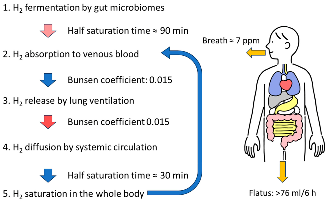
Figure 2.
Membrane potential between the intracellular and extracellular fluids. Unequal distribution of ions across the membrane creates the resting potential. Na+; sodium ion, K+; potassium ion.
Figure 2.
Membrane potential between the intracellular and extracellular fluids. Unequal distribution of ions across the membrane creates the resting potential. Na+; sodium ion, K+; potassium ion.
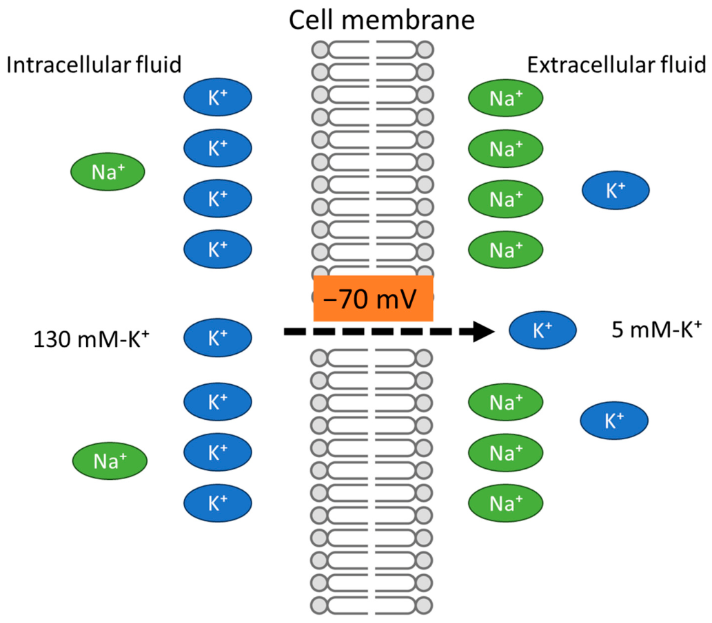
Figure 3.
The redox ladder displays the order of reaction in the electron transport chain. These redox gradients are reflected from the ratio of oxidized/reduced redox couples and the redox potentials of complexes. The redox potential of redox cofactors in the complexes are consistent for the SHE, but those varies for the RHE. FMN; flavin mononucleotide, Fe-S; iron-sulfur clusters, CoQH2; ubiquinol, Cyt; cytochrome.
Figure 3.
The redox ladder displays the order of reaction in the electron transport chain. These redox gradients are reflected from the ratio of oxidized/reduced redox couples and the redox potentials of complexes. The redox potential of redox cofactors in the complexes are consistent for the SHE, but those varies for the RHE. FMN; flavin mononucleotide, Fe-S; iron-sulfur clusters, CoQH2; ubiquinol, Cyt; cytochrome.
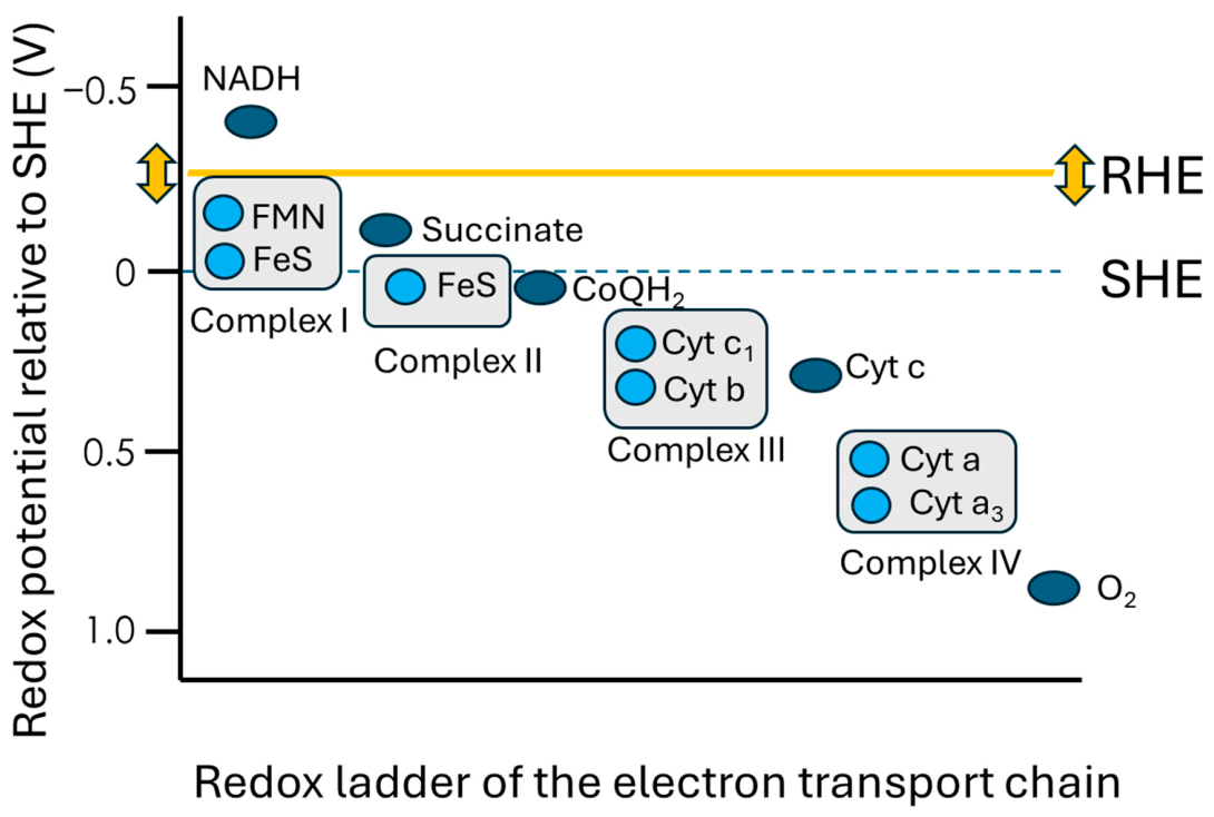
Figure 4.
Variables influencing the catalytic kinetics of the electron transport chain (ETC). The catalytic constant of ETC could be monitored using breath H2 as an external variable, regardless of the difficulty of controlling the other variables.
Figure 4.
Variables influencing the catalytic kinetics of the electron transport chain (ETC). The catalytic constant of ETC could be monitored using breath H2 as an external variable, regardless of the difficulty of controlling the other variables.
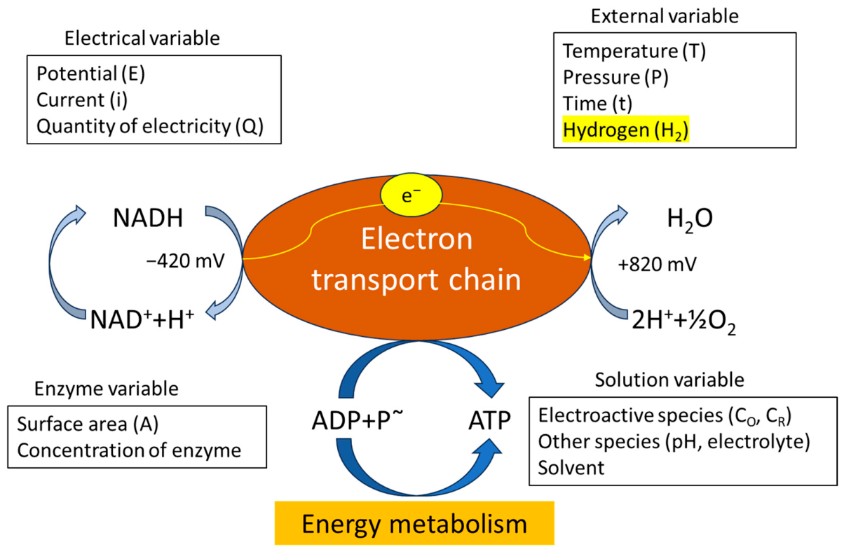
Figure 5.
Conceptual framework illustrating breath H2 as a measurable or controllable external variable in the systemic redox environment. Changes in breath H2 may influence the catalytic kinetics of redox reactions in the human body and may be used to evaluate electrochemical current-potential characteristics through physiological responses, such as the respiratory exchange ratio and markers of oxidative stress.
Figure 5.
Conceptual framework illustrating breath H2 as a measurable or controllable external variable in the systemic redox environment. Changes in breath H2 may influence the catalytic kinetics of redox reactions in the human body and may be used to evaluate electrochemical current-potential characteristics through physiological responses, such as the respiratory exchange ratio and markers of oxidative stress.
Disclaimer/Publisher’s Note: The statements, opinions and data contained in all publications are solely those of the individual author(s) and contributor(s) and not of MDPI and/or the editor(s). MDPI and/or the editor(s) disclaim responsibility for any injury to people or property resulting from any ideas, methods, instructions or products referred to in the content. |
© 2026 by the authors. Licensee MDPI, Basel, Switzerland. This article is an open access article distributed under the terms and conditions of the Creative Commons Attribution (CC BY) license (http://creativecommons.org/licenses/by/4.0/).
Copyright: This open access article is published under a Creative Commons CC BY 4.0 license, which permit the free download, distribution, and reuse, provided that the author and preprint are cited in any reuse.