
Submitted:
31 July 2026
Posted:
03 August 2026
You are already at the latest version
Abstract
Background and Purpose: Abnormality in the motion of the inferior papillary muscle was found, in the past, in Mitral Valve Prolapse (MVP). We visually observed an odd movement not only in the inferior wall but also in the anterolateral and inferolateral basal walls in several MVP patients as well. Therefore, the purpose of this work was to analyze this findings with the help of Strain (Speckle Tracking). Methods: 60 MVP patients, having a superior displacement of the posterior leaflet, the whole or part of, of at least 2 mm, and 20 normal controls were included. Longitudinal Strain was calculated in the 3 apical views (Philips iE 33 Matrix - implemented Strain software). The Strain in the 3 above mentioned basal segments was measured at 160 msec from the beginning of systole (mid-systole) and then averaged to form a “triad” that could be compared among the groups for statistical calculations. The Global Longitudinal Strain (GLS) was calculated for all as well. The Mann-Whitney U-test for comparisons was used. A “p” value ≤ 0.05 was considered significant. Results: According with the Strain patterns, we could identify two Groups of MVP patients: Group 1: having a “paradoxical” Strain at 160 msec (positive, that means “stretching”) in at least one of the three told basal segments (n = 35), and Group 2: not having a “paradoxical” Strain (n = 25) in any of the told three basal segments at the same mid-systolic point. Statistical significance was found between Groups 1 and 2 at 160 msec and at the peak (p < 0.0001 for both) and for GLS (p < 0.01) as well. Group 1 vs controls, of course, displayed significant difference in the Strain at160 msec (p < 0,0001) while Group 2 vs controls did not do and even not at the peak. Conclusions: According with our results, we found that there is a MVP population (more than half in our series) having a “paradoxical” Strain (= stretching) in at least one of the basal inferior-to-lateral segments in mid-systole. The passive continuous stretching of myocardial cells is known to lead, with time, to the replacement with fibrosis (and fibrosis is a condition that is well known to be a source of arrhythmias, and even malignant). Cardiac Magnetic Resonance (CMR) already demonstrated the presence of fibrosis just in the same basal wall segments in some MVP patients. Thus, considering our results in the Strain, in MVP patients with ventricular arrhythmias it is mandatory to analyze the Strain (not expensive tool), and then, if the Strain were to be found to be “paradoxical” even in only one segment of this previously called “triad”, a further evaluation with CMR (even if expensive) is highly recommended to look for the presence of fibrosis. The Strain is highly recommended because the topic is implicated in prognosis, in the further study of arrhythmias, and in the choice of a proper therapy a well.
Keywords:
mitral valve prolapse
; strain
; speckle tracking
; ventricular arrhythmias in mitral valve prolapse
Introduction
Mitral Valve prolapse (MVP) is a relatively frequent finding with a prevalence in the general population ranging from 0.6% to 2.4%. Patients with MVP show a very heterogeneous clinical spectrum [1] varying from asymptomatic status to different degrees of cardiac complications: mitral regurgitation (MR) of several degrees, cardiac rhythm disturbances ranging from simple premature supraventricular beats or atrial fibrillation or premature ventricular beats or ventricular tachycardia and even sudden cardiac death (SCD) [2,3].
MVP can present itself with a wide spectrum of valve changes: from an insignificant systolic superior displacement of one, or more than one, scallops or a part of, and to a more significant modifications like important leaflet thickening, elongated cords, and severe thickening and huge superior displacement of the leaflets due to myxomatous degeneration, and along with several degrees of regurgitation.
The Mitral Valve is a complex anatomical structure that include not only the valve itself, but also the ring, the papillary muscles, and the myocardium below the ring: all the above are contributing to the normal function of the mitral apparatus. Although the leaflets are the most commonly involved structures, MVP may involve any of the mitral apparatus components including the ventricular wall portion beneath the valve, which has been analysed in the present Study.
A very informative old study described an abnormal movement of the inferior papillary muscle in MVP [4].
In our clinical daily practice, we visually observed, in several patients, an odd movement in the basal segments, not only in the inferior, but also in the anterolateral and inferolateral walls, all below the mitral ring.
Therefore, the purpose of our study was to go inside this not normal movement of the walls by using two-dimensional (2D) Strain that is the Speckle Tracking (STE).
Materials and Methods
The Study was made accordingly with the Helsinki Declaration, and a verbal informed consent to participate in was obtained by all of the Patients.
Echocardiography for Strain analysis was made in all the 3 apical views for enrolling, provided having good images. Thus, we consequently enrolled 60 MVP subjects (defined as a superior displacement of at least 2 mm in one or more leaflets in toto or in a part of). 20 normal subjects formed the control group. We excluded patients with: 1) moderate or severe MR, 2) global wall motion abnormalities, 3) known or suspected coronary artery disease, 4) patients with more than mild abnormalities of the other valves that could affect the left ventricle (LV) dynamics, 5) patients with more than mild diastolic dysfunction, 6) patients having pericardial disease, 7) use of cardio-toxic drugs, 8) systemic disorders which might result in direct cardiac complications (such as diabetes, cancer, kidney failure, clinical obstructive/restrictive respiratory diseases, thyroid disease, autoimmune diseases), 9) patients with mitral-annular disjunction (MAD) which could interfere with the dynamics of the basal wall.
The control group was composed by subjects without MVP who met the same exclusion criteria (Table 1).
Clinical characteristics of the study and control groups, showing no statistically significant differences. LVEF: left ventricular ejection fraction; LA: left atrium dimension obtained in parasternal long-axis view; BMI: body mass index; SBP: systolic blood pressure; DBP: diastolic blood pressure.
All patients underwent 2D standard echocardiography with a Philips iE33 Matrix machine (The Netherlands). STE analysis was performed by using the implemented software in the machine with standard settings. Longitudinal STE was calculated in the three apical views: 2-chamber (2C), 3-chamber (3C), and 4-chamber (4C). After the first semiautomatic processing, we manually carefully adjusted the region of interest (ROI) to encompass the entire thickness of the myocardium throughout the whole cardiac cycle. As far as we observed that the basal inferior (BI), basal anterolateral (BAL) and basal inferolateral (BIL) walls were, one or more, involved in this altered wall movement, to make a statistical analysis we averaged the numbers and we called these three segments as a” triad”. So, to test our hypothesis, we registered the longitudinal strain (LS) in each basal segment of the “triad”, in each patient, at 160 msec from the beginning of systole ( mid-systole) and then we averaged the values to form the Groups. Subsequently, we compared the averaged strain value at 160 msec but also the Global Longitudinal Strain (GLS) among the Groups.
We choose the cut-off of 160 msec because it was the best point of the mid-systole in our population according to the mean heart rate and the STE software.
Statistics
Normally distributed variables are presented as mean ± SD. Statistical analysis was performed using the SPSS-22 software. Differences among groups were assessed using the Mann-Whitney U-test. P value ≤ 0.05 was considered statistically significant.
Results
According with our results in STE, we could observe, in all MVP patients, two Groups having different Strain patterns: Group 1 having a “paradoxical” strain at 160 msec in at least one of the segments of the basal “triad” (n = 35) (that means positive = stretching)( Figure 1), and a Group 2 not having “paradoxical” strain (n = 25) at the same 160 msec in anyone of the segments of that basal “triad” (Figure 2, bull-eye panel B). The clinical characteristics were the same between group 1 and group 2 (Table 2).
Bull-eye view of strain in the different groups: panel A: MVP paradoxical strain vs normals at 160 msec; panel B: MVP paradoxical strain vs non-paradoxical strain at 160 msec; panel C: MVP non-paradoxical strain vs normals at 160 msec; panel D: MVP paradoxical strain vs normals at peak, (blue: paradoxical strain).
Clinical characteristics between group 1 and group 2, showing no statistically significant differences. LVEF: left ventricular ejection fraction; LA: left atrium dimension obtained in parasternal long-axis view; BMI: body mass index; SBP: systolic blood pressure; DBP: diastolic blood pressure.
Concerning the Group 1, 19 patients showed the paradoxical strain at 160 msec in only one segment of the basal triad, 10 patients in two segments, and 6 patients in all the three segments. The most involved segment was the BI observed in 27 subjects, followed by the BIL in 21 subjects, and then the BAL in 9 subjects.
Statistical difference in strain was significant between the two MVP groups (Group 1 and Group 2) at 160 msec ( Figure 3) and at peak (p < 0.0001) (Figure 4) and between Group 1 and control Group at 160 msec and at peak (p < 0.0001) (Figure 2); conversely, MVP patients without paradoxical strain (Group 2) did not show any significant differences for the same parameters compared to the control Group (Figure 2) (Table 3).
Mean strain values at 160 msec and peak for the basal segments ( the “triad”) in each group. Statistical significance was found between group 1 vs control group and between group 2 vs group 1. ST: strain. GLS :Global Longitudinal Strain.
Concerning the GLS we found significant differences between Group 1 and 2 but not between Group 2 and controls (Table 3).
As far as the averaged STE value in the “triad” was obtained by averaging the strain at 160 msec in all the 3 segments measured for each patient, and also because in Group 1 some patients showed a paradoxical (positive) strain just in only one segment of the “triad”, a “negative” averaged value of the strain (only statistically not “positive”) at 160 msec was found in Group 1 (Table 3, line 1).
Finally, no gross differences between the Groups were found for the anterior wall strain values (Figure 2, bull-eye).
Discussion
Mitral valve prolapse is a complex anatomical and physiological disorder in which the exact underlying mechanisms of the disease and the progression of, remain unclear in many respect. Its clinical spectrum may vary from asymptomatic situations up to a constellation of symptoms or other physical abnormalities so that they are referred as “MVP syndrome” [5]. Although the mitral leaflets are usually the most involved structures, MVP complications can be the result of the involvement of all the other components of the mitral apparatus including the cords, the papillary muscles and the basal inferior ventricular wall.
Patients having MVP may sometimes present themselves with cardiac arrhythmias and even (rarely) with SCD even without any evidence of hemodynamic impairment. Fibrosis of the papillary muscles and of basal inferior wall have been previously described as the possible structural hallmarks of arrhythmias. However, the exact anatomical substrate of electric instability and their role in risk stratification remain unclear.
Interestingly, Sanfilippo et all [4] formerly observed an odd upward systolic movement of the inferior papillary muscle on echocardiography: in particular, they described an upward movement of the tip of the inferior papillary muscle toward the annulus moving away from the apex in systole (that is not normal). However, they could not explain further the cause of this abnormal papillary muscle motion due to the limited technological resources available at that time.
In the present study, we observed an altered movement not only in the basal inferior wall but also in the basal posterior and lateral walls and then we wanted to analyse these odd wall movements with the help of STE.
Some studies showed kinds of distinct patterns of mitral annulus excursion in healthy adults during the cardiac cycle using STE quantification: particularly, they observed that the mitral annulus excursion remained stable for a short time during the isovolumic contraction while it was rapidly moving towards the apex during the ejective systolic phase [6,7].
In the present study, we analysed the movement of the basal wall “triad” (as previously described formed by) with STE.
In our study on MVP population, 58% of MVP showed a “paradoxical” strain (= positive = stretching of myocardial fibres) in at least one of the basal “triad” segments in mid-systole, while the rest of the population (42%) had not. And so we found two distinct Groups of MVP patients: 1) one having a paradoxical strain in mid-systole, and 2) a second one not having a paradoxical strain in mid-systole.
But, we asked ourselves what is the clinical importance to separate these two populations of MVP patients that is one having paradoxical strain and another not having it ?
The paradoxical strain of the basal segments of the inferior, anterolateral and inferolateral walls in mid-systole is causing evidently a continuous mechanical myocardial cells’ stretch. A study on MVP patients with malignant arrhythmias, undergoing Cardiac Magnetic Resonance (CMR), described an unusual systolic movement of the posterior mitral ring on the adjacent myocardium, primarily downward, which they called “systolic curling”, associated with a systolic stretch in the LV inferior-basal segments closely linked to the valve [8].
The moving of the basal segments, toward the opposite direction than normal, leads to a mid-systolic dya ssynchrony. As a consequence, this might result in an early reduction in cardiac contractility, and ultimately in reduced GLS. These mechanisms may explain our results observed in the MVP Group1 (with paradoxical strain) compared with group 2 and the normals.
These mechanisms have also been described in a previous study carried out on patients with severe ischemic mitral regurgitation who had to undergo a mitral repair: similarly, STE analysis showed an extensive abnormal strain in all LV segments of the inferior wall, especially at the basal level [9].
The main implication of our Study is that the continuous stretching in mid-systole of these basal segments, observed in our group of paradoxical strain-MVP patients, may also represent “per se” a reason of arrhythmias. In fact, the continuous stretching of a myocardial cells (as it happens, on the other hand, in all the excitable cells of the human body) may trigger premature electrical potentials and then lead to premature excitement of the cell and, in the cases of the heart, to arrhythmias [10].
Moreover, the continuous passive mechanical stretching of the wall ( meaning the cells) in mid-systole, leads, time after time and time, to the replacement of that wall with fibrosis (and, in fact, this is the area in which fibrosis is described in CMR studies [8,11]). So, these part of the LV wall, in the presence of paradoxical strain, are doubly prone to arrhythmias both because of the cells’ stretching and also because of the fibrosis replacement. The presence of this paradoxical strain in these segments in mid systole, in our opinion, has also something to do with the presence, in some MVP patients, with the so-called “Pickelhaube Sign” that is a particular Tissue Doppler aspect of the movement of the mitral annulus showing a positive spike in the late systole which probably is partly due to the lack of muscle contraction in mid-systole and not only due of a traction by the mitral leaflet, and this is a sign which is known to be associated with arrhythmic risk especially in the presence of a MAD (anyway, we excluded MVP with MAD in our Study).
At the end, we make some “economical” considerations.
CMR is well known to be a very useful tool to quantify regional replacement of the wall with fibrosis in several cardiac disease and in MVP too and so it is able to furtherly identify patients who are at major risk for arrhythmias, as reported [12]. Anyway, CMR is an expensive exam which needs big structures to be performed, and cannot be made at bedside.
On the contrary, STE analysis is a cheap tool which also has been recently included in the “basic” necessary evaluation of an echo exam and has to be performed for a complete evaluation in several pathologies [13].
In our opinion, according with the present study, STE is mandatory to be performed in MVP patients with arrhythmias in order to look for the presence of “paradoxical” STE and, if it should be present, a further evaluation with CMR (even if expensive) is strongly indicated.
Patients’ follow-up for arrhythmic events was not in our purpose, in fact we did not enroll only patients having had undergo a 24 h Holter ECG exam, anyway, for future research, it could be helpful if made by multiple centres with a multicenter protocol.
The outcome of MVP patients, up to now, have been mainly described as influenced by the presence of identifiable primary and secondary risk factors [1]; anyway, according with our results, one could add the STE analysis to the risk factors and then improving the risk stratification.
In any case, in our opinion, STE in these MVP patients is already highly recommended because the topic is implicated in prognosis, in the further study of arrhythmias, and in the choice of a proper therapy as well.
Author Contributions
Conceptualization, Antonio Auriti, Lucio Monaco, Elisa Rauseo and Furio Colivicchi; Methodology, Antonio Auriti, Lucio Monaco and Elisa Rauseo; Software, Antonio Auriti and Lucio Monaco; Validation, Antonio Auriti; Formal analysis, Antonio Auriti and Lucio Monaco; Investigation, Antonio Auriti and Elisa Rauseo; Resources, Antonio Auriti and Elisa Rauseo; Data curation, Antonio Auriti and Lucio Monaco; Writing—original draft, Antonio Auriti and Lucio Monaco; Writing—review & editing, Antonio Auriti, Lanfranco Antonini and Furio Colivicchi; Visualization, Antonio Auriti and Furio Colivicchi; Supervision, Antonio Auriti and Furio Colivicchi; Project administration, Antonio Auriti and Furio Colivicchi. All authors have read and agreed to the published version of the manuscript.
Funding
This research received no external funding.
Institutional Review Board Statement
The study was conducted in accordance with the principles set forth in the Declaration of Helsinki. As this was a retrospective observational study based on examinations performed and scheduled for reasons unrelated to the study, approval from the ethics committee was not required.
Informed Consent Statement
Verbal informed consent for data collection was obtained whenever possible.
Data Availability Statement
The data presented in this study are available on request from the corresponding author upon a reasonable request.
Acknowledgments
The Abstract has been approved as a Poster at the EACVI Congress in 2024 (Berlin) and published on the European Heart Journal - Cardiovascular Imaging (2025) 26 (Suppl 1).
Conflicts of Interest
The authors declare no conflict of interest.
References
- Avierinos, J.-F.; Gersh, B.J.; Melton, LJ3rd; et al. Natural History of Asymptomatic Mitral Valve Prolapse in the Community. Circulation 2002, 106, 1355–1361. [Google Scholar] [CrossRef]
- Winkle, R.A.; Lopes, M.G.; Fitzgerald, J.W.; et al. Arrhythmias in patients with mitral valve prolapse. Circulation 1975, 52, 73–81. [Google Scholar] [CrossRef]
- Van Der Wall, E.E.; Schalij, M.J. Mitral valve prolapse: A source of arrhythmias? Int. J. Cardiovasc Imaging 2010, 26, 147–149. [Google Scholar] [CrossRef] [PubMed]
- Sanfilippo, A.J.; Harrigan, P.; Popovic, A.D.; et al. Papillary muscle traction in mitral valve prolapse: Quantitation by two-dimensional echocardiography. J. Am. Coll. Cardiol. 1992, 19, 564–571. [Google Scholar] [CrossRef] [PubMed]
- Amici, E.; Salustri, A.; Trambaiolo, P.; et al. Il prolasso valvolare mitralico. G. Ital. Cardiol. 2006, 7, 4–22. [Google Scholar]
- Bai, W.; Li, H.; Tang, H.; et al. Assessment of aortic and mitral annuli dynamics during the cardiac cycle using speckle tracking echocardiography. Echo Res. Pract. 2014, 1, 11–16. [Google Scholar] [CrossRef] [PubMed]
- Haruki, N.; Takeuchi, M.; Gerard, O.; et al. Accuracy of measuring mitral annular velocity by 2D speckle tracking imaging. J. Cardiol. 2009, 53, 188–195. [Google Scholar] [CrossRef] [PubMed]
- Perazzolo Marra, M.; Basso, C.; De Lazzari, M.; et al. Morphofunctional abnormalities of mitral annulus and arrhythmic mitral valve prolapse. Circ. Cardiovasc Imaging 2016, 9, e005030. [Google Scholar] [CrossRef] [PubMed]
- Gelsomino, S.; Van Garsse, L.; Lucà, F.; et al. Left ventricular strain in chronic ischemic mitral regurgitation in relation to mitral tethering pattern. J. Am. Soc. Echocardiogr. 2013, 26, 370–380. [Google Scholar] [CrossRef] [PubMed]
- Franz, M.R.; Cima, R.; Wang, D.; et al. Electrophysiological Effects of Myocardial Stretch and Mechanical Determinants of Stretch-Activated Arrhythmias. Circulation 1992, 86, 968–978. [Google Scholar] [CrossRef] [PubMed]
- Kitkungvan, D.; Nabi, F.; Kim, R.J.; et al. Myocardial Fibrosis in Patients With Primary Mitral Regurgitation With and Without Prolapse. J. Am. Coll. Cardiol. 2018, 72, 823–834. [Google Scholar] [CrossRef] [PubMed]
- Wei, M.; Thomas, M.; Do, D.; et al. Venticular Arrhythmias in Mitral Valve Prolapse. J. Am. Coll. Cardiol. 2019, 73 (Suppl 1, Abstr 493). [Google Scholar] [CrossRef]
- Thomas, J. D.; Edvardsen, T.; Abraham, T.; et al. Clinical Applications of Strain Echocardiography: A Clinical Consensus Statement From the American Society of Echocardiography Developed in Collaboration With the European Association of Cardiovascular Imaging of the European Society of Cardiology. J. Am. Soc. Echocardiogr. 2025, 38, 985–1020. [Google Scholar] [CrossRef] [PubMed]
Figure 1.
Example of paradoxical Strain in 4C.
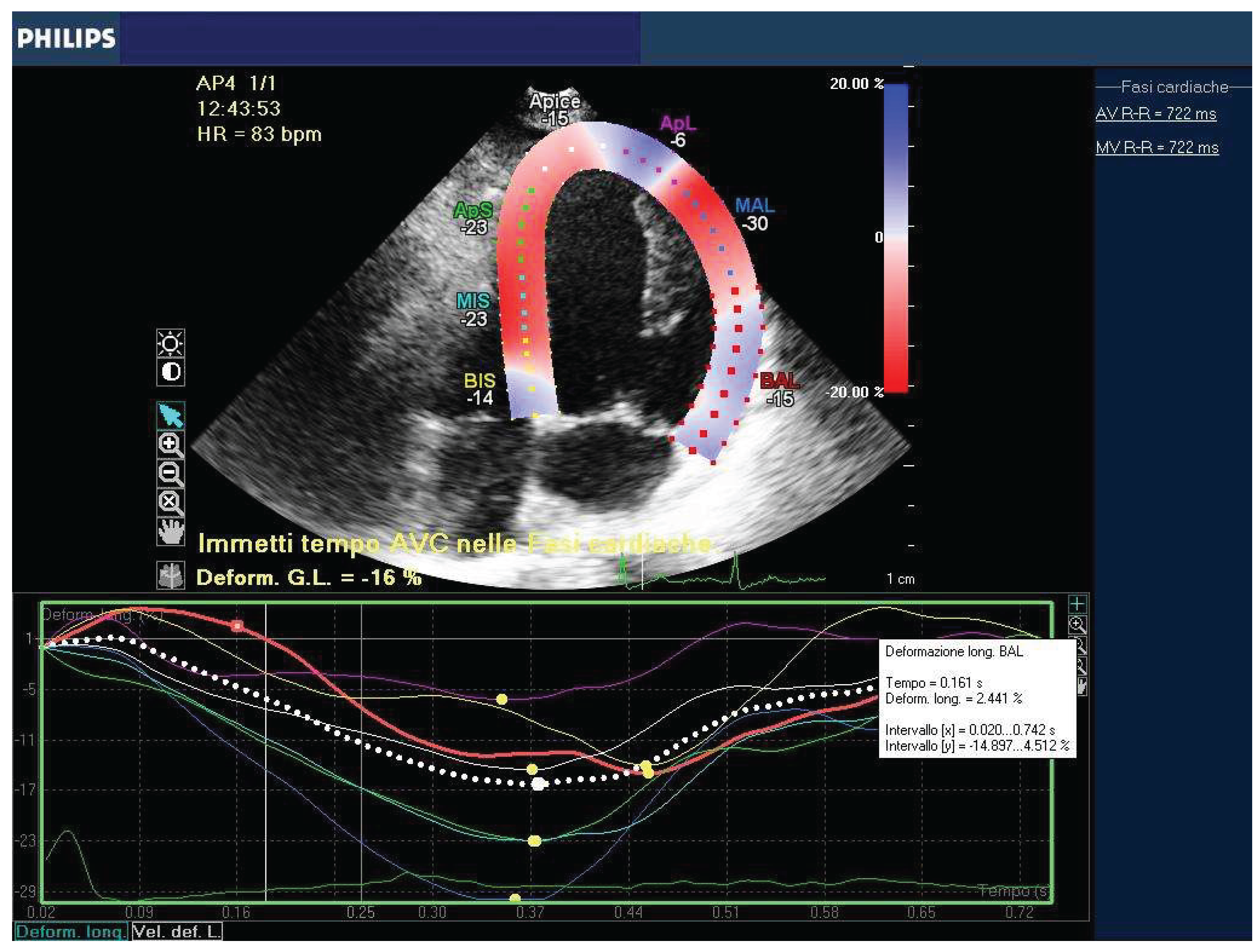
Figure 2.
Bull-eye of the Groups.
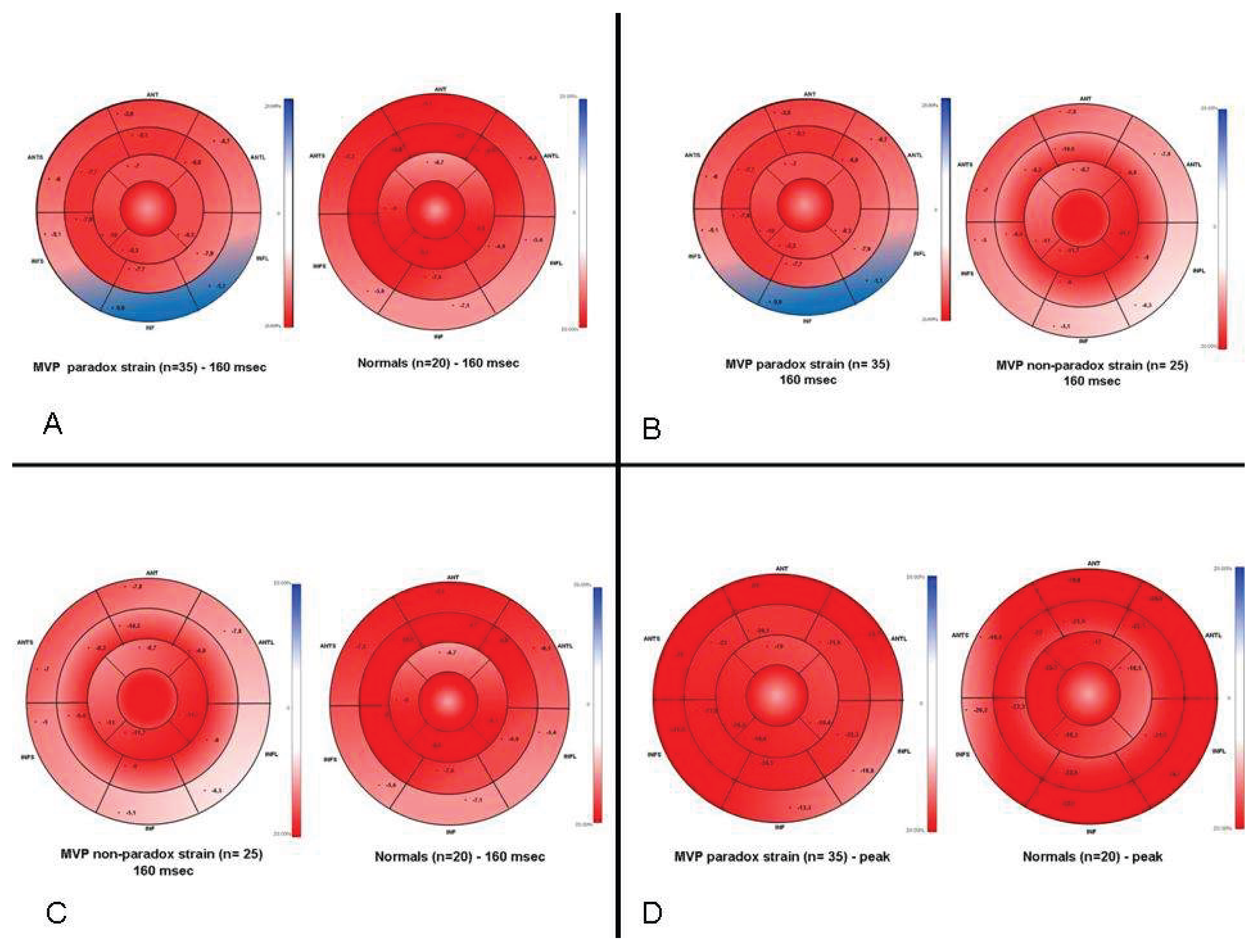
Figure 3.
STE at 160 msec in the Groups.
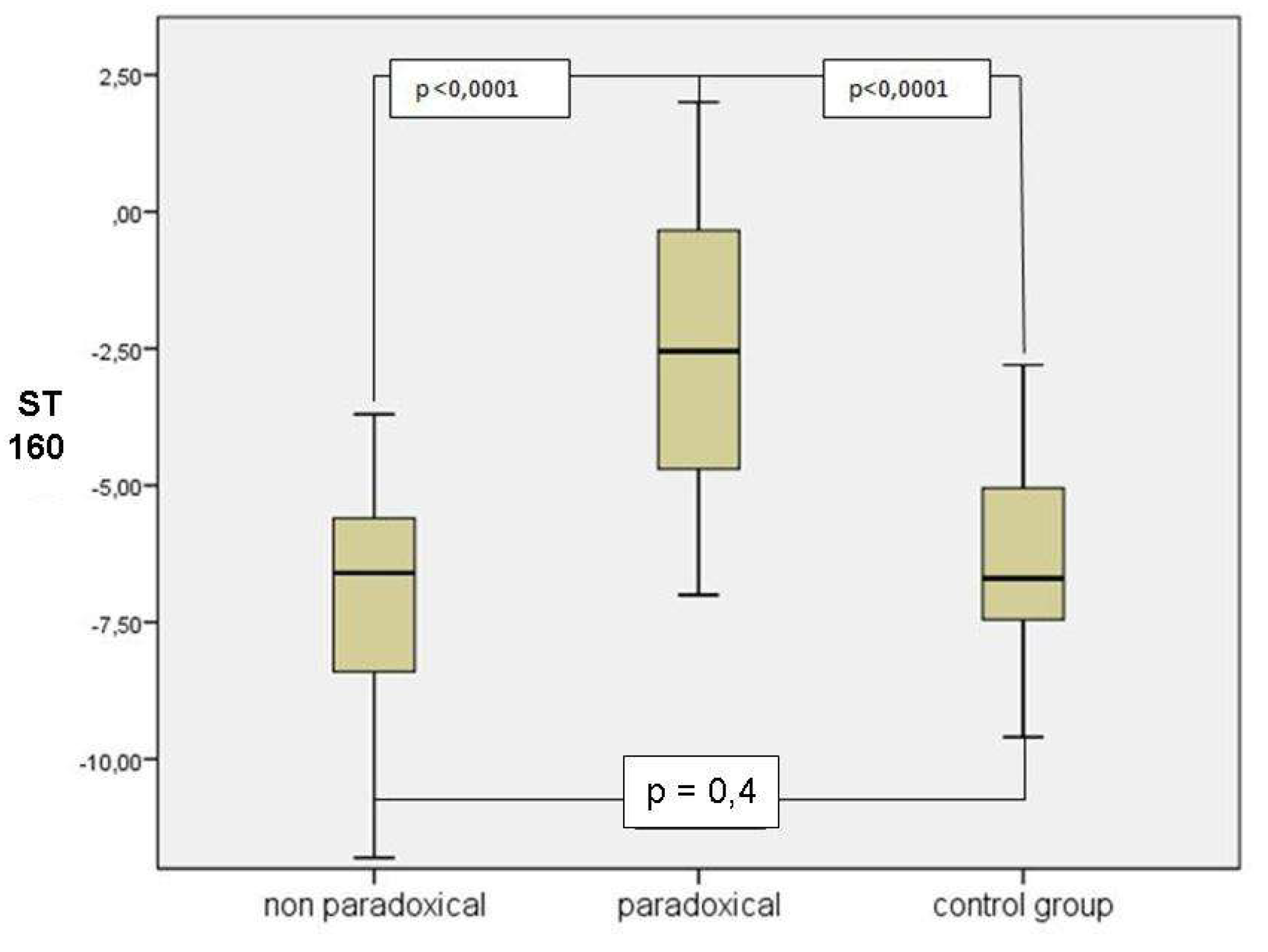
Figure 4.
Strain in the Groups at the peak.
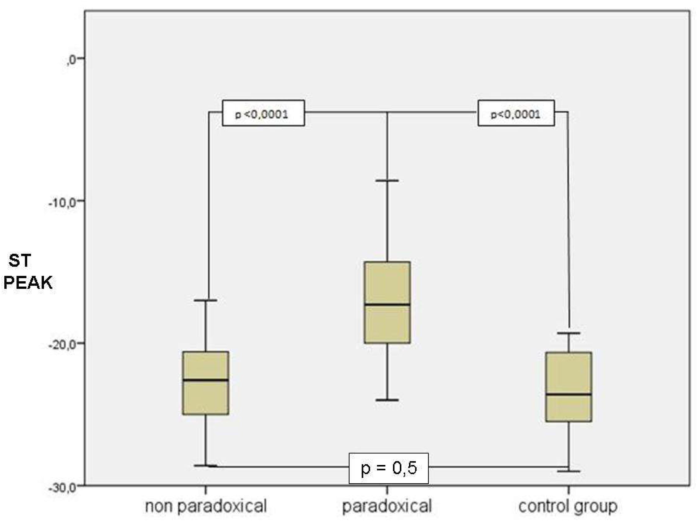

Table 1.
Clinical characteristics of MVP and control groups.
| MVP (n=60) | Control (n=20) | P value | |
| Males (%) | 27 (45) | 9 (45) | ns |
| Females (%) | 33 (55) | 11 (55) | ns |
| Age (years) | 45 ± 4.3 | 43 ± 3.6 | ns |
| LVEF (%) | 68 ± 7.3 | 65 ± 3.5 | ns |
| LA LAX (mm) | 40 ± 3.9 | 39 ± 2.8 | ns |
|
Prolapsing Leaflet (%) Posterior Anterior |
100 (60) 10 (6) |
00 | |
| BMI (Kg/m2) | 22.1 ± 1.54 | 22.1 ± 1.51 | ns |
| Heart Rate (beats/min) | 69 ± 5.8 | 71 ± 6 | ns |
|
SBP (mmHg) DBP (mmHg) |
115 ± 19 77 ± 4 |
112 ± 15 79 ± 4 |
ns ns |
Table 2.
Clinical characteristics of MVP Group 1 and Group 2.
| group 1 (n=35) | group 2 (n=25) | P | |
| Male (%) | 16 (55) | 11 (44) | Ns |
| Female (%) | 19 (55) | 14 (56) | Ns |
| Age (years) | 47 ± 2.8 | 42 ± 3.2 | Ns |
| LVEF (%) | 68 ± 6.7 | 65 ± 9.5 | Ns |
| LA LAX (mm) | 41 ± 3.7 | 39 ± 2.7 | Ns |
|
Prolapse (%) Posterior Anterior |
35 (100) 6 (17) |
25 (100)0 |
|
| MR grading (0-3) | 1 | 1 | Ns |
| BMI (Kg/m2) | 21.9 ± 1.4 | 22.4 ± 1.6 | Ns |
| Heart Rate (beats/minute) | 68.3 ± 5.9 | 70.3 ± 5.5 | Ns |
|
SBP (mmHg) DBP (mmHg) |
114.8 ± 18.4 78 ± 4 |
116 ± 21 75 ± 5.5 |
ns ns |
Table 3.
averaged Strain values of the basal segments (the “triad”) for each group.
| control group (20) | group 1 (35) | P | |
| ST at 160 msec | -6.3 ± 1.9 | -2.5 ± 2.3 | < 0.0001 |
| ST at peak | -23.4 ± 2.9 | -17.0 ± 3.6 | < 0.0001 |
| GLS | -20 ± 2 | -18.7 ± 2.4 | 0.01 |
| control group (20) | group 2 (25) | P | |
| ST at 160 msec | -6.3 ± 1.9 | -7.0 ± 2 | 0.4 |
| ST at peak | -23.4 ± 2.9 | -22.8 ± 3.1 | 0.5 |
| GLS | -20 ± 2 | -20 ± 2.5 | 0.9 |
| group 2 (25) | group 1 (35) | P | |
| ST at 160 msec | -7.0 ± 2 | -2.5 ± 2.3 | < 0.0001 |
| ST at peak | -22.8 ± 3.1 | -17.0 ± 3.6 | < 0.0001 |
| GLS | -20 ± 2.5 | -18.7 ± 2.4 | 0.01 |
Disclaimer/Publisher’s Note: The statements, opinions and data contained in all publications are solely those of the individual author(s) and contributor(s) and not of MDPI and/or the editor(s). MDPI and/or the editor(s) disclaim responsibility for any injury to people or property resulting from any ideas, methods, instructions or products referred to in the content. |
© 2026 by the authors. Licensee MDPI, Basel, Switzerland. This article is an open access article distributed under the terms and conditions of the Creative Commons Attribution (CC BY) license (http://creativecommons.org/licenses/by/4.0/).
Copyright: This open access article is published under a Creative Commons CC BY 4.0 license, which permit the free download, distribution, and reuse, provided that the author and preprint are cited in any reuse.