

Submitted:
03 August 2026
Posted:
03 August 2026
You are already at the latest version
Abstract
Silica nanoparticles (SiNPs) are common nanoparticles that are widely used in industrial and medical applications. Inhalation exposure to SiNPs is frequently inevitable in modern society. While the adverse effects of SiNPs on lung injury have been extensively documented, the intrinsic mechanisms underlying SiNPs-induced pulmonary inflammation remain incompletely understood. This study demonstrates that the stimulator of interferon genes (STING) plays an essential role in SiNPs-triggered lung inflammation. In vitro, SiNPs induced bone marrow derived macrophags (BMDMs) death and activated the STING pathway. Knockout the expression of STING alleviated SiNPs-induced inflammatory responses and subsequent cell death. Further mechanism investigations revealed that STING activation promotes nuclear translocation of NF-κB p65 and subsequent activation of the NF-κB pathway, ultimately driving the secretion of inflammatory factors. In vivo, SiNPs-induced NF-κB activation and inflammatory infiltration were significantly attenuated in STING-deficient (STING⁻/⁻) mice. These findings reveal that targeting the STING signaling pathway may represent a potential therapeutic strategy for mitigating lung inflammation caused by silica particles.
Keywords:
SiNPs
; cGAS-STING pathway
; NF-κB
; lung inflammation
1. Introduction
Natural silica exists in both crystalline and amorphous forms, and ranks among the most abundant compounds on the earth. Occupational exposure to crystalline silica has been unequivocally linked to the development of silicosis [1]. Although regulatory measures and exposure control strategies have been implemented, new cases of silicosis continue to emerge, partly due to non-traditional exposure sources such as silica nanoparticles (SiNPs) [2]. SiNPs represent a typical class of engineered nanomaterials, and are also one of the major forms of silica. At present, SiNPs have attracted considerable attention due to their large-scale production and extensive application. Based on their excellent properties, SiNPs are widely used in food processing, synthetic processes, medical diagnostics, and drug delivery, which substantially increases the risk of human exposure [3]. Although the toxicity of crystalline silica has been extensively studied, the toxicity of SiNPs remain largely unknown.
SiNPs can enter the human body through the respiratory tract, digestive tract, and skin, causing damage to the organism, and the lung is the most important and toxicologically affected target organ of SiNPs [3]. Existing studies have demonstrated that SiNPs are closely associated with various pulmonary diseases, including pneumonia, emphysema, pulmonary fibrosis, and even lung cancer, which can be life-threatening in severe cases [4]. However, further studies are still needed on the toxicity of SiNPs in both in vitro and in vivo. Alveolar macrophages, as key effector cells in the progression of silicosis, play a central role in initiating and sustaining pulmonary disease-related inflammation [5]. Upon inhalation, silica particles are phagocytosed and cleared by alveolar macrophages that migrate to deposition sites. However, due to the resistance of silica particles to degradation, persistent macrophage activation and continuous release of inflammatory mediators occur, ultimately leading to pulmonary inflammation and fibrotic responses [6].
STING (also known as TMEM173) is an immune adaptor protein that involved in regulating signal crosstalk across various physiological and pathological processes [7]. Although it is well-established that STING is transported from the endoplasmic reticulum (ER) to the Golgi apparatus upon recognition of DNA derivatives, emerging evidence indicates that STING can also be transported to different organelles, thereby determining its immune-dependent (type-I interferon and pro-inflammatory cytokine production) and/or immune-independent (autophagy activation, ER stress, cell death, and lipid metabolism) physiological functions [8]. Upon activation, STING binds and activates tank-binding kinase 1 (TBK1), leading to the aggregation and phosphorylation of interferon regulatory factor 3 (IRF3) [9]. Phosphorylated IRF3 translocates into the nucleus and promotes the transcription and synthesis of type-I interferons [10]. Additionally, STING activates the NF-κB signaling pathway, inducing the synthesis and release of pro-inflammatory cytokines such as IL-6 [11].
Studies have shown that the STING signaling pathway participates in regulating immune responses in various pulmonary inflammatory diseases [10,12]. It has been reported that macrophages produce sphingosine-1-phosphate (S1P) to suppress STING signaling, thereby alleviating inflammation-related vascular injury [13]. Furthermore, pharmacological inhibition of STING effectively reduces IFN-β production and associated tissue damage in mice [14]. Although some studies have addressed the pulmonary toxicity of SiNPs, the role of the STING signaling pathway in SiNPs-induced lung injury remains poorly understood.
In this current work, we aim to investigate the role of the STING signaling pathway in SiNPs-induced pulmonary inflammation through both in vivo and in vitro experiments, and to further investigate whether the NF-κB signaling pathway is involved in STING-mediated inflammatory responses triggered by SiNPs. Our findings demonstrate that knockout of STING suppresses NF-κB activation, thereby mitigating SiNPs-induced inflammatory responses in macrophages and subsequently ameliorating lung tissue injury and fibrosis in mice. These results suggest that SiNPs may induce pulmonary inflammation via the STING-NF-κB signaling axis.
2. Materials and Methods
2.1. Particle Preparation and Characterization
Prior to use, SiNPs (Sigma-Aldrich, USA) were acid washed in 1 M HCl at 100°C for 1 h to remove potential endotoxin contamination, washed three times with sterile water, and dried at 200°C. The actual size, shape and distribution of SiNPs were evaluated using transmission electron microscopy (TEM) (HT7700) and Image J software. Dynamic light scattering was performed with a Zetasizer (Malvern, UK) to determine the hydrodynamic size and zeta potential of SiNPs in dispersion media. Before being applied to cells or animals, SiNPs suspension were sonicated for 15 min to minimize aggregation.
2.2. Animal Maintenance and Exposure
Wild-type (WT) C57BL/6 mice and mice deficient for STING (STING-/-) were obtained from GemPharmatech (Jiangsu, China) and bred in Medical Laboratory Animal Center of Shandong Second Medical University. All animal experiments were approved by the Ethics Committee of the Laboratory Animal Administration of Shandong Second Medical University in Shandong Province under number 2023SDL231. For experiments, adult (male, 8-week-old, 20-22 g, n=6 per group) animals were freely provided with food and water under standard breeding conditions, with a 12 h reverse light/dark cycle and controlled temperature (22 ± 2℃).
Mice received a single intranasal instillation of either SiNPs saline suspension (1 mg/mouse) or saline vehicle. After 7 days, the mice were euthanized. The dosage and exposure duration were adopted from the previously published protocol that has been proven to cause acute lung injury [12]. The left lungs were fixed in 4% paraformaldehyde at least for 72 h, followed by paraffin embedding and subsequent histological analysis. The other parts of the lung tissue were stored at -80°C for western blot and real-time quantitative PCR (RT-qPCR) experiments.
2.3. BMDMs Extraction and Culture
BMDMs were generated as described previously with some modifications [12]. Briefly, C57BL/6 mice were sacrificed, and the femur and tibia bones were collected. Then bone marrow cells were obtained by flushing the bone marrow cavity with DMEM medium (Keygen Biotech, China), followed by filtration through a 70 μm cell filter. The cells were resuspended and cultured in DMEM medium supplemented with 10% FBS (Excell, China), 100 U/mL penicillin and 100 µg/mL streptomycin (Beyotime Biotechnology, China), and 30% (v/v) L929 conditioned medium as a source of M-CSF for 7 days differentiation period at 37°C in a humidified incubator containing 5% CO2. Following differentiation, the cells were washed and maintained in fresh medium for an additional 3 days prior to subsequent experiments.
2.4. Cell Viability and Lactate Dehydrogenase (LDH) Release Assay
For determination of the cell viability, a detection reagent composed of fresh culture medium containing 10% CCK-8 reagent was added to the wells and incubated for 1-4 h. The absorbance was measured at 450 nm using a microplate reader (Agilent BioTek, USA). Intracellular LDH release into the culture medium is an indicator of irreversible cell death due to damaged cell membranes. The release of LDH was measured using a commercial assay kit to evaluate the effect of SiNPs on cell membrane integrity. At the end of SiNPs exposure, cell medium was centrifuged to collect the supernatant. The LDH activity in the supernatant was then analyzed according to the manufacturer's instructions by measuring the absorbance at a wavelength of 450 nm. The amount of LDH released is expressed as the LDH activity (U/mL).
2.5. Western Blot Analysis
Protein extraction was performed as previously described [15]. The lung tissues and BMDMs were lysed with 1×RIPA lysis buffer [150 mM sodium chloride, 50 mM Tris-HCl (pH 7.4), 1% NP-40, 0.1% SDS, 0.5% sodium deoxycholate, 1×EDTA-free protease inhibitor cocktail (Selleck, USA), 1× phosphatase inhibitor cocktail (Selleck, USA)]. The lysates were centrifuged and the total protein concentration was quantified in the supernatant by a BCA assay kit (Dingguo Biotech, China). Proteins (30-50 µg per sample) were separated by SDS-polyacrylamide gel electrophoresis (SDS-PAGE) and then transferred onto nitrocellulose membranes. Membranes were blocked with 5% non-fat milk at room temperature for 2 h. Then the membranes were incubated with appropriate primary antibody overnight at 4°C on a shaker. The following primary antibodies were used: STING (#50494, Cell Signaling), cGAS (#31659, Cell Signaling), TBK1 (#3504, Cell Signaling), p-TBK1 (#5483, Cell Signaling), p-p65 (#bs-0982R, Bioss), p65 (#F0006, Selleck), Lamin B (#12987-1-AP, Proteintech), β-actin (#D110001, Sangon). After washing in 1×TBST three times, the membranes were further incubated with HRP-conjugated secondary antibodies (Sangon, China) for additional 1 h at room temperature. Protein bands were visualized using Super ECL Detection Reagent (Yeasen, China). Representative blots were chosen from at least three independent experiments and the proteins expression levels were calculated by Image J software. β-actin was used as a housekeeping gene.
2.6. RNA Extraction and RT-qPCR
Gene expression levels were measured by RT-qPCR. In short, total RNA was acquired from BMDMs or homogenized lung tissue using TRNzol Universal reagent (TIANGEN, China) according to the manufacturer’s protocol. The purified RNA was reverse transcribed into cDNA using Hifair® Ⅲ 1st Strand cDNA Synthesis SuperMix (Yeasen, China). RT-qPCR was performed on diluted cDNA with 2×SYBR Green qPCR Master Mix (Sellect, USA) to quantify the mRNA expression levels of different targeted proteins. Primers used for RT-qPCR are listed in Table S2. Relative gene expression levels from three independent experiments were calculated using the 2−△△Ct method and GAPDH was used as the internal control.
2.7. Nuclear and Cytoplasmic Protein Extraction
The BMDMs were washed with PBS for three times, then corresponding proteins were prepared using Nuclear Protein Extraction Kit (Solarbio, China) according to the manufacturer's protocol and our previous publication [16]. Briefly, 5×10⁶ cells were trypsinized, collected, centrifuged and washed with PBS. Then, cells were resuspended in the extraction solution for 20 min. After centrifugation at 2000 g for 5 min, the supernatant was transferred to another pre-cooled centrifuge tube, which was the cytoplasmic protein. The pellet was washed and then lysed to obtain nuclear protein. The protein concentrations were measured using the BCA method.
2.8. Immunofluorescence Assay of NF-κB p65 Nuclear Translocation
After exposure with SiNPs for 6 h, BMDMs were washed with PBS for three times and fixed with freshly prepared 4% paraformaldehyde for 20 min, permeabilized in 0.1% Triton X-100 for 10 min, following by blocking incubation in 3% BSA for 1 h. Then BMDMs were incubated with p65 antibody (#F0006, Selleck) overnight at 4°C, and then incubated with a secondary Cy3-conjugated antibody (#SA00009-2, Proteintech) for additional 1 h at room temperature. DAPI (MedChemExpress, USA) was incubated in a dark environment for 15 min. Finally, the images were obtained under a fluorescence microscope.
2.9. Histological Analysis
Histopathological analysis was performed following the standard laboratory procedures. Briefly, the embedded paraffin lung tissue blocks were sectioned into 4-μm-thick slices, which were then mounted on glass slides. hematoxylin and eosin (H&E) staining and Masson staining (Servicebio, China) were carried out according to the manufacturer’s instructions to evaluate pathological changes in lung tissue (inflammation and fibrosis). The images were captured using an optical microscope.
2.10. Immunohistochemistry Assay
Immunohistochemistry was used to detect pulmonary macrophage infiltration. Briefly, after dewaxing and antigen retrieval of paraffin sections, endogenous peroxidase was blocked with 3% hydrogen peroxide solution. Sections were blocked with 3% bovine serum albumin (BSA) (Sangon, China) for 1 h, then incubated with primary antibody at 4°C overnight. And then the sections were incubated with secondary antibody at 37 °C for 30 min. Diaminobenzidine (DAB) (Servicebio, China) was used as the chromogen, followed by hematoxylin counterstaining to visualize cell nuclei. The sections were imaged by optical microscope.
2.11. Statistical Analysis
The results are represented as the mean ± standard deviation (SD). Statistical analysis was performed using GraphPad Prism. Shapiro-Wilk test was applied for normality test. One-way ANOVA followed by Turkey’s multiple comparisons test was utilized in intergroup comparisons of more than two groups. p< 0.05 was considered statistically significant.
3. Results
3.1. Characterization of SiNPs
Prior to the toxicity investigation, the SiNPs were characterized. The morphology (Figure 1a) and actual size (Figure 1b) of SiNPs were measured by TEM. The chosen SiNPs were non-aggregated, near-spherical particles with size mainly concentrated in the range of 5-15 nm. Additionally, to assess the potential influence of the dispersion media on SiNPs, we subsequently measured the hydrodynamic sizes (Figure 1c) and zeta potentials (Figure 1d) of SiNPs in water, physiological saline, and DMEM complete culture medium. The results revealed that the hydrodynamic size of SiNPs in the dispersion media were larger than their original size. Zeta potential serves as a marker of colloidal stability, where a higher absolute value generally indicates greater resistance to aggregation. As shown in Figure 1d, SiNPs exhibited relatively favorable stability in water. However, their stability was somewhat compromised in saline solution and culture medium, indicating SiNPs were prone to aggregated.
3.2. SiNPs Activates STING Signaling Pathway
During the process of lung injury induced by silica particles, macrophages have the ability to engulf silica particles and secrete different cytokines to trigger multiple pulmonary stress responses [17]. Meanwhile, macrophages serve as typical host cells expressing STING [12]. Therefore, we selected BMDMs as the in vitro mode. Initially, we evaluated the cytotoxic effect of SiNPs on BMDMs. As shown in Figure 2a, SiNPs exposure resulted in a concentration-dependent decrease in the cell viability of BMDM, while concurrently promoting the release of intracellular LDH (Figure 2b). Western blot analysis demonstrated that SiNPs significantly upregulated the expressions of cGAS and STING, as well as the phosphorylated level of TBK1 in BMDMs (Figure 2c-2d). The mRNA expression levels of downstream target molecules in the STING signaling pathway, including interferon α4 (Ifna4), interferon β1 (Ifnb1), and C-X-C motif chemokine ligand 10 (Cxcl10), were also significantly increased (Figure 2e and Figures 2e and S1). These results indicate that the STING signaling pathway in BMDMs is activated after exposure to SiNPs.
3.3. SiNPs Exposure Causes an Inflammatory Response in BMDMs via STING
Previous studies have demonstrated SiNPs can trigger pulmonary inflammation [18], and the STING pathway has been shown to played an essential role as a mediator of inflammation in the settings of cellular stress and tissue injury [10]. Therefore, to investigate the inflammatory effects of SiNPs on BMDMs and the potential involvement of the STING pathway, we isolated BMDMs from STING-knockout (STING-/-) mice. We first examined the expression of Cxcl10, a downstream gene of STING. As shown in Figure 3a, SiNPs-induced Cxcl10 expression was significantly suppressed following STING knockout, confirming the effective blockade of the STING signaling pathway. IL-6, IL-1β, IL-8, et al. are known as the key inflammatory mediators [19]. The exposure of SiNPs caused the overexpression of the Il-6, Il-18 and Il-1β genes in BMDMs. Notably, genetic ablation of STING attenuated SiNPs-induced elevation in the expression levels of these inflammatory cytokines (Figure 3b-3d). Subsequent experiments further demonstrated STING deficiency effectively alleviated the cell death and LDH release caused by SiNPs (Figure 3e-3f). Collectively, these results indicate that Si NPs exposure triggers an inflammatory response in BMDMs through the STING pathway.
3.4. SiNPs Activates NF-κB via STING Signaling Pathway
We have confirmed that SiNPs induce BMDMs death and activate the STING pathway, leading to increased secretion of inflammatory cytokines. Previous studies have reported that STING activation can subsequently trigger the NF-κB pathway through TBK1 recruitment [20,21,22], and NF-κB serves as a key regulator of inflammatory cytokine expression [23]. Therefore, we next investigate whether SiNPs activate the NF-κB pathway and its potential relationship with STING signaling. To clarify SiNPs’ effect on NF-κB pathway, BMDMs were exposed to SiNPs in a time-dependent manner. Western blot analysis showed that phosphorylation of the NF-κB subunit p65 (p-p65) increased progressively with prolonged SiNPs exposure (Figure 4a), and quantitative analysis verified this time-dependent trend (Figure 4b). As nuclear translocation of p65 represents a hallmark of NF-κB activation, we next extracted nuclear and cytoplasmic proteins to analyze p65 distribution. With increased SiNPs treatment time, cytoplasmic p65 decreased while nuclear p65 increased (Figure 4c), indicating SiNPs effectively activate NF-κB pathway. Furthermore, STING deficiency significantly attenuated SiNPs-induced p65 phosphorylation (Figure 4d-4e) and markedly suppressed p65 nuclear translocation, as evidenced by immunofluorescence analysis (Figure 4f). In conclusion, our findings demonstrate that SiNPs activate the NF-κB pathway through STING signaling.
3.5. SiNPs Induces Inflammation Through the STING-NF-κB Signaling Pathway
We next investigate whether the induction of inflammatory factors by SiNPs via the STING pathway is dependent on the NF-κB pathway. BMDMs were treated with the NF-κB inhibitor JSH-23 prior to SiNPs exposure. Our results demonstrated that NF-κB pathway inhibition attenuated the SiNPs-induced decrease in cell viability (Figure 5a) and reduced LDH release (Figure 5b). Additionally, the gene expression levels of Il-6, Il-18 and Il-1β induced by SiNPs were significantly downregulated following JSH-23 treatment (Figure 5c). These findings collectively suggest that SiNPs induce inflammatory responses through the STING-NF-κB signaling pathway, providing crucial mechanistic insights into SiNPs-induced inflammatory responses.
3.6. SiNPs Exposure Causes Inflammatory Infiltration and Pulmonary Fibrosis in Mice
After confirming the cell damage caused by SiNPs in vitro, the lung injury was further evaluated in a murine model following exposure to SiNPs via intranasal instillation. The pulmonary pathological results of H&E staining (Figure 6a) revealed WT mice in SiNPs exposure group exhibited marked alveolar septa thickening, inflammatory cell infiltration into the alveoli, and alveolar structural disorder in lung tissues. Conversely, lung tissues from STING-/- mice treated with Si NPs displayed significant reduced inflammatory infiltration and relatively orderly alveolar structures. Masson staining results demonstrated extensive deposition of blue-stained collagen fibers in the lung tissues of WT mice in the Si NPs exposure group, indicating severe fibrosis; meanwhile, STING knockout alleviated collagen proliferation induced by SiNPs exposure, with improved tissue organization. Immunohistochemical staining for F4/80 (a macrophage marker) showed a large accumulation of macrophages in the lungs of WT mice exposed to SiNPs, whereas the number of macrophages was significantly decreased in STING deficient SiNPs group, suggesting that STING knockout inhibits Si NPs-induced macrophage infiltration. Moreover, the captured images visually demonstrated that STING knockout alleviated the swelling of lung tissues induced by SiNPs (Figure 6b).
In further in vivo mechanistic investigations, consistent with in vitro experiments, SiNPs exposure activated the STING-NF-κB signaling pathway in lungs, characterized by a significant increase in the phosphorylation levels of TBK1 and p65. The levels of p-TBK1 and p-p65 in STING-/- mice after SiNPs exposure are significantly reduced, indicating that STING deletion blocks the activation of the TBK1-NF-κB signaling pathway (Figure 6c-6e). We further detected the mRNA levels of inflammatory factors in lung tissues (Figure 6f), and the results showed that STING deficiency alleviates the significant upregulation of mRNA expression of Cxcl10, Il-6, Il-18, and Il-1β induced by SiNPs. In summary, SiNPs exposure induce inflammatory infiltration, fibrosis, and abnormal expression of multiple inflammatory factors in lung tissues by activating the STING-NF-κB signaling pathway (Figure 7).
4. Discussion
SiNPs are widely used in biomedical applications, cosmetics production, and general industrial processes, raising significant concerns regarding their biosafety [3,24,25]. The selected SiNPs primarily exhibit a particle size range of 5-15 nm. Particles smaller than 100 nm can be internalized by all cells types via endocytosis, having a better chance of transporting across biological barriers and interact with immune cells in blood, particularly the macrophages, resulting in an immune response [26,27]. After entering the blood, nanoparticles interact with biomolecules form a corona, such as a protein corona, which can mask or alter the surface properties of the nanoparticles and influence subsequent biological responses [28]. In this study, the measured hydrodynamic diameter of SiNPs in complete medium was larger than that in aqueous solution, suggesting the formation of a protein corona.
Although several studies have investigated SiNPs-induced pulmonary inflammation, the underlying molecular mechanisms have not completely understood [29]. Our study demonstrates that SiNPs induced lung inflammation and injury via STING-dependent activation of NF-κB signaling pathway. It is generally accepted that SiNPs exposure leads to pathological features including pulmonary fibrosis, mucus accumulation, and inflammatory cell infiltration [30,31,32]. Our experimental model exhibits characteristics consistent with those reported in prior investigations, confirming its suitability as a reliable tool for investigating SiNPs-induced pulmonary inflammation.
Recently, several excellent articles showed that the exposure of Silica induced the activation of STING pathway [12,17,33]. As an essential component of the innate immune system, STING pathway plays a key role in triggering inflammation, which promotes the expression of inflammatory cytokines [34]. After activating, STING recruits and phosphorylates TBK1 molecules to form a STING-TBK1 signalosome, resulting in initiation of IRF3 phosphorylation, and finally causing type-I interferon expression [35]. In addition, STING promotes the phosphorylation of IκB, an inhibitor of NF-κB, leading to IκB degradation by the ubiquitin-proteasome pathway and promoting NF-κB nuclear translocation [11]. NF-κB regulates the transcription of the inflammatory cytokines, which activation inducing a prominent pro-inflammatory cytokine response [36]. In our study, SiNPs exposure activates the STING pathway in BMDMs, promoting the nuclear translation of NF-κB and upregulating the gene expression of inflammatory cytokines. However, in STING-/- mice, SiNPs-induced inflammation was alleviated, indicating that the STING signaling pathway regulates SiNPs-induced inflammatory responses.
Previous studies have shown that TBK1 acts as a common upstream kinase mediating both IRF3 and NF-κB signaling [21]. However, emerging evidence indicates that IRF3 also plays a regulatory role in the NF-κB signaling pathway. There is evidence suggesting that IRF3, as an adaptor of immune signaling, engagements with STING at Ser358 and recruits TRAF6, resulting in the activation of NF-κB [37]. Fisher et al. reported that the activation of STING promotes the localization of the linear ubiquitin chain assembly complex E3 ligase (HOIP) to the LC3B-associated Golgi membrane, synthesizing M1-Ub chains to stimulate NF-κB pathway [38]. Zhang et al. found that NF-κB pathway also enhances STING signaling by inhibiting STING trafficking to lysosomes for degradation and causing ligand-independent STING autoactivation [23]. These finding highlight the extensive crosstalk between the STING and NF-κB signaling networks. Therefore, further studies are needed to elucidate the interaction and biological significance of the STING-NF-κB axis.
5. Conclusions
Our study elucidates the mechanisms of lung inflammation induced by small-sized silica nanoparticles. SiNPs exposure activates the STING-TBK1-NF-κB axis, triggering the release of inflammatory cytokines. Both knockdown of STING and pharmacological inhibition of NF-κB alleviated the SiNPs-induced inflammatory response in BMDMs. We also established a lung injury model induced by SiNPs in mice, in which STING knockout suppressed pulmonary fibrosis and inflammatory infiltration, as shown by H&E staining, Masson staining, and F4/80 immunohistochemistry. Further in vivo experiments confirmed that SiNPs induce lung inflammation via the STING-NF-κB signaling pathway. Our findings provide new insights and potential therapeutic targets for the treatment of silica particles-induced silicosis.
Supplementary Materials
The following supporting information can be downloaded at the website of this paper posted on Preprints.org, Figure S1: Ifna4, Ifnb1 and Cxcl10 transcripts measured by real-time PCR; Table S1: Summary of the physical properties of SiNPs; Table S2: Primer Sequences.
Author Contributions
Conceptualization, J.G. and Z.L.; methodology, F.W.; software, B.L.; validation, Y.J., H.D. and Z.L.; formal analysis, F.W.; investigation, J.G.; resources, Z.L.; data curation, J.G.; writing—original draft preparation, J.G.; writing—review and editing, Z.L.; visualization, D.W.; supervision, F.W.; project administration, Z.L.; funding acquisition, Z.L. All authors have read and agreed to the published version of the manuscript.
Funding
Please add: This research was funded by the Natural Science Foundation of Shandong Province (ZR2023MB025), Development Plan for Youth Innovation Teams in Higher Education Institutions in Shandong Province (2023KJ252), Medical and Health Science and Technology Development Project of Shandong Province (202402050463), College Students' Innovation and Entrepreneurship Training Program (X2026299), Research Startup Fund of Shandong Second Medical University (2022BKQ044).
Institutional Review Board Statement
The animal study protocol was approved by the Ethics Committee of the Laboratory Animal Administration of Shandong Second Medical University in Shandong Province (For mice, Approval Code: 2023SDL231, Approval Date: 21 March 2023).
Informed Consent Statement
Not applicable.
Data Availability Statement
The original contributions presented in this study are included in the article/Supplementary Materials. Further inquiries can be directed to the corresponding authors.
Conflicts of Interest
The authors declare no conflicts of interest.
References
- Song, M.Y.; Wang, J.X.; Sun, Y.L.; Han, Z.F.; Zhou, Y.T.; Liu, Y.; Fan, T.H.; Li, Z.G.; Qi, X.M.; Luo, Y.; et al. Tetrandrine alleviates silicosis by inhibiting canonical and non-canonical NLRP3 inflammasome activation in lung macrophages. Acta Pharmacol. Sin. 2022, 43, 1274–1284. [Google Scholar] [CrossRef] [PubMed]
- Requena-Mullor, M.; Alarcon-Rodriguez, R.; Parron-Carreno, T.; Martinez-Lopez, J.J.; Lozano-Paniagua, D.; Hernandez, A.F. Association between Crystalline Silica Dust Exposure and Silicosis Development in Artificial Stone Workers. Int. J. Env. Res. Public Health 2021, 18. [Google Scholar] [CrossRef] [PubMed]
- Liu, X.; Lu, B.; Fu, J.; Zhu, X.; Song, E.; Song, Y. Amorphous silica nanoparticles induce inflammation via activation of NLRP3 inflammasome and HMGB1/TLR4/MYD88/NF-kb signaling pathway in HUVEC cells. J. Hazard Mater. 2021, 404, 124050. [Google Scholar] [CrossRef] [PubMed]
- Ao, L.H.; Wei, Y.G.; Tian, H.R.; Zhao, H.; Li, J.; Ban, J.Q. Advances in the study of silica nanoparticles in lung diseases. Sci. Total Env. 2024, 912, 169352. [Google Scholar] [CrossRef] [PubMed]
- Li, S.; Xu, H.; Liu, S.; Hou, J.; Han, Y.; Li, C.; Li, Y.; Zheng, G.; Wei, Z.; Yang, F.; et al. Targeting Lp-PLA2 inhibits profibrotic monocyte-derived macrophages in silicosis through restoring cardiolipin-mediated mitophagy. Cell Mol. Immunol. 2025, 22, 776–790. [Google Scholar] [CrossRef] [PubMed]
- Zhou, Q.; Chang, M.; Guo, S.; Zhang, Y.; Qu, Q.; Zhou, Q.; Li, Z.; Yao, S. Honokiol ameliorates silica-induced lung fibrosis by inhibiting macrophage pyroptosis via modulating cGAS/STING signaling. Int. Immunopharmacol. 2025, 146, 113812. [Google Scholar] [CrossRef] [PubMed]
- Hopfner, K.P.; Hornung, V. Molecular mechanisms and cellular functions of cGAS-STING signalling. Nat. Rev. Mol. Cell Biol. 2020, 21, 501–521. [Google Scholar] [CrossRef] [PubMed]
- Zhang, R.; Kang, R.; Tang, D. STING1 in Different Organelles: Location Dictates Function. Front Immunol. 2022, 13, 842489. [Google Scholar] [CrossRef] [PubMed]
- Zhang, C.; Shang, G.; Gui, X.; Zhang, X.; Bai, X.C.; Chen, Z.J. Structural basis of STING binding with and phosphorylation by TBK1. Nature 2019, 567, 394–398. [Google Scholar] [CrossRef] [PubMed]
- Decout, A.; Katz, J.D.; Venkatraman, S.; Ablasser, A. The cGAS-STING pathway as a therapeutic target in inflammatory diseases. Nat. Rev. Immunol. 2021, 21, 548–569. [Google Scholar] [CrossRef] [PubMed]
- Chen, Q.; Sun, L.; Chen, Z.J. Regulation and function of the cGAS-STING pathway of cytosolic DNA sensing. Nat. Immunol. 2016, 17, 1142–1149. [Google Scholar] [CrossRef] [PubMed]
- Benmerzoug, S.; Rose, S.; Bounab, B.; Gosset, D.; Duneau, L.; Chenuet, P.; Mollet, L.; Le Bert, M.; Lambers, C.; Geleff, S.; et al. STING-dependent sensing of self-DNA drives silica-induced lung inflammation. Nat. Commun. 2018, 9, 5226. [Google Scholar] [CrossRef] [PubMed]
- Joshi, J.C.; Joshi, B.; Rochford, I.; Rayees, S.; Akhter, M.Z.; Baweja, S.; Chava, K.R.; Tauseef, M.; Abdelkarim, H.; Natarajan, V.; et al. SPHK2-Generated S1P in CD11b(+) Macrophages Blocks STING to Suppress the Inflammatory Function of Alveolar Macrophages. Cell Rep. 2020, 30, 4096–4109 e4095. [Google Scholar] [CrossRef] [PubMed]
- Gao, J.; Zheng, M.; Wu, X.; Zhang, H.; Su, H.; Dang, Y.; Ma, M.; Wang, F.; Xu, J.; Chen, L.; et al. CDK inhibitor Palbociclib targets STING to alleviate autoinflammation. EMBO Rep. 2022, 23, e53932. [Google Scholar] [CrossRef] [PubMed]
- Liu, Z.; Lv, X.; Xu, L.; Liu, X.; Zhu, X.; Song, E.; Song, Y. Zinc oxide nanoparticles effectively regulate autophagic cell death by activating autophagosome formation and interfering with their maturation. Part Fibre Toxicol. 2020, 17, 46. [Google Scholar] [CrossRef] [PubMed]
- Liu, Z.; Dong, W.; Yang, B.; Peng, L.; Xia, X.; Pu, L.; Zhang, N.; Song, E.; Song, Y. Tetrachlorobenzoquinone-Induced Nrf2 Confers Neuron-like PC12 Cells Resistance to Endoplasmic Reticulum Stress via Regulating Glutathione Synthesis and Protein Thiol Homeostasis. Chem. Res. Toxicol. 2018, 31, 1230–1239. [Google Scholar] [CrossRef] [PubMed]
- Ou, L.; Zhang, P.; Huang, Z.; Cheng, Y.; Miao, Q.; Niu, R.; Hu, Y.; Chen, Y. Targeting STING-mediated pro-inflammatory and pro-fibrotic effects of alveolar macrophages and fibroblasts blunts silicosis caused by silica particles. J. Hazard Mater. 2023, 458, 131907. [Google Scholar] [CrossRef] [PubMed]
- Wang, M.; Li, J.; Dong, S.; Cai, X.; Simaiti, A.; Yang, X.; Zhu, X.; Luo, J.; Jiang, L.H.; Du, B.; et al. Silica nanoparticles induce lung inflammation in mice via ROS/PARP/TRPM2 signaling-mediated lysosome impairment and autophagy dysfunction. Part Fibre Toxicol. 2020, 17, 23. [Google Scholar] [CrossRef] [PubMed]
- Guo, C.; Xia, Y.; Niu, P.; Jiang, L.; Duan, J.; Yu, Y.; Zhou, X.; Li, Y.; Sun, Z. Silica nanoparticles induce oxidative stress, inflammation, and endothelial dysfunction in vitro via activation of the MAPK/Nrf2 pathway and nuclear factor-kappaB signaling. Int. J. Nanomed. 2015, 10, 1463–1477. [Google Scholar] [CrossRef] [PubMed]
- Ouyang, Z.; Xu, J.; Liu, T.; Lin, S.; Sun, Y.; Huang, Y.; Zheng, Z.; Zeng, G.; Li, C.; Li, S.; et al. STING/TBK1 Regulates Inflammation in Macrophages and Titanium Particles-Induced Osteolysis. ACS Biomater. Sci. Eng. 2023, 9, 3273–3284. [Google Scholar] [CrossRef] [PubMed]
- Balka, K.R.; Louis, C.; Saunders, T.L.; Smith, A.M.; Calleja, D.J.; D'Silva, D.B.; Moghaddas, F.; Tailler, M.; Lawlor, K.E.; Zhan, Y.; et al. TBK1 and IKKepsilon Act Redundantly to Mediate STING-Induced NF-kappaB Responses in Myeloid Cells. Cell Rep. 2020, 31, 107492. [Google Scholar] [CrossRef] [PubMed]
- Bai, J.; Liu, F. The cGAS-cGAMP-STING Pathway: A Molecular Link Between Immunity and Metabolism. Diabetes 2019, 68, 1099–1108. [Google Scholar] [CrossRef] [PubMed]
- Zhang, L.; Wei, X.; Wang, Z.; Liu, P.; Hou, Y.; Xu, Y.; Su, H.; Koci, M.D.; Yin, H.; Zhang, C. NF-kappaB activation enhances STING signaling by altering microtubule-mediated STING trafficking. Cell Rep. 2023, 42, 112185. [Google Scholar] [CrossRef] [PubMed]
- Vance, M.E.; Kuiken, T.; Vejerano, E.P.; McGinnis, S.P.; Hochella, M.F., Jr.; Rejeski, D.; Hull, M.S. Nanotechnology in the real world: Redeveloping the nanomaterial consumer products inventory. Beilstein J. Nanotechnol. 2015, 6, 1769–1780. [Google Scholar] [CrossRef] [PubMed]
- Sun, J.; Zhou, Y.Q.; Xu, B.Y.; Li, J.Y.; Zhang, L.Q.; Li, D.Y.; Zhang, S.; Wu, J.Y.; Gao, S.J.; Ye, D.W.; et al. STING/NF-kappaB/IL-6-Mediated Inflammation in Microglia Contributes to Spared Nerve Injury (SNI)-Induced Pain Initiation. J. Neuroimmune Pharmacol. 2022, 17, 453–469. [Google Scholar] [CrossRef] [PubMed]
- Zhang, Y.; Chen, X.; Zhao, B.; Wu, H.; Yuan, L.; Zhang, H.; Dai, W.; He, B.; Xing, G.; Zhang, Q.; et al. Biosafety study and mechanism comparison on two types of silica with different nanostructures. Toxicol. Res. (Camb) 2017, 6, 487–498. [Google Scholar] [CrossRef] [PubMed]
- Croissant, J.G.; Fatieiev, Y.; Almalik, A.; Khashab, N.M. Mesoporous Silica and Organosilica Nanoparticles: Physical Chemistry, Biosafety, Delivery Strategies, and Biomedical Applications. Adv. Healthc. Mater. 2018, 7. [Google Scholar] [CrossRef] [PubMed]
- Zhang, H.; Peng, J.; Li, X.; Liu, S.; Hu, Z.; Xu, G.; Wu, R. A nano-bio interfacial protein corona on silica nanoparticle. Colloids Surf. B Biointerfaces 2018, 167, 220–228. [Google Scholar] [CrossRef] [PubMed]
- Adamcakova, J.; Mokra, D. New Insights into Pathomechanisms and Treatment Possibilities for Lung Silicosis. Int. J. Mol. Sci. 2021, 22. [Google Scholar] [CrossRef] [PubMed]
- Washimkar, K.R.; Kulkarni, C.; Tomar, M.S.; Verma, S.; Bhatt, D.; Verma, S.; Reddy, D.V.S.; Moharana, B.; Misra, A.; Bawankule, D.U.; et al. Metabolic dysregulation and temporal dynamics of NF-kappaB-p65/NLRP3, TXNIP, endoplasmic reticulum and mitochondrial stress in silica-induced pulmonary fibrosis in rats. J. Hazard Mater. 2025, 495, 138791. [Google Scholar] [CrossRef] [PubMed]
- Zhou, H.; Zhang, Q.; Liu, C.; Fan, J.; Huang, W.; Li, N.; Yang, M.; Wang, H.; Xie, W.; Kong, H. NLRP3 inflammasome mediates abnormal epithelial regeneration and distal lung remodeling in silica-induced lung fibrosis. Int. J. Mol. Med. 2024, 53. [Google Scholar] [CrossRef] [PubMed]
- Hasegawa, Y.; Franks, J.M.; Tanaka, Y.; Uehara, Y.; Read, D.F.; Williams, C.; Srivatsan, S.; Pitstick, L.B.; Nikolaidis, N.M.; Shaver, C.M.; et al. Pulmonary osteoclast-like cells in silica induced pulmonary fibrosis. Sci. Adv. 2024, 10, eadl4913. [Google Scholar] [CrossRef] [PubMed]
- Han, D.; Xu, T.; Lyu, X.; Li, K.; Xu, S. Smaller-Sized Silica Nanoparticles Exacerbated Cardiomyocyte Pyroptosis by Impairing Mitophagy to Activate mtDNA-cGAS-STING Signaling. J. Agric. Food Chem. 2025, 73, 9359–9369. [Google Scholar] [CrossRef] [PubMed]
- Zhong, S.; Zhou, Q.; Yang, J.; Zhang, Z.; Zhang, X.; Liu, J.; Chang, X.; Wang, H. Relationship between the cGAS-STING and NF-kappaB pathways-role in neurotoxicity. BioMed Pharmacother. 2024, 175, 116698. [Google Scholar] [CrossRef] [PubMed]
- Zhang, K.; Huang, Q.; Li, X.; Zhao, Z.; Hong, C.; Sun, Z.; Deng, B.; Li, C.; Zhang, J.; Wang, S. The cGAS-STING pathway in viral infections: a promising link between inflammation, oxidative stress and autophagy. Front Immunol. 2024, 15, 1352479. [Google Scholar] [CrossRef] [PubMed]
- Zhou, J.; Zhuang, Z.; Li, J.; Feng, Z. Significance of the cGAS-STING Pathway in Health and Disease. Int. J. Mol. Sci. 2023, 24. [Google Scholar] [CrossRef] [PubMed]
- Zhang, B.C.; Pedersen, A.; Reinert, L.S.; Li, Y.; Narita, R.; Idorn, M.; Hu, L.; Skouboe, M.K.; Li, S.; Maimaitili, M.; et al. STING signals to NF-kappaB from late endolysosomal compartments using IRF3 as an adaptor. Nat. Immunol. 2025, 26, 1916–1930. [Google Scholar] [CrossRef] [PubMed]
- Fischer, T.D.; Bunker, E.N.; Zhu, P.P.; Le Guerroue, F.; Hadjian, M.; Dominguez-Martin, E.; Scavone, F.; Cohen, R.; Yao, T.; Wang, Y.; et al. STING induces HOIP-mediated synthesis of M1 ubiquitin chains to stimulate NF-kappaB signaling. EMBO J. 2025, 44, 141–165. [Google Scholar] [CrossRef] [PubMed]
Figure 1.
Characterization of SiNPs. a. TEM image of SiNPs. Scale bar: 50 nm. b. Diameter distribution of SiNPs taken by TEM. SiNPs hydrated particle size (c) and Zeta potential (d) of SiNPs in water, saline or medium containing 10% FBS.
Figure 1.
Characterization of SiNPs. a. TEM image of SiNPs. Scale bar: 50 nm. b. Diameter distribution of SiNPs taken by TEM. SiNPs hydrated particle size (c) and Zeta potential (d) of SiNPs in water, saline or medium containing 10% FBS.
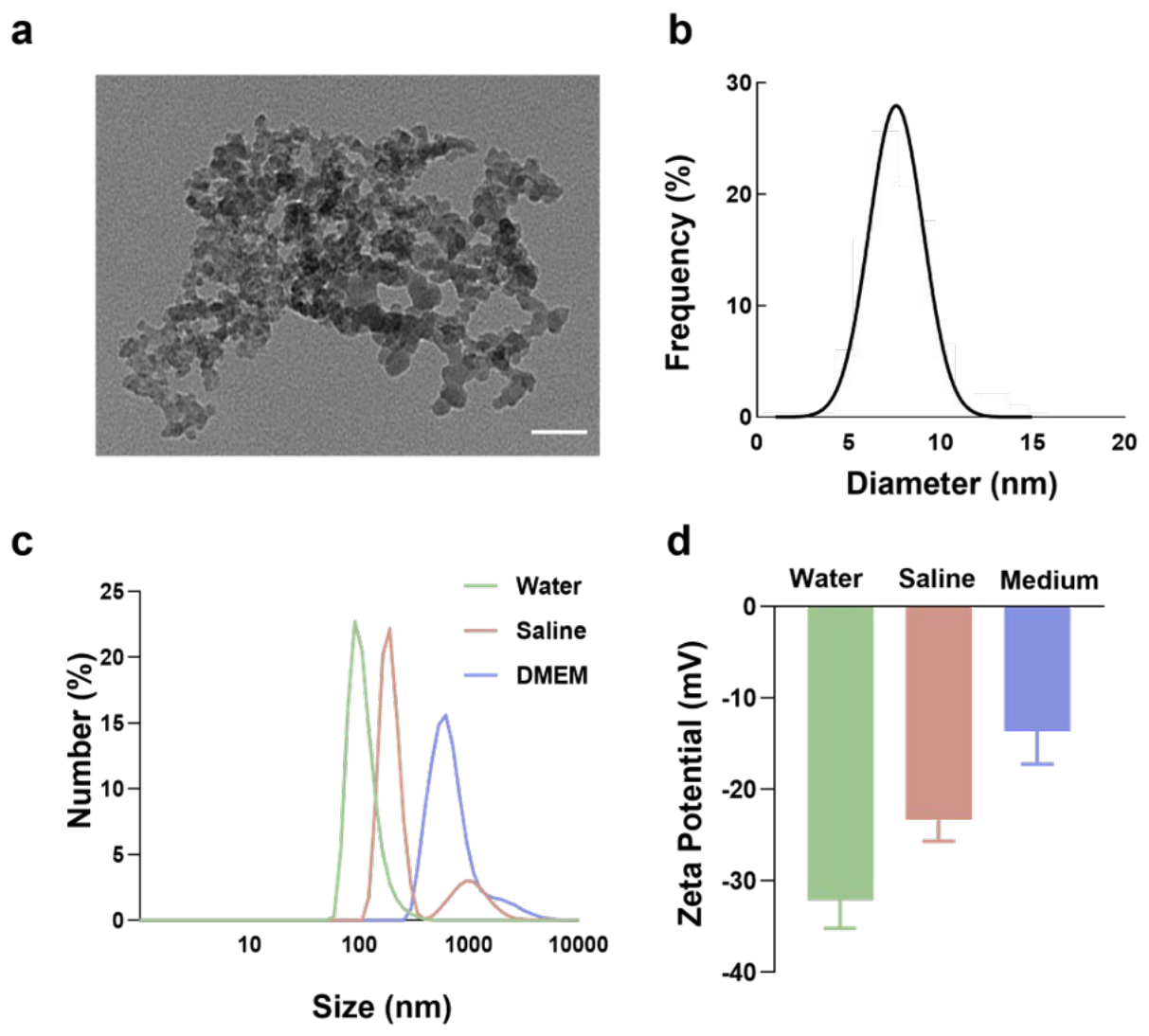
Figure 2.
Activation of the STING signaling pathway by SiNPs in BMDMs. Cell viability (a) and LDH release (b) of BMDMs were measured after treatment with different concentrations of SiNPs for 12 h. LDH release in the supernatant was indicated BMDMs injury. c. Immunoblots of cGAS, STING, p-TBK1 and TBK1 in BMDMs with β-actin as a reference. BMDMs were exposured to 25 μg/mL SiNPs for the indicated durations (0-12 h). d. Quantitative analysis of STING, cGAS and p-TBK1/TBK1 by Immunoblots. e. Time-dependent transcriptional induction of Ifna4, Ifnb1, and Cxcl10 measured by RT-qPCR after SiNPs exposure for 6 h. Data represent fold-changes relative to the untreated control, normalized to GAPDH (mean ± SD). *p < 0.05, **p < 0.01, ***p < 0.001 compared with the control group.
Figure 2.
Activation of the STING signaling pathway by SiNPs in BMDMs. Cell viability (a) and LDH release (b) of BMDMs were measured after treatment with different concentrations of SiNPs for 12 h. LDH release in the supernatant was indicated BMDMs injury. c. Immunoblots of cGAS, STING, p-TBK1 and TBK1 in BMDMs with β-actin as a reference. BMDMs were exposured to 25 μg/mL SiNPs for the indicated durations (0-12 h). d. Quantitative analysis of STING, cGAS and p-TBK1/TBK1 by Immunoblots. e. Time-dependent transcriptional induction of Ifna4, Ifnb1, and Cxcl10 measured by RT-qPCR after SiNPs exposure for 6 h. Data represent fold-changes relative to the untreated control, normalized to GAPDH (mean ± SD). *p < 0.05, **p < 0.01, ***p < 0.001 compared with the control group.
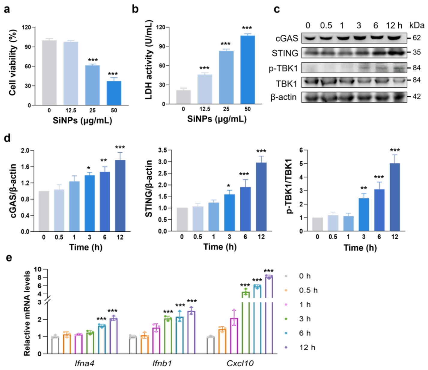
Figure 3.
STING-dependent inflammatory activation in BMDMs following SiNPs exposure. a-d. BMDMs from wild-type (WT) or STING-deficient (STING-/-) mice were unstimulated or stimulated with SiNPs (25 µg/mL) for 6 h. Cxcl10, Il-6, Il-18 and Il-1β transcripts were measured by RT-qPCR. Data represent fold-changes relative to the untreated control, normalized to GAPDH. Cell viability (e) and LDH release (f) of WT and STING-/- BMDMs were measured after 25 μg/mL SiNPs exposure for 12 h. Data are expressed as mean ± SD. ***p < 0.001 compared with the WT control group. ##p < 0.01, ###p < 0.001.
Figure 3.
STING-dependent inflammatory activation in BMDMs following SiNPs exposure. a-d. BMDMs from wild-type (WT) or STING-deficient (STING-/-) mice were unstimulated or stimulated with SiNPs (25 µg/mL) for 6 h. Cxcl10, Il-6, Il-18 and Il-1β transcripts were measured by RT-qPCR. Data represent fold-changes relative to the untreated control, normalized to GAPDH. Cell viability (e) and LDH release (f) of WT and STING-/- BMDMs were measured after 25 μg/mL SiNPs exposure for 12 h. Data are expressed as mean ± SD. ***p < 0.001 compared with the WT control group. ##p < 0.01, ###p < 0.001.
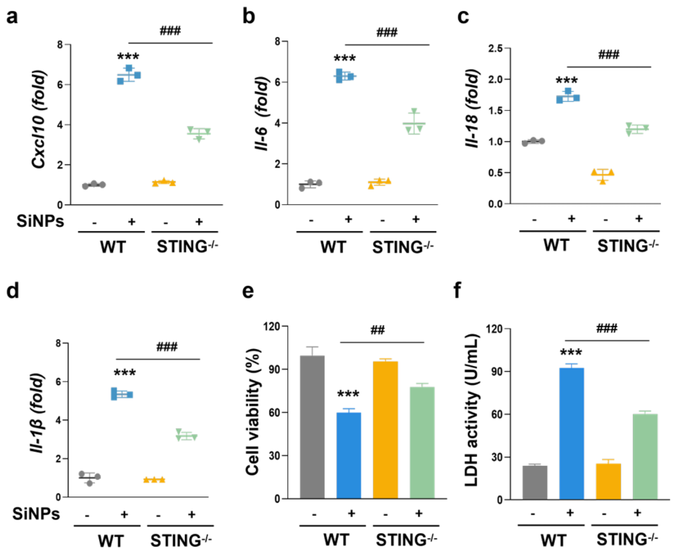
Figure 4.
SiNPs activates NF-κB p65 via STING signaling pathway. a. Immunoblots of p-p65 and p65 in BMDMs with β-actin as a reference. BMDMs were exposed to SiNPs (25 μg/mL) for the indicated times. b. Quantitative analysis of p-p65/p65 by Immunoblots. c. Western blot for NF-κB p65 expression in cytoplasmic and nuclear fractions of BMDMs exposed to SiNPs. d. Immunoblots of p-TBK1, TBK1, p65 and p-p65 in wild-type (WT) and STING-/- BMDMs with β-actin as a reference. BMDMs were exposured to 25 μg/mL SiNPs for 12 h. e. Quantitative analysis of p-p65/p65 and p-TBK1/TBK1 by Immunoblots. f. Immunofluorescence staining of p65 (red) and nuclei (blue) were detected, and merged images indicated the distribution of p65 after SiNPs exposure for 6 h. Scale bar: 20 μm. Data are expressed as mean ± SD. **p < 0.01, ***p < 0.001 compared with the WT control group. #p < 0.05, ###p < 0.001.
Figure 4.
SiNPs activates NF-κB p65 via STING signaling pathway. a. Immunoblots of p-p65 and p65 in BMDMs with β-actin as a reference. BMDMs were exposed to SiNPs (25 μg/mL) for the indicated times. b. Quantitative analysis of p-p65/p65 by Immunoblots. c. Western blot for NF-κB p65 expression in cytoplasmic and nuclear fractions of BMDMs exposed to SiNPs. d. Immunoblots of p-TBK1, TBK1, p65 and p-p65 in wild-type (WT) and STING-/- BMDMs with β-actin as a reference. BMDMs were exposured to 25 μg/mL SiNPs for 12 h. e. Quantitative analysis of p-p65/p65 and p-TBK1/TBK1 by Immunoblots. f. Immunofluorescence staining of p65 (red) and nuclei (blue) were detected, and merged images indicated the distribution of p65 after SiNPs exposure for 6 h. Scale bar: 20 μm. Data are expressed as mean ± SD. **p < 0.01, ***p < 0.001 compared with the WT control group. #p < 0.05, ###p < 0.001.
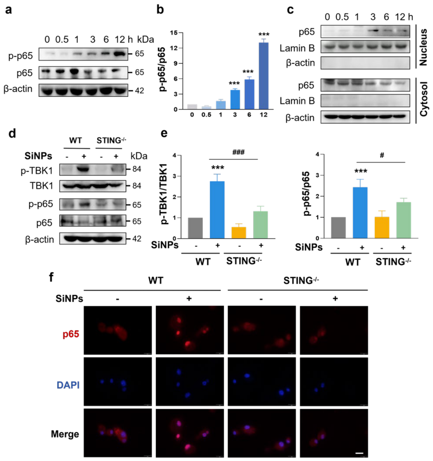
Figure 5.
SiNPs trigger inflammatory responses via STING-NF-κB p65 signaling axis. Cell viability (a) and LDH release (b) of BMDMs were determined following exposure to 25 μg/mL SiNPs for 12 h with or without JSH-23 (10 μM, NF-κB transcriptional activity inhibitor). c. Effect of the NF-κB inhibitor on SiNPs-induced Il-6, Il-18 and Il-1β mRNA expression. BMDMs were pre-treated with or without JSH-23 for 1 h followed by SiNPs exposure for 6 h. Data are expressed as mean ± SD. **p < 0.01, ***p < 0.001 compared with the control group. #p < 0.05, ##p < 0.01, ###p < 0.001.
Figure 5.
SiNPs trigger inflammatory responses via STING-NF-κB p65 signaling axis. Cell viability (a) and LDH release (b) of BMDMs were determined following exposure to 25 μg/mL SiNPs for 12 h with or without JSH-23 (10 μM, NF-κB transcriptional activity inhibitor). c. Effect of the NF-κB inhibitor on SiNPs-induced Il-6, Il-18 and Il-1β mRNA expression. BMDMs were pre-treated with or without JSH-23 for 1 h followed by SiNPs exposure for 6 h. Data are expressed as mean ± SD. **p < 0.01, ***p < 0.001 compared with the control group. #p < 0.05, ##p < 0.01, ###p < 0.001.
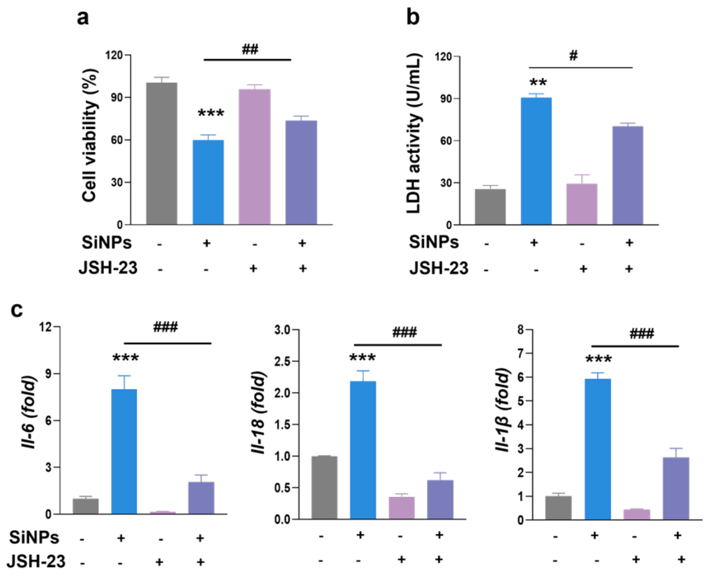
Figure 6.
STING-dependent pulmonary inflammatory infiltration triggered by SiNPs in mice. a. H&E, Masson and F4/80 representative images of lungs following SiNPs exposure in WT or STING-/- mice. Scale bar: 100 µm; b. Morphological alterations in pulmonary tissues across experimental groups. c. Western blot analysis of p-TBK1, TBK1, p-p65 and p65 expression in lungs of WT and STING-/- mice with β-actin as a reference. d,e. Quantitative analysis of p-TBK1/TBK1 and p-p65/p65 by western blot. f. Cxcl10, Il-6, Il-18 and Il-1β mRNA expression in lungs of WT and STING-/- mice. Data represent fold-changes relative to the untreated control, normalized to GAPDH. Data are expressed as mean ± SD. ***p < 0.001 compared to the WT control group. ##p < 0.01, ###p < 0.001.
Figure 6.
STING-dependent pulmonary inflammatory infiltration triggered by SiNPs in mice. a. H&E, Masson and F4/80 representative images of lungs following SiNPs exposure in WT or STING-/- mice. Scale bar: 100 µm; b. Morphological alterations in pulmonary tissues across experimental groups. c. Western blot analysis of p-TBK1, TBK1, p-p65 and p65 expression in lungs of WT and STING-/- mice with β-actin as a reference. d,e. Quantitative analysis of p-TBK1/TBK1 and p-p65/p65 by western blot. f. Cxcl10, Il-6, Il-18 and Il-1β mRNA expression in lungs of WT and STING-/- mice. Data represent fold-changes relative to the untreated control, normalized to GAPDH. Data are expressed as mean ± SD. ***p < 0.001 compared to the WT control group. ##p < 0.01, ###p < 0.001.
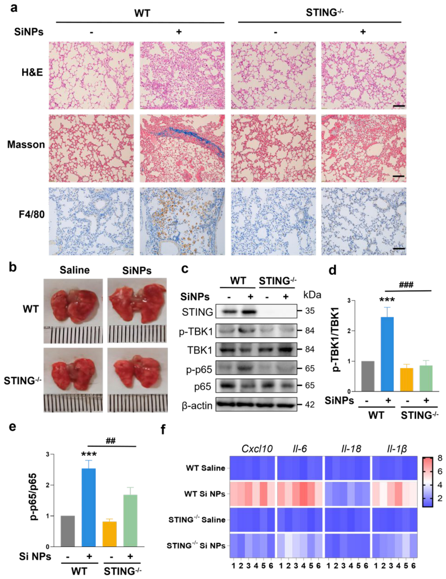
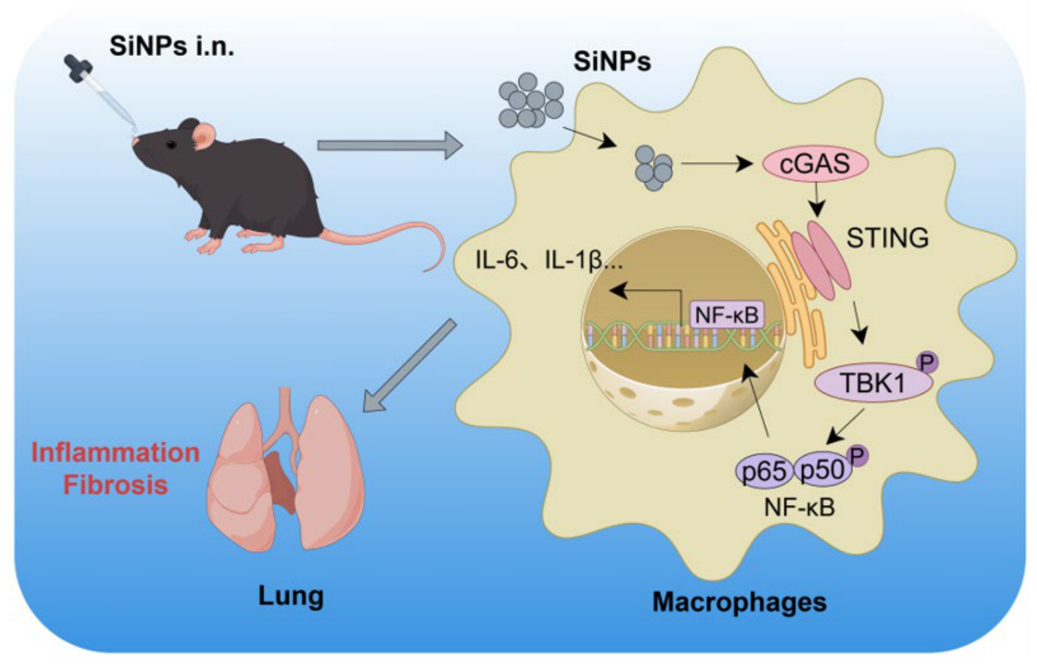
Figure 7.
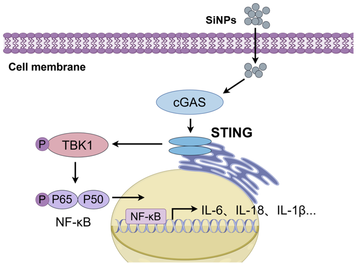
Schematic illustration that SiNPs induce pulmonary inflammation and fibrosis by activating STING-NF-κB signaling axis. SiNPs are inhaled and taken up by pulmonary macrophages. This triggers the activation of the STING pathway, which subsequently promotes the phosphorylation of TBK1 and NF-κB p65. The activated NF-κB then translocates into the nucleus to drive the expression of pro-inflammatory cytokines, ultimately leading to lung inflammation and fibrosis. The figure was drawn by Figdraw (www.figdraw.com).
Figure 7.
Schematic illustration that SiNPs induce pulmonary inflammation and fibrosis by activating STING-NF-κB signaling axis. SiNPs are inhaled and taken up by pulmonary macrophages. This triggers the activation of the STING pathway, which subsequently promotes the phosphorylation of TBK1 and NF-κB p65. The activated NF-κB then translocates into the nucleus to drive the expression of pro-inflammatory cytokines, ultimately leading to lung inflammation and fibrosis. The figure was drawn by Figdraw (www.figdraw.com).
Disclaimer/Publisher’s Note: The statements, opinions and data contained in all publications are solely those of the individual author(s) and contributor(s) and not of MDPI and/or the editor(s). MDPI and/or the editor(s) disclaim responsibility for any injury to people or property resulting from any ideas, methods, instructions or products referred to in the content. |
© 2026 by the authors. Licensee MDPI, Basel, Switzerland. This article is an open access article distributed under the terms and conditions of the Creative Commons Attribution (CC BY) license (http://creativecommons.org/licenses/by/4.0/).
Copyright: This open access article is published under a Creative Commons CC BY 4.0 license, which permit the free download, distribution, and reuse, provided that the author and preprint are cited in any reuse.


