

Submitted:
02 August 2026
Posted:
03 August 2026
You are already at the latest version
Abstract
Background: Acute kidney injury (AKI) affects 12-40% of critically ill newborns which in-creases mortality risk significantly. Current diagnosis relies on serum creatinine, which rises only after substantial renal damage. Early biomarkers enabling timely intervention are urgently needed for this vulnerable population. Serum cystatin C (sCysC) is a candi-date since it is constantly produced by all nucleated cells. Aim: To investigate sCysC diag-nostic performance for early neonatal AKI detection. Methods: This systematic review fol-lowed PRISMA-DTA guidelines and is registered with PROSPERO (CRD420261302574). We searched four major databases for studies measuring sCysC in neonates (0-28 days) using validated assays, with creatinine-based AKI definitions as the reference standard. Studies were categorized by time-related design as either concurrent diagnostic or early predictive accuracy. The risk of bias was assessed using the QUADAS-2. Results: Fifteen studies involving 53,751 neonates were included. sCysC showed strong early predictive accuracy (AUC 0.670-1.000) and identified AKI 24 hours to 4 days earlier than serum cre-atinine. In the largest cohort (52,333 neonates), sCysC achieved 70% sensitivity and 65% specificity for early AKI detection. Other diagnostic studies reported 85-89% sensitivity and 62-75% specificity (AUC 0.844-0.849). Optimal sCysC cut-offs ranged from 1.25 to 2.87 mg/L. Fourteen studies (93%) had a high risk of bias in at least one domain. Conclusion: Serum cystatin C allows earlier detection of neonatal AKI, offering a 1-4-day advantage over current methods and supporting timely intervention. Implementing standardized as-says and population-specific thresholds is essential for clinical use. This time advantage marks a shift toward preventive neonatal nephrology.
Keywords:
acute kidney injury
; serum cystatin C
; diagnostic accuracy
; neonate
; systematic review
1. Introduction
Acute kidney injury is an important complication among the neonatal population inside the neonatal intensive care unit (NICU). It is a significant cause of morbidity and mortality, therefore early detection is essential for timely intervention [1,2,3]. Nowadays, the assessment of AKI in neonates is challenging since the diagnosis relies primarily on traditional biomarkers, which include serum creatinine (sCr) levels, and urine output (oliguria/anuria) [4,5]. Currently, neonatal AKI is classified using standardized systems such as the neonatal-modified KDIGO (Kidney Disease: Improving Global Outcomes), AKIN (Acute Kidney Injury Network), and pRIFLE (pediatric Risk, Injury, Failure, Loss, End-Stage Renal Disease). [6,7] These systems define and stage AKI severity based on the magnitude of sCr increase from baseline and/or the duration of diminished urine output [6,7,8,9].
However, the traditional biomarkers are limited by multiple disadvantages such as:
- -
- maternal influence: in the first 24 to 72 hours of life, neonatal sCr represents maternal renal function and not the infants’, as creatinine freely crosses the placenta;
- -
- gestational age dependence: sCr levels decline and stabilize at varying rates over the first few weeks of life, meaning its baseline value is highly affected by gestational age (GA);
- -
- delayed detection: sCr is a late AKI marker as it measures function rather than actual tissue damage; sCr often does not rise until 48 to 72 hours after the renal injury, typically its levels rise only after almost half of the nephrons have already been compromised;
- -
Recent research has focused on serum cystatin C (sCysC) as a potential early biomarker for AKI in neonates. Serum Cystatin C is a 13.3 kDa cationic protein produced at a constant rate by all nucleated cells. Due to its small size, sCysC is freely filtered through the glomerular membrane and is subsequently completely reabsorbed and catabolized by the proximal renal tubules, without undergoing tubular secretion [13].
Serum Cystatin C has the following advantages over other biomarkers: it is minimally affected by muscle mass and maternal renal function, it is not significantly influenced by gender, gestational age, muscle mass and hydration status, and reacts rapidly to alterations in the glomerular filtration rate (GFR). [4,13,14] In order to achieve an early diagnosis, the use of sCysC instead of sCr in the neonatal population presents fundamental physiological advantages.
Despite no current consensus, multiple reviews and primary clinical studies suggest that sCysC may be an effective early biomarker for AKI in neonates, owing to its higher sensitivity than traditional biomarkers. [6,15,16,17,18,19] There is a growing interest in sCysC as a biomarker for neonatal AKI; however, essential aspects remain insufficiently clarified. The most important ones are when, how, and in which neonatal populations sCysC should be used. There are currently no standard threshold values for sCysC to accurately diagnose neonatal AKI. Another problem is the fact that most of the studies were conducted on a mixed population, both full-term and preterm neonates, who have different degrees of renal maturation.[16] Moreover, AKI diagnosis is usually established using modified neonatal KDIGO, AKIN, or pRIFLE criteria, but the reference standards (including measurement time points) vary considerably across studies.[20]
Previous reviews pooled data from studies with heterogeneous time points of sCysC measurements [16.17]. Among those reviews there was no distinction between the biomarker’s capacity to predict AKI before clinical manifestation and its ability to confirm AKI concurrently diagnosed with creatinine-based criteria. As the clinical implications of these two biomarkers are fundamentally different, this represents a gap in the literature.
Consequently, an important question remains unanswered: does sCysC perform better as an early predictor of neonatal AKI or as a concurrent diagnostic marker?
This review investigated the effectiveness and the role of sCysC in early AKI detection in newborns. We attained a structured narrative synthesis by systematically analysing the existing studies and separating early predictive performance from concurrent diagnostic values of this biomarker.
2. Patients and Methods
A. Protocol and Registration: this systematic review was conducted and reported in accordance with Preferred Reporting Items for Systematic Reviews and Meta-Analyses for Diagnostic Test Accuracy (PRISMA-DTA) guidelines. The completed PRISMA-DTA checklist is provided in Supplementary Material 1. Additionally, the narrative synthesis adhered to the Synthesis Without Meta-analysis (SWiM) reporting guidelines (Supplementary Material 2.) The protocol was prospectively registered in PROSPERO International Prospective Register of Systematic Reviews (Registration ID: (CRD420261302574).
B. Eligibility criteria
Studies were eligible for this systematic review if they met the following criteria:
a) Population: neonates from birth to 28 days of postnatal age. Both term (≥37 weeks) and preterm (<37 weeks) neonates across all clinical settings were considered (e.g., NICU, cardiac surgery). A minimum sample size of 10 neonates was required. This threshold was settled to minimize the risk of small studies effects. Exclusion criteria involved: mixed pediatric populations without extractable neonatal data; primary congenital kidney anomalies; studies focused exclusively on chronic kidney disease.
b) Index test: serum cystatin C (sCysC) measured by any validated method (e.g., PENIA, PETIA, ELISA). Given the potential validation in calibration between different assays, the specific measurement method was extracted from each study and treated as a potential source of methodological heterogeneity. Studies assessing only urinary cystatin C or reporting only cystatin C–based estimated glomerular filtration rate (eGFR) without extractable data for diagnostic accuracy were also excluded.
c) Reference standard: the reference standard used for AKI diagnosis was defined by validated sCr and/or urine output criteria, including the neonatal-modified KDIGO, original KDIGO, AKIN, RIFLE, or pRIFLE criteria. Baseline sCr was required to be defined using postnatal measurements obtained after the initial period of maternal creatinine influence (typically after 24-48 hours of life).
c) Target condition: neonatal AKI
d) Study design: Prospective and retrospective cohort studies, cross-sectional diagnostic accuracy studies reporting sensitivity, specificity, AUC-ROC (area under the receiver operating characteristic curve), cut off value.
e) Outcomes: diagnostic accuracy measured through various metrics including sensitivity, specificity, AUC-ROC and optimal threshold values
C. Information sources and search strategy:
a) Information Sources Databases:A comprehensive systematic search was conducted in the following electronic databases: PubMed/MEDLINE (1.02.2026), Cochrane Central Register of Controlled Trials/CENTRAL (2.02.2026), Web of Science[C9] (02.02.2026) and Scopus (8.03.2026).
b) Search strategy: An extensive literature search was conducted to identify studies evaluating the predictive and diagnostic value of sCysC for AKI in neonates. The search strategy was developed using three core concepts combined with Boolean operators: (1) neonatal population terms, (2) sCysC biomarker terms, and (3) AKI. Data specific syntax and controlled vocabulary were applied for each involved database, including Medical Subject Headings (MeSH).
Complete search strategies for all databases were provided in Supplementary Material 3.c) Limits and Restrictions: During the initial search no language or publication date restrictions were applied. Furthermore, no study design filters were applied.
D. Study Selection and Data Extraction:
All records retrieved from the database searches were imported into Zotero for de-duplication. Study selection was conducted in two sequential stages: (1) title and abstract screening, followed by (2) full-text eligibility assessment.
The data extraction after the screening was completed using a standardized extraction form, which was initially tested on a subset of studies to ensure consistency. Extracted data included study characteristics, population demographics, assay methods for sCysC, AKI definitions, timing of biomarker measurement, and diagnostic accuracy outcomes, including true positives, false positives, false negatives, true negatives, sensitivity, specificity, and AUC-ROC.
Both, screening stages and data extraction were performed independently by two reviewers using Rayyan, with reviewers blinded to each other’s decisions. Disagreements were resolved through discussion with a third reviewer.
E. Relationship between index test and reference standard according to timing:
The selected studies were grouped into two categories based on the timing between sCysC measurement (index test) and AKI diagnosis using sCr (reference standard):
- -
- Concurrent diagnostic accuracy studies: sCysC and sCr were measured simultaneously or within 24 hours, enabling direct comparison of the biomarkers. This design assessed the diagnostic accuracy of sCysC in detecting AKI, concurrently with the reference standard.
- -
- Predictive accuracy studies: sCysC was measured more than 24 hours before AKI was diagnosed using creatinine-based criteria. This design evaluated the ability of sCysC to identify newborns at risk of developing AKI before traditional biomarkers had abnormal levels.
This time-related classification highlighted the dual clinical utility of sCysC: earlier measurement assessed its value as a predictive biomarker, while concurrent measurement assessed its value as a diagnostic method. The 24-hour window was chosen based on previous studies that demonstrated that sCysC increases 1-2 days before serum creatinine in AKI among neonates.
F. Risk of Bias Assessment
The risk of bias of the included studies was evaluated by two independent assessors using the Quality Assessment of Diagnostic Accuracy studies 2 (QUADAS-2) tool, with signalling questions for the neonatal AKI topic. Any disagreements between the reviewers were resolved through discussion and consensus, or through discussion with a third reviewer. In particular, studies employing case-control designs that included completely healthy neonates as controls were flagged as having potential spectrum bias. Additionally, studies were evaluated for potential treatment-informed bias, defined as situations where sCysC results were available to clinicians and may have influenced clinical management, potentially affecting subsequent creatinine-based AKI diagnosis.
G. Data synthesis and analysis
Previous literature demonstrated significant heterogeneity, which prevented meta-analysis. This systematic review also identified substantial clinical and methodological heterogeneity among included studies. Key sources included variability in AKI definitions, differences in sCysC assessment and cut-off values, diverse study populations, and inconsistent timing between index test measurement and reference standard assessment. Following the SwiM reporting guideline, we described heterogeneity narratively rather than using statistical pooling. Reported diagnostic performance varied considerably; in the predictive group, AUC values ranged from 0.670 to 1.00, reflecting differences in population, clinical context, AKI definition, and measurement timing that could not be addressed through statistical pooling. Data were synthesized using a structured narrative synthesis approach in accordance with the Synthesis Without Meta-analysis (SWiM) reporting guideline. To address the primary research question and to preserve the distinction between clinical utilities, the studies evaluating concurrent diagnostic accuracy and those who assessed early predictive accuracy were analyzed separately.
Diagnostic performance metrics were summarized and they included sensitivity, specificity, positive and negative predictive values, and area under the receiver operating characteristic curve (AUC).
Sensitivity and Subgroup Analyses
To explore heterogeneity and assess the robustness of findings, the following narrative sensitivity analyses had been pre-specified: (1) exclusion of case-control studies to evaluate the impact of spectrum bias on diagnostic accuracy estimates; (2) stratification by sCysC assay method (e.g PENIA, ELISA, PETIA) to account for calibration variances; and (3) exclusion of studies with small sample size (n<30) to assess the influence of imprecise estimates on overall findings. Furthermore, predefined subgroup analysis was established to evaluate the diagnostic accuracy of sCysC separately for preterm and term neonates, because of the different degrees of renal maturation.
In addition, there were no deviations from the prospectively registered PROSPERO protocol occurring during the conduct of this systematic review.
3. Results
3.1. Study Selection
We conducted a systematic literature search using the selected electronic database and we identified a total of 568 records (PubMed 105 records, Web of Science 139 records, Cochrane Central 10 records and Scopus 314 records) (Figure 1). After removal of 203 duplicate records, 366 unique records remained for text and abstract screening.
During the initial screening phase, 346 studies were excluded based on the eligibility criteria: mixed pediatric population, absence of sCysC measurement, and no diagnostic study designs (e.g., reviews, meta-analysis, editorials, or study on animals). Therefore, we assessed the full text screening of the remaining 19 articles and 5 articles were excluded for specific reasons. Those reasons are detailed in the Supplementary Materials Table S1. The main reason of exclusion was a lack of mandatory diagnostic accuracy metrics.
A total of 15 studies met all inclusion criteria and were included in the final synthesis. The study selection process is illustrated in the PRISMA-DTA flow diagram (Figure 1).
3.2. Characteristics
Fifteen studies met the inclusion criteria. They were published between 2012 and 2025, showing a rising interest in the new AKI biomarkers regarding the neonatal population. Studies were conducted across 6 countries, including Egypt (n=8 studies), China (n=3 studies), Turkey (n=1 study), Greece (n=1 study), Slovenia (n=1 study), and Indonesia (n=1 study). The majority of study designs were prospective case-control (n=8 studies); one study had a mixed prospective case-control and cohort design. The rest of the studies employed either prospective cohorts (n=3 studies) or observational design (n=1 study), or retrospective (n=2 studies) cohorts designs.
Population. The included studies comprised a total of 53,751 neonates. Sample sizes ranged from 30 to 52,333 neonates (a median number of 90). Relating to gestational age, 8 studies enrolled only term neonates, 4 studies included only preterm neonates, and 3 studies included mixed populations (both term and preterm neonates). Clinical context varied across studies, with the most common conditions being perinatal asphyxia or HIE (n=5 studies), mixed critically conditions including sepsis (n=5 studies), RDS (n=4 studies), and severe hyperbilirubinemia (n=1 study).
Index test. The type of methods used for measuring sCysC across the included studies were the following: immunosorbent assay (ELISA) in 8 studies, particle-enhanced nephelometric immunoassay (PENIA) in 3 studies, particle enhanced turbidimetric immunoassay (PETIA) in 2 studies, immunonephelometry in 1 study and unspecified validated laboratory assay in 1 study. The timing of sCysC measurement varied, ranging from 0 hours (cord blood) to 72 hours of life. The threshold values ranged from 1.25 mg/L to 9.40 mg/L.
Reference standard. KDIGO-adapted neonatal criteria was used in 5 studies to define AKI, modified KDIGO criteria in 3 studies, pRIFLE/nRIFLE/AKIN criteria in 3 studies, and institution-specific definition or absolute sCr thresholds in 4 studies. The incidence of AKI across the included studies ranged from 12.1% to 61.5%.
Classification based on sampling timing. Based on timing between sCysC measurement and AKI diagnosis, 4 studies evaluated concurrent diagnostic accuracy (sCysC measured within the same 24 hours window as AKI diagnosis), 12 evaluated early predictive accuracy (sCysC measured ≥24 hours before AKI diagnosis) with 1 study [21] contributing data to both analyses due to its mixed timing design.
The detailed characteristics of all included studies are summarized in Table 1.
3.3. Risk of Bias and Diagnostic Accuracy
Risk of bias assessment. All 15 included studies were assessed for methodological quality using the Quality Assessment of Diagnostic Accuracy Studies-2 (QUADAS-2). Results are summarized in Figure 2.
3.3.1. Risk of Bias Domains
Patient selection. The risk of bias in patient selection was low in 4 studies and high in 11 studies. The main sources of bias were the use of case-control study designs comparing sick neonates with healthy controls, alongside non-consecutive patient enrollment, which collectively may lead to spectrum bias.
Index test. The risk of bias in the index test domain was high in 13 studies, low in 1 study, and unclear in 1 study. A notable concern was that sCysC thresholds were predominantly determined post-hoc using ROC curve analysis rather than testing a pre-defined cut-off value. Therefore, none of the included studies explicitly reported blinding of index test interpretation to reference standard results.
Reference standard. Regarding the reference standard, the risk of bias was low in 7 studies, high in 3 studies, and unclear in 5 studies. The use of sCr-based criteria is an imperfect gold standard in neonates due to delayed response to renal injury, maternal creatinine transfer and gestational age dependence. This limitation may underestimate the true diagnostic accuracy of sCysC.
Flow and Timing. Most of the studies showed appropriate time intervals between sCysC measurement and sCr assessment. There was one study, [23], that was rated as high risk of bias due to a small number of AKI cases (n=6) which may have affected the diagnostic accuracy estimates. Concerns in 2 other studies - Xu et al. [13], Hidayati et al. [27]- included variable timing of sample across the neonatal period.
3.3.2. Applicability Concerns
Patient selection. Applicability concerns were rated low across all studies for patient selection, as the population accurately reflected the targeted neonatal demographic.
Index test. Applicability concerns for index tests across all the included studies, reflecting that the laboratory assays for sCysC, were validated.
Reference standard. High or unclear concerns regarding reference standard applicability was noted in 7 studies due to non standardized AKI definitions or exclusion of urine output criteria from the diagnostic method.
3.3.3. Overall Assessment
Regarding overall risk, no single study was rated to have a low risk of bias across all four domains. Fourteen studies (93%) had high risk in at least one domain, while one study (Xu et al. [12]) presented a moderate overall risk of bias.
The high prevalence of methodological concerns across the included studies reflects three structural problems in the existing literature. First, spectrum bias appears in several studies that used case-control designs comparing critically ill neonates with healthy controls rather than with high-risk neonates without AKI. Second, post hoc threshold selection is common: 13 of the 15 studies derived the optimal sCysC value by analyzing their own outcome data rather than testing a predefined cut-off value, a practice that systematically overestimates performance. Third, and perhaps the most important problem, concerns the reference standard against which sCysC performance was measured. Serum creatinine, universally used across the included studies, rises substantially later than sCysC. Consequently, an early elevation of sCysC that precedes creatinine-defined AKI is recorded as a false positive rather than recognized as an early predictive signal.
3.4. Sensitivity and Subgroup Analyses
As specified in the predefined review protocol, a narrative synthesis was performed, including sensitivity analysis and subgroup analysis to address the existing methodological heterogeneity.
3.4.1. Sensitivity Analyses
To address the potential spectrum bias, a sensitivity analysis was performed by excluding case-control studies. Following the exclusion of these studies, 6 prospective or retrospective cohort studies remained, and the predictive value of sCysC was found to remain consistently high. The consistently high AUC values, ranging from 0.84 to 0.93, indicate that the conclusions remained robust and that the diagnostic performance of sCysC was not artificially inflated by the case-control design studies present in the literature.
In a secondary analysis, the extracted data were stratified according to the laboratory assay methodology used for sCysC measurement. ELISA assay (n=8) reported strong diagnostic performance, with AUC values ranging from 0.73 to 1.00, Sarafidis et al.[25] reporting an AUC of 0.73 and Diab et al.[29] showed AUC of 1.00). Comparable results with ELISA-based studies were noted in studies utilizing automated particle-enhanced approaches (PENIA, n=3, PETIA, n=2) which AUC values were between 0.67 (Zhang et al. [31]) to 0.97 (Abdelaal et al. [22]). These findings support that the diagnostic accuracy of sCysC is assay–independent across different population, provided that a validated calibration protocol is followed.
To assess the reliability of the findings, a sensitivity analysis excluded studies with fewer than 30 neonates. All included studies met this criterion, with the smallest cohorts comprising 30 neonates, as reported by Nour et al. [24] and El-Frargy et al. [26]. Therefore, no studies were excluded. The robustness of the synthesis is further supported by the substantial sample sizes within the cohort subset, particularly the 52,333 neonates analyzed by Xu et al. [13].
3.4.2. Subgroup Analysis: Term vs Preterm Neonates
To assess whether gestational age may modify the early predictive and diagnostic utility of sCysC, a predefined subgroup analysis comparing preterm and full-term neonates was conducted. Within the preterm subgroup, where RDS represented the predominant underlying condition – as evidenced by the studies of Abdelaal et al. [22], El-Gammacy et al. [10], Elmas et al. [23] and Afifi et al. [29] – sCysC demonstrated excellent early predictive accuracy, with AUC values ranging from 0.88 to 1.00. Among full term neonates, predominantly presenting with perinatal asphyxia or HIE – as documented by Sarafidis et al. [25], Treiber et al. [28], Refat et al. [2] – sCysC demonstrated strong discriminatory performance for AKI, with AUC values approaching 0.939.
Interventionary studies involving animals or humans, and other studies that require ethical approval, must list the authority that provided approval and the corresponding ethical approval code.
4. Diagnostic Accuracy Findings
Of the 15 studies, three were classified as concurrent, with sCysC measured within 24 hours of AKI diagnosis. Eleven were early predictive, with sCysC measured more than 24 hours before diagnosis. Abdelsattar et al. [21] was classified as both. Table 2 summarizes the diagnostic performance of all studies by assessment timing.
- a)
- Concurrent Diagnostic Accuracy:
Sensitivity and specificity. Analyses showed that sCys Csensitivity values ranged from 84,8% to 88,5%, while specificity ranged from 61.8% to 75%. The highest sensitivity (88.46%) and specificity (75%) were reported by Abdelsattar et al. [21] in neonates with sepsis associated AKI. Xu et al. [13], did not report sCysC sensitivity or specificity values.
Area under the ROC. Of the four concurrent diagnostic accuracy studies, two reported AUC values ranging from 0,844 to 0,849. All four studies demonstrated good discriminatory ability (AUC values ≥0.80) for sCysC. The highest AUC (0.849) was reported by Hidayati et al. [27] in critically ill newborns admitted in the NICU.
Optimal threshold values. Reported sCysC thresholds reflected a substantial variability. There were three thresholds ranging from 1.55 to 2.2 mg/L, consistent with published reference values. One study (Abdelsattar et al. [21]) reported a higher sCysCthreshold of >9.4 mg/L in newborns with sepsis-associated AKI. This significantly higher threshold might reflect the use of a non-standardized ELISA assay method and a heterogeneous neonatal population with severe septic shock.
- b)
- Early predictive diagnostic accuracy
Sensitivity and specificity. Serum cystatin C sensitivity varied substantially across the 12 early predictive studies, ranging from 16% to 100% (Table 2), that reflects methodological heterogeneity as much as true variation in biomarker performance. The lowest value in this range – 16%, reported by Elmas et al. [23] - in preterm neonates with RDS, using a cut off of ≥ 1.62 mg/L, derives from a study in which only 6 neonates developed AKI, a sample size that is insufficient to support a reliable sCysC sensitivity estimation and this result should not be read as representative of how the biomarker performs. Excluding it, sCysC sensitivity across the remaining studies ranged from 53.3% to 100%.
Two studies at the upper end of this range – Abdelaal et al. [22] and Afifi et al. [29], both reporting 100% sensitivity – used case-control designs that compared RDS neonates with healthy controls, a design known to introduce spectrum bias and to overestimate discriminatory performance.
Among studies less susceptible to spectrum bias, sCysC sensitivity ranged from 61.5% in asphyxiated neonates to 95% (Refat et al. [2], in a prospective cohort with perinatal asphyxia).
The reported sCysC specificity ranged from 69.3% to 100% across the same studies. Perfect specificity (100%) was reported by El-Sadek et al. [20] and Afifi et al. [29]; both used case-control designs incorporating healthy controls, a methodological choice that makes it substantially easier for any biomarker to achieve high specificity by comparing fundamentally different populations. In contrast, the lowest sCysCspecificity (69.3%), reported by Zhang et al. [31], was observed in a study where the comparison group consisted of asphyxiated neonates without AKI – a design that more accurately reflects the clinical setting in which the biomarker would be applied.
Area under the ROC. The AUC values were reported by 10 of the 12 early predictive studies, ranging from 0.670 to 1.00. Eight studies indicated good to excellent discriminatory ability (with ≥0.80). The lowest sCysCAUC (0.670) was reported by Zhang et al. [31] in asphyxiated neonates. Two studies did not reported AUC data (Nour et al. [24], El-Frargyet al. [26]).
Diagnostic lead time. Analysis showed that the time interval by which sCysC elevation preceded sCr-defined AKI ranged from 24 hours to approximately 4 days. The diagnostic lead time reported by most of the studies was 24 to 72 hours, with the longest interval (approx. 4 days) reported by Abdelaal et al. [22] in preterm neonates with RDS.
3. Discussion
This systematic review assessed the diagnostic performance of sCysC for AKI in neonates across 15 studies involving both term and preterm infants (Table 1). The evidence indicated that sCysC was a promising biomarker for neonatal AKI, demonstrating good to excellent diagnostic accuracy in various clinical settings and often providing earlier detection than sCr-based criteria (Table 2).
The main finding of this review is that sCysC may identify neonatal AKI earlier than sCr, with reported lead times ranging from approximately 24 hours to 4 days depending on the clinical population and timing of biomarker measurement [10,20,22,24,28,29]. This early predictive role was most evident in preterm neonates with RDS, where several studies reported high AUC values and showed that sCysC elevation preceded the delayed rise in sCr. In contrast, diagnostic performance was more variable among neonates with perinatal asphyxia or HIE, suggesting that sCysC interpretation may be influenced by clinical context, illness severity, and timing of sampling [2,24,25,28,31].This pattern suggests that sCysC may be most clinically informative when the timing of renal vulnerability is relatively predictable, whereas its performance becomes more variable in complex multisystem conditions such as asphyxia, HIE, sepsis, and critical illness.
Importantly, the findings also showed that sCysC should not be interpreted as a stand-alone replacement for sCr or established AKI criteria at this stage. Rather, the available evidence supported its potential role as an adjunctive biomarker for earlier recognition of renal dysfunction in high-risk neonates. However, heterogeneity in study populations, AKI definitions, assay methods, sampling times, and diagnostic cut-off values limited direct comparison between studies and highlighted the need for standardized neonatal thresholds before routine clinical implementation (Table 1 and Table 2).
The included studies evaluated heterogeneous neonatal populations, including preterm neonates with RDS, term neonates with perinatal asphyxia or HIE, critically ill neonates, septic neonates, and neonates with severe hyperbilirubinemia. Across these populations, the reported incidence of AKI varied substantially, ranging from 12.1% to 61.5%. This variability likely reflects differences in gestational age, underlying clinical condition, illness severity, timing of renal assessment, and AKI diagnostic criteria.
Among preterm neonates with RDS, AKI incidence ranged from 21.4% to 40%. The highest incidence in this subgroup was reported by Abdelaal et al., whereas lower rates were observed by El-Gammacy et al. and Elmas et al. [10,22,23]. Higher AKI rates were reported in studies including critically ill term neonates with severe perinatal asphyxia or sepsis. Sarafidis et al. [25] reported the highest AKI incidence among severely asphyxiated neonates, while Abdelsattar et al. [21] reported a high incidence in neonates with sepsis-associated AKI. In contrast, lower AKI rates were observed in the large multicenter cohort by Xu et al. [13], probably reflecting broader inclusion of hospitalized neonates with variable illness severity.
A key finding emerging from these studies was that the apparent incidence of neonatal AKI was strongly dependent on the diagnostic definition applied. In the large cohort reported by Xu et al. [13], sCysC-based criteria identified substantially more neonates with AKI than modified sCR-based KDIGO criteria. More specifically, CyNA criteria was reported to be 6.5 times more sensitive than modified sCr-based KDIGO criteria in identifying neonates at high risk of in-hospital mortality [13].The importance of this finding is that sCysC did not merely increase the apparent AKI rate; it identified a subgroup of neonates who were missed by sCr-based criteria but still had an increased risk of in-hospital mortality.This suggests that reliance on sCr alone may underestimate the burden of neonatal AKI, particularly during the early postnatal period, when sCr interpretation was limited by maternal transfer and delayed postnatal kinetics.Therefore, the study by Xu et al. (12) was central to the interpretation of the present review, as it challenges the assumption that sCr-based criteria should remain the sole reference framework for neonatal AKI detection.
A central finding of this review is that sCysC diagnostic performance depended strongly on the timing of biomarker measurement in relation to AKI diagnosis. Therefore, the included studies were classified into two clinically relevant categories: concurrent diagnostic studies, in which sCysC was measured simultaneously or within the same 24-hour window as AKI diagnosis, and early predictive studies, in which sCysC was measured more than 24 hours before creatinine-based AKI diagnosis. This distinction is important because it separated the potential role of cystatin C as a real-time diagnostic biomarker from its potential role as an earlier predictor of AKI.
Among the studies classified as concurrent diagnostic analyses, sCysC showed good and relatively consistent performance for identifying neonatal AKI. Reported AUC values were similar across the concurrent studies, ranging from 0.844 to 0.849, with sensitivities between 84.8% and 88.46% and specificities between 61.8% and 75.0% [21,27,30]. These findings suggest that sCysC may be useful as a complementary biomarker for real-time AKI detection in critically ill neonates, neonates with hyperbilirubinemia-related AKI, and neonates with sepsis-associated AKI.
However, the moderate specificity reported in some concurrent studies indicated that elevated sCysC should not be interpreted in isolation. In the study by Hidayati et al. [27], sCysC showed high sensitivity but lower specificity when compared with sCr-based estimated GFR, supporting its possible role as a screening biomarker rather than a stand-alone diagnostic test [27]. Similarly, Wang et al. [30] showed that sCysC contributed to the diagnosis of hyperbilirubinemia-related AKI, particularly when combined with other urinary biomarkers [30]. In the sepsis-associated AKI study by Abdelsattar et al. [21], sCysC also showed good diagnostic performance, but interpretation may be more complex in the inflammatory context of neonatal sepsis [21].
Overall, the concurrent diagnostic data suggested that sCysC performed consistently across different neonatal clinical settings, but its clinical use should be integrated with sCr, urine output, clinical condition, and other markers of illness severity.
The early predictive studies showed a wider range of diagnostic performance, with reported sCysC AUC values ranging from 0.670 to 1.00. This wider range likely reflects differences in study population, clinical context, AKI definition, assay method, timing of sampling, and cut-off derivation. Despite this heterogeneity, most early predictive studies showed that sCysC increased before sCr and could identify neonates at risk of AKI earlier than scCr-based criteria [2,10,20,22,23,24,25,28,29,31].
The strongest early predictive performance was observed in preterm neonates with RDS. The RDS studies were notable for their methodological convergence: most assessed sCysC around day 3 of life, when sCr remained relatively uninformative, and several reported later creatinine elevation during the first week. Abdelaal et al. reported an AUC of 0.97 for day-3 sCysC, with 100% sensitivity and 83.3% specificity at a cut-off of ≥1.28 mg/L. El-Gammacy et al. [10] similarly reported high predictive accuracy for day-3 sCysC, with an AUC of 0.92, sensitivity of 92.3%, and specificity of 96% at a cut-off of 1.3 mg/L. Elmas et al. and Afifi et al. also supported the predictive value of sCysC in preterm neonates with RDS [10,22,23,29].
By contrast, the predictive performance of sCysC was more variable in neonates with perinatal asphyxia or HIE. Zhang et al. [31] reported a lower AUC of 0.670 for sCysC measured 24 hours after birth, whereas Treiber et al. [28] reported stronger performance for umbilical cord cystatin C, with an AUC of 0.918. Refat et al. [2] also reported high predictive accuracy in neonates with perinatal asphyxia. In this clinical context, renal injury may begin antenatally or intrapartum, may be modified by resuscitation and therapeutic hypothermia, and may coexist with multiorgan dysfunction. Therefore, the optimal biomarker window may differ from that observed in preterm neonates with RDS. These differences suggest that, in asphyxiated neonates, the diagnostic value of sCysC may depend particularly on the timing of sampling, severity of hypoxic injury, and associated systemic illness.
Taken together, the early predictive studies support the main hypothesis of this review: sCysC can detect neonatal renal dysfunction earlier than sCr in several high-risk populations. Nevertheless, the wide range of reported AUC values and cut-off thresholds indicated that sCysC predictive performance was not uniform across all neonatal groups and requires population-specific interpretation.
One of the most clinically relevant findings of this review is the timing advantage of sCysC over sCr for the early detection of neonatal AKI. Across the included early predictive studies, sCysC elevation preceded creatinine-based AKI diagnosis by approximately 24 hours to 4 days, depending on the clinical population and timing of biomarker assessment [10,20,22,24,29]. This difference in time was most clearly observed in preterm neonates with RDS. Abdelaal et al. [22] reported that sCysC was significantly elevated on day 3 of life in neonates who developed AKI, whereas sCr became significantly higher only on day 7. Similarly, El-Gammacy et al. [10] and Afifi et al. [32] reported that day-3 sCysC was associated with subsequent AKI before the delayed rise in sCr. In critically ill neonates, El-Sadek et al. [20] reported that plasma cystatin C increased approximately 48 hours before sCr and renal resistive index changes became apparent [20]. Among neonates exposed to perinatal hypoxia or asphyxia, Treiber et al. [28] found that umbilical cord cystatin C had better discriminatory performance than sCr measured at birth or on day 3.
Several physiological and methodological factors may explain the delayed response of sCr in neonates. Early postnatal sCr levels partly reflect maternal creatinine, limiting their accuracy in assessing newborn renal function. Additionally, sCr is a functional marker and may not increase until significant glomerular filtration loss has occurred. Interpretation is further affected by gestational age, postnatal age, renal maturation, muscle mass, hydration status, and assay method, including interference from bilirubin in colorimetric assays [22,23,28]. Neonatal renal physiology may also contribute to the timing gap between cystatin C and creatinine. In preterm and critically ill neonates, renal perfusion is highly susceptible to hemodynamic instability, hypoxemia, ductal shunting, vasoactive medications, mechanical ventilation, and nephrotoxic drugs [33]. These factors can cause early changes in glomerular filtration or tubular stress before sCr reaches diagnostic thresholds.
In contrast, sCysC appears less affected by maternal transfer and may better reflect neonatal glomerular filtration during the immediate postnatal period [34]. This characteristic may explain why sCysC showed earlier predictive value in several included studies. Nevertheless, cystatin C should still be interpreted within the clinical context, because its values and diagnostic thresholds may vary according to illness severity, assay method, and timing of sampling; in septic neonates, systemic inflammation may further complicate interpretation [16].Top of FormBottom of Form
The diagnostic performance of sCysC varied across neonatal clinical contexts, suggesting that its interpretation should be population-specific. The most consistent results were observed in preterm neonates with RDS. In this subgroup, sCysC showed high predictive performance, particularly when measured on day 3 of life, and several studies reported earlier elevation compared with sCr s [10,22,23,29]. This may reflect the relatively defined timing of renal vulnerability in preterm neonates with RDS, where hypoxemia, hemodynamic instability, and reduced renal perfusion may contribute to early renal dysfunction. In this setting, early respiratory failure, surfactant deficiency, hypoxemia, mechanical ventilation, hemodynamic instability, and reduced renal perfusion occur during a relatively narrow postnatal window, which may explain why day-3 cystatin C performed consistently across studies.
In neonates with perinatal asphyxia or HIE, sCysC showed more variable diagnostic performance. Zhang et al. [31] reported lower discriminatory ability for sCysC measured 24 hours after birth, whereas Treiber et al. [28] reported stronger performance for umbilical cord cystatin C, and Refat et al. [2] reported high predictive accuracy in full-term neonates with perinatal asphyxia. This variability may be explained by differences in timing of biomarker measurement, severity of hypoxic ischemic injury, therapeutic hypothermia exposure, and the complex pattern of renal injury following asphyxia. Unlike RDS, renal injury after perinatal asphyxia may begin before birth or during the intrapartum period, making cord blood or first-24-hour sampling more biologically plausible than later postnatal sampling in some neonates.
In septic or critically ill neonates, sCysC also showed diagnostic value, but interpretation may be more complex. Hidayati et al. [27] found that sCysC had good sensitivity but only moderate specificity in critically ill neonates, supporting its use as a screening biomarker rather than a stand-alone diagnostic test. Abdelsattar et al. [21] reported good diagnostic performance in sepsis-associated AKI, although systemic inflammation may influence sCysC levels and should be considered when interpreting results in septic neonates. El-Sadek et al. [20] also supported the early predictive value of plasma cystatin C in critically ill neonates, reporting earlier elevation compared with sCr and renal resistive index.
Among neonates with severe hyperbilirubinemia, Wang et al. [30] reported that sCysC contributed to the early diagnosis of hyperbilirubinemia-related AKI, particularly when combined with urinary biomarkers such as uNGAL, KIM-1, TIMP – 2. This finding suggests that sCysC may be useful in clinical settings where sCr interpretation may be affected by assay-related limitations. However, the evidence in this subgroup is limited to a small number of studies, and further validation is needed before firm conclusions can be drawn.
A major limitation across the included studies was the heterogeneity of sCysC cut-off values. Most reported diagnostic thresholds ranged between 1.25 and 2.68 mg/L, but the optimal cut-off varied according to population, timing of measurement, assay method, and AKI definition [10,20,22,27,30]. For example, in preterm neonates with RDS, Abdelaal et al. [22] reported a day-3 cut-off of ≥1.28 mg/L, while El-Gammacy et al. [10] reported a similar cut-off of 1.3 mg/L. In contrast, El-Sadek et al. [20] reported a higher cut-off of 2.68 mg/L in critically ill neonates, suggesting that thresholds derived in one clinical population may not be directly applicable to another.
This variability was further increased by differences in assay methodology. Across the included studies, sCysC was measured using different laboratory methods, including ELISA, PENIA, PETIA, and other laboratory-based approaches. Because sCysC values may vary between assays, differences in measurement technique can affect both absolute values and derived diagnostic thresholds [21,22,23,27].
Another important source of heterogeneity was the use of post-hoc ROC-derived sCysC thresholds. In most studies, the optimal cut-off value was selected after data analysis rather than predefined before patient enrollment. Although this approach is useful for exploratory biomarker research, it may overestimate diagnostic performance and limited external validity. Few studies validated their proposed thresholds in an independent cohort. Therefore, the high AUC values reported in some studies, particularly small single-center or case-control studies, should be interpreted cautiously [2,10,22,29].
The outlying sCysC threshold reported by Abdelsattar et al. [21] in sepsis-associated AKI should also be interpreted with caution. This higher cut-off may reflect differences in assay method, study population, inflammatory status, or the specific diagnostic framework used in that study, rather than a generalizable sCysC threshold for neonatal AKI [21]. Overall, the current evidence does not support the use of a single universal sCysC cut-off for all neonates. Instead, future studies should aim to validate assay-specific, gestational-age-specific, and population-specific thresholds before sCysC can be routinely incorporated into neonatal AKI diagnostic pathways.
The optimal timing of sCysC measurement appears to be context-dependent rather than universal. Across the included studies, its diagnostic value was strongly influenced by the timing of measurement; the optimal sampling window appeared to differ according to neonatal population and clinical context.
In preterm neonates with RDS, day 3 of life emerged as the most consistent and clinically useful measurement point. Abdelaal et al. [22] reported that sCysC measured on day 3 predicted AKI earlier than sCr, which became significantly elevated only later in the first week of life. Similarly, El-Gammacy et al. [10] found that day-3 sCysC predicted AKI in preterm neonates with RDS, and Afifi et al. [32] also reported early day-3 elevation of sCysC in neonates who developed AK. Elmas et al. [23] further supported the value of early postnatal sCysC assessment in this population [23]. These findings suggest that day 3 may represent a useful balance between postnatal renal adaptation and early detection of evolving kidney injury in preterm neonates with RDS.
In neonates with perinatal asphyxia or HIE, earlier sampling may be more informative. Treiber et al. [28] reported strong diagnostic performance for cystatin C measured in umbilical cord blood, while Zhang et al. [31] assessed sCysC at 24 hours after birth. Refat et al. [2] also supported the early predictive value of sCysC in full-term neonates with perinatal asphyxia. These findings suggest that, in asphyxiated neonates, sCysC measurement at birth or during the first 24 hours of life may better capture early renal dysfunction related to hypoxic ischemic injury.
For critically ill neonates, the available evidence supports the value of early sampling at admission or within the first days of clinical deterioration, but the optimal timing remains less clearly defined. El-Sadek et al. [20] reported that plasma cystatin C increased approximately 48 hours before sCr and renal resistive index changes, while Hidayati et al. [27] evaluated sCysC as a concurrent diagnostic biomarker in critically ill neonates. In neonates with HIE undergoing or not therapeutic hypothermia, Nour et al. [24] used serial measurements and showed that ongoing kidney injury may persist despite improvement in sCr and urine output [24]. These findings suggest that serial monitoring may be more informative than a single measurement in unstable or critically ill neonates.
Overall, the available evidence suggests that sCysC measurement should be adapted to clinical context: day 3 of life may be most useful in preterm neonates with RDS, cord blood or first-24-hour sampling may be more appropriate after perinatal asphyxia, and serial monitoring may be preferable in critically ill neonates or those with evolving multiorgan dysfunction. However, these proposed timing windows require prospective validation before they can be translated into standardized neonatal AKI protocols.
The findings of this review are consistent with previous neonatal biomarker literature showing that sCr-based AKI diagnosis is limited and that alternative biomarkers may support earlier recognition of renal dysfunction [17,35]. Kuo et al. [17] found that several biomarkers, including sCysC, have been studied in premature infants, but meta-analysis was feasible only for urinary NGAL because many biomarkers were reported by single studies. This supports the interpretation that evidence on neonatal AKI biomarker remains promising but heterogeneous, and highlights the value of a cystatin C-focused synthesis.
The early rise of sCysC before creatinine-based AKI diagnosis in the present review is also consistent with concerns regarding sCr in neonates [11,35]. In the included studies, sCysC preceded sCr-based AKI diagnosis by approximately 24 hours to 4 days, especially in preterm neonates with RDS and in some neonates exposed to perinatal hypoxia or asphyxia [10,20,22,28,29]. Borloo et al. [11] showed that sCr trended in neonates undergoing whole-body hypothermia after perinatal asphyxia differ from reference patterns, supporting the difficulty of creatinine-based AKI assessment in this setting.
The more variable performance observed in asphyxia and HI may reflect differences in disease severity, timing of sampling, therapeutic hypothermia exposure, systemic illness, and creatinine kinetics. [11,35] By contrast, preterm neonates with RDS showed more consistent early predictive performance across the included studies. [10,22,23,29]
Evidence from sick neonates further supports the plausibility of sCysC as a renal function marker. Avong et al. [14] found that sCysC-derived estimated GFR approximated closer to an inulin reference compared to creatinine-derived estimates in sick neonates. [14] However, this evidence should be interpreted cautiously since it concerns GFR estimation rather than AKI diagnostic accuracy. The findings of Xu et al. [13] are particularly important in the context of existing literature as they challenge the assumption that sCr-based definitions should remain the only reference framework for neonatal AKI detection. In that large multicenter cohort, cystatin C-based criteria identified more neonates at increased risk of in-hospital mortality than modified KDIGO creatinine-based criteria, suggesting that the later definitions may miss clinically relevant renal dysfunction during the neonatal period.
A key contribution of this review is distinguishing concurrent diagnostic accuracy from early predictive accuracy. This distinction may help explain differences across studies and is clinically relevant because sCysC measured at AKI diagnosis and sCysC measured before sCr-based AKI diagnosis represent different biomarker applications. [17,35]
Overall, the evidence supports sCysC as a promising adjunctive biomarker for neonatal AKI, but not as a replacement for sCr or established AKI criteria. Heterogeneity in population, clinical setting, AKI definition, assay method, sampling time, and threshold derivation limits certainty; therefore, predefined, assay-specific and population-specific thresholds require validation before routine clinical implementation.
This review has several strengths. First, it is focused specifically on sCysC as a diagnostic and early predictive biomarker for neonatal AKI, a population in which sCr has important physiological and methodological limitations. Second, the review included different neonatal clinical contexts, including preterm neonates with RDS, neonates with perinatal asphyxia or HIE, critically ill neonates, septic neonates, and neonates with severe hyperbilirubinemia and these situations were addressed separately. This allowed a clinically relevant comparison of sCysC performance across different high-risk neonatal populations. Third, the review distinguished between concurrent diagnostic accuracy and early predictive accuracy, which is important because these two applications reflect different clinical uses of cystatin C. Fourth, the number of patients included in the review was consistent.
However, several limitations should be considered when interpreting the findings. The included studies were heterogeneous in terms of population characteristics, gestational age, illness severity, AKI definitions, timing of biomarker measurement, assay methods, and diagnostic thresholds. This heterogeneity limits direct comparison between studies and makes it difficult to define a single sCysC cut-off applicable to all neonates. In addition, most studies used post-hoc ROC-derived cut-off values rather than predefined thresholds, which may overestimate diagnostic accuracy and reduce external validity.
Also, several studies had relatively small sample sizes, single-center designs, or case-control methodology. These design features may increase the risk of selection bias and may partly explain the very high AUC values reported in some subgroups, particularly among preterm neonates with RDS [10,22,23,29]. By contrast, larger and more heterogeneous studies, such as those including critically ill neonates or broader hospitalized neonatal populations, reported more moderate or more variable diagnostic performance. [13,27]
Another important limitation is that sCr-based AKI criteria were frequently used as the reference standard. Because sCr itself is delayed and physiologically problematic in the neonatal period, diagnostic accuracy estimates for sCysC may be affected by imperfections in the comparator. Therefore, some apparent false-positive sCysC results may in fact represent renal dysfunction not captured by delayed sCr-based criteria. This is particularly relevant during the first days of life, when sCr may reflect maternal levels and may not accurately represent neonatal renal function. Finally, the lack of standardized neonatal sCysC reference intervals, assay-specific calibration, and gestational-age-specific thresholds limited the immediate clinical applicability of the findings.
The findings of this review support the potential clinical value of sCysC as an adjunctive biomarker for earlier recognition of AKI in high-risk neonates. Its main clinical advantage is the possibility of detecting renal dysfunction before sCr becomes abnormal, particularly in preterm neonates with RDS and in neonates exposed to perinatal hypoxia or asphyxia [10,22,28,29]. Earlier identification of neonates at risk of AKI may allow closer monitoring of renal function, optimization of fluid balance, avoidance or adjustment of nephrotoxic medications, and earlier consideration of nephrology consultation.
However, the current evidence does not support replacing sCr or established current AKI criteria with sCysC alone. Instead, sCysC should be considered a complementary biomarker that may improve early risk stratification when interpreted alongside sCr, urine output, gestational age, postnatal age, clinical condition, and exposure to nephrotoxic or hemodynamically significant events. Clinically, sCysC is more likely to be useful as part of a risk-stratification strategy or serial monitoring pathway than as a single isolated diagnostic threshold. This is particularly important since sCysC performance varied across clinical settings and diagnostic thresholds were not uniform between studies [13,21,27,30].
Future research should focus on prospective, adequately powered, multicenter studies using standardized sCysC assays and predefined diagnostic thresholds. Separate validation is needed for term and preterm neonates, as well as for specific clinical conditions such as RDS, perinatal asphyxia, sepsis, hyperbilirubinemia, and mixed critically ill neonatal cohorts. Future studies should also evaluate whether sCysC-guided monitoring improves clinically relevant outcomes, including progression of AKI, need for renal replacement therapy, duration of mechanical ventilation, length of NICU stay, mortality, and long-term kidney function. Importantly, future research should assess whether sCysC-guided clinical decisions, not merely sCysC measurement, improve neonatal outcomes.
Another important research priority is the development of assay-specific and gestational-age-specific neonatal reference intervals. Because several included studies used post-hoc thresholds, future studies should test sCysC predefined cut-offs in independent cohorts rather than deriving thresholds from the same population in which diagnostic accuracy is assessed. This would improve external validity and help determine whether sCysC can be incorporated into standardized neonatal AKI diagnostic algorithms.
4. Conclusions
In conclusion, sCysC is a promising adjunctive biomarker for early detection of neonatal AKI. Most studies reported good diagnostic performance, with sCysC often rising earlier than sCr. The strongest evidence was found in preterm neonates with RDS [10,22,23,29]. In cases of perinatal asphyxia, HIE, sepsis, critical illness, or hyperbilirubinemia-related AKI, sCysC also showed diagnostic potential, though results varied depending on clinical context, sampling time, and study methodology [2,21,24,25,27,28,30,31].
The identified timing advantage marks a shift toward preventive neonatal nephrology. Currently, sCysC should be used as a complementary biomarker, not as a replacement for sCr or established AKI criteria. Routine clinical use is limited by the absence of standardized assay methods, validated neonatal reference intervals, and population-specific diagnostic thresholds. Prospective multicenter studies are required to validate sCysC cutoffs and assess whether sCysC-guided monitoring improves neonatal outcomes..
Supplementary Materials
The following supporting information can be downloaded at the website of this paper posted on Preprints.org. All data extracted and analysed for this systematic review are included in the article and in the Supplementary Materials (PRISMA-DTA checklist — Supplementary Material 1; SWiM checklist — Supplementary Material 2; complete search strategies — Supplementary Material 3). The protocol is openly accessible in PROSPERO (CRD420261302574) at https://www.crd.york.ac.uk/prospero.
Author Contributions
Conceptualization: D.C.P., B.W.K., AND M.L.O.; Data search: I.A.R., D.T.A., and R.G.; Screening: R.A., I.A.R., and R.G.; Data collection: D.T.A., R.A., and D.C.P.; Risk of bias assessment: D.C.P., R.G.; Quality assessment: D.C.P., B.W.K., and M.L.O.; Data analysis and interpretation: D.C.P., I.A.R., D.T.A., AND M.L.O.; Writing – original draft: D.C.P., R.A.; Writing – review and editing: D.C.P., B.W.K., and M.L.O.; Supervision and project administration: D.C.P, and M.L.O. All authors have read and agreed to the published version of the manuscript..
Funding
This research received no external funding.
Institutional Review Board Statement
This systematic review of previously published data did not require ethical approval from an institutional review board. The protocol was prospectively registered with PROSPERO (CRD420261302574). All included studies reported ethical approval and/or informed consent appropriate to their original designs.
Informed Consent Statement
Not applicable.
Data Availability Statement
All data extracted and analysed for this systematic review are included in the article and in the Supplementary Materials (PRISMA-DTA checklist — Supplementary Material 1; SWiM checklist — Supplementary Material 2; complete search strategies — Supplementary Material 3). The protocol is openly accessible in PROSPERO (CRD420261302574) at https://www.crd.york.ac.uk/prospero.
Conflicts of Interest
The authors declare no conflicts of interest.:
Abbreviations
The following abbreviations are used in this manuscript:
| AKI | acute kidney injury |
| AKIN | Acute Kidney Injury Network |
| AUC | area under the receiver operating characteristic curve |
| eGFR | estimated glomerular filtration rate |
| ELISA | enzyme-linked immunosorbent assay |
| GA | gestational age |
| GFR | glomerular filtration rate |
| HIE | hypoxic-ischemic encephalopathy |
| KDIGO | Kidney Disease: Improving Global Outcomes |
| NICU | neonatal intensive care unit |
| PENIA | particle-enhanced nephelometric immunoassay |
| PETIA | particle-enhanced turbidimetric immunoassay |
| PRISMA-DTA | Preferred Reporting Items for Systematic Reviews and Meta-Analyses for Diagnostic Test Accuracy |
| pRIFLE | pediatric Risk, Injury, Failure, Loss, End-Stage Renal Disease |
| PROSPERO | International Prospective Register of Systematic Reviews |
| QUADAS-2 | Quality Assessment of Diagnostic Accuracy Studies 2 |
| RDS | respiratory distress syndrome |
| ROC | receiver operating characteristic |
| sCr | serum creatinine |
| sCysC | serum cystatin C |
| SWiM | Synthesis Without Meta-analysis |
References
- Askenazi, D.; Abitbol, C.; Boohaker, L.; Griffin, R.; Raina, R.; Dower, J.; Davis, T.K.; Ray, P.E.; Perazzo, S.; DeFreitas, M.; et al. Optimizing the AKI Definition during First Postnatal Week Using Assessment of Worldwide Acute Kidney Injury Epidemiology in Neonates (AWAKEN) Cohort. Pediatr. Res. 2019, 85, 329–338. [Google Scholar] [CrossRef] [PubMed]
- Refat, N.H.; Fathy, M.A.; Hafez, A.H.A.A.; Sabra, M.A.; Hamed, E.A.; Abdelraheem, Y.F. Early Predictor of Acute Kidney Injury in Newborns with Perinatal Asphyxia. J. Child Sci. 2023, 13, e104–e112. [Google Scholar] [CrossRef]
- Cucerea, M.; Simon, M.; Stoicescu, S.M.; Blaga, L.D.; Galiș, R.; Stamatin, M.; Olariu, G.; Ognean, M.L. Neonatal Resuscitation Practices in Romania: A Survey of the Romanian Association of Neonatology (ANR) and the Union of European Neonatal and Perinatal Societies (UENPS). J. Crit. Care Med. 2024, 10, 19–29. [Google Scholar] [CrossRef] [PubMed]
- Allegaert, K.; Smits, A.; Van Donge, T.; Van Den Anker, J.; Sarafidis, K.; Levtchenko, E.; Mekahli, D. Renal Precision Medicine in Neonates and Acute Kidney Injury: How to Convert a Cloud of Creatinine Observations to Support Clinical Decisions. Front. Pediatr. 2020, 8, 366. [Google Scholar] [CrossRef] [PubMed]
- Coleman, C.; Perez, A.; Selewski, D.; Steflik, H. Neonatal Acute Kidney Injury. Front. Pediatr. 2022, 10. [Google Scholar] [CrossRef] [PubMed]
- Shi, S.; Fan, J.; Shu, Q. Early Prediction of Acute Kidney Injury in Neonates with Cardiac Surgery. World J. Pediatr. Surg. 2020, 3, e000107. [Google Scholar] [CrossRef] [PubMed]
- Jetton, J.G.; Askenazi, D.J. Update on Acute Kidney Injury in the Neonate. Curr. Opin. Pediatr. 2012, 24, 191–196+281. [Google Scholar] [CrossRef] [PubMed]
- Mehta, R.L.; Kellum, J.A.; Shah, S.V.; Molitoris, B.A.; Ronco, C.; Warnock, D.G.; Levin, A. the Acute Kidney Injury Network Acute Kidney Injury Network: Report of an Initiative to Improve Outcomes in Acute Kidney Injury. Crit. Care 2007, 11, R31. [Google Scholar] [CrossRef] [PubMed]
- Akcan-Arikan, A.; Zappitelli, M.; Loftis, L.; Washburn, K.; Jefferson, L.; Goldstein, S. Modified RIFLE Criteria in Critically Ill Children with Acute Kidney Injury. [CrossRef] [PubMed]
- El-Gammacy, T.M.; Shinkar, D.M.; Mohamed, N.R.; Al-Halag, A.R. Serum Cystatin C as an Early Predictor of Acute Kidney Injury in Preterm Neonates with Respiratory Distress Syndrome. Scand. J. Clin. Lab. Invest. 2018, 78, 352–357. [Google Scholar] [CrossRef] [PubMed]
- Borloo, N.; Smits, A.; Thewissen, L.; Annaert, P.; Allegaert, K. Creatinine Trends and Patterns in Neonates Undergoing Whole Body Hypothermia: A Systematic Review. Children 2021, 8. [Google Scholar] [CrossRef] [PubMed]
- Capelli, I.; Vitali, F.; Zappulo, F.; Martini, S.; Donadei, C.; Cappuccilli, M.; Leonardi, L.; Girardi, A.; Aiello, V.; Galletti, S.; et al. Biomarkers of Kidney Injury in Very-Low-Birth-Weight Preterm Infants: Influence of Maternal and Neonatal Factors. Vivo 2020, 34, 1333–1339. [Google Scholar] [CrossRef] [PubMed]
- Xu, X.; Nie, S.; Xu, H.; Liu, B.; Weng, J.; Chen, C.; Liu, H.; Yang, Q.; Li, H.; Kong, Y.; et al. Detecting Neonatal AKI by Serum Cystatin C. J. Am. Soc. Nephrol. 2023, 34, 1253–1263. [Google Scholar] [CrossRef] [PubMed]
- Avong, O.; Abdulkadir, I.; Bugaje, M. Glomerular Filtration Rate in Sick Neonates: A Focus on Cystatin C. J. Clin. Neonatol. 2024, 13, 84–89. [Google Scholar] [CrossRef] [PubMed]
- Chirico, V.; Lacquaniti, A.; Tripodi, F.; Conti, G.; Marseglia, L.; Monardo, P.; Gitto, E.; Chimenz, R. Acute Kidney Injury in Neonatal Intensive Care Unit: Epidemiology, Diagnosis and Risk Factors. J. Clin. Med. 2024, 13, 3446. [Google Scholar] [CrossRef] [PubMed]
- Yang, H.; Lin, C.; Zhuang, C.; Chen, J.; Jia, Y.; Shi, H.; Zhuang, C. Serum Cystatin C as a Predictor of Acute Kidney Injury in Neonates: A Meta-Analysis. J. Pediatr. (Rio J.) 2022, 98, 230–240. [Google Scholar] [CrossRef] [PubMed]
- Kuo, J.; Akison, L.K.; Chatfield, M.D.; Trnka, P.; Moritz, K.M. Serum and Urinary Biomarkers to Predict Acute Kidney Injury in Premature Infants: A Systematic Review and Meta-Analysis of Diagnostic Accuracy. J. Nephrol. 2022, 35, 2001–2014. [Google Scholar] [CrossRef] [PubMed]
- Ratana, A.D.; Emilda, E.; Hendsun, H. The Role of CysC Levels as Biomarkers for Renal Function in the Use of Gentamicin for Preterm Infants Aged 28-36 Weeks With Neonatal Sepsis: A Narrative Literature Review. Biosci. Med. J. Biomed. Transl. Res. 2024, 8, 4230–4238. [Google Scholar] [CrossRef]
- Abdelmotaleb, G.S.; Diab, A.; Abdelhalem, W.A.; El-shaarawy, F. Early Detection of Acute Kidney Injury in Newborns With Respiratory Distress Syndrome by Measurement of Serum Cystatin C. Benha J. Appl. Sci. 2021, 5, 1–5. [Google Scholar] [CrossRef]
- El-sadek, A.E.; El-Gamasy, M.A.; Behiry, E.G.; Torky, A.A.; Fathy, M.A. Plasma Cystatin C versus Renal Resistive Index as Early Predictors of Acute Kidney Injury in Critically Ill Neonates. J. Pediatr. Urol. 2020, 16, 206.e1–206.e8. [Google Scholar] [CrossRef] [PubMed]
- Abdelsattar, S.; Al-Amodi, H.S.; Nazih, M.; Salem, E.H.M.; Mostafa, R.G.; Menshawy, S.S.; El-Banna, A.A.; Abdelgawad, B.M.; Nabih, O.S.; Mohsen, Y.; et al. Genotype–Phenotype Correlation of TNF-α (−238, Rs361525) and Cystatin C for Early Detection of Sepsis-Associated AKI and Its Severity in Critically Ill Neonates. Int. J. Mol. Sci. 2025, 26, 6738. [Google Scholar] [CrossRef] [PubMed]
- Abdelaal, N.A.; Shalaby, S.A.; Khashana, A.K.; Abdelwahab, A.M. Serum Cystatin C as an Earlier Predictor of Acute Kidney Injury than Serum Creatinine in Preterm Neonates with Respiratory Distress Syndrome. Saudi J. Kidney Dis. Transplant. Off. Publ. Saudi Cent. Organ Transplant. Saudi Arab. 2017, 28, 1003–1014. [Google Scholar] [CrossRef] [PubMed]
- Elmas, A.T.; Tabel, Y.; Elmas, O.N. Serum Cystatin C Predicts Acute Kidney Injury in Preterm Neonates with Respiratory Distress Syndrome. Pediatr. Nephrol. 2013, 28, 477–484. [Google Scholar] [CrossRef] [PubMed]
- Nour, I.; Elmaghraby, R.; Shehata, R.; El-Refaey, A.; Aldomiaty, H.; Mosbah, A.; Shouman, B.; Nasef, N. Selective Head Cooling and Acute Kidney Injury in Neonates with Hypoxic Ischemic Encephalopathy. J. Neonatal-Perinat. Med. 2020, 13, 21–30. [Google Scholar] [CrossRef] [PubMed]
- Sarafidis, K.; Tsepkentzi, E.; Agakidou, E.; Diamanti, E.; Taparkou, A.; Soubasi, V.; Papachristou, F.; Drossou, V. Serum and Urine Acute Kidney Injury Biomarkers in Asphyxiated Neonates. Pediatr. Nephrol. 2012, 27, 1575–1582. [Google Scholar] [CrossRef] [PubMed]
- El-Frargy, M.; El-Refaey, A.; Eid, R.; Hussien, M. Serum Cystatin-C and Beta 2-Microglobulin as Accurate Markers in the Early Diagnosis of Kidney Injury in Neonates: A Single Center Study. SAUDI J. KIDNEY Dis. Transplant. 2015, 26, 712–717. [Google Scholar] [CrossRef] [PubMed]
- Hidayati, E.L.; Utami, M.D.; Rohsiswatmo, R.; Tridjaja, B. Cystatin C Compared to Serum Creatinine as a Marker of Acute Kidney Injury in Critically Ill Neonates. Pediatr. Nephrol. Berl. Ger. 2021, 36, 181–186. [Google Scholar] [CrossRef] [PubMed]
- Treiber, M.; Gorenjak, M.; Pecovnik Balon, B. Serum Cystatin-C as a Marker of Acute Kidney Injury in the Newborn after Perinatal Hypoxia/Asphyxia. Ther. Apher. Dial. 2014, 18, 57–67. [Google Scholar] [CrossRef] [PubMed]
- Diab, A.M.; Abdelmotaleb, G.S.; Rashed, M.F.; Abd Elateef, W.; Elshaarawy, F.S.; Affi, W.E. Value of Serum NGAL Combined with Serum Cystatin C for Predicting Acute Kidney Injury in Preterm Neonates with Respiratory Distress Syndrome. Egypt. J. Hosp. Med. 2022, 89, 6895–6901. [Google Scholar] [CrossRef]
- Wang, Z.; Jin, L.; Shen, T.; Zhan, S. The Value of Urine NAG, NGAL Combined with Serum Cys-C in Early Diagnosis of Neonatal Hyperbilirubinemia-Related Acute Kidney Injury. Signa Vitae 2020, 16, 109–113. [Google Scholar] [CrossRef]
- Zhang, Y.; Zhang, B.; Wang, D.; Shi, W.; Zheng, A. Evaluation of Novel Biomarkers for Early Diagnosis of Acute Kidney Injury in Asphyxiated Full-Term Newborns: A Case-Control Study. Med. Princ. Pract. 2020, 29, 285–291. [Google Scholar] [CrossRef] [PubMed]
- Afifi, W.; Oneam, T.; Elfekky, O.; Abdel-Halim, W.A.; Assar, E. Relationship Between Body Weight Serum Creatinine, Cystatin C in Early Prediction of Acute Kidney Injury in Critically Ill Neonates. Geget 2021, 16, 60–70. [Google Scholar] [CrossRef]
- Jetton, J.G.; Askenazi, D.J. Acute Kidney Injury in the Neonate. Clin. Perinatol. 2014, 41, 487–502. [Google Scholar] [CrossRef] [PubMed]
- Cataldi, L.; Mussap, M.; Bertelli, L.; Ruzzante, N.; Fanos, V.; Plebani, M. Cystatin C in Healthy Women at Term Pregnancy and in Their Infant Newborns: Relationship Between Maternal and Neonatal Serum Levels and Reference Values. Am. J. Perinatol. 1999, 16, 287–295. [Google Scholar] [CrossRef] [PubMed]
- Evans, R.; Peterson, J.; Kim, J.; Mahaveer, A. How to Know When Little Kidneys Are in Trouble: A Review of Current Tools for Diagnosing AKI in Neonates. Front. Pediatr. 2023, 11. [Google Scholar] [CrossRef] [PubMed]
Figure 1.
PRISMA-DTA flow diagram of study identification, screening, eligibility, and inclusion.
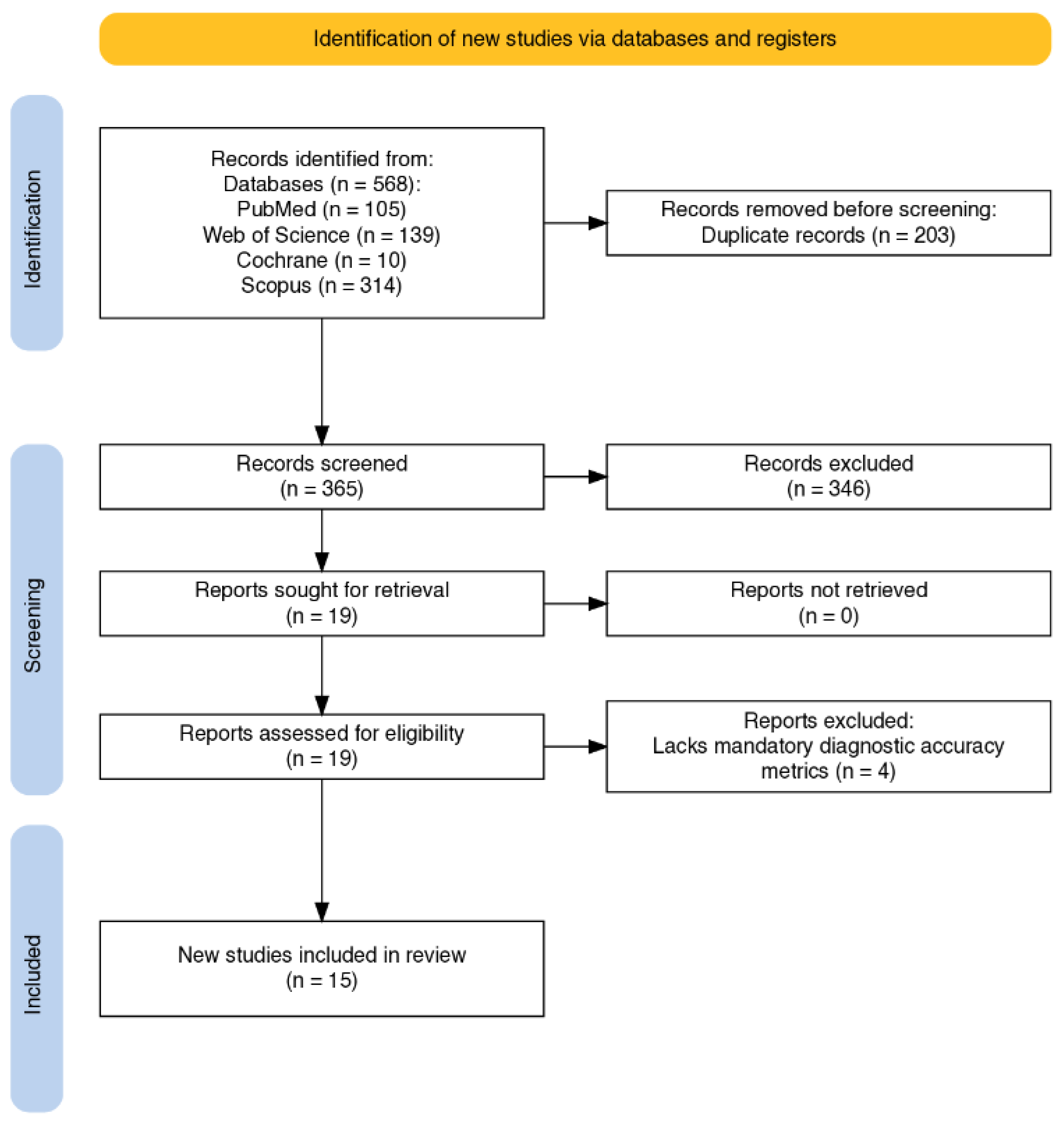
Figure 2.
QUADAS-2 risk of bias and applicability concerns summary for the 15 included studies across four domains (patient selection, index test, reference standard, flow and timing).
Figure 2.
QUADAS-2 risk of bias and applicability concerns summary for the 15 included studies across four domains (patient selection, index test, reference standard, flow and timing).
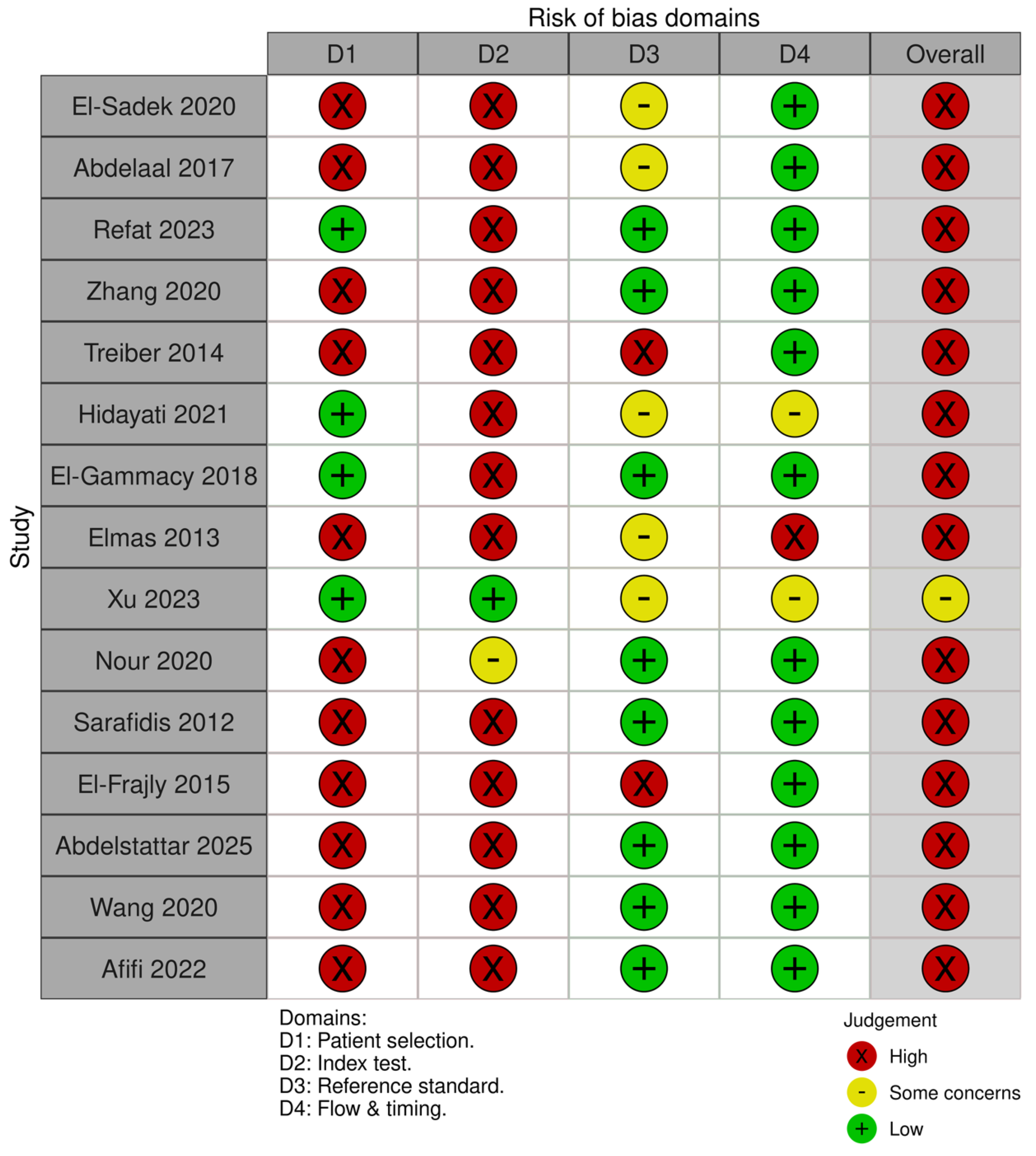

Table 1.
Characteristics of included studies.
| Study | Country | Design | Population | Clinical context | sCysC assay method | Timing of sCysC measurement | Cut-off (mg/L) | AKI reference standard |
|---|---|---|---|---|---|---|---|---|
| Abdelaal et al., 2017 [22] | Egypt | prospective case- control | preterm (100) | RDS | Jaffe method (Olympus AU640) | DOL 1, 3, 7 | DOL-3 ≥1.28 | KDIGO |
| El-Gammacy et al., 2018 [10] | Egypt | prospective | preterm (75) | RDS | ELISA | DOL 3 | 1.3 | Modified (pRIFLE) |
| El-sadek et al., 2019 [20] | Egypt | multicenter, prospective,case-controlled | term (90) | mixed | ELISA | day 1, 3, 5 of admission | 2.68 | KDIGO |
| Elmas et al., 2012 [23] | Turkey | prospective case control | preterm (62) | RDS | PENIA | DOL 3, 30 | 1.62 | Modified (pRIFLE) |
| Nour et al., 2020 [24] | Egypt | prospective observational | term (30) | HIE | ELISA | 24 h, DOL 4, 10 | 1.6 | modified AKIN criteria |
| Sarafidis et al., 2012 [25] | Greece | prospective case control | term/near term - 36 GA (35) | asphyxia | ELISA | DOL 1, 3, 10 | 2.87 | sCr based definition |
| El-Frargy et al., 2015 [26] | Egypt | prospective case control | term (30) | mixed | ELISA | day 1, day 3 of admission | 0.6 | sCr and BUN-based renal impairment |
| Refat et al., 2023 [2] | Egypt | prospective cohort study | term (70) | asphyxia | ELISA | DOL 1 | 1.43 | modified KDIGO |
| Hidayati et al., 2021 [27] | Indonesia | diagnostic test study | mixed (135) | mixed | PENIA | 0 – 28 DOL | 1.605 | neonatal RIFLE |
| Treiber et al., 2014 [28] | Slovenia | prospective case control | term (100) | asphyxia | PENIA | cord blood sample, DOL 3 | 1.67/1.69 | sCr based definition |
| Diab et al., 2022 [29] | Egypt | prospective case control | preterm (90) | RDS | ELISA | DOL 3 | 1.25 | KDIGO |
| Wang Z et al., 2020 [30] | China | observational cross-sectional diagnostic accuracy | term (196) | jaundice | PETIA | first 24 hours after admission | 1.55 | KDIGO |
| Zhang et al., 2020 [31] | China | case control | term (140) | asphyxia | Latex-enhanced immunoturbidimetric assay | 24 hours of life | 1.86 | AKIN |
| Xu et al., 2023 [13] | China | large multicenter retrospective cohort study | mixed (52333) | mixed | not reported | 0 – 28 DOL | 2.2 | modified KDIGO |
| Abdelsattar et al., 2025 [21] | Egypt | retrospective cohort study | mixed (200) | sepsis | ELISA | first 24 hours after admission | 9.4 | KDIGO |
Abbreviations: AKI, acute kidney injury; BUN, blood urea nitrogen; CyNA, cystatin C-detected neonatal acute kidney injury; ELISA, enzyme-linked immunosorbent assay; eGFR, estimated glomerular filtration rate; GA, gestational age; HIE, hypoxic-ischemic encephalopathy; nKDIGO, neonatal-modified KDIGO criteria; NICU, neonatal intensive care unit; PENIA, particle-enhanced nephelometric immunoassay; PETIA, particle-enhanced turbidimetric immunoassay; pRIFLE, pediatric RIFLE criteria; RDS, respiratory distress syndrome; sCr, serum creatinine; sCysC, serum cystatin C; UO, urine output; DOL, day of life.
Table 2.
Diagnostic performance of serum cystatin C across included studies based on time-related design.
Table 2.
Diagnostic performance of serum cystatin C across included studies based on time-related design.
| Study (Author, Year) | n (AKI/Total) |
AUC | Sensitivity (%) |
Specificity (%) |
Cut-off (mg/L) |
Lead time |
|---|---|---|---|---|---|---|
| Sarafidis et al., 2012 [25]* | 8/35 | 0.731 | 66.7 | 88.5 | 2.87 | 24 - 48 hours earlier |
| Elmas et al., 2013 [23]* | 28/62 | 0.88 | 16 | 82 | 1.62 | predicted AKI within 72 hours; sCr failed to differentiate groups on day 3 |
| Treiber et al., 2014 [28]* | 50/100 | 0.918 | 84 | 90 | 1.67 - 1.69 | 72 hours earlier |
| El-Frargy et al., 2015 [26]* | 20/30 | NR | 85 | 80 | 0.6 | 48 hours earlier |
| Abdelaal et al., 2017 [22]* | 24/100 | 0.97 | 100 | 83.3 | ≥1.28 | 4 days earlier |
| El-Gammacy et al., 2018 [10]* | 13/75 | 0.92 | 92.3 | 96 | 1.3 | 1 - 2 days earlier |
| El-Sadek et al., 2020 [20]* | 30/90 | 0.844 | 53.3 | 100 | 2.68 | 48 hours earlier |
| Nour et al., 2020 [24]* | 14/30 | NR | 86 | 75 | 1.6 | 24 hours earlier |
| Zhang et al., 2020 [31]* | 37/140 | 0.67 | 61.5 | 69.3 | 1.86 | not reported |
| Diab et al., 2022 [29]* | 18/90 | 1.00 | 100 | 100 | 1.25 | 48 hours earlier |
| Refat et al., 2023 [2]* | 21/70 | 0.939 | 95 | 94 | 1.43^ | 1 - 2 days earlier |
| Wang et al., 2020 [30]** | 27/196 | 0.844 | 85.2 | 62.9 | 1.55 | none |
| Hidayati et al., 2021 [27]** | 32/135 | 0.849 | 84.8 | 61.8 | 1.605 | none |
| Xu et al., 2023 [13]** | 6336/52333 | NR | NR | NR | 2.2 | none |
| Abdelsattar et al., 2025 [21]*** | 52/200 | 0.848 | 88.46 | 75.0 | 9.4 | precedes or concurrent (days 0, 2, 4) |
Legend: *Early predictive study; **Concurrent study; ***both early predictive and concurrent study; AKI, acute kidney injury; AUC, area under the curve; NR, not reported; NB: ^Refat et al. [2] reported the cut-off in ng/mL, converted to mg/L for comparison.
Disclaimer/Publisher’s Note: The statements, opinions and data contained in all publications are solely those of the individual author(s) and contributor(s) and not of MDPI and/or the editor(s). MDPI and/or the editor(s) disclaim responsibility for any injury to people or property resulting from any ideas, methods, instructions or products referred to in the content. |
© 2026 by the authors. Licensee MDPI, Basel, Switzerland. This article is an open access article distributed under the terms and conditions of the Creative Commons Attribution (CC BY) license.
Copyright: This open access article is published under a Creative Commons CC BY 4.0 license, which permit the free download, distribution, and reuse, provided that the author and preprint are cited in any reuse.


