

Submitted:
31 July 2026
Posted:
03 August 2026
You are already at the latest version
Abstract
Background: While non-specific pleural empyema in children can be successfully managed with minimally invasive drainage and pleural puncture in 85–90% of cases. Tuberculous pleural empyema (TB empyema) exhibits high clinical resistance and frequently requires extensive thoracic surgical interventions. The choice of surgical extent directly depends on the disease stage and the presence of complications. Methods: We conducted a retrospective analysis of diagnostic and surgical outcomes in 85 children and adolescents (aged 4 to 17 years) with tuberculous pleural empyema treated between 1984 and 2022. TB empyema staging was performed using the American Thoracic Society (ATS) classification was adapted for tuberculous etiology. Results: Stage I TB empyema was diagnosed in 3 patients, Stage II in 7 patients, and Stage III in 75 patients. Patients with Stage I and II disease underwent video-assisted thoracoscopic (VATS) debridement of the empyema cavity. In Stage III, 75 patients underwent 79 surgical interventions of varying extents. Postoperative complications occurred in 2 patients (2.5%). This occurred as delayed lung re-expansion, which was successfully managed with repeat thoracentesis in one patient and chest tube drainage in the other. The overall 30-day postoperative mortality rate was 0.0%. No recurrences were reported during the 3-year follow-up period.Conclusion: Comprehensive treatment combined with timely surgical intervention achieves clinical cure in all patients. Early minimally invasive VATS interventions during Stages I–II prevent the need for more traumatic procedures, such as pleurectomy and lung resection, which are often required in Stage III disease.
Keywords:
tuberculosis
; pediatric surgery
; pleural empyema
; VATS
; pleurectomy
; lung decortication
1. Introduction
Tuberculous pleural empyema (TB empyema) is characterized by the accumulation of mycobacterium-infected purulent exudate within the pleural cavity. Unlike non-specific empyema, the clinical course of TB empyema in pediatric patients is characterized by subtle, subclinical symptoms, a propensity for chronicity, and exceptionally high resistance to conventional conservative therapy and chest tube drainage.
In the international literature, the American Thoracic Society (ATS) classification is widely used to guide management strategies for pediatric pleural empyema [1]:
Stage I (Exudative phase): Characterized by the accumulation of low-viscosity fluid exudate without loculations.
Stage II (Fibrinopurulent phase): Marked by abundant fibrin deposition on the visceral and costal pleura, leading to loculation formation and early lung fixation.
Stage III (Organizational phase): Defined by the formation of a dense, non-expandable fibrous peel (rigid fibrothorax), causing persistent restriction of lung mobility, volume loss, and a residual cavity.
While Stage I non-specific empyema in children usually resolves rapidly with conservative management, tuberculous pleurisy and early empyema frequently progress to the organizational phase due to delayed diagnosis. Chest computed tomography (CT) is a critical differential diagnostic tool capable of identifying features such as the "split pleura sign", extrapleural fat proliferation, and calcifications.
2. Materials and Methods
2.1. Study Design and Patient Selection
This retrospective, cohort, observational study was approved by the Institutional Review Board (Ethics Committee) of Sechenov First Moscow State Medical University (Protocol No. 17-23). The medical records of 85 children and adolescents aged 4 to 17 years who underwent surgery for confirmed tuberculous pleural empyema between 1984 and 2022 were thoroughly reviewed.
Inclusion criteria: Confirmed diagnosis of tuberculous pleural empyema (Stages I–III); age from 4 years to 17 years and 11 months; complete surgical history.
Exclusion criteria: Pleural effusion of non-tuberculous etiology; age 18 years or older; patients who received exclusive conservative antituberculosis chemotherapy without surgical intervention.
2.2. Surgical Techniques
Stages I and II: Management focused on early video-assisted thoracoscopic (VATS) with debridement of the empyema cavity. Key steps included: (1) evacuation of seropurulent exudate; (2) disruption of individual loculations and purulent collections; (3) removal of fibrinous peel from the visceral and parietal pleura; (4) partial parietal pleurectomy (PPP) in areas with the most pronounced inflammatory changes for histological verification and to accelerate exudate resorption; and (5) adequate pleural cavity drainage.
Stage III: Due to the presence of an organized fibrothorax, open or video-assisted radical surgical procedures were required. Procedures included pleurectomy with lung decortication, segmental resection or lobectomy combined with decortication, and pleuropneumonectomy. In cases requiring extensive pneumolysis, particularly following previous surgeries, a modified lateral thoracotomy using double intercostal space incision (through the fifth and either the seventh or eighth intercostal spaces) was performed to optimize surgical exposure.
2.3. Statistical Analysis
Statistical processing was performed using IBM SPSS Statistics version 22.0. The normality of quantitative variables was assessed using the Shapiro-Wilk test. Normally distributed quantitative data are presented as Mean ± Standard Deviation (M ± SD); variables with non-normal distributions are expressed as Median and Interquartile Range (Me [Q1 – Q3]). Categorical data are described using absolute values and percentages. Comparison of distributions was performed using the non-parametric Mann-Whitney U test, and frequencies were compared using the Chi-square test or Fisher's exact test, with a significance level set at p < 0.05.
3. Results
3.1. Baseline Cohort Characteristics
The study included 85 patients: 47 boys (55.3%) and 38 girls (44.7%). The mean age of the patients was 11 ± 4 years (range: 4 to 17 years).
3.2. Preoperative Treatment and Disease Progression
The duration of ineffective conservative therapy prior to surgical intervention ranged from 1 to 16 months (median = 5 months [Q1–Q3: 3–7]). A direct statistical correlation was identified between prolonged conservative management and disease progression to severe fibrothorax (p < 0.001).
Delayed surgical referral resulted in disease progression in several cases:
In 3 cases, Stage I progressed to Stage II due to prolonged conservative observation lasting 1–2 months.
In 9 cases, Stage I progressed directly to Stage III (organizing fibrothorax) secondary to protracted non-surgical management lasting from 4 to 10 months (mean ± SD = 7 ± 2 months, 95% CI: 5–8).
3.3. Analysis of the Structure of Surgical Interventions
While all patients with Stage I (n = 3) and Stage II (n = 7) disease successfully underwent minimally invasive VATS debridement and sanitation of the empyema cavity, Stage III required advanced, extensive reconstructive surgeries (Table 3).
3.4. Postoperative Complications and Mortality
The 30-day postoperative mortality rate across all disease stages was 0.0%. No intraoperative complications were reported. Postoperative complications occurred and were successfully cured in two cases (2.5%) for 3rd stage TB empyema: one following VATS segmental resection and one following VATS lobectomy. These complications presented as delayed lung re-expansion and were fully resolved by repeat thoracocentesis and/or short-term chest tube drainage. Long-term follow-up over a 3-year period confirmed the absence of disease recurrence in all operated patients.
4. Case Reports and Illustrative Materials
4.1. Figure 1: Loculated Stage II Tuberculous Empyema (Patient K., 11 Years Old)
An 11-year-old boy presented with a loculated pleural effusion following one month of ineffective conservative treatment and therapeutic pleural punctures at a regional hospital. Pleurectomy with lung decortication was performed via a minithoracotomy approach. The imaging data for this clinical case are divided into two separate graphic files:
Panel A: Preoperative chest radiograph demonstrating a large left-sided loculation. Panels B, C, and D: Intraoperative stages of pleurectomy and lung decortication was performed via a minithoracotomy approach.
Panel E: Gross specimen of the loculation in cross-section, demonstrating caseopurulent contents. Panel F: Follow-up chest radiograph obtained on postoperative day 5, showing complete lung re-expansion and absence of pleural effusion.
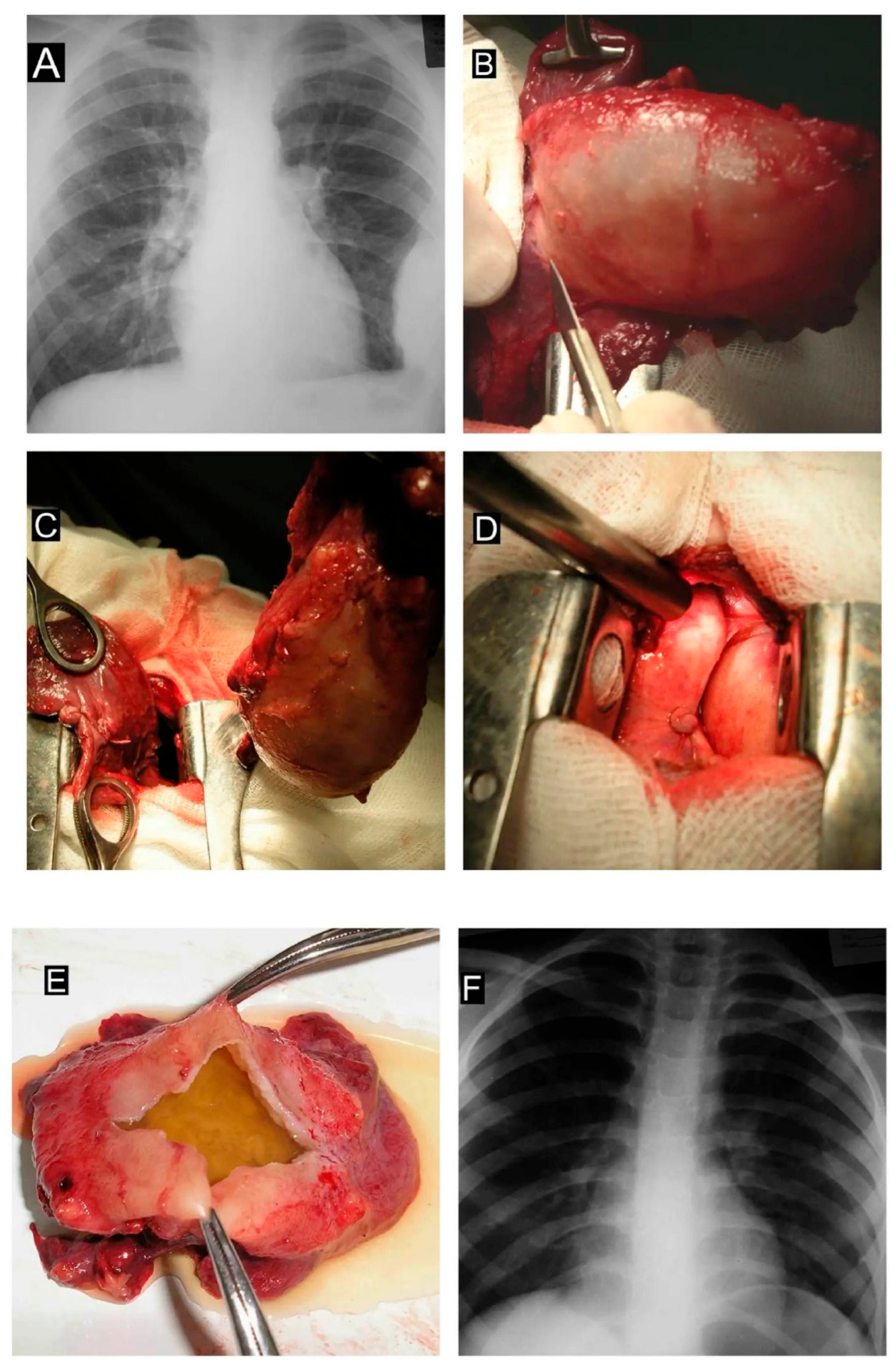
Figure 1.
2. Figure 2: Total Chronic Stage III Tuberculous Empyema with Secondary Pulmonary Cirrhosis (Patient M., 4 Years Old).
A 4-year-old girl presented with intrathoracic tuberculous lymphadenitis complicated by total chronic total left-sided TB empyema following 10 months of ineffective chest tube drainage. The imaging archive for this case is divided into two parts:
Panels A and B: Chest computed tomography (CT) scans demonstrate a total left-sided loculation and associated lung collapse. Panel C: Three-dimensional (3D) CT reconstruction of the lungs and tracheobronchial tree showing signs of left lung cirrhosis. Panel D: Intraoperative debridement.
Panel E: Pleurectomy with partial upper lobe resection. Panel F: 3D CT reconstruction at 1 month postoperatively, demonstrating incomplete lung re-expansion due to pulmonary fibrosis. Panel G: Chest radiograph at the 2-year follow-up, showing marked compensatory hypertrophy. Panel H: Clinical photograph of the child demonstrating the absence of chest wall deformity.
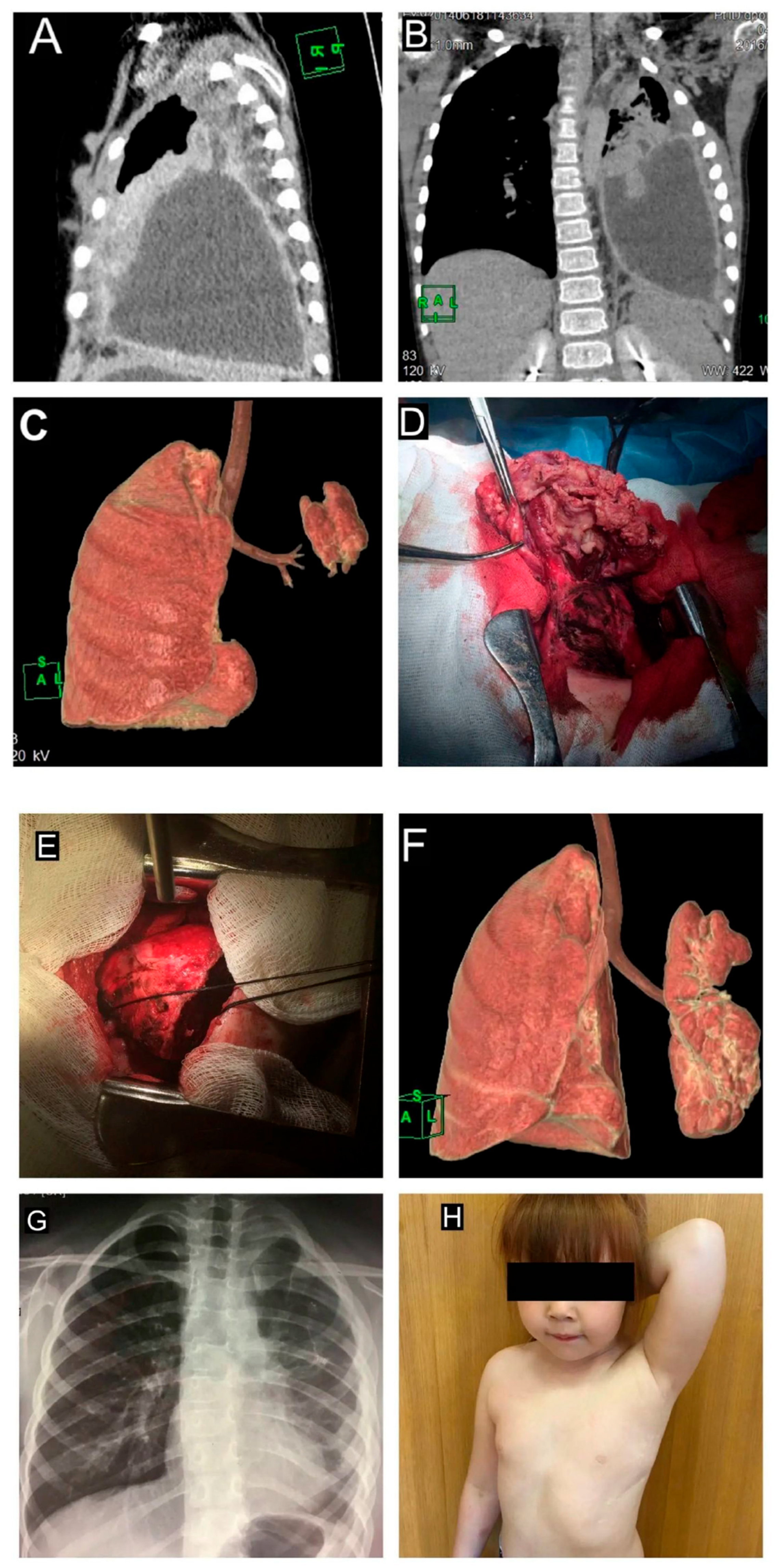
Figure 2.
3. Figure 3: Total Pneumolysis via a Double Intercostal Space Incision in Stage III Disease (Patient S., 17 Years Old).
A 17-year-old adolescent presented with recurrent organizing empyema following ineffective VATS performed at another institution. To achieve total pneumolysis, a surgical approach via a double intercostal space incision (the 5th and 7th intercostal spaces) was utilized. The illustrative material for this case is divided into two sections:
Panel A: Preoperative chest CT scan demonstrating massive multiloculated loculations. Panels B, C, and D: Intraoperative stages of dissecting the dense fibrous peel from the pulmonary surface.
Panel E: Intraoperative view of the surgical field via the double intercostal space approach. Panel F: Placement of three silicone chest tubes directed towards the apex, posterior sinus, and anterior sinus. Panel G: Gross specimen of the dense pleural sac, completely excised en bloc. Panel H: Chest radiograph obtained prior to hospital discharge.
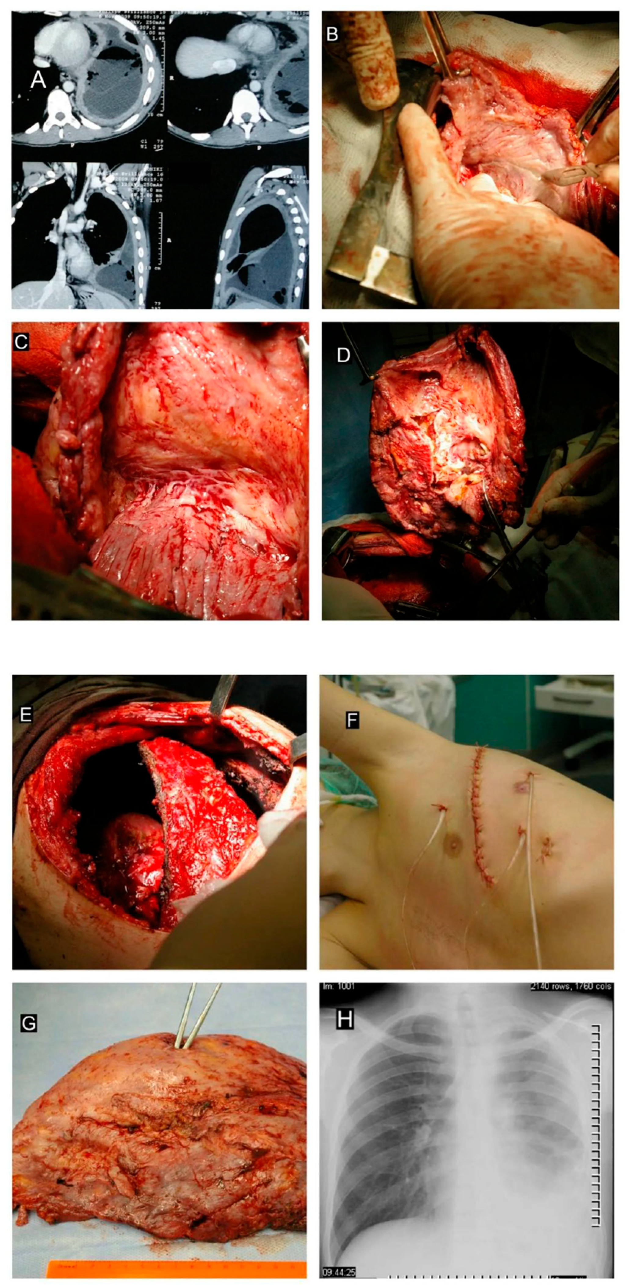
Figure 3.
4. Figure 4: Tuberculous Empyema Complicated by Calcified Constrictive Pericarditis (Concretio Cordis).
This section presents a severe complication of advanced tuberculosis involving extension of the inflammatory process to the pericardium, with subsequent calcification ("armoured heart"). All stages of the condition are illustrated in a single multipanel figure.:
Panel A: Chest CT scan (arrows indicate the dense calcified shell encasing the myocardium and the loculated empyema along the right pleura). Panel B: Surgical excision of the rigid, calcified pericardial layer. Panel C: Intraoperative view of the surgical field following subtotal pericardiectomy, demonstrating the liberated pulsating myocardium. Panel D: Gross specimen of the excised tissue (top: empyema sac; bottom: stony calcified pericardium).
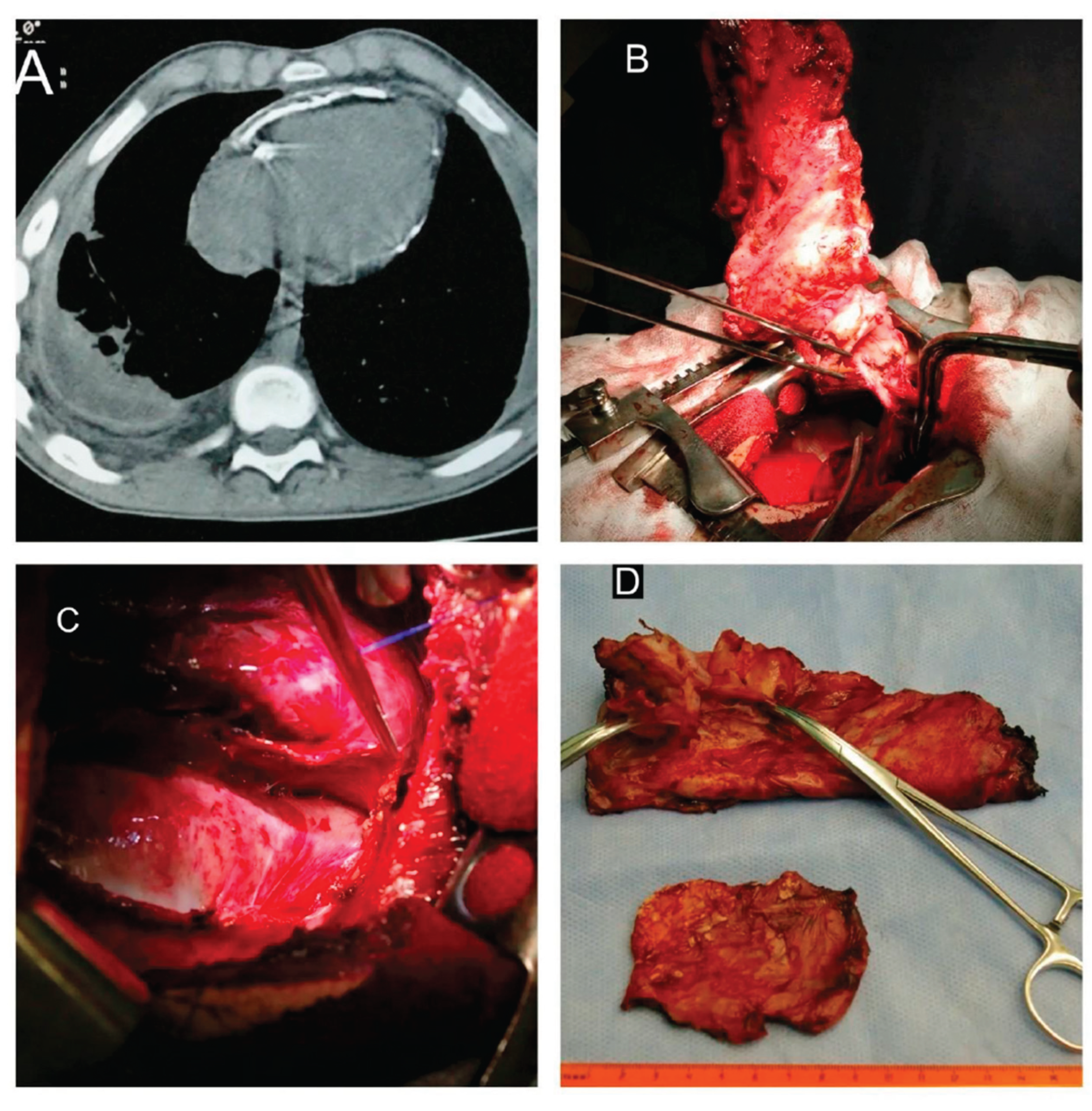
Figure 4.
5. Discussion
The management of pleural empyema in children and adolescents remains one of the most controversial and challenging issues in paediatric thoracic surgery. While international consensus guidelines and clinical recommendations for non-specific parapneumonic empyema give priority to conservative approaches, serial therapeutic thoracocentesis, and intrapleural fibrinolytic administration—with reported success rates of 80–84% [2,3,4]—this watchful waiting strategy is entirely ineffective in cases of specific tuberculous aetiology [5,6].
Pathogenetic differences determine markedly distinct rates of progression through the inflammatory phases. A high concentration of mycobacterial antigens within the closed space of the pleural cavity induces the expression of procoagulant factors, leading to rapid and accelerated fibrinogenesis [7]. As noted by Lee S.F. et al., therapeutic strategies for conventional infections are primarily aimed at eradicating the acute purulent focus [2], whereas tuberculous empyema requires combined surgical debridement and targeted multidrug antituberculosis chemotherapy [8,9].
Attempting to manage patients with Stage III chronic tuberculous empyema solely with therapeutic punctures or closed chest tube drainage inevitably results in a critical delay, persistent lung collapse, and the rapid development of irreversible pulmonary fibrosis [10,11]. Under these conditions, a classic rigid fibrothorax develops [7,12]. The ineffectiveness of prolonged conservative management in children with tuberculous exudative pleurisy was highlighted in the landmark studies of Kolpakchi M.I. and Khalikova E.M., who demonstrated that delayed radical surgery significantly worsens immediate treatment outcomes [13]. Motus I.Y. et al., analysing surgical strategies for tuberculous pleurisy, also emphasised the importance of timely differentiation between indications for conservative therapy and surgical debridement to prevent severe pleural fibrosis [14].
Our clinical experience convincingly demonstrates that early video-assisted thoracoscopic surgery (VATS) debridement during Stages I–II of empyema provides critical pathogenetic and practical advantages [15,16]:
1. Timely verification: This approach allows direct biopsy of abnormal pleural segments for rapid histological and molecular genetic (PCR) testing. This is critically important because, according to our data, 50% of patients did not have a confirmed diagnosis prior to surgery. Similar findings were reported by Malhotra P. et al., who noted that mycobacterial detection in pleural fluid via standard microscopy does not exceed 20% to 30% due to the low concentration of the pathogen [11].
2. Bacteriological control: VATS ensures the collection of high-quality operative specimens (pleural biopsies, fibrin clots) for Mycobacterium tuberculosis (MTB) culture isolation and rapid drug-susceptibility testing (MDR/XDR-TB). This aligns with the conclusions of Wang H. et al., who emphasized that precise microbiological verification is essential for the appropriate selection of antituberculosis drug therapy [17].
3. Decompression and detoxification: The procedure rapidly alleviates systemic intoxication, reduces total hospital stay, and effectively prevents the development of a rigid, restrictive fibrothorax. An early landmark publication by the Subcommittee on Surgery of the American Thoracic Society (ATS) in 1962 established that early liberation of the lung from its fibrin peel eliminates the need for traumatic decortications in the future [12]. Guided by these same principles, Yim A.P. was among the first worldwide to demonstrate the high efficacy and minimally invasive nature of thoracoscopic interventions in the management of various forms of pulmonary tuberculosis [15,16].
Despite the clear advantages of early minimally invasive surgery, the clinical profile of patients referred to specialised centres remains unsatisfactory. Nearly 90% of children and adolescents are referred to our department already in Stage III (the organising phase), when the effectiveness of isolated thoracoscopic debridement is largely exhausted, and extensive, traumatic surgical interventions are required. At this stage, dense fibrous peels, often several centimetres thick, cause marked restriction of chest wall mobility and fixed lung collapse. The development of such advanced forms is described in detail in the manual Pleural Diseases by Tyukhtin N.S., Stogova N.A., and Giller D.B., which demonstrates that prolonged persistence of tuberculous empyema inevitably transforms the pleural cavity into a focus of chronic purulent intoxication with caseous tissue degeneration [10].
When treating organising empyema (Stage III) in children, surgeons frequently encounter the problem of ineffective standard pneumolysis [18]. In cases where the lung is completely entrapped within a fibrous shell and anatomical landmarks are obliterated following previous ineffective drainage procedures, the operation becomes substantially more complex. Recent international studies by Xia Z. et al. demonstrate that, even in the presence of severe adhesions, uniportal VATS interventions can be successfully performed in children, offering reduced surgical trauma compared with the standard triportal approach [19].
However, in cases of extremely severe fibrothorax, we have successfully utilised a proprietary lateral thoracotomy approach through a double intercostal space incision for total pneumolysis. This approach optimises visualisation of all compartments of the pleural cavity, minimises rib traction, and reduces the incidence of postoperative pain. Similar challenges related to surgical exposure in advanced disease have been discussed by Pomerantz M. and Brown J., who noted that adequate exposure determines the radicality of intervention in extensive tuberculosis [20].
In cases of total lung destruction secondary to a chronic purulent process, debilitating procedures such as pleuropneumonectomy were required. In contemporary international literature, particularly in the work of Kuhtin O. et al., it is emphasised that such interventions (including thoracoplasty) in children should be considered strictly as salvage procedures (“operations of last resort”) when all other methods of lung re-expansion and source control have failed [21]. Our experience with eight pleuropneumonectomies without mortality demonstrates the feasibility of safely performing these procedures in a high-technology specialised hospital setting.
Long-term treatment outcomes warrant particular attention. The monograph Tuberculosis Surgery in Children and Adolescents, edited by Professor D.B. Giller, reports that the plasticity of the paediatric organism provides a high capacity for long-term cardiorespiratory compensation following pulmonary resection [8]. This is supported by our previous studies: an analysis of surgical treatment outcomes for respiratory tuberculosis in children published by Giller D.B., Martel I.I., and Ogai I.V. in 2013 [22], together with a detailed long-term follow-up evaluation in 2012 [23], demonstrated recovery of respiratory function and the absence of skeletal deformities in the majority of operated patients during growth.
A similar view was expressed by Grigoryan V.A., who emphasised the importance of preserving the maximum possible volume of healthy lung parenchyma during paediatric resections [24]. In our subsequent study, we demonstrated that the implementation of VATS technologies significantly reduced postoperative complication rates compared with the era of exclusively open thoracic surgery [9].
A comparative analysis of the epidemiological and clinical characteristics of paediatric tuberculous empyema across endemic regions worldwide allows our findings to be compared with international data. For example, a large-scale study by Yang G. et al., conducted in high tuberculosis-burden regions, demonstrated that children with tuberculous empyema have significantly longer periods of preoperative treatment and poorer pulmonary function parameters compared with children with conventional parapneumonic empyema [5,25].
Wu Y.H. et al. identified delayed radiological diagnosis and a high baseline bacterial burden as major risk factors for a prolonged course of pleural tuberculosis in children [25]. These findings correlate directly with our observations regarding the factors contributing to disease progression from early stages to fibrothorax. The importance of CT monitoring, surgical risk-factor assessment, and identification of specific radiological patterns has also been emphasised by other researchers [26,27,28,29,30].
The dilemma of selecting between conservative management and surgical intervention in children remains actively debated worldwide. For example, an analysis by Flausino F. et al. revealed considerable heterogeneity in the management of paediatric empyema in Brazil, where the absence of a unified surgical algorithm frequently contributes to disease progression and chronicity [30]. Meanwhile, Baram A. and Yaldo F. demonstrated that even combined intrapleural thrombolytic therapy is ineffective in the presence of dense fibrin deposition [3].
In tuberculosis, this situation is further complicated by the risk of severe complications, such as empyema necessitans (extension of empyema through the chest wall). Recent studies by Bendre A. [31] and Shieba Y. [32] have described clinical cases of this complication in children with tuberculosis. Our clinical protocol aims to prevent such complications through timely surgical debridement.
Although complex resectional and decortication procedures predominated among our interventions for Stage III disease, the implementation of our multidisciplinary approach—including optimised surgical exposure, precise pneumolysis, and targeted antituberculosis chemotherapy—resulted in a 100% clinical cure rate. Overall 30-day postoperative mortality was 0.0%, and the incidence of transient pleural complications did not exceed 2.5%; all complications were successfully managed using minimally invasive interventions. The three-year follow-up period confirmed the durability of these outcomes and the absence of recurrence. These findings support the revision of existing regional management strategies for children with pleural effusions towards the earlier adoption of VATS technologies.
6. Conclusions
1. Comprehensive management of tuberculous pleural empyema in children and adolescents, incorporating timely and mandatory surgical intervention, achieves durable clinical cure in all patients.
2. The degree of surgical trauma, the extent of the procedure, the incidence of postoperative complications, and the overall duration of hospitalisation are directly related to the stage of disease at the time of referral to a thoracic surgeon.
3. Children and adolescents with tuberculous exudative pleurisy or empyema at Stages I–II should be prioritised for early video-assisted thoracoscopic surgery (VATS) with pleural debridement, as this approach preserves functional lung volume and reduces the need for more extensive and debilitating interventions
Author Contributions
Conceptualization, X.X. and Y.Y.; methodology, X.X.; software, X.X.; validation, X.X., Y.Y. and Z.Z.; formal analysis, X.X.; investigation, X.X.; resources, X.X.; data curation, X.X.; writing—original draft preparation, X.X.; writing—review and editing, X.X.; visualization, X.X.; supervision, X.X.; project administration, X.X.; funding acquisition, Y.Y. All authors have read and agreed to the published version of the manuscript.
Funding
This research received no external funding.
Institutional Review Board Statement
This retrospective, cohort, observational study was approved by the Institutional Re-view Board (Ethics Committee) of Sechenov First Moscow State Medical University (Proto-col No. 17-23).
Informed Consent Statement
Data Availability Statement
The original contributions presented in this study are included in the article/supplementary material. Further inquiries can be directed to the corresponding author(s).
Conflicts of Interest
The authors declare no conflict of interest.
References
- Shen, K.R.; Bribriesco, A.; Crabtree, T.; et al. The American Association for Thoracic Surgery consensus guidelines for the management of empyema. J. Thorac. Cardiovasc Surg. 2017, 153(6), e129–e146. [Google Scholar] [CrossRef] [PubMed]
- Lee, S.F.; Lawrence, D.; Booth, H.; Morris-Jones, S.; Macrae, B.; Zumla, A. Thoracic empyema: current opinions in medical and surgical management. Curr. Opin. Pulm. Med. 2010, 16(3), 194–200. [Google Scholar] [CrossRef] [PubMed]
- Baram, A.; Yaldo, F. Pediatric Thoracic Empyema-Outcomes of Intrapleural Thrombolytics: Ten Years of Experience. Glob. Pediatr. Health 2020, 7, 2333794X20928200. [Google Scholar] [CrossRef] [PubMed]
- Elsheikh, A.; Bhatnagar, M.; Rahman, N.M. Diagnosis and management of pleural infection. Breathe (Sheff) 2023, 19(4), 230146. [Google Scholar] [CrossRef] [PubMed]
- Yang, G.; Wen, Y.; Chen, T.; Xu, C.; Yuan, M.; Li, Y. Comparison of pediatric empyema secondary to tuberculosis or non-tuberculosis community-acquired pneumonia in those who underwent surgery in high TB burden areas. Pediatr. Pulmonol. 2021, 56(10), 3321–3331. [Google Scholar] [CrossRef] [PubMed]
- Drebov, R.; Brankov, O.; Panov, M.; Kh, Shivachev; Iotov, I.; Sŭrbianova, N. Surgical aspects of primary tuberculosis in children: clinical presentation and indications for surgical treatment. Khirurgiia (Sofiia) 2006, (6), 12–15. [Google Scholar] [PubMed]
- Iguina, M.M.; Sharma, S.; Danckers, M. Thoracic Empyema. In StatPearls [Internet]; StatPearls Publishing: Treasure Island (FL), 24 Dec 2024. [Google Scholar] [PubMed]
- Giller, D.B. (Ed.) Tuberculosis Surgery in Children and Adolescents; (In Russ.); Aldi-print: Moscow, 2016; p. 457 p. [Google Scholar]
- Giller, D.B.; Martel, I.I.; Enilenis, I.I.; Koroyev, V.V.; Kesaev, OSh; Giller, B.D.; Shcherbakova, G.V. Surgical treatment of tuberculous empyema in children. Pirogov Russ. J. Surg. (In Russ.). 2019, (8), 22–28. [Google Scholar] [CrossRef] [PubMed]
- Tyukhtin, N.S.; Stogova, N.A.; Giller, D.B. Pleural Diseases; (In Russ.); Erokhin, V.V., Ed.; Meditsina: Moscow, 2010; p. 256 p. [Google Scholar]
- Malhotra, P.; Aggarwal, A.N.; Agarwal, R.; Ray, P.; Gupta, D.; Jindal, S.K. Clinical characteristics and outcomes of empyema thoracis in 117 patients: a comparative analysis of tuberculous vs. non-tuberculous aetiologies. Respir. Med. 2007, 101(3), 423–430. [Google Scholar] [CrossRef] [PubMed]
- Andrews, N.C.; Parker, E.F.; Shaw, R.P.; American Thoracic Society; et al. Management of nontuberculous empyema. A statement of the subcommittee on surgery. Am. Rev. Respir. Dis. 1962, 85, 935–936. [Google Scholar]
- Kolpakchi, M.I.; Khalikova, E.M. Immediate results of radical operations in children and adolescents with tuberculosis. Probl. Tuberk. In Russ.. 1971, (4), 32–35. [Google Scholar]
- Motus, I.Y.; Gaponyuk, P.F.; Krasnoborova, S.Y.; Saveliev, A.V.; Sokolov, V.Y. Surgical tactics in tuberculous pleurisy. Probl. Tuberk. (In Russ.). 2002, (11), 28–31. [Google Scholar]
- Yim, A.P. The role of video-assisted thoracoscopic surgery in the management of pulmonary tuberculosis. Chest 1996, 110(3), 829–832. [Google Scholar] [CrossRef] [PubMed]
- Yim, A.P.; Izzat, M.B.; Lee, T.W. Thoracoscopic surgery for pulmonary tuberculosis. World J. Surg. 1999, 23(11), 1114–1117. [Google Scholar] [CrossRef] [PubMed]
- Wang, H.; Lin, H.; Jiang, G. Pulmonary resection in the treatment of multidrug-resistant tuberculosis: a retrospective study of 56 cases. Ann. Thorac. Surg. 2008, 86(5), 1640–1645. [Google Scholar] [CrossRef] [PubMed]
- Zhang, J.; Liu, C.; Ding, W.; Zhang, J.; Tran, W. Video-assisted thoracic surgery in treatment of tuberculous empyema. China J. Endosc. 2004, 10(2), 47–49. [Google Scholar]
- Xia, Z.; Zheng, S.; Shi, Q.; Wang, H.; Ning, X.; Huang, P.; Wen, G.; Xiang, N.; Liao, M.; Ma, M.; Qiao, K. Uniportal versus triportal video-assisted thoracic surgery in the treatment of tuberculous empyema. J. Int. Med. Res. 2023, 51(5), 3000605231169901. [Google Scholar] [CrossRef] [PubMed]
- Pomerantz, M.; Brown, J. The surgical management of tuberculosis. Semin Thorac. Cardiovasc Surg. 1995, 7(2), 108–111. [Google Scholar] [PubMed]
- Kuhtin, O.; Veith, M.; Alghanem, M.; et al. Thoracoplasty—Current View on Indication and Technique. Thorac. Cardiovasc Surg. 2020, 68(4), 331–340. [Google Scholar] [CrossRef] [PubMed]
- Giller, D.B.; Martel, I.I.; Ogai, I.V. Results of surgical treatment of respiratory tuberculosis in children and adolescents. Tuberc. Lung Dis. In Russ.. 2013, (6), 23–24. [Google Scholar]
- Giller, D.B.; Ogai, I.V.; Martel, I.I.; Glotov, A.A.; Panova, L.V. Long-term results of surgical treatment of respiratory tuberculosis in children and adolescents. Tuberc. Lung Dis. In Russ.. 2012, (1), 30–38. [Google Scholar]
- Grigoryan, V.A. Lung surgery in children and adolescents with tuberculosis. Tuberc. Lung Dis. In Russ.. 2011, (4), 110. [Google Scholar]
- Wu, Y.H.; Wang, J.L.; Wang, M.S. Factors Associated With the Presence of Tuberculous Empyema in Children With Pleural Tuberculosis. Front Pediatr. 2021, 9, 751386. [Google Scholar] [CrossRef] [PubMed]
- Van Dyck, P.; Vanhoenacker, F.M.; Van den Brande, P.; De Schepper, A.M. Imaging of pulmonary tuberculosis. Eur. Radiol. 2003, 13(8), 1771–1785. [Google Scholar] [CrossRef] [PubMed]
- Zhou, M.; Ren, S.F.; Gong, H.Z.; Wang, M.S. Risk factors associated with surgical intervention in childhood pleural tuberculosis. Sci. Rep. 2021, 11(1), 3084. [Google Scholar] [CrossRef] [PubMed]
- Alioke, I.I.; Anumenechi, N.; Edaigbini, S.A. Treatment Outcomes of Tuberculous and Non-tuberculous Empyema Thoracis. J. West Afr. Coll. Surg. 2020, 10(1), 15–19. [Google Scholar] [CrossRef] [PubMed]
- Pillay, T.; Andronikou, S.; Zar, H.J. Chest imaging in paediatric pulmonary TB. Paediatr. Respir. Rev. 2020, 36, 65–72. [Google Scholar] [CrossRef] [PubMed]
- Flausino, F.; Manara, L.M.; Sandre, B.B.; Sawaya, G.N.; Maurici, R. Management of pediatric pleural empyema: a national survey of pediatric surgeons in Brazil. J. Bras. Pneumol. 2024, 50(2), e20230318. [Google Scholar] [CrossRef] [PubMed]
- Bendre, A.; Ramesh, T.; Shah, I. Beyond the lymph node: empyema necessitans in pediatric tuberculosis. Res. Conn. 2026, 1(2), vmag050. [Google Scholar] [CrossRef]
- Shieba, Y.; Ramadan, A. Empyema necessitans in a pediatric patient: A case study. Int. J. Surg. Case Rep. 2023, 112, 108932. [Google Scholar] [CrossRef] [PubMed]

Table 1.
Baseline Demographic and Clinical Characteristics by Empyema Stage.
| Characteristic | Stage I (n = 3) | Stage II (n = 7) | Stage III (n = 75) | p-value |
| Age (mean ± SD, years) | 9 ± 5 | 13 ± 2 | 11 ± 4 | 0.290 |
| Sex (n, %) | 0.279 | |||
| — Male | 3 (100.0%) | 4 (57.1%) | 40 (53.3%) | |
| — Female | 0 (0.0%) | 3 (42.9%) | 35 (46.7%) | |
| Duration of preoperative treatment (median [Q1–Q3], months) | 1 [1,1] | 1 [1,2] | 5 [4,5,6,7] | < 0.001* |
*The differences were statistically significant: p = 0.006 for Stage III vs. Stage I, and p < 0.001 for Stage III vs. Stage II.
Table 2.
Sex distribution of patients with 95% confidence intervals (CI).
| Sex | Absolute number (n) | Percentage (%) | 95% CI |
| Female | 38 | 44.7% | 33.9%–55.9% |
| Male | 47 | 55.3% | 44.1%–66.1% |
Table 3.
Structure of surgical procedures performed for Stage III tuberculous empyema in children (79 procedures in 75 patients).
Table 3.
Structure of surgical procedures performed for Stage III tuberculous empyema in children (79 procedures in 75 patients).
| Type of Surgical Procedure | Number of Procedures (n) | Percentage (%) | Complications (n) |
| VATS segmental resection with pleurectomy | 21 | 26.6% | 1 |
| Open segmental resection with pleurectomy | 1 | 1.3% | 0 |
| Thoracoscopic pleurectomy with lung decortication | 11 | 13.9% | 0 |
| VATS pleurectomy with lung decortication | 27 | 34.2% | 0 |
| Open pleurectomy with lung decortication | 1 | 1.3% | 0 |
| VATS lobectomy with decortication | 6 | 7.6% | 1 |
| VATS pleuropneumonectomy | 8 | 10.1% | 0 |
| VATS thoracomyoplasty | 2 | 2.5% | 0 |
| Transsternal occlusion of the main bronchus | 1 | 1.3% | 0 |
| Total procedures | 79 | 100.0% | 2 (2.5%) |
Disclaimer/Publisher’s Note: The statements, opinions and data contained in all publications are solely those of the individual author(s) and contributor(s) and not of MDPI and/or the editor(s). MDPI and/or the editor(s) disclaim responsibility for any injury to people or property resulting from any ideas, methods, instructions or products referred to in the content. |
© 2026 by the authors. Licensee MDPI, Basel, Switzerland. This article is an open access article distributed under the terms and conditions of the Creative Commons Attribution (CC BY) license (http://creativecommons.org/licenses/by/4.0/).
Copyright: This open access article is published under a Creative Commons CC BY 4.0 license, which permit the free download, distribution, and reuse, provided that the author and preprint are cited in any reuse.


