

Submitted:
28 July 2026
Posted:
03 August 2026
You are already at the latest version
Abstract
Background and Objectives: Post-traumatic elbow stiffness is a disabling condition often resulting from a combination of capsular contracture, osseous impingement, and residual instability, rendering it exceptionally challenging to treat. The aim of this study was to evaluate the outcomes of a comprehensive surgical approach for the treatment of post-traumatic elbow stiffness. Materials and Methods: A retrospective case series of 20 consecutive patients treated between 2018 and 2025 was conducted. Etiologies included distal humeral fractures (n = 9), terrible triad injuries (n = 4), elbow dislocations (n = 3), olecranon fractures (n = 2), and neglected radial head fractures (n = 2). All patients underwent anterior and posterior arthrolysis with triceps tenolysis. Additional procedures included ligament reconstruction, radial head arthroplasty, an-terior capsule repair, tricepsplasty, and ulnar nerve transposition when indicated. El-bow range of motion was assessed preoperatively and during follow-up at 2 weeks, 6 weeks, 3 months, 6 months, and 1 year postoperatively. Results: Mean flexion im-proved from 81.0° to 125.3°, while the extension deficit improved from 66.5° to 16.5°. Mean supination increased from 54.8° to 82.8°, and mean pronation from 67.0° to 84.5°. The mean flexion-extension arc improved from 14.5° to 108.8°, representing a mean gain of 94.3°. One transient sensory ulnar neuropathy, one case of postoperative triceps insufficiency requiring revision surgery were observed and one patient devel-oped postoperative skin blistering following a posterior approach. Conclusions: Com-prehensive, pathology-oriented surgical treatment resulted in substantial improve-ment in elbow motion and restoration of a functional range of movement.
Keywords:
stiff elbow
; flexion-extension arc
; arthrolysis
; triceps tenolysis
; ulnar transposition
1. Introduction
Traumatic elbow injuries represent some of the most challenging pathologies to treat due to the combination of capsular contracture, ligament retraction, osseous impingement, and residual instability. Loss of motion following trauma may severely compromise upper-limb function, affecting activities of daily living, work-related tasks, and overall quality of life. Despite advances in fracture fixation techniques and rehabilitation protocols, elbow stiffness remains a frequent complication after fractures and dislocations around the elbow (an incidence of 10-15%) [1] .
A functional elbow has traditionally been defined as a flexion-extension arc between 30° and 130° and a forearm rotation arc of at least 100° (50° in both supination and pronation) [2]. However, many patients fail to regain this functional range following trauma, particularly after complex injuries requiring surgical treatment. Distal humeral fractures, terrible triad injuries, elbow dislocations, olecranon fractures, and radial head fractures are among the most common causes of post-traumatic stiffness.
The development of elbow stiffness is rarely attributable to a single factor. For this reason, Morrey classified elbow stiffness into three groups, based on the anatomic location of the contracture: intrinsic ( intra-articular adhesions, intra-articular malunions, retained hard-ware, or loss of articular cartilage), extrinsic (capsular and ligamentous contractures, extra-articular malunions, heterotopic ossification and soft-tissue contractures) and combined [3]. In many cases, stiffness develops as the final consequence of inadequate initial treatment, delayed rehabilitation, insufficient fracture fixation, neglected injuries, persistent instability, or infections.
Stable fixation and early mobilization are widely recognized as the most important preventive measures. Nevertheless, prolonged postoperative immobilization remains a significant cause of stiffness, particularly after complex fractures and ligamentous injuries. Furthermore, neglected radial head fractures, chronic instability following terrible triad injuries, and inadequate osteosynthesis of distal humeral fractures may lead not only to loss of motion but also to persistent pain and functional impairment. O’Driscoll S.W. described four stages in development of stiffness after elbow injury: bleeding, oedema, granulation tissue, and fibrosis [4].
Several multicenter studies have demonstrated that stable osteosynthesis allowing early postoperative mobilization significantly decreases the incidence of post-traumatic elbow stiffness [5,6,7]. In addition, splinting, physiotherapy [8] and both corticosteroids [9] and nonsteroidal anti-inflammatory drugs [10] have been reported to reduce the likelihood of developing this complication.
Numerous surgical techniques have been described for the treatment of the stiff elbow. Open (lateral, medial, or combined approaches) and arthroscopic arthrolysis are the most commonly reported procedures; however, many patients require additional reconstructive interventions [11,12,13]. Restoration of motion often necessitates simultaneous management of capsular contracture, triceps adhesions, ligament insufficiency, radial head pathology, ulnar nerve involvement, and residual osseous abnormalities. Consequently, surgical treatment should be individualized according to the underlying pathology rather than relying on a single standardized procedure [14].
The purpose of this study was to evaluate the clinical outcomes of a comprehensive surgical approach in patients with post-traumatic elbow stiffness. All 20 patients underwent anterior and posterior arthrolysis combined with triceps tenolysis, while additional procedures, including collateral ligament reconstruction, radial head arthroplasty, anterior capsule repair, tricepsplasty, and ulnar nerve transposition, were performed when indicated. In our study, we aimed to demonstrate that a pathology-oriented reconstructive strategy could significantly improve elbow mobility and restore a functional range of motion in patients with complex post-traumatic stiffness.
2. Materials and Methods
2.1. Study Design
This study was designed as a retrospective consecutive case series evaluating the clinical and functional outcomes of surgical management in patients with post-traumatic elbow stiffness. The study included all eligible patients treated between January 2018 and December 2025 at a single tertiary orthopedic center. All surgical procedures were performed by the senior author, ensuring consistency in surgical indication, operative technique, and postoperative management.
The study was conducted in accordance with the principles of the Declaration of Helsinki and institutional ethical standards. Ethical approval was obtained from the local institutional review board, and all patients provided informed consent for surgical treatment and use of anonymized clinical data for research purposes.
Patients were included if they presented with clinically significant post-traumatic elbow stiffness associated with functional impairment in activities of daily living (an extension deficit greater than 30°, flexion less than 120°, or a forearm rotational arc of less than 100°) [2] and failure of a structured conservative treatment protocol, including supervised physiotherapy [15]. Functional limitation was defined as restriction in daily activities attributable to loss of elbow flexion-extension and/or forearm rotation.
Exclusion criteria included active or previous deep infection of the elbow, incomplete clinical or radiological follow-up, and cases in which stiffness was not primarily post-traumatic in origin (e.g., inflammatory arthropathy, neurological contractures, or congenital conditions).
2.2. Patient Characteristics
The study cohort consisted of 20 consecutive patients meeting the inclusion criteria, including 14 males and 6 females. The mean age at the time of surgery was 43.8 years (range: 24–66 years), reflecting a predominantly working-age population.
The etiological distribution included:
- Distal humeral fractures treated with prolonged immobilization and/or inadequate fixation constructs (n = 9)
- Terrible triad injuries, including neglected or inadequately managed cases
- (n = 4)
- Elbow dislocations treated with immobilization exceeding three weeks (n = 3)
- Olecranon fractures followed by prolonged postoperative immobilization (n = 2)
- Neglected radial head fractures (n = 2)
These conditions were associated with varying degrees of capsular contracture, heterotopic ossification, articular incongruity, and periarticular soft-tissue fibrosis.
2.3. Outcome Measures
Clinical outcomes were assessed using both objective and functional parameters. The primary outcome measure was improvement in elbow range of motion (ROM), specifically flexion-extension arc and forearm pronation-supination. Secondary outcome measures included Functional scores using the Mayo Elbow Performance Score (MEPS), Disability assessment using the Disabilities of the Arm, Shoulder and Hand (DASH) questionnaire, Pain evaluation using a Visual Analog Scale (VAS), assessment of ulnar nerve function and elbow stability. Range of motion was measured using a standardized universal goniometer by independent examiners not directly involved in the surgical procedures, when available.
2.4. Preoperative Assessment
All patients underwent a standardized preoperative evaluation including detailed clinical and imaging assessment. Clinical examination included measurement of active and passive ROM in flexion-extension and pronation-supination, recorded using a calibrated goniometer. Pain, joint stability, and functional limitations were systematically documented. Neurological evaluation focused on ulnar nerve symptoms, including sensory changes and motor deficits.
Radiographic evaluation included standard anteroposterior and lateral views of the elbow in all patients. Computed tomography (CT) was systematically performed to assess osseous anatomy, including malunion, intra-articular incongruity, osteophytes, heterotopic ossification, retained hardware, and mechanical impingement (Figure 1).
Magnetic resonance imaging (MRI) was selectively used in cases where soft tissue pathology was suspected, particularly ligament insufficiency, cartilage injury, or complex capsuloligamentous fibrosis. Preoperative findings were used to identify the dominant structural causes of stiffness and to define a tailored surgical strategy for each patient.
The optimal timing of surgical intervention for post-traumatic elbow stiffness remains controversial. While some authors advocate delaying surgery until approximately 12 months after injury to reduce the risk of secondary inflammation that may adversely affect postoperative outcomes [16]., others support earlier intervention, typically between 6 and 8 months after trauma, having found no significant differences in postoperative range of motion, functional outcomes, or complication rates between early and delayed treatment groups [17]. In the present series, all patients underwent surgery between 6 and 12 months after the initial traumatic event.
2.5. Surgical Technique
The surgical approach was individualized based on pathology, prior incisions, and imaging findings. A lateral approach was used in 7 patients, combined medial and lateral approaches in 4 patients, and a posterior approach in 9 patients.
All patients underwent a standardized core procedure consisting of:
- Anterior arthrolysis
- Posterior arthrolysis
- Triceps tenolysis
Additional procedures were performed as required:
- Lateral collateral ligament reconstruction (n = 2)
- Medial collateral ligament reconstruction (n=4)
- Radial head arthroplasty (n = 2)
- Anterior capsule repair or reinsertion (n = 4)
- Tricepsplasty (n = 8)
- Ulnar nerve anterior transposition (n = 12)
The surgical objective was restoration of a functional arc of motion while addressing capsular fibrosis, osseous impingement, malunion, soft tissue contracture, and instability when present.
The lateral approach involved dissection between the extensor carpi radialis longus and extensor carpi radialis brevis tendons, providing direct access to the anterior and lateral aspects of the elbow. Furthermore, it offered excellent exposure of the radiocapitellar joint, allowing both anterior and posterior arthrolysis to be performed through a single incision. [18]. The forearm was maintained in pronation throughout the deep dissection to protect the posterior interosseous nerve. In cases requiring lateral collateral ligament reconstruction, the Kocher interval between the anconeus and extensor carpi ulnaris was used to facilitate adequate exposure and graft passage. For reconstruction of the lateral collateral ligament, PushLock® anchors (Arthrex) were used. In one case, the neoligament was created using a previously harvested gracilis tendon, while in the other case FiberTape® (Arthrex) was utilized.
In most cases, a lateral approach provides adequate exposure for performing arthrolysis. This technique has been associated with high patient satisfaction rates and improved elbow range of motion, largely due to preservation of the lateral collateral ligament (when ligament reconstruction was not required). Subsequently, the origins of the extensor carpi radialis longus and brachioradialis muscles are detached from the lateral distal column of the humerus, allowing access to and release of the anterolateral capsule (Figure 2). At the end of the procedure, the extensor musculature was reattached.
Following capsular exposure, contracted capsular structures contributing to stiffness were systematically released. Osteophytes, loose bodies, and radiocapitellar impingement were addressed as indicated to restore motion. Both anterior and posterior capsular releases were performed through the same approach in order to achieve satisfactory improvement in the range of motion. In two cases, radial head arthroplasty was performed using the ExploR® Modular Radial Head System prosthesis (Zimmer Biomet) (Figure 3).
The main advantages of this approach included preservation of the extensor mechanism, limited soft-tissue dissection, reduced wound morbidity, and the ability to initiate early postoperative rehabilitation. This approach proved particularly useful in patients with predominantly lateral pathology and in cases that did not require ulnar nerve decompression or medial-sided reconstructive procedures [19].
In four patients, the lateral approach was not sufficient to expose the elbow joint, and a combined medial approach was therefore used. This decision was made to preserve the integrity of the triceps tendon, thereby avoiding secondary dysfunctions that may occur after its detachment and subsequent reconstruction.
The medial approach provided direct access to the anterior and posterior aspects of the ulnohumeral joint while allowing identification and protection of the ulnar nerve throughout the procedure. All procedures were performed by the senior author. A longitudinal incision centered over the medial epicondyle was utilized, and the ulnar nerve was routinely identified, carefully released, and protected during the operation. An anterior subcutaneous transposition of the ulnar nerve was systematically performed in all cases at the end of surgery, to minimize postoperative nerve irritation and to facilitate the extensive soft-tissue releases required for restoration of elbow motion.
Following exposure, the flexor-pronator mass was elevated as required to gain access to the joint capsule. Contracted anterior and posterior capsular structures contributing to stiffness were systematically released. Osteophytes involving the coronoid process, coronoid fossa, olecranon, and olecranon fossa were excised when present, and loose bodies were removed to eliminate mechanical blocks to motion. Particular attention was paid to restoring ulno-humeral congruity while preserving the integrity and stability of the medial collateral ligament complex. In four patients, reconstruction of the medial collateral ligament was required using SwiveLock® anchors and FiberWire® sutures from Arthrex.
The main advantages of the medial approach included excellent visualization of the ulno-humeral articulation, reliable identification and management of the ulnar nerve, and effective treatment of pathology involving the medial compartment of the elbow. This approach proved particularly useful in patients with severe flexion contractures requiring extensive anterior release and in cases in which concomitant ulnar nerve decompression and anterior transposition were indicated [20].
In four patients with a coronoid process defect and posterior subluxation, the anterior capsule was reattached to the remaining coronoid stump using Fastack anchors (Arthrex) (Figure 4).
At nine patients, a posterior approach to the elbow was preferred, as it offered superior exposure of both the distal humerus and the elbow joint, ensuring optimal visualization and access for the planned surgical procedure. This approach was chosen not only for its wide operative field but also for its versatility in managing complex intra-articular and periarticular pathology [21] (Figure 5).
A key advantage in these cases was the ability to perform simultaneous triceps tendon lengthening using a V-shaped tricepsplasty. This technique facilitated controlled mobilization of the extensor mechanism, allowing adequate joint exposure without necessitating complete detachment of the triceps insertion. As a result, it helped preserve the continuity and function of the extensor apparatus while minimizing the risk of postoperative weakness or dysfunction.
Overall, the posterior approach combined with V-shaped tricepsplasty provided a reliable balance between exposure and soft-tissue preservation, making it particularly suitable in cases requiring both complex access and careful protection of the triceps mechanism [22].
2.6. Postoperative Rehabilitation and Follow-Up
Early mobilization was initiated in all patients as soon as clinically tolerated, under supervision of a physiotherapist, knowing that the maximum range of motion is achieved during the first postoperative weeks [23,24,25,26]. The goal of postoperative rehabilitation is to maintain the maximum ROM gained during surgery. Adjuvant splinting can be used effectively to treat contractures resistant to standard exercise programs. Rehabilitation protocols were individualized depending on the extent of soft-tissue release and associated reconstructive procedures [27,28]. The program emphasized progressive restoration of flexion-extension and forearm rotation while protecting repaired structures when applicable.
In patients who underwent ligament reconstruction, flexion–extension mobilization during the first six weeks was performed exclusively in the sagittal plane, with varus and valgus stresses being avoided. In patients who underwent triceps lengthening, emphasis during the first six weeks was placed on passive flexion–extension exercises, while excessive flexion and overloading of the repair were avoided.
Follow-up evaluations were systematically performed at 2 weeks, 6 weeks, 3 months, 6 months, and 12 months postoperatively. The 12-month interval was strictly utilized as the definitive, uniform endpoint for all 20 consecutive cases to ensure statistical homogeneity during pre- and postoperative comparisons. For a subset of patients, extended clinical surveillance was maintained for up to 2 to 3 years to monitor long-term joint behavior.
2.7. Complications
Three complications were recorded during follow-up, corresponding to an overall complication rate of 15%. One patient developed postoperative sensory ulnar neuropathy in the setting of extensive heterotopic ossification surrounding the ulnar nerve. Symptoms gradually improved during follow-up without additional surgery. One patient developed postoperative triceps insufficiency following tricepsplasty and required revision surgery with triceps reinsertion. Final functional recovery was satisfactory.
In the remaining eight patients who underwent tricepsplasty, only mild triceps weakness was observed. However, this did not impair normal elbow function and did not require revision or additional reconstructive surgery.
One patient developed postoperative skin blistering following a posterior approach. This patient had a known history of systemic vasculitis and previous wound-healing complications after other surgical procedures (Figure 6). The skin lesions resolved with local wound care and observation without infection or further intervention (Table 1).
No deep infections, recurrent instability, implant failure, or recurrent stiffness requiring reoperation were observed.
3. Results
3.1. Patient Demographics and Baseline Characteristics
A total of 20 consecutive patients underwent surgical treatment for post-traumatic elbow stiffness and completed a minimum follow-up of 12 months. The cohort consisted of 14 males (70%) and 6 females (30%), with a mean age of 43.8 years (range, 24–66 years) (Table 2).
The etiologies of elbow stiffness included distal humeral fractures (45%), terrible triad injuries (20%), elbow dislocations (15%), olecranon fractures (10%), and neglected radial head fractures (10%) (Table 3).
All patients had failed nonoperative treatment, including physiotherapy, and presented with substantial functional limitations affecting activities of daily living.
3.2. Range of Motion Outcomes
Significant improvements were observed in all measured parameters of elbow and forearm motion following surgical treatment (Figure 7).
Mean elbow flexion improved from 81.0 ± 14.8° preoperatively to 125.3 ± 9.8° postoperatively (p < 0.0001).
The mean extension deficit improved from 66.5 ± 19.3° to 16.5 ± 13.9° (p < 0.0001).
Forearm supination improved from 54.8 ± 42.5° to 82.8 ± 18.2° (p = 0.0028), while pronation improved from 67.0 ± 29.5° to 84.5 ± 11.2° (p = 0.0019) (Table 4).
The overall flexion-extension arc increased from 14.5 ± 18.8° preoperatively to 108.8 ± 22.5° postoperatively, representing a mean gain of 94.3° (p < 0.001).
Most patients achieved a final functional arc exceeding 100°, allowing performance of the majority of activities of daily living.
3.3. Functional Outcomes
Substantial functional improvement was observed after surgery.
The mean Mayo Elbow Performance Score (MEPS) improved from 30.0 ± 7.6 preoperatively to 93.3 ± 6.1 postoperatively, corresponding to an improvement from a poor to an excellent functional category.
Similarly, the mean Disabilities of the Arm, Shoulder and Hand (DASH) score improved from 92.9 ± 10.3 preoperatively to 18.8 ± 13.4 postoperatively, indicating a marked reduction in upper-extremity disability (Table 5).
The mean postoperative Visual Analog Scale (VAS) score was 0.45 ± 0.69, reflecting minimal residual pain at final follow-up.
3.3. Effect Size Analysis
Continuous variables, including range of motion parameters, were expressed as means ± standard deviations (SD). The statistical significance of preoperative to postoperative improvements was evaluated using the paired t-test. To assess the clinical magnitude of the treatment effect, Cohen’s d effect sizes were calculated using standard pooled standard deviation formulas for paired samples. The resulting effect sizes were interpreted according to Cohen’s thresholds: 0.20 for a small effect, 0.50 for a medium effect, and 0.80 for a large effect, with values exceeding 2.0 considered indicative of an extremely large clinical impact. All mathematical computations were processed using standard digital statistical calculators.
Effect size analysis demonstrated substantial clinical improvement across all measured parameters. The largest treatment effect was observed for the flexion-extension arc (Cohen’s d > 4.0), followed by elbow flexion (d = 2.9) and correction of extension deficit (d = 2.4). Improvements in forearm supination (d = 0.77) and pronation (d = 0.81) corresponded to large effect sizes (Table 6).
These findings indicate that the observed improvements were not only statistically significant but also clinically meaningful.
3.4. Surgical Procedures
All patients underwent anterior arthrolysis, posterior arthrolysis, and triceps tenolysis.
Additional procedures were performed according to the underlying pathology:
- Ulnar nerve transposition: 12 patients (60%)
- Tricepsplasty: 8 patients (40%)
- Combined medial and lateral collateral ligament reconstruction: 4 patients (20%)
- Isolated lateral collateral ligament reconstruction: 2 patients (10%)
- Anterior capsule repair/reinsertion: 4 patients (20%)
- Radial head arthroplasty: 2 patients (10%) (Table 7).
To ensure maximum transparency regarding the tailored treatment approach, a patient-specific correlation matrix was developed. The distribution and severity of capsular contracture, heterotopic ossification (HO), articular incongruity, and periarticular soft-tissue fibrosis across individual patients, along with their corresponding combinations of adjunctive surgical procedures, are comprehensively detailed in Table 8. This matrix highlights how additional procedures were customized at the patient level to address specific secondary pathologies.
4. Discussion
4.1. Principal Findings
The main finding of this study is that open arthrolysis, combined with pathology-specific adjunctive procedures, led to significant and clinically relevant improvements in elbow mobility and function in patients with post-traumatic elbow stiffness. The advantage of open arthrolysis compared to other techniques, such as arthroscopy, is that it allows comprehensive access to both lateral and medial structures, with the possibility of ligament reconstruction when required [16]. In addition, the posterior approach enables triceps tendon lengthening, which contributes to a significant improvement in postoperative flexion.
The mean gain of 94.3° in the flexion-extension arc is particularly notable, demonstrating a substantial recovery of elbow mobility. Significant improvements in forearm pronation and supination were also observed, highlighting the effectiveness of a thorough and complete surgical release in restoring multi-planar forearm function.
The increase in MEPS from 30.0 to 93.3 points, alongside the reduction in DASH from 92.9 to 18.8 points, demonstrates that the gains in range of motion translated into meaningful improvements in daily function and overall quality of life
4.2. Comparison with Previous Literature
The treatment of post-traumatic elbow stiffness continues to be a demanding endeavor for upper-limb surgeons. Previous studies have demonstrated that successful treatment requires identification and correction of all contributing factors, including capsular fibrosis, heterotopic ossification, osseous impingement, muscle contracture, and residual instability [29,30,31,32]. The diverse etiology of our 20-case series, ranging from complex distal humerus fractures (45%) to terrible triads (20%) and neglected radial head fractures (10%), underscores the multi-factorial nature of arthrofibrosis development, as extensively mapped in bioinformatic and critical analyses by Morrey et al. [30] and Attum & Obremskey [31].
The cornerstone of achieving a functional range of motion (ROM) in these rigid joints is a comprehensive and structured surgical release. Our patients achieved an outstanding average gain in the total arc of flexion-extension of 94.3° (moving from a severely restricted 14.5° preoperatively to a functional 108.8° postoperatively). This massive gain likely reflects the comprehensive approach employed, including routine anterior and posterior arthrolysis, triceps tenolysis, and selective adjunctive reconstructive procedures.
When comparing our kinematics data to contemporary literature, our results align closely with the recent systematic review by Fabian et al., which validated the high success rates of open arthrolysis in large-scale data [34]. Furthermore, our approach to complex cases requiring associated reconstructions proved highly effective. For instance, our subset of patients who underwent radial head arthroplasty (n=2) or collateral ligament reconstructions (n=6) achieved stable, mobile joints without recurrent instability. This is consistent with the findings of Gang et al., who demonstrated that combining open arthrolysis with radial head arthroplasty yields durable, long-term results exceeding 8 years [35]. It also echoes the work of Marco et al. [33] regarding the necessity of restoring ligamentous stability (such as LUCL reconstruction) to secure long-term mobility and prevent failure.
Surgical approach and patient selection are critical modulators of the final outcome. In our cohort, the posterior approach was favored in 45% of cases, followed by the lateral (35%) and combined lateral-medial approach (20%). The choice of these exposures allowed excellent access to both the anterior and posterior compartments, minimizing the risk of incomplete release. This tactical customization is supported by Nikku et al., who emphasized that the surgical approach and strict adherence to mobilization protocols are paramount preoperative and intraoperative predictors of success [36].
Ultimately, the ultimate measure of surgical success lies in functional restoration and patient-reported outcomes. The postoperative MEPS of 93.3 points achieved in our cohort is fully comparable to the excellent outcomes reported in contemporary elbow arthrolysis literature [33,34,35,36,37,38], shifting our patients’ status from “Poor” to “Excellent”. This clinical success is further objectified by the drastic drop in the DASH score (from 93.0 to 18.8) and the near-complete eradication of pain, with the VAS score decreasing from 7.82 to 0.45. Similar long-term functional turnarounds and pain relief profiles after open contracture release were documented by Sun et al. [37] and in the long-term case series by Brittany et al..
The low rate of major complications in our series, with only one case of secondary triceps insufficiency following tricepsplasty, further reinforces the safety profile of our routine open release protocol when compared to the broader complication rates sometimes reported in historical cohorts.
4.3. Importance of Stability Restoration
Residual instability frequently contributes to both pain and recurrent stiffness, creating a paradoxical clinical challenge where a loose joint becomes fibrotic due to chronic inflammation and protective muscle guarding. In our series, ligament reconstruction was required in six patients (four combined LCL and MCL reconstructions, and two isolated LCL reconstructions) [39,40].
Failure to address instability at the time of arthrolysis may significantly compromise long-term outcomes and lead to early failure. Recent literature strongly reinforces this dual approach. Marco et al. [33] demonstrated excellent minimum 2-year outcomes when performing lateral ulnar collateral ligament (LUCL) reconstructions to address posterolateral rotatory instability, emphasizing that structural competence prevents mechanical wear and recurrent contracture. This paradigm is further supported by a comprehensive clinical review by Christopher et al. [41], which proved that ligament repairs and reconstructions in chronic instability settings not only effectively stabilize the joint but also directly yield significant improvements in the overall postoperative range of motion (ROM). [42]
Furthermore, in complex cases involving severe post-traumatic pathology or chronic dislocations, recent evidence by Anna E et al. [43] emphasizes that restoring normal anatomical relationships and reconstructing collateral ligament stability is just as critical as the open release itself to ensure stable functional recovery. More recently, innovative approaches by Mehmet F. et al. [44] regarding total collateral ligament reconstruction during stiff elbow release highlight that addressing ligamentous insufficiency is a viable, reliable, and mandatory solution to sustain the achieved surgical arc of motion. Consequently, our results strongly support the concept that restoration of elbow stability should be considered an essential, non-negotiable component of surgical management whenever ligament insufficiency is intraoperatively identified.
4.4. Ulnar Nerve Management
The ulnar nerve represents a particular concern during extensive elbow release procedures, as the acute increase in postoperative flexion places the nerve under sudden mechanical elongation. In our series, ulnar nerve transposition was performed in 60% of patients (12 out of 20 cases), resulting in only one transient postoperative neuropathy that fully resolved during follow-up. This excellent safety profile was maintained despite the high prevalence of severe contractures and heterotopic ossification within our cohort. These findings align with a recent comparative study by Li et al., which demonstrated that anterior transposition of the ulnar nerve yields vastly superior neurological outcomes and minimizes delayed traction neuritis in patients presenting with severe mechanical blocks or preoperative symptoms [45].
4.5. Functional Recovery and Complications
The dramatic improvement in both MEPS and DASH scores observed in this study indicates that gains in motion translated into meaningful improvements in daily activities, achieved without persistent pain as reflected by the very low postoperative VAS scores. Our overall complication rate of 15% is comparable to rates reported in previous studies of open elbow arthrolysis. Importantly, only one patient required revision surgery. The postoperative skin complication occurred in a patient with pre-existing systemic vasculitis and a known history of wound-healing difficulties, suggesting that this event was primarily related to patient-specific biological factors rather than surgical technique. No deep infections, recurrent instability, implant failures, or recurrent stiffness requiring reoperation were observed.
4.6. Limitations
This study has several limitations that should be acknowledged. First, its retrospective design introduces the potential for selection bias. Second, the sample size is relatively small (N = 20), reflecting the strict inclusion criteria and the specific nature of complex open arthrolysis performed at a single tertiary orthopedic center. Third, the study population demonstrated considerable heterogeneity in initial injury patterns (ranging from complex distal humeral fractures to neglected dislocations) and the subsequent combination of adjunctive surgical procedures. Because several etiological subgroups were represented by only two or three patients, meaningful subgroup analyses or direct intergroup statistical comparisons were precluded. Consequently, the sample size within each etiology subgroup remains insufficient to support robust statistical conclusions regarding whether specific injury mechanisms directly alter chronological kinematic recovery or final functional outcomes
Furthermore, although a detailed description of the core surgical technique is provided, the study population is limited by the lack of quantified pre-operative grading regarding the exact distribution and severity of capsular contracture, heterotopic ossification, and articular incongruity for each isolated patient level. Lastly, regarding the follow-up period, clinical evaluations were strictly standardized at fixed intervals (2 weeks, 6 weeks, 3 months, 6 months, and 12 months) to maintain chronological consistency and safeguard statistical homogeneity across the entire cohort for pre- and postoperative comparisons. While a 12-month endpoint provides valuable immediate insights into early functional gains, it is relatively short to evaluate the long-term durability of the surgical release. A longer standardized follow-up is critical to definitively monitor for late complications, such as the gradual recurrence of stiffness, progressive heterotopic ossification, delayed instability, or the eventual need for secondary procedures. Nevertheless, prospective, multicenter studies with larger, etiology-specific cohorts and longer standardized follow-up tracking remain necessary to definitive map the long-term outcomes of this comprehensive surgical approach.
5. Conclusions
Open arthrolysis combined with pathology-specific adjunctive procedures is a reliable and effective treatment for post-traumatic elbow stiffness.
The procedure resulted in significant improvements in elbow motion, forearm rotation, pain, and upper-extremity function. The mean flexion-extension arc improved by 94.3°, while MEPS increased from 30.0 to 93.3 points and DASH decreased from 92.9 to 18.8 points.
The large effect sizes observed across all motion parameters confirm that the improvements were not only statistically significant but also clinically meaningful.
When combined with appropriate rehabilitation and treatment of associated pathology, open arthrolysis can restore a functional range of motion and achieve excellent functional outcomes with an acceptable complication profile.
Author Contributions
Conceptualization, M.T.G..; methodology, M.T.G.; validation,D.G, .; formal analysis, S.T.; investigation, M.T.G.; resources, M.T.G.; data curation, M.T.G.; writing— original draft preparation, S.T.; writing—review and editing, M.T.G.; visualization, M.T.G..; supervision, D.G, . All authors have read and agreed to the published version of the manuscript.
Funding
This research received no external funding.
Informed Consent Statement
Not necessary.
Data Availability Statement
Conflicts of Interest
The authors declare no conflicts of interest.:
Abbreviations
The following abbreviations are used in this manuscript:
| MEPS | Mayo Elbow Performance Score |
| DASH | Disabilities of the Arm, Shoulder and Hand |
| VAS | Visual analog scale |
| ROM | Range of motion LCL Lateral collateral ligament MCL Medial collateral ligament |
The study was conducted in accordance with the Declaration of Helsinki, and the protocol was approved by the Scretary of the Ethics Committee of Roual Hospital Sf Lucia Clinic SRL, nr. 267 on 10.04.2026.
Informed consent was obtained from all subjects involved in the study.
References
- Ma CY, Zhai Y, Hu L. Research progress in treatment of post-traumatic elbow stiffness. J Clin Orthop Res. (2021) 6(4):250–2. [CrossRef]
- Morrey B.F., Askew L.J., Chao E.Y. A biomechanical study of normal functional elbow motion. J Bone Joint Surg Am. 1981 Jul;63(6):872–877. [CrossRef]
- Morrey B.F. Post-traumatic contracture of the elbow. Operative treatment, including distraction arthroplasty. J Bone Joint Surg Am. 1990 Apr;72(4):601–618. [CrossRef]
- O’Driscoll S.W., Giori N.J. Continuous passive motion (CPM): theory and principles of clinical application. J Rehabil Res Dev. 2000 Mar-Apr;37(2):179–188. Erratum in: J Rehabil Res Dev 2001 Mar-Apr;38(2):291.
- Iordens G.I., Van Lieshout E.M., Schep N.W. Early mobilisation versus plaster immobilisation of simple elbow dislocations: results of the FuncSiE multicentre randomised clinical trial. Br J Sports Med. 2017 Mar;51(6):531–538. Epub 2015 Jul 14. [CrossRef]
- Schreiber J.J., Paul S., Hotchkiss R.N., Daluiski A. Conservative management of elbow dislocations with an overhead motion protocol. J Hand Surg Am. 2015 Mar;40(3):515–519. Epub 2015 Jan 21. [CrossRef]
- Liu H.H., Wu K., Chang C.H. Treatment of complex elbow injuries with a postoperative custom-made progressive stretching static elbow splint. J Trauma. 2011 May;70(5):1268–1272. [CrossRef]
- Mark F S, Anna E 1, Eline M 1, Joost W , Denise E, Management of the stiff elbow: a literature review, EFORT Open Rev. 2023 May 9;8(5):351-360. [CrossRef]
- Desai M.J., Matson A.P., Ruch D.S., Leversedge F.J., Aldridge J.M., 3rd, Richard M.J. Perioperative glucocorticoid administration improves elbow motion in terrible triad injuries. J Hand Surg Am. 2017 Jan;42(1):41–46. [CrossRef]
- Sun Y., Cai J., Li F., Liu S., Ruan H., Fan C. The efficacy of celecoxib in preventing heterotopic ossification recurrence after open arthrolysis for post-traumatic elbow stiffness in adults. J Shoulder Elbow Surg. 2015 Nov;24(11):1735–1740. [CrossRef]
- Mansat P., Morrey B.F. The column procedure: a limited lateral approach for extrinsic contracture of the elbow. J Bone Joint Surg Am. 1998 Nov;80(11):1603–1615.
- Kasparyan N.G., Hotchkiss R.N. Dynamic skeletal fixation in the upper extremity. Hand Clin. 1997;13(4):643–663. [CrossRef]
- Junxi D , Guofeng Z, Shulin L, Jianguang X, Jiuzhou L, Arthroscopic Treatment of Posttraumatic Elbow Stiffness Due to Soft Tissue Problems, Orthop Surg. 2020 Oct;12(5):1464-1470. [CrossRef]
- Mihai Tudor Gavrilă, Vlad Cristea and Stefan Cristea Treatment of Neglected Elbow Dislocation with Secondary Heterotopic Ossification Diseases 2025, 13(11), 369. [CrossRef]
- Cohen MS, Schimmel DR, Masuda K, Hastings H, 2nd, Muehleman C. Structural and biochemical evaluation of the elbow capsule after trauma. J Shoulder Elbow Surg. (2007) 16(4):484–90. [CrossRef]
- Lanzerath F, Wegmann K, Hackl M, Uschok S, Ott N, Müller LP, et al. Surgical arthrolysis of the stiff elbow: a systematic review. Arch Orthop Trauma Surg. (2023) 143(5):2383–93. [CrossRef]
- Sun Z, Cui H, Liang J, Li J, Wang X, Fan C. Determining the effective timing of an open arthrolysis for post-traumatic elbow stiffness: a retrospective cohort study. BMC Musculoskelet Disord. (2019) 20(1):122. [CrossRef]
- Roulet S, Charruau B, Mazaleyrat M, Ferembach B, Marteau E, Laulan J, et al. Modified lateral approach of the elbow for surgical release and synovectomy. Tech Hand Up Extrem Surg. (2020) 25(2):84–8. [CrossRef]
- Guo YB, Zhang XL, Zheng YH, Chen ZM, Chen KY, Ye CX. Combined medial and lateral approach for surgical release in 23 cases of traumatic elbow stiffness. Chin J Bone Joint Inj. (2023) 38(7):758–60. [CrossRef]
- Andrea C, Luigi C, Extrinsic Post-traumatic Stiff Elbow: Limited Medial Approach, January 2022, Atlas of Elbow Surgery (pp.209-214). [CrossRef]
- Mark F Siemensma , Anna E van der Windt , Eline M van Es , Joost W Colaris , Denise Eygendaal, Management of the stiff elbow: a literature review, EFORT Open Rev . 2023 May 9;8(5):351–360. [CrossRef]
- Nuradeen A, Ahmad K, Results of Open Reduction and V-Y Triceps Plasty of Old Unreduced Elbow Dislocations in 38 Patients with Severe Extension Deformity, Asian Journal of Orthopaedic ResearchVolume 7, Issue 2, Page 71-79, 2024; Article no. AJORR.117182.
- Ahmad F, Torres-Gonzales L, Mehta N, Cohen MS, Simcock X, Wysocki RW. Progression patterns of range of motion progression after open release for post-traumatic elbow stiffness. JSES Int. (2022) 6(3):545–9. [CrossRef]
- Yau CCL Viveen J Eygendaal D & The B. Management of the stiff elbow. Orthopaedics and Trauma 202034206–212. [CrossRef]
- Myden C & Hildebrand K. Elbow joint contracture after traumatic injury. Journal of Shoulder and Elbow Surgery 20112039–44. [CrossRef]
- Iordens GIT, Van Lieshout EMM, Schep NWL, De Haan J, Tuinebreijer WE, Eygendaal D, Van Beeck E, Patka P, Verhofstad MH, Den Hartog D, et al. Early mobilisation versus plaster immobilisation of simple elbow dislocations: results of the FuncSiE multicentre randomised clinical trial. British Journal of Sports Medicine 201751 531–538. [CrossRef]
- Veltman ES Doornberg JN Eygendaal D & van den Bekerom MPJ. Static progressive versus dynamic splinting for posttraumatic elbow stiffness: a systematic review of 232 patients. Archives of Orthopaedic and Trauma Surgery 2015135613–617. [CrossRef]
- Doornberg JN Ring D & Jupiter JB. Static progressive splinting for posttraumatic elbow stiffness. Journal of Orthopaedic Trauma 200620400–404. [CrossRef]
- Myden C & Hildebrand K. Elbow joint contracture after traumatic injury. Journal of Shoulder and Elbow Surgery 20112039–44. [CrossRef]
- Morrey ME Abdel MP Riester SM Dudakovic A van Wijnen AJ Morrey BF & Sanchez-Sotelo J. Molecular landscape of arthrofibrosis: microarray and bioinformatic analysis of the temporal expression of 380 genes during contracture genesis. Gene 201761015–23. [CrossRef]
- Attum B & Obremskey W. Posttraumatic elbow stiffness: A critical analysis review. JBJS Reviews 20164. [CrossRef]
- Keschner MT & Paksima N. The stiff elbow. Bulletin of the NYU Hospital for Joint Diseases 20076524–28.
- Marco M S , et al. Lateral Ulnar Collateral Ligament Reconstruction for Posterolateral Rotatory Instability After Failed Common Extensor Origin Release: Outcomes at Minimum 2-Year Follow-up, Orthop J Sports Med. 2022 Feb 9;10(2):23259671211069340. [CrossRef]
- Fabian L. et al., Surgical arthrolysis of the stiff elbow: a systematic review, Arch Orthop Trauma Surg. 2022 Apr 28;143(5):2383–2393. [CrossRef]
- Gang LM, Ziyang S M, Juehong L , Zhenghua S, Wei W a, Cunyi F, Long-term outcomes of open arthrolysis combined with radial head arthroplasty for post-traumatic elbow stiffness: results are durable over 8 years, Journal of Shoulder and Elbow Surgery Volume 31, Issue 3, March 2022, Pages 509-521. [CrossRef]
- Nikku M, et. al The role of preoperative factors, surgical approach, and mobilization protocol for improved surgical outcomes after open elbow arthrolysis in post-traumatic elbow stiffness: an ambispective cohort study, ELBOWVolume 34, Issue 11P2610-2623November 2025. [CrossRef]
- Z. Sun, W. Liu, J. Li, C. Fan, Open elbow arthrolysis for posttraumatic elbow stiffness, Bone Joint Open 2020;1-9:576–584. [CrossRef]
- Brittany M , Gary U, Padmavathi P, April A, Analysis of Long-Term Outcomes Following Surgical Contracture Release of the Elbow: A Case Series, Cureus. 2021 Apr 26;13(4):e14691.
- Evan S. et al. Internal Joint Stabilizer: A Safe Treatment for Traumatic Elbow Instability, Journal of Orthopaedic Trauma 36(9):p 458-464, September 2022.
- Shengdi L. et al. Conservative and surgical management of simple elbow dislocations: SMDA-SEC consensus and guidelines, Orthop Rev (Pavia) . 2025 Apr 29;17:133988. [CrossRef]
- Christopher T L, Adam W D, Michael F, Amy W, Juma R, Wolfgang H., A clinical review of elbow ligament repairs and reconstructions in the acute and chronic settings, JSES Int . 2024 Mar 22;8(4):903–909. [CrossRef]
- Ziyang Sun et al., Clinical guideline on the open arthrolysis for post-traumatic elbow stiffness in adult patients, Journal of Shoulder and Elbow Surgery Volume 35, Issue 3, March 2026, Pages 811-825. [CrossRef]
- Anna E . Joost W , Dennis den H. Bertram T, Denise E , Persistent elbow dislocation, JSES Int . 2023 Jan 18;7(6):2605–2611. [CrossRef]
- Mehmet F , Muhammed Y, Erdem Ş1, Göker U, Total resection and reconstruction of collateral ligaments in severe elbow stiffness induced by heterotopic ossification: a novel approach and review of the literature, JSES Int. 2025 Dec 2;10(2):101418. [CrossRef]
- Jiajun X, Zhanchuan Y, Fanxiao L, Shun L, Lianxin L, Is anterior transposition of the ulnar nerve necessary for post-traumatic elbow stiffness? A retrospective study, J Orthop Surg Res. 2024 Nov 5;19(1):720. [CrossRef]
- Jiangyu C, Wei W, Hede Y, Yangbai S, Wei C, Shuai C, Cunyi F, Complications of Open Elbow Arthrolysis in Post-Traumatic Elbow Stiffness: A Systematic Review, PLoS One. 2015 Sep 18;10(9):e0138547.
Figure 1.
X-ray a) Fracture of olecranon, b) osteosynthesis with a plate and screws), c-d) secondary periarticular ossification, e-f), CT findings.
Figure 1.
X-ray a) Fracture of olecranon, b) osteosynthesis with a plate and screws), c-d) secondary periarticular ossification, e-f), CT findings.
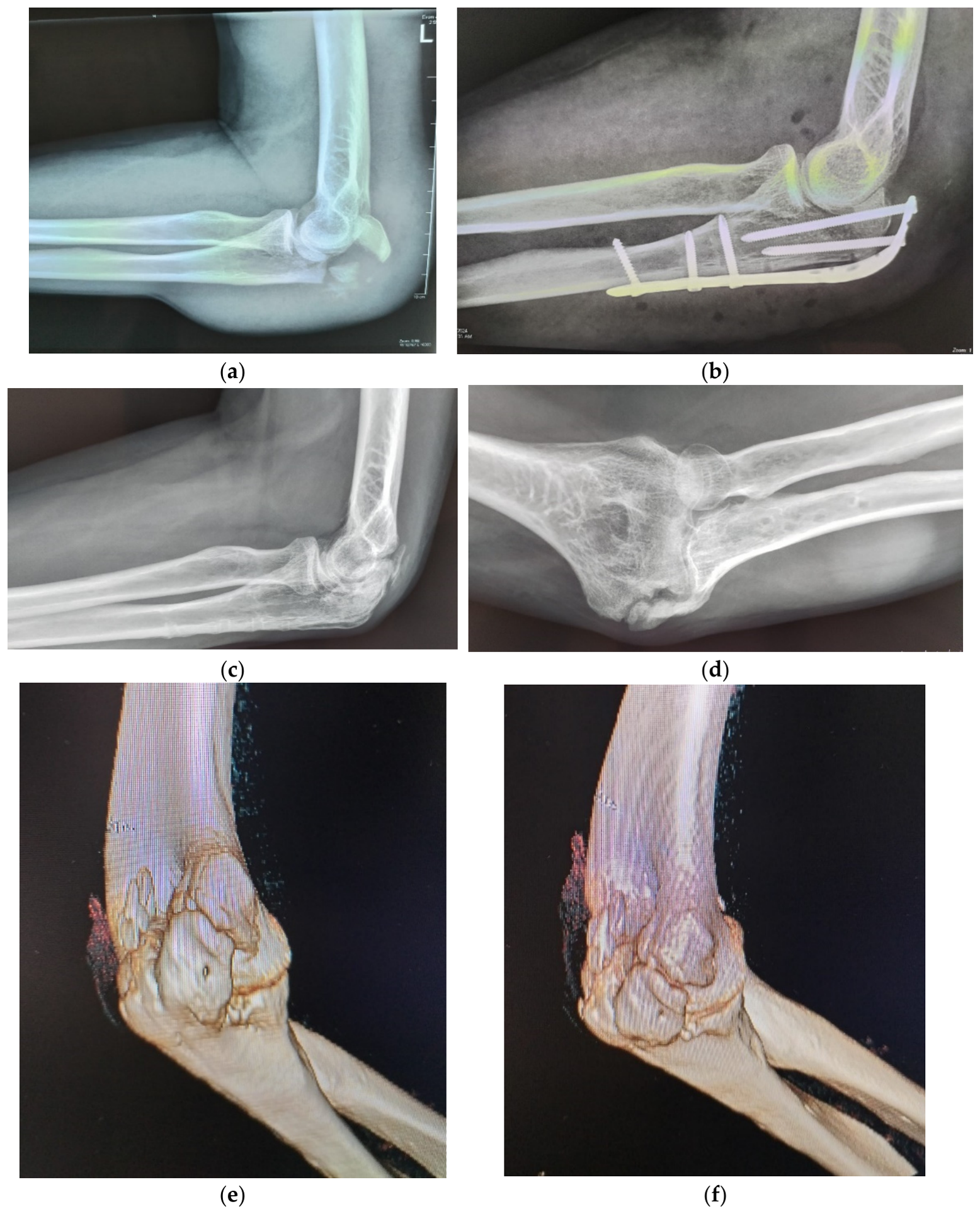
Figure 2.
a) Elbow stiffness at 90 degreesof flexion-extension following a conservatively treated radial head fracture. b-c) Postoperative result after lateral approach.
Figure 2.
a) Elbow stiffness at 90 degreesof flexion-extension following a conservatively treated radial head fracture. b-c) Postoperative result after lateral approach.
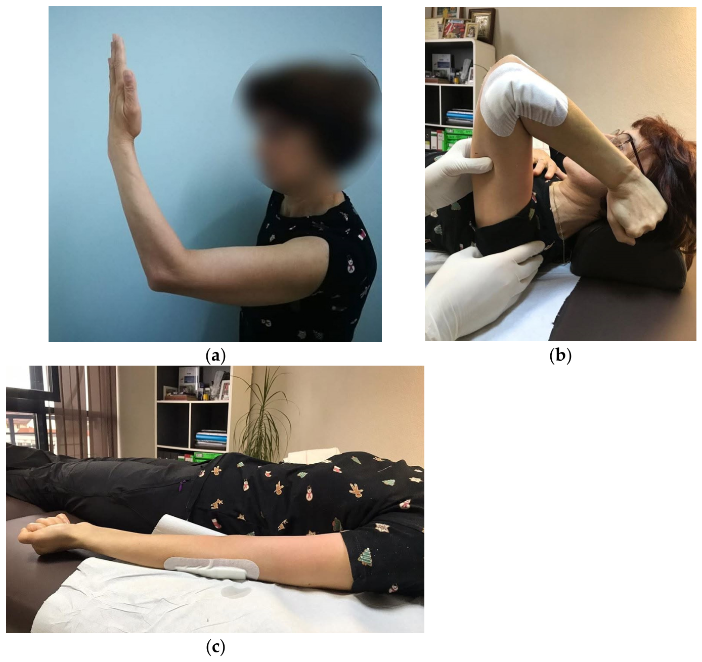
Figure 3.
a-b) radial head arthroplasty was performed using the ExploR® Modular Radial Head System prosthesis (Zimmer Biomet).
Figure 3.
a-b) radial head arthroplasty was performed using the ExploR® Modular Radial Head System prosthesis (Zimmer Biomet).
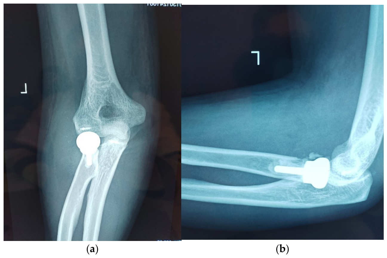
Figure 4.
a-c) Old neglected posterior elbow posterior luxation treated by open reduction, anterior capsulorrhaphy with a Fastak anchor, and lateral collateral ligament reconstruction using a gracilis autograft and SwiveLock anchors. d-h) Postoperative aspect at one year (lateral approach).
Figure 4.
a-c) Old neglected posterior elbow posterior luxation treated by open reduction, anterior capsulorrhaphy with a Fastak anchor, and lateral collateral ligament reconstruction using a gracilis autograft and SwiveLock anchors. d-h) Postoperative aspect at one year (lateral approach).
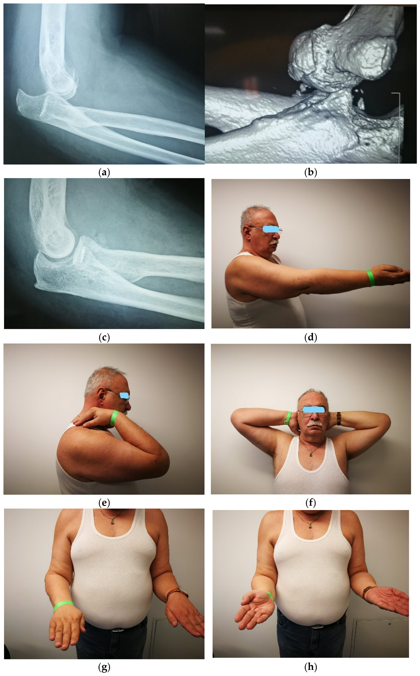
Figure 5.
Posterior approach with ulnar nerve isolation.
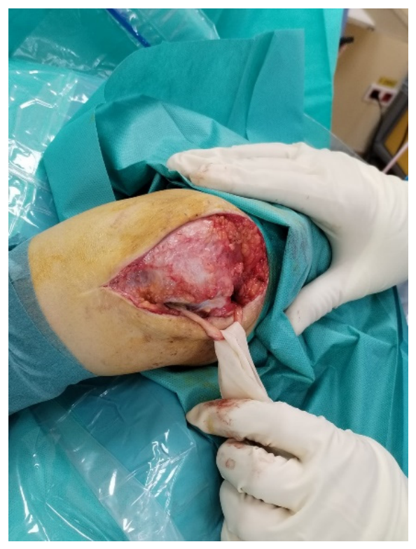
Figure 6.
a-b) Skin blistering following a posterior approach at a patient with history of systemic vasculitis. c-e) At 6 weeks postoperative view.
Figure 6.
a-b) Skin blistering following a posterior approach at a patient with history of systemic vasculitis. c-e) At 6 weeks postoperative view.
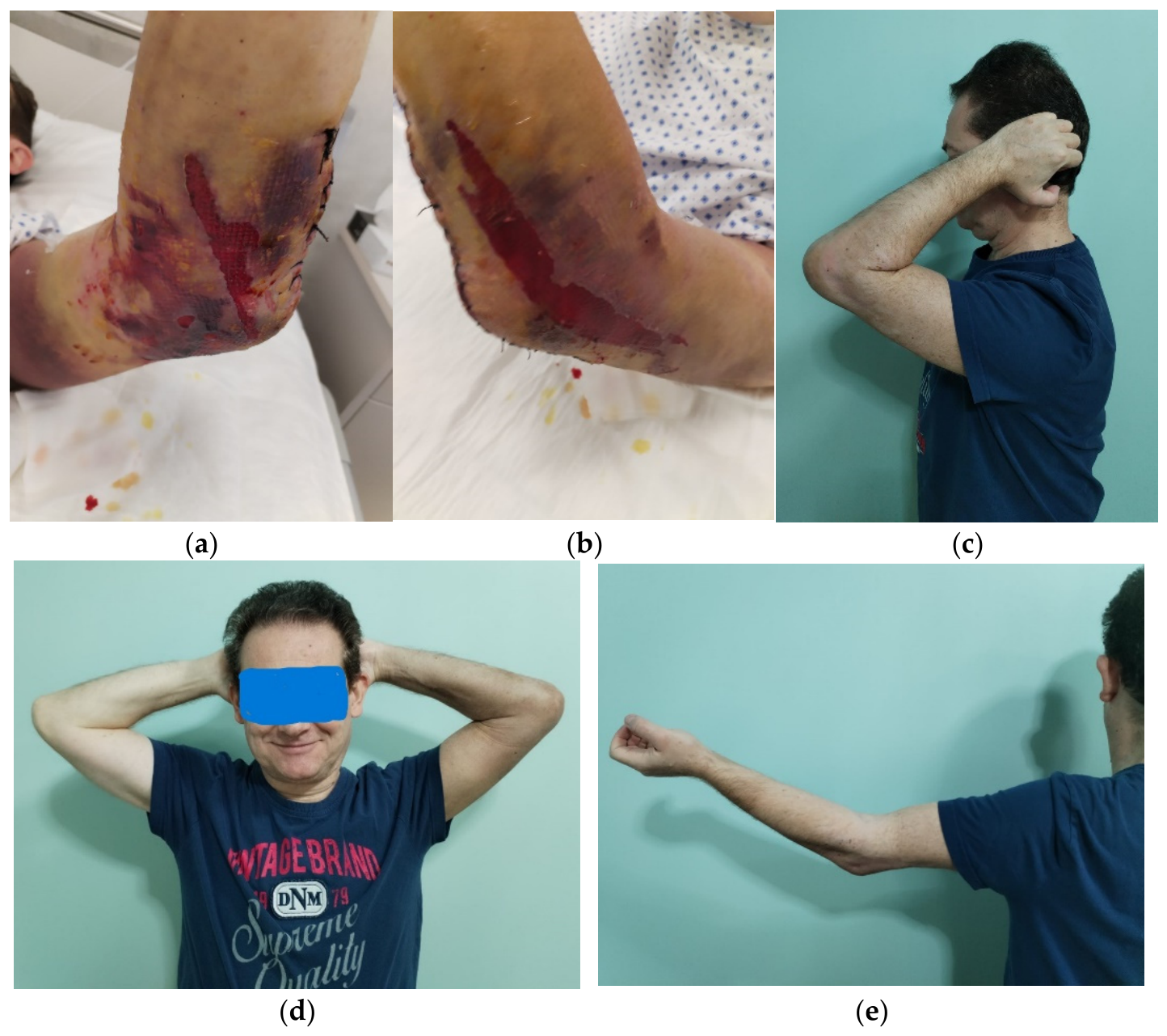
Figure 7.
Secondary elbow stiffness after posterior elbow dislocation treated conservatively. a-b) Preoperative flexion–extension arc: 70°–90°. c-g) Postoperative outcome at 6 weeks and h-i) 3 months (posterior approach).
Figure 7.
Secondary elbow stiffness after posterior elbow dislocation treated conservatively. a-b) Preoperative flexion–extension arc: 70°–90°. c-g) Postoperative outcome at 6 weeks and h-i) 3 months (posterior approach).
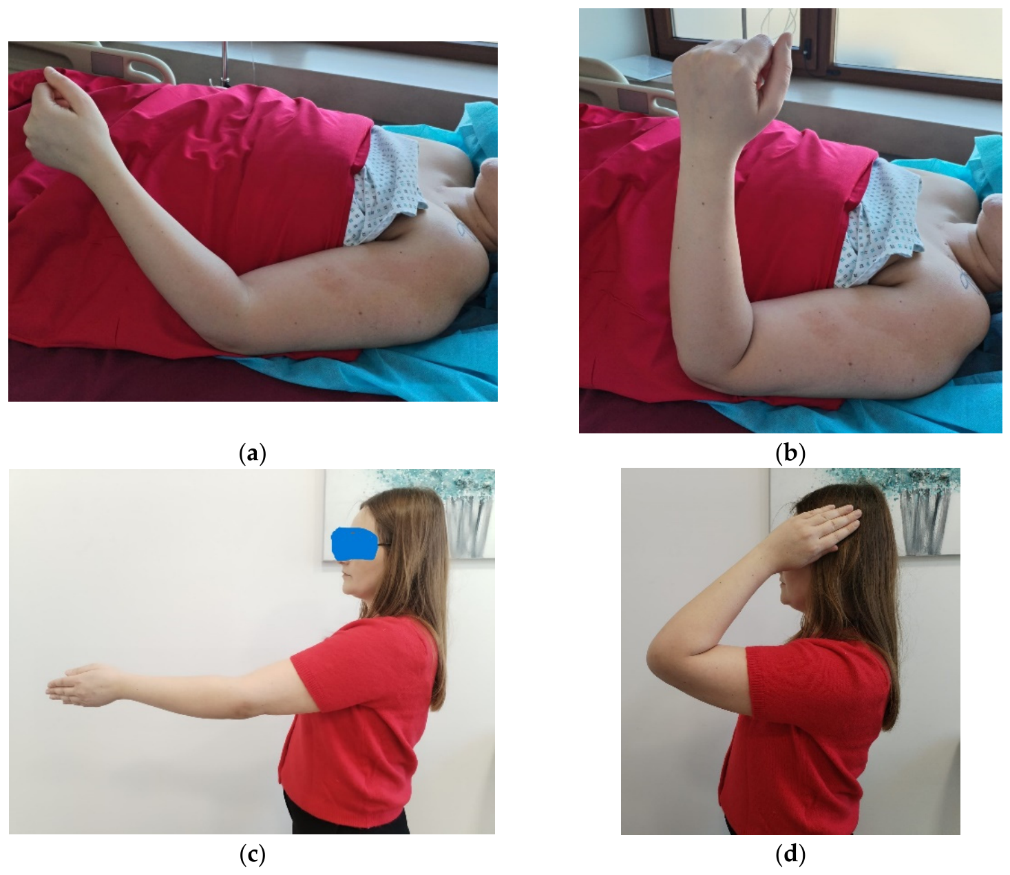
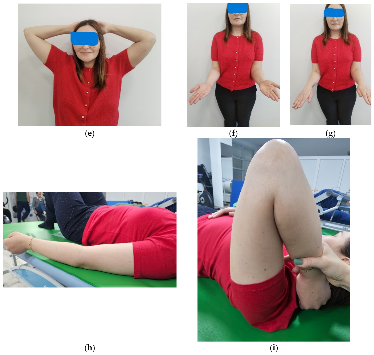

Table 1.
Complication.
| Complication | n (%) |
|---|---|
| Sensory ulnar neuropathy | 1 (5%) |
| Triceps insufficiency requiring revision | 1 (5%) |
| Skin blistering in patient with vasculitis | 1 (5%) |
| Total complications | 3 (15%) |
Table 2.
Patient Demographics.
| Variable | Value |
|---|---|
| Number of patients | 20 |
| Mean age (years) | 43.8 ± 11.6 |
| Male | 14 (70%) |
| Female | 6 (30%) |
Table 3.
Etiology of stiffness.
| Cause | n (%) |
|---|---|
| Distal humeral fracture | 9 (45%) |
| Terrible triad injury | 4 (20%) |
| Elbow dislocation | 3 (15%) |
| Olecranon fracture | 2 (10%) |
| Radial head fracture | 2 (10%) |
Table 4.
Range of Motion Outcomes.
| Parameter |
Preoperative (mean ± SD) |
Postoperative | Improvement | p-value |
|---|---|---|---|---|
| (mean ± SD) | ||||
| Flexion (°) | 81.0 ± 14.8 | 125.3 ± 9.8 | +44.3 | <0.001 |
| Extension deficit (°) | 66.5 ± 19.3 | 16.5 ± 13.9 | -50.0 | <0.001 |
| Flexion-extension arc (°) | 14.5 ± 18.8 | 108.8 ± 22.5 | +94.3 | <0.001 |
| Supination (°) | 54.8 ± 42.5 | 82.8 ± 18.2 | +28.0 | 0.003 |
| Pronation (°) | 67.0 ± 29.5 | 84.5 ± 11.2 | +17.5 | 0.002 |
Table 5.
Functional Outcomes.
| Outcome Measure | Preoperative (mean± SD) | Postoperative (mean ± SD) |
|---|---|---|
| MEPS | 30.0 ± 7.6 | 93.3 ± 6.1 |
| DASH | 92.9 ± 10.3 | 18.8 ± 13.4 |
| VAS | 7.82 ± 0.94 | 0.45 ± 0.69 |
1 MEPS: Mayo Elbow Performance Score, 2 DASH: Disabilities of the Arm, Shoulder and Hand, 3 VAS: Visual Analog Scale.
Table 6.
Effect Size Analysis.
| Parameter | Cohen’s d | Interpretation |
|---|---|---|
| Flexion | 3.53 | Extremely large |
| Extension deficit | 2.97 | Extremely large |
| Flexion-extension arc | 4.55 | Extremely large |
| Supination | 0.86 | Large |
| Pronation | 0.78 | Large |
Table 7.
Surgical Procedures.
| Procedure | n (%) |
|---|---|
| Ulnar nerve transposition | 12 (60%) |
| Tricepsplasty | 8 (40%) |
| Combined MCL + LCL reconstruction | 4 (20%) |
| Isolated LCL reconstruction | 2 (10%) |
| Anterior capsule repair | 4 (20%) |
| Radial head arthroplasty | 2 (10%) |
1 MCL: Medial collateral ligament, 2 LCL: Lateral collateral ligament.
Table 8.
Patient-specific distribution of baseline etiologies, findings, and adjunctive procedures (N=20)*.
Table 8.
Patient-specific distribution of baseline etiologies, findings, and adjunctive procedures (N=20)*.
| Pacient |
Etiology |
Soft tissue Pathology& Bony alterations & Adjunctive Surgical Severity Incongruity Procedures |
|---|---|---|
| P1 |
Distal humeral Fr |
Severe ant/post Olecranon fossa V-tricepsplasty, UN capsular osteophytes transposition fibrosis |
| P2 |
Distal Humeral Fr |
Severe global fibrosis, Coronoid osteophytes V-tricepsplasty, UN Triceps adhesions hardware transposition, HW removal |
| P3 |
Distal Humeral Fr |
Moderate ant capsular HO class I UN transposition contracture |
|
P4 |
Distal Humeral Fr |
Severe post capsular Olecranon fossa V-tricepsplasty, UN Contracture osteophytes Osteophyte excision |
| P5 |
Distal Humeral Fr |
Moderate global Retained hardware, HW removal periarticular no HO fibrosis |
| P6 |
Distal Humeral Fr |
Severe Ant contracture HO Class II minor V-tricepsplasty, UN Triceps shortening incongruity transposition |
| P7 |
Distal Humeral Fr |
Severe post capsular Olecranon/coronoid V-tricepsplasty, UN Contracture osteophytes transposition |
| P8 P9 P10 P11 |
Distal Humeral Fr Distal Humeral Fr Terrible Triad Terrible Triad |
Moderate Ant capsular HO Clas I UN transposition Contracture Moderate post Minor osteophytes, UN transposition Periarticular fibrosis no HO Severe global fibrosis Coronoid fr malunion MCl+LCL reconstruction LCL/MCL laxity subluxation Ant capsular repair Severe global fibrosis, Coronoid defect, MCL+LCL reconstruction Tissue retraction subluxation Ant capsular repair, UN transposition |
| P12 P13 P14 P15 P16 P17 P18 P19 P20 |
Terrible Triad Terrible Triad Elbow Dislocation Elbow Dislocation Elbow Dislocation Olecranon Fr Olecran Fr Neglected RH Fr Neglected RH Fr |
Severe Ant contracture Coronoid displacement, MCL+LCL reconstruction, Ligament tear HO Class I Ant capsular repair Severe global Humero-ulnar MCL+LCL recontruction contracture, tissue subluxation, Ant capsular repair tissue scarring HP Class II UN transposition Severe Ant capsular Minor periarticular UN transposition Contracture calcifications Moderate global Concentric joint None (Core procedure Periarticular fibrosis no, HO only) Severe Post capsular Minor olecranon UN transposition Contracture osteophypes Moderate Post Retained hardware V-tricepsplasty Capsular contracture Post impingement HW removal Moderate global Retainining tension HW removal Periarticular fibrosis band wiring Severe lateral Severe radiocapitellar RHA, LCL reconstruction contracture, LCL arthritis V-tricepsplasty, UN laxity transposition Severe lateral contracture Radiocapitellar RHA, LCL reconstruction LCL tear arthritis, loose V-tricepsplasty bodies |
*Note: All 20 patients underwent the standardized core procedure (Anterior and Posterior Arthrolysis + Triceps Tenolysis). Abbreviations: Ant, anterior; Post, posterior; Fr, fracture; HO, heterotopic ossification; HW, hardware; UN, ulnar nerve; MCL, medial collateral ligament; LCL, lateral collateral ligament; RHA, radial head arthroplasty; RH, radial head.
Disclaimer/Publisher’s Note: The statements, opinions and data contained in all publications are solely those of the individual author(s) and contributor(s) and not of MDPI and/or the editor(s). MDPI and/or the editor(s) disclaim responsibility for any injury to people or property resulting from any ideas, methods, instructions or products referred to in the content. |
© 2026 by the authors. Licensee MDPI, Basel, Switzerland. This article is an open access article distributed under the terms and conditions of the Creative Commons Attribution (CC BY) license (http://creativecommons.org/licenses/by/4.0/).
Copyright: This open access article is published under a Creative Commons CC BY 4.0 license, which permit the free download, distribution, and reuse, provided that the author and preprint are cited in any reuse.